BTEC HSC Unit 14 – Physiological Disorders Understanding physiological disorders.
Handbook of Physiological ResearchMethods in Health Psychology
Measurement of Cortisol
Contributors: Linda J. Luecken & Linda G. GalloPrint Pub. Date: 2008Online Pub. Date: June 22, 2009Print ISBN: 9781412926058Online ISBN: 9781412976244DOI: 10.4135/9781412976244Print pages: 37-75
This PDF has been generated from SAGE knowledge. Please note that the paginationof the online version will vary from the pagination of the print book.

University Of Arizona
Copyright ©2012 SAGE knowledge
Page 2 of 44 Handbook of Physiological Research Methods inHealth Psychology: Measurement of Cortisol
SAGE knowledge
Arizona State UniversitySan Diego State University10.4135/9781412976244
[p. 37 ↓ ]
Chapter 3: Measurement of Cortisol
Nancy A. Nicolson
Introduction to the Hypothalamic-Pituitary-Adrenocortical Axis
The hypothalamic-pituitary-adrenocortical (HPA) axis and its end product, cortisol, arethought to be important mediators of the relationship between stressful life experiencesand health outcomes. The HPA response is a component of the organism's adaptivesystem for maintaining function under changing environmental circumstances. Over thelong term, however, chronic overactivation following repeated stressors can give rise towear and tear or allostatic load (McEwen, 2003). Both maladaptive responses to stressand disturbances in the functioning of the HPA axis have been implicated in a widevariety of syndromes and illnesses, including cardiovascular illness, insulin resistancesyndrome and diabetes, cognitive decline during aging, fatigue and pain syndromes,and psychiatric disorders such as depression and posttraumatic stress disorder (PTSD),among others (Charmandari, Tsigos, & Chrousos, 2005).
As the name indicates, the main components of the HPA axis are the hypothalamus,the pituitary gland, and the adrenal cortex (see Figure 3.1). The hypothalamus releasescorticotropin-releasing hormone (CRH, also known as CRF) into the portal bloodvessels connecting the hypothalamus to the anterior pituitary. CRH, which workssynergistically with arginine vasopressin (AVP) released from the hypothalamus,then triggers the pituitary to secrete adrenocorticotropic hormone (ACTH) into thebloodstream. After reaching the adrenal cortex, ACTH stimulates the release ofglucocorticoids (GCs)—in humans, cortisol. This entire process takes place within a

University Of Arizona
Copyright ©2012 SAGE knowledge
Page 3 of 44 Handbook of Physiological Research Methods inHealth Psychology: Measurement of Cortisol
SAGE knowledge
matter of minutes. The HPA axis is regulated by a complex negative feedback system,with circulating glucocorticoids inhibiting activity at the level of the hippocampus, thehypothalamus, and the pituitary. In general, hippocampal structures exert inhibitoryinfluences on the axis at the level of the hypothalamus, whereas the amygdala playsan activating role (Herman & Cullinan, 1997). Mineralocorticoid (MR) and glucocorticoid(GR) receptors in the brain are thought to play different but complementary roles inregulating normal circadian activity, preparing the organism to respond to externalstimuli, and facilitating recovery of disturbed homeostasis after acutely stressfulsituations (de Kloet, 1991).
[p. 38 ↓ ]
Figure 3.1 Schematic Overview of the Hypothalamic-Pituitary-Adrenocortical (HPA) Axis
University Of Arizona
Copyright ©2012 SAGE knowledge
Page 4 of 44 Handbook of Physiological Research Methods inHealth Psychology: Measurement of Cortisol
SAGE knowledge
NOTE: CRH = corticotropin-releasing hormone, AVP = arginine vasopressin, ACTH =adrenocorticotropic hormone. Dashed lines indicate negative feedback effects.
Activity of the HPA axis shows a pronounced circadian rhythm, controlled by the primaryendogenous pacemaker, the suprachiasmatic nucleus. ACTH and cortisol are secretedin short pulsatile episodes, concentrated in the morning hours in humans, but occurringthroughout the day, even in the absence of stressors. In a 24-hour cycle, approximately15 to 18 ACTH pulses can be identified. In people who have a normal routine ofnocturnal sleep and daytime activity, cortisol levels are lowest between 10 p.m. and4 a.m. After a quiescent period of HPA activity lasting from 2.5 to 6 hours (Linkowskiet al., 1985), cortisol levels begin to rise several hours before awakening, with anadditional sharp increase in the 30 to 40 minutes following awakening. Thereafter,cortisol levels steadily decrease, except for a moderate rise following lunch. Althoughcortisol levels decline over the rest of the afternoon and throughout the evening untilsleep onset, the slope of the diurnal curve is relatively flat compared to the morninghours.
A Brief Overview of Research Approaches
Because of its central role in regulating the psychobiological stress response, the HPAaxis is one of the most heavily investigated physiological systems in health psychologyand psychiatry. Hans Selye's conception of the general adaptation syndrome, inparticular, [p. 39 ↓ ] called attention to the importance of the HPA axis in regulating awide range of bodily functions and their disturbance by acute physiological stressors,such as exposure to toxins (Selye, 1956). A deeper understanding of the effects ofpsychological stress on the HPA axis, however, began to emerge in the 1960s, whennow-classic studies employed new methods to assess endocrine responses to stress inrodents, nonhuman primates, and humans (Levine, 2000; Mason, 1968; Rose, 1984).The widespread involvement of the HPA axis in both healthy adaptation and commondisorders, combined with the increasing ease with which its activity can be measured,have led to an enormous growth over the last two decades in research on this system,in settings ranging from the laboratory to the community.
University Of Arizona
Copyright ©2012 SAGE knowledge
Page 5 of 44 Handbook of Physiological Research Methods inHealth Psychology: Measurement of Cortisol
SAGE knowledge
Research approaches include studies of spontaneous hormone secretion throughoutthe day, pharmacological manipulations to determine how feedback mechanismsare functioning, and studies of reactivity to acute real-life or experimental stressors.Assessment of the HPA axis at multiple levels is not feasible in most studies, becauseof the invasive procedures involved. Ignoring the vast literature on animal models andclinical research, this review focuses on methods that can be more generally appliedby health psychologists studying human subjects in a wide variety of real-life andlaboratory settings, without undue inconvenience or risk to the research participants orthe need for specialized medical personnel. This means that measures of CRH, ACTH,GC receptor characteristics, or responses of the HPA axis to challenge tests in whichCRH, ACTH, or other substances are administered are not covered, despite their utilityin psychoneuroendocrine research and clinical studies. Furthermore, this chapter doesnot discuss the rationale or procedures for measuring dehydroepiandrosterone (DHEA),a steroid hormone produced primarily by the adrenal cortex, although there is evidencethat DHEA may counteract some of the effects of elevated glucocorticoids and play arole in stress-related disorders such as depression and chronic fatigue (Goodyer, Park,Netherton, & Herbert, 2001; Khorram, 1996; Wolkowitz, Brizendine, & Reus, 2000).
This chapter focuses specifically on cortisol, the end product of the HPA axis. As acautionary note, it is important to realize that cortisol is a peripheral measure andsecretory patterns can be deviant in the statistical sense without necessarily reflectingdysregulation at a higher level. In some cases, apparent abnormalities may be theresult of an adaptive response to environmental demands. On the other hand, cortisollevels can also be perfectly normal when other probes indicate regulatory abnormalities;excessive CRH or ACTH secretion might, for example, be coupled with decreasedadrenal sensitivity. The HPA axis is a complex and dynamic system, and cortisolmeasures can provide only a partial window into how this system is regulated—ordysregulated.
University Of Arizona
Copyright ©2012 SAGE knowledge
Page 6 of 44 Handbook of Physiological Research Methods inHealth Psychology: Measurement of Cortisol
SAGE knowledge
Investigating Spontaneous Activity of theHPA Axis
Basal Cortisol Levels
Researchers have long been interested in obtaining overall basal measures ofglucocorticoid output, as overactivation of the HPA axis resulting from chronic stressor illness was expected to result in higher levels of circulating cortisol. Because of theinherent novelty of hospital settings as well as the trouble and expense of bringinghealthy subjects to the clinic, ambulatory procedures have distinct advantages.Numerous studies have used 24-hour urinary measures or repeated salivary samplingto examine genetic, developmental, and especially environmental influences onHPA activity in healthy adults and children. Others have investigated HPA [p. 40 ↓ ]abnormalities in stress-related disorders. It is now clear that not only hypercortisolismbut also hypocortisolism can occur, for example in PTSD, pain, and fatigue syndromes.The processes by which stress could lead to such divergent outcomes are stillpoorly understood, but recent reviews have summarized a number of hypotheses(Fries, Hesse, Hellhammer, & Hellhammer, 2005; Gunnar & Vazquez, 2001; Heim,Ehlert, & Hellhammer, 2000; Yehuda, 2002). For example, hypocortisolism couldbe the long-term effect of adverse early experiences that permanently alter theaxis. Down-regulation might even be seen as a protective mechanism, set in motionfollowing long-term hyperactivation to reduce the negative effects of allostatic load.Alternatively, hypocortisolism might represent a preexisting risk factor, of genetic orearly developmental origin, which later undermines the individual's ability to respondadaptively to trauma or chronic stressors.
University Of Arizona
Copyright ©2012 SAGE knowledge
Page 7 of 44 Handbook of Physiological Research Methods inHealth Psychology: Measurement of Cortisol
SAGE knowledge
Circadian Rhythm and Diurnal Patterns ofHPA Axis Activity
In addition to overall cortisol levels, the diurnal patterning of hormone secretion canprovide important clues to HPA axis dysregulation. Sophisticated chronobiologicalanalyses of circadian rhythms (see, e.g., Posener et al., 2000; Van Cauter, Leproult, &Kupfer, 1996) require more frequent sampling than is feasible in ambulatory settings,not to mention the problem of obtaining nocturnal measures. For this reason, simplermeasures of the shape of the diurnal curve are more frequently employed, in particularthe steepness of the decline in cortisol levels from morning to evening. Loss of diurnalvariation, as reflected in flatter slopes, has been reported in various disorders and at-risk groups (Bower et al., 2005; Sephton, Sapolsky, Kraemer, & Spiegel, 2000).
Even if the diurnal slope is not of direct relevance to the goals of a study, collectingseveral samples over the course of a day is good practice; differences between groupsbeing compared may be restricted to a certain time of day, which often cannot bepredicted on theoretical grounds. For this reason, studies with only a single diurnalsampling time will inevitably raise questions about how results generalize to the rest ofthe day.
Cortisol Response to Awakening
In recent years, interest has been growing in the cortisol awakening response (CAR).Cortisol levels rise sharply (50–160% in saliva) during the first 30 to 40 minutes afterwakeup, returning to the awakening baseline within 60 to 75 minutes, and decliningmore gradually thereafter (Clow, Thorn, Evans, & Hucklebridge, 2004; Pruessner etal., 1997; Wüst et al., 2000). The function of the CAR is not yet clear, but generalagreement is that this response is a discrete aspect of cortisol's circadian rhythm, withits own regulatory processes (Clow et al., 2004; Schmidt-Reinwald et al., 1999).
The CAR appears to be moderately stable within persons, from day to day and overlonger periods of several weeks to months, and it has a clear genetic component (Wüst

University Of Arizona
Copyright ©2012 SAGE knowledge
Page 8 of 44 Handbook of Physiological Research Methods inHealth Psychology: Measurement of Cortisol
SAGE knowledge
et al., 2000). Nevertheless, it can vary in relation to short-term influences, such asthe stressfulness of a workday compared to a weekend (Kunz-Ebrecht, Kirschbaum,Marmot, & Steptoe, 2004), or an early-shift compared to a late-shift workday (Williams,Magid, & Steptoe, 2005). In addition, the CAR may be either enhanced or blunted inchronic stress, burnout, depression, and other disorders (e.g., Bhagwagar, Hafizi, &Cowen, 2005; Grossi et al., 2005; Pruessner, Hellhammer, Pruessner, & Lupien, 2003;Stetler & Miller, 2005).
[p. 41 ↓ ]
Within-Person Variability
One important aspect of spontaneous cortisol secretion that has received relatively littleresearch attention, despite its potential significance as an index of HPA dysregulation,is within-person variability. Greater irregularity in within-day cortisol measures has beenobserved in affective disorders, even when overall levels are normal (Peeters, Nicolson,& Berkhof, 2004; Posener et al., 2004; Yehuda, Teicher, Trestman, Levengood, &Siever, 1996), and may predict worse clinical outcomes (Goodyer, Tamplin, Herbert, &Altham, 2000). There is some evidence that a subset of individuals lacks a consistentdiurnal slope pattern (Smyth et al., 1997), but day-to-day variation in cortisol measuresremains largely unexplored. One major obstacle is that investigating within-personvariability requires many more samples per person.
Summary
The degree of detail with which a given study is able to characterize spontaneouscortisol secretory patterns depends on its specific goals, but also on the availablebudget and logistical considerations. Thus, large epidemiological surveys are oftenrestricted to obtaining only a few samples per subject and may have to choose betweenthe response to awakening and/or a diurnal slope measure (either of which can beestimated with a minimum of two saliva samples; see, e.g., Young & Breslau, 2004),perhaps in combination with a urinary measures if nighttime or total cortisol secretionare of interest. At the other extreme, intensive daily process designs may collect 60 or
University Of Arizona
Copyright ©2012 SAGE knowledge
Page 9 of 44 Handbook of Physiological Research Methods inHealth Psychology: Measurement of Cortisol
SAGE knowledge
more saliva samples per subject in order to estimate not only overall levels and diurnalslopes, but also the association between cortisol at a particular point in time with currentmood, symptoms, daily hassles, and uplifts (Smyth et al., 1998; van Eck, Berkhof,Nicolson, & Sulon, 1996). As we don't yet know which measures of spontaneous cortisolsecretion are most relevant for understanding disease processes, a conservativeapproach would be to obtain reliable measures of cortisol basal levels, diurnal slopes,and the CAR in the same protocol (see Sampling Strategy under A Framework forDesigning a Study and Interpreting the Results).
The availability of noninvasive sampling methods (described in Measuring Activity ofthe HPA Axis) has greatly increased the range of research applications. These includecross-cultural field studies (Flinn, 1999; Hruschka, Kohrt, & Worthman, 2005), large-scale longitudinal studies in the community (Rosmalen et al., 2005), intervention studies(Carlson, Speca, Patel, & Goodey, 2004; Gaab et al., 2003), and prediction of diseaseoutcomes (Sephton et al., 2000).
Sensitivity of the HPA Axis to NegativeFeedback
Measuring the response of the HPA axis to synthetic glucocorticoids provides ameasure of the strength of negative feedback inhibition. Following an oral dose of 1mg dexamethasone late in the evening, cortisol levels are normally suppressed thenext day; incomplete suppression or early escape from suppression indicates deficitsin feedback regulatory mechanisms. The dexamethasone suppression test (DST)was originally developed as a diagnostic tool in major depression, a disorder in whichhypercortisolism is often observed (Carroll et al., 1981). A low-dose (0.25–0.5 mg)version of the DST has been used to investigate more subtle deficits in feedbackregulation in individuals with chronic stress (Powell et al., 2002) or to determinewhether sensitivity of the HPA axis to glucocorticoid negative feedback is heightened indisorders in which hypocortisolism is more frequently observed, such as PTSD (Yehudaet al., 1993) or chronic fatigue syndrome (Gaab et al., 2002).
[p. 42 ↓ ]
University Of Arizona
Copyright ©2012 SAGE knowledge
Page 10 of 44 Handbook of Physiological Research Methods inHealth Psychology: Measurement of Cortisol
SAGE knowledge
Response of the HPA Axis to AcuteStressors
Early studies of the HPA axis focused on the hormonal response to acute stressors, andthis remains one of the primary interests of health psychologists. In humans, the cortisolresponse to stress can be studied in real life or under more controlled conditions inthe laboratory. Compared to the quick but short-lived response of the catecholamines,the cortisol response to acute stress is relatively slow. Within minutes of the onset ofa discrete stressful stimulus or event, such as public speaking, cortisol levels begin torise, superimposed on the diurnal profile of basal HPA activity. After termination of thestressor, cortisol levels gradually return to their prestress baseline; full recovery cantake an hour or more, in part reflecting the approximately one-hour half-life of cortisol inblood or saliva.
Basal levels of glucocorticoids act permissively to prepare the individual to respond toa stressful episode. The cortisol response to stress mobilizes energy for coping withthe stressor, but also shuts down the initial fight or flight responses of the sympatheticnervous and immune systems to prevent them from overshooting and damagingthe organism (Munck, 2000). Glucocorticoid release during stress is thus primarily aprotective response. If, however, cortisol levels are delayed in their poststress recovery,or repeated stress exposures result in sensitization instead of habituation of the HPAaxis, a chronic hyperactivation of this system can be maladaptive, leading to stress-related disorders (McEwen, 2003).
What Kinds of Stimuli Activate the HPAAxis?
It is a common misconception, probably going back to the work of Hans Selye (1956),that the HPA axis will respond to all types of stressful experiences and the acutecortisol response can therefore serve as the gold standard for determining whether aparticular experience was stressful. Many physiological systems are involved in stress
University Of Arizona
Copyright ©2012 SAGE knowledge
Page 11 of 44 Handbook of Physiological Research Methods inHealth Psychology: Measurement of Cortisol
SAGE knowledge
responses, and each system varies in terms of the types of stressors that activate it, itstemporal dynamics, and its relations to other systems (Baum & Grunberg, 1995). Forexample, aversive stimuli that activate the sympathetic nervous system and adrenalmedulla, producing elevations in heart rate, blood pressure, and catecholamines, do notnecessarily lead to measurable changes in cortisol.
Certain types of psychosocial stressors do have consistent effects. Reviews ofearly studies in humans, rodents, and nonhuman primates concluded that situationscharacterized by novelty, unpredictability, or low perceived control were most likelyto activate the HPA axis (Mason, 1968; Rose, 1984). A recent metaanalysis ofexperimental studies showed that social-evaluative threat during task performance andlow control over the situation were the two best predictors of acute cortisol responses inhumans (Dickerson & Kemeny, 2004). Although an individual's appraisal of the stressor,coping, and degree of distress are predicted, on the basis of transactional stresstheory (Lazarus & Folkman, 1984), to moderate or (in the case of distress) mediate thecortisol response, laboratory studies have shown surprisingly low correlations betweenindividual self-reports of these variables and cortisol measures.
Physical stressors such as intense exercise also activate the HPA axis. The observationthat cortisol elevations are often greater during competitive sports than during trainingat the same level of physical exertion (Cook, Ng, Read, Harris, & Riad-Fahmy, 1987)indicates that physical and psychosocial components of competition have additiveeffects. Cortisol levels also increase following experimentally induced pain (al'Absi,Petersen, & Wittmers, 2002).
[p. 43 ↓ ]
University Of Arizona
Copyright ©2012 SAGE knowledge
Page 12 of 44 Handbook of Physiological Research Methods inHealth Psychology: Measurement of Cortisol
SAGE knowledge
Measuring Activity of the HPA Axis
Salivary Cortisol
Background
The first assays for salivary steroids were described in 1959, but this method didnot gain widespread acceptance until researchers at the Tenovus Institute in Walesdeveloped reliable assays for steroids in small volumes of whole saliva (Riad-Fahmy,Read, Walker, & Griffiths, 1982; Walker, Riad-Fahmy, & Read, 1978). Over the past20 years, there has been an explosive growth in the number of studies using salivarymeasures to assess cortisol levels in a wide variety of applications in psychology,psychiatry, endocrinology, and beyond. The advantages of salivary cortisol sampling,compared to traditional procedures for blood sampling, have been summarized inseveral reviews (Kirschbaum & Hellhammer, 1989, 1994; Vining, McGinley, Maksvytis,& Ho, 1983). In addition to the ease and noninvasive nature of sample collection, thefact that salivary cortisol is “free,” unbound by corticosteroid-binding globulin (CBG) orother carriers, is advantageous, as free cortisol thus represents the biologically activefraction of the hormone (Mendel, 1989).
As noted earlier, salivary cortisol is ideal for assessing acute responses to experimentalstressors. In addition, repeated measurement by subjects in their daily environmentallows a good estimate of basal levels, diurnal variation, and response to awakening;some naturalistic designs also permit individual estimates of day-to-day variability andstress reactivity.
Comparison with Blood Measures
Cortisol levels measured in saliva correlate highly with free cortisol in blood. However,because of partial conversion of cortisol to cortisone during passage through the
University Of Arizona
Copyright ©2012 SAGE knowledge
Page 13 of 44 Handbook of Physiological Research Methods inHealth Psychology: Measurement of Cortisol
SAGE knowledge
salivary glands, the absolute level of free cortisol in saliva is 10% to 35% lower than itis in blood (Vining et al., 1983). Correlations with total blood concentrations (bound andfree fractions) are also high, but the slope of the regression line becomes steeper athigher cortisol concentrations, after CBG-binding sites in blood are fully occupied. CBGlevels can vary both within and between individuals, for example during pregnancy orwith oral contraceptive use.
Movement of cortisol from blood to saliva occurs by passive diffusion, so that salivarylevels are independent of the flow rate of saliva (Vining et al., 1983). Changes in plasmaand salivary cortisol levels are closely synchronized. After injections of cortisol, salivarylevels increased within 1 minute (Walker, 1984), and peak concentrations in bloodare seen 2 to 3 minutes later in saliva (Kirschbaum & Hellhammer, 2000). Cortisolresponses to awakening and to meals appear to be more pronounced in salivarythan in plasma measures, and salivary cortisol returns to baseline more slowly afterpsychosocial stressors (Kirschbaum & Hellhammer, 2000).
Collection
The popularity of salivary cortisol measures is largely due to the ease of collectingsamples from participants in both laboratory and field settings. A number of differenttechniques for collecting saliva samples have been described; which is mostappropriate for a given research question will depend on characteristics of theparticipants, the setting, and frequency with which samples will be collected, andwhether other substances will be measured in the same samples.
Saliva samples are usually obtained from infants and toddlers with pipettes or otherdevices that aspirate saliva from the floor of the mouth, cotton ropes, swabs, orsponges held by the researcher or parent (Gunnar & Talge, 2007). In older children andadults, [p. 44 ↓ ] cotton dental rolls—including the widely used salivettes® (Sarstedt,Nümbrecht, Germany)—have convenient features for both research participants andlaboratory personnel. Because of aspecific binding to the swabs or interference by othersubstances such as phytoestrogens that may be present, cotton salivettes or swabsshould not be used when other steroids (e.g., DHEA, testosterone, progesterone) orsalivary immunoglobulin A (IgA) are also being measured (Shirtcliff, Granger, Schwartz,
University Of Arizona
Copyright ©2012 SAGE knowledge
Page 14 of 44 Handbook of Physiological Research Methods inHealth Psychology: Measurement of Cortisol
SAGE knowledge
& Curran, 2001). In 2007 Sarstedt began production of a new synthetic salivette,designed to eliminate the risk of batch-to-batch variation in the performance of cottonswabs. Swabs are unnecessary if participants can collect saliva by drooling into a tube,either directly or through a straw. Drooling may be less acceptable in studies whererepeated samples need to be collected as rapidly and unobtrusively as possible, forexample, during participants' daily activities outside the home. In one comparison,subjects collected adequate amounts of saliva in 1 to 2 minutes with cotton salivettesor cellulose-cotton tip “eyespears,” whereas passive drooling took from 1 to 15 minutesto produce the same amount (Strazdins et al., 2005). For all collection methods, it isimportant that the plastic storage tubes and stoppers are made of materials, such asunrecycled polypropylene (IBL, 2006), that do not absorb the hormone. Stoppers alsoneed to fit tightly, because evaporation of saliva will lead to inaccurate cortisol results.
Most cortisol assays require only 20 to 50 µl of saliva per tube, and therefore twicethese amounts for a duplicate assay. In practice, larger volumes of saliva need tobe collected when cotton-based methods are used, because up to 450 µl of salivacan remain in the cotton after centrifuging (de Weerth, Graat, Buitelaar, & Thijssen,2003). Specialized techniques make it possible to extract cortisol from smaller samplevolumes, which may be a great advantage in studies of infants (de Weerth et al.,2003). In subgroups with low spontaneous flow rates (e.g., babies and small children,depressed patients, the elderly), saliva flow can be stimulated with powdered drink mixcrystals, candies containing citric acid, or lemon juice. Salivettes prepared with citricacid are also meant to stimulate salivary flow. Extreme caution is warranted in usingsuch procedures, however, as they can lower the pH of the resulting saliva sample.Many currently available immunoassays produce false high values when sample pHis lower than 3.5 to 4 (Kirschbaum & Hellhammer, 2000; Schwartz, Granger, Susman,Gunnar, & Laird, 1998; Talge, Donzella, Kryzer, Gierens, & Gunnar, 2005; Vialard-Miguel, Belaidi, Lembeye, & Corcuff, 2005). Chewing on an inert substance (e.g., plainsalivette, sugarless chewing gum, parafilm) or just making chewing movements aregood alternatives for stimulating salivary flow.
University Of Arizona
Copyright ©2012 SAGE knowledge
Page 15 of 44 Handbook of Physiological Research Methods inHealth Psychology: Measurement of Cortisol
SAGE knowledge
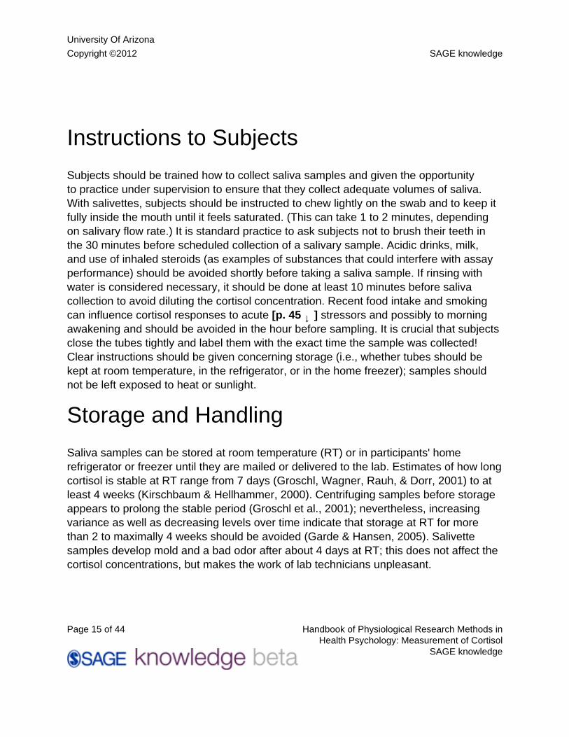
Instructions to Subjects
Subjects should be trained how to collect saliva samples and given the opportunityto practice under supervision to ensure that they collect adequate volumes of saliva.With salivettes, subjects should be instructed to chew lightly on the swab and to keep itfully inside the mouth until it feels saturated. (This can take 1 to 2 minutes, dependingon salivary flow rate.) It is standard practice to ask subjects not to brush their teeth inthe 30 minutes before scheduled collection of a salivary sample. Acidic drinks, milk,and use of inhaled steroids (as examples of substances that could interfere with assayperformance) should be avoided shortly before taking a saliva sample. If rinsing withwater is considered necessary, it should be done at least 10 minutes before salivacollection to avoid diluting the cortisol concentration. Recent food intake and smokingcan influence cortisol responses to acute [p. 45 ↓ ] stressors and possibly to morningawakening and should be avoided in the hour before sampling. It is crucial that subjectsclose the tubes tightly and label them with the exact time the sample was collected!Clear instructions should be given concerning storage (i.e., whether tubes should bekept at room temperature, in the refrigerator, or in the home freezer); samples shouldnot be left exposed to heat or sunlight.
Storage and Handling
Saliva samples can be stored at room temperature (RT) or in participants' homerefrigerator or freezer until they are mailed or delivered to the lab. Estimates of how longcortisol is stable at RT range from 7 days (Groschl, Wagner, Rauh, & Dorr, 2001) to atleast 4 weeks (Kirschbaum & Hellhammer, 2000). Centrifuging samples before storageappears to prolong the stable period (Groschl et al., 2001); nevertheless, increasingvariance as well as decreasing levels over time indicate that storage at RT for morethan 2 to maximally 4 weeks should be avoided (Garde & Hansen, 2005). Salivettesamples develop mold and a bad odor after about 4 days at RT; this does not affect thecortisol concentrations, but makes the work of lab technicians unpleasant.
University Of Arizona
Copyright ©2012 SAGE knowledge
Page 16 of 44 Handbook of Physiological Research Methods inHealth Psychology: Measurement of Cortisol
SAGE knowledge
The benefits of refrigeration at 4° to 5°C, compared to RT, are unclear. In one study(Groschl et al., 2001), cortisol levels decreased in samples refrigerated for 11 daysor longer; in contrast, Garde and Hansen (2005) found no change in cortisol levels inpolyester salivettes refrigerated up to 3 months. Freezing clearly prolongs the stabilityof salivary cortisol. In samples frozen at either -20° or -80°C, cortisol concentrationsremain stable for 9 months (Aardal & Holm, 1995) to 1 year (Garde & Hansen, 2005);freezing for as long as 2 years is probably possible.
In settings where there is no access to refrigerators or freezers, stability of samplescan be prolonged by adding preservatives such as sodium azide (Groschl et al., 2001),citric acid (alone or with sodium benzoate), or ethyl and propyl paraben (Nimmagudda,Ramanathan, & Putcha, 1997). Cortisol in samples treated with citric acid and sodiumbenzoate remained stable for 180 days at RT (Nimmagudda et al., 1997). As notedearlier, preservatives, especially those that lower pH, may invalidate certain assays.Blood spots offer an alternative to saliva when extended storage at RT is necessary(Worthman & Stallings, 1997). (See Blood Spot Measures, below.)
Salivary cortisol levels are relatively insensitive to repeated thawing and refreezing;in recent studies, cortisol levels remained stable in samples undergoing up to three(Groschl et al., 2001) or four (Garde & Hansen, 2005) freeze/thaw cycles prior to assay.In the laboratory, samples collected by passive drool are frozen and thawed at leastonce before assaying in order to break down mucins that can interfere with pipetting(Vining & McGinley, 1986). Centrifuging helps remove particulate matter that caninterfere with immunoassay. In salivettes, clear saliva collects in the bottom of the outertube after centrifuging.
There is normally no need to transport samples to the laboratory on ice (Clements &Parker, 1998). However, when the time in transit is more than a few days, shipping ondry ice will prevent molding and may be required by some laboratories. (For informationon international shipping, see International Air Transport Association IATA regulations;adjustments made in 2005 exempt saliva samples from regulations for hazardousbiological substances.)
Prior to assay, saliva samples should be checked for blood contamination, as thiscan artificially elevate the cortisol concentration. Deficient diet, poor oral hygiene, and

University Of Arizona
Copyright ©2012 SAGE knowledge
Page 17 of 44 Handbook of Physiological Research Methods inHealth Psychology: Measurement of Cortisol
SAGE knowledge
overly strenuous toothbrushing can cause bleeding gums. In a recent study (Kivlighanet al., 2004), subjects first brushed their teeth [p. 46 ↓ ] vigorously and then collectedsaliva by direct drool. Minor injuries to the oral cavity led to detectable blood leakagein the samples, as assessed by three different methods: trans-ferrin immunoassay,dipsticks for detecting hemoglobin in urine, and visual inspection. A moderate degree ofblood contamination (samples visibly pink) had a negligible effect on cortisol levels, butdarker saliva samples were more problematic. Visual inspection and discarding of salivasamples that are discolored therefore appears to be adequate to control this source oferror under normal circumstances. This is good news, because assay of transferrin—the most accurate method for assessing blood contamination—is relatively expensive,and dipsticks can yield false-positive results (Worthman & Stallings, 1997).
Types of Assays
Free cortisol in the blood represents only 4% to 5% of total cortisol released; moreover,during passive diffusion into the salivary glands, approximately one-third of thefree cortisol is lost through conversion to cortisone. Sensitive assay proceduresare therefore necessary. Several methods currently allow reliable measurementof salivary cortisol without the necessity of extraction procedures. These includeradioimmunoassay (RIA), enzyme-linked immunosorbent assay (ELISA), fluorescenceimmunoassay (FIA), and chemiluminescence immunoassay (LIA). The last three arenonradioactive assays in microtiter plate format, which can be run either manually or onautomated equipment. Special laboratory equipment is required. Before the late 1990s,salivary cortisol assays were often adaptations of protocols designed for plasma/serummeasures. Currently, assay kits developed for salivary determinations have standardssuspended in a saliva matrix (in contrast to a serum or buffer matrix). Regardless of theassay used, the following procedures are recommended: (1) assay samples in duplicate
and use the mean value in statistical analyses,1 (2) repeat the assay for samples withduplicate values that differ by more than 20%, and (3) measure all samples from a givensubject in the same assay run.

University Of Arizona
Copyright ©2012 SAGE knowledge
Page 18 of 44 Handbook of Physiological Research Methods inHealth Psychology: Measurement of Cortisol
SAGE knowledge
Choosing a Lab
Over the last several years, commercial labs have proliferated, in some cases offeringassay services for salivary cortisol as well as kits for use in the investigator's ownlab. Details are available through the laboratories' websites, for example http://www.salimetrics.com, http://www.ibl-hamburg.com, and http://www.dslabs.com. Inaddition, many hospital and research labs have expertise in salivary assays; some usecommercially available kits, while others have developed their own in-house assays.Price is an important consideration; the costs of a duplicate cortisol determination canrange from roughly $6 to $30, often with a discount for large quantities. Quality shouldalso enter into the choice, as not all assays are equally sensitive or reliable. Fortunately,assay quality and cost are likely to be inversely related, because laboratories withtailored salivary assays also tend to have more experience, higher volumes, andautomated procedures.
Sensitivity refers to the minimum concentration of cortisol that can be distinguishedfrom zero. Salivary cortisol assays generally have a lower detection limit of less than .01µg/dL, which is below the concentration normally observed until late in the eveningwhen the HPA axis becomes quiescent. The reliability of an assay, which is evenmore crucial for most research questions, is reflected in the intra- and interassaycoefficients of variation (CV). The intra-assay CV can be calculated by dividing thestandard deviation by the mean and then multiplying this figure by [p. 47 ↓ ] 100, over arepresentative subsample (high, medium, and low cortisol concentrations) of duplicatemeasures from the same assay run. The interassay CV is calculated over severalassay runs; laboratories should be able to provide this information on request. BothCVs should be under 12% to 15% (most are lower). In general, CVs are higher forcortisol concentrations in the lower part of the range. Even when intra- and interassaycoefficients of variation are acceptable, some laboratories may obtain higher or lowerabsolute cortisol values than others (Hansen, Garde, Christensen, Eller, & Netterstrom,2003; Kraemer et al., 2006). For this reason, it is inadvisable to switch from one type ofassay to another or from one laboratory to another during the same study.
Other indicators of assay performance are range of calibration, range of linearity ofthe assay (the linear region of the standard curve should cover the range in which
University Of Arizona
Copyright ©2012 SAGE knowledge
Page 19 of 44 Handbook of Physiological Research Methods inHealth Psychology: Measurement of Cortisol
SAGE knowledge
most salivary cortisol values are found), spike recovery, and specificity (the percentageof crossreactivity with other endogenous or exogenous substances, like cortisoneor prednisone, that are or may be present in saliva). Laboratories should be able toprovide this information for their assays. Reviewers are likely to request more detailedinformation for in-house assays. Recent implementation of voluntary quality assessmentprograms for salivary assays (IBL, 2005) will hopefully make it easier for researchers toevaluate and compare laboratories.
Urinary Cortisol
Background
Urinary measures of glucocorticoid metabolites (17-hydroxycorticoids) were among thefirst techniques available for studying activity of the HPA axis in humans, going back tothe 1950s. An impressive body of knowledge emerged from the early psychoendocrinestudies of 17-HOCs levels (Mason, 1968). New techniques soon allowed researchersto measure cortisol directly, as small amounts are excreted as free cortisol in the urine(UFC). Urine samples collected over 24 hours provide an integrated measure of totalfree cortisol excretion. Mean UFC values are approximately 20µg/24 h (range 3–43µg/24 h) in healthy adult women (Murphy, 2003).
To reduce participant burden, collection over shorter periods may prove adequate for aspecific research question. In addition, urine collection can be scheduled in such a waythat more refined analyses are possible. As an example, Jerjes and colleagues wereable to investigate diurnal patterns of HPA activity by having subjects collect urine every3 hours for 15 hours (Jerjes et al., 2006). Another recent study compared women'surinary cortisol levels when they were at home, at work, or asleep (Dettenborn et al.,2005).
A distinct advantage of urinary measures is that they allow assessment of nighttimecortisol levels, which may be crucial in certain disorders in which daytime levels areoften normal (anxiety: Abelson & Curtis, 1996; PTSD: Yehuda, 2002). Urinary measuresalso have some disadvantages, which explain why they are less popular than salivary
University Of Arizona
Copyright ©2012 SAGE knowledge
Page 20 of 44 Handbook of Physiological Research Methods inHealth Psychology: Measurement of Cortisol
SAGE knowledge
cortisol. First, integrated measures are not very informative for research questionsconcerning acute stress responses. Second, the burden of collecting complete urinesamples should not be underestimated, as it can lead to low participation in studies orpoor compliance. Finally, transporting large volumes of urine from field to laboratory iscumbersome.
Collection, Storage, and Handling
At the beginning of the sampling period, subjects void and discard the first urine. Allurine produced thereafter is collected in large [p. 48 ↓ ] plastic containers designedfor this purpose, or several containers if the study entails separate measurements.Samples can be kept at room temperature, without preservatives, for at least 24 hourswithout degradation of glucocorticoids (Gouarne, Foury, & Duclos, 2004).
Assays
Urinary free cortisol represents a small fraction of total cortisol released by the adrenalcortex. Commercially available RIA kits for measuring UFC may yield falsely highvalues, as results can be influenced by the presence of cortisol metabolites as well asother interfering substances; UFC values obtained with these assays are potentiallytwo to four times higher than the true values established with chromatography (Murphy,2002). Immunoassays have been reported to show particularly low specificity and poorprecision at low cortisol concentrations, leading to widely discrepant results in studiesof adrenal suppression (Fink et al., 2002). In choosing a laboratory, it is thereforeimportant to make sure that the assay has been validated and is monitored accordingto established standards for UFC; details concerning the assay (accuracy, recovery,precision, antibody used, crossreactivity, extraction method) should also be reportedin publications. Accurate methods, for example liquid chromatography/tandem massspectrometry (McCann, Gillingwater, & Keevil, 2005; Turpeinen & Stenman, 2003), arebecoming more widely available and affordable. Because the tiny percentage (2–3%) ofUFC in relation to total urinary cortisol metabolites may vary due to changes in steroidmetabolism, measuring urinary cortisone, the ratio of cortisone to cortisol, or total
University Of Arizona
Copyright ©2012 SAGE knowledge
Page 21 of 44 Handbook of Physiological Research Methods inHealth Psychology: Measurement of Cortisol
SAGE knowledge
cortisol metabolites may provide additional insights into HPA axis function (Gouarne,Groussard, Gratas-Delamarche, Delamarche, & Duclos, 2005; Jerjes et al., 2006). UFCresults are often corrected for creatinine levels.
Blood Spot Measures
Background
Finger-prick blood spot sampling provides an alternative to salivary measures ofcortisol; this technique combines the advantages of traditional blood samples, interms of the range of substances that can be measured, with greater ease of samplecollection and more convenient storage and handling procedures (Wong, Yan, Donald,& McLean, 2004; Worthman & Stallings, 1997). Using devices designed to allowdiabetics to monitor their own glucose levels, collection of finger-prick samples incapillary blood is quick and minimally invasive. Because of the tiny amount of bloodrequired, obtaining repeated samples from an individual is feasible. Blood spot cortisolis highly correlated with serum levels. The method also has some disadvantages: not allparticipants can be trained to collect their own samples, so that research personnel mayhave to be involved; finger-pricks are not entirely painless, and recruitment of subjectsmay be more difficult for this reason; subjects may be concerned about the safety of theprocedure; and so on.
Collection, Handling, and Storage
Capillary blood from a finger-prick is dropped, without blotting or smearing, ontospecially designed filter paper of the sort widely used in neonatal screening programs.One drop (yielding a blood spot of approximately 50 µL of whole blood) is sufficient forcortisol determination. After samples on filter paper are air-dried for several hours, theycan be easily stored in plastic bags for transport and even be mailed directly to the labby ordinary post.
University Of Arizona
Copyright ©2012 SAGE knowledge
Page 22 of 44 Handbook of Physiological Research Methods inHealth Psychology: Measurement of Cortisol
SAGE knowledge
Assays
Special, highly sensitive assays have been developed to determine cortisol levels in [p.49 ↓ ] blood spots. These assays are currently performed by an increasing number oflaboratories, including commercial laboratories such as Salimetrics and DSL. Additionalhormones and other substances can be measured in blood spots, including those notmeasurable in saliva, such as prolactin and markers of immune function (McDade et al.,2000). Accurate measures of estradiol and progesterone can also be obtained, enablingthe researcher reliably to assess the stage of the menstrual cycle (Shirtcliff, Reavis,Overman, & Granger, 2001), for example.
A Framework for Designing a Study andInterpreting the Results
The first key to designing an effective study with clear results is awareness of thetemporal dynamics of HPA axis activity, as these will dictate the sampling strategy.For studies of stress reactivity, the choice of a stressor can have a major impact onthe results and their interpretation. Finally, the design should take into considerationthe range of possible moderators, mediators, and confounders that might affect thehypothesized relationship between HPA measures and biopsychosocial variablesof interest. This review attempts to summarize current recommendations andpractice, without claiming that evidence in all cases is so consistent and complete thatresearchers have reached a consensus.
Sampling Strategy
As previously described, integrated (in urine, UFC) and momentary (in saliva orblood) measures of cortisol are available. For UFC, the main decision is whether tocollect samples over 24 hours or shorter time periods; the choice should be basedon theoretical grounds, but subject burden and logistics often play a role. For salivarycortisol and blood spots, the optimal number and timing of samples depends on the
University Of Arizona
Copyright ©2012 SAGE knowledge
Page 23 of 44 Handbook of Physiological Research Methods inHealth Psychology: Measurement of Cortisol
SAGE knowledge
aspects of HPA activity being investigated (e.g., basal levels, diurnal variation, responseto awakening, negative feedback inhibition, or response to acute stressors) and thestability of these measures over time.
Basal Cortisol Levels and Diurnal Variation
Although investigators seem to agree that cortisol should be measured several timesa day for a number of days to get reliable estimates of mean basal levels and diurnalslope (Goodyer et al., 2001; Stewart & Seeman, 2000), clear recommendations withsupporting data are difficult to find. Hruschka and colleagues (2005) recently presentedformulas for determining these figures on the basis of variance estimates from multilevelregression models. Based on data from a number of studies using different samplingprotocols, these calculations suggested that—depending on the spacing of the samplesin time—as few as four samples taken on one day might be adequate for estimatingindividual mean levels, but that 14 or more days of sampling with four to five samplesper day might be necessary to obtain a reliable estimate of an individual's diurnalslope. In contrast, a study in an older population with very good protocol compliancefound that five samples per day for 3 days provided a reliable estimate of daytimeslope; moreover, slopes based on as few as two daily time points (wake and 9:00p.m.) correlated highly with those based on four points, and additional days did notsubstantially increase reliability (Kraemer et al., 2006). These findings underscore theneed for more analyses of existing “daily profile” datasets. To establish an optimalsampling protocol for a specific population, a pilot study with at least [p. 50 ↓ ] 50participants is desirable (Kramer et al., 2006). Until more empirical results are available,a conservative recommendation would be to collect three to five samples a day for atleast 3 days if basal levels are of primary interest and for 6 to 7 days if diurnal variationis a major focus. Increasing the number of subjects can increase statistical power whenreliability of the cortisol measures is not optimal.
Because individual differences in sleep patterns may be associated with shifts in thecircadian cycle, some researchers have chosen to collect samples at fixed intervalsfrom the habitual time of awakening, instead of at fixed times of day. Another elegantbut logistically simpler design is to sample at fixed times of day and then model effectsof time since awakening statistically (Cohen et al., 2006). In all cases, efforts should be
University Of Arizona
Copyright ©2012 SAGE knowledge
Page 24 of 44 Handbook of Physiological Research Methods inHealth Psychology: Measurement of Cortisol
SAGE knowledge
made to obtain accurate information concerning the actual sample collection times. Themost foolproof method is some form of electronic monitoring, for example, devices thatrecord whenever a participant opens a vial to remove a cotton swab (Broderick, Arnold,Kudielka, & Kirschbaum, 2004; Jacobs et al., 2005; Kudielka, Broderick, & Kirschbaum,2003) or handheld computers that generate time stamps with which participants mustlabel their tubes (Stetler, Dickerson, & Miller, 2004). Awareness that complianceis being monitored increases the probability that samples will be taken as directed(Kudielka et al., 2003). Prompting participants with an audible or vibrating signal canalso help. Finally, instructions to participants should emphasize the importance ofaccuracy and honesty in reporting actual collection times. In older adults, self-reportedcollection times were close to automatically recorded times, and test-retest reliability ofslope estimates was actually slightly better when based on self-reported times (Kraemeret al., 2006).
Cortisol Awakening Response (CAR)
The time course of this response has been well characterized. The peak responseoccurs 30 to 45 minutes after awakening; by 60 minutes, cortisol levels are decreasingand may no longer be reliably distinguishable from the levels at awakening. At least twosamples (at awakening and either 30 or 45 minutes later) are needed to characterizethe response; more samples (e.g., at 0, 30, 45, and 60 minutes) may increase reliabilityand allow calculation of AUC measures (see Statistical Analysis). The CAR shouldpreferably be measured on at least 2 days. Given the narrow window of response,accurate timing of samples is crucial. Because participants appear to have difficultyin taking early morning samples as directed (Kudielka et al., 2003), it may be wise toreduce the sample burden to the minimum, emphasizing quality rather than quantity.Some kind of alarm device is useful to remind the participant to collect samples at theappropriate times, and compliance should be monitored electronically (see above) if thisis possible. Activity monitors can be helpful in confirming time of awakening, but this isnot considered essential for all studies.
Methodological issues relevant to study design have been summarized by Clow andcolleagues (2004). Instructions to subjects should be standardized along the followinglines:
University Of Arizona
Copyright ©2012 SAGE knowledge
Page 25 of 44 Handbook of Physiological Research Methods inHealth Psychology: Measurement of Cortisol
SAGE knowledge
• Place all materials next to your bed before going to sleep.• Take the first sample in bed immediately after awakening, with lights on and
eyes open.• Do not go back to sleep; get out of bed (within 15 minutes) before taking
another sample.• The second sample should be taken [n] minutes after awakening (and so on
for each sample).• Do not brush your teeth, smoke, eat, or drink anything except water until you
have finished taking the [n] morning samples.• Remember to record the exact time each sample was taken on the tube,
even if this was not the scheduled time.
Dexamethasone Suppression as a Measureof Negative Feedback Sensitivity
Dexamethasone (DEX) can be safely ingested by participants at home and its effectsmeasured in salivary cortisol (Lindley, Carlson, & Benoit, 2004; Powell et al., 2002). Inmost studies, cortisol measures on a control day are compared with measures takenat the same times of day following intake of 0.25 to 0.5 mg (low dose) or 1.0 mg (highdose) dexamethasone late the previous evening (at 11 p.m. or an agreed-on bedtime).The original dexamethasone suppression test (DST), developed as a diagnostic test formajor depression, was scored as positive if, following administration of 1 mg DEX at 11p.m. on day 1, cortisol at 4 p.m. on day 2 was above an established cutoff point (Carrollet al., 1981). For research purposes, analyzing the cortisol results as continuous insteadof dichotomized measures yields more information. The optimal timing of the samplesdepends on the DEX dosage, as cortisol will “escape” from suppression earlier withlower doses. Collecting a number of post-DEX saliva samples at intervals of severalhours will increase reliability of the results and gives added information about the timecourse of feedback inhibition.
University Of Arizona
Copyright ©2012 SAGE knowledge
Page 26 of 44 Handbook of Physiological Research Methods inHealth Psychology: Measurement of Cortisol
SAGE knowledge
The Cortisol Response to Acute Stressors
Cortisol responses to acute stressors can be studied in the laboratory and in reallife, where anticipated as well as unanticipated stressors occur. Design issues varyaccording to the setting. In the laboratory, important decisions include the best timeof day to schedule the experiment, how many samples are needed to characterizethe stress response, the timing of these samples in relation to the stress task, thenature of the task, how to control for effects of novelty and anticipation, and habituationto repeated stressors. For a detailed overview of many of these design issues, seeDickerson and Kemeny (2004).
Time of Day. Although the HPA axis is capable of responding to acute stress at anypoint in the diurnal cycle (Kudielka, Schommer, Hellhammer, & Kirschbaum, 2004),scheduling experiments in the mid to late afternoon (roughly between 3 and 6 p.m.)has advantages. First, the cortisol response to stress is more readily distinguishable inthe afternoon than in the morning from background noise in the form of spontaneouspulsatile episodes and the natural decline in basal levels over the morning hours; themetaanalysis performed by Dickerson and Kemeny (2004) showed moderate effectsizes for cortisol response to stress tasks performed in the afternoon, compared tosmall effect sizes in the morning. Second, cortisol responses are easier to provoke inthe afternoon than in the late evening, when the HPA axis becomes quiescent. Third,effects of potential confounders such as recent awakening and lunch (see below)are easier to exclude. Thus far, study design has been influenced mainly by suchpractical considerations, and little attention has been paid to theoretically importantissues, such as the consequences of differential activation of MR and GR systems bystressors occurring at the trough versus the peak of the diurnal cycle (Dallman, Akana,Bhatnagar, Bell, & Strack, 2000).
Number and Timing of Samples. To characterize the cortisol response to an acutestressor, samples are taken at fixed intervals during baseline (30–40 minutes),stress exposure (10–20 minutes), and recovery (40–60 [p. 52 ↓ ] minutes) periods.Participants thus need to remain at the laboratory for a total of 1½ to 2 hours. Thestress exposure includes both the preparation period, if there is one, and the actualtask performance. Peak cortisol levels are usually observed 20 to 40 minutes after
University Of Arizona
Copyright ©2012 SAGE knowledge
Page 27 of 44 Handbook of Physiological Research Methods inHealth Psychology: Measurement of Cortisol
SAGE knowledge
task onset, depending on the intensity and duration of the task, with a gradual returnto baseline levels over the next hour or longer (Dickerson & Kemeny, 2004). Evenwith an identical task, however, there are marked individual differences in latency topeak response (Gunnar & Talge, 2007). If time and budget allow, an optimal designwould include two to three baseline measures, one to two measures shortly after andpossibly during the stress task, and two to three measures during the recovery period.Minimalistic designs with one prestress and one poststress measure run the risk ofmissing the peak response and provide no information about speed of recovery.
Controlling for Novelty and Anticipation. Prestress baseline measures are highlysensitive to the novelty of the setting. Previous visits to the lab or an extendedacclimation period (30 minutes or more) after arrival can reduce the probabilityof elevated baseline levels. Anxiety in anticipation of the task remains difficult tocontrol. Because information provided earlier as part of informed consent and pretaskinstructions can either heighten or reduce anxiety, procedures need to be fullystandardized in terms of both content and timing. Obtaining a saliva sample at home,at the same time on another day, is very useful in determining whether lab baselinelevels are elevated (Nicolson, Storms, Ponds, & Sulon, 1997). This is important to know,because high baseline cortisol is often associated with a blunted response to stress(Kudielka, Schommer, et al., 2004; Young & Nolen-Hoeksema, 2001). Interestingly,infants and young children tend to show lower cortisol levels at lab arrival than at home(Gunnar & Talge, 2007).
Type of Stressor. Health psychology studies most frequently apply psychosocialstress tasks, as these are thought to have the greatest ecological validity. The HPAaxis can be activated by physical stressors such as intense exercise or pain, or bypharmacological challenges; individuals' responses to different classes of stressors,however, do not appear to be highly intercorrelated. Among the psychosocial stressors,performance tasks with elements of social-evaluative threat, uncontrollability, or bothproduce the largest and most consistent increases in cortisol. Less consistent resultsare found for passive tasks (e.g., watching a film or other emotion induction procedures,noise exposure) and performance tasks without evaluative threat or uncontrollability(Dickerson & Kemeny, 2004).
University Of Arizona
Copyright ©2012 SAGE knowledge
Page 28 of 44 Handbook of Physiological Research Methods inHealth Psychology: Measurement of Cortisol
SAGE knowledge
Only a few stress tasks have been described in sufficient detail that results can becompared across studies and populations. The best known of these is the Trier SocialStress Test (TSST) (Kirschbaum, Pirke, & Hellhammer, 1993). The widespread useof the TSST reflects the fact that it has been extensively studied, can be applied insubjects varying in age and educational status, and induces a cortisol response in themajority of participants. For the TSST and other tasks with a combination of social-evaluative threat and uncontrollability, effect sizes, on average, are large (Dickerson &Kemeny, 2004). Results are sensitive to changes in the procedure, however: when theinterval between instructions to subjects about the task and performance was extendedfrom 10 minutes to 1 hour, the cortisol response to the task was obliterated (Young &Nolen-Hoeksema, 2001).
Interpretation of Results. Although laboratory stress tasks are probably the single most[p. 53 ↓ ] common approach to investigating the HPA axis in health psychology, resultscan be difficult to interpret. Failure to detect a statistically significant cortisol responseis a rather common occurrence, even when the task appears to be experienced asstressful. Weaknesses in study design are responsible for some of these negativefindings. With adequate sample size and the right choice of stress task and timing ofmeasures, however, the majority of subjects are likely to show an increase in cortisolfrom baseline to posttask. Theoretically, one would expect the magnitude of thisresponse to reflect the individual's experience of the situation, in terms of appraisedthreat, coping possibilities, and the intensity of emotional distress. Unfortunately, thisis rarely the case with laboratory tasks, possibly because self-report instruments arenot sensitive to the relevant processes or because other aspects of the situation—forexample, its novelty—are more salient. Another discouraging finding is that cortisolresponses often show no correlation with personality traits linked to stress reactivity,such as neuroticism (Schommer, Kudielka, Hellhammer, & Kirschbaum, 1999).Furthermore, little is known about the generalizability of lab reactivity measures to real-life situations (for studies involving cortisol, see Houtman & Bakker, 1987; Lundberg,Melin, Fredrikson, Tuomisto, & Frankenhaeuser, 1990; van Eck, Nicolson, Berkhof,& Sulon, 1996). Giving a speech on a particular topic or performing mental arithmeticbefore an audience may also have different meanings for individuals or groups,depending on variables such as cognitive ability, occupation, and cultural background.The challenge for researchers is to design laboratory stressors that convincingly tap
University Of Arizona
Copyright ©2012 SAGE knowledge
Page 29 of 44 Handbook of Physiological Research Methods inHealth Psychology: Measurement of Cortisol
SAGE knowledge
into the processes of interest in a given population and also reliably activate the HPAaxis. Examples include a task involving competition with an antagonistic peer in children(van Goozen et al., 1998) and a standardized lecture in student teachers (Houtman &Bakker, 1987).
Test-retest reliability of acute stress response measures appears to be low, probablybecause of both the underestimated noise introduced by spontaneous pulsatile activity(Young, Abelson, & Lightman, 2004) and the tendency of the cortisol response tohabituate following repeated exposures. Low reliability of cortisol outcome measuresmeans that laboratory stress experiments are particularly vulnerable to Type 2 error.This issue is important in all studies, but especially needs to be taken into account inintervention studies, where stress reactivity is compared pre- and postintervention. Moreanalyses are needed to determine how many cortisol measures per session and howmany repeated sessions are necessary to obtain reliable measures of stress reactivityfor different subject populations and stressors (Gunnar & Talge, 2007; Hruschka et al.,2005).
Habituation and Sensitization. If the cortisol response to experimental stressors is tobe considered a valid indicator of what goes on in daily life, it is important to know whathappens following repetitive stressful experiences. The acute response is adaptive, butis expected to habituate over repeated exposures as novelty decreases and controlincreases. Failure to habituate or sensitization to repeated stressors, in contrast, isregarded as maladaptive, contributing in the long run to allostatic load. Habituationversus sensitization of the HPA response has been extensively examined in animalmodels (e.g., Pitman, Ottenweller, & Natelson, 1990), but comparatively little researchhas been conducted in humans (exceptions include al'Absi et al., 1997; Epel et al.,2000; Gerra et al., 2001; Gunnar, Hertsgaard, Larson, & Rigatuso, 1991; Kirschbaum,Prüssner et al., 1995; Wüst, Federenko, van Rossum, Koper, & Hellhammer, 2005).Findings indicate [p. 54 ↓ ] rapid habituation from the first to the second and laterexposures, but also show marked individual differences, with a subset of individualsfailing to habituate after repeated exposures. In addition, correlations between thecortisol response and trait characteristics may increase (Kirschbaum, Prüssner et al.,1995) or decrease (al'Absi & Lovallo, 1993) over successive task performances. Thesefindings suggest that repeated testing (for example, three times in a week, or once
University Of Arizona
Copyright ©2012 SAGE knowledge
Page 30 of 44 Handbook of Physiological Research Methods inHealth Psychology: Measurement of Cortisol
SAGE knowledge
a week for 3 weeks) is much more informative than single exposures in elucidatingindividual differences in stress reactivity that are relevant to long-term health outcomes.
Naturalistic Experiments. Responses of the HPA axis can also be investigated inresponse to real-life activities that entail some level of challenge or threat. Examplesinclude exams, parachuting, sports competitions, musical performances, andoccupational stressors, to name just a few. Tension-reducing activities such as yoga,meditation, and massage, on the other hand, may lower cortisol levels. Like labexperiments, these activities are usually scheduled or at least anticipated in advance,so that baseline, response, and recovery measures can be obtained. Certain activities,like parachute jumping, also lend themselves to studies of the habituation process(Deinzer, Kirschbaum, Gresele, & Hellhammer, 1997; Levine, 1978). Correlationsbetween cortisol responses and subjective distress measures may be higher for real-lifethan for laboratory stressors (Nicolson, 1992).
HPA axis responses to life events can also be investigated, although prestress baselinemeasures are rarely available because of the unpredictable nature of individuallife events (e.g., rape, sudden death of a family member) and natural or manmadecatastrophes (e.g., earthquakes, hurricanes, war). In this case, cortisol levels inexposed individuals are compared with those in an unexposed comparison group andare often examined longitudinally, in relation to symptoms.
Daily Hassles and Emotions. Combining repeated self-reports with salivary measures,experience sampling or ecological momentary assessment studies have investigatedcortisol reactivity to daily life hassles and uplifts and accompanying emotions. Real-life stressors vary widely in duration, and participants are often unable to report exactlywhen a stressful situation began or when it ended. Although the timing of cortisolmeasures in relation to daily hassles is therefore imprecise, multilevel regressionanalyses can assess associations between the two. The association between dailyevents and cortisol is probably mediated by changes in negative affect. The finding ofhigher salivary cortisol in association with daily hassles or negative affects has beenreplicated in several samples of adults (Hanson, Maas, Meijman, & Godaert, 2000;Peeters, Nicholson, & Berkhof, 2003; Smyth et al., 1998; van Eck, Berkhof et al., 1996)and children (Adam, 2006). In some but not all studies, positive affects were associatedwith lower cortisol (Adam, 2005; Polk, Cohen, Doyle, Skoner, & Kirschbaum, 2005).
University Of Arizona
Copyright ©2012 SAGE knowledge
Page 31 of 44 Handbook of Physiological Research Methods inHealth Psychology: Measurement of Cortisol
SAGE knowledge
Moderators and Confounders
Interpretation of cortisol results can be facilitated by considering a number of between-and within-individual factors that can influence HPA axis activity. These include age,gender-related variables, and ethnicity; somatic variables such as illness, medications,and obesity; daily lifestyle variables such as food intake, smoking, and sleep patterns;psychosocial variables related to stress; and genetic differences.
Age and Gender-Related Variables
Findings concerning the effects of age and gender on the HPA axis vary from study to[p. 55 ↓ ] study in magnitude and sometimes even in direction. It remains essential toconsider and if necessary to control for the independent effects of these two variablesand their possible interactions. In brief summary of the most consistent findings, cortisollevels increase with age, especially in the very old, and changes also occur, sometimesdependent on gender, in circadian amplitude and phase (Van Cauter et al., 1996). Age-related differences in acute stress reactivity have also been reported (Nicolson et al.,1997; Otte et al., 2005).
Gender appears to have a negligible effect on basal cortisol levels or diurnal slopes,but males and females often show different responses to experimental stressors. Assummarized in recent reviews (Kajantie & Phillips, 2006; Kudielka & Kirschbaum, 2005),gender differences in cortisol reactivity have been attributed to the influence of femalereproductive hormones as well as exogenous estrogens, but also to differences incognitive, emotional, and behavioral responses to specific stressors (e.g., Kirschbaum,Klauer, Filipp, & Hellhammer, 1995; Stroud, Salovey, & Epel, 2002). Basal levels remainstable throughout the menstrual cycle, but both menstrual phase and oral contraceptiveuse may influence cortisol reactivity to stressors. Although findings are conflicting,current evidence indicates that salivary cortisol responses to psychosocial stress areblunted during the follicular as compared to the luteal phase of the menstrual cycle,whereas responses of women in the luteal phase are similar to those of men. Oralcontraceptive users show responses similar to those observed in the follicular phase
University Of Arizona
Copyright ©2012 SAGE knowledge
Page 32 of 44 Handbook of Physiological Research Methods inHealth Psychology: Measurement of Cortisol
SAGE knowledge
(Kajantie & Phillips, 2006; Kudielka & Kirschbaum, 2005). Pubertal development in girls(Netherton, Goodyer, Tamplin, & Herbert, 2004), pregnancy (de Weerth & Buitelaar,2005), and menopause (Kajantie & Phillips, 2006; Kudielka, Buske-Kirschbaum,Hellhammer, & Kirschbaum, 2004) have all been reported to moderate either basalcortisol levels or stress reactivity. Female hormonal and reproductive status shouldtherefore be taken into account in study design and analysis.
Race/Ethnicity
Studies examining ethnic differences in cortisol measures have produced mixed results(Bennett, Merritt, & Wolin, 2004; Cohen et al., 2006; Polk et al., 2005; Reynolds et al.,2006). Given race differences in other physiological measures and their relevance todisparities in health outcomes, additional research is needed.
Somatic Variables
Illness. Participants who are acutely ill, with fever and malaise, should be excludedor rescheduled after full recovery. Chronic disorders such as Type 1 diabetes andother endocrine disorders, epilepsy, autoimmune disorders, and severe psychiatricdisorders are often excluded because of their known or suspected direct effects onthe HPA axis or effects of associated medications. Adrenal disorders should clearly beexcluded, and it is standard practice to exclude other severe or unmanaged chronicdisorders. For more prevalent and manageable disorders, exclusion criteria should beconsidered in light of study objectives and population. In a community sample of oldermen and women, for example, exclusion of all with hypertension, Type 2 diabetes,asthma, fibromyalgia, osteoarthritis, or a lifetime or family history of psychiatric disorderwould leave few participants, and results of such a study would not be generalizable.Self-reports of these conditions are also unreliable, as many of these illnesses remainundiagnosed.
Medication. A similar situation applies to confounding effects of medications. These [p.56 ↓ ] should not be underestimated, and a conservative approach (as in most clinicalstudies of the HPA axis) would require all subjects to be drug free. However, due to the
University Of Arizona
Copyright ©2012 SAGE knowledge
Page 33 of 44 Handbook of Physiological Research Methods inHealth Psychology: Measurement of Cortisol
SAGE knowledge
range of medications in widespread use and their small or as yet unknown effects of theHPA axis, it is not feasible in health psychology studies to exclude all subjects who takeany medication. All medications should therefore be carefully recorded. Some classesof drugs, in particular systemic GCs like prednisone, prednisolone, and hydrocortisone,can have long-term effects on the HPA feedback system, and individuals whohave used them in the past 6 months should be excluded. Anticonvulsants suchas phenytoin and carbamazepine should also be excluded (Kunzel et al., 2003),as well as pure agonist opioids (Hibel et al., 2006). Use of low-dose GC inhalers,intranasal sprays, and topically applied creams can lead to mild suppression of the HPAaxis in some individuals (Masharani et al., 2005), but this is unlikely to be a seriousconfounder (Hibel et al., 2006). Clinical dosages of zolpidem, a frequently used non-benzodiazepine hypnotic, does not alter cortisol rhythms (Copinschi et al., 1995). Useof antidepressants, low-dose benzodiazepines, non-steroidal anti-inflammatory drugs(NSAIDs), antihypertensives, and even over-the-counter drugs such as acetylsalicylicacid and acetaminophen (Hibel et al., 2006) should be evaluated in light of study goalsand controlled for as necessary.
Body Weight. A comprehensive review of the literature on cortisol in human obesity(Björntorp & Rosmond, 2000) indicates that cortisol secretion rate is elevated in obesity,but cortisol is removed more rapidly from the circulation; the net effect is normal orlower-than-normal basal levels. However, studies that take the type of obesity intoaccount have found that hypercortisolemia and dysregulation of the HPA axis are morecommon in central, abdominal obesity than in peripheral obesity. Men and womenwith central obesity often show elevated cortisol responses to laboratory stressors andto food intake, but there is marked heterogeneity, also in patterns of diurnal salivarycortisol secretion (Rosmond & Björntorp, 2001). Depending on the population andresearch questions, waist-to-hip ratio (WHR) may be a more informative measure thanbody mass index (BMI) (Epel et al., 2000; Ljung et al., 2000). BMI is a useful indexof abnormally low body weight due to fasting or malnutrition, which has also beenassociated with HPA axis irregularities.
University Of Arizona
Copyright ©2012 SAGE knowledge
Page 34 of 44 Handbook of Physiological Research Methods inHealth Psychology: Measurement of Cortisol
SAGE knowledge
Daily Activities and Lifestyle
Sleep Patterns. The circadian cycle is sensitive to disturbances and individualdifferences in the sleep-wake cycle. Studies of diurnal variation or the CAR shouldcertainly control for effects of wake-up time, sleep duration, acute sleep loss (Leproult,Copinschi, Buxton, & Van Cauter, 1997), and disturbances in the sleep-wake pattern,including those due to jet lag and shift work. Related variables include individualdifferences in morningness-eveningness and seasonal changes in zeitgebers that affectHPA axis activity directly or through changes in sleep and activity patterns (Polk etal., 2005; Touitou et al., 1983). Seasonal effects may be more pronounced in certaindisorders (Sher et al., 2005).
Food Intake. Controlled experiments have shown that food intake, particularly atlunch, increases cortisol secretion. In studies where salivary cortisol was repeatedlysampled over the day under naturalistic conditions, recent food intake at any time ofday was associated with higher cortisol (Peeters et al., 2003; van Eck, Berkhof, etal., 1996). Studies have shown that the magnitude of the response depends on themacronutrient composition of the meal. Protein-rich meals lead to an [p. 57 ↓ ] increaseof 50% to 100% in cortisol concentrations, with levels starting to rise approximately 30minutes after meal onset, peaking around 60 minutes, and returning to baseline within2 hours (Gibson et al., 1999; Slag, Ahmed, Gannon, & Nuttall, 1981). Glucose intakeenhances cortisol response to acute stressors (Gonzalez-Bono, Rohleder, Hellhammer,Salvador, & Kirschbaum, 2002), which implies that food intake prior to undergoingan experimental stressor should be carefully controlled. Researchers might consideroffering participants a standardized snack, with uniform caloric and carbohydratecontent, an hour before baseline measures or else ask participants to refrain fromeating. Similarly, subjects should not eat before completing all assessments of the CAR.For assessment of diurnal profiles with fixed schedules, researchers should choosesampling times long enough after usual meal times to minimize the effects of foodintake (e.g., 11 a.m., 4 p.m., and 9 p.m. instead of 9 a.m., 2 p.m., and 7 p.m.). In studieswith more frequent measures over the entire day, participants should be asked to recordwhether they have eaten in the past hour.

University Of Arizona
Copyright ©2012 SAGE knowledge
Page 35 of 44 Handbook of Physiological Research Methods inHealth Psychology: Measurement of Cortisol
SAGE knowledge
Caffeine Intake. Dietary doses of caffeine have been shown to increase cortisolsecretion under experimental conditions (Lovallo, Al'Absi, Blick, Whitsett, & Wilson,1996). In a recent study, acute cortisol responses to cumulative caffeine administrationduring a single day were reduced but not eliminated when subjects had consumedcaffeine on the preceding 5 days, compared to a placebo condition (Lovallo et al.,2005). This suggests that even regular coffee drinkers may display some degree ofHPA axis activation, especially in the afternoon. A single cup of coffee or tea, on theother hand, is unlikely to trigger an acute cortisol response (Quinlan, Lane, & Aspinall,1997). Taken together, these results suggest that habitual caffeine consumption maybe a relevant trait variable to assess, especially if one suspects that groups beingcompared might differ in percentage of coffee drinkers. Caffeine intake in the past houris not likely to be a serious confounder in experimental studies.
Smoking. A number of studies in different age groups have reported higher cortisollevels in habitual smokers than in nonsmokers. For example, teenagers who smokedmore than 10 cigarettes a day had higher basal cortisol levels than nonsmokers or lightsmokers; this effect was especially pronounced in girls (Canals, Colomina, Domingo,& Domenech, 1997). In college student smokers, serum cortisol levels were higherthan in nonsmokers (Gilbert, Stunkard, Jensen, Detwiler, & Martinko, 1996). Habitualsmoking was similarly associated with higher serum cortisol levels in postmenopausalwomen; this affect was not attributable to acute effects of smoking cigarettes duringthe test day (Baron, Comi, Cryns, Brinck Johnsen, & Mercer, 1995). In middle-agedmen and women, salivary cortisol levels were higher throughout the day in smokersthan in nonsmokers, and smokers' cortisol responses to awakening were also greater(Steptoe & Ussher, 2006). Smokers in the process of quitting show an acute reductionin cortisol levels in the early weeks, with levels gradually returning to somewhat underthe preabstinence baseline after 4 to 6 weeks (Frederick et al., 1998; Steptoe & Ussher,2006). Because smoking status can be an important confounder and may also partiallymediate effects of other variables of interest (for examples, see Cohen et al., 2006;Olff et al., 2006), it should always be assessed, including number of cigarettes or othersources of nicotine per day and recent cessation or reduction.
Smoking may have greater trait than state influences on cortisol. One-day abstinencecompared to ad libitum smoking had no effect on cortisol measures in habitual smokers(al'Absi, Amunrud, & Wittmers, 2002). [p. 58 ↓ ] On the other hand, several studies

University Of Arizona
Copyright ©2012 SAGE knowledge
Page 36 of 44 Handbook of Physiological Research Methods inHealth Psychology: Measurement of Cortisol
SAGE knowledge
have reported that recent smoking can cause transient cortisol elevations (Baron et al.,1995; Kirschbaum, Wust, & Strasburger, 1992) and attenuates the cortisol response toacute psychosocial stressors (Kirschbaum, Strasburger, & Langkrar, 1993; Rohleder& Kirschbaum, 2006; Tsuda, Steptoe, West, Fieldman, & Kirschbaum, 1996). Subjectsshould therefore be asked to refrain from smoking for at least an hour prior to cortisolmeasurements in the laboratory or scheduled sampling in real life. With randomsampling, participants should record whether they have smoked in the last hour so thatpossible effects can be controlled for in the statistical analysis.
Alcohol Intake. Acute alcohol intake is associated with an increase in cortisol in lightdrinkers; this response is attenuated in heavy drinkers (King, Houle, de Wit, Holdstock,& Schuster, 2002). In a study of non-alcohol-dependent binge drinkers, moderateconsumption of white wine (4 glasses over roughly 2 hours) reduced the cortisolresponse to food intake (Kokavec & Crowe, 2001). Moderate alcohol consumption doesnot appear to significantly influence basal cortisol levels, but alcohol dependence canalter both basal activity and reactivity of the HPA axis. Compared to alcohol-dependentindividuals who were currently abstinent, those who were recently intoxicated displayedelevated cortisol, which increased further during withdrawal (Adinoff, Ruether, Krebaum,Iranmanesh, & Williams, 2003). Depending on study goals and population, thesefindings suggest that subjects should be screened for alcohol or other substancedependence. This is often an exclusion criterion.
Physical Activity. Both recent physical exertion and habitual athletic training caninfluence HPA activity. Experiments have shown that an hour of high-intensity exercise(at 70% VO2
peak) leads to pronounced increases in cortisol levels, whereas lower intensities orshorter durations had no significant effects (Jacks, Sowash, Anning, McGloughlin,& Andres, 2002). Moderate increases in physical activity are thus unlikely to have ameasurable effect on cortisol. Postural changes (e.g., from supine to standing) do notaffect cortisol measures (Hucklebridge, Mellins, Evans, & Clow, 2002).
University Of Arizona
Copyright ©2012 SAGE knowledge
Page 37 of 44 Handbook of Physiological Research Methods inHealth Psychology: Measurement of Cortisol
SAGE knowledge
Psychosocial Variables
Past and current exposure to life events and chronic stressors are known to moderatebasal cortisol levels and stress reactivity, as do individual traits like neuroticism andhabitual coping styles. Depending on the research question, it may be useful to assesschildhood adversity, recent life events, chronic stress (low socioeconomic status,difficulties with work, family, or other life domains), neuroticism, trait positive affect,coping styles, and current symptoms of depression, anxiety, or fatigue. Psychologicalstate at the time samples are collected is also important to assess, including separatemeasures of positive and negative affect, and recent as well as anticipated stressors.
Genetic Polymorphisms
There is growing evidence of individual differences in genetic vulnerability to stress, aswell as gene-environment interactions (Caspi et al., 2003). Polymorphisms in genesinvolved in HPA axis regulation (DeRijk, Schaaf, & de Kloet, 2002; Wüst, DeRijk, etal., 2005) or in other stress-sensitive systems may predispose individuals to showdifferent patterns of cortisol response to environmental stressors. DNA can be obtainednoninvasively from buccal swabs, salivettes, or mouth rinses (Etter, Neidhart, Bertrand,Malafosse, & Bertrand, 2005).
[p. 59 ↓ ]

University Of Arizona
Copyright ©2012 SAGE knowledge
Page 38 of 44 Handbook of Physiological Research Methods inHealth Psychology: Measurement of Cortisol
SAGE knowledge
Statistical Analysis of Cortisol Data
Preparing the Data for Analysis
Checking the Cortisol Distributions
Before statistical analysis, cortisol data should be screened to eliminate outliers,in particular those values exceeding the normal physiological range. The highestunstimulated salivary cortisol levels observed in healthy subjects are approximately45 to 50 nmol/L, most likely to occur in the early morning. Cortisol outliers can also bedefined statistically, for example, as values greater than 4 standard deviations abovethe mean. The researcher should also define criteria for excluding individuals who havea relatively high percentage of suspect measures, even if some samples are in thenormal range.
Even after excluding physiologically abnormal data points, cortisol values displayskewed distributions, especially in the early morning and evening hours. Data areusually logarithmically transformed prior to analysis, to avoid violating assumptionsof common statistical procedures. Transformation, when necessary, should be doneon the measure (e.g., area under the curve, or AUC) actually being entered into theanalysis.
Checking Compliance with Timing ofSamples
If saliva collection is unsupervised, it may be necessary to exclude samples nottaken close enough to the intended collection time. This is essential for accuratemeasurement of the cortisol response to awakening, as the peak response occurswithin a narrow time window (30–45 minutes after awakening); participants who collect

University Of Arizona
Copyright ©2012 SAGE knowledge
Page 39 of 44 Handbook of Physiological Research Methods inHealth Psychology: Measurement of Cortisol
SAGE knowledge
saliva samples too late will thus falsely appear to have a blunted response. Later inthe day, when cortisol levels are fairly stable over periods of a few hours, deviationsfrom planned collection times are less likely to have a serious impact on the results;here, it is appropriate to exclude samples outside preestablished windows of 30 to60 minutes on either side of the scheduled collection time. Some researchers defineeven larger windows of acceptability (Cohen et al., 2006). In multilevel regressionapproaches, the diurnal curve can be more accurately modeled using actual collectiontimes (self-reported or electronically monitored) (Ranjit et al., 2005), and in this case,cortisol measures taken later than scheduled need not be excluded. Intensive, semi-random sampling schedules appear to enhance participant compliance and improve thereliability of cortisol results obtained over the day, even when participants believe thattheir compliance is not being monitored (Jacobs et al., 2005).
Statistical Methods
Total Cortisol Concentration and DiurnalVariation
Urinary cortisol provides an integrated measure of total output over the collectioninterval. Momentary assessments of cortisol in saliva or blood, however, need tobe aggregated or statistically modeled in order to test effects of other variables ondaily cortisol levels or slopes. Individual summary measures of basal levels or diurnalslope are also needed when cortisol is an independent variable in the analysis—forexample, in longitudinal studies where cortisol measures are examined as predictors ofhealth outcomes (Sephton et al., 2000). There are several options. For total levels, thesimplest approach is to calculate the sum or average of two or more samples on a givenday. With three or more daily samples, calculating an AUC for total levels takes intoaccount that time intervals between samples may not be equal. Because the precedingmeasures will be heavily influenced by morning cortisol (when normative levels arehigh), [p. 60 ↓ ] other researchers have standardized cortisol values relative to thesample mean at each time of day before calculating a daily average cortisol measure
University Of Arizona
Copyright ©2012 SAGE knowledge
Page 40 of 44 Handbook of Physiological Research Methods inHealth Psychology: Measurement of Cortisol
SAGE knowledge
(DAC; Gunnar, Morison, Chisholm, & Schuder, 2001). An underlying assumption of thisapproach is that the biological “meaning” of a given cortisol level may vary according tothe time of day, as studies in animal models suggest (Dallman et al., 2000). Advantagesand disadvantages of aggregated measures have been discussed (Hruschka et al.,2005; Kraemer et al., 2006; Rosmalen et al., 2005). The most important drawback isthat these measures ignore and obscure diurnal variation, which is fundamental tounderstanding the nature of HPA axis dysregulation. The current consensus that cortisollevels and slopes represent different constructs argues against combining them in thesame measure (Kraemer et al., 2006).
The simplest measure of diurnal change entails calculating the difference between themorning and the evening values. When cortisol is the dependent variable, analysis ofvariance for repeated measures also allows differentiation between overall basal leveland diurnal variation. A practical problem is that missing data at one of the samplingtimes results in exclusion of the entire day. When multiple days are sampled, it may bepossible to reduce the percentage of missing data in the analysis by first aggregatingdata at each time of day, but this approach is far from elegant. In short, these models donot allow accurate modeling of diurnal variation in cortisol secretion.
In comparison to these traditional approaches, multilevel regression (hierarchical linearmodeling) offers more accurate and statistically powerful approaches to analyzingcortisol data. The multilevel model is a variant of multiple linear regression, appropriatefor data sets with a hierarchical structure (Raudenbush & Bryk, 2002). These methodsoffer many advantages for analyzing cortisol data, when the hormone measures arenested within days, and days are often nested within participants. First, they allowestimation of effects of independent variables on overall cortisol level, slope, and othercharacteristics of diurnal activity, as well as individual estimates of these parameters.Second, because discrete or continuous explanatory variables can enter the model atany level of the hierarchy, trait and state influences on cortisol can be teased apart.Models can thus be extended to test associations between cortisol and such time-varying covariates as emotional state or recent food intake, as well as individualcharacteristics like age or gender. Third, the multilevel model makes maximum useof the data, as it can deal flexibly with missing data and does not require fixed timeintervals between successive measures. This is particularly useful in studies outside thelaboratory, in which missing data are inevitable when participants are asleep, forget, or
University Of Arizona
Copyright ©2012 SAGE knowledge
Page 41 of 44 Handbook of Physiological Research Methods inHealth Psychology: Measurement of Cortisol
SAGE knowledge
are unable to comply with the sampling schedule. Moreover, the problem of subjects'failing to collect samples on schedule is reduced, as actual collection times can beaccommodated in the analysis (Ranjit et al., 2005). Fourth, multilevel models explicitlytake into account the dependencies among repeated cortisol measures taken withindays and within individuals, thus allowing valid inferences. Finally, statistical power isincreased compared to analyses of aggregated data.
Hruschka and colleagues (2005) and Ranjit and colleagues (2005) provide moredetailed rationales for using multilevel methods in the analysis of cortisol data, aswell as examples. Initially applied in experience sampling (ecological momentaryassessment) studies with semi-random sampling intervals (Smyth et al., 1998; vanEck, Berkhof et al., 1996), multilevel modeling is also ideal for analysis of diurnal profiledata collected at fixed time points. Despite its many advantages, multilevel modelinghas the drawback of requiring more statistical expertise than some more familiarmethods. Accurately [p. 61 ↓ ] modeling the diurnal cortisol curve, for example, is morecomplex than it might first appear. Moreover, although recent versions of standardstatistical software packages (SAS, Stata, SPSS) offer more extensive procedures formixed models, consultation with a statistician remains advisable for all but the simplestanalyses.
Cortisol Response to Awakening (CAR)
The CAR is usually operationalized as the absolute change in cortisol levels fromawakening to either a fixed time point (e.g., 30 minutes) or the peak value of repeatedmeasurements over the first hour. Alternatively, an AUC can be calculated, either asthe response from waking baseline or as the total area relative to zero (Pruessner,Kirschbaum, Meinlschmid, & Hellhammer, 2003). Results should be presented in sucha way that it is clear whether a larger CAR is due to relatively low cortisol at awakening.Although cortisol generally shows a pronounced increase following awakening,declining levels (negative CARs) are also observed and are not necessarily artifactual(Wüst et al., 2000). Given the current lack of consensus concerning the best way tocharacterize the CAR and the extent to which nonresponse is due to confounders,researchers are urged to provide as much information as possible (Clow et al., 2004).The CAR should be considered a discrete part of the circadian cycle, and samples
University Of Arizona
Copyright ©2012 SAGE knowledge
Page 42 of 44 Handbook of Physiological Research Methods inHealth Psychology: Measurement of Cortisol
SAGE knowledge
taken to assess the response should be excluded from analyses of basal levels anddiurnal slopes.
Cortisol Response to Acute Stress
For assessing cortisol responses to experimental stressors, traditional statisticalapproaches such as repeated measures ANOVA/ANCOVA are often used. Theliterature varies considerably in how constructs such as baseline, peak response,total response, and recovery are operationalized. An AUC can be computed, using atrapezoidal integration, as a measure of total response. The AUC is usually consideredto be the area above the baseline level, but should also be allowed to take negativevalues (Grice & Jackson, 2004; Gunnar & Talge, 2007; Pruessner et al., 2003). Stressresponse measures should be corrected for baseline levels, as higher baseline is oftenassociated with an attenuated response. A recent study used growth curve analysis (aform of multilevel modeling) to characterize changes in salivary cortisol levels duringthe TSST over three baseline samples, two stress response samples, and four recoverysamples for each subject (Taylor et al., 2006). Estimates for the cortisol intercept andslopes of baseline, reactivity, and recovery measures were all significant and variedfrom person to person (random effects); to address the study's hypotheses, effectsof between-subject predictors were tested in an extension of this model (in this case,oxytocin level at baseline was a significant predictor of cortisol intercept and baselineslope, whereas the presence of an audience was associated with steeper reactivityand recovery slopes). Given the general advantages of multilevel modeling mentionedearlier, plus the specific advantages of being able to model separate and theoreticallyimportant aspects of cortisol variability in the laboratory, this new statistical approach islikely to become standard practice.
How to Report Findings
Laboratories often report cortisol concentrations in metric units (either µg/dL or ng/dL), whereas research journals may prefer molar units of measurement. For salivarymeasures, for example, the conversion from metric (ng/ dL) to molar units (nmol/L)

University Of Arizona
Copyright ©2012 SAGE knowledge
Page 43 of 44 Handbook of Physiological Research Methods inHealth Psychology: Measurement of Cortisol
SAGE knowledge
simply entails dividing by 36.2. Similarly, urinary free cortisol can be expressed inmicrograms/24 [p. 62 ↓ ] hours or nanomoles/24 hours (or per unit time for shorterperiods); micrograms/24 hours divided by 3.62 transforms the value to nanomoles/24hours.
The methods section of papers should briefly mention the following: exclusion criteria(for subjects or samples) intended to reduce confounding of HPA axis measures,frequency and timing of measures, whether compliance was electronically monitored,sample collection method, storage conditions, type of assay performed (including nameand location of manufacturer if a commercial kit was used), and assay performancecharacteristics (inter- and intraassay coefficients of variation and lower detection limit;for in-house assays, reviewers will expect more details, e.g., spike recovery). Forlaboratory studies, a detailed description of the stress task is essential, including thetime of day when experiments were performed.
The results section should include information about compliance with ambulatorycollection procedures and times of awakening or meals, if these could have influencedthe cortisol results. Presentation of statistical results should include informationconcerning effect sizes and observed power, so that the meaning of nonsignificantresults can be judged. Providing intraclass correlations (ICCs) for mixed modelsenables readers to evaluate the strength of the results (what percentage of the variancein cortisol is explained by within- or between-person variables, after controlling for timeof day effects) and to estimate sample sizes needed for future studies (see Hruschka etal., 2005).
Conclusion
The availability of reliable and noninvasive methods for measuring cortisol, incombination with an appreciation of the pivotal role of stress in psychiatric andpsychosomatic disorders, has spurred many researchers to consider incorporatingcortisol measures into their investigations. The goals of this chapter were to describebasic features of the HPA axis and to alert the reader to possibilities and constraints ateach stage in the process from study design, sample collection, assays, and statisticalanalysis through interpretation and presentation of results. Attention to these details can
University Of Arizona
Copyright ©2012 SAGE knowledge
Page 44 of 44 Handbook of Physiological Research Methods inHealth Psychology: Measurement of Cortisol
SAGE knowledge
help ensure that new studies will continue to extend our understanding of how the HPAaxis contributes to human health and disease.
Specialized Reviews on Related Topics
• Clow, A., Thorn, L., Evans, P., & Hucklebridge, F. (2004). The awakeningcortisol response: Methodological issues and significance. Stress, 7(1), 29–37.
• de Kloet, E. R., Joëls, M., & Holsboer, F. (2005). Stress and the brain: Fromadaptation to disease. National Review of Neurosciences, 6, 463–475.
• Dickerson, S. S., & Kemeny, M. E. (2004). Acute stressors and cortisolresponses: A theoretical integration and synthesis of laboratory research.Psychology Bulletin, 130(3), 355–391.
• Goodyer, I. M., Park, R. J., Netherton, C. M., & Herbert, J. (2001). Possiblerole of cortisol and dehydroepiandrosterone in human development andpsychopathology. British Journal of Psychiatry, 179, 243–249.
• Gunnar, M. R., & Talge, N. M. (2007). Neuroendocrine measures indevelopmental research. In L. A. Schmidt & S. J. Segalowitz (Eds.),Developmental psychophysiology (pp. 343–366). Cambridge, UK: CambridgeUniversity Press.
• Heim, C, Ehlert, U., & Hellhammer, D. H. (2000). The potential role ofhypocortisolism in the pathophysiology of stress-related bodily disorders.Psychoneuroendocrinology, 25(1), 1–35.
• Kirschbaum, C, & Hellhammer, D. H. (2000). Salivary cortisol. In G. Fink(Ed.), Encyclopedia of stress (Vol. 3, pp. 379–383). New York: AcademicPress.
10.4135/9781412976244.n3