Hand injury
61
Hand Injury Soft Tissue Hossam Elkafrawi M.D.
-
Upload
hossam-elkafrawi -
Category
Health & Medicine
-
view
181 -
download
3
Transcript of Hand injury

Hand InjurySoft Tissue
Hossam Elkafrawi M.D.

• Hand injuries are common and account for 5-10% of emergency department visits nationwide
• Good physical examination skills, and knowledge of indications for treatment are indispensable for the emergency physician.

Anatomy
Bony anatomy:
• The wrist is composed of 8 carpal bones arranged in 2 rows of 4. The flexor retinaculum together with the carpal bones forms the carpal tunnel
• The metacarpal bones articulate with the wrist at the carpometacarpal (CMC) joints
• The thumb has only 1 interphalangeal (IP) joint, while the rest of the digits have proximal interphalangeal (PIP) and distal interphalangeal(DIP) joints.


• Intrinsic muscles of the hand: • They can be divided into 4 groups as follows: • The thenar eminence is formed by the extensor pollicis brevis and the 3
short thenar muscles: the abductor pollicis brevis, flexor pollicis brevis, and opponens pollicis. innervated by the recurrent branch of the median nerve. The superficial location of this branch renders it vulnerable to seemingly trivial trauma to the thenar eminence.
• hypothenar (little finger),• Lumbricals: flex the digits at the MCP joints and extend the IP joints. They
place the fingers in the writing position. • Seven interosseous muscles are located between the metacarpal bones; 3
are palmar and 4 are dorsal. The palmar interossei adduct, while the dorsal interossei abduct.
• The adductor pollicis. It is innervated by the ulnar nerve.

Evaluation of inuries
• Skin
• Circulation
• Tendons
• Nerves
• Bones and joints

Principle of treatment
• Anaesthesia
• Tourniquet
• Careful wound toilet.
• Splints and dressing
• Avoidance of swelling and stiffness

• Tidy hand Injuries
• Untidy Hand Injuries
• Undeterminable hand injuries

Skin loss
• Finger tip injuries
• Skin loss without exposure of deeper structures
• Skin loss with exposure to deeper structures


Tendon injuries
• Early :
– Primary repair
• Delayed:
– Secondary Repair
– Tendon grafts
– Tendon transferes

Anatomy of the flexor tendons
• Superficialis tendons maintain constant arrangement in the distal wrist:
– the tendons to the middle and ring fingers lie palmar to those of the index and little fingers.
• Profundus tendons travel in a single layer deep to the superficialis tendons in the wrist and the palm.

Anatomy of the flexor tendons
• The lumbrical muscles originate from the FDP distal to the carpal tunnel.

Anatomy of the flexor tendons
•Over the proximal phalanx, the FDS tendon splits into two slips around the FDP tendon and then reunite deep to it with decussation of half of the fibers (Camper’s chiasma).

• The pulleys of the fingers consists of a palmar aponeurosis pulley, five annular pulleys and three cruciate pulleys.
• The annular pulleys A2 and A 4 are crucial for normal digital function, they prevent tendon bowstringing and provide optimal joint flexion.

Verdan’s flexor tendon zones
• The actual level of tendon injury in relation to its surrounding tissue is of significance in estimating the prognosis.
• No man’s land of Bunnell.

Diagnosis of flexor tendon injury:

Surgical repair of flexor tendon:
1- Primary. (within 24 hours).
2- Delayed primary. (1-14 days).
3- Early secondary. (2-5 weeks).
4- Late secondary. (> 5 weeks).
5- Tendon grafts.
6- Tenolysis.

1- Adequate anaesthesia.
2- complete aseptic technique.
3- Bloodless field. (tourniquet).
4- Good magnification.
5- Atraumatic technique.
6- Adequate exposure.
Repair of flexor tendons should be carried
out under optimal conditions:

Retrieval of flexor tendons:
Multiple maneuvers:
• Blind retrieval.
• Catheter retrieval.
• Tendon retrievers.
• Endoscopy.

Suture techniques:
• Any suture technique should include:
1- Core suture.
2- Circumferential epitenon suture.
• Many suture techniques for flexor tendon repairs have been advocated; Bunnell, modified Kessler-Tajima, augmented Becker, Strickland, Kleinert, Pulvertaft and epitenon-first.

Suture techniques:
• Modified Kessler

Suture Techniques
• Kleinert suture

Suture Techniques
• Strickland

Postoperative mobilization
• There are three methods of postoperative motion:
1- Controlled passive motion. (Duran & Houser)
Non-compliant.
2- Controlled active extension. (Kleinert)
Compliant patients
3- Early active motion. (Chow)
Highly motivated patient.

• Duran &Houser
Postoperative mobilization

Postoperative mobilization
• Kleinert

Early Active Mobilization
• This is carried out under strict supervision of a physiatrist then by the patient.
• The aim is to do selective 5 daily active FDP and FDS flexions separately.
• This will help the differential function of the separate muscles.

Tendon Grafts
• 2-staged (tendon spacers)
• Intercalary
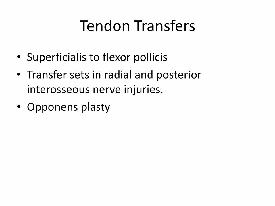
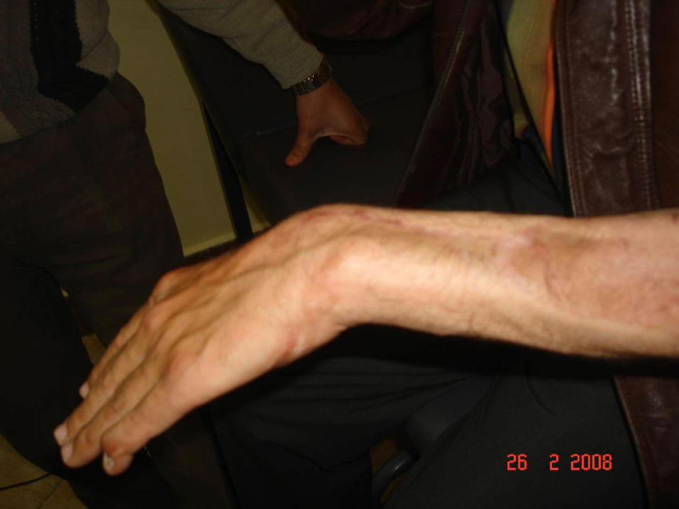
Tendon Transfers
• Superficialis to flexor pollicis
• Transfer sets in radial and posterior interosseous nerve injuries.
• Opponens plasty


Nerve iujuries
• Primary repair
• Delayed repair


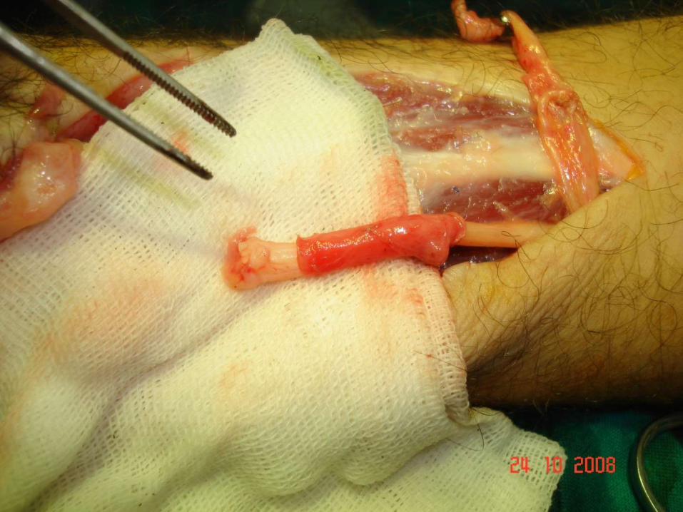
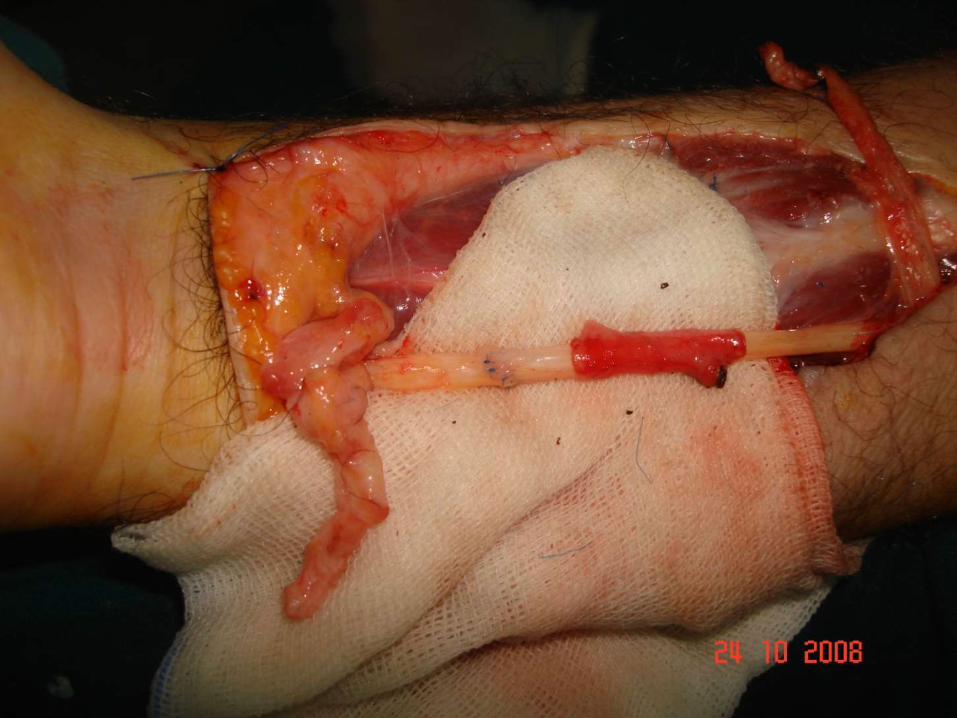


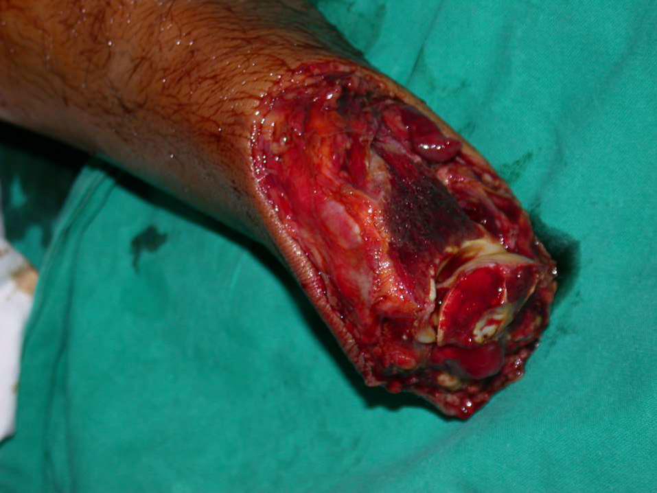
Vascular injuries
• Re-vasucularization
• Replantation

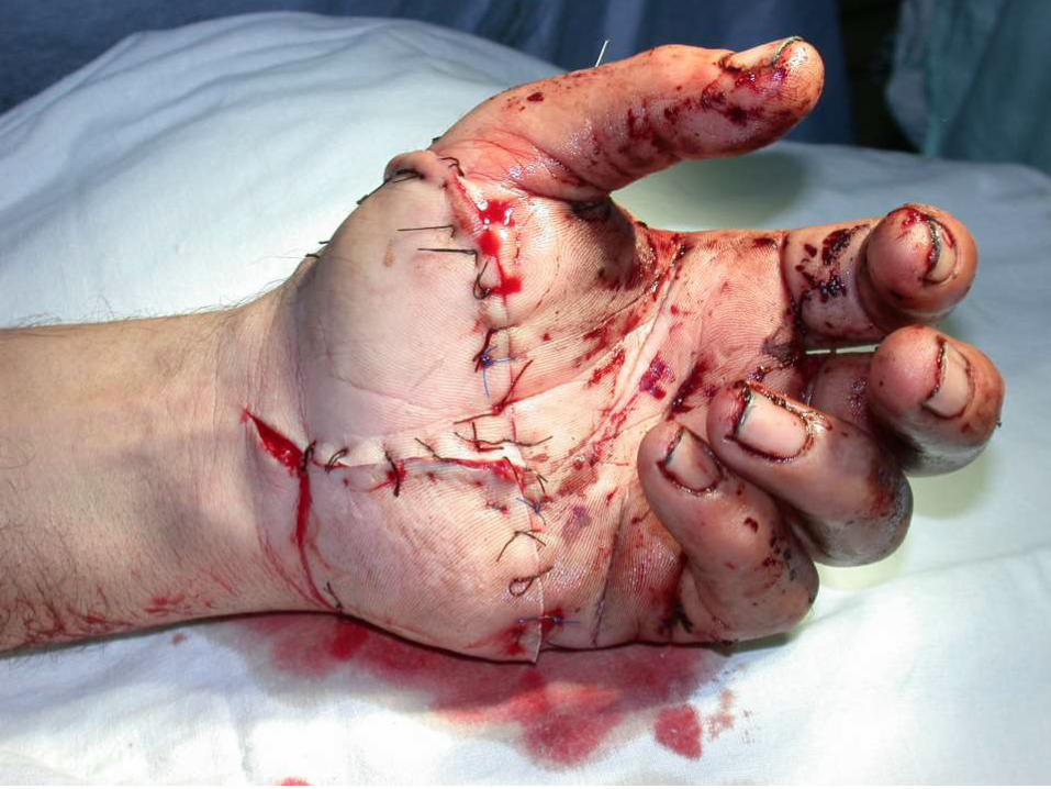


Replantation
• Replantation is the reattachment of a completely detached body part.
• Fingers and thumbs are the most common but the Hands, ear, scalp, hand, arm and penis have all been replanted.


• Generally replantation involves restoring blood flow, restoring the bony skeleton and connecting tendons and nerves as required.
• Initially, success was defined in terms of a survival of the amputated part alone.
• However, as more experience was gained in this field, surgeons began to understand that survival of the amputated piece was not enough
• In this way, functional demands of the amputated specimen became paramount in guiding which amputated pieces should and should not be replanted.