Hamzeh Alsalhi - HUMSC
16
Transcript of Hamzeh Alsalhi - HUMSC


▼Without treatment, recurring bleeding is common in survivors & prognosis worsens with each episode of bleeding. ►90% of saccular aneurysms occur in the anterior circulation
aneurysms multiple); 9-23Fnear major arterial branch points (exist in 20% to 30% of cases.
Ì Although they are sometimes referred to as congenital, they are not present at birth but develop over time because of underlying defects in the arterial media. ÌThere is an Ï risk of aneurysms (1) in individuals with
; & in autosomal dominant polycystic kidney disease(2) in an association with disorders of ECM
Ì The probability of rupture K with the aneurysm size, aneurysms >1cm have a roughly 50% risk of bleeding per year.
/ In the healing phase of subarachnoid H, meningeal
obstruction occur, sometimes leading to fibrosis & scarringinterruption of the normal pathways flow as well as of CSF
of CSF resorption.
_←←-
=
ismoreprone to rupture . @% 1 walls# aneurysm 11 f- >
⇒-
→ Leading toHydrocephat.IT
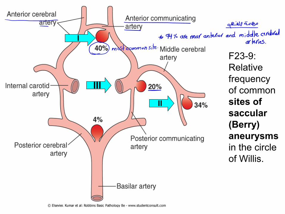
F23-9: Relative frequency of common sites of saccular (Berry) aneurysms in the circle of Willis.
l
ll
IIl
= = ↳sg-m_
* 94% are near anterior and middle cerebralarteries
.
② most common site
-

►GROSSLY, Ì Berry saccular aneurysm is a thin-walled outpouching of an artery. At the neck of the aneurysm, the muscular wall & intimal elastic lamina stop short & are absent from the aneurysm sac itself and the sac is made up of thickened hyalinized intima. The adventitia covering the sac
). 10-23Fis continuous with that of the parent artery (/ Rupture usually occurs at the apex of the sac with
, subarachnoid spaceextravasation of blood (mostly) into the .brain, or in bothor, less commonly in the substance of the
type of most common Berry saccular aneurysms are the Ä
intracranial aneurysm, in addition, Ð Less commonly, other types of intracranial aneurysms, include atherosclerotic (fusiform, mostly of the basilar artery,
the Mycotic, Traumatic, & Dissecting aneurysms,); & 9.39F latter 3, as with saccular aneurysms, are most often found in the anterior circulation. / The 4 aneurysms usually present with cerebral infarction from vascular occlusion instead of subarachnoid H.
#
↳@"1.* brains↳In-.€- ↳
intracranial .br
←

F23-10: Berry saccular aneurysms. A, View of the base of the brain, dissected to show the circle of Willis with an aneurysm of the anterior cerebral artery (arrow). B, Dissected circle of Willis to show the large aneurysm. C, Section through a saccular aneurysm showing the hyalinized fibrous vessel wall (H&E).
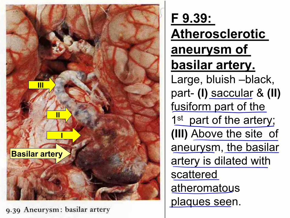
: 9.39F Atherosclerotic aneurysm of
basilar artery. Large, bluish –black, part- (I) saccular & (II) fusiform part of the 1st part of the artery; (III) Above the site of aneurysm, the basilar artery is dilated with scattered atheromatous plaques seen.
III
II
I
Basilar artery

Vascular Malformations Vascular malformations of the brain are classified into 4 main
of the abnormal vessels: nature types, based on the (1) Arteriovenous malformations (AVM), (2) Cavernous angiomas, (3) Capillary telangiectasias, (4) Venous angiomas. (1) Arteriovenous malformations (AVM), the most common, affect males twice as frequently as females; the lesion is most often recognized clinically between the ages of 10 & 30 years,
(intracerebral H) 2(epilepsy), Or ( seizure) 1as a ( presentingor subarachnoid, depending on their location). ÌLarge AVMs occurring in the newborn period can lead to high-output congestive heart failure because of blood shunting directly from arteries to veins. 1 The risk of bleeding makes AVM the most dangerous type
. )35-9& 11-23F(of vascular malformation
-
→The most common .
☐→ tumor. _&w@j
↳ Is >←€☒°&Hemorrhage.MTÉsb⑨.b
OR(FAnemiaate.ae#-ausI-ormation
or thyrotoxicosis, or Pagetdisease

F23-11: Arteriovenous malformation in subarachnoid space.
AVM
e-

F 9-35: Arteriovenous malformation (hamartoma) : brain. A large complex intracerebral AVM is present within the thalamus & basal ganglia. The greyish-white vessels are thick-walled &
many are thrombosed. The adjacent brain contains much brown hemosiderin pigment as a result of previous hemorrhages. ¥0

►GROSSLY, the AVMs resemble a tangled network of wormlike vascular channels involve vessels either in the
extending into brain )11-23Fsubarachnoid space ( (I)parenchyma or they may occur (II) exclusively within the brain (F9-35).
� H, they are enlarged BV separated by gliotic tissue, often with evidence of previous H. Some BVs can be recognized as arteries with duplicated & fragmented internal elastic lamina, while others show marked thickening or partial replacement of the media by hyalinized connective tissue.
(2) Cavernous hemangiomas consist of aggregates of distended, loosely organized vascular channels with thin
devoid of intervening nervous tissuecollagenized walls; (thus distinguishing them from capillary telangiectasias). They occur most often in the cerebellum, pons, & subcortical regions. Old foci of H , infarction, & calcification frequently surround the abnormal lesion.
É-→⇒
⇒-
Malformation wit
§9 tumorof Blts m
andno__capsule .
-e-- -

(3) Capillary telangiectasias (� 4.40) are microscopic foci of dilated, thin-walled vascular channels separated by relatively normal brain parenchyma & occurring mostly in the pons. (4) Venous angiomas (varices) consist of aggregates of ectatic (dilated) venous channels (F 9-34). Lesions 3 & 4 are unlikely to bleed or cause symptoms, & are most commonly discovered incidentally. Hypertensive cerebrovascular disease
-Over the past few decades there has been a Ð threshold for treatment of hypertension & more extensive screening for early disease, both of which have contributed to an overall Ð in the incidence of these complications. Nevertheless, hypertension continues to be important, due to poor patient compliance or inadequate access to health care. ►Hypertension affects the deep penetrating arteries & arterioles that supply the basal ganglia & hemispheric white matter & the brain stem. /4 Most important effects of hypertension on the brain are:
"⑨? -1*-4%-084 * ectasia :- .←É→
-aid:L£÷÷÷.D Ibibio £8
brain tissue .
¥*HhUMÉ↳&M&FmIighyprt *

� 4.40: Capillary telangiectasia: Brain X 80. A solitary lesion, consists of abnormally dilated capillaries, each with a very thin wall (arrow), surrounded by thin layer of eosinophilic hyaline amorphous material. The capillaries are separated by neural tissue & not by fibromuscular tissue (compare with those seen in an ordinary capillary/ cavernous hemangiomas). Complete resection of this lesion may be difficult or, impossible.
↳
↳%___

F 9-34: Venous angioma: brain, forming a complex tangle of dilated & thrombosed veins within the leptomeninges (arachnoid & pia mater) over the left parietal lobe. - This rare lesion is unlikely to bleed or cause symptoms
& is most commonly discovered incidentally.

(1) Massive hypertensive intracerebral H (2) Lacunar infarcts (3) Slit H (4) Hypertensive encephalopathy
in which hypertensive intracerebral H (see above), Massive) 1(
chronic hypertension is associated with the development of in vessels that microaneurysms Bouchard-Charcot minute
are less than 300 μm in ∅, these aneurysms can rupture, resulting in Spontaneous intraparenchymal H.
become arterioleswhich in, Hyaline arteriolar sclerosis) 2(
weaker than normal & are more vulnerable to rupture; the important clinical & pathologic outcome of which is the
. These small lacunar infarctsor lacunesdevelopment of cavitary infarcts are <15 mm, are found most commonly in deep gray matter (basal ganglia & thalamus); internal capsule, deep white matter, & pons, & they consist of tissue
laden macrophages & -, with scattered lipidcavitiesloss surrounding gliosis. Depending on their location in the CNS, lacunes can either be clinically silent or cause significant neurologic impairment.
a--
-
O-
--
€•÷:¥

(3) Hypertension also gives rise to rupture of the small-caliber penetrating vessels & the development of small H. In time, these H resorb, leaving behind a slitlike cavity (slit hemorrhage) surrounded by brownish discoloration. (4) Acute hypertensive encephalopathy is a clinicopathologic syndrome characterized by diffuse cerebral dysfunction, including headaches, confusion, vomiting, & convulsions, sometimes leading to coma. ÄRapid intervention to reduce the accompanying K intracranial pressure is required, since the syndrome does not usually remit spontaneously. 1 Postmortem examination of fatal cases show an edematous brain, with/or without transtentorial or tonsillar herniation. � H, Fibrinoid necrosis of arterioles & petechiae (of malignant hypertension) may be seen microscopically in the gray & white matter.
_I→_#
÷"-÷÷**-
①
µgµe**db.gs#&-M--.---

Vasculitis ►A variety of inflammatory processes that involve BV may lead to luminal narrowing & cerebral infarcts. Infectious arteritis of small & large BV was previously seen in association with syphilis & tuberculosis, but now more commonly occurs in the setting of immunosuppression & opportunistic infection (such as toxoplasmosis, aspergillosis, & CMV encephalitis). Ì Some of the systemic forms of vasculitis, such as polyarteritis nodosa (PAN), may involve cerebral BV & cause single or multiple infarcts throughout the brain.
ÌPrimary angiitis of the CNS is an inflammatory disorder that involves multiple, small to medium-sized parenchymal & subarachnoid vessels, & is characterized by chronic inflammation, multinucleated giant cells (with or without granuloma formation), & destruction of the BV wall. Affected individuals manifest a diffuse encephalopathic clinical picture, often with cognitive dysfunction; improvement occurs with steroid & immunosuppressive treatment.
goend- arteritis tuberuns
.
€.
=