H-FIB: Adjunctive Renal Denervation to Modify Hypertension ...
57
Transcript of H-FIB: Adjunctive Renal Denervation to Modify Hypertension ...

aleep
Typewritten Text
H-FIB: Ad
aleep
Typewritten Text
aleep
Typewritten Text
aleep
Typewritten Text
aleep
Typewritten Text
aleep
Typewritten Text
junctive Renal Denervation to Modify Hypertension and Sympathetic Tone as Upstream Therapy in the Treatment of Atrial Fibrillation
aleep
Typewritten Text
aleep
Typewritten Text
aleep
Typewritten Text
aleep
Typewritten Text
aleep
Typewritten Text
aleep
Typewritten Text
aleep
Typewritten Text
aleep
Typewritten Text
aleep
Typewritten Text
aleep
Typewritten Text
aleep
Typewritten Text
PI: Vivek Reddy, MD NCT01635998 Document Date: July 8, 2016
aleep
Typewritten Text

Vivek Reddy, M.D. H-FIB Protocol
Version 5.9 July 2016
1
PROTOCOL
H-FIB:
ADJUNCTIVE RENAL DENERVATION TO MODIFY HYPERTENSION AND SYMPATHETIC TONE AS
UPSTREAM THERAPY IN THE TREATMENT OF ATRIAL FIBRILLATION (HFIB)
Version 5.9
July 2016
Principal Investigator: Vivek Reddy, M.D.
Data Coordinating Center: Electrophysiology Clinical Research Group
Icahn School of Medicine at Mount Sinai New York

Vivek Reddy, M.D. H-FIB Protocol
Version 5.9 July 2016 2
STUDY NETWORK Data Coordinating Center Electrophysiology Clinical Research Group, Icahn School of Medicine at Mount Sinai (Vivek Reddy, MD, Betsy Ellsworth, MSN, ANP, Sam Cammack, MA, MPH) Study Chair Vivek Reddy, MD; Icahn School of Medicine at Mount Sinai Study Sponsors Vivek Reddy, MD Funding Agency Boston Scientific, Inc. Protocol Development Committee Vivek Reddy, MD Marc Miller, MD Emilia Bagiella, PhD Sam Cammack, MA, MPH Betsy Ellsworth, MSN, ANP

Vivek Reddy, M.D. H-FIB Protocol
Version 5.9 July 2016 3
TABLE OF CONTENTS 1. ABBREVIATIONS ................................................................................................................................... 8
2. ABSTRACT ............................................................................................................................................ 10
TABLE 1. SCHEDULE OF TREATMENTS AND TESTS ................................................................ 11
3. OBJECTIVES .......................................................................................................................................... 8
4. BACKGROUND ...................................................................................................................................... 9
5. SPECIFIC AIMS ................................................................................................................................... 12
6. STUDY DESIGN ................................................................................................................................... 13
7. RANDOMIZATION ............................................................................................................................. 13
8. MASKING ............................................................................................................................................. 14
9. STUDY POPULATION ........................................................................................................................ 20
10. TREATMENT INTERVENTIONS .................................................................................................... 21
11. DEFINITIONS AND MEASUREMENT OF ENDPOINTS ............................................................ 20
12. CLINICAL CENTERS ....................................................................................................................... 32
13. SCREENING AND BASELINE DATA COLLECTION ................................................................ 33
14. POST-RANDOMIZATION DATA COLLECTION ........................................................................ 36
15. DATA MANAGEMENT ..................................................................................................................... 38
16. ANALYTICAL PLAN ........................................................................................................................ 40
17. ORGANIZATION OF THE STUDY ................................................................................................. 50
18. REFERENCES .................................................................................................................................... 52
APPENDIX I: NEW YORK HEART ASSOCIATION CLASSIFICATION (NYHA) ....................... 51
APPENDIX II: QUALITY OF LIFE MEASURES ................................................................................ 52
APPENDIX III: TRANSTHORACIC DOPPLER ECHOCARDIOGRAPHY .................................... 54
APPENDIX IV: BLOOD PRESSURE TREATMENT GUIDELINES ................................................. 55
APPENDIX V: STUDY SITES ................................................................................................................. 55
APPENDIX VI: LIST OF APPROVED ATRIAL FIBRILLATION ABLATION DEVICES ........... 55

Vivek Reddy, M.D. H-FIB Protocol
Version 5.9 July 2016 4
1. ABBREVIATIONS
AAD Anti-arrhythmic drugs ACC American College of Cardiology ACT Activated Clotting Time AE Adverse Event AF Atrial Fibrillation AFEQT Atrial Fibrillation Effect on Quality-of-Life AFL Atrial flutter AHA American Heart Association ALT Alanine transaminase ARBs Angiotensin receptor blockers ARDS Acute Respiratory Distress Syndrome AST Aspartate aminotransferase AV Atrioventricular BNP Brain natriuretic peptide BP Blood Pressure BSA Body surface area CABG Coronary artery bypass grafting CFAE Complex fractionated atrial electrograms CHF Congestive heart failure CMC Clinical Management Committee CT Computerized tomography CTI Cardiac Troponin I CV Curriculum Vitae CVP Central Venous Pressure DC Direct current DCC Data Coordinating Center DSMB Data Safety Monitoring Board EAC Event Adjudication Committee ECG Electrocardiogram eCRFs Electronic Case Report Forms EDC Electronic Data Capture eGFR Estimated Glomerular Filtration Rate EP Electrophysiology FDA Food and Drug Administration GCP Good Clinical Practice HIPAA Health Insurance Portability and Accountability Act HRS Heart Rhythm Society ICU Intensive Care Unit INR International Normalized Ratio IRB Institutional Review Board I/O Input/Output
Vivek Reddy, M.D. H-FIB Protocol
Version 5.9 July 2016 5
InCHOIR International Center for Health Outcomes & Innovation Research JNC-7 Joint National Committee 7 LA Left atrium LAA Left atrial appendage LBBB Left bundle branch block LMW Low molecular weight LV Left ventricle LVEF Left ventricular ejection fraction MACE Major adverse cardiac event MDRD Modification of Diet in Renal Disease MI Myocardial infarction MR Mitral regurgitation MRI Magnetic resonance imaging MV Mitral valve NIHSS National Institutes of Health Stroke Scale NYHA New York Heart Association PCI Percutaneous coronary intervention PV Pulmonary vein OHRP Office of Human Research Protections OR Operating Room QoL Quality of Life RBC Red blood cells RF Radiofrequency RVAD Right Ventricular Assist Device SAE Serious Adverse Event SF-12 Short Form – 12 SSL Secure Socket Layer ST-T ST segment/T wave SVC Superior vena cava TDI Tissue Doppler Imaging TEE Trans-esophageal echocardiography TIA Transient Ischemic Attack TTE Trans-thoracic echocardiography UADE Unanticipated adverse device effect UP Unanticipated Problem URL Upper Reference Limit

Vivek Reddy, M.D. H-FIB Protocol
Version 5.9 July 2016 6
2. ABSTRACT Objectives Determine the role of adjunctive blood pressure management and sympathetic modulation
attained via renal sympathetic denervation in the prevention of AF recurrence in patients with hypertension scheduled for an AF ablation procedure. Patients will be randomized to either AF catheter ablation (usual therapy) or AF catheter ablation plus renal sympathetic denervation.
Study Design Prospective, placebo-controlled, double-blind, randomized (1:1) clinical trial; patients randomized to either catheter ablation (control group) or a catheter ablation + renal denervation (study group).
Target Population History of documented significant hypertension (defined as SBP ≥160 mm Hg and/or DBP ≥100 mmHg) and receiving treatment with at least one anti-hypertensive medication, or a known history of hypertension and receiving treatment with at least two anti-hypertensive medications (specifically for hypertension).
Rx arms AF catheter ablation alone versus AF catheter ablation plus renal sympathetic denervation Sample Size Total: 300 patients: provides 80% power to detect a 40% decrease in AF recurrence (24%
versus 40%) (measured at 12 months) This study is divided into two phases: an initial PILOT phase (to include 50 patients) followed by the remaining 250 patients to a total of 300 patients. The PILOT phase will be conducted at up to 10 US sites. An interim safety analysis will be conducted when all patients in the PILOT phase (50 subjects) have completed the 3-month follow up visit. The purpose of this analysis is only for safety. The efficacy/outcome data for the primary endpoint will not be examined; this will permit pooling these 50 patients with the subsequent 250 patients for the final efficacy analysis.
Duration 24 months follow-up following randomization. 1 Endpoints Single-procedure freedom from AF recurrence off all AADs (after the 90 day blanking
period) at 12 months. 2 Endpoints AAD-free single-procedure freedom from AF recurrence 24 months (not-including
recurrences within the first 90 days of the initial ablation procedure); freedom from AF recurrence at 24 months (not-including a 90 day blanking period) despite taking AADs; blood pressure control between the two groups as compared to baseline at t = 6 months, 12 months and 24 months; Major Adverse Cardiac Events (MACE); differences in measures of LV hypertrophy (LV wall thickness, mitral inflow parameters, LV mass index2.7) and LA size recorded via TTE at baseline and at 12 months for each patient; procedure adverse events; total number of anti-hypertensive medications at study end, compared between the 2 treatment arms; QOL.
Inclusion Criteria Age ≥ 18; history of AF and planned for a catheter ablation procedure; history of hypertension and on at least one anti-hypertensive medication; accessibility of renal vasculature (as determined by intra-procedural renal angiography); Ability to understand the requirements of the study; Willingness to adhere to study restrictions and comply with all post-procedural follow-up requirements
Exclusion Criteria Inability to undergo AF catheter ablation (e.g., presence of a left atrial thrombus, contraindication to all anticoagulation); Prior left atrial ablation for an atrial arrhythmia (before this index procedure); NYHA class IV congestive heart failure; Individual has known secondary hypertension; Individual has renal artery anatomy that is ineligible for treatment including: a) Inability to access renal vasculature, b) Main renal arteries < 3 mm in diameter or < 20 mm in length., c) Hemodynamically or anatomically significant renal artery abnormality or stenosis in either renal artery which, in the eyes of the operator, would interfere with safe cannulation of the renal artery or meets standards for surgical repair or interventional dilation, d) History of prior renal artery intervention including balloon angioplasty or stenting that precludes a possibility of ablation treatment; Individual has an estimated glomerular filtration rate (eGFR) of less than 45mL/min/1.73m2, using the MDRD calculation; Individual has a single functioning kidney (either congenitally or iatrogenically); Individual is pregnant or nursing; Life expectancy <1 year for any medical condition

Vivek Reddy, M.D. H-FIB Protocol
Version 5.9 July 2016 7
Table 1. Schedule of Treatments and Tests
Baseline Procedure Discharge 3, 6, 12, 18 & 24 mos
Event Driven
Type of visit Office Hospital Office
Informed Consent X
Brief History & Physical X X X
Office BP Measurement X X
Medications X X X
Blood chemistry X 6 & 12 mo only
Quality-of-Life Assessment X 12 & 24 mo only
TTE ф X 12 mo only
ECG X X
Continuous ECG Monitoring (7 days each)
3 mo, 6 mo, 12 mo, 24 mo
only
Renal Doppler 6 mo only
Blinding assessment 12 mo only
Randomization* X
Hospital Admission X X
EP Mapping & AF Ablation X X
Renal Angiography X X Renal Sympathetic Denervation § X
Adverse Events X
AF recurrence X
Missed Visit Assessment X
Mortality X
Study Completion/Early Termination X ф Performed within 3 months of randomization (i.e., within 3 months of the planned ablation procedure). * Randomization will occur after renal angiogram. § The blinding of the patient is maintained by the fact that the renal angiogram and renal denervation steps will be performed with the same sedation as used for the ablation procedure: either deep sedation or general anesthesia (per the usual protocol of the EP laboratory).
Vivek Reddy, M.D. H-FIB Protocol
Version 5.9 July 2016 8
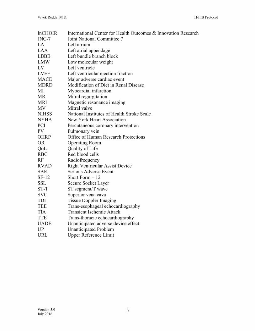
3. OBJECTIVES Purpose of the Study The objective of the H-FIB trial is to determine the role of renal sympathetic denervation in the prevention of Atrial Fibrillation (AF) recurrence in patients with hypertension for whom a catheter-based AF ablation procedure is planned. Patients will be randomized to either AF catheter ablation (usual therapy) or AF catheter ablation plus renal sympathetic denervation.
3.1 Primary Hypothesis In hypertension patients with atrial fibrillation, there is ample evidence of a strong association between the chronic hypertension and arrhythmia-inducing cardiac substrate modifications; the association seems related to both hypertension itself (along with derangements in the renin-angiotensin-aldosterone system), and elevated sympathetic tone. It is hypothesized that a treatment strategy targeting both these ancillary conditions (hypertension and elevated sympathetic tone) and atrial fibrillation triggers themselves may lead to greater and more durable procedural success post-catheter ablation. In hypertensive patients undergoing catheter ablation for AF, we hypothesize that there will be a difference through 12 months between the two treatment arms in AAD-free freedom from recurrent AF after a 90 day blanking period following a single AF ablation procedure. The null hypothesis of principal interest is that there will be no difference through 12 months between the two treatment arms in single-procedure freedom from AF off of all AADs after a 90 day blanking period in patients randomized to AF ablation with a single renal sympathetic denervation procedure compared to patients randomized to AF ablation alone.
3.2 Secondary Aims
1. Compare AAD-free single-procedure freedom from AF recurrence through 24 months (not-including recurrences within the first 90 days of the initial ablation procedure)
2. Compare freedom from AF recurrence through 24 months regardless of antiarrhythmic drug (AAD) use (excluding the 90 day blanking period)
3. Examine blood pressure control between the two groups as compared to baseline at 6 months, 12 months and 24 months
4. Examine differences between treatment groups in measures of LV hypertrophy (LV wall thickness, mitral inflow parameters) and LA size by TTE at baseline and at 12 months for each patient
5. Procedure-related adverse events 6. Major Adverse Cardiac Events (MACE) 7. Serious Adverse Events 8. Compare anti-hypertensive medication use in the two treatment arms at 24
months. 9. Compare quality of life between treatment arms and differences in quality of life
from baseline using a disease specific scale (AFEQT) at 12 and 24 months.

Vivek Reddy, M.D. H-FIB Protocol
Version 5.9 July 2016 9
4. BACKGROUND There is a wealth of epidemiological, pre-clinical and clinical data that suggests that both hypertension and sympathetic overdrive have important effects on the cardiovascular system that increases the propensity for atrial fibrillation. Furthermore, there is pre-clinical and early clinical data that indicate that this relationship can be modulate by catheter-based renal sympathetic denervation (by virtue of its effects on both the renin-angiotensin-aldosterone system and sympathetic tone). The following details some of these preliminary data which provides the scientific basis for this proposed HFIB clinical trial. Hypertension and Atrial Fibrillation Aside from its obvious impact on renal function, chronic hypertension significantly increases the risk for stroke, coronary artery disease, heart failure, and vascular disease, and is believed to mediate the pathogenesis and progression of atrial fibrillation (AF) via its remodeling effects on cardiac anatomy. This inextricable link between hypertension and cardiovascular health has been well described, as has their combined effect on up to 40% of the aging, obesity-battling Western world. Furthermore, from the perspective of a cardiovascular electrophysiologist, the prevalence of hypertension has important implications for the prognosis of patients with cardiac arrhythmias—particularly, drug-resistant AF. Despite significant advances in the field of catheter ablation for AF, the prevailing evidence suggests that even when procedural success can be obtained acutely, a considerable rate of PV electrical reconnection is observed over time. This is further supported by a 15–50% rate of clinical AF recurrence during follow-up; and as demonstrated by several recent studies which have extended follow-up to 6 years, clinical recurrence is even more likely with increasing scrutiny and duration of follow-up. The situation is more complicated for patients with persistent AF—the treatment of which has been less effective with PV isolation alone. Although the pathogenesis is not completely understood, hypertension is believed to mediate the pathogenesis and progression of paroxysmal to persistent AF, via its remodeling effects on cardiac anatomy. This includes left ventricular hypertrophy, impaired ventricular filling, left atrial enlargement, and alteration of atrial electrophysiology. It is therefore reasonable to assume that limiting or reversing such morphological changes could have a profound effect on the effectiveness and durability of currently available ablative therapies for AF. Indeed, there is a substantial (albeit at times, a conflicting) body of evidence suggesting that treatment with “positively” remodeling agents (such as antagonists of the renin-angiotensin system) may decrease AF recurrence in patients after cardioversion or as adjunctive treatment with antiarrhythmic drugs. Renal sympathetic denervation is a therapeutic modality that has been shown to affect the sympathetic state in various pre-clinical and clinical disease states. While Symplicity HTN-3 was overall a negative study for the control of resistant hypertension, there are

Vivek Reddy, M.D. H-FIB Protocol
Version 5.9 July 2016 10
compelling data suggesting that one of the most important reasons for the negative outcome was technical: namely, the inability of point-by-point radiofrequency ablation to achieve circumferential denervation of the peri-renal sympathetic fibers. To this point, as opposed to the initial (and somewhat crude) spot radiofrequency ablation catheter employed in that trial, there are now a number of other renal denervation systems specifically designed to safely deliver a circumferential array of ablative lesions (that in pre-clinical animal models, is very effective in achieving excellent renal denervation). Potential Role of renal sympathetic denervation as an adjunct therapy in the treatment of AF: Potential benefits beyond BP control.
Multiple pre-clinical and clinical studies have demonstrated that the sympathetic nervous system (SNS) plays an important role in the initiation and maintenance of AF, especially in patients with any degree of structural heart disease 3,4. Increases in sympathetic tone frequently precede the onset of AF 5, 6, 7, and excessive sympathetic nervous activation (assessed by 123I-mIBG scintigraphy) can predict recurrences of AF after catheter ablation 8. One explanation for the ‘pro-fibrillatory’ effects of the sympathetic nervous system is the distribution of cardiac autonomic innervations. The PV-LA junction has abundant sympathetic innervations, which become the dominant sites for the origin of focal atrial tachyarrhythmias in response to heightened sympathetic tone 9. Low concentrations of tyramine, which activates sympathetic neurons by releasing norepinephrine from nerve endings, can initiate spontaneous AF by early after-depolarization-mediate triggered activity from the LA-PV junction 11. Following catheter ablation, or rapid atrial pacing, the PV-LA junction can undergo further neural remodeling (i.e. sympathetic hyperinnervation), potentially making these sites even more sensitive to adrenergic stimuli 11,12. The importance of the autonomic nervous system in mediating AF is further strengthened by the results of interventions intended to inhibit sympathetic outflow to the heart. Low-level vagus nerve stimulation, which suppresses stellate ganglion nerve activities, significantly reduces the frequency of paroxysmal atrial tachyarrhythmias in ambulatory canines 13. This excessive sympathetic nervous activation, which contributes to the pathogenesis of atrial fibrillation, is a potential target of renal sympathetic denervation, beyond just the control of blood pressure.
An additional consideration is the effect of renal sympathetic denervation on
cardiac structure and function. In patients with essential hypertension, increased left ventricular mass (LVM) is an independent predictor of both AF and cardiovascular outcomes. Furthermore, reductions in LVM are associated with prognostic benefits, independent of reductions in blood pressure 14,15. To this end, Brandt and colleagues recently demonstrated that renal sympathetic denervation significantly reduced LVM and improved diastolic function; two established risk factors for the development and maintenance of AF 16. Interestingly, even in a small group of BP ‘non-responders’, there was marked reductions on LVM and improvements in diastolic parameters. Regression of LVM during antihypertensive therapy is associated with a lower likelihood of new-onset AF, independent of blood pressure lowering and treatment modality in essential hypertension 17. In total, these studies suggest that renal sympathetic denervation has the

Vivek Reddy, M.D. H-FIB Protocol
Version 5.9 July 2016 11
potential to reduce the frequency of AF recurrences in patients with significant essential hypertension who are undergoing catheter ablation of AF. Furthermore, there is now a substantial body of pre-clinical data demonstrating that in animal models, renal denervation has profound effects on both atrial electrophysiology and atrial ultrastructure (decreasing fibrosis). And as a consequence, renal denervation has favorable effects on AF initiation and duration in animal models of AF. Preliminary Data & Rationale for this Study A single-center randomized controlled trial examined the effects of adjunctive renal denervation in a group of patients with resistant hypertension and atrial fibrillation 18. In this study, 27 patients were enrolled, 14 were randomized to AF ablation only and 13 were randomized to AF ablation with renal artery denervation. Nine of the 13 patients (69%) treated with PVI with renal denervation were AF-free at the 12-month post-ablation follow-up examination versus 4 (29%) of the 14 patients in the PVI-only group (p = 0.033). While this was admittedly a small study, and included the unusual population of patients with both AF and resistant hypertension, it nonetheless was the proof-of-principle that adjunctive renal denervation can have potent antiarrhythmic effects. Furthermore, a more recent study examined the link between sympathetic system overdrive and atrial arrhythmogenesis, independent of hypertension control.19 In this study of patients undergoing catheter ablation of AF, the investigators tested whether moxonidine, a centrally-acting sympathoinhibitory agent, can similarly lead to a reduction in post-ablation AF recurrence. In this prospective, double-blinded study, 291 hypertensive patients were randomized to receive either moxonidine (0.2-0.4 mg daily) or placebo, along with standard antihypertensive treatment. Despite the fact that there were no significant differences in blood pressure between the two groups, the 12-month AF recurrence rates were 36.9% in the AF ablation alone group and 20.0% in the AF ablation + moxonidine group (p=0.007). This study provides addition clinical proof-of-principle that modification of the sympathetic system would provide ameliorative anti-arrhythmic effects post-AF ablation even without significant effects on the blood pressure. Finally, we conducted a pilot H-FIB study (FDA IDE# G120025) which revealed interesting outcomes. This single-blind, multicenter (10 sites) study enrolled patients with a history of significant hypertension (defined as a SBP ≥ 160 mmHg and/or DBP ≥ 100 mmHg) and receiving treatment with at least one anti-hypertensive medication, and scheduled to undergo AF ablation. After AF ablation, patients were randomized to no further therapy or renal denervation using an off-the-shelf radiofrequency ablation catheter (Celsius Thermocool, Biosense-Webster, Inc). Briefly, 13 patients were randomized to AF ablation + renal denervation (study group) and 17 patients to AF ablation only (control group). After a mean of 15 months of follow-up, freedom from AF was achieved in 77% (10/13) of the study group compared to 53% (9/17) of Controls. While not statistically significant, these data are nonetheless quite compelling. However, there were significant safety events that prompted us to halt the study: 3 patients with

Vivek Reddy, M.D. H-FIB Protocol
Version 5.9 July 2016 12
renal artery dissection, and 3 patients with significant post-ablation renal artery stenosis (>60%). Thus, while the off-the-shelf ablation catheter had been safe in our single-center experience, we were not able to replicate these favorable safety results in the multicenter study. Together, these data suggest that the H-FIB study should be re-started utilizing a dedicated renal denervation catheter. To this point, the H-FIB trial will re-start using the Vessix renal denervation system. Patients meeting the inclusion / exclusion criteria will undergo a catheter ablation procedure for AF according to the standard practice. During the procedure, they will additionally undergo a renal angiogram to assess the accessibility of the renal vasculature. Assuming that the anatomy is suitable for renal denervation, the patient will be randomized to receive either: (1) AF ablation + catheter-based renal sympathetic denervation, or (2) AF ablation alone. Both groups will be followed for two years. The primary endpoint is single-procedure survival free from AF (without taking AADs) through 12 months, with a 90 day blanking period. The H-FIB trial will be conducted in up to 20 participating sites in the United States and Europe (10 sites for the initial 50 patients). The aim of H-FIB is to assess the potential therapeutic and quality-of-life benefit of performing hypertension-associated cardiac substrate modification, in addition to the standard practice of catheter ablation of atrial fibrillation. It is believed that because both hypertension and sympathetic overdrive both independently (and likely synergistically) allow for the genesis and/or propagation of atrial fibrillation, significantly reversing and/or reducing the effects of such atrial remodeling by renal denervation will improve the durability of procedural success after an ablation. 5. SPECIFIC AIMS
5.1 Primary Endpoint Efficacy The primary endpoint of this study is AAD-free single-procedure freedom from AF recurrence through 12 months (not including a 90 day blanking period).
5.2 Secondary Endpoints AAD-free single-procedure freedom from AF recurrence through 24 months (not-including recurrences within the first 90 days of the initial ablation procedure). Freedom from AF recurrence through 24 months (not-including the pre-defined 90 day blanking period) despite taking AADs. Blood pressure control between the two groups as compared to baseline at 6 months, 12 months and 24 months.

Vivek Reddy, M.D. H-FIB Protocol
Version 5.9 July 2016 13
Major adverse cardiac events (MACE)
- defined as a non-weighted composite score of: death, stroke, CHF hospitalization, clinically diagnosed thromboembolic events other than stroke and hemorrhage requiring transfusion within 12 months of randomization
Serious Adverse Events Differences in measures of LV hypertrophy (LV wall thickness, mitral inflow parameters) and LA size Procedure adverse events Total number of anti-hypertensive medications at study end, compared between the two treatment arms Quality of Life 6. STUDY DESIGN This is a prospective, multi-center, double-blinded, randomized trial. The trial will be conducted in up to 20 clinical sites from both within and outside the United States, for a total of 300 randomized patients. During the enrollment of the initial 50 patients, the number of sites will be 10. Accrual is expected to take 24 months, and all patients will be followed for 24 months post randomization.
7. RANDOMIZATION
All patients who have given informed consent for the study will undergo catheter ablation for AF, as this is the standard care they would receive regardless of enrollment in the study. During the procedure, a renal angiogram will be performed to assess the accessibility of the renal vasculature, and its appropriateness for catheter-based renal sympathetic denervation. If the renal anatomy meets the protocol defined dimensions, the patient will then undergo randomization. Patients will be randomized to one of two treatment groups:
Group 1: catheter ablation of atrial fibrillation plus renal sympathetic denervation Group 2: catheter ablation of atrial fibrillation (control group)
Patients will be randomized in a 1:1 fashion. By this arrangement, 50% of the patients will undergo renal sympathetic denervation (Group 1) and 50% will not (Group 2). All patients will undergo AF catheter ablation.

Vivek Reddy, M.D. H-FIB Protocol
Version 5.9 July 2016 14
Randomization Procedure A DCC representative will be available to discuss any questions regarding patient eligibility. Once the site investigator has confirmed that the patient meets all eligibility criteria for participation in the trial, and has completed the eligibility forms in the Electronic Data Capture (EDC) system, randomization will be performed electronically. Randomization to the study assignment will be generated by the EDC system once the checklist of inclusion and exclusion criteria has been completed and verified. For the purpose of the primary analysis, patients are considered enrolled in the study once they have signed informed consent.
8. MASKING The operating physician will be aware of the randomization assignment, but the patient will not. The blinding of the patient is maintained by the fact that the renal angiogram and renal denervation steps will be performed with the same sedation as used for the ablation procedure. This is typically either deep sedation or under general anesthesia (per the usual protocol of the EP laboratory); this amount of anesthesia used for the AF ablation procedure is sufficient for the discomfort felt during the renal denervation procedure. This approach has been selected to minimize (1) study bias and (2) placebo effect. The randomization schema will be blocked by size for each site. Core labs will also be blinded to treatment assignment. Adverse events will be adjudicated by an Event Adjudication Committee and trial oversight will be provided by an independent DSMB. Those assessing the primary outcome will be blinded to patients' treatment assignment. 9. STUDY POPULATION
Characterization of Patient Population The patient population for this trial consists of patients with atrial fibrillation and a history of hypertension. All patients should be planned for a catheter ablation procedure for atrial fibrillation. All patients who meet the eligibility criteria may be included in the study regardless of gender, race or ethnicity. Inclusion Criteria
1. Age ≥ 18 years of age 2. History of AF (paroxysmal or persistent) and planned for a guideline-
supported catheter ablation procedure 3. History of significant hypertension (defined as SBP ≥160 mm Hg and/or
DBP ≥100 mmHg) and receiving treatment with at least one anti-hypertensive medication,
or Clinical History of hypertension and receiving treatment with at least two anti-hypertensive medications (specifically for blood pressure reduction)
4. Renal vasculature is accessible as determined by intra-procedural renal angiography.

Vivek Reddy, M.D. H-FIB Protocol
Version 5.9 July 2016 15
5. Ability to understand the requirements of the study 6. Willingness to adhere to study restrictions, comply with all post-
procedural follow-up requirements and to sign informed consent
Exclusion Criteria 1. Inability to undergo AF catheter ablation (e.g., presence of a left atrial
thrombus, contraindication to all anticoagulation) 2. Prior left atrial ablation for an atrial arrhythmia (before this index
procedure) 3. Patients with NYHA class IV congestive heart failure 4. Individual has known secondary hypertension 5. Individual has renal artery anatomy that is ineligible for treatment
including: a. Inability to access renal vasculature b. Main renal arteries < 3 mm in diameter or < 20 mm in length. c. Hemodynamically or anatomically significant renal artery abnormality
or stenosis in either renal artery which, in the eyes of the operator, would interfere with safe cannulation of the renal artery or meets standards for surgical repair or interventional dilation.
d. A history of prior renal artery intervention including balloon angioplasty or stenting that precludes a possibility of ablation treatment.
6. Individual has an estimated glomerular filtration rate (eGFR) of less than 45mL/min/1.73m2, using the MDRD calculation.
7. Individual has a single functioning kidney (either congenitally or iatrogenically).
8. Individual is pregnant or nursing. 9. Life expectancy <1 year for any medical condition
Recruitment Strategies Recruitment strategies may include: mailings to referring physicians of the study hospitals, symposia and health care events targeted towards this population; as well as telephone calls to neighboring health care facilities. The DCC will regularly assess actual enrollment in relation to pre-specified goals, and additional interventions to increase enrollment will be implemented as needed. The Screening Log will identify numbers of patients screened and reasons for non-enrollment in the trial. Roll-in phase Each site has the option of enrolling two patients as part of a roll-in phase. Each roll-in patient will have AF ablation, renal angiography and renal sympathetic denervation performed. Patient data on these roll-in patients will not be used for the final analysis.
• Roll-in patients will be required to sign the study ICF, but will not be randomized.
• Although the data will not be used for the primary analysis, patients will asked to adhere to the same follow up schedule of visits as randomized patients.

Vivek Reddy, M.D. H-FIB Protocol
Version 5.9 July 2016 16
• As per FDA, roll-in patients will count toward the enrollment ceiling.
• The CRF must be adjusted to clearly reflect roll-in patients from randomized patients.
10. TREATMENT INTERVENTIONS Catheter Ablation for Atrial Fibrillation
• The approach to catheter ablation of AF should be performed according to the EP lab standards. A few general comments (and restrictions) are noted below:
• Patient anesthesia will be administered according to standard EP lab protocol. • Arterial and venous access will be achieved through cannulation of the right
and/or left femoral arteries and veins as per the usual practice of the treating electrophysiologist.
• Full systemic anticoagulation will be instituted as per standard hospital
procedures to a target ACT of approximately 300 seconds or greater (or equivalent level depending on testing system performed). As per the most recent AF consensus document 20, pre-procedural anticoagulation management should include performing the ablation on therapeutic oral anticoagulation – e.g.,: warfarin with a goal INR 2-3, or one of the newer oral anticoagulants: dabigatran, rivaroxaban, apixaban, or edoxaban (either continuing one of these agent to the time of the procedure, or discontinuing it 3 to 5 days prior to ablation, followed by periprocedural bridging using low-molecular-weight heparin). These recommendations apply to the cardiac ablation. Consistent with the Symplicity-1 and -2 trials, there are no specific additional recommendations pertaining to pre-procedural anticoagulation prior to or following renal sympathetic denervation.
Patients in the study will include patients with paroxysmal AF and non-paroxysmal AF. The AF ablation procedure will be performed with any catheter approved for that such specific indication or per clinical practice at the study site for that indication (i.e., for non-paroxysmal AF). For US and OUS sites, a list of recommended catheters is in Appendix V. OUS sites may use catheters which have been approved in their jurisdiction for that indication. • All Patients: Pulmonary vein isolation with an approved irrigated radiofrequency
ablation catheter, laser ablation or cryoballoon ablation catheter (as per the usual practice of the treating physician). PV isolation must be confirmed by a multi-electrode mapping catheter after placing the last ablation of the PV-isolating lesion set.
➢ Paroxysmal AF Patients:
▪ Additional ablation may be performed to target non-PV triggers ▪ SVC isolation is permitted (as per the physician’s usual practice)

Vivek Reddy, M.D. H-FIB Protocol
Version 5.9 July 2016 17
▪ A typical flutter CT isthmus line may be placed in patients with either a history of ECG-determined typical flutter or induced typical flutter during the procedure
▪ One should not perform additional targeting of CFAE sites or the placement of linear lesions
➢ Non-Paroxysmal AF Patients:
▪ This category includes all patients with AF sustained for ≥ 7 days, and requiring termination by pharmacological or electrical cardioversion
▪ Additional ablation may be performed to target non-PV triggers, SVC isolation, linear lesions (e.g., CT isthmus line, Mitral Isthmus line, Roof line)
▪ Additional targeting of rotor activity using approved mapping technology is permitted
▪ Ablation of CFAE sites is not permitted
• As per usual practice of the treating physician, an approved catheter navigation system (such as the robotic or magnetic navigation system) may be employed for AF ablation.
• During radiofrequency ablation, power titration should be performed as per the physician’s usual practice to avoid thromboembolic complications or perforation. One reasonable strategy includes careful power titration to achieve an impedance drop not exceeding 10%. In general, the tip temperature should not exceed 45°C, and preferably should stay below 42°C.
• During cryoballoon ablation, ablation lesions should be placed as per usual practice (2-4 min lesions/PV). During ablation of the right pulmonary veins, as per the center’s usual practice, a strategy should be used to minimize the possibility of phrenic nerve injury. Two examples of such appropriate strategies include: i) pacing the phrenic nerve from the SVC during ablation and monitoring of diaphragmatic excursion to indicate phrenic nerve integrity, and ii) monitoring diaphragmatic excursion with free breathing during ablation to indicate phrenic nerve integrity.
• During laser ablation, the CardioFocus HeartLight Catheter must be used according to the guidelines set in the FDA approved Instructions for Use 21. Phrenic nerve pacing is recommended to avoid injury to the phrenic nerve. As per the IFU, one method for conducting pacing is to pace from the superior vena cava once the Balloon is inflated near the right superior pulmonary vein (RSPV) and if desired, the right inferior pulmonary vein (RIPV) to assess the location of the phrenic nerve by observing any “capture” at the target energy delivery site. It is advised that pacing be conducted at high output to ensure capture of the phrenic nerve. When pacing prior to energy delivery, the site of diaphragmatic stimulation (and therefore the approximate phrenic nerve location) can be

Vivek Reddy, M.D. H-FIB Protocol
Version 5.9 July 2016 18
compared via fluoroscopy or ICE with the target energy delivery site. Energy should not be delivered to any area where there is phrenic nerve capture. Alternatively, concomitant phrenic nerve pacing can be performed using a pacing catheter in the SVC during laser balloon energy delivery to the RSPV (or RIPV). When used, energy delivery must be discontinued if movement of the diaphragm slows or changes during energy delivery.
• During the ablation, an intravenous heparin infusion will be maintained for a
target ACT >300 seconds. ACTs should be monitored every 30-60 minutes (per usual practice).
Renal Angiography
• A renal angiogram will be performed after the AF ablation procedure is complete. Once the renal arteries are found to be acceptable for ablation, randomization will occur. Once angiography is performed, no additional atrial ablation is permitted.
• Following completion of the cardiac ablation and renal angiography, if the patient
was randomized to the control group, the heparin infusion will be discontinued. The arterial and venous sheaths will be removed and hemostasis achieved with manual pressure (protamine may be used to facilitate this) and/or percutaneous vascular closure devices (as per the institution’s practice). If the patient was randomized to the renal sympathetic denervation arm (study group), these steps will be performed after the renal artery ablation has been completed.
Renal Sympathetic Denervation
• After confirmation of eligibility, the radiofrequency ablation catheter will be introduced into each renal artery.
• Despite some variability in renal vascular anatomy, the method for performing the procedure is fairly straightforward. An aortogram is performed to visualize the renal vessels and deem suitability (no significant renal artery stenosis or atherosclerosis, etc.).
• The renal ablation procedure will be performed using the VessixTM Renal Denervation System (Boston Scientific, Quincy, Massachusetts). The operating physician will manipulate the ablation catheter into the renal arteries as per standard operating procedures.

Vivek Reddy, M.D. H-FIB Protocol
Version 5.9 July 2016 19
• The Vessix catheter is available in various sizes, and can be used to treat arteries between 3 mm and 7 mm in diameter and more than 20 mm in length. Radiofrequency energy is delivered via 8 electrodes to four distinct sites within the artery in a bipolar fashion. As with other renal denervation procedures, this catheter delivers four lesions in the artery which are separated along the length of the artery and are also separated orthogonally in different planes (anterior, inferior, posterior, and superior).
• The Vessix generator is pre-programmed with a temperature-controlled treatment algorithm. Energy is delivered to achieve a temperature of 68oC. Treatment time is 30 seconds. Average power is approximately 1 watt per electrode and is displayed on the generator screen during treatment. The system is designed to automatically deliver the proper amount of energy to achieve the appropriate temperature. The user does not need to (nor can he/she) adjust or set any of the biophysical parameters when using this system. The generator uses a proprietary algorithm (including impedance measurement) to determine appropriate apposition with the vessel wall. Electrodes which do not have appropriate apposition (such as those extending into the aorta) are automatically disabled by the generator.
• Only one catheter placement is required in most renal arteries. If the operator determines that the initial placement is unsatisfactory to treat the full length of the artery, a second activation will be permitted in a different location. No more than two full catheter activations will be allowed in each renal artery; however, if a lesion is terminated early (i.e., in <15 sec), then an additional ablation lesion is permitted to compensate for this.
• After delivery of a complete lesion (30 seconds), no further testing is required to ensure renal denervation. Physiologic endpoints for renal denervation have not been standardized.
Post-procedure Hospitalization
• As per usual practice, post-ablation patients will be admitted overnight to a telemetry unit for monitoring. ECG recordings will be obtained from all patients.

Vivek Reddy, M.D. H-FIB Protocol
Version 5.9 July 2016 20
If intracardiac echo was not used during the procedure (and at procedure end) to assess for pericardial effusion, patients will undergo trans-thoracic echocardiography to assess for pericardial effusion.
Anticoagulation • As per the ACC/AHA/HRS Guidelines on AF ablation, systemic anticoagulation
should be administered to the patient for a minimum of 2 months after the procedure. Options include:
1. Warfarin: for a target INR 2-3. 2. One of the novel Thrombin or Factor Xa inhibitors 3. LMW heparin formulations (such as Fragmin/Lovenox) injected
subcutaneously • As per the ACC/AHA/HRS Guidelines on AF ablation, long-term management of
anticoagulation should be according to the baseline risk status for embolic stroke Blanking Period and Second Procedures
• The duration of the blanking period for all patients is 90 days.
• All Class I and III antiarrhythmic drugs should be stopped no later than 90 days after ablation of paroxysmal and non-paroxysmal AF patients. The use of any Class I or III AADs after the 90 day blanking period will be considered a primary failure.
• Any repeat AF ablation will cause the patient to be considered a primary failure.
• No repeat renal denervation procedures will be allowed.
Risks and Benefits Pre-clinical 22 studies using similar lesion location and lesion duration parameters demonstrated no evidence of clinically significant adverse renal artery damage/stenosis. Our protocol utilizes an average power of 1 watt per electrode.
11. DEFINITIONS AND MEASUREMENT OF ENDPOINTS
11.1 Primary Endpoint Efficacy The primary endpoint of this study is AAD-free freedom from AF recurrence through 12 months (not including the pre-defined 90 day blanking period). This will be defined by absence of any electrocardiographically documented AF, clinically documented AF or AF determined by Holter monitoring.
o Patients who die prior to the 12-month assessment will be considered as treatment failures (not free from AF).

Vivek Reddy, M.D. H-FIB Protocol
Version 5.9 July 2016 21
o The use of any Class I or III AADs after the 90 day blanking period will be considered a primary failure.
11.2 Secondary Endpoints
AAD-free single-procedure freedom from AF recurrence through 24 months (not-including recurrences within the first 90 days of the initial ablation procedure). Recorded at the 12 and 24 month visits for each patient. Freedom from AF recurrence through 24 months (not-including the pre-defined 90 day blanking period) despite taking AADs. Freedom from AF recurrence will be defined by absence of any electrocardiographically documented AF, clinically documented AF or AF determined by Holter monitoring.
o Patients who die prior to the 12-month assessment will be considered as treatment failures (not free from AF).
Blood pressure control between the two groups as compared to baseline at 6 months, 12 months and 24 months. Determined by office visit blood pressure readings.
Differences in measures of LV hypertrophy (LV wall thickness, mitral inflow parameters) and LA size Recorded via TTE at baseline and at 12 months for each patient. Serious Adverse Events Major Adverse Cardiac Events
o Major adverse cardiac events (MACE) defined as a non-weighted composite score of: death, stroke, CHF hospitalization, clinically diagnosed thromboembolic events other than stroke and hemorrhage requiring transfusion within 12 months of randomization
Total number of anti-hypertensive medications at study end, compared between the two treatment arms Recorded at the 24 month visit for each patient. Quality of Life

Vivek Reddy, M.D. H-FIB Protocol
Version 5.9 July 2016 22
The change in QoL from baseline to 12 months will be measured using one quality of life instrument (AFEQT). The AFEQT is a 20-item disease-specific scale developed to capture subjective ratings of AF disease and treatment burden. The AFEQT questionnaire can be found in Appendix II. For this trial, the AFEQT is available in English, Spanish, French, German, and Czech. Inability to read and complete the instrument in the available languages does not preclude a patient from enrollment in the trial.
11.3 Safety
Adverse Events The incidence of serious adverse events over the course of the trial will be compared between the two treatment groups. All serious and all protocol-defined adverse events (AE) will be adjudicated by an Event Adjudication Committee (EAC). The endpoints for safety will be reported as the frequencies of occurrence of each adverse event, the rate of adverse events per patient/year and time to each event. In addition, the number of patients with each serious adverse event type will be recorded. Safety data will be collected throughout this study and the incidence of each event type will be computed along with the 95% confidence intervals. An adverse event (AE) is any undesirable experience (sign, symptom,
illness, abnormal laboratory value, or other medical event) occurring to a
subject during the course of the study. Physical findings (including vital
signs) observed at follow-up, or pre-existing physical findings that worsen
compared to baseline, are adverse events if the investigator determines
they are clinically significant.
AF recurrence by itself is considered a recurrence of disease (pre-existing
condition), and, therefore, does not meet the definition of an AE.
Recurrence of pre-existing AFL/AT is also considered recurrence of disease,
and does not meet the definition of an AE.
The following clinical events will not be considered an adverse event for this clinical study:
• Minor pericarditis attributable to the ablation procedure defined as
pleuritic chest discomfort with or without pericardial rub and ECG changes.

Vivek Reddy, M.D. H-FIB Protocol
Version 5.9 July 2016 23
• AF/AFL/AT recurrence requiring pharmacological or synchronized electrical cardioversion during the hospitalization for the index ablation procedure, or throughout the duration of the study. However, new onset of left atrial flutter occurring post-ablation is an AE.
• Re-ablation for AF or pre-existing AFL/AT
• Standard follow-up or exacerbation of pre-existing conditions unrelated to the cardiovascular system including diabetes, cancer/tumors, multiple sclerosis, allergies, osteoporosis, arthritis, emphysema, vision or aural problems and rectal polyps.
• Physical trauma determined to be unrelated to the ablation or renal sympathetic denervation procedures including pain due to musculoskeletal injuries, muscle aches due to over-exertion, joint degeneration, tendonitis and bursitis and burns from external causes.
• Newly developed disease or illness unrelated to AF, the ablation or renal sympathetic denervation procedures, a study device or the drugs used to treat cardiovascular illness including gastrointestinal ulcers, hemorrhoids, immune system deficiencies like HIV or AIDS, infectious diseases like hepatitis or herpes viruses, neurological/psychological disorders such as Alzheimer’s, Parkinson’s, dementia, obsessive/compulsive or eating disorders.
• Other illness, if clearly isolated from the cardiovascular disease, including jaundice unrelated to systemic infection, infection in extremities unrelated to the procedure, bacterial infection or cellulitis.
• Common ailments unrelated to the cardiovascular system, the ablation or renal sympathetic denervation procedures, a study device or the drugs used to treat cardiovascular illness including common headache, muscle pain, food poisoning, superficial infections, rashes, warts, shingles, constipation, eczema, hernias, upper respiratory infections or influenza.
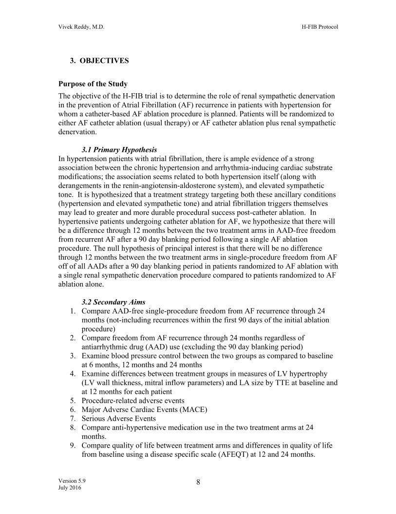
Serious Adverse EventA serious adverse event (SAE) is any event that meets one or more of the following criteria:
Leads to a death Leads to a serious deterioration in the health of a subject that resulted in:
a life-threatening illness or injury a permanent impairment of a body structure or a body function
Vivek Reddy, M.D. H-FIB Protocol
Version 5.9 July 2016 24
in-patient hospitalization or prolongation of an existing hospitalization*
medical or surgical intervention to prevent life-threatening illness or injury or permanent impairment to body structure or a body function
Leads to fetal distress, fetal death or a congenital abnormality or birth defect.
This definition excludes hospitalization solely due to arrhythmia recurrence or non-medically urgent cardioversion.
*Planned hospitalization for a condition present prior to the participant’s
enrollment in the study will not meet the definition of an SAE. An AE would meet the criterion of “hospitalization” if the event necessitated an
admission to a health care facility (e.g., an overnight stay). Emergency room visits that do not result in admission to the hospital should be evaluated for one of the other serious outcomes.
Unanticipated Serious Adverse Event An unanticipated (unexpected) serious adverse event is any serious adverse event that is not protocol-defined or documented in the patient consent form. Expedited reporting is required for serious adverse events that are unexpected.
Unanticipated Adverse Device Effects An unanticipated adverse device effects (UADE) is any serious adverse effect on health or safety or any life-threatening problem caused by, or associated with, a device, if that effect, problem, or death was not previously identified in nature, severity, or degree of incidence in the investigational plan, or any other unanticipated serious problem associated with the device that relates to the rights, safety, or welfare of subjects.
Unanticipated Problems According to the Office for Human Research Protections (OHRP), an Unanticipated Problem (UP) generally includes any incident, experience, or outcome that meets all of the following criteria: (1) Unexpected (in terms of nature, severity, or frequency) given (a) the research procedures that are described in the protocol-related documents, such as the IRB-approved research protocol and informed consent document; and (b) the characteristics of the subject population being studied; and (2) Related or possibly related to participation in the research (in this guidance document, possibly related means there is a reasonable possibility that the incident, experience, or outcome may have been

Vivek Reddy, M.D. H-FIB Protocol
Version 5.9 July 2016 25
caused by the procedures involved in the research); and (3) Suggests that the research places subjects or others at a greater risk of harm (including physical, psychological, economic, or social harm) than was previously known or recognized. Based on the definitions above and as illustrated below (per OHRP guidance), many adverse events are not unanticipated problems, and many unanticipated problems are not adverse events. However, some adverse events are also unanticipated problems. For example, a serious adverse event that is unexpected and at least possibly related to study participation is also by definition an unanticipated problem. As stated above, an unanticipated problem may not necessarily be an adverse event, which is the case when the problem does not cause actual physical harm to participants. For example, if a laptop computer with sensitive, identifiable study data is stolen, this theft places the participants at greater risk of psychological or social harm; this is an unanticipated problem that is not an adverse event. Another example of an unanticipated problem that is not an adverse event is if the FDA announces that one of the study drugs is tainted (e.g., with paint chips), yet no participant experiences any adverse effects. Event Recording The following adverse events will be captured throughout the period of trial participation:
o Protocol-defined (as described below) o Serious unanticipated events (serious “Other” adverse events)
Causality The investigator will assess the relationship of an adverse event to the intervention. The investigator should distinguish the relationship between the event and (a) ablation procedure (i.e. AF ablation procedure or other concomitant procedures), and (b) renal sympathetic denervation. Causality will be defined as follows:
Probable Adverse events that, after careful medical evaluation, are considered with a high degree of certainty to be related to the intervention (AF ablation ± renal sympathetic denervation). The following characteristics will apply:
o A reasonable temporal relationship exists between the event and the intervention, and
o The event is a known reaction to the intervention, and cannot be explained by an alternative etiology commonly occurring in the population/individual.
Possible Adverse events that, after careful medical evaluation, do not meet the criteria for a probable relationship to the intervention, but for which a connection cannot be ruled out with certainty. The following characteristics will apply:
o The event occurs after intervention, and o The event is not a known reaction to intervention, but cannot be explained
by a commonly occurring alternative etiology

Vivek Reddy, M.D. H-FIB Protocol
Version 5.9 July 2016 26
Unlikely Adverse events that, after careful medical evaluation, do not meet the criteria for a possible or probable relationship to intervention and for which a connection is unlikely. The following characteristics will apply:
o The event does not follow a reasonable temporal sequence from administration of the intervention, or
o May have been produced by environmental factors, and there is no apparent pattern of response to the intervention.
Reporting of Serious Adverse Events and Unanticipated Adverse Device Effects All investigators must report both expected (protocol-defined) and unexpected SAEs. All protocol defined SAEs must be reported directly to the clinical center’s IRB and the DCC within 10 business days of knowledge of the event, or as dictated by the specific IRB policy, whichever is sooner. All deaths, UADEs, and unexpected SAEs that are possibly or probably related to the renal sympathetic denervation must be reported to the DCC and the clinical center’s IRB within 24 hours of knowledge of the event, or as dictated by the specific IRB policy, whichever is sooner. All unexpected SAEs that are unlikely related to the study intervention must be reported to the DCC and the clinical center’s IRB within 5 business days of knowledge of the event, or as dictated by the specific IRB policy, whichever is sooner. The DCC will report these events to the DSMB chair within 72 hours of notification. All SAEs will be reported to the DSMB at least semi-annually, at the discretion of the DCC medical monitor. Reporting of Unanticipated Problems All UPs that are also SAEs, which are at least possibly related to the study intervention, must be reported to the DCC within 24 hours of knowledge of the event. All UPs that are not SAEs must be reported to the DCC within 5 calendar days of knowledge of the event, or as dictated by the specific IRB policy, whichever is sooner. DCC Reporting to FDA The DCC will report unexpected SAEs that are possibly or probably related to the investigational device or UADEs to FDA as appropriate. The DCC will send an initial IDE safety report communication to the FDA within 2 business days of notification from the site. The DCC will submit a follow-up safety communication to the FDA, based on source documentation or PI Report from the site, within 10 business days from notification of a UADE for this IDE trial. Specific Adverse Event Definitions Allergic Reactions
Allergic reactions can occur to the local anesthetic, sedatives, x-ray dye, heparin, protamine, or other agents administered during the procedure. 23-29

Vivek Reddy, M.D. H-FIB Protocol
Version 5.9 July 2016 27
Arterial or Venous Injury
Arterial or venous injury including arterial dissection, thrombosis, occlusion or hemorrhage at the catheter insertion sites or at other sites along the vessels (risk <1%). 30-31 This may result in hemorrhage, hematoma or ischemic injury to an extremity or major organ.
Bleeding
A bleeding event is defined by any one of the following: o Transfusion of > 2 units RBC within the first 24 hours following ablation o Death due to hemorrhage o Re-operation for hemorrhage or tamponade
NOTE: Hemorrhagic stroke is considered a neurological event and not as a separate bleeding event.
Major Infection
A new clinical infection accompanied by pain, fever, drainage and/or leukocytosis that is treated by anti-microbial agents (non-prophylactic). A positive culture from the infected site or organ should be present unless strong clinical evidence indicates the need for treatment despite negative cultures. The general categories of infection are listed below:
Sepsis
Evidence of systemic involvement by infection, manifested by positive blood cultures and hypotension. In addition, we will record systemic antibiotic use for presumptive sepsis.
Localized Infection
Infection localized to any organ system or region, including the mediastinum, pericardium, or endocardium, without evidence of systemic involvement (see sepsis definition), ascertained by standard clinical methods and either associated with evidence of bacterial, viral, fungal or protozoal infection, and/or requiring empirical treatment.
Occlusion of Coronary Artery
Radiofrequency current or cryoenergy may cause occlusion of a coronary artery, either by direct thermal damage, spasm, or thrombosis (risk <0.5%).30-31 Coronary arterial occlusion could produce myocardial infarction, angina or death. Should occlusion of a coronary artery occur for any reason, the physician will attempt to restore coronary blood flow through pharmacological, catheter and/or surgical intervention as medically indicated.
Damage to AV Conduction System

Vivek Reddy, M.D. H-FIB Protocol
Version 5.9 July 2016 28
The application of radiofrequency or cryoenergy current close to the AV node or His bundle could damage or destroy the normal AV conduction system, producing complete heart block.
Embolization of Thrombus
The placement of linear lines of ablation may require a large number of RF lesions to be delivered to the left ventricle. The risk of stroke due to embolization of thrombus is approximately 1%. To minimize this risk, patients will be heparinized to an activated clotting time of greater than 300 seconds during the procedure.
Cardiac Perforation
Cardiac perforation may result from catheter manipulation or application of radiofrequency current or cryoenergy (risk is <1%).30-31 This may result in cardiac tamponade and may require percutaneous pericardial drainage or surgical repair. Significant hemodynamic compromise can result in neurological injury or death. An increased risk of cardiac perforation may be associated with the use of saline-irrigated electrode catheter due to its creation of a larger, deeper RF lesion.
Valvular Injury
Injury to a cardiac valve may result from catheter manipulation or the application of radiofrequency current or cryoenergy (risk <1%).30-31 This may produce valvular insufficiency and possibly require surgical valve replacement.
Pulmonary Vein Stenosis
While advances in technique and increased physician experience have made pulmonary vein stenosis unusual (risk 1.3%), 30-31 it may occur in patients who undergo catheter ablation.
Radiation Exposure Radiation exposure during the fluoroscopic imaging of the catheters may result in an increase in the lifetime risk of developing a fatal malignancy (0.1%) or a genetic defect in offspring (0.002%).30, 31-33
Pneumonia
Rales, pulmonary edema, ARDS and asthmatic attack can be sequelae of aspiration pneumonia and nosocomial pneumonia. Aspiration of gastric contents can occur when subjects are intubated, and nosocomial pneumonia is well documented as the second most common nosocomial infection. The incidence of aspiration pneumonia and nosocomial pneumonia after AF ablation is rare (<1%).
Phrenic Nerve Damage
Phrenic nerve damage can be a sequela of pulmonary vein isolation; also rare (<1%) with RF catheter ablation.

Vivek Reddy, M.D. H-FIB Protocol
Version 5.9 July 2016 29
Cardiac Arrhythmias Any documented arrhythmia (excluding atrial fibrillation and atrial flutter) that results in clinical compromise (e.g., hemodynamic compromise, oliguria, pre-syncope or syncope) that requires hospitalization. Cardiac arrhythmias are classified as: 1. Sustained ventricular arrhythmia requiring defibrillation or cardioversion
(either DC or chemical) 2. Cardiac conduction abnormalities requiring permanent pacemaker
Pericardial Fluid Collection
Accumulation of fluid or clot in the pericardial space that requires surgical intervention or percutaneous catheter drainage. This event will be subdivided into those with clinical signs of tamponade (e.g. increased central venous pressure and decreased cardiac output) and those without signs of tamponade.
Pleural Effusion Accumulation of fluid or clot in the pleural space documented by chest radiogram or chest CT that requires evacuation with surgical intervention or chest tube placement.
Heart Failure
New onset of signs or symptoms of congestive heart failure or worsening of pre-existing heart failure by ≥ 1 NYHA class.
Myocardial Infarction Myocardial infarction (MI) should be classified when there is evidence of myocardial necrosis in a clinical setting consistent with myocardial ischemia. Under these conditions, any one of the following criteria meets the diagnosis for myocardial infarction.35 Myocardial Infarction Detection of rise and/or fall of cardiac biomarkers (preferably troponin) with at least one value above the 99th percentile of the upper reference limit (URL) together with evidence of myocardial ischemia with at least one of the following: o Symptoms of ischemia; o ECG changes indicative of new ischemia (new ST-T changes or new left
bundle branch block [LBBB]); o Development of pathological Q waves in the ECG; o Imaging evidence of new loss of viable myocardium or new regional wall
motion abnormality. Sudden unexpected cardiac death, involving cardiac arrest, often with symptoms suggestive of myocardial ischemia, and accompanied by presumed new ST elevation or new LBBB, and/or evidence of fresh thrombus by coronary

Vivek Reddy, M.D. H-FIB Protocol
Version 5.9 July 2016 30
angiography and/or autopsy, with death occurring before blood samples obtained, or at a time before the expected appearance of cardiac biomarkers in blood will be classified as a mortality due to MI.
Atrioesophageal Fistula
Atrioesophageal fistula is a rare (risk < 1%) but often fatal risk of RF catheter ablation.36-37
Neurologic Dysfunction
Any new, temporary or permanent, focal or global neurological deficit ascertained by a standard neurological examination (administered by a neurologist or other qualified physician and documented with appropriate diagnostic tests and consultation note). The examining physician will distinguish between a transient ischemic attack (TIA), which is fully reversible within 24 hours (and without evidence of infarction), and a stroke, which lasts longer than 24 hours (or less than 24 hours if there is evidence of infarction). Each neurological event must be subcategorized as: Transient Ischemic Attack Defined as an acute event that resolves completely within 24 hours with no imaging evidence of infarction. Ischemic or Hemorrhagic Stroke (Cerebrovascular Accident) Defined as an event that persists beyond 24 hours or less than 24 hours associated with infarction on an imaging study. Hemorrhagic conversion of an ischemic stroke should be classified as ischemic. Toxic Metabolic Encephalopathy Defined as a disorder of the brain function that arises from abnormal systemic metabolism or exogenous substances, altering awareness and/or consciousness, in which there is a non-focal neurological examination and a negative brain image. Other Neurologic Dysfunction
Renal Events
Two categories of renal events will be identified: Renal Dysfunction Abnormal kidney function defined as serum creatinine increase >50% or >0.3 mg/dl rise in serum creatinine (Cr) from baseline. Renal Failure New requirement for hemodialysis related to renal dysfunction. This definition excludes aquapheresis for volume removal alone.

Vivek Reddy, M.D. H-FIB Protocol
Version 5.9 July 2016 31
Respiratory Failure Impairment of respiratory function requiring re-intubation, tracheostomy or the inability to discontinue ventilatory support within 48 hours post-catheter intervention. This excludes intubation for re-operation or temporary intubation for diagnostic or therapeutic procedures.
Arterial Non-CNS Thromboembolism An acute systemic arterial perfusion deficit in any non-cerebrovascular organ system due to thromboembolism confirmed by one or more of the following:
o Standard clinical and laboratory testing o Operative findings o Autopsy findings
This definition excludes neurological events.
Venous Thromboembolic Event
Evidence of venous thromboembolic event (e.g. deep vein thrombosis, pulmonary embolism) by standard clinical and laboratory testing.
Other
An event that causes clinically relevant changes in the patient’s health, or any event that is life-threatening, results in a fatality, results in permanent disability, requires hospitalization, or prolongs an existing hospital stay.
As specifically related to the renal denervation procedure, additional potential adverse ANTICIPATED events that may occur include: Occlusion of Renal Artery
Radiofrequency current may cause occlusion of a renal artery, either by direct thermal damage, spasm, or thrombosis (risk <0.5%). Renal arterial occlusion could produce renal infarction, acute renal failure, possibly resulting in the need for hemodialysis. Should occlusion of a renal artery occur for any reason, the physician will attempt to restore blood flow through pharmacological, catheter and/or surgical intervention as medically indicated.
Renal Artery Stenosis
Renal artery stenosis (defined as ≥ 60% reduction) by renal duplex ultrasonography will be defined as a i) renal-aortic ratio of 3.5 or more or a ii) peak systolic velocity of more than 200 cm/s. 38-40 Renal artery stenosis may also occur insidiously over time, and may not manifest until weeks after the ablation procedure. The risk of this is minimized by longitudinally staggering the ablation lesions such circumferential lesions are not placed in the same segment of the artery.

Vivek Reddy, M.D. H-FIB Protocol
Version 5.9 July 2016 32
12. CLINICAL CENTERS The study will be conducted in up to 20 sites both in and outside the United States. Each clinical center will be required to obtain IRB approval for the protocol and consent (and their revisions) in a timely fashion, to recruit patients, to collect data and enter it accurately in the electronic data capture (EDC) system, to faithfully follow the protocol and adhere to the standards of Good Clinical Practice (GCP). In addition, centers will be required to provide the DCC with the information necessary for interim, annual, and final reports, to provide source documents, data and regulatory documents for study monitors, provide prompt responses to DCC inquiries, and to participate in analyses and reporting of study results. Investigator Profile All surgeons, cardiologists, coordinators and other investigators in the study must complete the Investigator Profile form, including hospital affiliation, address, telephone, fax, beeper and email information. The physicians and coordinators must email their CV, Conflict of Interest Statement and Financial Disclosure Certification, and Institutional Health Insurance Portability and Accountability Act (HIPAA) Certificates to the DCC. Qualifications and Training Clinical investigators will be electrophysiologists with expertise in atrial fibrillation ablation. The certified operator will either perform the ablation and renal denervation on their own patient, or participate in the ablation and renal denervation of an enrolled patient. The clinical site Principal Investigator will be responsible for overseeing the ongoing performance of the other participating investigators at that site over the course of the study. All clinical site investigators and coordinators will be trained by the DCC in the specifics of the protocol at a site initiation visit in advance of patient enrollment. In addition, the investigators and coordinators will undergo a separate training session to gain familiarity with the electronic data capture system. Signature Verification Investigators will input an electronic signature into the electronic data capture system (EDC). It will be updated throughout the study as new site personnel are approved. Conflict of Interest and Financial Disclosure Agreement This statement verifies that all investigators have no conflict of interest with any institution that may influence their participation in this study. All investigators need to complete this statement. Investigators will also submit a financial disclosure agreement. Site Approval The following documents must be collected prior to site approval:

Vivek Reddy, M.D. H-FIB Protocol
Version 5.9 July 2016 33
• Clinical Study Agreement
• Clinical site IRB roster
• Clinical site IRB approval, version and date for protocol and consent
• Clinical Center Laboratory Certification
A signed agreement between the clinical site and the DCC (Icahn School of Medicine at Mount Sinai) is required prior to site initiation. Prior to enrolling a patient, representatives from the DCC will conduct a site initiation for all investigators, coordinators, and any other health care professionals who may be involved in the study (e.g. engineers, social workers). Patient Confidentiality All patients’ records will be kept confidential according to HIPAA guidelines. Study Investigators, site Institutional Review Boards (IRBs), and the DCC may review source documentation as necessary, but all unique patient and hospital identifiers will be removed. The aggregate data from this study may be published as per publication policy documented in the trial agreements; however, no data with patient identifiers will be published. 13. SCREENING AND BASELINE DATA COLLECTION Pre-Screening Failure Form Prior to informed consent Prior to approaching a patient to begin the informed consent process, the study personnel will review data on prospective patients to determine eligibility for inclusion in the trial. All pre-screened patients (patients who are not consented) who are not enrolled are recorded in the Pre-Screening Failure form. For these patients, reasons for failing the pre-screening process are documented. The data collected is HIPAA compliant and does not include patient identifiers but only includes screening quarter, screening year, age, gender and reason for not being eligible. The reasons for screening failure for patients who were consented are collected on the Eligibility Evaluation Form. Consent Prior to screening data collection and protocol-defined procedures (initiated ≥24 hours prior to randomization) Prior to screening, a thorough explanation of the risks and benefits of the study will be outlined by the investigator to the patient and the patient will be asked to sign a consent form. Release of Medical Information Prior to screening data collection and protocol defined procedures The patient must sign the Release of Medical Information form or equivalent that authorizes release of medical records to the study investigators, monitors, and the FDA.

Vivek Reddy, M.D. H-FIB Protocol
Version 5.9 July 2016 34
Demographics Form At initiation of screening A screened patient is defined as an individual (a consented patient) who was referred to, or identified at a clinical site for consideration of entry into the study, and for whom some preliminary (i.e. medical record) data have been collected and/or reviewed. For all patients screened, the first, middle, and last initial, date of birth, ethnic origin, and sex will be captured on the screening log and registration form. The EDC will generate a unique 8-digit identification code that will identify the patient throughout the course of the study. Medical History Within 60 days prior to randomization This form captures the information pertaining to the medical history, including but not limited to previous myocardial infarction, myocardial revascularization, arrhythmias, automatic implantable cardioverter-defibrillator, permanent right ventricular or biventricular pacemaker, stroke and other comorbidities such as diabetes and peripheral vascular disease. Information regarding the current medical condition is also captured, including but not limited to disposition at time of screening (outpatient, inpatient, ICU monitoring, etc.). New York Heart Association Classification Within 60 days prior to randomization Functional status will be assessed using the NYHA classification. NYHA classification will be determined by investigative center personnel and documented on the “New York Association Classification” form. The NYHA classification scheme is detailed in Appendix I. Medications Within 60 days prior to randomization This form captures all medications taken within 30 days prior to randomization. Physical Examination Within 60 days prior to randomization This form captures the comprehensive physical examination including vital signs cardiopulmonary examination, abdominal examination, and anthropometrics (height, weight and BSA). Quality of Life Within 30 days prior to randomization AFEQT (Appendix II) Questionnaire will be completed by the patient and used to assess QoL. Data regarding completeness of QoL data collection and reasons for missing responses to questionnaires will be collected. Office Blood Pressure Measurement The auscultatory method of BP measurement will be used. Persons will be seated quietly for at least five minutes in a chair (rather than on an exam table), with feet on the floor

Vivek Reddy, M.D. H-FIB Protocol
Version 5.9 July 2016 35
and arm supported at heart level. Caffeine, exercise and smoking will be avoided for at least 30 minutes prior to measurement. The bladder of the cuff will encircle at least 80 percent of the arm circumference. Three readings will be taken at intervals of at least 1 minute, and the average of those readings will be used to represent the patient’s blood pressure. This approach is the standard approach recommended by JCN-7. 41-42 Laboratory Assessment Within 30 days prior to randomization
• Hematology • Coagulation • Blood chemistry • Liver function tests • Glomerular Filtration Rate • Urine or beta serum HcG (women of childbearing potential)
Eligibility Criteria/Eligibility Evaluation Form Prior to randomization The inclusion and exclusion criteria will be documented by the clinical site study coordinator and verified with the site Principal Investigator in the Eligibility Evaluation Form. All screened patients (patients who are consented) who are not randomized in the trial will have the reasons for non-randomization documented in the Eligibility Evaluation Form. The data collected is HIPAA compliant and includes reason for not being randomized. A representative from the DCC will be available to discuss any questions regarding patient eligibility. Electrocardiogram (ECG) Within 3 days prior to randomization An ECG will be done at baseline. This tracing will be used to assess heart rhythm on all patients and provide part of the documentation needed to establish the diagnosis of longstanding persistent AF. Transthoracic Echocardiography Must be performed within three months prior to randomization Transthoracic Doppler echocardiography will be used to measure LA function (a) atrial filling, (b) LA ejection fraction, (c) atrial systolic mitral annular velocity (TDI). MV function, mitral regurgitation and LV function and LV mass. The LV mass will be derived from 2D TTE imaging or MR imaging. Two-dimensionally guided M-mode echocardiography will be performed from a parasternal long-axis view. Using the leading edge–to–leading edge technique, the left ventricular end-diastolic dimension (LVED), posterior wall thickness, and interventricular septal thickness will be measured at end-diastole at the level just below the mitral valve leaflets. LV mass will be calculated using the following equation: 0.8{1.04[(LVED + left ventricular posterior wall thickness + interventricular septal thickness) − LVED]} + 0.6. The LV mass index

Vivek Reddy, M.D. H-FIB Protocol
Version 5.9 July 2016 36
will be calculated from this by dividing the LV mass by the height exponentiated to the power of 2.7.
14. POST-RANDOMIZATION DATA COLLECTION Ablation Procedures Initial catheter intervention and event driven Includes information regarding specific primary procedure, operative variables, device used for ablation, lesion sets created, additional procedures performed at time of initial operation, intra-operative pharmacological agents. The procedure form will also include a section where the investigator can depict the lesion set created by annotating a diagram; this form will be mailed/faxed to the DCC. Hospitalizations Index hospitalization and event driven For all patients the index (baseline) hospitalization and all subsequent hospital admissions (for any reason) must be reported on the Hospitalization form. Patient Pre-discharge Prior to discharge
▪ Medications All cardiovascular medications are recorded.
▪ Physical Examination (including vital signs and cardiopulmonary examination) ▪ Adverse Events
Detailed information regarding adverse events is recorded at the time an event occurs during hospitalization. Investigators are asked to make a judgment as to the seriousness and relationship of the event to the intervention.
Holter Monitoring 3, 6, 12 and 24 months. Prior to each time point, patients will be given a Holter device and instructed in its use. Follow-up Visits 3 months (±14 days); 6 and 9 months (±14days); 12, 18 and 24 months (±30 days) Follow-up visits will be conducted by investigative site personnel to document that the patient is alive and to assess the following:
▪ 7-day Holter Monitoring (3, 6, 12, 24 months only) The Holter device provided to patients prior to each time point will be used for this assessment.
▪ Laboratory Assessment (6, 12 months only) • Blood chemistry
Vivek Reddy, M.D. H-FIB Protocol
Version 5.9 July 2016 37
▪ Medications All cardiovascular, inotropic, antiplatelet agents and anticoagulants will be recorded. Medications (including AADs) will be recorded at each follow-up, and also as indicated at the time of associated adverse events. We will also record response to DC cardioversions (success or failure; failure defined as the inability to restore sinus rhythm for ≥ 30 seconds post-electrical cardioversion20).
▪ Adverse Events, Anti-arrhythmic Interventions, and Hospitalizations Patients will be asked to recall any adverse events (including stroke and bleeding) and the number of hospitalizations that occurred out of network since the previous contact. Patients will also be asked if they had a permanent pacemaker insertion, cardioversion or subsequent ablation since the last contact.
▪ Renal Doppler (6 months only) All patients will undergo follow-up renal Doppler echocardiography at the investigative center. If the 6 month renal duplex ultrasonography indicates significant renal stenosis (≥60%) and/or image quality is inadequate to assess the severity of stenosis, than an MRI or CT should be performed as confirmatory testing.
▪ Transthoracic Doppler Echocardiogram (12 months only)
All patients will undergo follow-up transthoracic Doppler echocardiography at the investigative center (See Appendix III). Quality of Life The AFEQT questionnaire (Appendix II) will be used to assess QoL. The patients themselves must complete this questionnaire and return it to the investigative center.
▪ Functional Status The NYHA class will be documented on the “New York Heart Association Classification” form.
▪ Adverse Events Patients will be asked to recall any adverse events (including stroke and bleeding) and the number of hospitalizations that occurred out of network since the last contact. Patients will also be asked if they had a permanent pacemaker insertion, cardioversion or subsequent ablation since the last contact.
▪ Blinding assessment (12 months only) Patients will be asked to determine which treatment(s) they believe they have received.
▪ Follow-up Treating Physician Blinding assessment (12 months only) A patient’s follow-up treating physician will be asked to determine which treatment(s) the physician believes that the patient has received.

Vivek Reddy, M.D. H-FIB Protocol
Version 5.9 July 2016 38
Event Driven Data Collection Follow-Up Surgical Procedures Event Driven All operations following the initial procedure(s) must be reported on the procedure form within 48 hours of the knowledge of the event. If the operation is to address a complication, the coordinator must also complete an adverse event report. All intra-operative transfusion requirements must be documented. Adverse Events Event Driven Detailed information regarding adverse events will be recorded at the time an adverse event occurs. Investigators will be asked to make a judgment as to the seriousness and relationship of the event to the procedure using the guidance provided. All adverse events will be recorded until completion of the trial. All serious and all protocol-defined adverse events will be adjudicated by an EAC. Missed Visit Assessment Event Driven If a patient is unable to attend any follow-up visit before the closure of a study visit window, a missed visit assessment must be completed that captures the reason for missing the follow-up. Mortality Event Driven within 24 hours of knowledge of event The investigator will record the date of death, immediate cause of death, primary underlying cause of death, notation of autopsy being performed, and clinical narrative of the event. Study Completion/Early Termination Event Driven or End of Study This form records the date and reason for study completion or early termination. The reasons for a patient to be withdrawn from this study at the patient’s request or at the physician’s discretion will also be documented on this form. Investigator’s Statement End of study The Principal Investigator will review all of the electronic case report forms (eCRFs) and patient summaries at that center. His/her electronic signature will attest to the accuracy and completeness of the data collected. 15. DATA MANAGEMENT

Vivek Reddy, M.D. H-FIB Protocol
Version 5.9 July 2016 39
All study data will be entered in the web-based electronic data capture (EDC) system (specified in detail in the operations manual). Study personnel requiring access will have their own Login/Password. Access to clinical study information will be based on individuals' roles and responsibilities. The application provides hierarchical user permission for data entry, viewing, and reporting options. For optimum security, the system operates Secure Socket Layer (SSL) 128-bit encryption protocol over Virtual Private Networks. This application is designed to be in full compliance with International Conference on Harmonization and Good Clinical Practices (ICH-GCP), the FDA’s Code of Federal Regulations (CFR) Number 21 Part 11 Electronic Record and Electronic Signatures, the FDA's "Guidance For Industry: Computerized Systems Used in Clinical Trials", and the Privacy Rule of the Health Insurance Portability and Accountability Act of 1996 (HIPAA).
15.1 Quality Assurance
The data quality assurance tool has been designed as an automatic feature of the EDC system. When a form is submitted the system conducts instantaneous validation and cross-form validation checks. A query is generated and sent to the site coordinator electronically so that data may be verified and corrected. All changes made to a form are stored in an audit log.
15.2 On-Site Monitoring The primary objectives of the DCC in monitoring clinical sites are to educate, support, identify and resolve issues related to the clinical trial. The monitors will discuss the protocol in detail, and clarify any areas of uncertainty. At initiation of the study, the monitors will conduct a tutorial on the EDC system. The coordinators will practice entering data so that the monitors can confirm that the coordinators are proficient in all aspects of data entry, query response, and communication with the data management team. The DCC will employ a risk-based approach to monitoring for this study. This will be accomplished via centralized or remote monitoring of data via the EDC with a focus on safety, study endpoints, data completion and data outliers. The DCC will also centrally monitor study logs including the Informed Consent Log, the Protocol Violation/Deviation Log and the Serious Adverse Event/IND Safety Report Log periodically to ensure that the sites are adhering to the study protocol and procedures. The DCC will generate performance metrics to analyze site characteristics such as recruitment rates and timeliness of data entry. This will allow the DCC to identify trends across sites and to address low-performing sites appropriately. The DCC will also conduct one on-site monitoring visit each year for every clinical site for the duration of the study. In addition, interim monitoring will be conducted on an ongoing basis using the EDC system. During the annual on-site monitoring visits, the monitor will collect copies of any source documents (de-identified) for all deaths and AEs. Prior to each EAC meeting copies of any source documents (de-identified) requested by the DCC must be sent via FedEx or secure fax line to the DCC.

Vivek Reddy, M.D. H-FIB Protocol
Version 5.9 July 2016 40
The monitors will review the source documents to determine whether the data reported in the EDC system are complete and accurate. They will also verify that all serious and protocol-defined adverse events exist on the source documents, are consistent with the protocol and are documented in the appropriate format. Source documents include medical charts, initial hospital admission reports, operative procedure records, discharge and re-admission reports, consult notes, radiology reports, lab reports, clinic records, and other study-related notes. The study monitors reserve the right to copy de-identified records in support of all adverse events and outcomes. The monitors will also confirm that the regulatory (administrative) binder is complete and that all associated documents are up to date. The regulatory binder should include all revisions of the protocol and informed consent, IRB roster, IRB approvals for all of the above documents, IRB correspondence, investigator’s agreements, CVs of all study personnel, institutional HIPAA certificates, monitor site visit log, telephone contact log, a study device log and correspondence with the DCC. If a problem is identified during the visit (i.e., poor communication with the DCC, inadequate or insufficient staff to conduct the study, missing study documents, etc.), then the monitor will assist the site in resolving the issue. Some issues may require input from the Steering Committee or the Principal Investigator, as well as the sponsor. The combination of yearly on-site monitoring and ongoing monitoring using the EDC system that includes instantaneous electronic validation, and visual cross-validation to detect complex errors, it is anticipated that the best possible quality and most complete data will be collected. The monitor will verify a minimum of the following variables for all patients: initials, date of birth, sex, signed informed consent, eligibility criteria, date of randomization, anticoagulation, serious and protocol-defined adverse events, mortality, NYHA Classifications and QoL. These data will be 100% source data verified. All other data collection will be monitored as indicated by the data completeness and accuracy at each clinical site.
16. ANALYTICAL PLAN
16.1 Statistical Analysis Plan General Design Issues This is a prospective, multi-center, double-blinded, randomized trial. Patients with hypertension and planning to undergo an AF ablation procedure will be randomized using a 1:1 allocation to either AF catheter ablation (usual therapy) or AF catheter ablation plus renal sympathetic denervation. Patients will be blinded to their group assignment (control vs. study group) throughout the duration of the study. The physician responsible for blood pressure management in the post-operative period (who will not be the same as the operator who performed the procedure) will also be blinded to group assignment. The

Vivek Reddy, M.D. H-FIB Protocol
Version 5.9 July 2016 41
operator performing the procedure will not be blinded to the treatment. Outcomes will be assessed and recorded by study personnel masked to treatment assignment. The trial’s primary goal is comparing two treatment arms: an AF ablation control and AF ablation + renal sympathetic denervation study group. The null hypothesis of principal interest is that there will be no difference through 12 months between the two treatment arms in single-procedure freedom from AF off of all AADs after a 90 day blanking period. There are a number of secondary objectives, including 1) assessing the 24-month single procedure freedom from AF off of all AADs after a 90 day blanking period; 2) measuring the impact of arrhythmia event reduction on Quality of Life, and 3) other surrogate measures of potential benefit from renal denervation (such blood pressure differences, differences in LV mass and the number of anti-hypertensive medications at study end). Sample size 16.2 Randomization Design and Procedure This trial will randomize a total of 300 patients in equal proportions to AF ablation with renal sympathetic denervation versus AF ablation alone. The randomization will be stratified by clinical center and use a random permuted block design, with blocks of size 4 or 8 chosen at random. Randomization will be implemented as described in section eight. A total of three hundred patients provides 80% power to detect a relative 40% decrease in the one-year incidence of atrial fibrillation (from 40% to 24%) in patients treated with AF ablation with renal sympathetic denervation compared to AF ablation alone based on a two-tailed 0.05 level chi-squared test. The sample size accounts for two planned interim analyses. This study is divided into two phases: a PILOT phase (to include 50 patients) followed by the remaining 250 patients to a total of 300 patients. The PILOT phase will be conducted at up to 10 US sites: TBD. An interim safety analysis will be conducted when all patients in the PILOT phase (50 subjects) have completed the 3-month follow up visit. The purpose of this analysis is only for safety. The efficacy/outcome data for the primary endpoint will not be examined; this will permit pooling these 50 patients with the subsequent 250 patients for the final efficacy analysis. 16.3 Data Monitoring and Analysis Methods of Analysis The primary efficacy outcome of this randomized clinical trial is single procedure freedom from AF over 12 months post randomization, excluding a 90 day blanking period. Patients who die within 12 months will be considered treatment failures (not free of AF). The null hypothesis is that there is no difference in the proportion of patients meeting the primary outcome between patients randomized to AF ablation plus renal sympathetic denervation or AF ablation alone. That is, we will test
H0: π0 = π1 versus H1: π0 ≠ π1

Vivek Reddy, M.D. H-FIB Protocol
Version 5.9 July 2016 42
where π0 and π1 represent the proportion free from AF at 12 months assessment (following a single AF ablation procedure and off AADs after 90 days) among patients randomized to receive either AF ablation plus a single renal sympathetic denervation procedure or AF ablation alone. The primary null hypothesis will be tested in an intent-to-treat analysis at the 0.05 level. To compare the proportion of patients free from AF in the two treatment groups, we will use a mixed model in which the type of catheter is considered as a random effect to account for the heterogeneity that could be brought about by the use of different catheters. The benefit of AF ablation plus renal sympathetic denervation to AF ablation alone will be quantified by the relative risk and associated 95% confidence interval (adjusted for two interim looks). Patients who die prior to the 12-month assessment, or who are determined by an independent adjudicator to be too ill to undergo AF measurement, will be considered as treatment failures (not free of AF). In the primary analysis, patients in both treatment arms who undergo ablation therapy for AF subsequent to the index procedure will be considered treatment failures (not free of AF). Patients missing the 12-month assessment, for reasons other than death or illness, will have their primary outcome imputed using a multiple imputation algorithm described below. Imputed data will be rounded to 0 (free of AF) or 1 (AF present).
An implicit assumption of the proposed primary analysis is that the results for all of the study centers can be pooled or combined. We will assess the validity of this assumption by a variety of means, recognizing that establishing poolability merits both clinical and statistical examination.
We do not anticipate that pooling of results across study centers will be threatened due to any clinically important differences in trial conduct. Centers participating in the study have been selected for their experience and demonstrated success in conduct of clinical trials and adherence to study protocols. Frequent site monitoring and regular contact including teleconferences involving site coordinators and investigators, as well as in person meetings, ensure that all centers strictly follow trial protocols.
Additionally, we will conduct several statistical analyses to assess poolability of results across centers. These include center specific analyses of the primary endpoint to assess the consistency of results, and similarity of patient demographics and baseline characteristics across centers. In addition to uncovering potential poolability issues that would require further exploration, differences may indicate the need for additional, more complex, statistical analysis. The effect of center will be explicitly assessed by a secondary analysis testing the interaction of treatment with center via a logistic regression model incorporating centers through a series of indicator variables. Power for detecting an interaction may be low, so we will use a p-value of 0.15 to assess interaction, and also consider the qualitative analysis of center specific results to assess the extent the heterogeneity of treatment by center. Interim Analysis

Vivek Reddy, M.D. H-FIB Protocol
Version 5.9 July 2016 43
We plan to perform two interim analyses with respect to the primary outcome to give the option of stopping early should results strongly favor one arm or the other. The proposed timing of the two analyses is at 0.33 and 0.67 on the information scale, i.e., after 33% and 67% of patients reach the primary endpoint. The utility of performing these analyses will depend on the rate of accrual of patients into the trial. A group sequential procedure allows for flexibility in the number and timing of interim analyses should the DSMB choose to modify the proposed plan, or should accrual mitigate the usefulness of an interim look. We will use the Lan-DeMets approach, implementing an O'Brien-Fleming-type spending function that allots most of the type I error to the final look. 43 In addition to the ethical concern of continuing a trial that shows a clear benefit in favor of one treatment, there is also a corresponding ethical concern of continuing a trial that has little chance of ever showing a benefit of one treatment compared to the other. We propose that the trial’s conditional power, under the original alternative hypothesis, be computed at the interim look and that the DSMB use this as a basis to determine whether randomization, if not completed, be halted for futility. We propose that consideration be given to halting the trial for futility if, given the data up to the point of the interim analysis, the probability of detecting an absolute 16% benefit for patients randomized to AF ablation + renal sympathetic denervation is less than 40%. In addition an initial safety review will be performed after 40 patients have been enrolled and ablated. The proposed analysis after 40 patients have been ablated will only assess safety concerns. The results of this analysis, a summary and comparison of adverse events by treatment arm, will be provided to FDA for review. We propose to continue enrollment during this period of FDA review, up to a maximum of 80 patients. If 80 patients have been randomized, and FDA has not concluded its review, enrollment would cease pending FDA approval for study continuation. No study modifications are proposed based on this initial review. Assessment of Balance of the Randomization The success of the randomization procedure in balancing important covariates between randomization groups will be assessed at the interim analysis and at the final analysis. Continuous measures will be compared using t-tests, while chi-squared tests will be used to compare categorical variables such as age and ethnicity. As 300 patients will be randomized, no substantial imbalances are expected. However, should any covariate differ significantly between treatment groups at the 0.01 level, and be substantively large, we will adjust for those covariates in all analyses. Additional sensitivity analysis will be employed to determine the effect on the trial’s primary analysis result when adjusted for any covariate found to differ between groups at the 0.15 level. Secondary Analyses A number of secondary analyses are planned to supplement the primary analysis. Comparisons will be based on chi-squared test for categorical variables and t-tests for continuous variables. Incidence rates of any serious adverse event will be compared using Poisson regression. These secondary analyses will be performed at a two-sided 0.01 level.

Vivek Reddy, M.D. H-FIB Protocol
Version 5.9 July 2016 44
• Compare AAD-free single-procedure freedom from AF recurrence at 24
months (not-including recurrences within the first 90 days of the initial ablation procedure)
• Compare freedom from AF recurrence at 24 months regardless of AAD use
(excluding the 90 day blanking period)
• Examine blood pressure control between the two groups as compared to baseline, 6 months, 12 months and 24 months
• Compare Major adverse cardiac events (MACE)
- defined as a non-weighted composite score of: death, stroke, CHF hospitalization, clinically diagnosed thromboembolic events other than stroke and hemorrhage requiring transfusion within 12 months of randomization
• Serious Adverse events
Demographic, baseline, and outcome characteristics will be summarized numerically and graphically. For continuous variables numerical summaries will include the mean, standard deviation, median, inter-quartile range and range. Graphical summaries will include histograms and box plots. Categorical variables will be summarized by observed frequency distributions and bar graphs. Clinical reports will provide summaries by clinical sites, and by region (U.S. versus Europe). Additional descriptive analyses include: Anti-arrhythmic Interventions In addition to use of AADs after 90 days post-randomization, anti-arrhythmic interventions include the requirement for permanent pacemaker placement, electrical cardioversion, and any AF ablations (surgical or percutaneous) at any point following the treatment intervention. We will compare the need for anti-arrhythmic interventions between treatment groups. Anti-hypertensive medication use in the two treatment arms at 24 months Measures of LV hypertrophy (LV wall thickness, mitral inflow parameters) and LA size by TTE at baseline and at 12 months for each patient Procedure related adverse events (Safety) Differences in the incidence of individual protocol-defined and serious adverse events within 24 months of randomization will be summarized using exact 95% confidence intervals (based on the Poisson distribution) for the risk ratios for individual adverse events for treatment with AF ablation with renal sympathetic denervation versus treatment with AF ablation alone will be computed.

Vivek Reddy, M.D. H-FIB Protocol
Version 5.9 July 2016 45
Quality of Life Compare quality of life between treatment arms and differences in quality of life from baseline using a disease specific scale (AFEQT) at 12 and 24 months. Mortality Survival will be assessed by all-cause mortality at 24 months. The proportion of deaths between randomization groups will be primarily compared by a continuity-corrected chi-squared test. Time to death will also be described by Kaplan-Meier curves and differences between randomization groups will be assessed via the log-rank test. Analysis of other Atrial Fibrillation Endpoints Imputation Procedure for Missing Data Randomized patients missing the primary efficacy endpoint is a potential problem in all clinical trials. Adherence to the intention-to-treat principle requires that all randomized patients be included in the primary analysis. We will report reasons for missing data for each randomization group and compare the reasons qualitatively. The effect that any missing data might have on results will be assessed via sensitivity analysis of augmented data sets (for example, best-case or worst-case scenarios). Patients missing the trial’s primary endpoint will be included in the analysis by modern imputation methods for missing data. Data imputation methods generally require several assumptions to support their validity theory. One such assumption is that data are missing at random 45, i.e., the missing nature of the variable is independent of value of the variable given the observed data. While it is not possible to explicitly verify assumptions about the missing data mechanism, it may be possible to detect when data are not missing at random. If there is no evidence that the data are not missing at random, the imputation method described below will be implemented. If the assumption of missing at random appears untenable, then additional procedures including selection modeling and pattern mixture modeling will be considered. The main feature of the imputation approach is the creation of a set of clinically reasonable imputations for the respective outcome for each dropout. This will be accomplished using a set of repeated imputations created by predictive models based on the majority of participants with complete data. The imputation models will reflect uncertainty in the modeling process and inherent variability in patient outcomes, as reflected in the complete data. The specific imputation model to be used will be specified prior to the examination of any outcome data. After the imputations are completed, all of the data (complete and imputed) will be combined and the analysis performed for each imputed-and-completed dataset. Rubin’s method of multiple (i.e., repeated) imputation will be used to estimate treatment effect.

Vivek Reddy, M.D. H-FIB Protocol
Version 5.9 July 2016 46
We propose to use 15 datasets (an odd number to allow use of one of the datasets to represent the median analytic result). A tipping point analysis will also be performed. These methods are preferable to simple mean imputation, or simple “best-worst” or “worst-worst” imputation, because the categorization of patients into clinically meaningful subgroups, and the imputation of their missing data by appropriately different models, accords well with best clinical judgment concerning the likely outcomes of the dropouts, and therefore will enhance the trial’s results. Crossovers Crossovers (patients who after randomization switch from the allocated treatment to the non-allocated treatment) are not expected to occur in this trial, given the nature of the interventional therapy.
17. ORGANIZATION OF THE STUDY Event Adjudication Committee The charge of the Event Adjudication Committee (EAC) is to review source documents and adjudicate all adverse events and causes of mortality. The individuals who will serve on the committee are unaffiliated with the clinical sites and the DCC, and will be appointed by the DCC. The committee will consist, at least, of a statistician, a cardiologist, and a nephrologist. The EAC will meet every 6 months or as needed to review outcomes data for each subject enrolled.
Data and Safety Monitoring Board To meet the study's ethical responsibility to its subjects, an independent data safety monitoring board (DSMB) will monitor results during the study. The board consists of physicians and a biostatistician who have no formal involvement or conflict of interest with the subjects, the investigators, or the DCC, and will be appointed by the Executive Steering Committee. The DSMB will act in a senior advisory capacity to the DCC regarding data and safety matters throughout the duration of the study. In addition, the DSMB will review interim summary results of the accumulating data from the Event Adjudication Committee every 6 months. These data include adverse events (e.g., infection, bleeding, right heart failure) and mortality. They will communicate their findings directly with the DCC. The FDA will be provided a copy of any written communication from the DSMB to the study sponsor related to safety concerns, or changes to the study protocol, procedures or informed consent document. The DSMB will be provided a copy of any letter from the FDA to the study sponsor related to safety concerns, or changes to the study protocol, procedures or informed consent document. The clinical centers will have no contact with the members of DSMB regarding this trial and no voting member of the committee may participate in the study as an investigator. Non-DSMB members will not be allowed during DSMB closed meetings.

Vivek Reddy, M.D. H-FIB Protocol
Version 5.9 July 2016 47
Data Coordinating Center A university-based DCC bears responsibility for study design, monitoring interim data, and analyzing the study's results in conjunction with the investigators. The DCC will coordinate and monitor the trial. The DCC will administrate the DSMB and EAC. Electrophysiology Core Lab All Holter rhythms will be centrally analyzed by the Helmsley Electrophysiology Center Core Lab directed by Joseph Anthony Gomes, MD, located at the Icahn School of Medicine at Mount Sinai, New York, NY. Executive Steering Committee The Executive Steering Committee (with the assistance of the protocol development committee) will provide the overall scientific direction for the study. The responsibilities of the Steering Committee are to: (a) maintain contact with study investigators to ensure high quality data collection; (b) approve and implement major protocol changes in response to advice from the DSMB; (c) collaborate in data analysis, interpretation, and publication; (d) establish criteria for authorship on all manuscripts, publications and presentations that arise from the study.

Vivek Reddy, M.D. H-FIB Protocol
Version 5.9 July 2016 48
18. REFERENCES
1. Krum H, Schlaich M, Whitbourn R, Sobotka PA, Sadowski J, Bartus K, Kapelak B, Walton A, Sievert H, Thambar S, Abraham WT, Esler M. Catheter-based renal sympathetic denervation for resistant hypertension: a multicentre safety and proof-of-principle cohort study. Lancet. 2009 Apr 11;373(9671):1275-81.
2. Esler MD, Krum H, Sobotka PA, et al. Renal sympathetic denervation in patients with treatment-resistant hypertension (the Symplicity HTN-2 Trial): A randomised controlled trial. Lancet. 2010; 376:1903-1909.
3. Akutsu, Y, et al. Association between left and right atrial remodeling with atrial fibrillation recurrence after pulmonary vein catheter ablation in patients with paroxysmal atrial fibrillation: a pilot study. J Circ Cardiovasc Imaging. 2011; 4 (5): 524-31.
4. Coumel, P. Autonomic influences in atrial tachyarrhythmias. J Cardiovasc Electrophysiology. 1996; 7 (10): 999-1007.
5. Dimmer, C, et al. Variations of autonomic tone preceding onset of atrial fibrillation after coronary artery bypass grafting. Amer J Cardiol. 1998; 82 (1): 22-25.
6. Coccagna, G, et al. Paroxysmal atrial fibrillation in sleep. J Sleep. 1997; 20 (6): 396-398.
7. Huang, JL, et al. Changes of autonomic tone before the onset of paroxysmal atrial fibrillation. Int J Cardiol. 1998; 66: 275-83.
8. Arimoto, T, et al. High washout rate of Iodine-123-Metaiodobenzylguanidine imaging predicts the outcome of catheter ablation of atrial fibrillation. J Cardiovasc Electrophysiol. 2011; 22 (12): 1297-1304.
9. Tan, AY, et al. Ectopic atrial arrhythmias arising from canine thoracic veins in vivo stellate ganglia stimulation. Am J Physiol Heart Circ Physiol. 2008; 295 (2): H691-H698.
10. Numata, A, et al. Spontaneous atrial fibrillation initiated by tyramine in canine atrial with increased sympathetic nerve sprouting. J Cardiovasc Electrophysiol. 2011; doi: 10.1111/j.1540-8167.2011.02197.x.
11. Jayachandran, JV, et al. Role of the Na(+)/H(+) exchanger in short-term atrial electrophysiological remodeling. Circulation. 2000; 101 (15): 1861.
12. Chen, PS and AY Tan. Autonomic nerve activity and atrial fibrillation. Heart Rhythm. 2007; 4 (3 Suppl.): S-61-64.
13. Shen, MJ, et al. Continuous low-level vagus nerve stimulation reduces stellate ganglion nerve activity and paroxysmal atrial tachyarrhythmias in ambulatory canines. Circulation. 2011; 123 (20): 2204-12.
14. Verdecchia, P, et al. Atrial fibrillation in hypertension: predictors and outcome. Hypertension. 2003; 41( 2): 218-23.
15. Verdecchia, P, et al. When can the practicing physician suspect white coat hypertension? Statement from the Working Group on Blood Pressure Monitoring of the European Society of Hypertension. Am J Hypertens. 2003; 16 (1): 87-91.
16. Brandt, MC, et al. Renal sympathetic denervation reduces left ventricular hypertrophy and improves cardiac function in patients with resistant hypertension. J Am Coll Cardiol. 2012; 59: 901-909.

Vivek Reddy, M.D. H-FIB Protocol
Version 5.9 July 2016 49
17. Okin, PM, et al. Regression of electrocardiographic left ventricular hypertrophy and decreased incidence of new-onset atrial fibrillation in patients with hypertension. JAMA. 2006; 296 (10): 1242-8.
18. Pokushalov, E, et al. A Randomized Comparison of Pulmonary Vein Isolation With Versus Without Concomitant Renal Artery Denervation in Patients With Refractory Symptomatic Atrial Fibrillation and Resistant Hypertension. J Am Coll Cardiol 2012; 60 (13): 1163-1170.
19. Giannopoulos, G, et al. Central Sympathetic Inhibition to Reduce Postablation Atrial Fibrillation Recurrences in Hypertensive Patients: A Randomized, Controlled Study. Circulation. 2014; 130: 1346-1352.
20. Calkins, H, et al. 2012 HRS/EHRA/ECAS Expert Consensus Statement on Catheter and Surgical Ablation of Atrial Fibrillation: Recommendations for Patient Selection, Procedural Techniques, Patient Management and Follow-up, Definitions, Endpoints, and Research Trial Design: A report of the Heart Rhythm Society (HRS) Task Force on Catheter and Surgical Ablation of Atrial Fibrillation. Developed in partnership with the European Heart Rhythm Association (EHRA), a registered branch of the European Society of Cardiology (ESC) and the European Cardiac Arrhythmia Society (ECAS); and in collaboration with the American College of Cardiology (ACC), American Heart Association (AHA), the Asia Pacific Heart Rhythm Society (APHRS), and the Society of Thoracic Surgeons (STS). Endorsed by the governing bodies of the American College of Cardiology Foundation, the American Heart Association, the European Cardiac Arrhythmia Society, the European Heart Rhythm Association, the Society of Thoracic Surgeons, the Asia Pacific Heart Rhythm Society, and the Heart Rhythm Society. Heart Rhythm. 2012, Mar 1. Available online at: http://www.ncbi.nlm.nih.gov/pubmed/22386883.
21. CardioFocus, Inc. HeartLight Endoscopic Ablation Catheter, Endoscope and Balloon Fill Media Instructions for Use. ECN-2754 REV 02. Available online at: http://www.accessdata.fda.gov/cdrh_docs/pdf15/P150026c.pdf.
22. Sievert, H, et al. Renal denervation with a percutaneous bipolar radiofrequency balloon catheter in patients with resistant hypertension: 6-month results from the REDUCE-HTN clinical study. EuroIntervention. 2015; (10): 1213-1220.
23. Kochiadakis GE, Igoumenidis NE, Marketou ME, et al. Low-dose amiodarone versus sotalol for suppression of recurrent symptomatic atrial fibrillation. Am J Cardiol. 1998;81:995–998.
24. Tieleman RG, De Langen C, Van Gelder IC, et al. Verapamil reduces tachycardia-induced electrical remodeling of the atria. Circulation. 1997;95:1945–1953.
25. Simone A, Stabile G, Vitale DF, et al. Pretreatment with verapamil in patients with persistent or chronic atrial fibrillation who underwent electrical cardioversion. J Am Coll Cardiol. 1999;34:810–814.
26. Fortuño MA, Ravassa S, Etayo JC, et al. Overexpression of Bax protein and enhanced apoptosis in the left ventricle of spontaneously hypertensive rats: effects of AT1 blockade with losartan. Hypertension. 1998; 32:280–286.
27. López B, Querejeta R, Varo N, et al. Usefulness of serum carboxyterminal propeptide of procollagen type I in assessment of the cardioreparative ability of antihypertensive treatment in hypertensive patients. Circulation. 2001;104:286–291.

Vivek Reddy, M.D. H-FIB Protocol
Version 5.9 July 2016 50
28. Capucci A, Villani GQ, Aschieri D, et al. Oral amiodarone increases the efficacy of direct-current cardioversion in restoration of sinus rhythm in patients with chronic atrial fibrillation. Eur Heart J. 2000;21:66–73.
29. Members of the Sicilian Gambit. New approaches to antiarrhythmic therapy: emerging therapeutic applications of the cell biology of cardiac arrhythmias. Eur Heart J;. 2001;22:2148–2163.
30. Cappato, R, et al. Worldwide survey on the methods, efficacy and safety of catheter ablation for human atrial fibrillation. Circulation. 2005; 111: 1110-5.
31. Holmes, DR, et al. Pulmonary vein stenosis complicating ablation for atrial fibrillation: clinical spectrum and interventional considerations. J Am Coll Cardiol Intv. 2009; 2: 267-276.
32. Pedersen OD, Bagger H, Kober L, et al. Trandolapril reduces the incidence of atrial fibrillation after acute myocardial infarction in patients with left ventricular dysfunction. Circulation. 1999;100:376–380.
33. Nakashima H, Kumagai K, Urata H, et al. Angiotensin II antagonist prevents electrical remodeling in atrial fibrillation. Circulation. 2000;101:2612–2617.
34. Roy D, Talajic M, Dorian P, et al. Amiodarone to prevent recurrence of atrial fibrillation. Canadian Trial of Atrial Fibrillation Investigators. N Engl J Med. 2000;342:913–920.
35. Joint ESC/ACCF/AHA/WHF Task for the Redefinition of Myocardial Infarction, Circulation.2007;116:0-0.
36. Doll, N, et al. Esophageal perforation during left atrial radiofrequency ablation: is the risk too high? J Thorac Cardiovasc Surg. 2003; 125: 836-842.
37. Pappone, C., et al. Atrio-esophageal fistula as a complication of percutaneous catheter transcatheter ablation of atrial fibrillation. Circulation. 2004; 109: 2724-2726.
38. Olin, JW, et al. The utility of duplex ultrasound scanning of the renal arteries for diagnosing significant renal artery stenosis. Ann Intern Med. 1995; 122: 833-838.
39. Williams, GJ, et al. Comparative accuracy of renal duplex sonographic parameters in the diagnosis of renal artery stenosis: paired and unpaired analysis. AJR. 2007; 188: 798-811.
40. Dworkin, LD and CJ Cooper. Clinical practice: renal artery stenosis. N Eng J Med. 2009; 361 (20): 1972-8.
41. U.S. Department of Health and Human Services. Complete Report: the seventh report of the Joint National Committee on Prevention, Detection, Evaluation and Treatment of High Blood Pressure. 2004; NIH Publication No. 04-5230.
42. Pickering, TP, et al. Recommendations for blood pressure management in humans and experimental animals: Part 1: blood pressure in humans: a statement for professionals from the Subcommittee of Professional and Public Education of the American Heart Association Council on High Blood Pressure Research. Hypertension. 2005; 45 (1): 142-61.
43. Lan, K.K.G.; DeMets, D.L. Discrete sequential boundaries for clinical trials. Biometrika 1983, 70(3), 659-663.
44. Wu, M.C. and Bailey, K. Estimation and comparison of changes in the presence of informative right censoring: Conditional linear model. Biometrics. 1989;45:939-955.
45. Little RJ, Rubin DB. Causal effects in clinical and epidemiological studies via potential outcomes: concepts and analytical approaches. Annu Rev Public Health. 2000;21:121-45.

Vivek Reddy, M.D. H-FIB Protocol
Version 5.9 July 2016 51
Appendix I: New York Heart Association Classification (NYHA)
Class Patient Symptoms
Class I (Asymptomatic) No limitation of physical activity. Ordinary physical activity does not cause undue fatigue, palpitation, or dyspnea (shortness of breath).
Class II (Mild) Slight limitation of physical activity. Comfortable at rest, but ordinary physical activity results in fatigue, palpitation, or dyspnea.
Class III (Moderate) Marked limitation of physical activity. Comfortable at rest, but less than ordinary activity causes fatigue, palpitation, or dyspnea.
Class IV (Severe) Unable to carry out any physical activity without discomfort. Symptoms of cardiac insufficiency at rest. If any physical activity is undertaken, discomfort is increased.
References: The Criteria Committee of the New York Heart Association. Nomenclature and Criteria for Diagnosis of Diseases of the Heart and Great Vessels. 9th ed. Boston, Mass: Little, Brown & Co; 1994:253-256.

Vivek Reddy, M.D. H-FIB Protocol
Version 5.9 July 2016 52
Appendix II: Quality of Life Measures

Vivek Reddy, M.D. H-FIB Protocol
Version 5.9 July 2016 53
References: Spertus J, Dorian P, Bubien R, Lewis S, Godejohn D, and Burk C. Validation of the atrial fibrillation effect on quality-of-life (AFEQT) questionnaire for atrial fibrillation (AF): A new disease-specific health status measure. Heart Rhythm, 7 (5), May Supplement 2010: S214.

Vivek Reddy, M.D. H-FIB Protocol
Version 5.9 July 2016 54
Appendix III: Transthoracic Doppler Echocardiography Transthoracic Doppler echocardiography will be used to measure LA function (a) atrial filling, (b) LA ejection fraction, (c) atrial systolic mitral annular velocity (TDI). MV function, mitral regurgitation and LV function and LV mass. The LV mass will be derived from 2D TTE imaging or MR imaging. Two-dimensionally guided M-mode echocardiography will be performed from a parasternal long-axis view. Using the leading edge–to–leading edge technique, the left ventricular end-diastolic dimension (LVED), posterior wall thickness, and interventricular septal thickness will be measured at end-diastole at the level just below the mitral valve leaflets. LV mass will be calculated using the following equation: 0.8{1.04[(LVED + left ventricular posterior wall thickness + interventricular septal thickness) − LVED]} + 0.6. The LV mass index will be calculated from this by dividing the LV mass by the height exponentiated to the power of 2.7.

Vivek Reddy, M.D. H-FIB Protocol
Version 5.9 July 2016 55
Appendix IV: Study Sites Icahn School of Medicine at Mount Sinai Massachusetts General Hospital Na Homolce Hospital Medical University of South Carolina Others to be determined

Vivek Reddy, M.D. H-FIB Protocol
Version 5.9 July 2016 56
Appendix V: List of Recommended Atrial Fibrillation Ablation Devices Biosense Webster Celsius Thermocool Ablation Catheter Biosense Webster Celsius Thermocool SF Ablation Catheter Biosense Webster Navistar Thermocool Ablation Catheter Biosense Webster Navistar Thermocool SF Ablation Catheter Biosense Webster Thermocool Smarttouch Ablation Catheter Medtronic Arctic Front Cryoballoon Ablation Catheter St Jude Medical Tacticath Ablation Catheter CardioFocus HeartLight Ablation Catheter