Gut Microflora Influences Pathology in the Kawasaki ...wcm/@sop/... · Gut Microflora Influences...
16
Gut Microflora Influences Pathology in the Kawasaki Disease (KD) Vasculitis Mouse Model Daiko Wakita 1 , Yosuke Kurashima 2 , Yoshihiro Takasato 2 , Youngho Lee 1 , Kenichi Shimada 1 , Shuang Chen 1 , Timothy R. Crother 1 , Michael C. Fishbein 3 , Thomas J.A. Lehman 4 , Hiroshi Kiyono 2 and Moshe Arditi 1 1 Pediatrics and Infectious Diseases, Cedars-Sinai Medical Center, Los Angeles, CA. 2 Division of Mucosal Immunology, The University of Tokyo, Tokyo, Japan. 3 Department of Medicine Pathology and Laboratory Medicine, University of Los Angeles, Los Angeles, CA. 4 Pediatric Rheumatology, Hospital for Special Surgery, New York, NY.
Transcript of Gut Microflora Influences Pathology in the Kawasaki ...wcm/@sop/... · Gut Microflora Influences...
Gut Microflora Influences Pathologyin the Kawasaki Disease (KD) Vasculitis Mouse Model
Daiko Wakita1, Yosuke Kurashima2, Yoshihiro Takasato2, Youngho Lee1, Kenichi Shimada1, Shuang Chen1,
Timothy R. Crother1, Michael C. Fishbein3, Thomas J.A. Lehman4, Hiroshi Kiyono2 and Moshe Arditi1
1Pediatrics and Infectious Diseases, Cedars-Sinai Medical Center, Los Angeles, CA. 2Division of Mucosal Immunology, The University of Tokyo, Tokyo, Japan.
3Department of Medicine Pathology and Laboratory Medicine, University of Los Angeles, Los Angeles, CA. 4Pediatric Rheumatology, Hospital for Special Surgery, New York, NY.

Daiko Wakita, PhD
Gut Microflora Influences Pathology in the Kawasaki Disease
Vasculitis Mouse Model
FINANCIAL DISCLOSURE:
No relevant financial relationship exists
Presenter Disclosure Information

Intestinal Microbiota and Disease
Health Disease
Immune system
Metabolism
IBD(inflammatory Bowel disease)
allergy
Arteriosclerosis
The Biodesign Institute at Arizona State UniversityPNAS

Changes in intestine of KD patients
A wide variety of bacteria was isolated from jejunal biopsies in the acute phase of KDas compared with those from control children
KD patients had a significantly lower incidence of Lactobacillus than disease control patients
Macrophage/dendritic cells and activated CD4+ T cells were significantly increased in the lamina propria of KD patients in the acute phase.
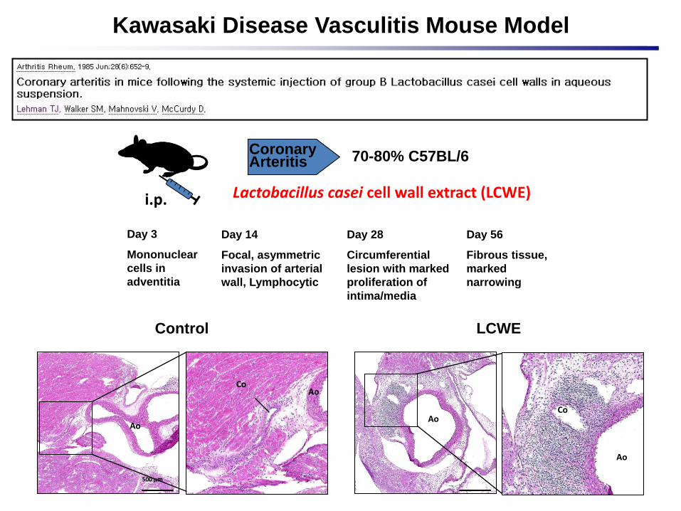
Coronary Arteritis 70-80% C57BL/6
Day 14
Focal, asymmetric
invasion of arterial
wall, Lymphocytic
Day 28
Circumferential
lesion with marked
proliferation of
intima/media
Day 56
Fibrous tissue,
marked
narrowing
Kawasaki Disease Vasculitis Mouse Model
Lactobacillus casei cell wall extract (LCWE) i.p.
Day 3
Mononuclear
cells in
adventitia
Control LCWE
500 mm
AoAo
Ao
AoCo
Co
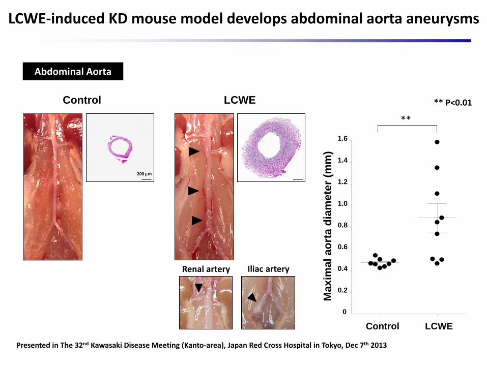
Abdominal Aorta
Renal artery Iliac artery
200 mm
Control LCWE
0
0.2
0.4
0.6
0.8
1.0
1.2
1.4
1.6
Max
ima
l a
ort
a d
iam
ete
r (m
m)
**
LCWE-induced KD mouse model develops abdominal aorta aneurysms
Control LCWE
Presented in The 32nd Kawasaki Disease Meeting (Kanto-area), Japan Red Cross Hospital in Tokyo, Dec 7th 2013
** P<0.01

NOD2-/- and Dectin-1-/- mice are protected from LCWE-induced KD vasculitis
FungiBacteria
NOD2
Inflammatory cytokines
Peptidoglycan
Dectin-1
β-1,3-glucan
WT NOD2-/- Dectin-1-/-
WT NOD2-/- Dectin-1-/-
0
5
10
15
Heart
ve
ss
els
Infl
am
mati
on
sc
ore
WT NOD2-/- Dectin-1-/-
0
25
50
75
100
Incid
en
ce (
%)
***
*****
**
11/12
0/12
2/7
** P<0.01, ***P<0.001

LCWE i.p.
Day7
HeartAbdominal Aorta
SPF(n=13)
GermFree
(n=13)
Germ-Free mice develop markedly decreased cardiovascular lesionsin KD mouse model
SPF GF
500 μm
SPF GF
0
2
4
6
8
10
12
He
art
ve
ss
els
Infl
am
mati
on
sco
re
SPF GF
0
10
20
30
40
50
60
70
80
Incid
en
ce (
%)
10/13
4/13
***** P<0.01 * P<0.05
(Coronary lesions)
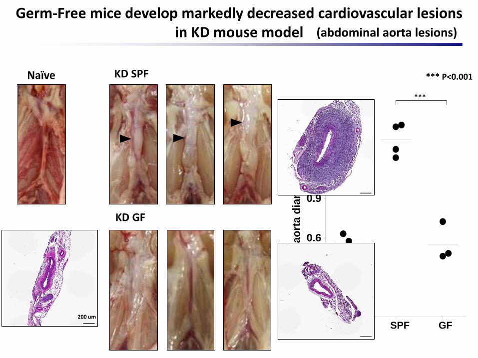
Naive SPF GF0.0
0.3
0.6
0.9
1.2
1.5
Max
ima
l a
ort
a d
iam
ete
r (m
m)
***
*** P<0.001KD SPF
KD GF
Naïve
Germ-Free mice develop markedly decreased cardiovascular lesions in KD mouse model
200 um
(abdominal aorta lesions)

Pregnant Birth 5 wk
LCWE i.p.
6 wk
HeartAbdominal Aorta
Anti-fungal drug (Fluconazole; Fluc)
and/or
Antibiotics cocktail (Abx)(Neomycin, Ampicillin, Vancomycin, Metronidazole)
Depletion of commensal fungi and bacteria with fluconazole and antibiotic treatment
Control Abx0
1
2
3
4
5
Ba
cte
ria
l 1
6S
rD
NA
(Re
lati
ve
to
to
tal
DN
A)
***
Bacteria amount
Control Fluc0.00
0.25
0.50
0.75
1.00
Fu
ng
al
ITS
1-2
DN
A
(Re
lati
ve
to
to
tal
DN
A)
*
Fungi amount
*** P<0.001* P<0.05

Control Fluc Abx Fluc+Abx
Control Fluc Abx Fluc+Abx0
5
10
15
He
art
ve
ss
els
Infl
am
mati
on
sco
re
***
*
Fluconazole and/or antibiotics treatment decreased cardiovascular lesions in KD mouse model
* P<0.05** P<0.01

Control Fluc Abx Fluc+Abx
Control Fluc Abx Fluc+Abx
0.0
0.2
0.4
0.6
0.8
1.0
1.2
1.4
Max
imal ao
rta d
iam
ete
r (m
m)
*
*
***
*
Fluconazole and/or antibiotics treatment decreased cardiovascular lesions in KD mouse model
* P<0.05, *** P<0.001

Bacteria
Fungi
Products
Metabolites
Gut Microflora
Inflammatory diseases
Translocation of intestinal microflora
Intestinal barrier dysfunction
Host
Intestinal permeability and disease development

LCWE injection increases intestinal permeability
Control 8 hr 20 hr0
0.2
0.4
0.6
0.8
1.0
1.2
FIT
C-D
extr
an
(u
g/m
l)
Hours after LCWE injection
LCWE (i.p.)
FITC-Dextran(p.o.)
Serum
4hr
20hr
FITC-Dextran(p.o.)
16hr
Serum
8hr
**
* P<0.05

Conclusions
LCWE-induced cardiovasculitis was decreased in germ free mice
Depletion of gut commensal fungi and bacteria diminished KD vasculitis
LCWE injection increased intestinal permeability
Gut MicrofloraLCWE
? Role of microbiome in KD pathogenesis, new diagnostic/therapuetic strategies
KD vasculitisBacteria
Fungi
Products
Metabolites
Permeability

Acknowledgement
Cedars-Sinai Medical centerPediatrics
Moshe Arditi Lab
Young Ho Lee
Shuang Chen
Timothy R. Crother
Kenichi Shimada
Wenxuan Zhang
Ganghua HuangMicheal Fishbein
University of CaliforniaLos Angeles
The University of Tokyo
Hiroshi Kiyono
Yosuke Kurashima
Yoshihiro Takasato
Grant supports
NIH AI072726 and NIH AI1070162 to Dr ArditiAHA fellowship to Dr Wakita
Thomas Lehman
Hospital for special surgery, New YorkCornell Medical College