Gul Moonis, M.D Department of Radiology Columbia ... · reconstruction Unlike myringoplasty, middle...
13
Gul Moonis, M.D Department of Radiology Columbia University Medical Center NY, NY Indications ,Contraindications,Normal Imaging Findings and Complications of common temporal bone surgical procedure Chronic Otitis Media with or without cholesteatoma Otospongiosis Severe B/L SNHL-Cochlear Implant Vestibular schwannoma Myringotomy Myringoplasty Tympanoplasty Canalplasty Meatoplasty Ossiculoplasty Mastoidectomy Surgical procedure in which a tiny incision is created in the tympanic membrane ,for drainage of middle ear effusion Followed by placement of pressure equalization(PE) tube (also known as grommet, ear tubes, tympanostomy tube-T tube, myringotomy tubes or ventilation tubes) Material -Plastic, metal or Teflon Maybe Subannular versus transtympanic Subannular tube is put between the bony wall of the EAC and the annulus (i.e. lateral to the annulus); used when long term ventilation of the middle ear is needed A “regular” transtympanic tube is placed through the TM, i.e., medial (central) to the annulus via myringotomy incision Axial and coronal CT demonstrate a sub annular PE tube between bony wall of EAC and annulus of TM(arrow)
Transcript of Gul Moonis, M.D Department of Radiology Columbia ... · reconstruction Unlike myringoplasty, middle...

Gul Moonis, M.DDepartment of Radiology
Columbia University Medical CenterNY, NY
Indications ,Contraindications,Normal Imaging Findings and Complications of common temporal bone surgical procedure Chronic Otitis Media with or without cholesteatoma Otospongiosis Severe B/L SNHL-Cochlear Implant Vestibular schwannoma
Myringotomy Myringoplasty Tympanoplasty Canalplasty Meatoplasty Ossiculoplasty Mastoidectomy
Surgical procedure in which a tiny incision is created in the tympanic membrane ,for drainage of middle ear effusion
Followed by placement of pressure equalization(PE) tube (also known as grommet, ear tubes, tympanostomy tube-T tube, myringotomy tubes or ventilation tubes)
Material -Plastic, metal or Teflon Maybe Subannular versus transtympanic
Subannular tube is put between the bony wall of the EAC and the annulus (i.e. lateral to the annulus); used when long term ventilation of the middle ear is needed
A “regular” transtympanic tube is placed through the TM, i.e., medial (central) to the annulus via myringotomy incision
Axial and coronal CT demonstrate a sub annular PE tube between bony wall of EAC and annulus of TM(arrow)

Myringoplasty refers to reconstruction of a perforation in the tympanic membrane without inspection of the ossicular chain
Assumes normal middle ear mucosa and ossicles
TM perforation in the pars tensa (arrow)
Material used : temporalis fascia, perichondrium, cartilage, periosteum, and adipose tissue.
Cartilage TM Graft
Procedure which involves lifting of the TM and removal of middle ear disease followed by TM reconstruction
Unlike myringoplasty, middle ear pathology is also addressed; like cholesteatoma,chronic OM, adhesions, scars, ossicular erosions
If ossicles are also surgically addressed at the same time , it is referred to as Ossiculoplasty or OssicularChain Reconstruction (OCR)
Tympanoplasty can be performed with or without ossiculoplasty, but all ossiculoplasty’s involve a tympanoplasty(lifting of the TM)
Tympanoplasty can be performed with or without mastoidectomy
Axial CT image demonstrates a TM graft (arrow) and a Canal Wall Up mastoidectomy(star)
Canalplasty is widening of the bony portion of the external ear canal.
In order to visualise the tympanic annulus, particularly in anterior or subtotal perforations, canalplasty is essential and may be an integral part of myringoplasty or tympanoplasty.
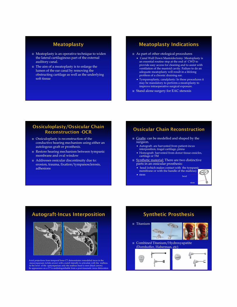
Meatoplasty is an operative technique to widen the lateral cartilaginous part of the external auditory canal.
The aim of a meatoplasty is to enlarge the lumen of the ear canal by removing the obstructing cartilage as well as the underlying soft tissue
As part of other otological procedures Canal Wall Down Mastoidectomy :Meatoplasty is
an essential routine step at the end of CWD to provide easy access for cleaning and to assist with ventilation of the mastoid cavity. Failure to do an adequate meatoplasty will result in a lifelong problem of a chronic draining ear.
Tympanoplasty, canalplasty: In these procedures it may be mandatory to perform a meatoplasty to improve intraoperative surgical exposure.
Stand alone surgery for EAC stenosis
Ossiculoplasty is reconstruction of the conductive hearing mechanism using either an autologous graft or prosthesis.
Restore hearing mechanism between tympanic membrane and oval window
Addresses ossicular discontinuity due to: erosion, trauma, fixation/tympanosclerosis, adhesions
Grafts: can be modelled and shaped by the surgeon. Autograft- are harvested from patient-incus
interposition, tragal cartilage, pinna Homograft- harvested from donor tissue-ossicles,
cartilage or TM Synthetic material: There are two distinctive
parts in an ossicular prosthesis: head (which makes contact with the tympanic
membrane or with the handle of the malleus) stem
head
stem
Axial projections from temporal bone CT demonstrates remodeled incus in themesoympanum (white arrow) with a notch laterally to articulate with the malleus. At the level of the epitympanum only the malleus head is seen (black arrow)Its appearance on a CT is undistinguishable from a post-traumatic incus dislocation
Titanium
Combined Titanium/Hydroxyapatite (Dornhoffer, Haberman, etc)
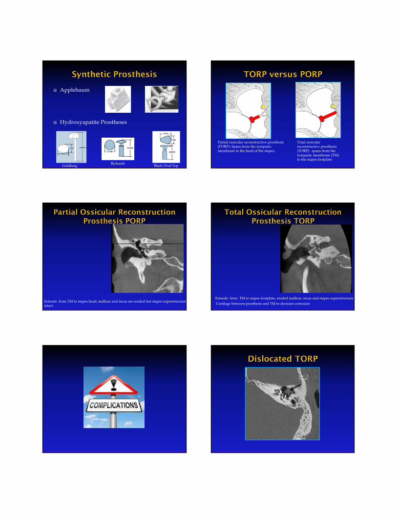
Applebaum
Hydroxyapatite Prostheses
GoldbergRichards
Black Oval Top
Total ossicularreconstructive prosthesis (TORP): spans from the tympanic membrane (TM) to the stapes footplate
Partial ossicular reconstructive prosthesis (PORP): Spans from the tympanic membrane to the head of the stapes.
Extends from TM to stapes head, malleus and incus are eroded but stapes superstructureintact Cartilage between prosthesis and TM to decrease extrusion
Extends from TM to stapes footplate, eroded malleus, incus and stapes superstructure

Chronic otorrhea refractory to medical therapy Obtain access to middle ear to correct middle
ear pathology (e.g., ossicular discontinuity causing a conductive hearing loss)
Obtain access to remove a cholesteatoma
Two major types Canal wall up (CWU) Canal wall down(CWD)
Some other terms that we may come across:
Cortical Mastoidectomy- a form of CWU, consists of opening the mastoid cortex and identifying the aditus ad antrum.
Radical Mastoidectomy –a form of CWD, involves removal of ossicles
Modified radical mastoidectomy – the most common form of CWD, CWD except the middle ear space and native tympanic membrane are not manipulated
Tympanomastoidectomy- Term used when tympanoplasty and mastoidectomy are performed together
Removal of all of the mastoid air cells along the tegmen, sigmoid sinus, presigmoid dural plate, and posterior wall of the external auditory canal. Koerner’s septum removed.The posterior wall of the external auditory canal is preserved.
A canal wall down mastoidectomy : CWU + removal of the posterior and superior osseous external auditory canal.
Meatoplasty is essential part of the surgery
External auditory canal, mastoid, and epitympanum become one common cavity –Mastoid Bowl
The tympanic membrane is reconstructed to separate the mucosal lined middle ear space from the mastoid cavity and ear canal.
ADVANTAGE DISADVANTAGE
Rapid healing Easier long term care No water precautions
Technically difficult Recurrent disease 2nd look needed Residual disease harder
to detect
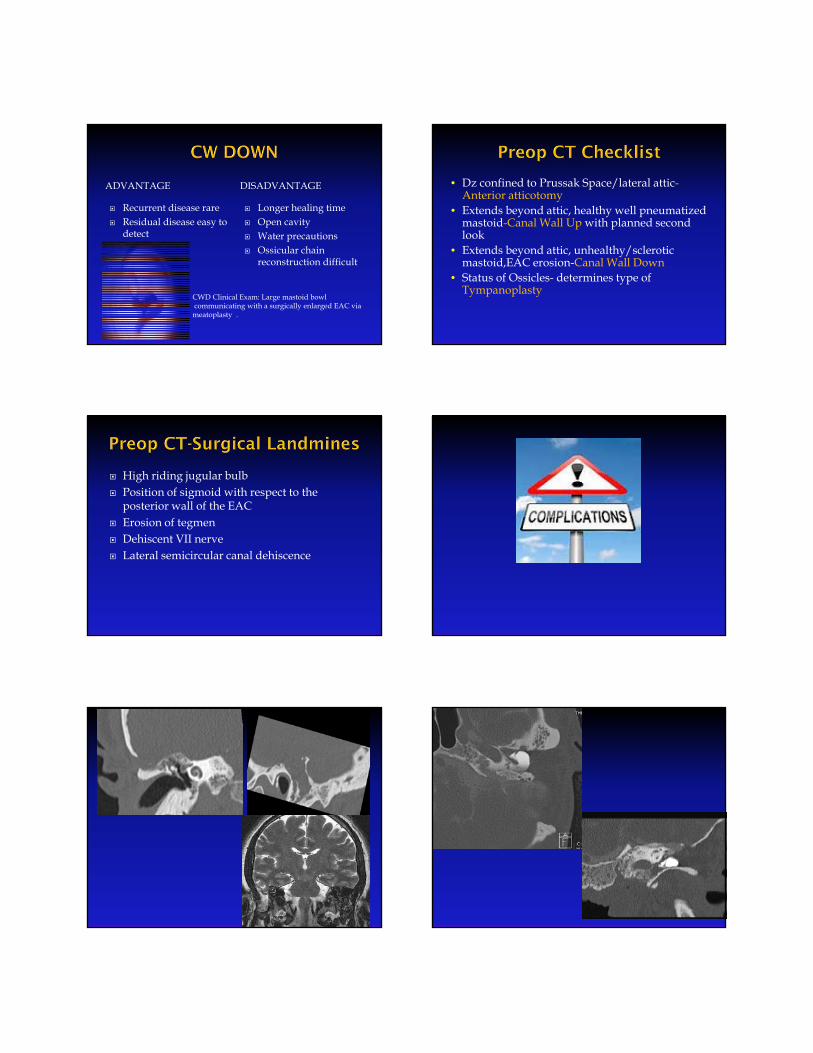
ADVANTAGE DISADVANTAGE
Recurrent disease rare Residual disease easy to
detect
Longer healing time Open cavity Water precautions Ossicular chain
reconstruction difficult
CWD Clinical Exam: Large mastoid bowlcommunicating with a surgically enlarged EAC via
meatoplasty .
• Dz confined to Prussak Space/lateral attic-Anterior atticotomy
• Extends beyond attic, healthy well pneumatizedmastoid-Canal Wall Up with planned second look
• Extends beyond attic, unhealthy/sclerotic mastoid,EAC erosion-Canal Wall Down
• Status of Ossicles- determines type of Tympanoplasty
High riding jugular bulb Position of sigmoid with respect to the
posterior wall of the EAC Erosion of tegmen Dehiscent VII nerve Lateral semicircular canal dehiscence
T1 pre T1 post
Coronal multishot HASTE DWI T2 T1 postT1 pre
FIESTA
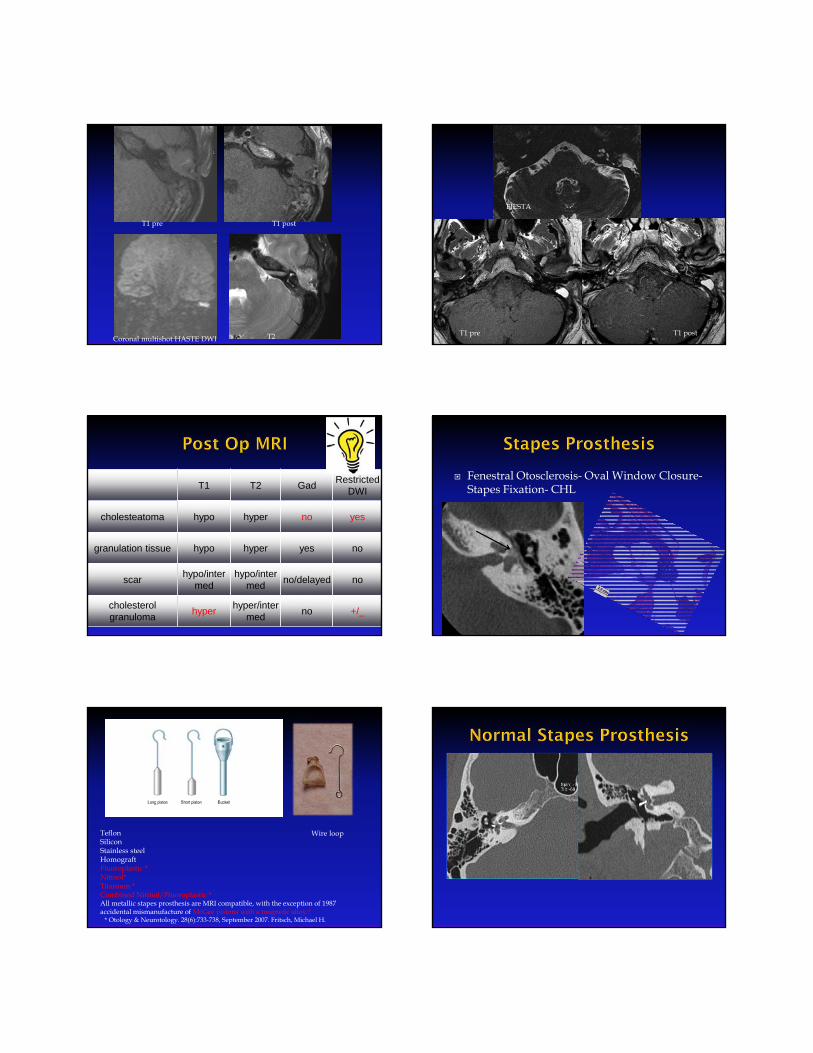
T1 T2 GadRestricted
DWI
cholesteatoma hypo hyper no yes
granulation tissue hypo hyper yes no
scar hypo/inter
medhypo/inter
medno/delayed no
cholesterol granuloma
hyperhyper/inter
medno +/_
Fenestral Otosclerosis- Oval Window Closure-Stapes Fixation- CHL
Teflon SiliconStainless steelHomograftFluoroplastic *Nitinol*Titanium *Combined Nitinol/Fluoroplastic * All metallic stapes prosthesis are MRI compatible, with the exception of 1987 accidental mismanufacture of McGee pistons with a magnetic alloy.*
* Otology & Neurotology. 28(6):733-738, September 2007. Fritsch, Michael H.
Wire loop

1. A microphone, which picks up sound from the environment.
2. A speech processor, which selects and arranges sounds picked up by the microphone.
3-4. A transmitter and receiver/stimulator, which receive signals from the speech processor and convert them into electric impulses.
5. An electrode array, which is a group of electrodes that collects the impulses from the stimulator and sends them to different regions of the cochlear nerve.
•1) A “well” is created in the parietal occipetal bone for the reciever-stimulator complex•2) Simple Canal Wall Up mastoidectomy•3)Electrode enters middle earthrough facial nerve recess•4)Electrode enters cochlea through round window/cochleostomy and makes 1.5 turns in scalatympani.
1
2 34
The absolute requirements for cochlear implantation are the presence of a cochlea (either normal or malformed) and of a cochlear nerve.
Absence of cochlea is best seen on temporal bone ct.
Absence of cochlear nerve is best seen on sagittal MRI images through the IAC.

The Baha(bone anchored hearing aide) works on a principle of efficient coupling of the sound processor to the underlying bone through 1) a small connector across the skin,
and 2) an implant that directly bonds with
the underlying bone This allows the bone to transfer
sound directly to a functioning cochlea rather than via the middle ear
Indication: Conductive and mixed hearing loss( EAC atresia/chronic infection of the middle ear)
Middle cranial Fossa approach Suboccipetal Translabyrinthine
The MCF approach Small tumorsMainly intracanalicularPatients with good hearing.
Adv:Hearing preservationSuperior access to fundus IAC
Disadv:Limited exp to CPATemporal lobe retraction
http://radiographics.rsna.org/content/29/7/1955.full
Suboccipital (Retrosigmoid) ApproachPost wall IAC removedAdv:No size limitationGreater access to the cerebellopontine angle while maintaining the option of hearing preservation. Better facial n exposureDisadv:Limited access to the lateral IAC Cerebellum retracted
The translabyrinthine approach
Mastoidectomy, removal of post wall IAC, sig sinus plate,SCC, labyrinthectomyFat packing to eliminate csf leakAdv:Widest exposure ,lowest tumor recurrence rate
Addresses tumor in cochlea/vestibuleDisadv: Eliminates hearing

Mastoidectomy and Tympanoplasty with ossicular chain reconstuction( Incus interposition/PORP/TORP for COM
Stapes Prosthesis for Fenestral Otosclerosis Presence of Cochlear Nerve is absolute
indication for CI Translabyrinthine approach for VS eliminates
hearing and should be used in patients with large tumors and no hearing