Guidelines for the Management of Oncologic Emergencies in ...
74
Guidelines for the Management of Oncologic Emergencies in Adult Cancer Patients Full Version
Transcript of Guidelines for the Management of Oncologic Emergencies in ...
Guidelines for the Management of
Oncologic Emergencies in Adult Cancer Patients
DepartmentDivisionSection
Full Version
i
Guidelines for the Managem
ent of O
ncologic Emergencies in Adult Cancer Patients
Guidelines for the Managem
ent of O
ncologic Emergencies in Adult Cancer Patients
AcknowledgementsThe majority of this practice guideline has been adopted, with permission, from Oncologic Emergencies, A Guide for Family Physicians, Guideline Utilization Resource Unit, Alberta Health Services, 2010.1
Cancer Care Nova Scotia (CCNS) expresses appreciation to the members of the Oncologic Emergencies Working Groups who refined and/or developed sections of the guidelines:
Emergency Health Services (EHS) – Oncologic Emergencies Working Group:Dr Tete Ago, Head, Radiation Oncology, Capital Health Cancer Care Program Dr. Sam Campbell, Emergency Medicine, Capital District Health AuthoritySteven Carrigan, Policy Analyst, Nova Scotia Department of Health & Wellness Dr. Mark Doreen, Head, Medical Oncology, Capital Health Cancer Care ProgramDr. Bahram Forouzesh, Medical Oncologist, Cape Breton Cancer CentreTracey George, Nurse Practitioner, Cape Breton District Health AuthorityDr William Harless, Medical Director, Cape Breton Cancer CentreDr. Wanda Hasegawa, Hematologist, Capital Health Cancer Care ProgramKara Henman, Oncology Practice Consultant, CCNSDebbie MacDonald, Acting Emergency Clinical Nurse EducatorMeg McCallum, Provincial Manager Education & Patient Navigation, CCNSDr. Tanya Munro, Emergency Medicine, Colchester East Hants Health AuthorityChris Nickerson, Director, Emergency Health Services, Nova Scotia Department of Health & Wellness Dr. Christian Pugh, Emergency Medicine, South Shore HealthMichele Rogez, Oncology Practice Consultant, CCNSDr. Andrew Travers, Medical Director, Emergency Health Services, Nova Scotia Department of Health & Wellness Dr. Sandra Wajstaub, Radiation Oncologist, Cape Breton District Health Authority
Febrile Neutropenia Working Group:Dr. Andrea Kew, Hematologist, Capital Health Cancer Care Program, Working Group Chair Dr. Shoaib Ansari, Infectious Diseases, Cape Breton District Health Authority Shelly Brown, Nurse Manager, Capital Health Cancer Care ProgramLori Butts, Clinical Nurse Educator, Cape Breton Cancer CentreDr. Sam Campbell, Emergency Medicine, Capital District Health AuthorityDr. Ian Davis, Infectious Diseases, Capital District Health AuthorityDr. Mark Doreen, Head, Medical Oncology, CDHA Cancer Care ProgramDonna Gamble, Nurse Manager, Capital Health Cancer Care ProgramTracy George, Nurse Practitioner, Cape Breton Cancer CentreDr William Harless, Medical Director, Cape Breton Cancer CentreKara Henman, Oncology Practice Consultant, CCNSTina Lewis, Emergency Nursing, Annapolis Valley HealthDebbie MacDonald, Acting Emergency Clinical Nurse EducatorDebbie MacIssac, Emergency Nursing, Guysboro, Antigonish, Strait Health AuthorityFlo MacLellan, Hematology Nurse Educator, CDHA Cancer Care ProgramMeg McCallum, Provincial Manager Education & Patient Navigation, CCNSDr. Brian Moses, Internal Medicine, South West HealthDr. Tanya Munro, Emergency Medicine, Colchester East Hants Health AuthorityDr. Christian Pugh, Emergency Medicine, South Shore HealthDr. Heather Robertson, General Practitioner (Oncology), South Shore HealthMichele Rogez, Oncology Practice Consultant, CCNSMegan Rolle, Clinical Pharmacist, Capital Health Cancer Care Program Dr. Kathy Slater, Infectious Diseases, Capital District Health AuthorityAngela Whynot, Clinical Nurse Educator, Capital Health Cancer Care Program
Oncologic Emergencies Psychosocial Health Needs Working Group:Dr. Janice Howes, Clinical Lead, Supportive Care, CCNSMarianne Arab, Manager, Supportive Care, CCNSMeg McCallum, Provincial Manager Education & Patient Navigation, CCNS
CCNS also acknowledges the valuable contributions of cancer patients and health professionals from throughout Nova Scotia who participated in the guideline development process.
ii
Guidelines for the Managem
ent of O
ncologic Emergencies in Adult Cancer Patients
Guidelines for the Managem
ent of O
ncologic Emergencies in Adult Cancer Patients
Recommended Citation
Cancer Care Nova Scotia (2014). Guidelines for the Management of Oncologic Emergencies in Adult Patients – Full Version. © Crown copyright, Province of Nova Scotia.
May be reprinted with permission from CCNS, 1-866-599-2263 or [email protected].
Review
This guideline will be reviewed in three years from publication date or earlier if important new evidence becomes available.
1
Guidelines for the Managem
ent of O
ncologic Emergencies in Adult Cancer Patients
Table of Contents
Preamble . . . . . . . . . . . . . . . . . . . . . . . . . . . . . . . . . . . . . . . . . . . . . . . . . . . . . . . . . . . . . . . . . . . . . . . . . . . . . . . 2
Introduction . . . . . . . . . . . . . . . . . . . . . . . . . . . . . . . . . . . . . . . . . . . . . . . . . . . . . . . . . . . . . . . . . . . . . . . . . . . . . . . . . . 3
EHS Special Patient Designation . . . . . . . . . . . . . . . . . . . . . . . . . . . . . . . . . . . . . . . . . . . . . . . . . . . . . . . . . . . . . . . . 4
Bleeding in a Cancer Patient . . . . . . . . . . . . . . . . . . . . . . . . . . . . . . . . . . . . . . . . . . . . . . . . . . . . . . . . . . . . . . . . . . . . 5Gastrointestinal (GI) bleeding . . . . . . . . . . . . . . . . . . . . . . . . . . . . . . . . . . . . . . . . . . . . . . . . . . . . . . . . . . . . . . . . . 6Hematuria . . . . . . . . . . . . . . . . . . . . . . . . . . . . . . . . . . . . . . . . . . . . . . . . . . . . . . . . . . . . . . . . . . . . . . . . . . . . . . . . . . 7Hemoptysis . . . . . . . . . . . . . . . . . . . . . . . . . . . . . . . . . . . . . . . . . . . . . . . . . . . . . . . . . . . . . . . . . . . . . . . . . . . . . . . . . 8 Vaginal bleeding. . . . . . . . . . . . . . . . . . . . . . . . . . . . . . . . . . . . . . . . . . . . . . . . . . . . . . . . . . . . . . . . . . . . . . . . . . . . . 8
Brain Metastases, Increased Intracranial Pressure and Seizures . . . . . . . . . . . . . . . . . . . . . . . . . . . . . . . . . . . 14
Disseminated Intravascular Coagulation (DIC) . . . . . . . . . . . . . . . . . . . . . . . . . . . . . . . . . . . . . . . . . . . . . . . . . . . 17
Febrile Neutropenia (High Risk and Low Risk) . . . . . . . . . . . . . . . . . . . . . . . . . . . . . . . . . . . . . . . . . . . . . . . . . . . . 20 Hyperviscosity Syndrome . . . . . . . . . . . . . . . . . . . . . . . . . . . . . . . . . . . . . . . . . . . . . . . . . . . . . . . . . . . . . . . . . . . . . . 25
Malignancy Associated Hypercalcemia (MAH) . . . . . . . . . . . . . . . . . . . . . . . . . . . . . . . . . . . . . . . . . . . . . . . . . . . . 28
Malignant Airway Obstruction . . . . . . . . . . . . . . . . . . . . . . . . . . . . . . . . . . . . . . . . . . . . . . . . . . . . . . . . . . . . . . . . . . 31
Malignant Epidural Spinal Cord Compression . . . . . . . . . . . . . . . . . . . . . . . . . . . . . . . . . . . . . . . . . . . . . . . . . . . . 34
Superior Vena Cava Obstruction . . . . . . . . . . . . . . . . . . . . . . . . . . . . . . . . . . . . . . . . . . . . . . . . . . . . . . . . . . . . . . . . 38
Syndrome of Inappropriate Antidiuretic Hormone Secretion . . . . . . . . . . . . . . . . . . . . . . . . . . . . . . . . . . . . . . . 41
Tumor Lysis Syndrome . . . . . . . . . . . . . . . . . . . . . . . . . . . . . . . . . . . . . . . . . . . . . . . . . . . . . . . . . . . . . . . . . . . . . . . . 44Hyperuricemia . . . . . . . . . . . . . . . . . . . . . . . . . . . . . . . . . . . . . . . . . . . . . . . . . . . . . . . . . . . . . . . . . . . . . . . . . . . . . 46Hyperkalemia . . . . . . . . . . . . . . . . . . . . . . . . . . . . . . . . . . . . . . . . . . . . . . . . . . . . . . . . . . . . . . . . . . . . . . . . . . . . . . 46Hyperphosphatemia . . . . . . . . . . . . . . . . . . . . . . . . . . . . . . . . . . . . . . . . . . . . . . . . . . . . . . . . . . . . . . . . . . . . . . . . 46 Hypocalcemia . . . . . . . . . . . . . . . . . . . . . . . . . . . . . . . . . . . . . . . . . . . . . . . . . . . . . . . . . . . . . . . . . . . . . . . . . . . . . . 46
Psychosocial Health Needs of Patients and Families Experiencing Oncologic Emergencies . . . . . . . . . . . . 49
References . . . . . . . . . . . . . . . . . . . . . . . . . . . . . . . . . . . . . . . . . . . . . . . . . . . . . . . . . . . . . . . . . . . . . . . . . . . . . . . . . . . 50
Appendix 1 Guideline Development Process . . . . . . . . . . . . . . . . . . . . . . . . . . . . . . . . . . . . . . . . . . . . . . . . . . . . . 52
Appendix 2 Initial Stakeholder Survey Instrument . . . . . . . . . . . . . . . . . . . . . . . . . . . . . . . . . . . . . . . . . . . . . . . . 56
Appendix 3 Patient Survey Results . . . . . . . . . . . . . . . . . . . . . . . . . . . . . . . . . . . . . . . . . . . . . . . . . . . . . . . . . . . . . . 59
Appendix 4 Second Stakeholder Survey Instrument . . . . . . . . . . . . . . . . . . . . . . . . . . . . . . . . . . . . . . . . . . . . . . 67
2
Guidelines for the Managem
ent of O
ncologic Emergencies in Adult Cancer Patients
Preamble
These guidelines focus on the management of adult patients with a suspected oncologic
emergency who present to EHS Paramedics or Emergency Departments.
• Adult patients who present with a suspected oncologic emergency within the Cape Breton Cancer Centre (CBCC) or the QEII Cancer Program (QEII) could be initially managed within the unit/clinic, following these guidelines, and transferred, as clinically indicated.
• Inpatients, in facilities other than the CBCC or QEII, who experience a suspected oncologic emergency can, if clinically appropriate, be initially managed on the unit, following these guidelines, and should be transferred to the ICU, Regional Hospital, CBCC or QEII, as required.
• Adult patients who present with a suspected oncologic emergency in any other setting should be immediately transported to an Emergency Department (ED) to be managed according to these guidelines. It is strongly recommended that practitioners call the Emergency Department to advise them that a patient with a suspected oncologic emergency (specify the nature of the emergency) is being transported to their facility.
• Some community EDs may elect to transport patients to a regional or tertiary ED for more advanced emergency care.
For information concerning the management of pediatric oncologic emergencies, please refer to APPHON/ROHPPA Emergency Room Supportive Care Guidelines binder or visit www.apphon-rohppa.com.
While cancer patients are at increased risk for bowel obstruction, pericardial tamponade and venous thromboembolism (VTE), these situations are not unique to the cancer patient population. Therefore, they are not included in this guideline. Clinicians encountering these emergency situations should manage them according to established practice guidelines, consulting Oncology as required.
Practice guidelines are intended to assist health care professionals with decisions throughout the spectrum of the cancer experience. This guideline is intended to assist health care professionals to care for adult cancer patients who experience oncologic emergencies. Management should be customized to meet the unique needs of individuals and their families. Guidelines should never replace specific decisions for individual patients, and do not substitute for the shared decisions between any patient and health professional which are unique to each circumstance. However, guidelines do provide evidence-based background information, consensus-based recommendations for similar situations, and a context for each individual decision.
These guidelines are designed for health professionals, working in a variety of settings. A Quick Reference Version of the guidelines is available on the Cancer Care Nova Scotia (CCNS) website, www.cancercare.ns.ca.
We recommend that patients, families and other non-health care professionals be referred to information regarding oncologic emergencies designed for the public, such as the Living Well With Cancer resources, available on the CCNS website, www.cancercare.ns.ca, the Canadian Cancer Society’s Cancer Information Service, 1-888-939-3333, www.cancer.ca or the National Cancer Institute’s Patient Version PDQ’s®, www.cancer.gov/cancertopics/pdq.
For further information on this, or any other Oncology Practice Guideline, please contact CCNS, 1-866-599-2267 or [email protected]
3
Guidelines for the Managem
ent of O
ncologic Emergencies in Adult Cancer Patients
Introduction 1, 2, 3, 4
Cancer is a leading cause of morbidity and mortality in Canada. Nova Scotia has high cancer
incidence and mortality rates amongst both males and females compared to the national rates. Given the complex nature of the disease and the cytotoxcity of treatment, cancer patients may experience a range of potentially life-threatening conditions that require urgent intervention.
In general, an oncologic emergency may be defined as any acute, potentially life-threatening incident, directly or indirectly related to a patient’s cancer or its treatment. Oncologic emergencies may result in permanent morbidity or the death. While some oncologic complications are subtle and may take weeks or even months to develop, others can manifest in a few hours, and quickly lead to severe negative outcomes, including paralysis, coma, and death. Prompt identification and intervention can prolong survival and improve quality of life.
Cancer patients are not immune from any medical emergency that may be experienced by an individual without a cancer diagnosis. Other non-neoplastic conditions must be considered in the differential diagnosis of every oncologic emergency.
Oncologic emergencies are not confined to the period of initial diagnosis and active treatment. They can occur at any time from pre-diagnosis to end-stage disease. In situations of recurrent malignancies, these emergencies can occur years after a cancer patient has been transferred from an oncologist to a primary care provider. Thus, it is critical for health professionals caring for cancer patients and survivors to be aware of a patient's cancer history and the related potential complications.
Once recognized, the aggressiveness of the management of any oncologic emergency should be influenced by the reversibility of the immediate event, the probability of long-term survival and cure, the ability to offer effective palliative treatment, the patient's/family's wishes/goals and/or advance directives.
Patients experiencing oncologic emergencies and their families will, undoubtedly, experience some degree of distress. Please refer to page 49, for information about addressing the psychosocial health needs of patients and families, and information regarding the support of and referral of those who are distressed and having difficulty coping.
4
Guidelines for the Managem
ent of O
ncologic Emergencies in Adult Cancer Patients
Emergency Health Services Special Patient Designation
In order to streamline pre-hospital care and transport the patient to the most appropriate
facility, Oncologists may elect to designate complex cancer patients at particularly high risk for experiencing an oncologic emergency as an Emergency Health Services (EHS) “Special Patient”. The EHS Special Patient program enables the Oncologist to specify a tailored treatment and transport protocol for a high risk patient. The Special Patient protocol supersedes EHS’ normal medical protocols. This program may be particularly helpful for patients residing in remote communities.
The EHS Special Patient application, to be completed by the Oncologist, can be accessed via the EHS website www.gov.ns.ca/health/ehs/pmd/special-patient.asp. Send completed applications to Emergency Health Services, Special Patient Program to 237 Brownlow Ave, Suite 160 Dartmouth, NS B3B 2C5, fax (902) 424-1781, or email [email protected].
The application will be reviewed by the EHS Provincial Medical Director. The EHS Provincial Medical Director may consult with the Oncologist, as necessary, to approve and finalize the Special Patient Protocol. A copy of the approved Special Patient card is sent to the Oncologist. In the case where an application is declined, the EHS Provincial Medical Director will send a letter of explanation to the Oncologist.
Once the Special Patient Protocol is approved, the EHS Communication Centre enters the information from the application into their communication system. This enables paramedics to access the patient’s information electronically when en route to a call.
An EHS paramedic will hand deliver the Special Patient card to the patient’s residence, confirm any information that may not have been given to them at the time of the application and review the program and card with the patient and/or next of kin. The patient and/or next of kin is advised to keep this card with them at all times.
Should the patient or next of kin call 911, EHS will follow the protocol on the Special Patient card, including contacting the receiving hospital, as soon as possible, to prepare for the patient’s care.
Oncologists should reserve the designation of “Special Patient” for complex cancer patients at particularly high risk for experiencing an oncologic emergency.
5
Guidelines for the Managem
ent of O
ncologic Emergencies in Adult Cancer Patients
Bleeding in a Cancer Patient 1, 2
Bleeding in cancer patients can be caused by the underlying malignancy, cancer treatment or
non-malignancy related factors.
In patients with hematologic cancers, bleeding is the second most frequent cause of death. Amongst metastatic cancer patients, bleeding is the third most frequent cause of death after organ failure from tumor invasion and infection. As many as 10% of cancer patients being treated with chemotherapy experience one or more significant bleeding episodes.
Patients experiencing acute bleeding and their families will, undoubtedly, experience some degree of distress. Please refer to page 49 for information about addressing the psychosocial health needs of patients and families, and information regarding the support of and referral of those who are distressed and having difficulty coping.
EHS Out-Of Hospital Care
Assessment/Management
Paramedics responding to a cancer patient with significant bleeding:• Assessment• Airway management• IV resuscitation• Symptom management• Determine the need to initiate massive
transfusion protocol
Transport
• Destination protocol• Pre hospital activation of ED system of care
In-Hospital Care
Immediate Management
• Resuscitate• If on anticoagulation, use appropriate reversal• Symptom control
Investigations
• Determine the source of the bleeding• CBC• Electrolytes• Urea• Creatinine• INR• PTT• Type and screen• Chest x-ray in the case of hemoptysis• Urinalysis in case of hematuria• Others as clinically indicated
6
Guidelines for the Managem
ent of O
ncologic Emergencies in Adult Cancer Patients
Bleeding in a Cancer Patient (continued)
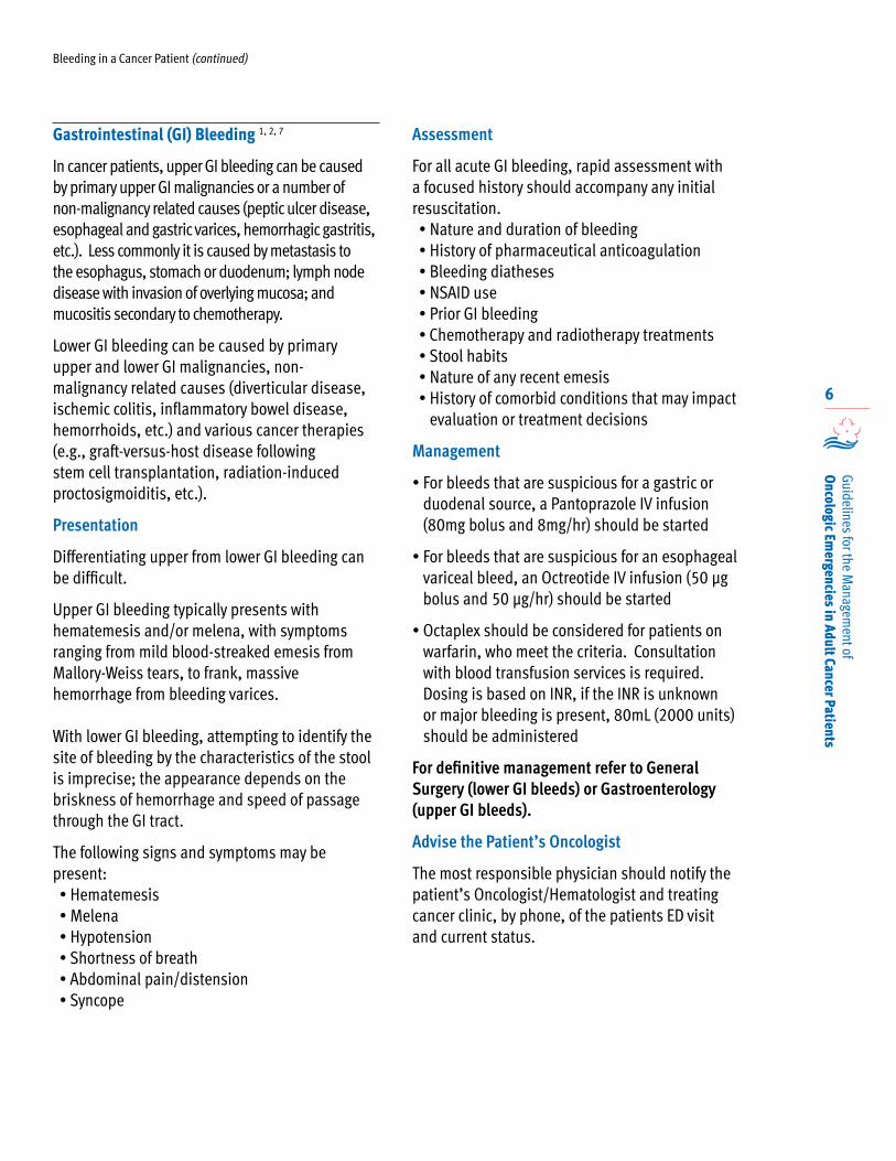
Gastrointestinal (GI) Bleeding 1, 2, 7
In cancer patients, upper GI bleeding can be caused by primary upper GI malignancies or a number of non-malignancy related causes (peptic ulcer disease, esophageal and gastric varices, hemorrhagic gastritis, etc.). Less commonly it is caused by metastasis to the esophagus, stomach or duodenum; lymph node disease with invasion of overlying mucosa; and mucositis secondary to chemotherapy.
Lower GI bleeding can be caused by primary upper and lower GI malignancies, non-malignancy related causes (diverticular disease, ischemic colitis, inflammatory bowel disease, hemorrhoids, etc.) and various cancer therapies (e.g., graft-versus-host disease following stem cell transplantation, radiation-induced proctosigmoiditis, etc.).
Presentation
Differentiating upper from lower GI bleeding can be difficult.
Upper GI bleeding typically presents with hematemesis and/or melena, with symptoms ranging from mild blood-streaked emesis from Mallory-Weiss tears, to frank, massive hemorrhage from bleeding varices.
With lower GI bleeding, attempting to identify the site of bleeding by the characteristics of the stool is imprecise; the appearance depends on the briskness of hemorrhage and speed of passage through the GI tract.
The following signs and symptoms may be present:• Hematemesis• Melena• Hypotension• Shortness of breath• Abdominal pain/distension• Syncope
Assessment
For all acute GI bleeding, rapid assessment with a focused history should accompany any initial resuscitation. • Nature and duration of bleeding• History of pharmaceutical anticoagulation• Bleeding diatheses• NSAID use• Prior GI bleeding• Chemotherapy and radiotherapy treatments• Stool habits• Nature of any recent emesis• History of comorbid conditions that may impact
evaluation or treatment decisions
Management
• For bleeds that are suspicious for a gastric or duodenal source, a Pantoprazole IV infusion (80mg bolus and 8mg/hr) should be started
• For bleeds that are suspicious for an esophageal variceal bleed, an Octreotide IV infusion (50 μg bolus and 50 μg/hr) should be started
• Octaplex should be considered for patients on warfarin, who meet the criteria. Consultation with blood transfusion services is required. Dosing is based on INR, if the INR is unknown or major bleeding is present, 80mL (2000 units) should be administered
For definitive management refer to General Surgery (lower GI bleeds) or Gastroenterology (upper GI bleeds).
Advise the Patient’s Oncologist
The most responsible physician should notify the patient’s Oncologist/Hematologist and treating cancer clinic, by phone, of the patients ED visit and current status.
7
Guidelines for the Managem
ent of O
ncologic Emergencies in Adult Cancer Patients
Bleeding in a Cancer Patient (continued)
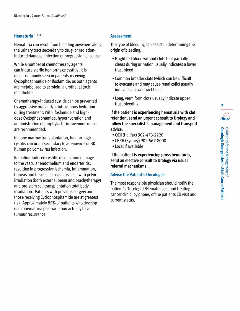
Hematuria 1, 2, 8
Hematuria can result from bleeding anywhere along the urinary tract secondary to drug- or radiation-induced damage, infection or progression of cancer.
While a number of chemotherapy agents can induce sterile hemorrhage cystitis, it is most commonly seen in patients receiving Cyclophosphamide or Ifosfamide; as both agents are metabolized to acrolein, a urothelial toxic metabolite.
Chemotherapy-induced cystitis can be prevented by aggressive oral and/or intravenous hydration during treatment. With Ifosfamide and high-dose Cyclophosphamide, hyperhydration and administration of prophylactic intravenous mesna are recommended.
In bone marrow transplantation, hemorrhagic cystitis can occur secondary to adenovirus or BK human polyomavirus infection.
Radiation-induced cystitis results from damage to the vascular endothelium and endarteritis, resulting in progressive ischemia, inflammation, fibrosis and tissue necrosis. It is seen with pelvic irradiation (both external beam and brachytherapy) and pre-stem cell transplantation total body irradiation. Patients with previous surgery and those receiving Cyclophosphamide are at greatest risk. Approximately 85% of patients who develop macrohematuria post-radiation actually have tumour recurrence.
Assessment
The type of bleeding can assist in determining the origin of bleeding:
• Bright red blood without clots that partially clears during urination usually indicates a lower tract bleed
• Common broader clots (which can be difficult to evacuate and may cause renal colic) usually indicates a lower tract bleed
• Long, vermiform clots usually indicate upper tract bleeding
If the patient is experiencing hematuria with clot retention, send an urgent consult to Urology and follow the specialist’s management and transport advice. • QEII (Halifax) 902-473-2220• CBRH (Sydney) 902-567-8000• Local if available
If the patient is experiencing gross hematuria, send an elective consult to Urology via usual referral mechanisms.
Advise the Patient’s Oncologist
The most responsible physician should notify the patient’s Oncologist/Hematologist and treating cancer clinic, by phone, of the patients ED visit and current status.
8
Guidelines for the Managem
ent of O
ncologic Emergencies in Adult Cancer Patients
Bleeding in a Cancer Patient (continued)
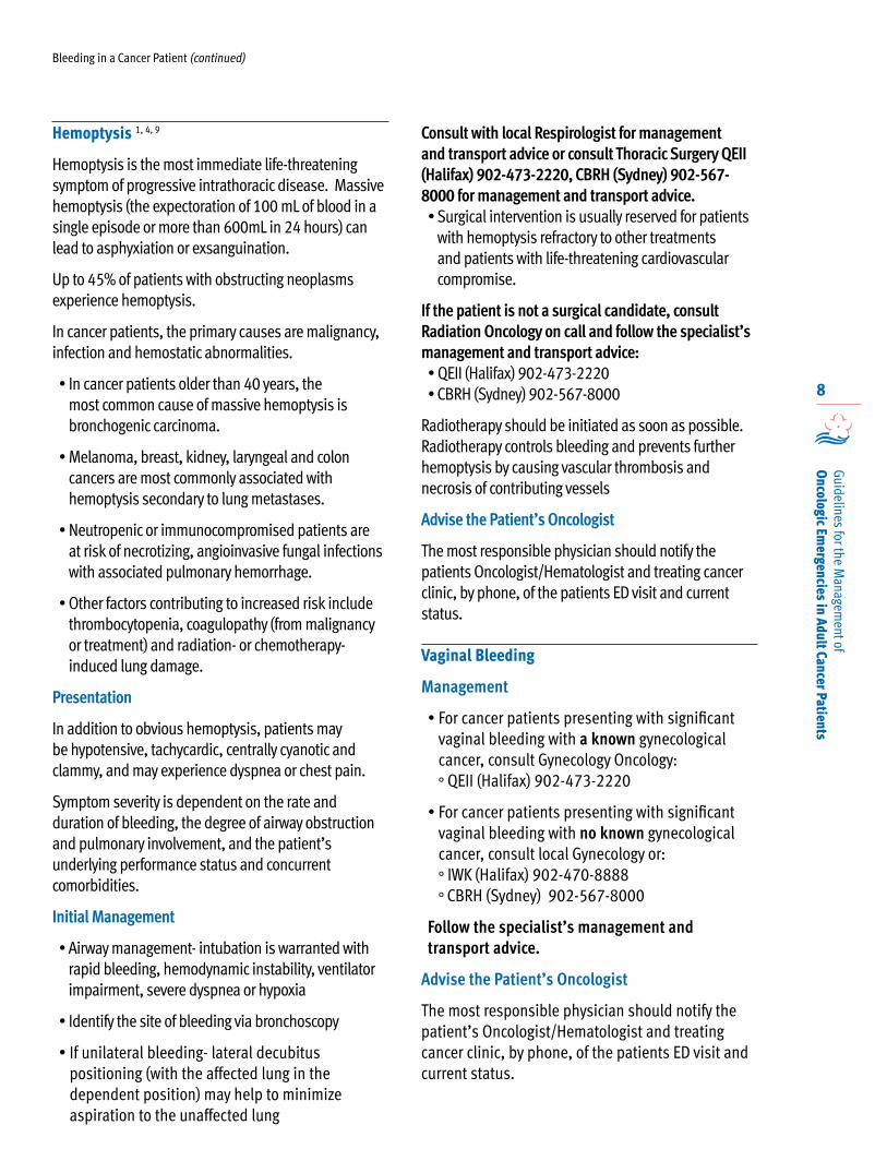
Hemoptysis 1, 4, 9
Hemoptysis is the most immediate life-threatening symptom of progressive intrathoracic disease. Massive hemoptysis (the expectoration of 100 mL of blood in a single episode or more than 600mL in 24 hours) can lead to asphyxiation or exsanguination.
Up to 45% of patients with obstructing neoplasms experience hemoptysis.
In cancer patients, the primary causes are malignancy, infection and hemostatic abnormalities.
• In cancer patients older than 40 years, the most common cause of massive hemoptysis is bronchogenic carcinoma.
• Melanoma, breast, kidney, laryngeal and colon cancers are most commonly associated with hemoptysis secondary to lung metastases.
• Neutropenic or immunocompromised patients are at risk of necrotizing, angioinvasive fungal infections with associated pulmonary hemorrhage.
• Other factors contributing to increased risk include thrombocytopenia, coagulopathy (from malignancy or treatment) and radiation- or chemotherapy-induced lung damage.
Presentation
In addition to obvious hemoptysis, patients may be hypotensive, tachycardic, centrally cyanotic and clammy, and may experience dyspnea or chest pain.
Symptom severity is dependent on the rate and duration of bleeding, the degree of airway obstruction and pulmonary involvement, and the patient’s underlying performance status and concurrent comorbidities.
Initial Management
• Airway management- intubation is warranted with rapid bleeding, hemodynamic instability, ventilator impairment, severe dyspnea or hypoxia
• Identify the site of bleeding via bronchoscopy
• If unilateral bleeding- lateral decubitus positioning (with the affected lung in the dependent position) may help to minimize aspiration to the unaffected lung
Consult with local Respirologist for management and transport advice or consult Thoracic Surgery QEII (Halifax) 902-473-2220, CBRH (Sydney) 902-567-8000 for management and transport advice.• Surgical intervention is usually reserved for patients
with hemoptysis refractory to other treatments and patients with life-threatening cardiovascular compromise.
If the patient is not a surgical candidate, consult Radiation Oncology on call and follow the specialist’s management and transport advice: • QEII (Halifax) 902-473-2220• CBRH (Sydney) 902-567-8000
Radiotherapy should be initiated as soon as possible. Radiotherapy controls bleeding and prevents further hemoptysis by causing vascular thrombosis and necrosis of contributing vessels
Advise the Patient’s Oncologist
The most responsible physician should notify the patients Oncologist/Hematologist and treating cancer clinic, by phone, of the patients ED visit and current status.
Vaginal Bleeding
Management
• For cancer patients presenting with significant vaginal bleeding with a known gynecological cancer, consult Gynecology Oncology: ° QEII (Halifax) 902-473-2220
• For cancer patients presenting with significant vaginal bleeding with no known gynecological cancer, consult local Gynecology or: ° IWK (Halifax) 902-470-8888 ° CBRH (Sydney) 902-567-8000
Follow the specialist’s management and transport advice.
Advise the Patient’s Oncologist
The most responsible physician should notify the patient’s Oncologist/Hematologist and treating cancer clinic, by phone, of the patients ED visit and current status.
9
Guidelines for the Managem
ent of O
ncologic Emergencies in Adult Cancer Patients
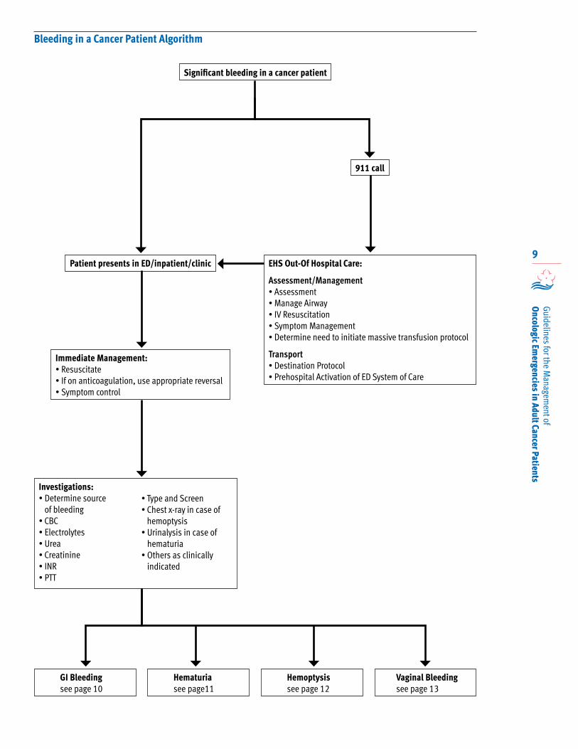
Bleeding in a Cancer Patient Algorithm
Patient presents in ED/inpatient/clinic
911 call
EHS Out-Of Hospital Care:
Assessment/Management• Assessment• Manage Airway• IV Resuscitation• Symptom Management• Determine need to initiate massive transfusion protocol
Transport• Destination Protocol• Prehospital Activation of ED System of Care
Immediate Management:• Resuscitate• If on anticoagulation, use appropriate reversal• Symptom control
Investigations:• Determine source
of bleeding• CBC• Electrolytes• Urea• Creatinine• INR• PTT
• Type and Screen• Chest x-ray in case of
hemoptysis• Urinalysis in case of
hematuria• Others as clinically
indicated
Significant bleeding in a cancer patient
GI Bleedingsee page 10
Hemoptysissee page 12
Hematuriasee page11
Vaginal Bleedingsee page 13
10
Guidelines for the Managem
ent of O
ncologic Emergencies in Adult Cancer Patients
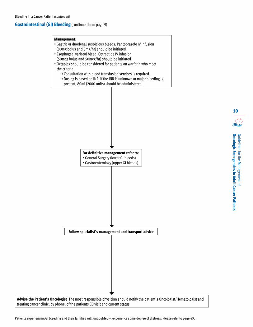
Management:• Gastric or duodenal suspicious bleeds: Pantoprazole IV infusion
(80mg bolus and 8mg/hr) should be initiated• Esophageal variceal bleed: Octreotide IV infusion
(50mcg bolus and 50mcg/hr) should be initiated• Octaplex should be considered for patients on warfarin who meet
the criteria. ° Consultation with blood transfusion services is required. ° Dosing is based on INR, if the INR is unknown or major bleeding is
present, 80ml (2000 units) should be administered.
For definitive management refer to: • General Surgery (lower GI bleeds) • Gastroenterology (upper GI bleeds)
Follow specialist’s management and transport advice
Advise the Patient’s Oncologist The most responsible physician should notify the patient’s Oncologist/Hematologist and treating cancer clinic, by phone, of the patients ED visit and current status
Patients experiencing GI bleeding and their families will, undoubtedly, experience some degree of distress. Please refer to page 49.
Gastrointestinal (GI) Bleeding (continued from page 9)
Bleeding in a Cancer Patient (continued)
11
Guidelines for the Managem
ent of O
ncologic Emergencies in Adult Cancer Patients
Bleeding in a Cancer Patient (continued)
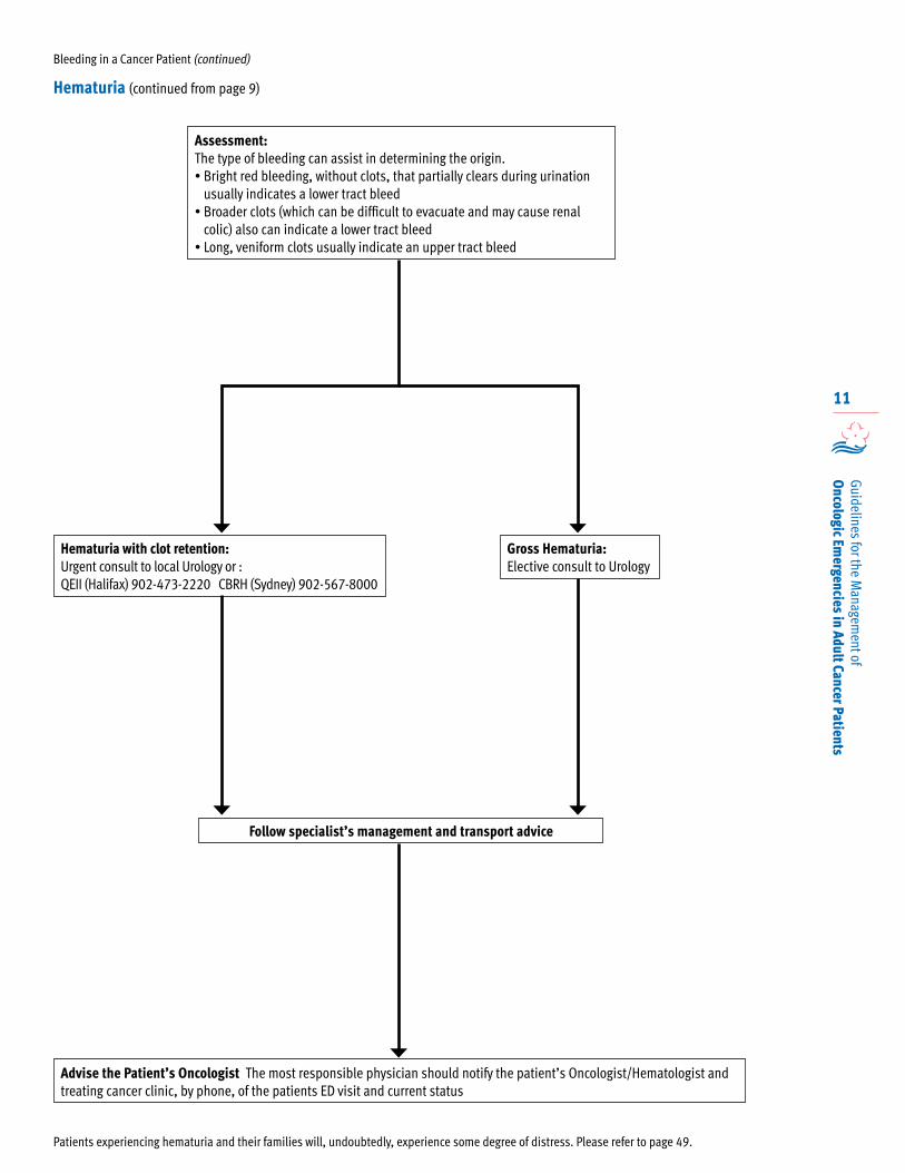
Assessment:The type of bleeding can assist in determining the origin.• Bright red bleeding, without clots, that partially clears during urination
usually indicates a lower tract bleed• Broader clots (which can be difficult to evacuate and may cause renal
colic) also can indicate a lower tract bleed• Long, veniform clots usually indicate an upper tract bleed
Patients experiencing hematuria and their families will, undoubtedly, experience some degree of distress. Please refer to page 49.
Gross Hematuria:Elective consult to Urology
Hematuria with clot retention: Urgent consult to local Urology or :QEII (Halifax) 902-473-2220 CBRH (Sydney) 902-567-8000
Follow specialist’s management and transport advice
Advise the Patient’s Oncologist The most responsible physician should notify the patient’s Oncologist/Hematologist and treating cancer clinic, by phone, of the patients ED visit and current status
Hematuria (continued from page 9)
12
Guidelines for the Managem
ent of O
ncologic Emergencies in Adult Cancer Patients
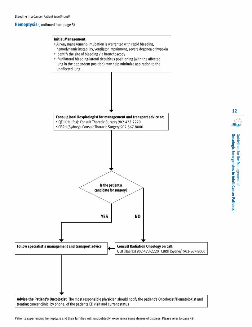
Initial Management:• Airway management- intubation is warranted with rapid bleeding,
hemodynamic instability, ventilator impairment, severe dyspnea or hypoxia• Identify the site of bleeding via bronchoscopy• If unilateral bleeding-lateral decubitus positioning (with the affected
lung in the dependent position) may help minimize aspiration to the unaffected lung
Patients experiencing hemoptysis and their families will, undoubtedly, experience some degree of distress. Please refer to page 49.
Advise the Patient’s Oncologist The most responsible physician should notify the patient’s Oncologist/Hematologist and treating cancer clinic, by phone, of the patients ED visit and current status
Consult local Respirologist for management and transport advice or:• QEII (Halifax): Consult Thoracic Surgery 902-473-2220 • CBRH (Sydney): Consult Thoracic Surgery 902-567-8000
YES NO
Consult Radiation Oncology on call: QEII (Halifax) 902-473-2220 CBRH (Sydney) 902-567-8000
Follow specialist’s management and transport advice
Hemoptysis (continued from page 5)
Bleeding in a Cancer Patient (continued)
Is the patient a candidate for surgery?
13
Guidelines for the Managem
ent of O
ncologic Emergencies in Adult Cancer Patients
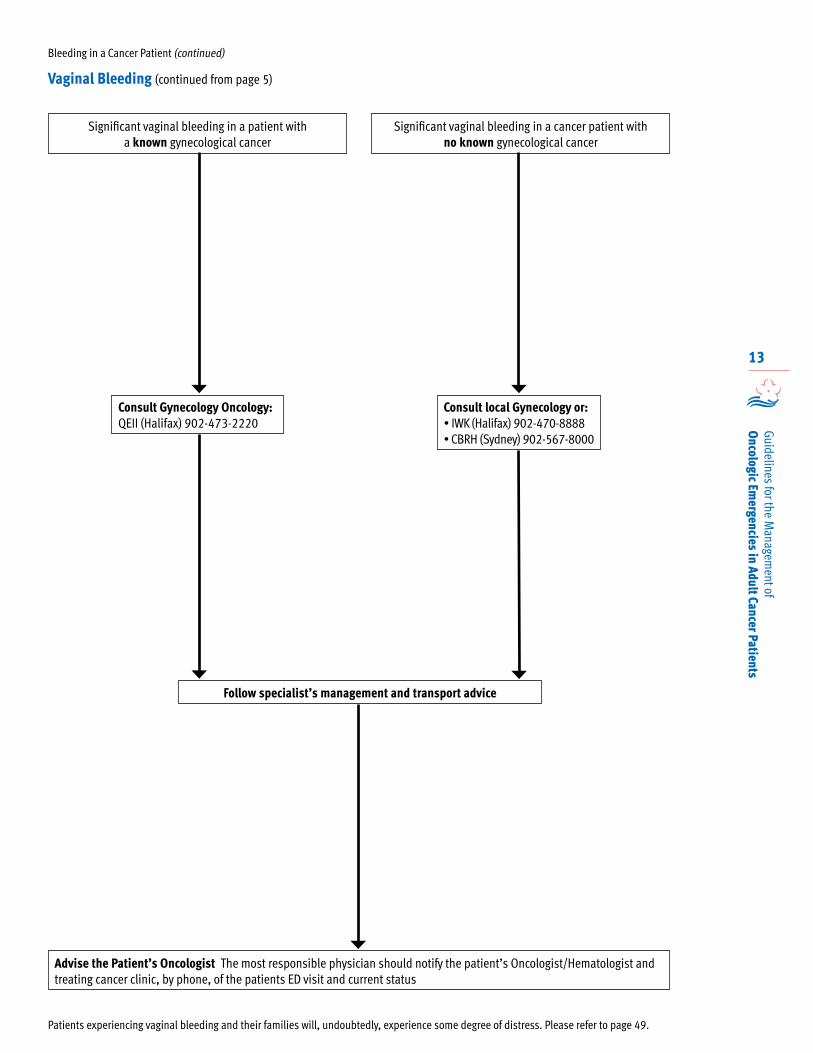
Significant vaginal bleeding in a cancer patient with no known gynecological cancer
Significant vaginal bleeding in a patient with a known gynecological cancer
Patients experiencing vaginal bleeding and their families will, undoubtedly, experience some degree of distress. Please refer to page 49.
Advise the Patient’s Oncologist The most responsible physician should notify the patient’s Oncologist/Hematologist and treating cancer clinic, by phone, of the patients ED visit and current status
Consult local Gynecology or:• IWK (Halifax) 902-470-8888 • CBRH (Sydney) 902-567-8000
Consult Gynecology Oncology:QEII (Halifax) 902-473-2220
Follow specialist’s management and transport advice
Vaginal Bleeding (continued from page 5)
Bleeding in a Cancer Patient (continued)
14
Guidelines for the Managem
ent of O
ncologic Emergencies in Adult Cancer Patients
Brain metastases are the most common type of brain malignancy, occurring in 20-40% of
adult cancer patients. Although any tumour can metastasize to the brain, lung cancer, breast cancer, and melanoma are the most common, accounting for 70-90% of brain metastases. Other common primary tumours include colorectal cancer and renal cell carcinoma. Melanoma and lung cancer are most frequently associated with multiple brain metastases; breast, colorectal, and renal cancers are more likely to be associated with a solitary metastasis.
Brain metastases can lead to neurologic deficits and seizures, and become an oncologic emergency in cases of increased intracranial pressure and status epilepticus.
Patients with a primary brain tumour and patients with edema resulting from treatment with whole-brain radiotherapy or some chemotherapeutic agents can also show signs of increased intracranial pressure.
Untreated, patients experiencing increased intracranial pressure have a median survival of approximately 4 weeks. Prognosis is dependent on Karnofsky performance status, the presence of systemic disease, and the primary tumor.
Increased intracranial pressure and seizures lasting longer than 30 minutes are considered oncologic emergencies.
Status epilepticus is defined as more than 30 minutes of continuous seizure activity or two or more sequential seizures without full recovery between seizures.
Patients experiencing brain metastases, increased intracranial pressure or seizures and their families will, undoubtedly, experience some degree of distress. Please refer to page 49, for information about addressing the psychosocial health needs of patients and families.
Presentation
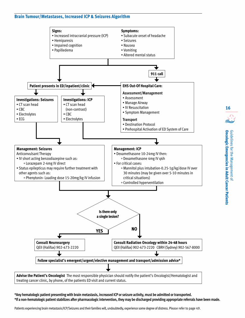
Patients with brain metastases may experience a variety of neurological symptoms. About 50% of cases experience subacute onset of headache. Other common symptoms include altered mental status, hemiparesis, impaired cognition, increased intracranial pressure, and seizures.
Patients with increased intracranial pressure due to brain metastases or a primary brain tumour classically present with headache, nausea, and vomiting, all of which may be most severe in the morning and when supine. In addition, papilledema detected on physical examination almost always indicates increased intracranial pressure.
EHS Out-Of Hospital Care
Paramedics responding to a cancer patient with ICP or seizures:
Assessment/Management
• Assessment• Airway management• IV resuscitation• Symptom management
Transport
• Destination protocol• Pre hospital activation of ED system of care
Brain Tumour/Metastases, Increased Intracranial Pressure (ICP) & Seizures 1, 4, 10
15
Guidelines for the Managem
ent of O
ncologic Emergencies in Adult Cancer Patients
In-Hospital Care
Investigations: ICP
• CT scan head (non contrast)• CBC• Electrolytes
Investigations: Seizures
• CT scan head (non contrast)• CBC• Electrolytes• ECG
ICP Management
Initial treatment of elevated ICP is with Dexamethasone, because it is the most lipid-soluble of all the steroids. Dexamathasone 10-24 mg IV, followed by 4 mg IV every 6 hours should be administered.
• In the most severe cases, Mannitol 0.25-1g/kg/dose IV over 30 minutes (may be given over 5-10 minutes in critical situations) in addition to intubation and controlled hyperventilation may be used to decrease cerebral edema, but this is reserved for critical cases in patients with rapidly declining clinical states.
Seizure Management
Status epilepticus is a medical emergency requiring the immediate assessment of airway, breathing, and circulation.
• Anticonvulsant therapy with a short-acting benzodiazepine should be administered to halt seizure activity: ° Lorazepam 2-4 mg IV direct
• Patients with status epilepticus may also require further treatment with other anticonvulsants such as: ° Phenytonin-loading dose 15-20 mg/kg IV
infusion
• Anticonvulsants are associated with significant adverse effects and are not recommended for prophylaxis in patients with brain metastases without a history of seizures.
For select patients with a good performance status and well-controlled systemic disease, more definitive treatments of brain metastases may include surgery, whole brain radiotherapy, stereotactic radiosurgery or a combination.
• If the CT scan reveals only a single lesion, Neurosurgery should be consulted: ° QEII (Halifax) 902-473-2220
• If the CT scan reveals multiple lesions, Radiation Oncology should be consulted within 24-48 hours: ° QEII (Halifax) 902-473-2220 ° CBRH (Sydney) 902-567-8000
Follow the specialist’s emergent/urgent/elective management and transport/admission advice.*
* Any hematology patient presenting with brain metastasis, increased ICP or seizure activity must be admitted or transported.
* If a non-hematologic patient stabilizes after pharmacologic intervention, they may be discharged providing the appropriate referrals have been made.
Advise the Patient’s Oncologist
The most responsible physician should notify the patients Oncologist/Hematologist and treating cancer clinic, by phone, of the patients ED visit and current status.
Brain Tumour/Metastases, Increased ICP & Seizures (continued)
16
Guidelines for the Managem
ent of O
ncologic Emergencies in Adult Cancer Patients
Brain Tumour/Metastases, Increased ICP & Seizures Algorithm
Signs:• Increased intracranial pressure (ICP)• Hemiparesis• Impaired cognition• Papilledema
Symptoms:• Subacute onset of headache • Seizures• Nausea• Vomiting• Altered mental status
Patient presents in ED/inpatient/clinic
911 call
EHS Out-Of Hospital Care:
Assessment/Management• Assessment• Manage Airway• IV Resuscitation• Symptom Management
Transport• Destination Protocol• Prehospital Activation of ED System of Care
Investigations: Seizures• CT scan head• CBC• Electrolytes• ECG
Investigations: ICP• CT scan head
(non-contrast)• CBC• Electrolytes
Management: SeizuresAnticonvulsant Therapy• IV short acting benzodiazepine such as:
° Lorazepam 2-4mg IV direct• Status epilepticus may require further treatment with
other agents such as:° Phenytonin- Loading dose 15-20mg/kg IV infusion
Management: ICP• Dexamethasone 10-24mg IV then:
° Dexamethasone 4mg IV q6h• For critical cases:
° Mannitol plus intubation-0.25-1g/kg/dose IV over 30 minutes (may be given over 5-10 minutes in critical situations)
° Controlled hyperventilation
Is there only a single lesion?
YES NO
Consult Neurosurgery QEII (Halifax) 902-473-2220
Consult Radiation Oncology within 24-48 hours QEII (Halifax) 902-473-2220 CBRH (Sydney) 902-567-8000
Follow specialist’s emergent/urgent/elective management and transport/admission advice*
Advise the Patient’s Oncologist The most responsible physician should notify the patient’s Oncologist/Hematologist and treating cancer clinic, by phone, of the patients ED visit and current status.
*Any hematologic patient presenting with brain metastasis, increased ICP or seizure activity, must be admitted or transported.*If a non-hematologic patient stabilizes after pharmacologic intervention, they may be discharged providing appropriate referrals have been made.
Patients experiencing brain metastasis/ICP/Seizures and their families will, undoubtedly, experience some degree of distress. Please refer to page 49.
17
Guidelines for the Managem
ent of O
ncologic Emergencies in Adult Cancer Patients
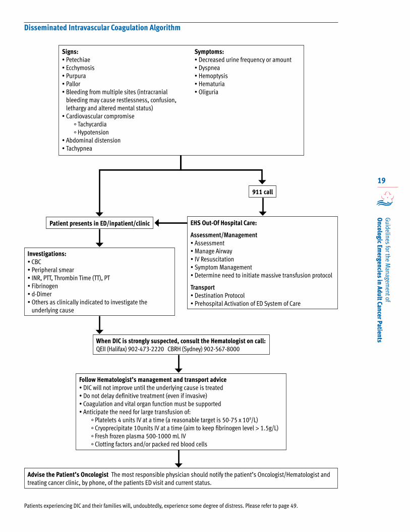
Disseminated Intravascular Coagulation (DIC) 1, 2, 5, 6
DIC, arising from inappropriate thrombin activation, results in the rapid formation
of fibrin clots in the microcoagulation, consumption of clotting factors and clot degradation. Continuous bleeding and clotting continues until clotting factors are completely consumed, resulting in uncontrollable bleeding. In cancer patients, DIC is most commonly seen with acute myelocytic leukemia, adenocarcinoma, septicemia or transfusion reactions.
Patients experiencing disseminated intravascular coagulation and their families will, undoubtedly, experience some degree of distress. Please refer to page 49, for information about addressing the psychosocial health needs of patients and families.
Presentation
Acute DIC typically presents with prolonged PT and PTT times, D-dimer, low fibrinogen, fragments (schistocytes) seen on peripheral blood film, and evidence of hemolytic anemia with increased LD, increased indirect bilirubin, increased reticulocyte count, low haptoglobin, and/or decreased platelet counts.
Patients may present with the following symptoms:• Dyspnea • Oliguria• Hematuria• Hemoptysis • Decreased urine frequency or amount
The following signs may be present: • Abdominal distention • Bleeding from multiple sites • Ecchymosis• Purpura• Tachypnea• Pallor • Petechiae• Cardiovascular compromise
Patients with intracranial bleeding may present with restlessness, confusion, lethargy and altered mental status.
In patients with overwhelming infection, purpura fulminans (DIC in association with symmetric limb ecchymosis and skin necrosis) may be observed.
Cardiovascular compromise presents as tachycardia with hypotension.
With tumour-initiated DIC, a hypercoagulability state is seen more often than hemorrhage.
Overt manifestations include DVT, pulmonary embolus and thrombosis in the central nervous system and abdominal organs.
Patients with laboratory evidence of DIC may be asymptomatic, but with progression of the underlying condition, can rapidly become symptomatic.
With sepsis-induced DIC, patients more commonly present with bleeding rather than thrombosis.
EHS Out-Of Hospital Care
Paramedics responding to a cancer patient with suspected DIC:
Assessment/Management
• Assessment• Airway management• IV resuscitation• Symptom management• Determine the need to initiate massive
transfusion protocol
Transport
• Destination protocol• Pre hospital activation of ED system of care
18
Guidelines for the Managem
ent of O
ncologic Emergencies in Adult Cancer Patients
Disseminated Intravascular Coagulation (continued)
In Hospital Care
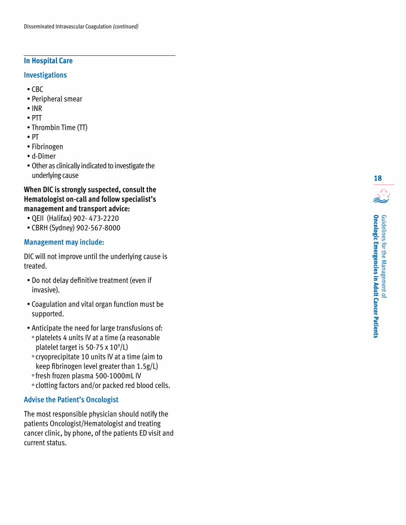
Investigations
• CBC• Peripheral smear• INR• PTT• Thrombin Time (TT)• PT• Fibrinogen• d-Dimer • Other as clinically indicated to investigate the
underlying cause
When DIC is strongly suspected, consult the Hematologist on-call and follow specialist’s management and transport advice:• QEII (Halifax) 902- 473-2220• CBRH (Sydney) 902-567-8000
Management may include:
DIC will not improve until the underlying cause is treated.
• Do not delay definitive treatment (even if invasive).
• Coagulation and vital organ function must be supported.
• Anticipate the need for large transfusions of: ° platelets 4 units IV at a time (a reasonable platelet target is 50-75 x 109/L) ° cryoprecipitate 10 units IV at a time (aim to keep fibrinogen level greater than 1.5g/L) ° fresh frozen plasma 500-1000mL IV ° clotting factors and/or packed red blood cells.
Advise the Patient’s Oncologist
The most responsible physician should notify the patients Oncologist/Hematologist and treating cancer clinic, by phone, of the patients ED visit and current status.
19
Guidelines for the Managem
ent of O
ncologic Emergencies in Adult Cancer Patients
Disseminated Intravascular Coagulation Algorithm
Patient presents in ED/inpatient/clinic
911 call
EHS Out-Of Hospital Care:
Assessment/Management• Assessment• Manage Airway• IV Resuscitation• Symptom Management• Determine need to initiate massive transfusion protocol
Transport• Destination Protocol• Prehospital Activation of ED System of Care
Investigations: • CBC• Peripheral smear• INR, PTT, Thrombin Time (TT), PT• Fibrinogen• d-Dimer• Others as clinically indicated to investigate the
underlying cause
When DIC is strongly suspected, consult the Hematologist on call: QEII (Halifax) 902-473-2220 CBRH (Sydney) 902-567-8000
Follow Hematologist’s management and transport advice• DIC will not improve until the underlying cause is treated• Do not delay definitive treatment (even if invasive)• Coagulation and vital organ function must be supported• Anticipate the need for large transfusion of:
° Platelets 4 units IV at a time (a reasonable target is 50-75 x 109/L)° Cryoprecipitate 10units IV at a time (aim to keep fibrinogen level > 1.5g/L)° Fresh frozen plasma 500-1000 mL IV° Clotting factors and/or packed red blood cells
Signs:• Petechiae• Ecchymosis• Purpura • Pallor• Bleeding from multiple sites (intracranial
bleeding may cause restlessness, confusion, lethargy and altered mental status)
• Cardiovascular compromise° Tachycardia° Hypotension
• Abdominal distension• Tachypnea
Symptoms:• Decreased urine frequency or amount• Dyspnea• Hemoptysis• Hematuria• Oliguria
Advise the Patient’s Oncologist The most responsible physician should notify the patient’s Oncologist/Hematologist and treating cancer clinic, by phone, of the patients ED visit and current status.
Patients experiencing DIC and their families will, undoubtedly, experience some degree of distress. Please refer to page 49.
20
Guidelines for the Managem
ent of O
ncologic Emergencies in Adult Cancer Patients
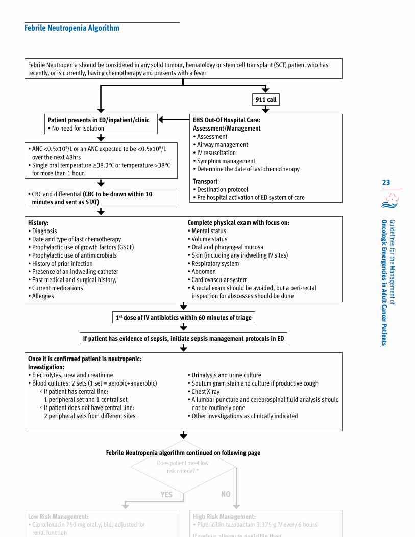
Febrile Neutropenia1, 11, 12, 13, 14, 15, 16, 17, 18, 19, 20
Febrile neutropenia is one of the most common complications related to cancer therapy, and is
considered a potentially life-threatening medical emergency. The risk for development of febrile neutropenia is approximately 25 to 40% in adult cancer patients, and is based on the type, duration and intensity of the chemotherapy regimen.
Febrile neutropenia should be considered in any solid tumour, hematology or stem cell transplant (SCT) patient who has recently, or is currently having chemotherapy and presents with a fever.
The mortality rate associated with febrile neutropenia in cancer patients is between 5-20%, therefore timely recognition of symptoms and administration of antibiotics is critical for the prevention of sepsis and death. In addition, the development of febrile neutropenia in a patient with cancer can lead to the decision to reduce or delay subsequent chemotherapy cycles, thereby leading to negative outcomes for patients being treated with curative intent.
Neutropenia is defined by an absolute neutrophil count (ANC) less than 500 cells/microlitre (< 0.5 x10⁹/L) or ANC expected to be less than 500 cells/microlitre (< 0.5 x10⁹/L) over the next 48 hours.
The ANC is calculated as follows: ANC=(neutrophils + Bands) x WBC.
Fever is defined by a single oral temperature greater than or equal to 38.3°C (101°F) or temperature greater than 38°C (100.4°F) for more than 1 hour. Note: Tympanic temperature is generally considered to be 0.5-1° higher than oral.
More than 70% of patients presenting with febrile neutropenia have an underlying hematological disease (leukemia, lymphoma, multiple myeloma or post-stem cell transplant) and the remaining 30% have solid tumors. The cause of myelosuppression is usually chemotherapy or can be the presenting symptom of a new hematological diagnosis.
International guidelines advocate the administration of empiric antibacterial therapy within 60 minutes of presentation in all patients presenting with neutropenic fever.
All stem cell transplant (SCT), hematology and solid tumour patients who have recently or are currently receiving chemotherapy in Nova Scotia are issued a “yellow card” which identifies them to be at risk of febrile neutropenia and provides management guidelines.
Patients experiencing febrile neutropenia and their families will, undoubtedly, experience some degree of distress. Please refer to page 49, for information about addressing the psychosocial health needs of patients and families.
EHS Out of Hospital Care
Paramedics responding to a cancer patient with suspected febrile neutropenia:
Assessment/Management
• Assessment• Airway management• IV resuscitation• Symptom management• Determine date of last chemotherapy treatment
Transport
• Destination protocol• Pre hospital activation of ED system of care
In Hospital care
Key Timelines
• CBC to be drawn within 10 minutes and sent as STAT
• Door to administration of first dose of IV antibiotics = 60 minutes
Presentation and Diagnosis
Given the inability of most immunocompromised patients to mount an adequate response to infection, the classic signs and symptoms of infection, other than fever, may be minimal. A focused history and physical examination is therefore required.
21
Guidelines for the Managem
ent of O
ncologic Emergencies in Adult Cancer Patients
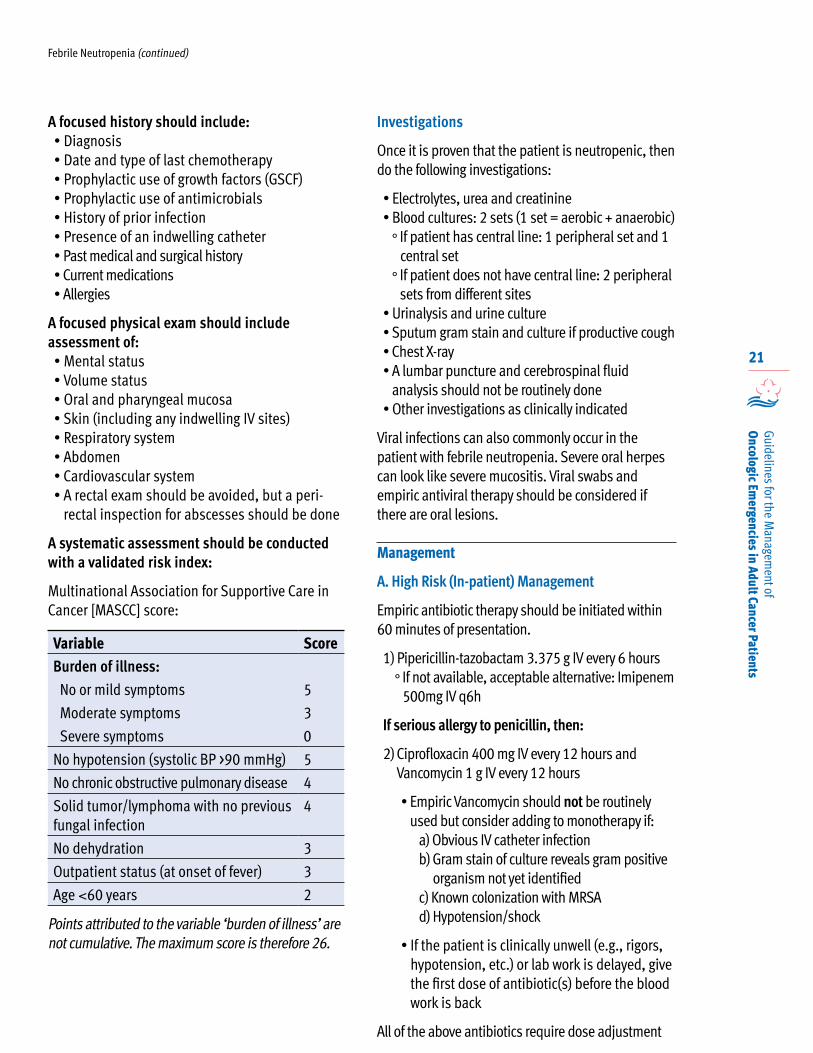
A focused history should include:• Diagnosis• Date and type of last chemotherapy• Prophylactic use of growth factors (GSCF)• Prophylactic use of antimicrobials• History of prior infection• Presence of an indwelling catheter• Past medical and surgical history• Current medications• Allergies
A focused physical exam should include assessment of:• Mental status• Volume status• Oral and pharyngeal mucosa• Skin (including any indwelling IV sites)• Respiratory system• Abdomen• Cardiovascular system • A rectal exam should be avoided, but a peri-
rectal inspection for abscesses should be done
A systematic assessment should be conducted with a validated risk index:
Multinational Association for Supportive Care in Cancer [MASCC] score:
Variable ScoreBurden of illness:
No or mild symptoms 5
Moderate symptoms 3
Severe symptoms 0
No hypotension (systolic BP >90 mmHg) 5
No chronic obstructive pulmonary disease 4
Solid tumor/lymphoma with no previous fungal infection
4
No dehydration 3
Outpatient status (at onset of fever) 3
Age <60 years 2
Points attributed to the variable ‘burden of illness’ are not cumulative. The maximum score is therefore 26.
Investigations
Once it is proven that the patient is neutropenic, then do the following investigations:
• Electrolytes, urea and creatinine• Blood cultures: 2 sets (1 set = aerobic + anaerobic)
° If patient has central line: 1 peripheral set and 1 central set ° If patient does not have central line: 2 peripheral
sets from different sites• Urinalysis and urine culture• Sputum gram stain and culture if productive cough • Chest X-ray• A lumbar puncture and cerebrospinal fluid
analysis should not be routinely done• Other investigations as clinically indicated
Viral infections can also commonly occur in the patient with febrile neutropenia. Severe oral herpes can look like severe mucositis. Viral swabs and empiric antiviral therapy should be considered if there are oral lesions.
Management
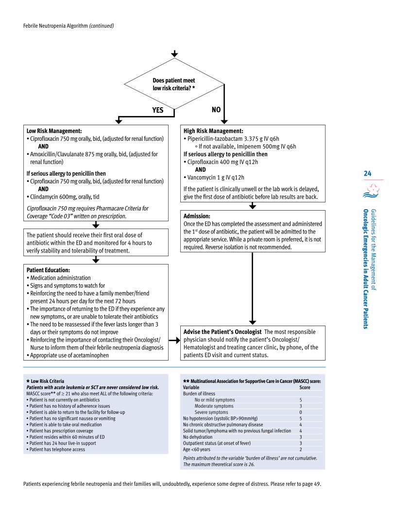
A. High Risk (In-patient) Management
Empiric antibiotic therapy should be initiated within 60 minutes of presentation.
1) Pipericillin-tazobactam 3.375 g IV every 6 hours ° If not available, acceptable alternative: Imipenem
500mg IV q6h
If serious allergy to penicillin, then:
2) Ciprofloxacin 400 mg IV every 12 hours and Vancomycin 1 g IV every 12 hours
• Empiric Vancomycin should not be routinely used but consider adding to monotherapy if:
a) Obvious IV catheter infection b) Gram stain of culture reveals gram positive organism not yet identified c) Known colonization with MRSA d) Hypotension/shock
• If the patient is clinically unwell (e.g., rigors, hypotension, etc.) or lab work is delayed, give the first dose of antibiotic(s) before the blood work is back
All of the above antibiotics require dose adjustment
Febrile Neutropenia (continued)
22
Guidelines for the Managem
ent of O
ncologic Emergencies in Adult Cancer Patients
Febrile Neutropenia (continued)
in the presence of renal insufficiency, but this relates to subsequent doses and no adjustments are needed for the first dose.
Rectal temperatures, suppositories, enemas and intramuscular injections should be avoided in febrile neutropenia patients.
If the patient has evidence of sepsis, then sepsis management and protocols should begin immediately in the ED, without delay.
Once the ED physician has completed the assessment and administered the first dose of antibiotics, the patient will be admitted to the appropriate service.
For admission, while a private room is preferred, it is not required. The more expediently the patient can be admitted, the better. In addition, reverse isolation is not recommended.
Advise the Patient’s Oncologist
The most responsible physician should notify the patients Oncologist/Hematologist and treating cancer clinic, by phone, of the patients ED visit and current status.
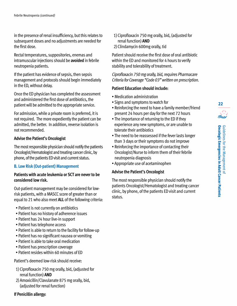
B. Low Risk (Out-patient) Management
Patients with acute leukemia or SCT are never to be considered low risk.
Out-patient management may be considered for low-risk patients, with a MASCC score of greater than or equal to 21 who also meet ALL of the following criteria:
• Patient is not currently on antibiotics• Patient has no history of adherence issues• Patient has 24 hour live-in support• Patient has telephone access• Patient is able to return to the facility for follow-up• Patient has no significant nausea or vomiting• Patient is able to take oral medication• Patient has prescription coverage • Patient resides within 60 minutes of ED
Patient’s deemed low-risk should receive:
1) Ciprofloxacin 750 mg orally, bid, (adjusted for renal function) AND
2) Amoxicillin/Clavulanate 875 mg orally, bid, (adjusted for renal function)
If Penicillin allergy:
1) Ciprofloxacin 750 mg orally, bid, (adjusted for renal function) AND
2) Clindamycin 600mg orally, tid
Patient should receive the first dose of oral antibiotic within the ED and monitored for 4 hours to verify stability and tolerability of treatment.
Ciprofloxacin 750 mg orally, bid, requires Pharmacare Criteria for Coverage “Code 03” written on prescription.
Patient Education should include:
• Medication administration• Signs and symptoms to watch for• Reinforcing the need to have a family member/friend
present 24 hours per day for the next 72 hours• The importance of returning to the ED if they
experience any new symptoms, or are unable to tolerate their antibiotics
• The need to be reassessed if the fever lasts longer than 3 days or their symptoms do not improve
• Reinforcing the importance of contacting their Oncologist/Nurse to inform them of their febrile neutropenia diagnosis
• Appropriate use of acetaminophen
Advise the Patient’s Oncologist
The most responsible physician should notify the patients Oncologist/Hematologist and treating cancer clinic, by phone, of the patients ED visit and current status.
23
Guidelines for the Managem
ent of O
ncologic Emergencies in Adult Cancer Patients
Febrile Neutropenia Algorithm
Febrile Neutropenia should be considered in any solid tumour, hematology or stem cell transplant (SCT) patient who has recently, or is currently, having chemotherapy and presents with a fever
Patient presents in ED/inpatient/clinic• No need for isolation
911 call
EHS Out-Of Hospital Care:Assessment/Management• Assessment• Airway management• IV resuscitation• Symptom management • Determine the date of last chemotherapy
Transport• Destination protocol• Pre hospital activation of ED system of care
• ANC <0.5x10⁹/L or an ANC expected to be <0.5x10⁹/L over the next 48hrs
• Single oral temperature ≥38.3°C or temperature >38°C for more than 1 hour.
History: • Diagnosis• Date and type of last chemotherapy• Prophylactic use of growth factors (GSCF)• Prophylactic use of antimicrobials• History of prior infection• Presence of an indwelling catheter• Past medical and surgical history, • Current medications• Allergies
Complete physical exam with focus on:• Mental status• Volume status• Oral and pharyngeal mucosa• Skin (including any indwelling IV sites)• Respiratory system• Abdomen• Cardiovascular system • A rectal exam should be avoided, but a peri-rectal
inspection for abscesses should be done
If patient has evidence of sepsis, initiate sepsis management protocols in ED
Once it is confirmed patient is neutropenic:Investigation:• Electrolytes, urea and creatinine• Blood cultures: 2 sets (1 set = aerobic+anaerobic)
° If patient has central line: 1 peripheral set and 1 central set
° If patient does not have central line: 2 peripheral sets from different sites
• Urinalysis and urine culture• Sputum gram stain and culture if productive cough • Chest X-ray• A lumbar puncture and cerebrospinal fluid analysis should
not be routinely done• Other investigations as clinically indicated
Does patient meet low risk criteria? *
YES NO
Low Risk Management:• Ciprofloxacin 750 mg orally, bid, adjusted for
renal functionAND
High Risk Management:• Pipericillin-tazobactam 3.375 g IV every 6 hours
If serious allergy to penicillin then
1st dose of IV antibiotics within 60 minutes of triage
Febrile Neutropenia algorithm continued on following page
• CBC and differential (CBC to be drawn within 10 minutes and sent as STAT)
24
Guidelines for the Managem
ent of O
ncologic Emergencies in Adult Cancer Patients
Febrile Neutropenia Algorithm (continued)
Low Risk Management:• Ciprofloxacin 750 mg orally, bid, (adjusted for renal function)
AND• Amoxicillin/Clavulanate 875 mg orally, bid, (adjusted for
renal function)
If serious allergy to penicillin then• Ciprofloxacin 750 mg orally, bid, (adjusted for renal function)
AND• Clindamycin 600mg, orally, tid
Ciprofloxacin 750 mg requires Pharmacare Criteria for Coverage “Code 03” written on prescription.
The patient should receive their first oral dose of antibiotic within the ED and monitored for 4 hours to verify stability and tolerability of treatment.
Patient Education:• Medication administration• Signs and symptoms to watch for• Reinforcing the need to have a family member/friend
present 24 hours per day for the next 72 hours• The importance of returning to the ED if they experience any
new symptoms, or are unable to tolerate their antibiotics• The need to be reassessed if the fever lasts longer than 3
days or their symptoms do not improve • Reinforcing the importance of contacting their Oncologist/
Nurse to inform them of their febrile neutropenia diagnosis• Appropriate use of acetaminophen
High Risk Management:• Pipericillin-tazobactam 3.375 g IV q6h
° If not available, Imipenem 500mg IV q6hIf serious allergy to penicillin then• Ciprofloxacin 400 mg IV q12h
AND • Vancomycin 1 g IV q12h
If the patient is clinically unwell or the lab work is delayed, give the first dose of antibiotic before lab results are back.
Admission:Once the ED has completed the assessment and administered the 1st dose of antibiotic, the patient will be admitted to the appropriate service. While a private room is preferred, it is not required. Reverse isolation is not recommended.
Advise the Patient’s Oncologist The most responsible physician should notify the patient’s Oncologist/Hematologist and treating cancer clinic, by phone, of the patients ED visit and current status.
* Low Risk CriteriaPatients with acute leukemia or SCT are never considered low risk.MASCC score** of ≥ 21 who also meet ALL of the following criteria:• Patient is not currently on antibiotics• Patient has no history of adherence issues• Patient is able to return to the facility for follow-up• Patient has no significant nausea or vomiting• Patient is able to take oral medication• Patient has prescription coverage • Patient resides within 60 minutes of ED• Patient has 24 hour live-in support• Patient has telephone access
** Multinational Association for Supportive Care in Cancer (MASCC) score:Variable ScoreBurden of illness
No or mild symptoms 5Moderate symptoms 3Severe symptoms 0
No hypotension (systolic BP>90mmHg) 5No chronic obstructive pulmonary disease 4Solid tumor/lymphoma with no previous fungal infection 4No dehydration 3Outpatient status (at onset of fever) 3Age <60 years 2
Points attributed to the variable ‘burden of illness’ are not cumulative. The maximum theoretical score is 26.
Patients experiencing febrile neutropenia and their families will, undoubtedly, experience some degree of distress. Please refer to page 49.
YES NO
Does patient meet low risk criteria? *
25
Guidelines for the Managem
ent of O
ncologic Emergencies in Adult Cancer Patients
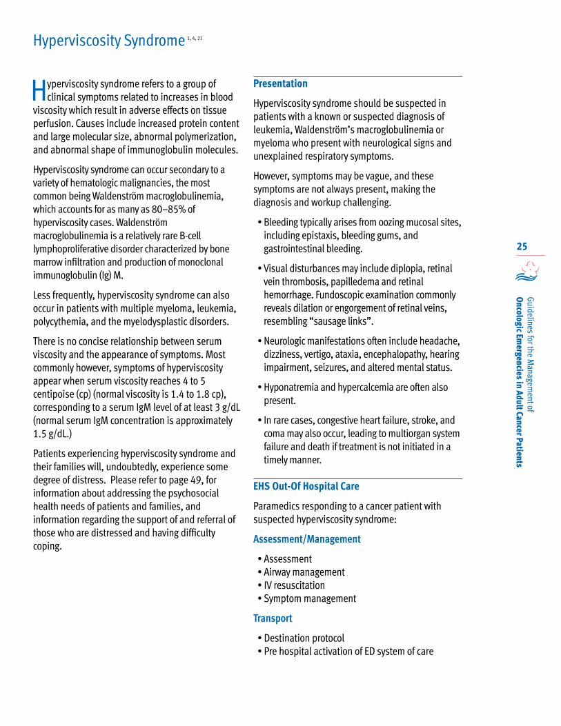
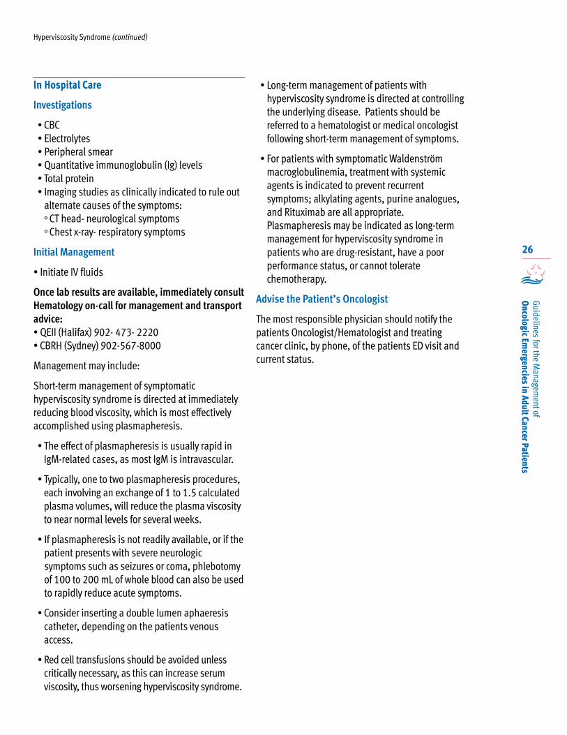
Hyperviscosity Syndrome 1, 4, 21
Hyperviscosity syndrome refers to a group of clinical symptoms related to increases in blood
viscosity which result in adverse effects on tissue perfusion. Causes include increased protein content and large molecular size, abnormal polymerization, and abnormal shape of immunoglobulin molecules.
Hyperviscosity syndrome can occur secondary to a variety of hematologic malignancies, the most common being Waldenström macroglobulinemia, which accounts for as many as 80–85% of hyperviscosity cases. Waldenström macroglobulinemia is a relatively rare B-cell lymphoproliferative disorder characterized by bone marrow infiltration and production of monoclonal immunoglobulin (Ig) M.
Less frequently, hyperviscosity syndrome can also occur in patients with multiple myeloma, leukemia, polycythemia, and the myelodysplastic disorders.
There is no concise relationship between serum viscosity and the appearance of symptoms. Most commonly however, symptoms of hyperviscosity appear when serum viscosity reaches 4 to 5 centipoise (cp) (normal viscosity is 1.4 to 1.8 cp), corresponding to a serum IgM level of at least 3 g/dL (normal serum IgM concentration is approximately 1.5 g/dL.)
Patients experiencing hyperviscosity syndrome and their families will, undoubtedly, experience some degree of distress. Please refer to page 49, for information about addressing the psychosocial health needs of patients and families, and information regarding the support of and referral of those who are distressed and having difficulty coping.
Presentation
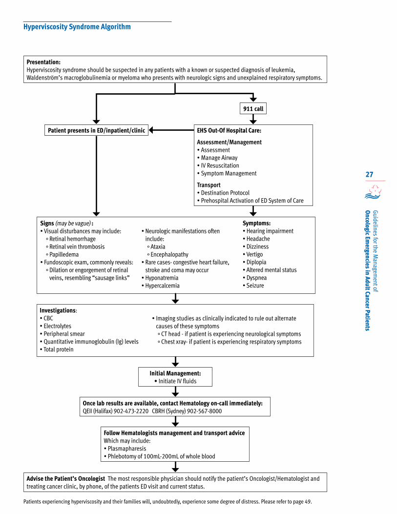
Hyperviscosity syndrome should be suspected in patients with a known or suspected diagnosis of leukemia, Waldenström’s macroglobulinemia or myeloma who present with neurological signs and unexplained respiratory symptoms.
However, symptoms may be vague, and these symptoms are not always present, making the diagnosis and workup challenging.
• Bleeding typically arises from oozing mucosal sites, including epistaxis, bleeding gums, and gastrointestinal bleeding.
• Visual disturbances may include diplopia, retinal vein thrombosis, papilledema and retinal hemorrhage. Fundoscopic examination commonly reveals dilation or engorgement of retinal veins, resembling “sausage links”.
• Neurologic manifestations often include headache, dizziness, vertigo, ataxia, encephalopathy, hearing impairment, seizures, and altered mental status.
• Hyponatremia and hypercalcemia are often also present.
• In rare cases, congestive heart failure, stroke, and coma may also occur, leading to multiorgan system failure and death if treatment is not initiated in a timely manner.
EHS Out-Of Hospital Care
Paramedics responding to a cancer patient with suspected hyperviscosity syndrome:
Assessment/Management
• Assessment• Airway management• IV resuscitation• Symptom management
Transport
• Destination protocol• Pre hospital activation of ED system of care
26
Guidelines for the Managem
ent of O
ncologic Emergencies in Adult Cancer Patients
In Hospital Care
Investigations
• CBC• Electrolytes• Peripheral smear• Quantitative immunoglobulin (Ig) levels• Total protein• Imaging studies as clinically indicated to rule out
alternate causes of the symptoms: ° CT head- neurological symptoms ° Chest x-ray- respiratory symptoms
Initial Management
• Initiate IV fluids
Once lab results are available, immediately consult Hematology on-call for management and transport advice:• QEII (Halifax) 902- 473- 2220• CBRH (Sydney) 902-567-8000
Management may include:
Short-term management of symptomatic hyperviscosity syndrome is directed at immediately reducing blood viscosity, which is most effectively accomplished using plasmapheresis.
• The effect of plasmapheresis is usually rapid in IgM-related cases, as most IgM is intravascular.
• Typically, one to two plasmapheresis procedures, each involving an exchange of 1 to 1.5 calculated plasma volumes, will reduce the plasma viscosity to near normal levels for several weeks.
• If plasmapheresis is not readily available, or if the patient presents with severe neurologic symptoms such as seizures or coma, phlebotomy of 100 to 200 mL of whole blood can also be used to rapidly reduce acute symptoms.
• Consider inserting a double lumen aphaeresis catheter, depending on the patients venous access.
• Red cell transfusions should be avoided unless critically necessary, as this can increase serum viscosity, thus worsening hyperviscosity syndrome.
• Long-term management of patients with hyperviscosity syndrome is directed at controlling the underlying disease. Patients should be referred to a hematologist or medical oncologist following short-term management of symptoms.
• For patients with symptomatic Waldenström macroglobulinemia, treatment with systemic agents is indicated to prevent recurrent symptoms; alkylating agents, purine analogues, and Rituximab are all appropriate. Plasmapheresis may be indicated as long-term management for hyperviscosity syndrome in patients who are drug-resistant, have a poor performance status, or cannot tolerate chemotherapy.
Advise the Patient’s Oncologist
The most responsible physician should notify the patients Oncologist/Hematologist and treating cancer clinic, by phone, of the patients ED visit and current status.
Hyperviscosity Syndrome (continued)
27
Guidelines for the Managem
ent of O
ncologic Emergencies in Adult Cancer Patients
Presentation:Hyperviscosity syndrome should be suspected in any patients with a known or suspected diagnosis of leukemia, Waldenström’s macroglobulinemia or myeloma who presents with neurologic signs and unexplained respiratory symptoms.
Patient presents in ED/inpatient/clinic
911 call
EHS Out-Of Hospital Care:
Assessment/Management• Assessment• Manage Airway• IV Resuscitation• Symptom Management
Transport• Destination Protocol• Prehospital Activation of ED System of Care
Patients experiencing hyperviscosity and their families will, undoubtedly, experience some degree of distress. Please refer to page 49.
Initial Management: • Initiate IV fluids
Once lab results are available, contact Hematology on-call immediately:QEII (Halifax) 902-473-2220 CBRH (Sydney) 902-567-8000
Follow Hematologists management and transport advice Which may include:• Plasmapharesis• Phlebotomy of 100mL-200mL of whole blood
Advise the Patient’s Oncologist The most responsible physician should notify the patient’s Oncologist/Hematologist and treating cancer clinic, by phone, of the patients ED visit and current status.
Signs (may be vague) :• Visual disturbances may include:
° Retinal hemorrhage° Retinal vein thrombosis° Papilledema
• Fundoscopic exam, commonly reveals:° Dilation or engorgement of retinal
veins, resembling “sausage links”
• Neurologic manifestations often include:° Ataxia° Encephalopathy
• Rare cases- congestive heart failure, stroke and coma may occur
• Hyponatremia• Hypercalcemia
Symptoms:• Hearing impairment• Headache• Dizziness• Vertigo• Diplopia• Altered mental status• Dyspnea• Seizure
Investigations:• CBC• Electrolytes• Peripheral smear• Quantitative immunoglobulin (Ig) levels• Total protein
• Imaging studies as clinically indicated to rule out alternate causes of these symptoms° CT head - if patient is experiencing neurological symptoms° Chest xray- if patient is experiencing respiratory symptoms
Hyperviscosity Syndrome Algorithm
28
Guidelines for the Managem
ent of O
ncologic Emergencies in Adult Cancer Patients
While unexpected hypercalcemia of unknown etiology may be an indicator of malignancy, this
guideline focuses on the identification and management of hypercalcemia in the patient already known to have cancer.
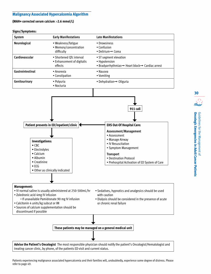
MAH is defined as a corrected serum calcium > 2.6 mmol/L.
• Calcium circulates in the blood in a biologically active ionized form (50%), a protein-bound (biologically inactive) fraction (40%) and in a form complexed to assorted anions (10%). Thus, changes in albumin levels can alter total calcium levels.
• Most labs measure total serum calcium (not ionized calcium), which must be “corrected” in the setting of hypo-or hyperalbuminemia to compare total calcium values against the normal range (every 10 g/L decrease in albumin corresponds to a 0.2 mmol/L increase in calcium).
MAH occurs in up to 30% of cancer patients, most commonly among those with breast, lung and head/neck tumours, and those with hematologic malignancies (particularly multiple myeloma and adult T-cell leukemia/lymphoma).
• Humoral hypercalcemia accounts for most cases (80%). It results from secretion of parathyroid hormone related protein (PTHrP) or other cytokines which bind to the PTH receptor and mimics the physiological effects of PTH (i.e., increased bone resorption, enhanced renal retention of calcium).
• Osteolytic bone metastases account for 20% of cases.
• Other causes (such as ectopic PTH section, vitamin D secreting lymphomas, etc.) account for less than 1%.
• Many cancer therapies (e.g., antineoplastic agents, vitamin D analogues) can induce or exacerbate hypercalcemia, particularly when used in combination.
In cancer patients hospitalized with hypercalcemia, the 30-day mortality rate of has been shown to approach 50%.
Patients experiencing MAH and their families will, undoubtedly, experience some degree of distress. Please refer to page 49, for information about addressing the psychosocial health needs of patients and families.
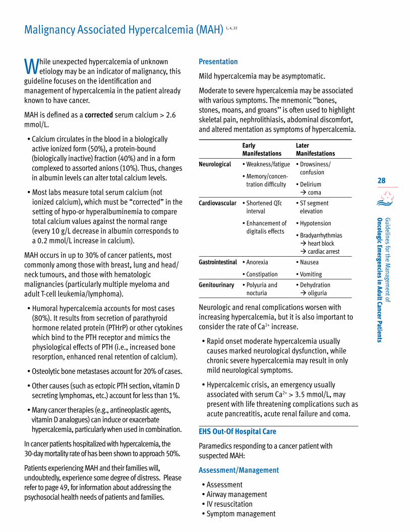
Presentation
Mild hypercalcemia may be asymptomatic.
Moderate to severe hypercalcemia may be associated with various symptoms. The mnemonic ‘‘bones, stones, moans, and groans’’ is often used to highlight skeletal pain, nephrolithiasis, abdominal discomfort, and altered mentation as symptoms of hypercalcemia.
Early Manifestations
Later Manifestations
Neurological • Weakness/fatigue
• Memory/concen-tration difficulty
• Drowsiness/ confusion
• Delirium coma
Cardiovascular • Shortened QTc interval
• Enhancement of digitalis effects
• ST segment elevation
• Hypotension
• Bradyarrhythmias heart block cardiac arrest
Gastrointestinal • Anorexia
• Constipation
• Nausea
• Vomiting
Genitourinary • Polyuria and nocturia
• Dehydration oliguria
Neurologic and renal complications worsen with increasing hypercalcemia, but it is also important to consider the rate of Ca2+ increase.
• Rapid onset moderate hypercalcemia usually causes marked neurological dysfunction, while chronic severe hypercalcemia may result in only mild neurological symptoms.
• Hypercalcemic crisis, an emergency usually associated with serum Ca2+ > 3.5 mmol/L, may present with life threatening complications such as acute pancreatitis, acute renal failure and coma.
EHS Out-Of Hospital Care
Paramedics responding to a cancer patient with suspected MAH:
Assessment/Management
• Assessment• Airway management• IV resuscitation• Symptom management
Malignancy Associated Hypercalcemia (MAH) 1, 4, 22
29
Guidelines for the Managem
ent of O
ncologic Emergencies in Adult Cancer Patients
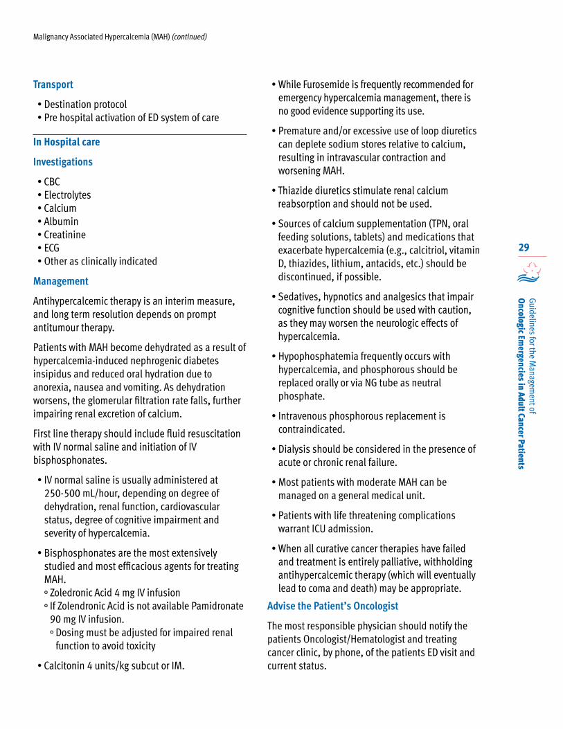
Transport
• Destination protocol• Pre hospital activation of ED system of care
In Hospital care
Investigations
• CBC• Electrolytes• Calcium• Albumin• Creatinine• ECG• Other as clinically indicated
Management
Antihypercalcemic therapy is an interim measure, and long term resolution depends on prompt antitumour therapy.
Patients with MAH become dehydrated as a result of hypercalcemia-induced nephrogenic diabetes insipidus and reduced oral hydration due to anorexia, nausea and vomiting. As dehydration worsens, the glomerular filtration rate falls, further impairing renal excretion of calcium.
First line therapy should include fluid resuscitation with IV normal saline and initiation of IV bisphosphonates.
• IV normal saline is usually administered at 250-500 mL/hour, depending on degree of dehydration, renal function, cardiovascular status, degree of cognitive impairment and severity of hypercalcemia.
• Bisphosphonates are the most extensively studied and most efficacious agents for treating MAH. ° Zoledronic Acid 4 mg IV infusion ° If Zolendronic Acid is not available Pamidronate
90 mg IV infusion. ° Dosing must be adjusted for impaired renal
function to avoid toxicity
• Calcitonin 4 units/kg subcut or IM.
• While Furosemide is frequently recommended for emergency hypercalcemia management, there is no good evidence supporting its use.
• Premature and/or excessive use of loop diuretics can deplete sodium stores relative to calcium, resulting in intravascular contraction and worsening MAH.
• Thiazide diuretics stimulate renal calcium reabsorption and should not be used.
• Sources of calcium supplementation (TPN, oral feeding solutions, tablets) and medications that exacerbate hypercalcemia (e.g., calcitriol, vitamin D, thiazides, lithium, antacids, etc.) should be discontinued, if possible.
• Sedatives, hypnotics and analgesics that impair cognitive function should be used with caution, as they may worsen the neurologic effects of hypercalcemia.
• Hypophosphatemia frequently occurs with hypercalcemia, and phosphorous should be replaced orally or via NG tube as neutral phosphate.
• Intravenous phosphorous replacement is contraindicated.
• Dialysis should be considered in the presence of acute or chronic renal failure.
• Most patients with moderate MAH can be managed on a general medical unit.
• Patients with life threatening complications warrant ICU admission.
• When all curative cancer therapies have failed and treatment is entirely palliative, withholding antihypercalcemic therapy (which will eventually lead to coma and death) may be appropriate.
Advise the Patient’s Oncologist
The most responsible physician should notify the patients Oncologist/Hematologist and treating cancer clinic, by phone, of the patients ED visit and current status.
Malignancy Associated Hypercalcemia (MAH) (continued)
30
Guidelines for the Managem
ent of O
ncologic Emergencies in Adult Cancer Patients
Malignancy Associated Hypercalcemia Algorithm
Signs/Symptoms:
System Early Manifestations Late Manifestations
Neurological • Weakness/fatigue• Memory/concentration
difficulty
• Drowsiness• Confusion• Delirium➞ Coma
Cardiovascular • Shortened QTc interval• Enhancement of digitalis
effects
• ST segment elevation• Hypotension• Bradyarrhythmias➞ Heart block➞ Cardiac arrest
Gastrointestinal • Anorexia• Constipation
• Nausea• Vomiting
Genitourinary • Polyuria• Nocturia
• Dehydration➞ Oliguria
Patient presents in ED/inpatient/clinic
911 call
EHS Out-Of Hospital Care:
Assessment/Management• Assessment• Manage Airway• IV Resuscitation• Symptom Management
Transport• Destination Protocol• Prehospital Activation of ED System of Care
Patients experiencing malignance associated hypercalcemia and their families will, undoubtedly, experience some degree of distress. Please refer to page 49.
Advise the Patient’s Oncologist The most responsible physician should notify the patient’s Oncologist/Hematologist and treating cancer clinic, by phone, of the patients ED visit and current status.
(MAH= corrected serum calcium >2.6 mmol/L)
Investigations:• CBC• Electrolytes• Calcium • Albumin • Creatinine• ECG• Other as clinically indicated
Management:• IV normal saline is usually administered at 250-500mL/hr• Zoledronic acid 4mg IV infusion
° If unavailable Pamidronate 90 mg IV infusion• Calcitonin 4 units/kg subcut or IM• Sources of calcium supplementation should be
discontinued if possible
• Sedatives, hypnotics and analgesics should be used with caution
• Dialysis should be considered in the presence of acute or chronic renal failure
These patients may be managed on a general medical unit
31
Guidelines for the Managem
ent of O
ncologic Emergencies in Adult Cancer Patients
Airway obstruction in cancer patients is most commonly caused by direct extension from an
adjacent tumour, particularly an inoperable or recurrent tumour in the mediastinum or a primary tumour of the head and neck. Although rare in non-thoracic malignancies, 20- 30% of lung cancer patients will experience airway compromise or obstruction.
In esophageal, renal, breast, or thyroid cancers, airway obstruction secondary to metastatic malignancies has also been reported.
Tumour encroachment and tumour-associated airway edema or hemorrhage can also cause airway obstruction.
Patients experiencing airway obstruction and their families will, undoubtedly, experience some degree of distress. Please refer to page 49, for information about addressing the psychosocial health needs of patients and families, and information regarding the support of and referral of those who are distressed and having difficulty coping.
Presentation
Progressive symptoms of malignant airway obstruction represent a true medical emergency. The most common presenting symptoms of malignant airway obstruction include:
• Dyspnea• Hemoptysis• Wheezing• Hoarseness• Difficulty clearing secretions• Cough• Stridor (most marked on inspiration)
These non-specific symptoms may be mistaken for exacerbation of chronic obstructive pulmonary disease, asthma, infections, bronchitis, or heart disease.
When present, stridor is most marked on inspiration, and can progress to a near-complete obstruction as a result of infection, inflammation, or manipulation of the airway.
Up to 45% of patients with obstructing tumours report hemoptysis.
EHS Out-Of Hospital Care
Paramedics responding to a cancer patient with suspected airway obstruction:
Assessment/Management
• Assessment• Airway management• IV resuscitation• Symptom management
Transport
• Destination protocol• Pre hospital activation of ED system of care
In Hospital care
Management
• Airway management • Administer Dexamethasone 20 mg IV followed by
4mg q6h OR q12h• Use paralytics with caution
Investigations
• In most cases, physical examination, accompanied by direct visualization with a laryngoscope or bronchoscope is sufficient to make a diagnosis, depending on the location of the lesion. Consider consulting with anesthesia.
• To guide endotracheal tube placement, laryngoscopy or bronchoscopy may be required.
• A neck and chest CT scan is the most effective initial study to determine the location and extent of airway obstruction.
• Pulse oximetry.
If the obstruction is laryngeal or above, consult appropriate local service regarding an emergency cricothyroidotomy or tracheostomy, and follow the specialist’s management and transport advice.
Malignant Airway Obstruction 1, 2, 4, 23
32
Guidelines for the Managem
ent of O
ncologic Emergencies in Adult Cancer Patients
If the obstruction is below laryngeal, consult Thoracic Surgery, and follow the specialist’s management and transport advice.• QEII (Halifax) 902-473-2220• CBRH (Sydney) 902-567-8000
Management depends on:• The tumour histologic type• Tumour stage• Tumour location• Urgency of patient’s presentation• Patient’s performance status
Neodymium-yttrium-aluminum-garnet (Nd:YAG) lasers, in combination with rigid bronchoscopy, can also be used to open the airway in patients with malignant intrinsic airway obstruction.
Following the establishment of a patent airway, more definitive tumour control measures should follow, including airway surgery, stenting, radiotherapy, and endobronchial brachytherapy.
• Stent placement, particularly self-expanding metal stents, is indicated for the relief of acute airway obstruction in patients with extrinsic tumour compression or with tracheoesophageal fistulas.
• Stents can be used as a supplemental therapy to laser ablation or photodynamic therapy, or as a second line therapy following failure of local control with laser, photodynamic, or radiation therapy.
• For patients with upper respiratory obstruction, the following may be helpful: ° Intravenous corticosteroids to reduce edema° Supplemental humidified oxygen° If bronchospasm is present, patients may
benefit from bronchodilators
If the patient is not a candidate for surgery, consult Radiation Oncology and follow the specialist’s management and transport advice.• QEII (Halifax) 902-473-2220• CBRH (Sydney) 902-567-8000
Short course, multi-fractionated external-beam radiotherapy at a dose of 20 Gy/5 fractions is recommended for the palliation of intrathoracic symptoms, and is preferred over single-fraction treatment.
Endobronchial brachytherapy may be considered as an adjuvant to external beam radiotherapy, and also in select patients with lung cancer previously treated with external beam radiotherapy who become obstructed due to secondary disease.
Advise the Patient’s Oncologist
The most responsible physician should notify the patients Oncologist/Hematologist and treating cancer clinic, by phone, of the patients ED visit and current status.
Malignant Airway Obstruction (continued)
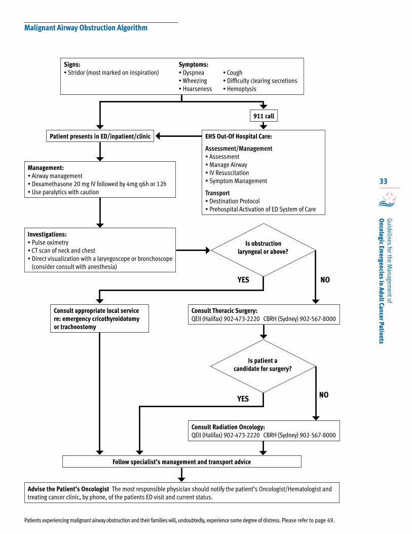
33
Guidelines for the Managem
ent of O
ncologic Emergencies in Adult Cancer Patients
Patient presents in ED/inpatient/clinic
911 call
EHS Out-Of Hospital Care:
Assessment/Management• Assessment• Manage Airway• IV Resuscitation• Symptom Management
Transport• Destination Protocol• Prehospital Activation of ED System of Care
Investigations:• Pulse oximetry• CT scan of neck and chest• Direct visualization with a laryngoscope or bronchoscope
(consider consult with anesthesia)
Management:• Airway management• Dexamethasone 20 mg IV followed by 4mg q6h or 12h• Use paralytics with caution
YES NO
Consult appropriate local service re: emergency cricothyroidotomy or tracheostomy
Consult Thoracic Surgery: QEII (Halifax) 902-473-2220 CBRH (Sydney) 902-567-8000
Follow specialist’s management and transport advice
Advise the Patient’s Oncologist The most responsible physician should notify the patient’s Oncologist/Hematologist and treating cancer clinic, by phone, of the patients ED visit and current status.
Patients experiencing malignant airway obstruction and their families will, undoubtedly, experience some degree of distress. Please refer to page 49.
Signs:• Stridor (most marked on inspiration)
Is patient a candidate for surgery?
YES NO
Consult Radiation Oncology: QEII (Halifax) 902-473-2220 CBRH (Sydney) 902-567-8000
Symptoms:• Dyspnea• Wheezing• Hoarseness
• Cough• Difficulty clearing secretions• Hemoptysis
Is obstruction laryngeal or above?
Malignant Airway Obstruction Algorithm
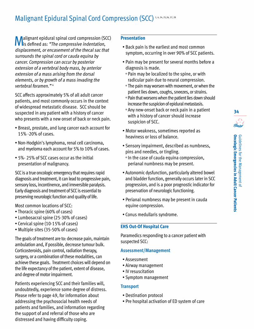
34
Guidelines for the Managem
ent of O
ncologic Emergencies in Adult Cancer Patients
Malignant epidural spinal cord compression (SCC) is defined as: “The compressive indentation,
displacement, or encasement of the thecal sac that surrounds the spinal cord or cauda equina by cancer. Compression can occur by posterior extension of a vertebral body mass, by anterior extension of a mass arising from the dorsal elements, or by growth of a mass invading the vertebral foramen.” 4
SCC affects approximately 5% of all adult cancer patients, and most commonly occurs in the context of widespread metastatic disease. SCC should be suspected in any patient with a history of cancer who presents with a new onset of back or neck pain.
• Breast, prostate, and lung cancer each account for 15% -20% of cases.
• Non-Hodgkin's lymphoma, renal cell carcinoma, and myeloma each account for 5% to 10% of cases.
• 5%- 25% of SCC cases occur as the initial presentation of malignancy.
SCC is a true oncologic emergency that requires rapid diagnosis and treatment, it can lead to progressive pain, sensory loss, incontinence, and irreversible paralysis. Early diagnosis and treatment of SCC is essential to preserving neurologic function and quality of life.
Most common locations of SCC:• Thoracic spine (60% of cases)• Lumbosacral spine (25-30% of cases) • Cervical spine (10-15% of cases)• Multiple sites (35-50% of cases)
The goals of treatment are to: decrease pain, maintain ambulation and, if possible, decrease tumour bulk. Corticosteroids, pain control, radiation therapy, surgery, or a combination of these modalities, can achieve these goals. Treatment choices will depend on the life expectancy of the patient, extent of disease, and degree of motor impairment.
Patients experiencing SCC and their families will, undoubtedly, experience some degree of distress. Please refer to page 49, for information about addressing the psychosocial health needs of patients and families, and information regarding the support of and referral of those who are distressed and having difficulty coping.
Presentation
• Back pain is the earliest and most common symptom, occurring in over 90% of SCC patients.
• Pain may be present for several months before a diagnosis is made. ° Pain may be localized to the spine, or with
radicular pain due to neural compression. ° The pain may worsen with movement, or when the
patient lies down, coughs, sneezes, or strains. ° Pain that worsens when the patient lies down should
increase the suspicion of epidural metastasis. ° Any new-onset back or neck pain in a patient
with a history of cancer should increase suspicion of SCC.
• Motor weakness, sometimes reported as heaviness or loss of balance.
• Sensory impairment, described as numbness, pins and needles, or tingling. ° In the case of cauda equina compression,
perianal numbness may be present.
• Autonomic dysfunction, particularly altered bowel and bladder function, generally occurs later in SCC progression, and is a poor prognostic indicator for preservation of neurologic functioning.
• Perianal numbness may be present in cauda equine compression.
• Conus medullaris syndrome. EHS Out-Of Hospital Care
Paramedics responding to a cancer patient with suspected SCC:
Assessment/Management
• Assessment• Airway management• IV resuscitation• Symptom management
Transport
• Destination protocol• Pre hospital activation of ED system of care
Malignant Epidural Spinal Cord Compression (SCC) 1, 4, 24, 25,26, 27, 28
35
Guidelines for the Managem
ent of O
ncologic Emergencies in Adult Cancer Patients
In Hospital care
As soon as SCC is suspected corticosteroids should be administered. Because the most important prognostic indicator for ambulatory outcome is the pre-treatment motor function, immediate initiation of therapy is critical.
• Administer Dexamethasone 10-20mg IV followed by 4-6 mg IV every 4 hours.° Dexamethasone rapidly reduces spinal cord
edema and back pain, and may also improve neurologic functioning.
Assessment
The initial diagnosis should involve a medical history and physical examination.
• The ability to ambulate must be assessed – this is a highly predictive finding of the chance of recovery:° >80% of SCC patients who were ambulatory
prior to SCC treatment will be ambulatory post-treatment
° <50% of SCC patients who experienced weakness prior to SCC treatment will be ambulatory post-treatment
° <10% of SCC patients who experienced paraplegia prior to SCC treatment will be ambulatory post-treatment
• A sensory level may be evident on pinprick examination and may help to focus on the suspected level of cord compression.
• Reflexes may be absent at the level of the compression and hyperactive below.
• Digital Rectal Exam (DRE) may demonstrate a lax anal sphincter tone in late stage SCC.
Investigations
• MRI is the preferred imaging study. ° If the history and clinical findings do not suggest
metastasis to the cervical spine, it is appropriate to obtain an MRI of the thoracic and lumbosacral spinal regions only.
° A CT scan can also be used if MRI is contraindicated or not available.
Management
If the SCC is related to a previously diagnosed malignancy immediately consult Radiation Oncology and follow the specialist’s advice for management and transport.• QEII (Halifax) 902-473-2220• CBRH (Sydney) 902-567-8000
• External beam radiation therapy can provide significant pain relief, with up to one-third of patients achieving complete pain relief at the treated site
• Radiation therapy with or without decompressive surgery demonstrated improvements in pain, ambulatory ability, urinary continence, duration of continence, functional status, and overall survival.
If the SCC is related to an undiagnosed malignancy, consult with Neurosurgery and follow the specialist’s advice for management and transport.• QEII (Halifax) 902-473-2220
• Surgery should be considered for patients with a good prognosis who are medically and surgically operable.
• Decompressive surgery followed by postoperative radiotherapy has been shown to be superior to radiotherapy alone for select patients with SCC.
• Patients treated with surgery were twice as likely to regain ambulation as patients treated with radiotherapy alone.
The decision whether to take emergency action must be guided by the patient’s overall condition, the nature of the cancer and its prognosis, the burden associated with any proposed treatment, the distress caused by the symptoms and the patient’s and family’s desires.
Advise the Patient’s Oncologist
The most responsible physician should notify the patients Oncologist/Hematologist and treating cancer clinic, by phone, of the patients ED visit and current status.
Malignant Epidural Spinal Cord Compression (SCC) (continued)
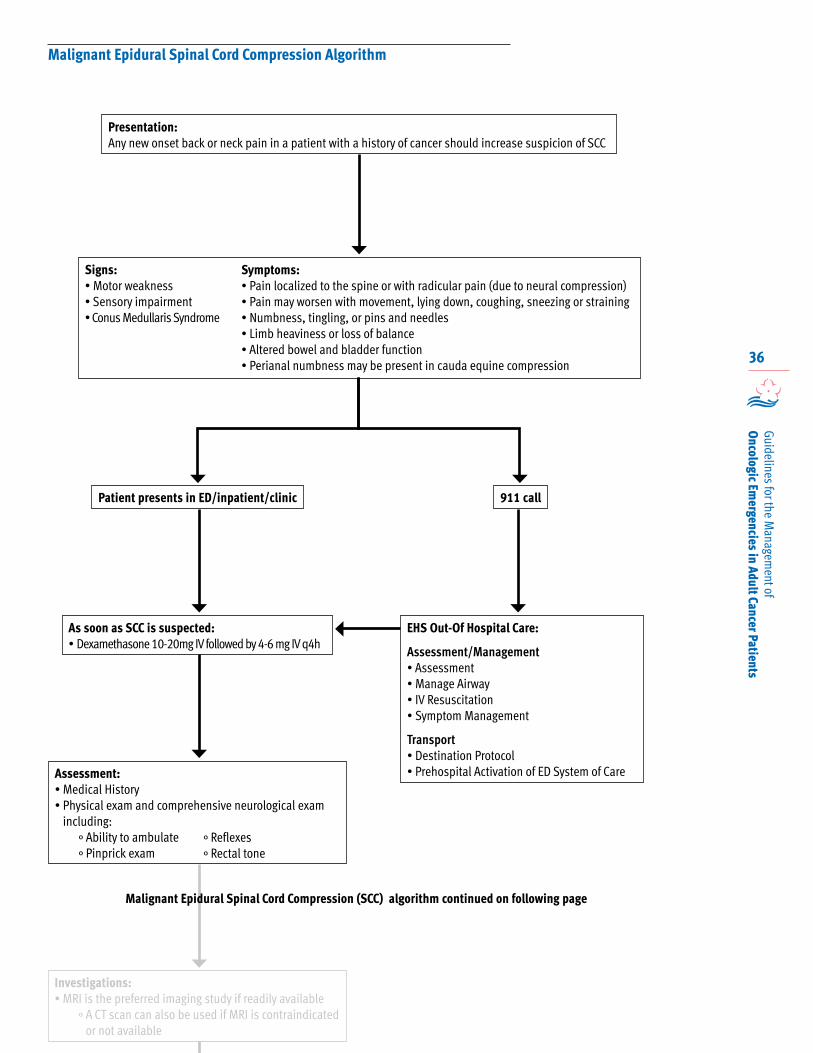
36
Guidelines for the Managem
ent of O
ncologic Emergencies in Adult Cancer Patients
Malignant Epidural Spinal Cord Compression Algorithm
Patient presents in ED/inpatient/clinic 911 call
EHS Out-Of Hospital Care:
Assessment/Management• Assessment• Manage Airway• IV Resuscitation• Symptom Management
Transport• Destination Protocol• Prehospital Activation of ED System of Care
As soon as SCC is suspected:• Dexamethasone 10-20mg IV followed by 4-6 mg IV q4h
Presentation:Any new onset back or neck pain in a patient with a history of cancer should increase suspicion of SCC
Signs:• Motor weakness• Sensory impairment• Conus Medullaris Syndrome
Symptoms:• Pain localized to the spine or with radicular pain (due to neural compression)• Pain may worsen with movement, lying down, coughing, sneezing or straining• Numbness, tingling, or pins and needles• Limb heaviness or loss of balance• Altered bowel and bladder function• Perianal numbness may be present in cauda equine compression
Assessment:• Medical History• Physical exam and comprehensive neurological exam
including:° Ability to ambulate° Pinprick exam
° Reflexes° Rectal tone
Investigations:• MRI is the preferred imaging study if readily available
° A CT scan can also be used if MRI is contraindicated or not available
Malignant Epidural Spinal Cord Compression (SCC) algorithm continued on following page
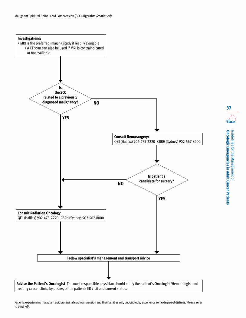
37
Guidelines for the Managem
ent of O
ncologic Emergencies in Adult Cancer Patients
Malignant Epidural Spinal Cord Compression (SCC) Algorithm (continued)
NO
Consult Radiation Oncology: QEII (Halifax) 902-473-2220 CBRH (Sydney) 902-567-8000
Consult Neurosurgery: QEII (Halifax) 902-473-2220 CBRH (Sydney) 902-567-8000
Follow specialist’s management and transport advice
Advise the Patient’s Oncologist The most responsible physician should notify the patient’s Oncologist/Hematologist and treating cancer clinic, by phone, of the patients ED visit and current status.
Patients experiencing malignant epidural spinal cord compression and their families will, undoubtedly, experience some degree of distress. Please refer to page 49.
NO
YES
YES
Investigations:• MRI is the preferred imaging study if readily available
° A CT scan can also be used if MRI is contraindicated or not available
Is patient a candidate for surgery?
Is the SCC
related to a previously diagnosed malignancy?
38
Guidelines for the Managem
ent of O
ncologic Emergencies in Adult Cancer Patients
Superior vena cava obstruction (SVCO) refers to a constellation of signs and symptoms resulting
from partial or complete obstruction of blood flow through the superior vena cava to the right atrium. Compression, invasion, thrombosis, or fibrosis of this vessel may cause the obstruction.
Eighty-five percent of all cases of SVCO occur within patients diagnosed with lung cancer, both small cell and non-small cell, and non-Hodgkin lymphoma.
• 10% of patients with small cell lung cancer will develop SVCO
• 2-4% of patients with non-small cell lung cancer will develop SVCO
• 2-4% of patients with non-Hodgkin lymphoma will develop SVCO.
An increasing number of cases of SVCO in cancer patients are due to thrombosis of indwelling central venous catheter devices and/or pacemaker leads.
Patients experiencing SVCO and their families will, undoubtedly, experience some degree of distress. Please refer to page 49, for information about addressing the psychosocial health needs of patients and families, and information regarding the support of and referral of those who are distressed and having difficulty coping.
Presentation
• Facial or neck swelling• Dilated chest vessels• Stridor (which may indicate laryngeal edema)
Other symptoms may include:• Facial fullness• Swelling of trunk and upper extremeties• Headache or confusion (which may indicate
cerebral edema)• Cough• Dyspnea• Orthopnea
Sudden onset of SVCO is rare, but is considered a true oncologic emergency because the rapid elevation of pressure in the superior vena cava causes increased intracranial pressure, resulting in cerebral edema, intracranial thrombosis or bleeding, and death.
Symptoms of SVCO more typically present insidiously over a period of day to weeks.
EHS Out-Of Hospital Care
Paramedics responding to a cancer patient with suspected SVCO:
Assessment/Management
• Assessment• Airway management• IV resuscitation, left arm preferred• Symptom management
Transport
• Destination protocol• Pre hospital activation of ED system of care
Superior Vena Cava Obstruction (SVCO) 1, 20, 30
39
Guidelines for the Managem
ent of O
ncologic Emergencies in Adult Cancer Patients
In Hospital Care
Investigations
• Contrast-enhanced CT scan of the chest is the most useful imaging study, as it clearly identifies the level and extent of the blockage, as well as an evaluation of collateral pathways of drainage
• MRI is useful for patients who cannot tolerate the CT contrast medium
• CBC
• Pulse oximetry
• Venography is generally only warranted when an intervention such as stent placement or surgery is planned
Management
Initial management should include:
• Dexamethasone 10mg IV followed by 4mg q6-8h
• Symptom control
• Anticoagulation is NOT appropriate if the patient does not have a previously diagnosed malignancy.
• Emergency treatment with radiotherapy is indicated if a histologic diagnosis cannot be established in a timely manner, and in rare life-threatening situations, such as airway obstruction, spinal cord compression, or increased intracranial pressure.
If patient presents with SVCO without a prior cancer diagnosis, every effort should be made to obtain biopsies and histologic diagnosis before any treatment decisions are made. This will aid in the decision of whether a definitive curative course of therapy versus palliative treatment is most appropriate.
Treatment of patients experiencing SVCO can vary widely. Management is guided by the severity of the symptoms and the underlying malignancy and the anticipated response to the treatment.
If the SVCO is not related to a previously diagnosed malignancy, consult Thoracic Surgery to obtain urgent cytology/histology.• QEII (Halifax) 902-473-2220• CBRH (Sydney) 902-567-8000
If the SVCO is related to a previously diagnosed malignancy, consult the appropriate specialist:• Small cell/NE malignancy- consult Medical
Oncology • Hematology malignancy- consult Hematology • Other malignancy- consult Radiation Oncology
° QEII (Halifax) 902-473-2220° CBRH (Sydney) 902-567-8000
Radiotherapy is the preferred initial treatment, particularly for those with SVCO caused by recurrent disease after chemotherapy or with tumours insensitive to chemotherapy, such as non-small cell lung cancer.
• While there is insufficient evidence to guide practice on radiotherapy dose fractionation, a retrospective review of SVCO, due to any histology, reported that an initial dose greater than or equal to 3Gy/fraction yields a higher rate of symptomatic relief in less than two weeks than a conventional dose of 2Gy/fraction.
Chemotherapy can also be used as the initial treatment of SVCO caused by malignancies sensitive to chemotherapy (ie small cell lung cancer and non-Hodgkin lymphoma).
Stent insertion provides rapid relief of associated symptoms, particularly for patients who fail to respond to chemotherapy or radiotherapy.
• Because the stent can be placed before a histologic diagnosis is made, it is a useful and effective option for patients with severe symptoms such as respiratory distress.
Advise the Patient’s Oncologist
The most responsible physician should notify the patients Oncologist/Hematologist and treating cancer clinic, by phone, of the patients ED visit and current status.
Superior Vena Cava Obstruction (continued)
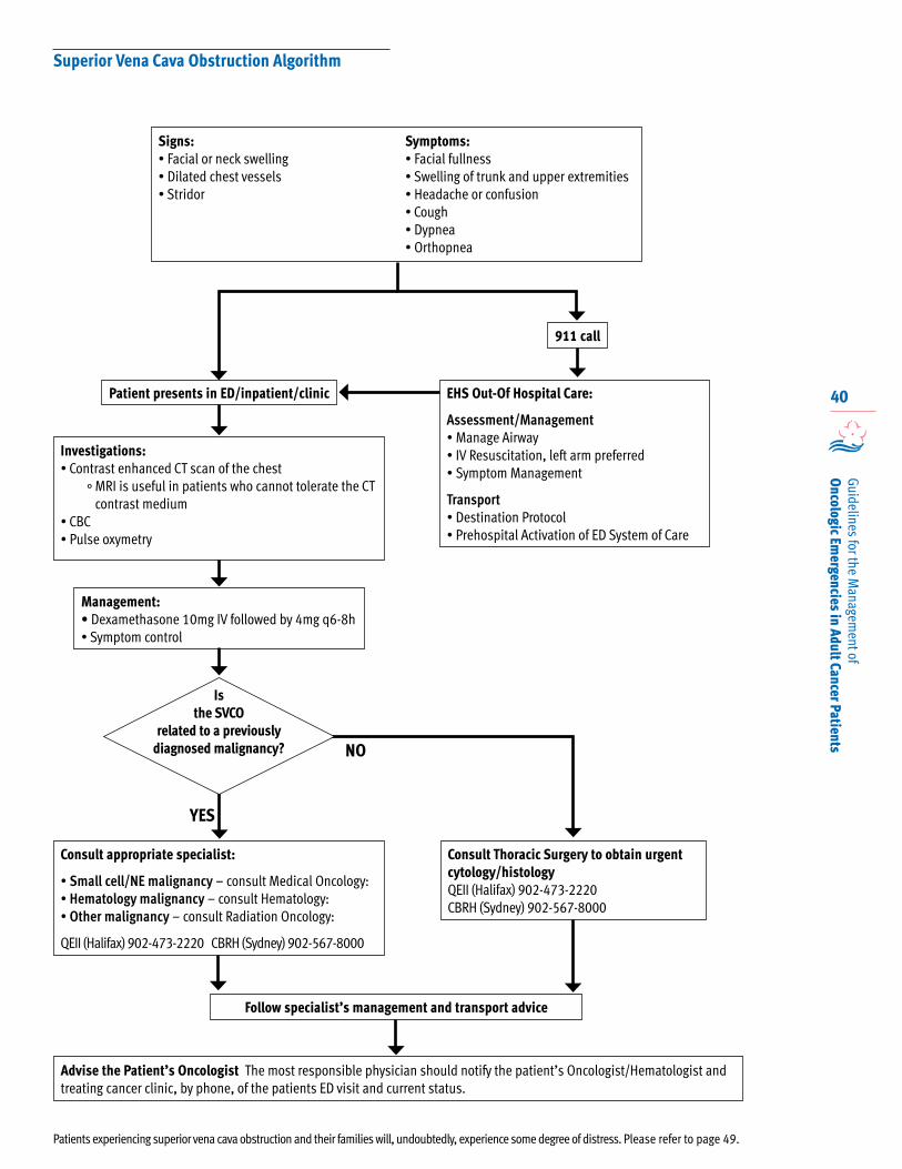
40
Guidelines for the Managem
ent of O
ncologic Emergencies in Adult Cancer Patients
Superior Vena Cava Obstruction Algorithm
Patient presents in ED/inpatient/clinic
911 call
EHS Out-Of Hospital Care:
Assessment/Management• Manage Airway• IV Resuscitation, left arm preferred• Symptom Management
Transport• Destination Protocol• Prehospital Activation of ED System of Care
Investigations:• Contrast enhanced CT scan of the chest
° MRI is useful in patients who cannot tolerate the CT contrast medium
• CBC• Pulse oxymetry
Management: • Dexamethasone 10mg IV followed by 4mg q6-8h• Symptom control
YES
NO
Consult appropriate specialist:
• Small cell/NE malignancy – consult Medical Oncology: • Hematology malignancy – consult Hematology: • Other malignancy – consult Radiation Oncology:
QEII (Halifax) 902-473-2220 CBRH (Sydney) 902-567-8000
Consult Thoracic Surgery to obtain urgent cytology/histologyQEII (Halifax) 902-473-2220 CBRH (Sydney) 902-567-8000
Follow specialist’s management and transport advice
Advise the Patient’s Oncologist The most responsible physician should notify the patient’s Oncologist/Hematologist and treating cancer clinic, by phone, of the patients ED visit and current status.
Signs:• Facial or neck swelling• Dilated chest vessels• Stridor
Symptoms:• Facial fullness• Swelling of trunk and upper extremities• Headache or confusion• Cough• Dypnea• Orthopnea
Patients experiencing superior vena cava obstruction and their families will, undoubtedly, experience some degree of distress. Please refer to page 49.
Is the SVCO
related to a previously diagnosed malignancy?
41
Guidelines for the Managem
ent of O
ncologic Emergencies in Adult Cancer Patients
Syndrome of Inappropriate Antidiuretic Hormone Secretion (SIADH) 1, 4, 31
Syndrome of Inappropriate Antidiuretic Hormone Secretion (SIADH) results from the
inappropriate production and secretion of antidiuretic hormone (ADH also known as arginine vasopressin) which results in water retention/intoxication, hyponatremia and hypoosmolality.
In cancer patients, SIADH can be caused by the ectopic production of ADH by tumour tissue.
Small cell lung cancer is most often associated with SIADH.
Less common malignancies associated with SIADH include:
• Breast• Carcinoid tumours • Head and neck• Duodenal• Esophageal • Lymphoma and
leukemia
• Neuroblastoma • Non-small cell lung
cancer• Ovarian• Prostate• Pancreatic• Thymoma
Other possible causes of SIADH include cytotoxic chemotherapy and agents, including:
• Cisplatin
• Cyclophosphamide
• Ifosfamide
• Imatinib
• Melphalan
• NSAIDs, thiazide diuretics, barbiturates and anesthetic agents can increase the effects of ADH on the renal tubules
• Opioid analgesics
• Selective serotonin reuptake inhibitors can cause increase ADH production
• Tricyclic antidepressants
• Vinca alkaloids (Vinblastine, Vinorelbine, Vincristine, and Vindesine)
Patients experiencing SIADH and their families will, undoubtedly, experience some degree of distress. Please refer to page 49, for information about addressing the psychosocial health needs of patients and families, and information regarding the support of and referral of those who are distressed and having difficulty coping.
Presentation
Despite the water intoxication of SIADH, signs and symptoms of fluid overload (hypertension, peripheral edema, ascites, heart failure) are rarely observed, due to the excess water diffusing into the intracellular fluid, with only a portion is retained in the intravascular and interstitial spaces.
Early symptoms of SIADH can include:
• Abdominal cramping• Anorexia• Ataxia • Diarrhea• Difficulty concentrating• Headache • Vomiting
Early signs include:• Hypoactive reflexes
• Irritability• Lethargy• Muscle cramps and
weakness • Nausea• Oliguria• Thirst
Severe hyponatremia (serum sodium < 125 mmol/L) has serious and potentially life threatening consequences, particularly if onset is rapid (within 1-3 days), including:
• Confusion• Hallucinations• Delirium• Seizures• Decerebrate posturing• Coma• Respiratory arrest
If SIADH has a slower onset, patients can remain asymptomatic.
42
Guidelines for the Managem
ent of O
ncologic Emergencies in Adult Cancer Patients
EHS Out-Of Hospital Care
Paramedics responding to a cancer patient with suspected SIADH:
Assessment/Management
• Assessment• Airway management• IV access• Symptom management
Transport
• Destination protocol• Pre hospital activation of ED system of care
In Hospital Care
Investigations
• CBC• Electrolytes• Urea• Creatinine• Serum osmolality• Urine osmolality• Urine sodium
Diagnosis
Hyponatremia alone is not sufficient to diagnose SIADH, essential features include:
• Decreased effective serum osmolality (<275 mOsm/kg of water)
• Urine osmolality >100 mOsm/kg of water during hypotonicity
• Clinical euvolemia (no evidence of volume depletion or excessive volume of extracellular fluid)
• Urinary sodium >40 mmol/L with normal dietary salt intake and normal thyroid and adrenal function.
Management
When SIADH is suspected, immediately consult the Internist on call for management and transport advice.
The only definitive treatment is elimination of the underlying cause:
• Antineoplastic therapy that causes regression of chemosensitive tumours usually resolves SIADH
• If brain metastases are an underlying cause, corticosteroids and radiation therapy may be effective
• If other pharmacologic agents are the cause, their discontinuation usually quickly resolves the syndrome
Treatment decisions should be guided by the severity and duration of hyponatremia, and the presence or absence of symptoms.
Rapid treatment is indicated for symptomatic patients with severe hyponatremia known to have developed acutely (within 48 hours).
• The goal of therapy is to increase serum sodium by 1-2 mmol/L per hour (for the first 3-4 hours then reduce correction rate to 0.5 mEq/L per hour) via 3% hypertonic saline infusion.
• Sodium correction should not exceed 8-10 mmol/L in 24 hours or 18-25 mmol/L in 48 hours*
• A central line is required for infusions longer than 6 hours.
Patients with hyponatremia of unknown duration and nonspecific or absent symptoms require more cautious management.
• Modest correction (0.5 to 1.0 mmol/L per hour, to a maximum of 8 mmol/L in 24 hours and 18 mmol/L in 48 hours) via 0.9% saline infusion.
• Consider use of salt tablets, loop diuretics.
* Rapid correction of serum sodium may cause osmotic demyelination characterized by lethargy and affective changes, mutism or dysarthria, spastic quadraparesis and pseudobulbular palsy.
* In all patients, frequent monitoring of serum sodium (as often as every 2-3 hours) is indicated to avoid overcorrection.
Advise the Patient’s Oncologist
The most responsible physician should notify the patients Oncologist/Hematologist and treating cancer clinic, by phone, of the patients ED visit and current status.
Syndrome of Inappropriate Antidiuretic Hormone Secretion (continued)
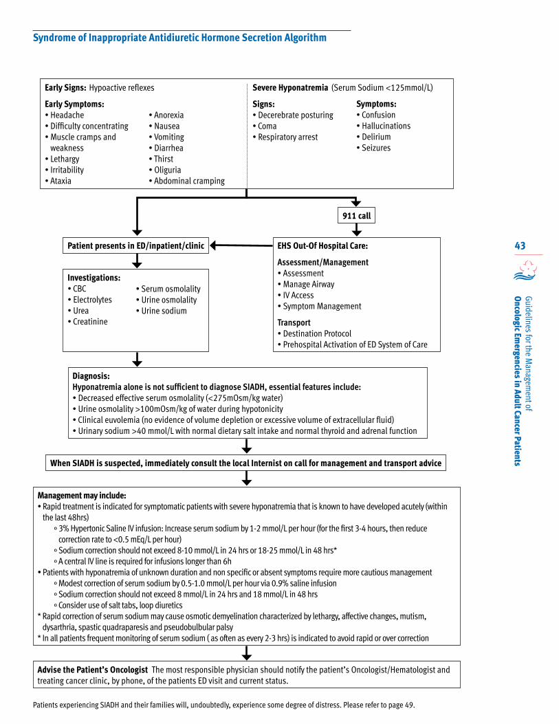
43
Guidelines for the Managem
ent of O
ncologic Emergencies in Adult Cancer Patients
Syndrome of Inappropriate Antidiuretic Hormone Secretion Algorithm
Patient presents in ED/inpatient/clinic EHS Out-Of Hospital Care:
Assessment/Management• Assessment• Manage Airway• IV Access• Symptom Management
Transport• Destination Protocol• Prehospital Activation of ED System of Care
Investigations:• CBC• Electrolytes• Urea• Creatinine
• Serum osmolality• Urine osmolality• Urine sodium
Diagnosis:Hyponatremia alone is not sufficient to diagnose SIADH, essential features include:• Decreased effective serum osmolality (<275mOsm/kg water)• Urine osmolality >100mOsm/kg of water during hypotonicity • Clinical euvolemia (no evidence of volume depletion or excessive volume of extracellular fluid)• Urinary sodium >40 mmol/L with normal dietary salt intake and normal thyroid and adrenal function
Management may include:• Rapid treatment is indicated for symptomatic patients with severe hyponatremia that is known to have developed acutely (within
the last 48hrs)° 3% Hypertonic Saline IV infusion: Increase serum sodium by 1-2 mmol/L per hour (for the first 3-4 hours, then reduce
correction rate to <0.5 mEq/L per hour)° Sodium correction should not exceed 8-10 mmol/L in 24 hrs or 18-25 mmol/L in 48 hrs*° A central IV line is required for infusions longer than 6h
• Patients with hyponatremia of unknown duration and non specific or absent symptoms require more cautious management° Modest correction of serum sodium by 0.5-1.0 mmol/L per hour via 0.9% saline infusion° Sodium correction should not exceed 8 mmol/L in 24 hrs and 18 mmol/L in 48 hrs° Consider use of salt tabs, loop diuretics
* Rapid correction of serum sodium may cause osmotic demyelination characterized by lethargy, affective changes, mutism, dysarthria, spastic quadraparesis and pseudobulbular palsy
* In all patients frequent monitoring of serum sodium ( as often as every 2-3 hrs) is indicated to avoid rapid or over correction
Advise the Patient’s Oncologist The most responsible physician should notify the patient’s Oncologist/Hematologist and treating cancer clinic, by phone, of the patients ED visit and current status.
When SIADH is suspected, immediately consult the local Internist on call for management and transport advice
Patients experiencing SIADH and their families will, undoubtedly, experience some degree of distress. Please refer to page 49.
Hypoactive reflexes (Serum Sodium <125mmol/L)Early Signs:
Early Symptoms:• Headache• Difficulty concentrating• Muscle cramps and
weakness• Lethargy• Irritability• Ataxia
• Anorexia • Nausea• Vomiting• Diarrhea• Thirst• Oliguria• Abdominal cramping
Severe Hyponatremia
Signs:• Decerebrate posturing• Coma• Respiratory arrest
Symptoms:• Confusion• Hallucinations• Delirium• Seizures
911 call
44
Guidelines for the Managem
ent of O
ncologic Emergencies in Adult Cancer Patients
Tumour lysis syndrome (TLS) is a complication of massive cellular breakdown in rapidly
proliferating, bulky or highly chemo-radiosensitive tumours and the subsequent release of intracellular contents into the bloodstream.
TLS should be suspected in patients with a known malignancy (hematologic or solid tumor) who present with fluid overload, decreased urine output, lethargy, muscle cramps, arrhythmias or seizure.
TLS is characterized by hyperuricemia and major electrolyte disturbances (hyperkalemia, hyperphosphatemia and hypocalcemia).
Tumor lysis syndrome is considered an emergency because it disturbs hemodynamics. Without timely intervention, it can quickly lead to oliguric renal failure, seizures, cardiac arrhythmias and death.
While common in hematologic malignancies, TLS is relatively rare in solid tumours. Patients most at risk are those with acute lymphoproliferative disorders with a high proliferative rate and high tumour sensitivity to chemotherapy (e.g., Burkitt’s lymphoma, B-cell acute lymphoblastic leukemia).
Predisposing co-morbidities include pre-treatment elevations in serum uric acid levels, pre-existing renal disease, tumour infiltration in the kidney, obstructive uropathy and advanced age.
Highly active, cycle specific drugs (e.g., cytarabine, etoposide, cisplatin) are most frequently associated with TLS, as are corticosteroids (likely implicated because they are used as primary therapy for highly proliferating lymphoid disease).
TLS has also been reported after treatment with other agents, including intrathecal methotrexate, monoclonal antibodies (e.g., rituximab), radiotherapy, interferon, thalidomide, hydroxyurea, fludarabine, imatinib and bortezomib.
TLS may also arise spontaneously prior to the initiation of anti-tumour therapy.
Patients experiencing TLS and their families will, undoubtedly, experience some degree of distress. Please refer to page 49, for information about addressing the psychosocial health needs of patients and families, and information regarding the support of and referral of those who are distressed and having difficulty coping.
Presentation
The syndrome is often divided into laboratory TLS and clinical TLS.
The clinical manifestations of TLS are variable, and may include:
• Arrhythmias • Congestive heart failure• Decreased urine output• Fluid overload • Lethargy• Muscle cramps• Nausea
• Paresthesias• Renal failure• Seizures• Syncope• Tetany• Vomiting
In patients with obstructive uropathy secondary to hyperuricemia, hematuria, flank or back pain and hypertension may be present.
Severe hypocalcemia is one of the most critical manifestations of TLS and may cause:• Cardiovascular (ventricular arrhythmias, heart
block, hypotension)• Muscular (cramps, spasms)• Neurological ( confusion, delirium, hallucinations,
seizures)
EHS Out-Of Hospital Care
Paramedics responding to a cancer patient with suspected TLS:
Assessment/Management
• Assessment• Airway management• IV resuscitation• Symptom management
Transport
• Destination protocol• Pre hospital activation of ED system of care
Tumor Lysis Syndrome 1, 2, 32, 33, 34
45
Guidelines for the Managem
ent of O
ncologic Emergencies in Adult Cancer Patients
In Hospital Care
Investigations• CBC• Electrolytes• Urea• Creatinine• Uric Acid (if post Rasburicase, sample must be
sent on ice)• Phosphorus• Calcium• Albumin• ECG
Routine evaluation and diagnostic workup should include: strict monitoring of weight, blood pressure and urine output.
Renal ultrasound to rule out obstructive uropathy may be considered.
While there is no universally accepted specific definition or grading system for laboratory TLS, the Cairo and Bishop grading scale19 is commonly cited; where TLS is defined as two or more of the following metabolic abnormalities within 3 days before or 7 days after the initiation of chemotherapy:
• Serum uric acid > 476 μmol/L (or 25% increase from baseline)
• Serum potassium ≥ 6.0 mmol/L (or 25% increase from baseline)
• Serum phosphorous ≥ 1.45 mmol/L in adults (or 25% increase from baseline)
• Secondary hypocalcemia (corrected serum calcium ≤ 1.75 mmol/L or 25% decrease from baseline)
Management
When the lab results are available, immediately consult the Medical Oncologist/Hematologist on call and follow specialist’s management and transport advice.• QEII (Halifax) 902- 473- 2220• CBRH (Sydney) 902-567-8000
The basic principles of TLS management are aggressive prophylaxis and early recognition and correction of metabolic abnormalities to prevent or minimize clinical manifestations.
• In the absence of acute renal dysfunction and oliguria, vigorous IV hydration (3 L/m² per day) and diuresis (≥ 100 mL/m²/hour) should be maintained.
• Diuretics (Furosemide, Mannitol) may be required to maintain urine output and prevent fluid overload, but should only be used if there is no evidence of acute obstructive uropathy or hypovolemia.
• Urine specific gravity should be kept below 1.010.
• Potassium, calcium and phosphate should not initially be added to hydration fluids.
• Dose modifications of drugs that are primarily renally cleared may be required during acute TLS.
• Hemodialysis or hemofiltration may be required with increasing renal dysfunction; uncontrolled hyperphosphatemia, hyperkalemia, hyperuricemia, hypocalcemia, volume overload or hypertension; severe acidosis and/or severe uremia with pericarditis or CNS toxicity.
• It is important to note that while rare, TLS associated with solid tumours has a higher mortality rate compared to that observed with hematological malignancies. This may be due, in part, to time of onset. While TLS associated with hematological malignancies generally occurs soon after treatment begins, TLS in solid tumours can occur days or weeks later. Awareness and vigilance are essential to preventing a delay in or omission of effective prophylaxis.
Tumor Lysis Syndrome (continued)
46
Guidelines for the Managem
ent of O
ncologic Emergencies in Adult Cancer Patients
Hyperuricemia 1, 32, 33
Acute kidney injury (AKI) secondary to precipitation of uric acid in renal tubules is a potential complication of hyperuricemia.
Urine alkalinization via sodium bicarbonate administration is no longer recommended given accumulating evidence of ineffectiveness and the potential to actually induce AKI. Rasburicase 6mg IV infusion daily for 1-7 days is the treatment of choice for all patients with TLS-associated hyperuricemia and for initial prophylaxis in patients at high risk of TLS. Rasburicase reduces the formation of new uric acid, and also degrades uric acid formed prior to treatment initiation.
Allopurinol is recommended for patients with no hyperuricemia at intermediate risk of TLS. It should be initiated 12-24 hours before anti-cancer therapy and continued until the patient can be classified as low risk.
Hyperkalemia 1, 33
Potassium levels should be immediately verified with a second sample to rule out hemolysis during phlebotomy.
Close evaluation of ECG, cardiac rhythm and electrolytes is warranted.
Oral and IV supplemental potassium should be discontinued.
In asymptomatic patients, the preferred treatment is sodium polystyrene sulphonate.• 15-30g orally• 30-50g rectally
In symptomatic patients, regular insulin 10 units IV and a 20-50g dextrose IV should be administered.
• Sodium bicarbonate 1mEq/kg/IV direct can be given to induce the influx of potassium into cells
• Calcium gluconate 1g IV over 5 minutes (and with continuous ECG monitoring for bradycardia) may be used for life-threatening arrhythmias
• Consider dialysis
Hyperphosphatemia 1, 33, 34
• Initial management includes removing phosphate from IV fluids and administering oral or nasogastric phosphategnify binders (e.g., aluminum hydroxide or aluminum carbonate) for up to two days.° Longer treatment carries the risk of aluminum
toxicity
• Calcium infusions should be withheld
• In severe cases, continuous peritoneal dialysis, hemodialysis, or continuous venovenous hemofiltration may be required
Symptomatic Hypocalcemia 1, 33
Treatment is not recommended in asymptomatic patients as the risk of precipitating metastatic calcifications is high, especially with hyperphosphatemia, and hypocalcemia will generally resolve as tumour lysis improves.
Severe hypocalcemia is one of the most critical manifestations of TLS and may cause the following signs: • Cardiovascular (ventricular arrhythmias, heart
block, hypotension) • Muscular (cramps, spasms) • Neurological (confusion, delirium,
hallucinations, seizures)
Symptomatic hypocalcemia is treated with calcium gluconate 1-2g IV, though patients must be monitored for acute obstructive uropathy.
If phosphate is also high, a renal consult should be considered.
Advise the Patient’s Oncologist
The most responsible physician should notify the patients Oncologist/Hematologist and treating cancer clinic, by phone, of the patients ED visit and current status.
Tumor Lysis Syndrome (continued)
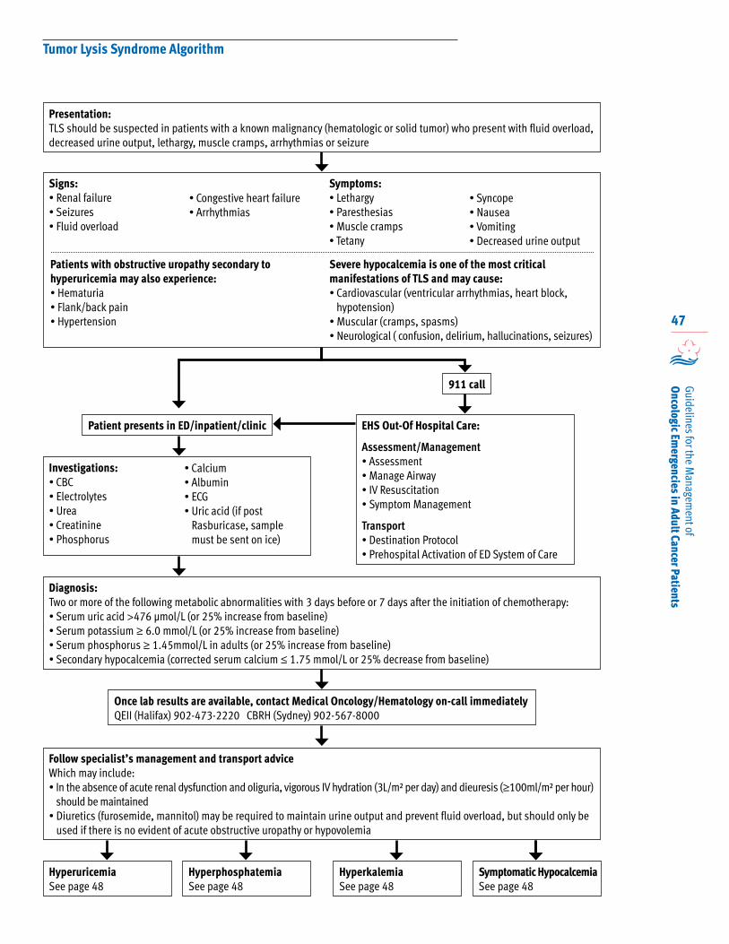
47
Guidelines for the Managem
ent of O
ncologic Emergencies in Adult Cancer Patients
Tumor Lysis Syndrome Algorithm
Patient presents in ED/inpatient/clinic
911 call
EHS Out-Of Hospital Care:
Assessment/Management• Assessment• Manage Airway• IV Resuscitation• Symptom Management
Transport• Destination Protocol• Prehospital Activation of ED System of Care
Investigations:• CBC• Electrolytes• Urea• Creatinine• Phosphorus
• Calcium• Albumin• ECG• Uric acid (if post
Rasburicase, sample must be sent on ice)
Diagnosis:Two or more of the following metabolic abnormalities with 3 days before or 7 days after the initiation of chemotherapy:• Serum uric acid >476 μmol/L (or 25% increase from baseline)• Serum potassium ≥ 6.0 mmol/L (or 25% increase from baseline)• Serum phosphorus ≥ 1.45mmol/L in adults (or 25% increase from baseline)• Secondary hypocalcemia (corrected serum calcium ≤ 1.75 mmol/L or 25% decrease from baseline)
Follow specialist’s management and transport adviceWhich may include:• In the absence of acute renal dysfunction and oliguria, vigorous IV hydration (3L/m² per day) and dieuresis (≥100ml/m² per hour)
should be maintained• Diuretics (furosemide, mannitol) may be required to maintain urine output and prevent fluid overload, but should only be
used if there is no evident of acute obstructive uropathy or hypovolemia
Once lab results are available, contact Medical Oncology/Hematology on-call immediatelyQEII (Halifax) 902-473-2220 CBRH (Sydney) 902-567-8000
HyperuricemiaSee page 48
Signs:• Renal failure• Seizures • Fluid overload
• Congestive heart failure• Arrhythmias
Symptoms:• Lethargy• Paresthesias• Muscle cramps• Tetany
• Syncope• Nausea • Vomiting• Decreased urine output
Patients with obstructive uropathy secondary to hyperuricemia may also experience:• Hematuria• Flank/back pain• Hypertension
Severe hypocalcemia is one of the most critical manifestations of TLS and may cause:• Cardiovascular (ventricular arrhythmias, heart block,
hypotension)• Muscular (cramps, spasms)• Neurological ( confusion, delirium, hallucinations, seizures)
HyperkalemiaSee page 48
HyperphosphatemiaSee page 48
Symptomatic HypocalcemiaSee page 48
Presentation:TLS should be suspected in patients with a known malignancy (hematologic or solid tumor) who present with fluid overload, decreased urine output, lethargy, muscle cramps, arrhythmias or seizure
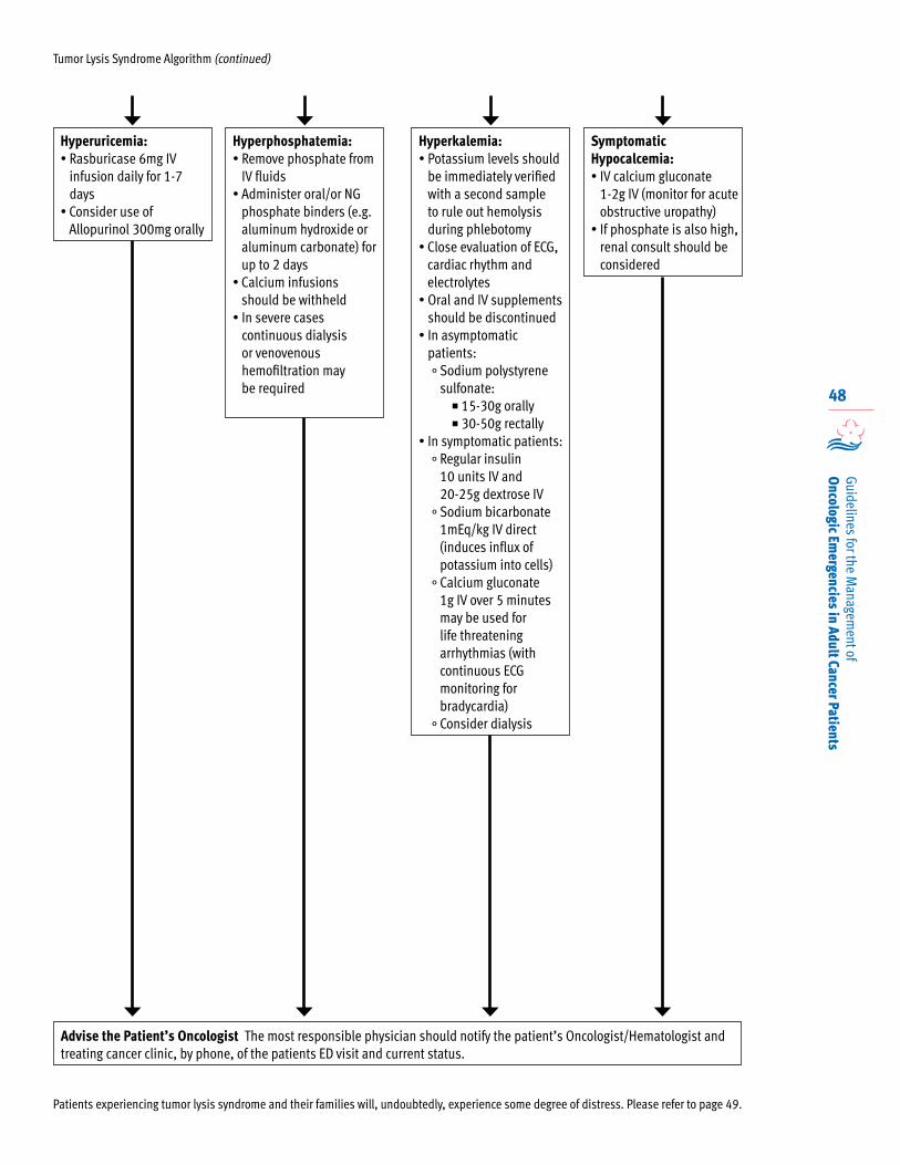
48
Guidelines for the Managem
ent of O
ncologic Emergencies in Adult Cancer Patients
Tumor Lysis Syndrome Algorithm (continued)
Hyperuricemia:• Rasburicase 6mg IV
infusion daily for 1-7 days
• Consider use of Allopurinol 300mg orally
Hyperkalemia:• Potassium levels should
be immediately verified with a second sample to rule out hemolysis during phlebotomy
• Close evaluation of ECG, cardiac rhythm and electrolytes
• Oral and IV supplements should be discontinued
• In asymptomatic patients:° Sodium polystyrene
sulfonate: 15-30g orally 30-50g rectally
• In symptomatic patients:° Regular insulin
10 units IV and 20-25g dextrose IV
° Sodium bicarbonate 1mEq/kg IV direct (induces influx of potassium into cells)
° Calcium gluconate 1g IV over 5 minutes may be used for life threatening arrhythmias (with continuous ECG monitoring for bradycardia)
° Consider dialysis
Hyperphosphatemia:• Remove phosphate from
IV fluids• Administer oral/or NG
phosphate binders (e.g. aluminum hydroxide or aluminum carbonate) for up to 2 days
• Calcium infusions should be withheld
• In severe cases continuous dialysis or venovenous hemofiltration may be required
Symptomatic Hypocalcemia:• IV calcium gluconate
1-2g IV (monitor for acute obstructive uropathy)
• If phosphate is also high, renal consult should be considered
Advise the Patient’s Oncologist The most responsible physician should notify the patient’s Oncologist/Hematologist and treating cancer clinic, by phone, of the patients ED visit and current status.
Patients experiencing tumor lysis syndrome and their families will, undoubtedly, experience some degree of distress. Please refer to page 49.
49
Guidelines for the Managem
ent of O
ncologic Emergencies in Adult Cancer Patients
Psychosocial Health Needs of Patients and Families Experiencing Oncologic Emergencies
35, 36, 37, 38
The needs and concerns of adult cancer patients vary from the time of initial diagnosis through treatment
and survivorship, advanced disease and death and dying. Their needs include physical, emotional, psychological, practical, informational, social, and spiritual issues, and all are important in the provision of person-centred care.
Thirty-five to 45% of cancer patients display clinically significant levels of distress at some point during their cancer experience. Family members also experience clinically significant distress, at levels equivalent to or greater than patients. Cancer-related distress is defined as “a multi-factorial unpleasant emotional experience of a psychological (cognitive, behavioral, emotional), social and / or spiritual nature that may interfere with the ability to cope effectively with cancer, its physical symptoms and its treatment. Distress extends along a continuum, ranging from common normal feelings of vulnerability, sadness, and fears to problems that can become disabling such as depression, anxiety, panic, social isolation and existential and spiritual crisis”.
Distress is now recognized as the sixth Vital Sign of cancer care. In Nova Scotia, identification of patient distress through screening and management of the cancer-related distress is a standard of care.
Various points in the illness continuum, such as initial diagnosis, end of medical treatment, time of medical procedures, change in disease status, during end of life care and times of personal transition, are associated with increased distress and uncertainty for patients and families. Dealing with oncologic emergencies, that are potentially life-threatening, or potentially limiting in regard to quality of life, are especially stressful times for patients and families. The sudden onset and outcome of these emergency events can be traumatic and distressing for patients and families. Providing person-centered, culturally competent care to patients experiencing oncologic emergencies can reduce the distress experienced by patients and families.
When dealing with an oncologic emergency, patients and families will benefit from supportive communication (e.g., clear communication, provision of relevant information, active listening, empathy), supportive counselling (e.g., provision of support, minimizing symptoms, making patients and families aware of resources) and symptom management (supportive carea), as appropriate.
For those patients and families who experience a high level of distress and are having difficulty coping with an oncologic emergency, referral for specialized psychosocial care (psychosocial oncologyb), is recommended. The patient/family should be referred to the most appropriate healthcare professional available in the clinic/hospital. For example, if the patient with an oncologic emergency is seen in the Emergency Department, referral may be initially to Mental Health Triage, Social Worker assigned to Emergency Department, or to the Cancer Patient Navigator for assessment and referral to available resources. If the emergency occurs while patient is hospitalized, or being followed on an outpatient basis, referral may be to the QEII Psychosocial Oncology Team, Psychiatrist, Psychologist, Social Worker, Advanced Practice Nurse, Spiritual Care, or Cancer Patient Navigator, dependent on the resources available in the district.
The cost of ambulance services is not an insured service. The cost of ambulance transport may create a financial hardship for patients/families. Patients/families may be eligible for support through the Ambulance Fee Assistance Program and should be referred to the Emergency Health Services billing office, (902) 832-8337 or toll-free 1-888-280-8884. Further information concerning ambulance fees can be accessed at www.gov.ns.ca.
For further information and guidance concerning the management of cancer-related distress, refer to the CCNS Best Practice Guideline for the Management of Cancer-Related Distress in Adults.
a Supportive care services are defined as addressing “a range of needs, including informational and counselling needs related to the management of symptoms and specific practical or functional issues. A variety of disciplines may be involved in provision of supportive care, such as nursing, medicine, nutrition and rehabilitation services.”37
b Psychosocial oncology is defined as “a specialty in cancer care concerned with understanding and treating the social, psychological, emotional, spiritual, quality-of-life and functional (practical) aspects of cancer, from prevention through bereavement. It is a whole-person approach to cancer care that addresses a range of very human needs. Psychosocial Oncology focuses on the emotional distress aspects of cancer care and is particularly concerned with the assessment and treatment of distress...and the management of complex issues” 37
50
Guidelines for the Managem
ent of O
ncologic Emergencies in Adult Cancer Patients
References
1. Guideline Utilization Resource Unit, Alberta Health Services (2010). Oncologic Emergencies A Guide for Family Physicians. Edmonton: Alberta Health Services. Retrieved from www.albertahealthservices.ca.
2. Morris, J.C. and Holland, J.F. (2000). Oncologic Emergencies. In: Bast, R.C., Kufe, D.W., Pollock, R.E., et al., (editors). Holland-Frei Cancer Medicine, Chapter 158, 5th edition. Hamilton (ON): B.C. Decker. Retrieved from www.ncbi.nlm.nih.gov.
3. Canadian Cancer Society’s Steering Committee on Cancer Statistics (2012). Canadian Cancer Statistics 2012.Toronto, ON: Canadian Cancer Society.
4. Lewis, M.A., Hendrickson, A.W. and Moynihan, T.J. (2011). Oncologic emergencies: Pathophysiology, presentation, diagnosis, and treatment. CA a Cancer Journal for Clinicians. doi: 10.3322/caac.20124.
5. Hyman D.M., Soff G.A. and Kampel L.J. (2011).Disseminated Intravascular Coagulation with Excessive Fibrinolysis in Prostate Cancer: A Case Series and Review of the Literature. Oncology. 81:119–125.
6. Saba, H.I., Morelli, G.A. and Saba, R.I. (2009) Disseminated Intravascular Coagulation (DIC) in Coagulation in Cancer, Cancer Treatment and Research. Volume 148: 137-156.
7. Jervoise, H., Andreyev,N., Davidson,S.E., Gillespie,C., Allum,W.H. and Swarbrick, E. (2012). Practice guidance on the management of acute and chronic gastrointestinal problems arising as a result of treatment for cancer. Gut. 61:179-192.
8. Berkey, F. J. (2010). Managing the adverse effects of radiation therapy. American Family Physician, 82(4), 381-8, 394.
9. Tsao, M.N., Rades, D., Wirth, A., Lo, S.S., Danielson, B.L., Gaspar,L.E., Sperduto, P., Vogelbaum,W.A., Radawski,J.D., Wang, J.Z., Gillin, M.T., Mohideen, N, Hahn, C.A. and Chang,E.L. (2012). Radiotherapeutic and surgical management for newly diagnosed brain metastases: An American Society for Radiation Oncology evidence-based guideline. Practical Radiation Oncology. 2:210–225.
10. Grosu, H.B., Casal, R.F., Morice, R.C, Nogueras-González, G.M., Eapen, G.A., Ost, D., Sarkiss, M.G. and Jimenez, C.A.. (2013). Bronchoscopic Findings and Bleeding Control Predict Survival in Patients with Solid Malignancies Presenting with Mild Hemoptysis. Annals of the American Thoracic Society.10 (4): 342-349.
11. Antimicrobial Agents Subcommittee, District Drugs and Therapeutics Committee, Capital District Health Authority (2012). Antimicrobial Handbook. Halifax, Nova Scotia: Capital District Health Authority.
12. Guideline Utilization Resource Unit, Alberta Health Services (2012). Management of Febrile Neutropenia in Adult Cancer Patients. Edmonton: Alberta Health Services. Retrieved from www.albertahealthservices.ca.
13. Flowers, C.R., Seidenfeld, J., Bow, E.J., Karten, C., Gleason, C., Hawley,D.K., Kuderer, N.M., Langston, A.A., Marr, K.A., Rolston, K.V.I. and Ramsey, S.D. (2013). Antimicrobial Prophylaxis and Outpatient Management of Fever and Neutropenia in Adults Treated for Malignancy: American Society of Clinical Oncology Clinical Practice Guideline. Journal of Clinical Oncology doi:10.1200/JCO.2012.45.8661.
14. Klastersky, J., Paesmans, M., Rubenstein, E.B., Boyer, M., Elting, L., Feld, R., Gallagher, J., Herrstedt, J., Rapoport, B., Rolston, K. and Talcott, J., for the Study Section on Infections of Multinational Association for Supportive Care in Cancer. (2000). The Multinational Association for Supportive Care in Cancer Risk Index: A Multinational Scoring System for Identifying Low-Risk Febrile Neutropenic Cancer Patients. Journal of Clinical Oncology, 18(16):3038-3051.
15. Talcott, J.A., Siegel, R.D., Finberg, R., et al. (1992). Risk assessment in cancer patients with fever and neutropenia: A prospective, two-center validation of a prediction rule. Journal of Clinical Oncology.10:316-322.
16. Bullard, M.J., Unger, B., Spence, J., Grafstein, E. and the CTAS National Working Group(2008). Canadian Journal of Emergency Medicine. 10(2):136-42.
17. National Comprehensive Cancer Network, Inc. (2012). Fever and Neutropenia, in Prevention and Treatment in Cancer Related Infections. NCCN Clinical Practice Guidelines in Oncology.
18. de Naurois,J., Novitzky-Basso, I., Gill, M. J., Marti Marti, F., Cullen, M. H. & Roila, F. (2010). Management of febrile neutropenia: ESMO Clinical Practice Guidelines. Annals of Oncology. 21 (Supplement 5): v252–v256.
19. Freifeld,A.G., Bow, E.J., Sepkowitz, K.A., Boeckh,M.J., Ito, J.I., Mullen, C.A., Raad, I.I., Rolston, K.V., Young, J.H. and Wingard, J.R. Clinical Practice. (2011). Guideline for the Use of Antimicrobial Agents in Neutropenic Patients with Cancer: 2010 Update by the Infectious Diseases Society of America. Clinical Infectious Diseases.52(4):e56–e93.
51
Guidelines for the Managem
ent of O
ncologic Emergencies in Adult Cancer Patients
20. Cape Breton Cancer Centre (2008). Febrile Neutropenia Management Tool. Sydney: Cape Breton Cancer Centre
21. Stone, M.J. and Bogen, S.A. (2012). Evidence-based focused review of management of hyperviscosity syndrome. Blood. 119:10, 2205-2208.
22. Rosner, M.H. and Dalkin,A.C. (2012). Onco-Nephrology: The Pathophysiology and Treatment of Malignancy-Associated Hypercalcemia. Clinical Journal of the American Society of Nephrology. doi: 10.2215/ CJN.02470312. Accessed from cjasn.asnjournals.org.
23. Inchingolo, R., Sabharwal, T.,Spiliopoulos, S.,Krokidis, M., Dourado,R., Ahmed, I., King, J. and Adam, A. (2012).Tracheobronchial Stenting for Malignant Airway Disease: Long-Term Outcomes From a Single-Center Study. American Journal of Hospice Palliative Care, doi: 10.1177/1049909112462861. Accessed from ajh.sagepub.com.
24. Lutz, S., Korytko, T., Nguyen, J., Khan, L., Chow, E., & Corn, B. (2011). Palliative radiotherapy for bone metastases: An ASTRO Evidence based Guideline. International Journal of Radiation Oncology, Biology, Physics. Vol. 79 (4): 965–976.
25. Ribas, E.S.C. and Schiff, D. (2012). Spinal Cord Compression. Current Treatment Options in Neurology. 14:391–401, doi: 10.1007/s11940-012-0176-7.
26. Loblaw, D.A, Mitera, G., Ford, M. and Laperriere, N.J.(2012). A 2011 Updated Systematic Review and Clinical Practice Guideline for the Management of Malignant Extradural Spinal Cord Compression. International Journal of Radiation Oncology, Biology, Physics. 84 (2), 312-317. http://www.sciencedirect.com/science/article/pii/S0360301612000570 - item1#item1http://www.sciencedirect.com/science/article/pii/S0360301612000570 - item2#item2
27. Conway, R. Graham, J., Kidd, J., and Levack, P.; Scottish Spinal Cord Compression Group. (2007). What happens to people after malignant spinal cord compression? Survival, function, quality of life, emotional well being, and place of care one month after diagnosis. Clinical Oncology (Royal College of Radiologists). 19(1), 56-62.
28. Fowell, A. and Stuart, N.S.A. (2011). Emergencies in Palliative Medicine. Medicine. 39 (11), 660-663.
29. Gompelmann, D., Eberhardt, R. and Herth, F.J.F. (2011). Advanced Malignant Lung Disease: What the Specialist Can Offer. Respiration. 82:111–123. doi: 10.1159/000329703
30. Wan, J.A. and Bezjak, A. (2009). Superior Vena Cava Syndrome. Emergency Medicine Clinics of North America. 27(2): 243–255.
31. Peri, A., Pirozzi N., Parenti G., Festuccia F. And Menè P. (2010). Hyponatremia and the syndrome of inappropriate secretion of antidiuretic hormone (SIADH). Journal of Endocrinological Investigation. 33(9):671-82. http://www.sciencedirect.com/science/article/pii/S0733862709000169 - item1#item1.
32. Cairo, M.S. and Bishop, M. (2004). Tumour lysis syndrome: new therapeutic strategies and classification. British Journal of Haematology. 127(1):3-11.
33. Coiffier, B., Altman, A., Pui, C.H., Younes, A., Cairo, M.S. (2008). Guidelines for the management of pediatric and adult tumor lysis syndrome: an evidence-based review. Journal of Clinical Oncology. 1;26(16):2767-2778.
34. Firwana,B.M.; Hasan, R.; Hasan, N.; Alahdab, F.; Alnahhas, I; Hasan, S.; Varon,J. (2012). Tumor Lysis Syndrome: A Systematic Review of Case Series and Case Reports. Postgraduate Medicine.124 (2). DOI:10.3810/ pgm. 2012.03.2540
35. National Comprehensive Cancer Network, Inc. (2013). Distress Management, version 2.2013. NCCN Clinical Practice Guidelines in Oncology.
36. Cancer Care Nova Scotia (2013). Best Practice Guideline for the Management of Cancer-Related Distress in Adults. Halifax: Province of Nova Scotia.
37. Canadian Association of Psychosocial Oncology. (2010). Standards of Psychosocial Health Services for Persons with Cancer and their Families. Toronto: CAPO.
38. Nova Scotia Department of Health (2005). A Cultural Competence Guide for Primary Health Care Professionals in Nova Scotia. Halifax, N.S: Province of Nova Scotia.
References (continued)
52
Guidelines for the Managem
ent of O
ncologic Emergencies in Adult Cancer Patients
Appendix 1
Guideline Development Process
Affirming the Need for Guidelines
In 2011, Cancer Care Nova Scotia (CCNS) identified the lack of consistent province-wide
guidelines for the assessment and management of oncologic emergencies. A literature review revealed that Alberta Health Services (AHS) had produced a guide in 2010.1
CCNS staff reviewed the AHS resource, Oncologic Emergencies, A Guide for Family Physicians, and deemed it appropriate for adaptation to the Nova Scotia (NS) practice context. CCNS then sought the expert opinion of Dr. Sam Campbell, Chief of Emergency Medicine, Capital District Health Authority (CDHA). Dr. Campbell validated the need for guidelines to assess and manage common oncologic emergencies and also verified that the AHS guide was appropriate for adaptation.
CCNS sought and received permission from the Guideline Utilization Resource Unit, Alberta Health Services to adapt their guide.
Development Process
Prior to engaging provincial stakeholders in the development process, CCNS determined that to be most relevant for the Nova Scotia practice context a number of adaptations to the AHS resource would need to occur:
• The AHS resource is directed to family physicians. CCNS elected to broaden the target audience for the guideline to include all health professionals involved in the care of adult cancer patients, particularly Emergency Health Services (EHS) and Emergency Medicine and Emergency Nursing.
• Addressing the psychosocial health needs of cancer patients and their families within guideline documents is a CCNS standard. Thus, a related section was added to the guideline.
• In keeping with CCNS practice, algorithms were developed for each oncologic emergency.
In 2012, CCNS conducted a survey (refer to Appendix 2) to assess stakeholders perception of the AHS resource and CCNS’s proposed adaptations; determine interest in participating in the adaptation process and provide an opportunity for stakeholders to tailor the guidelines to the Nova Scotia practice context. The survey was distributed electronically to the CDHA and Cape Breton District Health Authority’s Heads of Medical and Radiation Oncology, Head of Hematology, Chiefs of Infectious Disease and Managers of Oncology Pharmacies; District Chiefs of Emergency Medicine and Family Practice, GPOs, and the Medical and Program Directors of Emergency Health Services (EHS). Sixteen responses were received (50% response rate).
There was general consensus amongst the respondents regarding:
• The need for provincial guidelines for oncologic emergencies
• The exclusion of emergency situations not unique to cancer patients
• Re-titling the document to focus on the role of all Health Professionals caring for Oncology Patients, not solely Family Physicians
• Reinforcing the patient population as adult oncology patients
• Including appropriate psychosocial care measures to take/referrals to make in a oncologic emergency situation
• Adding relevant information concerning early detection of Oncologic Emergencies, including reference to appropriate patient/family education resources
• Including referral pathways for each Oncologic Emergency
Respondents also noted some specific recommendations regarding:
• Drugs and dosages
• The importance of articulating the role of EHS
• The management of febrile neutropenia
53
Guidelines for the Managem
ent of O
ncologic Emergencies in Adult Cancer Patients
Respondents recommendations were then incorporated into the draft NS guidelines. In addition, practice guidelines that had been published since 2010 were reviewed, incorporated into and/or referenced in the draft NS guideline.
Current Nova Scotian health information systems do not readily enable the measurement of the incidence of oncologic emergencies. In 2013, CCNS asked Oncology Managers and Chiefs of Emergency throughout the province to estimate the annual number of occurrences of each oncologic emergency, included in the guidelines, in clinic, inpatient and Emergency Room settings. For those oncologic emergencies that the respondents were able to estimate, febrile neutropenia was, by far, the most frequent presentation, followed by brain metastases, seizures, spinal cord compression, MAH, superior vena cava compression, SIADH and tumor lysis syndrome.
In 2013, CCNS established a working group to articulate the role of EHS in Oncologic Emergencies led by Dr. Andrew Travers, Medical Director, EHS. The group was comprised of representatives from EHS, Medical Oncology, Hematology, Radiation Oncology and CCNS Staff (a list of members is included in the Acknowledgements section of this document, page i). The group’s work was informed by the findings of the stakeholder survey. This group further refined the guidelines and defined the EHS response for each oncologic emergency included in the guideline and highlighted how high risk cancer patients could be supported by the EHS Special Patient Designation.
CCNS also established a working group in 2013 to refine the Febrile Neutropenia guideline led by Dr. Andrea Kew, Hematologist, QEII Cancer Care Program. The group was comprised of representatives from Medical Oncology, Hematology, Radiation Oncology, Emergency Medicine (rural and urban), Internal Medicine, Infectious Diseases, General Practitioners (Oncology), Oncology Pharmacy, Emergency Nursing (rural and urban), Oncology Nursing, and CCNS Staff. (a list of members is included in the
Acknowledgements section of this document). The group’s work was informed by the findings of the stakeholder survey. This group created guidelines for both low risk and high risk patients. The group also updated the patient’s fever card which is designed to provide febrile cancer patients and Emergency Department Staff with instructions regarding the care of febrile neutropenic patients. An important outcome of the group’s work was attaining consensus that the fever card would be used province wide, previously it had only been in use on mainland Nova Scotia. The group also updated the CCNS Living Well With Cancer Neutropenia patient education resource to align with the guidelines.
The EHS and Febrile Neutropenia working groups’ recommendations were incorporated into the guideline.
Patient Engagement
CCNS is committed to engaging patients in cancer system improvement initiatives. In keeping with this commitment, CCNS elicited feedback from patients, cancer survivors and family members regarding their oncologic emergency experiences. An electronic survey was sent to members of the CCNS Cancer Patient Family Network as well as community based cancer organizations (refer to Appendix 3).
The survey revealed a number of misconceptions held by patients that affected their willingness and appreciation of the need to go to the Emergency Department (ED), as well as their experience within the ED. The survey also highlighted a number of challenges faced by cancer patients experiencing oncologic emergencies. The findings of the survey clearly reinforced the need for provincial guidelines for common oncologic emergencies, the need to clarify communication mechanisms between EDs, on-call Oncology/Hematology and the patient’s Oncologist/Hematologist and the need for consistent patient education regarding oncologic emergencies.
The findings of the survey (refer to Appendix 2 for the full report) informed the development of the guidelines, the yellow fever card and the neutropenia patient resource.
Appendix 1 Guideline Development Process (continued)
54
Guidelines for the Managem
ent of O
ncologic Emergencies in Adult Cancer Patients
Appendix 1 Guideline Development Process (continued)
Patients and families were also involved in a review of the updated CCNS Living Well With Cancer Neutropenia patient education resource. We elected not to have patients review the updated fever card as the same information is contained within the neutropenia patient resource and any suggestions provided for the resource could be replicated on the fever card. In January 2014, an electronic survey regarding the neutropenia patient resource was sent to members of the CCNS Cancer Patient Family Network. Twelve patients responded to the survey, their responses influenced the content and format of the patient resource. The key findings were as follows:
• 84% would have found it very helpful to have this resource at the time of their cancer treatment
• 75% rated the resource as “very easy” to understand
• 77% thought the resource contained the “right amount” of information
• 100% felt all the information included was necessary
• 17% suggested information regarding growth factors ought to be included
• 83% noted they would refer to the resource throughout their treatment
• 50% indicated they would share the resource with family members
Stakeholder Review
In 2014, CCNS constructed a second survey (refer to Appendix 4) to assess a broad group of stakeholders perception of:
• The Oncologic Emergencies guidelines
• The role of EHS outlined for each Oncologic Emergency
• The extent to which current practice is aligned with the guidelines
• Barriers that may encountered in aligning practice with the guidelines
• The resources/actions required to overcome the barriers
The guidelines and survey were distributed electronically to stakeholders representing Cancer Centre Administrators, Oncologists, Oncology Nurses and Nurse Practitioners, Oncology Pharmacists, Emergency Medicine, Emergency Nursing and Diagnostics. Respondents were invited to respond individually or as a group representing their district. Thirty-two responses were received, representing all District Health Authorities.
Thirty-nine percent of respondents rated the preamble and introduction as “very helpful”. The remaining sixty-one percent rated the sections “somewhat helpful” and suggested some minor editorial changes which were incorporated into the final documents.
Sixty-four percent of respondents found the EHS Special Patient Designation information very helpful.
Sixty-four percent of respondents rated the Addressing the Psychosocial Needs section as “very helpful”. Seven percent found it too vague to be helpful. Based on this feedback, CCNS strengthened this section providing more information about referrals to appropriate services.
There was consensus that the role of EHS outlined for each oncologic emergency was appropriate.
Respondents concurred that the guidelines for each oncologic emergency were appropriate. Minor editorial suggestions were incorporated into the final documents.
Respondents noted that current practice within their districts would only require moderate modification in order to be in alignment with the guidelines. One district noted their EDs do not carry the recommended drugs for TLS (hyperuricemia) and it would be cost-prohibitive to do so. The TLS guideline highlights a consultation that will take place with a Medical Oncologist or Hematologist. During this consultation alternative medication and/or transport considerations would be discussed. Thus, we elected not to change the suggested medication.
55
Guidelines for the Managem
ent of O
ncologic Emergencies in Adult Cancer Patients
One district noted that limited MRI availability may affect their ability to practice in alignment with the spinal cord compression guideline. The guideline indicates that a CT scan can also be used if MRI is contraindicated or not available. Thus, we did not modify the guideline.
Respondents indicated that educational sessions for health professionals would enable their district to practice in accordance with the guidelines. Respondents provided a number of suggestions to support the implementation of the guidelines including which were considered by CCNS:
• Widespread distribution of the guidelines
• CME events for physicians
• Grand Rounds presentations
• Nursing education sessions
• In-services and workshop resources (i.e. slide decks, speaking points, participant handouts) for local educators
• Online modules
• Electronic versions of the guidelines
• Webinars
In summary, the stakeholder review provided valuable feedback to inform the final version of the guidelines, confirmed health professionals interest in having such guidelines to support practice and indentified mechanisms to support the implementation of the guidelines.
Implementation Process
A multi-faceted implementation plan was created; elements included:
• Collaborating with the District Health Authorities to address District specific implementation issues
• Offering CME accredited Oncologic Emergency workshops based on guidelines
• Collaborating with EHS to provide in-services for EHS personnel
• Offering in-services for Emergency Physicians and Nurses
• Hosting an Oncology Grand Rounds session re: Oncologic Emergencies and using the opportunity to promote the guidelines
• Presenting a guidelines overview at the Provincial Cancer Network Meeting, District Family Practice and ED meetings
• Posting the guidelines on the CCNS website
• Promoting the guidelines via CCNS facebook, twitter, newsletter, the Doctors Nova Scotia newsletter and website and EHS communication channels
• Integrating the guidelines into Provincial Emergency Department and EHS Toolkits
• Reworking current patient education processes to ensure the relevant information pertaining to oncologic emergencies is included.
Monitoring and Evaluation Process
Members of the Oncologic Emergencies EHS Working Group were asked to identify key performance indicators for the guideline. However, a number of the indicators of interest are not currently captured by health information systems. The indicators of interest were shared with the CCNS Indicator Working Group, this group will be establishing a number of indicators and processes to monitor system performance relative to all CCNS guidelines and standards.
One year following the implementation of the guidelines, CCNS intends to conduct a follow-up survey of stakeholders to assess the utility of the Oncologic Emergencies Guidelines and identify opportunities for improvement.
Appendix 1 Guideline Development Process (continued)
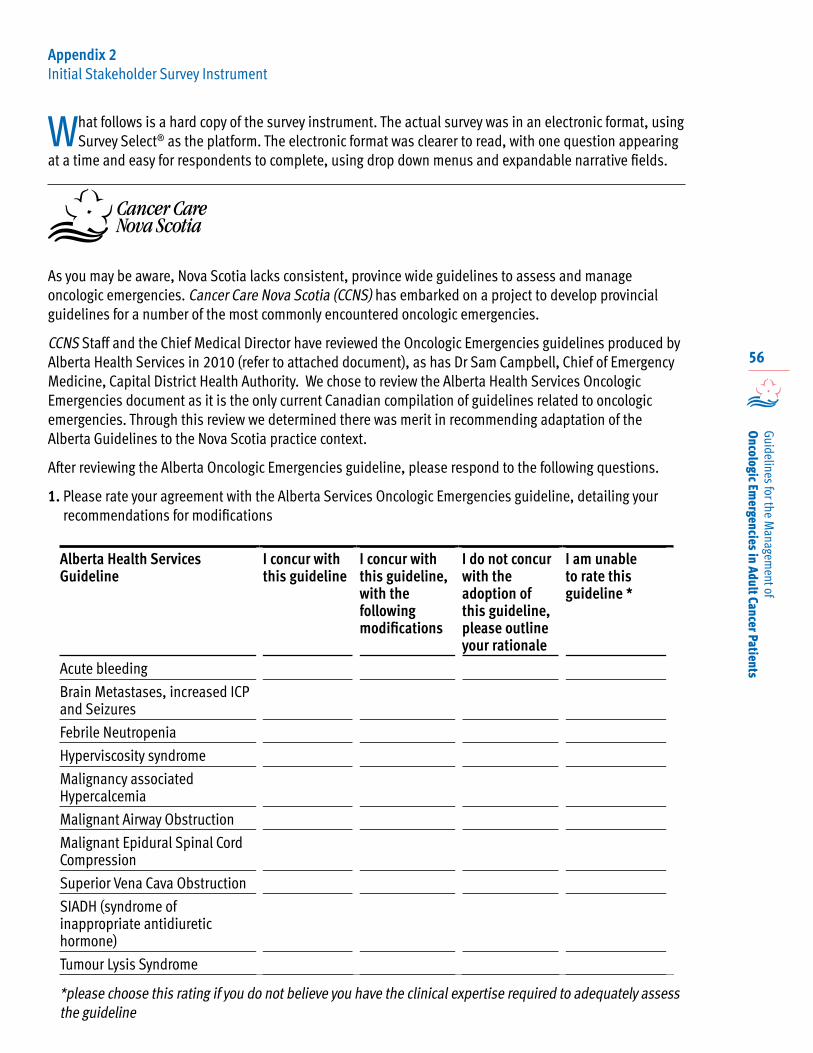
56
Guidelines for the Managem
ent of O
ncologic Emergencies in Adult Cancer Patients
Appendix 2 Initial Stakeholder Survey Instrument
What follows is a hard copy of the survey instrument. The actual survey was in an electronic format, using Survey Select® as the platform. The electronic format was clearer to read, with one question appearing
at a time and easy for respondents to complete, using drop down menus and expandable narrative fields.
As you may be aware, Nova Scotia lacks consistent, province wide guidelines to assess and manage oncologic emergencies. Cancer Care Nova Scotia (CCNS) has embarked on a project to develop provincial guidelines for a number of the most commonly encountered oncologic emergencies.
CCNS Staff and the Chief Medical Director have reviewed the Oncologic Emergencies guidelines produced by Alberta Health Services in 2010 (refer to attached document), as has Dr Sam Campbell, Chief of Emergency Medicine, Capital District Health Authority. We chose to review the Alberta Health Services Oncologic Emergencies document as it is the only current Canadian compilation of guidelines related to oncologic emergencies. Through this review we determined there was merit in recommending adaptation of the Alberta Guidelines to the Nova Scotia practice context.
After reviewing the Alberta Oncologic Emergencies guideline, please respond to the following questions.
1. Please rate your agreement with the Alberta Services Oncologic Emergencies guideline, detailing your recommendations for modifications
Alberta Health Services Guideline
I concur with this guideline
I concur with this guideline, with the following modifications
I do not concur with the adoption of this guideline, please outline your rationale
I am unable to rate this guideline *
Acute bleeding
Brain Metastases, increased ICP and Seizures
Febrile Neutropenia
Hyperviscosity syndrome
Malignancy associated Hypercalcemia
Malignant Airway Obstruction
Malignant Epidural Spinal Cord Compression
Superior Vena Cava Obstruction
SIADH (syndrome of inappropriate antidiuretic hormone)
Tumour Lysis Syndrome
*please choose this rating if you do not believe you have the clinical expertise required to adequately assess the guideline
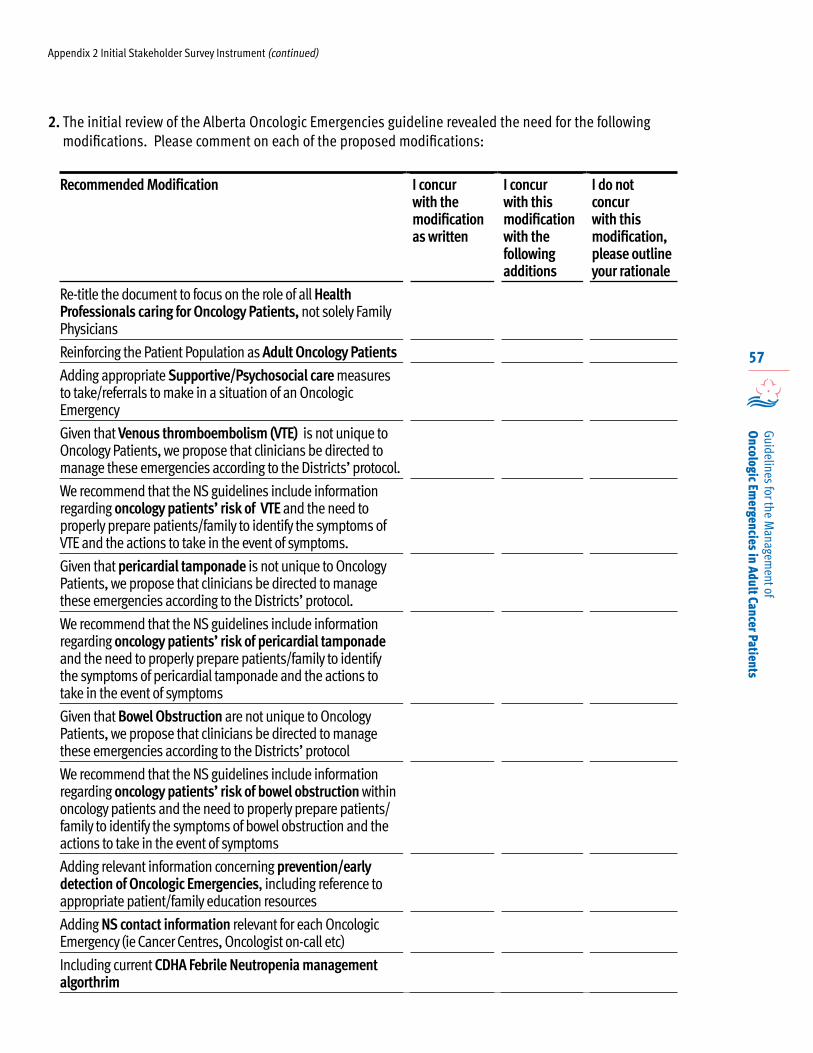
57
Guidelines for the Managem
ent of O
ncologic Emergencies in Adult Cancer Patients
2. The initial review of the Alberta Oncologic Emergencies guideline revealed the need for the following modifications. Please comment on each of the proposed modifications:
Recommended Modification I concur with the modification as written
I concur with this modification with the following additions
I do not concur with this modification, please outline your rationale
Re-title the document to focus on the role of all Health Professionals caring for Oncology Patients, not solely Family Physicians
Reinforcing the Patient Population as Adult Oncology Patients
Adding appropriate Supportive/Psychosocial care measures to take/referrals to make in a situation of an Oncologic Emergency
Given that Venous thromboembolism (VTE) is not unique to Oncology Patients, we propose that clinicians be directed to manage these emergencies according to the Districts’ protocol.
We recommend that the NS guidelines include information regarding oncology patients’ risk of VTE and the need to properly prepare patients/family to identify the symptoms of VTE and the actions to take in the event of symptoms.
Given that pericardial tamponade is not unique to Oncology Patients, we propose that clinicians be directed to manage these emergencies according to the Districts’ protocol.
We recommend that the NS guidelines include information regarding oncology patients’ risk of pericardial tamponade and the need to properly prepare patients/family to identify the symptoms of pericardial tamponade and the actions to take in the event of symptoms
Given that Bowel Obstruction are not unique to Oncology Patients, we propose that clinicians be directed to manage these emergencies according to the Districts’ protocol
We recommend that the NS guidelines include information regarding oncology patients’ risk of bowel obstruction within oncology patients and the need to properly prepare patients/family to identify the symptoms of bowel obstruction and the actions to take in the event of symptoms
Adding relevant information concerning prevention/early detection of Oncologic Emergencies, including reference to appropriate patient/family education resources
Adding NS contact information relevant for each Oncologic Emergency (ie Cancer Centres, Oncologist on-call etc)
Including current CDHA Febrile Neutropenia management algorthrim
Appendix 2 Initial Stakeholder Survey Instrument (continued)
58
Guidelines for the Managem
ent of O
ncologic Emergencies in Adult Cancer Patients
3. In addition to the modifications you have noted in questions 1 and 2, do you have other recommendations that would enhance the alignment of the Alberta Services Oncologic Emergencies guideline with practice context in Nova Scotia?
4. Please outline other Oncologic Emergencies, not included in the Alberta guideline that ought to be included in the NS guideline. Please provide a rationale for each suggestion
5. Please add any other further commentary you have concerning developing NS Oncologic Emergency Guidelines
6. Would you be interested in meeting with other stakeholders to refine and validate the NS Oncologic Emergencies Guidelines? Y N If yes, provide contact info:
Appendix 2 Initial Stakeholder Survey Instrument (continued)
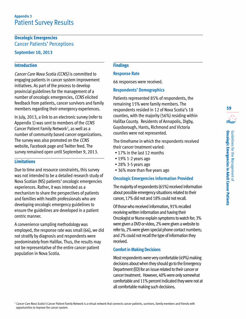
59
Guidelines for the Managem
ent of O
ncologic Emergencies in Adult Cancer Patients
Appendix 3
Patient Survey Results
Oncologic EmergenciesCancer Patients’ Perceptions
September 10, 2013
Introduction Cancer Care Nova Scotia (CCNS) is committed to engaging patients in cancer system improvement initiatives. As part of the process to develop provincial guidelines for the management of a number of oncologic emergencies, CCNS elicited feedback from patients, cancer survivors and family members regarding their emergency experiences.
In July, 2013, a link to an electronic survey (refer to Appendix 1) was sent to members of the CCNS Cancer Patient Family Network3, as well as a number of community based cancer organizations. The survey was also promoted on the CCNS website, Facebook page and Twitter feed. The survey remained open until September 9, 2013.
Limitations
Due to time and resource constraints, this survey was not intended to be a detailed research study of Nova Scotian (NS) patients’ oncologic emergencies experiences. Rather, it was intended as a mechanism to share the perspectives of patients and families with health professionals who are developing oncologic emergency guidelines to ensure the guidelines are developed in a patient centric manner.
A convenience sampling methodology was employed, the response rate was small (66), we did not stratify by diagnosis and respondents were predominately from Halifax. Thus, the results may not be representative of the entire cancer patient population in Nova Scotia.
Findings
Response Rate
66 responses were received.
Respondents’ Demographics
Patients represented 85% of respondents, the remaining 15% were family members. The respondents resided in 12 of Nova Scotia’s 18 counties, with the majority (56%) residing within Halifax County. Residents of Annapolis, Digby, Guysborough, Hants, Richmond and Victoria counties were not represented.
The timeframe in which the respondents received their cancer treatment varied:• 17% in the last 12 months• 19% 1-2 years ago• 28% 3-5 years ago• 36% more than five years ago
Oncologic Emergencies Information Provided
The majority of respondents (65%) received information about possible emergency situations related to their cancer, 17% did not and 18% could not recall.
Of those who received information, 91% recalled receiving written information and having their Oncologist or Nurse explain symptoms to watch for; 3% were given a DVD or video, 2% were given a website to refer to, 2% were given special phone contact numbers; and 2% could not recall the type of information they received.
Comfort in Making Decisions
Most respondents were very comfortable (49%) making decisions about when they should go to the Emergency Department (ED) for an issue related to their cancer or cancer treatment. However, 40% were only somewhat comfortable and 11% percent indicated they were not at all comfortable making such decisions.
3 Cancer Care Nova Scotia’s Cancer Patient Family Network is a virtual network that connects cancer patients, survivors, family members and friends with opportunities to improve the cancer system.
60
Guidelines for the Managem
ent of O
ncologic Emergencies in Adult Cancer Patients
Those who responded “not at all” or “somewhat” were asked to describe what would have better prepared them to make a decision about when to go to the ED. Respondents offered a number of suggestions, most related to providing something in writing with detailed information about potential emergency situations and how best to respond. Respondents highlighted the critical importance of also having the Oncologist or Nurse verbally explain this information to them, more than once, during their cancer treatment.
Two respondents noted some hesitancy on the part of ED Staff to contact their Oncologist or the Oncologist on-call and suggested enhancing these communication mechanisms.
…on several occasions I had spoken to residents on the phone at home prior to my visit to emerg. I
could not get the emergency staff to page them to tell them I was there. I started paging the residents on my cell phone to let them know I was in emerg…
A number of respondents noted they didn’t want to misuse the ED.
I guess I was hesitant to rush in unnecessarily. I had a yellow card to take with me if my
temperature went over a certain amount. It reached that amount, but didn’t go over and I didn’t go to Emergency. If I had, perhaps the problem would have been cleared up sooner.
When getting treatment, I was at the hospital so often that I almost felt guilty if I had to go on my
own accord for an emergency.
Those who responded “very comfortable” were asked what helped them become very comfortable identifying situations when they should go to the ED. Predominately, respondents noted that the thorough explanations and instructions provided by the Oncologist and Nurse, as well as the written information they were given increased their confidence. A number of respondents indicated that being able to call the on-call Nurse or Physician to discuss the symptoms they were experiencing was very helpful. A small number noted that being a nurse themselves, having a family member who was a nurse or having previous experience with the cancer system increased their confidence.
I trusted the instructions that my medical team gave me and acted upon them…
Sometimes it was hard to tell (i.e. is this a real fever or a symptom of the chemo). I called the nurse on-call to clarify and that really helped I
found.
I actually called the on call doctor and discussed it and so felt confident that that was what I needed
to do.
Use of the “Yellow Card”
At the time of this survey, all cancer patients receiving chemotherapy and bone marrow transplant patients in mainland NS were to receive a laminated “yellow card” to present to ED staff if they experienced a fever. The card provided instructions for both the patient and the ED staff. Forty-six percent of the respondents from mainland NS received a “yellow card”, 43% did not and 11% could not recall.
Those who received the card were asked to express their understanding of the purpose of the card (refer to Figure 1).
There appears to be a misunderstanding regarding the purpose of the “yellow card”. The majority understood correctly that the card was to ensure they were seen quickly if they had a fever (80%) and to ensure ED staff identified them as a cancer patient (76%). However, only 52% understood the card also provided the ED staff with treatment information. Interestingly, 44% mistakenly understood that the card would ensure they were seen quickly for any reason and 80% mistakenly understood the card would ensure they were isolated from other patients.
Those who received the “yellow card” were also asked if their Nurse or Oncologist explained that, while cancer patients are a high priority, they may have to wait in the ED while other sicker patients are cared for. Fifty-six percent responded yes, 20% responded no and 24% could not recall.
One respondent from Cape Breton noted they received a white card which they understood was to inform ED staff that they were a cancer patient, ensure they were seen quickly for any reason and ensure they were isolated from ill people in the waiting room.
Appendix 3 Patient Survey Results (continued)
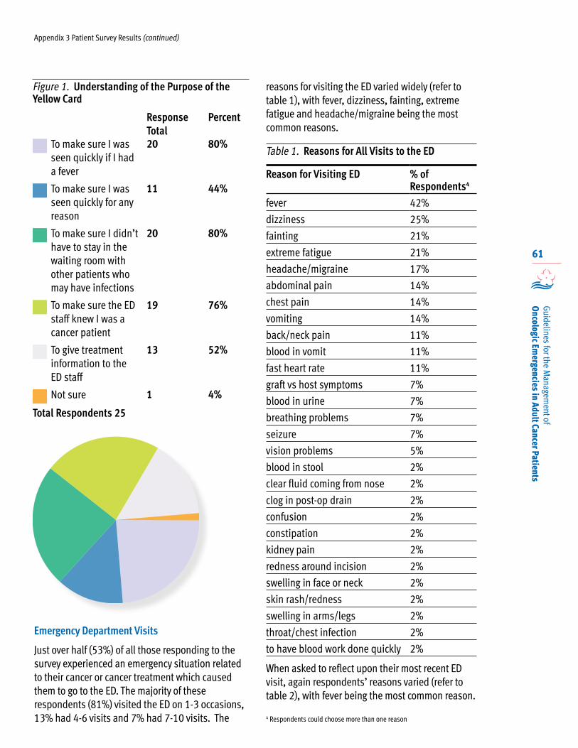
61
Guidelines for the Managem
ent of O
ncologic Emergencies in Adult Cancer Patients
Emergency Department Visits
Just over half (53%) of all those responding to the survey experienced an emergency situation related to their cancer or cancer treatment which caused them to go to the ED. The majority of these respondents (81%) visited the ED on 1-3 occasions, 13% had 4-6 visits and 7% had 7-10 visits. The
reasons for visiting the ED varied widely (refer to table 1), with fever, dizziness, fainting, extreme fatigue and headache/migraine being the most common reasons.
Table 1. Reasons for All Visits to the ED
Reason for Visiting ED % of Respondents4
fever 42%
dizziness 25%
fainting 21%
extreme fatigue 21%
headache/migraine 17%
abdominal pain 14%
chest pain 14%
vomiting 14%
back/neck pain 11%
blood in vomit 11%
fast heart rate 11%
graft vs host symptoms 7%
blood in urine 7%
breathing problems 7%
seizure 7%
vision problems 5%
blood in stool 2%
clear fluid coming from nose 2%
clog in post-op drain 2%
confusion 2%
constipation 2%
kidney pain 2%
redness around incision 2%
swelling in face or neck 2%
skin rash/redness 2%
swelling in arms/legs 2%
throat/chest infection 2%
to have blood work done quickly 2%
When asked to reflect upon their most recent ED visit, again respondents’ reasons varied (refer to table 2), with fever being the most common reason.
Appendix 3 Patient Survey Results (continued)
Figure 1. Understanding of the Purpose of the Yellow Card
Response Total
Percent
To make sure I was seen quickly if I had a fever
20 80%
To make sure I was seen quickly for any reason
11 44%
To make sure I didn’t have to stay in the waiting room with other patients who may have infections
20 80%
To make sure the ED staff knew I was a cancer patient
19 76%
To give treatment information to the ED staff
13 52%
Not sure 1 4%
Total Respondents 25
4 Respondents could choose more than one reason
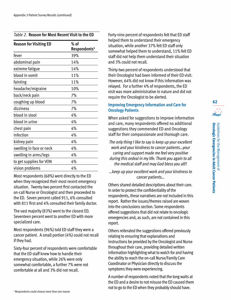
62
Guidelines for the Managem
ent of O
ncologic Emergencies in Adult Cancer Patients
Table 2. Reason for Most Recent Visit to the ED
Reason for Visiting ED % of Respondents4
fever 39%
abdominal pain 14%
extreme fatigue 14%
blood in vomit 11%
fainting 11%
headache/migraine 10%
back/neck pain 7%
coughing up blood 7%
dizziness 7%
blood in stool 4%
blood in urine 4%
chest pain 4%
infection 4%
kidney pain 4%
swelling in face or neck 4%
swelling in arms/legs 4%
to get supplies for VON 4%
vision problems 4%
Most respondents (68%) went directly to the ED when they recognized their most recent emergency situation. Twenty-two percent first contacted the on-call Nurse or Oncologist and then proceeded to the ED. Seven percent called 911, 6% consulted with 811 first and 4% consulted their family doctor.
The vast majority (83%) went to the closest ED. Seventeen percent went to another ED with more specialized care.
Most respondents (96%) told ED staff they were a cancer patient. A small portion (4%) could not recall if they had.
Sixty-four percent of respondents were comfortable that the ED staff knew how to handle their emergency situation, while 26% were only somewhat comfortable, a further 7% were not comfortable at all and 3% did not recall.
Forty-nine percent of respondents felt that ED staff helped them to understand their emergency situation, while another 37% felt ED staff only somewhat helped them to understand, 11% felt ED staff did not help them understand their situation and 3% could not recall.
Thirty-two percent of respondents understood that their Oncologist had been informed of their ED visit. However, 64% did not know if this information was relayed. For a further 4% of respondents, the ED visit was more administrative in nature and did not require the Oncologist to be alerted.
Improving Emergency Information and Care for Oncology Patients
When asked for suggestions to improve information and care, many respondents offered no additional suggestions they commended ED and Oncology staff for their compassionate and thorough care.
The only thing I like to say is keep up your excellent work and your kindness to cancer patients…your caring and support made me feel very positive
during this ordeal in my life. Thank you again to all the medical staff and may God bless you all!!
…keep up your excellent work and your kindness to cancer patients…
Others shared detailed descriptions about their care. In order to protect the confidentiality of the respondents, these narratives are not included in this report. Rather the issues/themes raised are woven into the conclusions section. Some respondents offered suggestions that did not relate to oncologic emergencies and, as such, are not contained in this report.
Others reiterated the suggestions offered previously relating to ensuring that explanations and instructions be provided by the Oncologist and Nurse throughout their care, providing detailed written information highlighting what to watch for and having the ability to reach the on-call Nurse/Family Care Coordinator or Physician directly to discuss the symptoms they were experiencing.
A number of respondents noted that the long waits at the ED and a desire to not misuse the ED caused them not to go to the ED when they probably should have.
Appendix 3 Patient Survey Results (continued)
4 Respondents could choose more than one reason
63
Guidelines for the Managem
ent of O
ncologic Emergencies in Adult Cancer Patients
Appendix 3 Patient Survey Results (continued)
…I often could not tolerate the long waits as there was no comfortable place to lie down to wait to
be seen…
…at times I was too sick to go to emerg to wait for care….
…I became very weak and was only comfortable lying on the floor so my friend went to get
assistance. No one came but luckily after some time, I finally felt stronger so we left. In hindsight, it would have been better if I had seen a staff member
to make sure that I was fine to leave the hospital.
Perhaps some assurance that it’s OK to go to ER if you think something’s wrong.
…I waited in the lobby area from 8am to 3pm…This was very tiring and also worrisome since I
was around people with colds, etc.
A few implored other patients and families to respond quickly and to ensure the patient had improved before they left the ED:
If you suspect there is a problem don’t wait around before going to emergency…
Not so much did we question whether or not we should go to the emergency room or not, but if we were to go through this again, I would ensure that
the ER did not discharge my family member without ensuring all was still well...
A small number of respondents suggested that patients should come directly to the Cancer Centre rather than their local ED.
A number of respondents suggested that ED staff respond more promptly when the yellow card is presented.
Others emphasized how stressful and complex the cancer journey can be and that accessing prompt; consistent care in the ED would reduce some of that distress.
…staff need to look at old and current chart and not count on patient to recall many dates in their
medical history.
I feel that when Emergency staff place you in a individual room, it would be advisable to be
checked on more frequently as the condition can worsen quickly...
Conclusions
Although the survey has a number of limitations (refer to page 1), there are some general themes which will be helpful to the Oncologic Emergencies Working Group. Perhaps the most significant outcome of the survey is that the findings affirm the issues that had been previously anecdotally identified by the Working Group.
The survey findings reinforce the need for provincial guidelines for common oncologic emergencies. These guidelines will ensure that patients receive consistent care in the most appropriate setting. Moreover, the guidelines will clarify communication mechanisms between EDs, on-call Oncology/Hematology and the patient’s Oncologist/Hematologist.
All Oncology/Hematology patients and families should receive verbal and written detailed explanations of the potential emergency situations associated with their diagnosis and treatment, the symptoms to watch for and the actions to take. These explanations should be repeated throughout the cancer patients’ journey.
Oncology/Hematology health professionals should reassure patients and families that in certain situations it is appropriate for them to go to the ED and not to delay going in an emergency situation.
Where feasible, patients should be given an on-call number to phone when they have questions/concerns about the symptoms they are experiencing.
Admission instructions need to be clarified for ED staff so cancer patients do not stay in the ED waiting for a private and/or on-service bed.
The “yellow card” should be used province-wide. The card and the associated patient teaching should be redesigned to ensure both the patient and ED Staff understand the purpose of the card.

64
Guidelines for the Managem
ent of O
ncologic Emergencies in Adult Cancer Patients
Patient Survey Instrument
What follows is a hard copy of the survey instrument. The actual survey was in an electronic format, using Survey Select ® as the platform. The electronic format was clearer to read, with one question appearing at a time and easy for respondents to complete, using drop down menus and expandable narrative fields.
Thank you for your interest in helping Cancer Care Nova Scotia develop provincial guidelines for the most common emergency situations adult cancer patients experience.
We want to hear from adult cancer patients to learn how we can better prepare patients to identify emergency situations related to their cancer or cancer treatment and improve the emergency care provided for cancer patients. You don’t have to have had an emergency situation in order to complete the survey.
Family members of adult cancer patients are also encouraged to share their feedback.
You can complete the survey on your own or with a family member.
1. I am a Cancer Patient Family Member
2. I live in county
3. I received my/my family member received their cancer treatment (circle one):In the last twelve months 1-2 years ago 3-5 years ago More than 5 years ago
4. Did your Cancer Nurse/Doctor give you information about possible emergency situations related to your cancer or cancer treatment? Yes No Can't recall
If yes, what type of information were you given (circle all that apply):Written instructionsMy Nurse/Doctor told me what to watch forWebsite to refer toDVD/VideoOther (please specify)
5. During your cancer treatment, how comfortable were you making decisions about when you should go to the Emergency Department for an issue related to your cancer or your cancer treatment? Not at all comfortable Somewhat comfortable Very comfortable
If you rated not or somewhat above– what would have better prepared you to you identify situations when you should go to the Emergency Department?
If you rated very above– what helped you become very comfortable identifying situations when you should go to the Emergency Department?
Appendix 3 Patient Survey Results (continued)
65
Guidelines for the Managem
ent of O
ncologic Emergencies in Adult Cancer Patients
6. Did your Cancer Nurse/Doctor give you a yellow card to give to Emergency Department staff? Yes No Can't recall
If yes, what was your understanding of the purpose of the yellow card? (choose all that apply) to make sure I was seen quickly if I had a fever to make sure I was seen quickly for any reason to make sure I didn’t have to stay in the waiting room with other patients who may have infections to make sure the Emergency Department staff knew I was a cancer patient to give treatment information to the Emergency Department staff other (please specify)
If you received a yellow card. Did your Cancer Nurse/Doctor explain that, while cancer patients are a high priority, you may have to wait in the Emergency Department while other sicker patients are cared for? Yes No Can't recall
7. During your cancer treatment did you experience an emergency situation related to your cancer or your cancer treatment which caused you to go to the Emergency Department? Yes, go to question 8 No, go to question 17
8. During your cancer treatment how many times did you go to the Emergency Department for an issue related to your cancer or your cancer treatment? 1-3 times 4-6 times 7-10 times More than 10 times
9. Thinking about ALL of your emergency department visits related to your cancer or cancer treatment, what signs or symptoms caused you to go to the Emergency Department? (choose all that apply)
abdominal pain back pain chest pain blood in stool blood in urine blood in vomit coughing up blood breathing problems
confusion constipation diarrhea dizziness extreme fatigue fainting fast heart rate fever
headache/migraine swelling in face or neck swelling in legs or arms seizure vision problems weakness or numbness
in arms or legs vomiting
other (please specify):
10. Thinking about your MOST RECENT emergency department visits related to your cancer or cancer treatment, what signs or symptoms caused you to go to the Emergency Department?
abdominal pain back pain chest pain blood in stool blood in urine blood in vomit coughing up blood breathing problems
confusion constipation diarrhea dizziness extreme fatigue fainting fast heart rate fever
headache/migraine swelling in face or neck swelling in legs or arms seizure vision problems weakness or numbness
in arms or legs vomiting
other (please specify):
Appendix 3 Patient Survey Results (continued)
66
Guidelines for the Managem
ent of O
ncologic Emergencies in Adult Cancer Patients
11. During your most recent emergency situation, before you went to the emergency department, did you (check all that apply): Call 911 Call 811 Call the Cancer Clinic or your Cancer Doctor for advice Call or visit your Family Doctor/walk in clinic for advice Other (please specify):
None of the above, I went straight to the emergency department
12. During your most recent emergency situation did you go to the closest emergency department another emergency department that has more specialized care
13. During your most recent emergency situation, did you tell the Emergency Department staff that you were a cancer patient? Yes No Don't recall
14. During your most recent emergency situation, were you comfortable that the Emergency Department staff knew how to handle your emergency situation? Yes No Somewhat Don't recall
15. During your most recent emergency situation, did Emergency Department staff help you understand your emergency situation? Yes No Somewhat Don't recall
16. During your most recent emergency situation, was your Cancer Doctor informed that you were seen in the Emergency Department? Yes No Don't know Don't recall
17. Thank you for completing this survey. Is there anything else you would like to share that would improve emergency information or care for cancer patients?
Appendix 3 Patient Survey Results (continued)
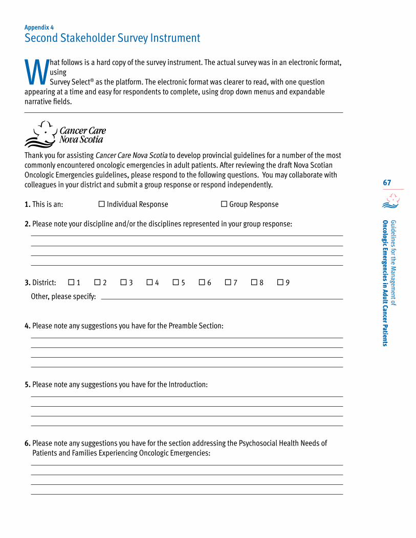
67
Guidelines for the Managem
ent of O
ncologic Emergencies in Adult Cancer Patients
What follows is a hard copy of the survey instrument. The actual survey was in an electronic format, using Survey Select® as the platform. The electronic format was clearer to read, with one question
appearing at a time and easy for respondents to complete, using drop down menus and expandable narrative fields.
Thank you for assisting Cancer Care Nova Scotia to develop provincial guidelines for a number of the most commonly encountered oncologic emergencies in adult patients. After reviewing the draft Nova Scotian Oncologic Emergencies guidelines, please respond to the following questions. You may collaborate with colleagues in your district and submit a group response or respond independently.
1. This is an: Individual Response Group Response
2. Please note your discipline and/or the disciplines represented in your group response:
3. District: 1 2 3 4 5 6 7 8 9 Other, please specify:
4. Please note any suggestions you have for the Preamble Section:
5. Please note any suggestions you have for the Introduction:
6. Please note any suggestions you have for the section addressing the Psychosocial Health Needs of Patients and Families Experiencing Oncologic Emergencies:
Appendix 4
Second Stakeholder Survey Instrument
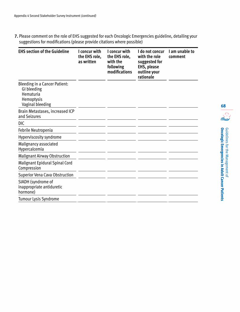
68
Guidelines for the Managem
ent of O
ncologic Emergencies in Adult Cancer Patients
7. Please comment on the role of EHS suggested for each Oncologic Emergencies guideline, detailing your suggestions for modifications (please provide citations where possible)
EHS section of the Guideline I concur with the EHS role, as written
I concur with the EHS role, with the following modifications
I do not concur with the role suggested for EHS, please outline your rationale
I am unable to comment
Bleeding in a Cancer Patient:GI bleedingHematuriaHemoptysisVaginal bleeding
Brain Metastases, increased ICP and Seizures
DIC
Febrile Neutropenia
Hyperviscosity syndrome
Malignancy associated Hypercalcemia
Malignant Airway Obstruction
Malignant Epidural Spinal Cord Compression
Superior Vena Cava Obstruction
SIADH (syndrome of inappropriate antiduretic hormone)
Tumour Lysis Syndrome
Appendix 4 Second Stakeholder Survey Instrument (continued)
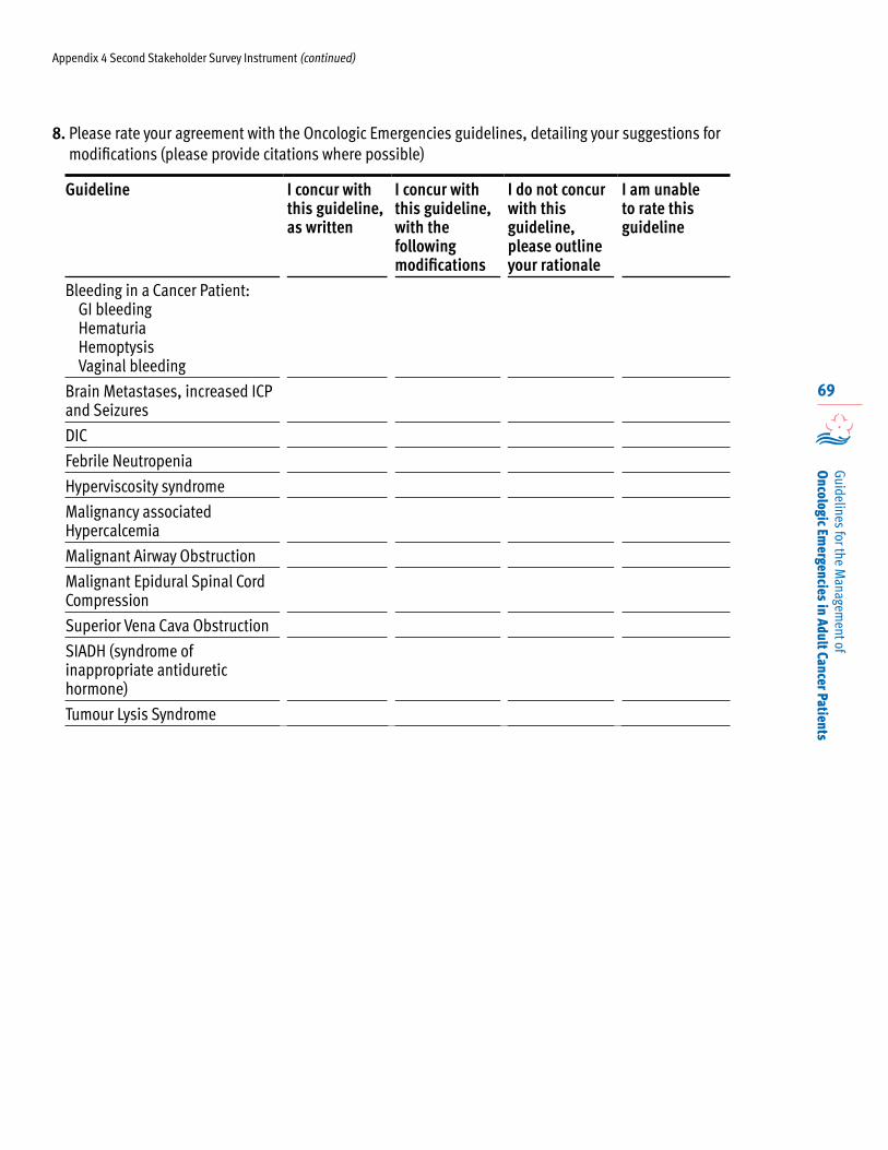
69
Guidelines for the Managem
ent of O
ncologic Emergencies in Adult Cancer Patients
8. Please rate your agreement with the Oncologic Emergencies guidelines, detailing your suggestions for modifications (please provide citations where possible)
Guideline I concur with this guideline, as written
I concur with this guideline, with the following modifications
I do not concur with this guideline, please outline your rationale
I am unable to rate this guideline
Bleeding in a Cancer Patient:GI bleedingHematuriaHemoptysisVaginal bleeding
Brain Metastases, increased ICP and Seizures
DIC
Febrile Neutropenia
Hyperviscosity syndrome
Malignancy associated Hypercalcemia
Malignant Airway Obstruction
Malignant Epidural Spinal Cord Compression
Superior Vena Cava Obstruction
SIADH (syndrome of inappropriate antiduretic hormone)
Tumour Lysis Syndrome
Appendix 4 Second Stakeholder Survey Instrument (continued)
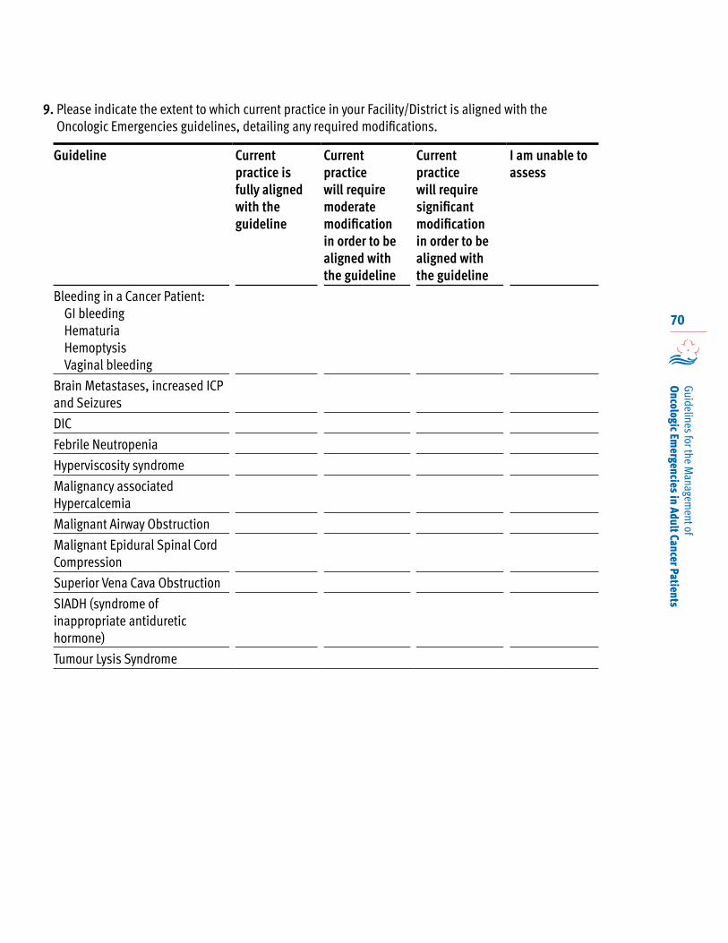
70
Guidelines for the Managem
ent of O
ncologic Emergencies in Adult Cancer Patients
9. Please indicate the extent to which current practice in your Facility/District is aligned with the Oncologic Emergencies guidelines, detailing any required modifications.
Guideline Current practice is fully aligned with the guideline
Current practice will require moderate modification in order to be aligned with the guideline
Current practice will require significant modification in order to be aligned with the guideline
I am unable to assess
Bleeding in a Cancer Patient:GI bleedingHematuriaHemoptysisVaginal bleeding
Brain Metastases, increased ICP and Seizures
DIC
Febrile Neutropenia
Hyperviscosity syndrome
Malignancy associated Hypercalcemia
Malignant Airway Obstruction
Malignant Epidural Spinal Cord Compression
Superior Vena Cava Obstruction
SIADH (syndrome of inappropriate antiduretic hormone)
Tumour Lysis Syndrome
71
Guidelines for the Managem
ent of O
ncologic Emergencies in Adult Cancer Patients
10. Please highlight any barriers your Facility/District may encounter in aligning practice with the guidelines and note the resources/actions required to overcome the barriers
Guideline Implementation Barriers Resources/Actions required to overcome the barriers
Bleeding in a Cancer Patient:GI bleedingHematuriaHemoptysisVaginal bleeding
Brain Metastases, increased ICP and Seizures
DIC
Febrile Neutropenia
Hyperviscosity syndrome
Malignancy associated Hypercalcemia
Malignant Airway Obstruction
Malignant Epidural Spinal Cord Compression
Superior Vena Cava Obstruction
SIADH (syndrome of inappropriate antiduretic hormone)
Tumour Lysis Syndrome
11. In addition to the modifications you have noted above do you have other recommendations that would enhance the guidelines?