Guidelines for the interpretation of the neonatal electrocardiogram
16
European Heart Journal (2002) 23, 1329–1344 doi:10.1053/euhj.2002.3274, available online at http://www.idealibrary.com on Task Force Report Guidelines for the interpretation of the neonatal electrocardiogram A Task Force of the European Society of Cardiology P. J. Schwartz 1 (Chair), A. Garson, Jr 2 , T. Paul 3 , M. Stramba-Badiale 4 , V. L. Vetter 5 , E. Villain 6 and C. Wren 7 1 Department of Cardiology, University of Pavia and IRCCS Policlinico S. Matteo, Pavia, Italy; 2 University of Virginia, Charlottesville, VA, U.S.A.; 3 The Children’s Heart Program of South Carolina, Medical University of South Carolina, Charleston, SC, U.S.A.; 4 Pediatric Arrhythmias Center, IRCCS Istituto Auxologico Italiano, Milan, Italy; 5 Division of Pediatric Cardiology, Department of Pediatrics, Children’s Hospital of Philadelphia, University of Pennsylvania School of Medicine, Philadelphia, PA, U.S.A.; 6 Division of Pediatric Cardiology, Department of Pediatrics, Ho ˆpital Necker Enfants Malades, Paris, France; 7 Department of Paediatric Cardiology, Freeman Hospital, Newcastle upon Tyne, U.K. Introduction.............................................................1329 Normal electrocardiogram in the newborn .............1330 Normal values......................................................1330 Technology ..........................................................1330 Artefacts...............................................................1332 Electrocardiographic measurements ....................1332 Heart rate.............................................................1332 P wave..................................................................1332 QRS complex .......................................................1332 QT interval ..........................................................1333 ST segment and T wave ......................................1333 Abnormal electrocardiogram in the newborn .........1333 Heart rate.............................................................1333 Sinus arrhythmia ..............................................1333 Sinus tachycardia .............................................1333 Sinus bradycardia.............................................1335 Other bradycardias...........................................1335 P wave..................................................................1335 Atrioventricular conduction.................................1335 Complete (3rd) atrioventricular block .............1335 1st and 2nd atrioventricular block ...................1336 Intraventricular conduction .................................1336 Bundle branch block ........................................1336 Non-specific intraventricular conduction abnormalities ....................................................1336 Wolff–Parkinson–White syndrome ..................1336 QRS axis and amplitude ......................................1338 Right ventricular hypertrophy .........................1338 Left ventricular hypertrophy ............................1338 Low QRS voltage.............................................1338 Ventricular repolarization ....................................1338 QT prolongation: differential diagnosis ...........1339 Long QT syndrome ..........................................1339 ST segment elevation .......................................1341 Atrial and ventricular arrhythmias ......................1341 Atrial/junctional ...............................................1341 Premature atrial beats ..................................1341 Supraventricular tachycardia........................1342 Atrial flutter .................................................1342 Ventricular arrhythmias ...................................1342 Premature ventricular beats..........................1342 Ventricular tachycardia ................................1343 Accelerated ventricular rhythm ....................1343 Conclusion ...............................................................1343 Acknowledgements ..................................................1343 References ................................................................1343 Introduction Most cardiologists who care for adults have no or minimal experience with electrocardiograms (ECGs) recorded in infants. So far, this has had no practical implications because only seldom are they requested to examine a neonatal ECG. This situation, however, may change as some European countries have begun to consider the possibility of introduc- ing in their National Health Services the performance of an ECG during the first month of life in all newborns, as part of a cardiovascular screening programme. Correspondence: Peter J. Schwartz, MD, FESC, FACC,, FAHA, Professor & Chairman, Department of Cardiology, Policlinico S. Matteo IRCCS, Viale Golgi, 19-27100 Pavia, Italy. 0195-668X/02/$35.00 2002 Published by Elsevier Science Ltd on behalf of The European Society of Cardiology
Transcript of Guidelines for the interpretation of the neonatal electrocardiogram

European Heart Journal (2002) 23, 1329–1344doi:10.1053/euhj.2002.3274, available online at http://www.idealibrary.com on
Task Force Report
Guidelines for the interpretation of the neonatalelectrocardiogram
A Task Force of the European Society of Cardiology
P. J. Schwartz1 (Chair), A. Garson, Jr2, T. Paul3, M. Stramba-Badiale4,V. L. Vetter5, E. Villain6 and C. Wren7
1Department of Cardiology, University of Pavia and IRCCS Policlinico S. Matteo, Pavia, Italy; 2University ofVirginia, Charlottesville, VA, U.S.A.; 3The Children’s Heart Program of South Carolina, Medical University ofSouth Carolina, Charleston, SC, U.S.A.; 4Pediatric Arrhythmias Center, IRCCS Istituto Auxologico Italiano,Milan, Italy; 5Division of Pediatric Cardiology, Department of Pediatrics, Children’s Hospital of Philadelphia,University of Pennsylvania School of Medicine, Philadelphia, PA, U.S.A.; 6Division of Pediatric Cardiology,
Department of Pediatrics, Hopital Necker Enfants Malades, Paris, France; 7Department of Paediatric Cardiology,Freeman Hospital, Newcastle upon Tyne, U.K.
Correspondence: Peter J. Schwartz, MD, FESC, FACC,, FAHA,Professor & Chairman, Department of Cardiology, Policlinico S.Matteo IRCCS, Viale Golgi, 19-27100 Pavia, Italy.
0195-668X/02/$35.00 � 2002 Published by
Introduction
Most cardiologists who care for adults have noor minimal experience with electrocardiograms(ECGs) recorded in infants. So far, this has had nopractical implications because only seldom are theyrequested to examine a neonatal ECG. This situation,however, may change as some European countrieshave begun to consider the possibility of introduc-ing in their National Health Services the performanceof an ECG during the first month of life in allnewborns, as part of a cardiovascular screeningprogramme.
Introduction.............................................................1329Normal electrocardiogram in the newborn .............1330
Normal values......................................................1330Technology ..........................................................1330Artefacts...............................................................1332Electrocardiographic measurements ....................1332Heart rate.............................................................1332P wave..................................................................1332QRS complex.......................................................1332QT interval ..........................................................1333ST segment and T wave ......................................1333
Abnormal electrocardiogram in the newborn .........1333Heart rate.............................................................1333
Sinus arrhythmia..............................................1333Sinus tachycardia .............................................1333Sinus bradycardia.............................................1335Other bradycardias...........................................1335
P wave..................................................................1335Atrioventricular conduction.................................1335
Complete (3rd) atrioventricular block .............13351st and 2nd atrioventricular block...................1336
Intraventricular conduction .................................1336Bundle branch block ........................................1336Non-specific intraventricular conductionabnormalities....................................................1336Wolff–Parkinson–White syndrome ..................1336
QRS axis and amplitude......................................1338Right ventricular hypertrophy .........................1338
E
Left ventricular hypertrophy............................1338Low QRS voltage.............................................1338
Ventricular repolarization....................................1338QT prolongation: differential diagnosis ...........1339Long QT syndrome..........................................1339ST segment elevation .......................................1341
Atrial and ventricular arrhythmias......................1341Atrial/junctional ...............................................1341
Premature atrial beats ..................................1341Supraventricular tachycardia........................1342Atrial flutter .................................................1342
Ventricular arrhythmias ...................................1342Premature ventricular beats..........................1342Ventricular tachycardia ................................1343Accelerated ventricular rhythm ....................1343
Conclusion...............................................................1343Acknowledgements ..................................................1343References................................................................1343
lsevier Science Ltd on behalf of The European Society of Cardiology

1330 Task Force Report
The background of this evolution is multiple, but itlies largely in the realization that early identificationof life-threatening arrhythmogenic disorders, whichoften manifest in infancy, childhood or even later,may allow initiation of effective preventive therapy. Alarge prospective study has indicated that some in-fants with prolonged QT interval in the first week oflife had sudden death, and would have previouslybeen labelled as victims of the Sudden Infant DeathSyndrome[1]. Furthermore, in infants with this diag-nosis, post-mortem molecular screening may revealthe presence of the long QT syndrome (LQTS)[2]. Aswith most screening tests, a single ECG must be putinto context (e.g. family history, etc.). Additionally, itis traditional to examine neonatal ECGs looking forthose with parameters below the 2nd or exceedingthe 98th percentile. While it is true that thesevalues are ‘abnormal’ in a strict statistical sense, veryoften ‘abnormality’ does not imply the presence of adisease, or of a risk for clinically relevant events. Thisdepends largely on the parameter under examination.However, also the reverse may be true and, in theneonate, a completely normal ECG may be seen withmultiple types of congenital heart defects and with theentire spectrum of arrhythmias. This call for cautiondoes not detract from the valid concept that theidentification of ECG abnormalities in the newborncan be the first step toward a meaningful act ofpreventive medicine.
Should this neonatal screening indeed be intro-duced as part of National Health Services, then hos-pital cardiologists — most of whom are unfamiliarwith neonatal ECGs — would be asked to read thesetracings. The European Society of Cardiology (ESC)has realized the potential implications for Europeancardiologists and for health care, and has acted ac-cordingly. Through the Committee for PracticeGuidelines and Policy Conferences, chaired byWerner Klein, it has instituted this Task Force. Theexperts were designated by the Guidelines Committeeand approved by the Board of the ESC. The panelwas composed of physicians and scientists involved inclinical practice in University and non-University hos-pitals. Members were selected to represent experts ofdifferent European countries; in addition, two non-European members were included for their worldwiderecognized expertise in the field of pediatric electro-cardiography.
The ESC considers medical education and theimprovement of clinical practice among its majorobligations. The main objective of the present reportis to present adult cardiologists with a consensusdocument designed to provide guidelines for theinterpretation of the neonatal ECG, focusing onthe most clinically relevant abnormalities and on theensuing management and referral options. Thisdocument aims also at providing paediatricians andneonatologists with updated information of clinicalrelevance that can be detected from a neonatalECG.
Eur Heart J, Vol. 23, issue 17, September 2002
The procedure used for developing and issuing theseguidelines was in accordance with the recently issued‘Recommendations for Task Force creation and reportwriting’, (http://www.escardio.org/scinfo/guidelines_recommendations.htm) which is a position document ofthe ESC Committee for Practice Guidelines and PolicyConferences.
This document was reviewed and approved by theCommitee for Practice Guidelines and Policy Confer-ences. It was endorsed by the Board of the ESC andrepresents the official position of the ESC with regard tothis subject. These guidelines will be reviewed two yearsafter publication and considered as current unless the‘Guidelines’ Committee revises or withdraws them fromcirculation.
This Task Force was financed by the budget of theCommittee for Practice Guidelines and Policy Confer-ences of the ESC and was independent of any commer-cial, health or governmental authorities.
Normal electrocardiogram in thenewborn
Normal values
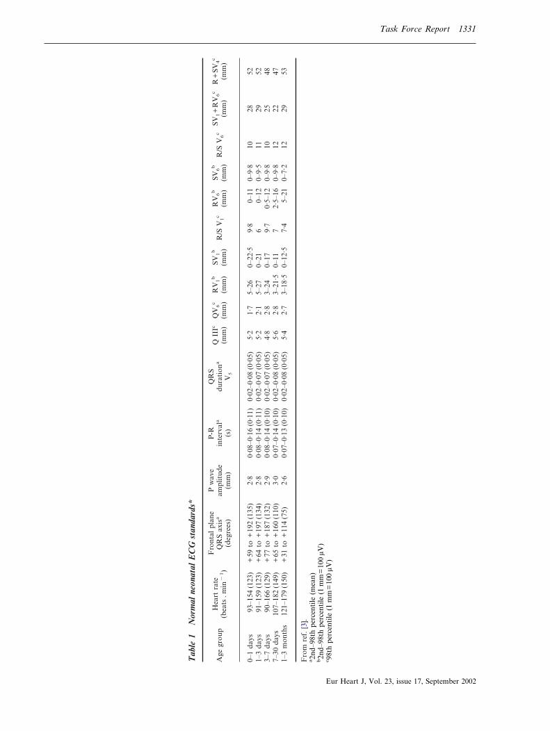
Changes occur in the normal ECG from birth to adultlife. They relate to developmental changes in physiology,body size, the position and size of the heart relative tothe body, and variations in the size and position of thecardiac chambers relative to each other. The majorchanges in the paediatric ECG occur in the first year oflife with the majority of normal adult values beingabnormal in the newborn. Likewise, many normal new-born values and patterns would be abnormal in theadult. Normal electrocardiographic values in the paedi-atric population traditionally derive from those pub-lished in 1979 by Davignon et al.[3]. From the ECGs of1027 infants less than 1 year of age and among these, 668in the first month of life, the percentile distribution ofelectrocardiographic variables was calculated. It is im-portant to refer to tables of normal values as shown inTable 1. A recent large study by Rijnbeek et al.[4]
included an extremely low number of neonates (n=44)but no one below 3 weeks of age. Thus, the percentiletables published by Davignon are recommended for usein clinical practice. Other references on the reading ofECGs in neonates and children are available[5,6].
Technology
The normal newborn ECG should include 12 leads.Other leads, V3R, V4R and V7, may provide additionalinformation to evaluate possible congenital heartlesions.
The current use of computerized digital ECG systemsaffects newborn ECGs to a greater extent than those ofolder children or adults[7]. The newborn ECG may have
Task Force Report 1331
Tab
le1
Nor
mal
neon
atal
EC
Gst
anda
rds*
Age
grou
pH
eart
rate
(bea
ts.m
in�
1)
Fro
ntal
plan
eQ
RS
axis
a
(deg
rees
)
Pw
ave
ampl
itud
e(m
m)
P-R
inte
rval
a
(s)
QR
Sdu
rati
ona
V5
QII
Ic
(mm
)Q
V6
c
(mm
)R
V1
b
(mm
)SV
1b
(mm
)R
/SV
1c
RV
6b
(mm
)SV
6b
(mm
)R
/SV
6c
SV1+
RV
6c
(mm
)R
+SV
4c
(mm
)
0–1
days
93–1
54(1
23)
+59
to+
192
(135
)2·
80·
08–0
·16
(0·1
1)0·
02–0
·08
(0·0
5)5·
21·
75–
260–
22·5
9·8
0–11
0–9·
810
2852
1–3
days
91–1
59(1
23)
+64
to+
197
(134
)2·
80·
08–0
·14
(0·1
1)0·
02–0
·07
(0·0
5)5·
22·
15–
270–
216
0–12
0–9·
511
2952
3–7
days
90–1
66(1
29)
+77
to+
187
(132
)2·
90·
08–0
·14
(0·1
0)0·
02–0
·07
(0·0
5)4·
82·
83–
240–
179·
70·
5–12
0–9·
810
2548
7–30
days
107–
182
(149
)+
65to
+16
0(1
10)
3·0
0·07
–0·1
4(0
·10)
0·02
–0·0
8(0
·05)
5·6
2·8
3–21
·50–
117
2·5–
160–
9·8
1222
471–
3m
onth
s12
1–17
9(1
50)
+31
to+
114
(75)
2·6
0·07
–0·1
3(0
·10)
0·02
–0·0
8(0
·05)
5·4
2·7
3–18
·50–
12·5
7·4
5–21
0–7·
212
2953
Fro
mre
f.[3
].a2n
d–98
thpe
rcen
tile
(mea
n)b2n
d–98
thpe
rcen
tile
(1m
m=
100
�V)
c 98th
perc
enti
le(1
mm
=10
0�V
)
Eur Heart J, Vol. 23, issue 17, September 2002
1332 Task Force Report
a higher voltage and shorter duration QRS complexesresulting in a higher percentage of high frequency com-ponents. The recommendations of a number of groupsvary as to the best bandwidth cutoffs and samplingfrequency to reduce error[8,9]. Higher bandwidth cutoffsmay alter amplitude of signals by as much as 46%[10].This would make standards determined from analoguesignals or digitized signals at lower sampling rates andlower frequency cutoffs different from those at highersettings. The current American Heart Association rec-ommendation for paediatric ECGs is 150 Hz as a mini-mum bandwidth cutoff and 500 Hz as a minimumsampling rate[11]. The Rijnbeek study reported normalsusing a higher sampling rate of 1200 Hz. Compared toDavignon’s study, which used a sampling rate of 333 Hz,the newborn upper limits in Rijnbeek’s study were12–25% higher than in Davignon’s[3,4].
Artefacts
Artefacts are common in newborn ECGs and includelimb lead reversal and incorrect chest lead positioning.In addition, electrical interference, usually 60 cycles,can occur in hospital settings from bedside monitors,warmers or other equipment.
Other artefacts occur because of various types ofpatient movement common in neonates. These artefactsmay be random as with hiccoughs or limb movement.Normal complexes are seen along with the artefacts, andthe intrinsic rhythm of the patient is not affected. Othercommon artefacts include a fine, often irregular undu-lation of the baseline from muscle tremors or jitteriness.Again, the intrinsic rhythm is not affected. The size ofthe QRS complex and the baseline may wander in acyclic fashion with respirations. It should be noted thatthe neonate breathes from 30–60 times per min.
The main clue in determining the presence of anartefact is to evaluate whether it affects the intrinsicrhythm and if it is timed such that it could be a truedepolarization. A signal within 80 ms from a true QRScomplex could not occur from an electrophysiologicpoint of view.
Electrocardiographic measurements
Because of the current limitations of electronic measure-ments in newborn ECGs, intervals should be handmeasured as the computerized systems are often inaccu-rate in the newborn. Intervals in children increase withincreasing age, reaching most of the adult normal valuesby 7–8 years of age.
Heart rate
Heart rate can be determined by a variety of methods. Itshould be noted that normal neonates may have rates
Eur Heart J, Vol. 23, issue 17, September 2002
between 150–230 beats . min�1, especially if they arecrying or agitated. Over 200 beats . min�1, one-halfsmall box can make an appreciable difference in heartrates.
Heart rates between the 2nd and 98th percentile in thefirst year of life are shown in Table 1. The normal heartrate increases from the first day of life, it reaches a peakbetween the first and the second month and then de-clines returning to the values recorded at birth by thesixth month. During the following 6 months, it remainsrather stable and then slowly declines after 1 year due tomaturation of vagal innervation of the sinus node[12].Clinically significant gender differences in heart rate arenot seen in the neonatal period.
P wave
The P wave axis is a vector indicating the direction ofactivation, which is away from the site of origin. Byidentifying the quadrant location of the P wave axis onecan determine the site of origin of the rhythm. Forexample, sinus rhythm originates in the high rightatrium transcribing a P wave with an axis in the quad-rant bordered by 0 and +90�. Measurements are avail-able for P wave amplitude (Table 1). The P wave isgenerally pointed in lead II and aVF and more roundedin other leads. Lead V1 may be diphasic.
The PR interval is measured from the onset of the Pwave to the Q or R wave if no Q wave is present. The PRinterval, measured in lead II, increases with age anddecreases with heart rate. The normal neonatal PRinterval ranges from a minimum of 70 ms to a maximumof 140 ms, with a mean of 100 ms.
QRS complex
The normal full-term neonate has an axis between 55�and 200� but by 1 month, the normal upper limit hasfallen to 160� or less. Although one might identify anaxis of 120� as right axis deviation in an adult, it is anormal finding in a newborn. The QRS axis in thepremature newborn ECG ranges between 65� and 174�.
The duration of the QRS complex is measured fromthe beginning to the end of the ventricular depolariz-ation complex and it should be measured in a lead withan initial Q wave[5]. QRS duration in the newborn andinfant is narrow (<80 ms). Normal QRS duration in-creases with age. Normal values for QRS complexduration in lead V5 are displayed in Table 1.
QRS morphology in the newborn may have morenotches and direction changes than seen in older chil-dren or adults. The direction of the Q wave in theprecordial or horizontal plane indicates the direction ofseptal depolarization. Normally, there is a Q wave inleads V5–V6 indicating depolarization from left to right.Normal values of Q wave amplitudes vary with the leadand with age. Q wave amplitudes may be as high
Task Force Report 1333
as 0·55 mV in lead III or 0·33 mV in aVF at 1 month.Q wave duration >30 ms is abnormal. The appearanceof secondary r waves (r� or R�) in the right chest leadsis frequent in normal neonates.
Davignon et al.[3] provided ‘normal’ values in infants.The use of 2nd and 98th percentiles to define normalityimplies that 4% of the population are ‘abnormal’ for anygiven single measurement, so ‘normal’ ranges have to beinterpreted with caution (Table 1). Thomaidis et al.published normal voltages from healthy term andpremature neonates[13].
QT interval
The QT interval is the interval between the beginning ofthe QRS complex and the end of the T wave. The QTmeasurement should be made in leads II, V5, and V6
with the longest value being used. The main difficulty liesin identifying correctly the point where the descendinglimb of the T wave intersects the isoelectric line. Due tothe fast heart rate of infants the P wave may besuperimposed on the T wave, particularly when the QTinterval is prolonged. In this case, the end of the T waveshould be extrapolated by drawing a tangent to thedownslope of the T wave and considering its intersectionwith the isoelectric line.
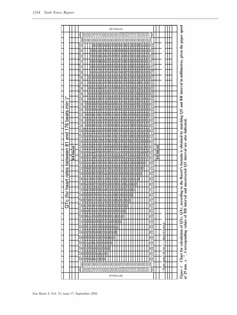
The QT interval duration changes with rate and it isusually corrected (QTc) by using Bazett’s formula. Cor-rection of the QT interval requires a stable sinus rhythmwithout sudden changes in the RR interval. QTc is equalto QT interval in seconds divided by the square root ofthe preceding RR interval in seconds. To avoid time-consuming calculations, a simple chart (Fig. 1) wherethe value of QTc is easily obtained by matching QT andRR interval in millimetres (given the paper speed at25 mm . s�1) has been produced. When heart rate isparticularly slow or fast the Bazett’s formula may not beaccurate in the correction but it remains the standard forclinical use.
The mean QTc on the 4th day of life is 400�20 ms[1]
and, at variance with the adult, no gender differences arepresent[14]. Therefore, the upper normal limit of QTc (2standard deviations above the mean, corresponding tothe 97·5 percentile) is 440 ms. By definition, 2·5% ofnormal newborns are expected to have a QTc greaterthan 440 ms. In healthy infants there is a physiologicalprolongation of QTc by the second month (mean410 ms) followed by a progressive decline[15], so that bythe sixth month QTc returns to the values recorded inthe first week.
Pitfalls with QT measurement. Despite its apparentsimplicity the measurement of the QT interval is fraughtwith errors. The simple fact that a small square on theECG paper is equivalent to 40 ms explains why healthyscepticism should accompany claims of clinical impor-tance attached to very small degrees of ‘QT prolon-gation’. An attempt should be made to measure with
10 ms (1/4 of a mm) while we recognize that this may bewithin measurement error.
ST segment and T wave
ST segment elevations >1 mm above the isoelectric lineare uncommon in the newborn. In neonates and infantsit is better to consider as the isoelectric line the TPsegment instead of the PQ segment. T waves are nor-mally quite variable in the first week of life. After1 week, the T wave is negative in lead V1 and positivein V5–V6.
Abnormal electrocardiogram in thenewborn
Heart rate
Sinus arrhythmiaSince sinus arrhythmia is less pronounced at fast heartrate, neonates show a more regular rhythm than youngchildren and adolescents, particularly in the first week oflife. Sinus arrhythmia should be differentiated fromwandering pacemaker, which manifests itself with agradual change of P wave axis and morphology and thatis due to a shift of the pacemaker from the sinus nodeto the atrium and the atrioventricular (AV) junction.Although wandering pacemaker may accompany othertypes of bradyarrhythmia, it has no pathologic meaning.Work-up
No work-up should be necessary unless significantbradycardia coexists.
Sinus tachycardiaSinus tachycardia is a sinus rhythm with a heart rateabove the normal limit for age. In the newborn theupper normal limit (98th percentile) is 166 beats . min�1
in the first week and 179 beats . min�1 in the firstmonth. After the sixth month the upper normal limitdeclines to approximately 160 beats . min�1 and at1 year is 151 beats . min�1. These values have beenmeasured from ECGs recorded when infants were awakeand quiet. It has to be noted that newborn infants maytransiently reach a heart rate up to 230 beats . min�1.
Causes. Sinus tachycardia may be a sign of any con-dition associated with an increase of cardiac output. Themost frequent causes of sinus tachycardia in the neo-natal period are represented by fever, infection, anae-mia, pain, and dehydration (hypovolaemia). Othercauses of sinus tachycardia include neonatal hyper-thyroidism and myocarditis, particularly when it is notproportionate to the level of fever. Myocarditis isusually, but not necessarily, associated with other clini-cal signs, such as gallop rhythm, or ECG abnormalities,including T wave changes and conduction disturbances.
Eur Heart J, Vol. 23, issue 17, September 2002
1334 Task Force Report
Eur Heart J, V
Fig
ure
1C
hart
for
calc
ulat
ion
ofQ
Tc.
QT
c,ac
cord
ing
toth
eB
azet
t’s
form
ula
isob
tain
edby
mat
chin
gQ
Tan
dR
Rin
terv
alin
mill
imet
res,
give
nth
epa
per
spee
dat
25m
m.s
�1 .
Cor
resp
ondi
ngva
lues
ofR
Rin
terv
alan
dun
corr
ecte
dQ
Tin
terv
alar
eal
soin
dica
ted.
ol. 23, issue 17, September 2002

Task Force Report 1335
Finally, several drugs that are commonly used duringinfancy, e.g. beta adrenergic agonists or theophyllin,may induce sinus tachycardia. In the newborn, thesemay have been transmitted across the placenta orthrough breast milk.Work-up
The evaluation of these patients should be performedaccording to the underlying condition. If myocarditis issuspected an echocardiogram should be performed. Appro-priate acute treatment of causes of tachycardia may beconsidered. Persistence of elevated rates should be furtherevaluated.
Sinus bradycardiaSinus bradycardia is defined as a sinus rhythm witha heart rate below the normal limit. In the neonatalperiod the lower normal limit (2nd percentile) is91 beats . min�1 during the first week and107 beats . min�1 in the first month of life. At the firstmonth the lower limit increases to 121 beats . min�1 anddeclines to approximately 100 beats . min�1 in the fol-lowing months. At 1 year the lower normal limit is89 beats . min�1. These values apply to an ECG re-corded in the awake state when heart rate is measuredover two respiratory cycles.
Causes. Central nervous system abnormalities, hypo-thermia, hypopituarism, increased intracranial pressure,meningitis, drugs passed from the mother to infant,obstructive jaundice, and typhoid fever represent causesof sinus bradycardia. As a consequence when sinusbradycardia is present on the surface ECG such con-ditions should be excluded. Hypothyroidism is anothercause of bradycardia and is often associated with theso-called ‘mosque sign’, a dome-shaped symmetric Twave in the absence of a ST segment. Transient sinusbradycardia has been observed in newborns from anti-Ro/SSA positive mothers, especially women with lupuserythematosus or other connective diseases.
A lower than normal heart rate has been described inpatients affected by LQTS, a phenomenon which isevident in the neonatal period[16]. It may sometimesrepresent the first sign of the disease during the foetalperiod[17].Work-up
24-h Holter monitoring may be helpful for furtherevaluation when a heart rate below 80–90 beats . min�1 ispresent on surface ECG during infancy. Evaluation forunderlying conditions should be performed.
Other bradycardiasSinus pauses in newborns may last from 800 to 1000 ms.Pauses >2 s are abnormal. Sinus pauses may be followedby escape beats which arise from the atria or from theAV-junction. It has to be noted that even healthyneonates may show periods of junctional rhythm, i.e. asequence of narrow QRS complexes in the absence ofpreceding P waves.
Causes. Infants with autonomic nervous system dysfunc-tion consisting of augmented vagal tone may have sinus
bradycardia, or significant sinus pauses of severalseconds. These generally occur during feeding, sleep,defecation, or other times of increased vagal tone.
Apparent life-threatening events (ALTE), described asloss of consciousness accompanied by pallor and hypo-tonia, have been related to vagal overactivity which maymanifest as sinus pauses or abrupt bradycardia. ALTEmay be associated with apneic episodes, or gastro-esophageal reflux, that may precede severe bradycardia.
Infants with LQTS not only tend to have sinusbradycardia but may also have sinus pauses.Work-up
24-h Holter monitoring may be useful for the assess-ment of significant bradycardia. Long pauses secondary toexcessive vagal tone may be eliminated by the use ofatropine, and rarely require pacemakers. Treatment ofother underlying diseases should be undertaken.
P wave
Abnormal P waves may be seen in infants with atrialenlargement or non-sinus origin of the P wave. Ectopicatrial rhythms originate most commonly from the lowright atrium (0 to �90�), high left atrium (+90 to+180�) or the low left atrium (+180 to +270�).
Right atrial enlargement and/or hypertrophy typicallyproduces increased P wave amplitude with a normal Pwave duration. The P wave axis usually remains normalso the effect is usually best seen in lead II.
Left atrial enlargement and/or hypertrophy typicallyproduces an increased and prolonged negative terminaldeflection of the P wave in lead V1 (generally accepted as>40 ms in duration and 0·1 mV in amplitude). Left atrialenlargement also causes exaggerated notching of the Pwave in lead II although this is not a specific sign.Work-up
An echocardiogram should be performed when clinicallyindicated.
Atrioventricular conduction
During atrial tachycardia, it is possible to observe 1/1conduction through the atrio-ventricular node at ratesover 300 beats . min�1.
Complete (third degree) atrioventricular blockComplete AV block implies complete absence of con-duction from atrium to ventricle. ECG shows normalatrial activation and slower dissociated regular QRScomplexes. Congenital complete block is observed incomplex congenital heart malformations[18].
Approximately one out of every 15 000 to 20 000 livebirths results in a baby with isolated AV block. Theassociation between isolated neonatal AV block andmaternal connective tissue disease is well established andascribed to the presence of anti Ro/SSA and La-SSBantibodies in the mothers. Nearly every mother with anaffected child has circulating antibodies. However, only2 to 5% of women with known antibodies will have a
Eur Heart J, Vol. 23, issue 17, September 2002
1336 Task Force Report
first child with AV block[19]. Mortality rate in patientswith neonatal AV block is still high, especially duringthe first 3 months of life[20]. Acquired complete AVblock is rare in neonates. It is mainly infective (viralmyocarditis, HIV infection) or may be related totumours.
First and second degree atrioventricular blockNeonates may present with first or second degree AVblock and rare reports exist demonstrating progressionto complete AV block after birth in children with andwithout antibody mediated conduction disorders[21].
Long QT syndrome is occasionally complicated byimpaired atrioventricular conduction, mostly 2:1 AVblock[22,23]. Functional AV block can be observed inneonates because they have a fast atrial rate and the Pwave falls within the very prolonged T wave. Cases ofinfra-Hisian block location at the His-Purkinje levelhave been demonstrated[24,25]. In spite of different treat-ment modes including the use of high doses of beta-blockers and pacing, there is still significant mortality.
Heart block associated with prolonged QT intervalhas been described in neonates and infants receivingcisapride. Second degree AV block due to QT intervalprolongation has been also reported with the use ofother agents such as diphemanil[26] or doxapram inpremature infants.Work-up
In neonates and infants with AV conduction abnormali-ties clinical history of autoimmune disease and plasmatitres of maternal antibodies (anti Ro/SSA and antiLa/SSB) should be performed. When neonates have abnormalAV nodal conduction without maternal antibodies, anECG should also be performed on the parents and siblings(see intraventricular abnormalities). Neonates with firstdegree AV block should be followed with additional ECGsin the following months. Neonates and infants with secondor third degree AV block need a complete paediatriccardiologic work-up, including an echocardiogram. Theonly effective treatment of congenital complete AV blockin neonates with symptoms or a low ventricular escaperhythm is permanent artificial pacing.
Intraventricular conduction
Bundle branch blockCongenital isolated complete right (RBBB) and leftbundle branch block are very rare in neonates. Southallet al. found only one case of complete RBBB in apopulation of 3383 apparently healthy newborn in-fants[27]. The classical ECG in Ebstein’s anomaly of thetricuspid valve displays a prolonged PR interval and awide RBBB. Left anterior fascicular block is found inassociation with congenital heart malformations such asatrio-ventricular canal defects and tricuspid atresia. Insevere cardiomyopathy, interruption of the left bundle,which results from the involvement of the left ventricleand/or its conduction system, has been reported andcarries a poor prognosis[28].
Eur Heart J, Vol. 23, issue 17, September 2002
Hereditary bundle branch block is an autosomaldominant genetic disease that was mapped in somefamilies to the long arm of chromosome 19[29,30]. Af-fected individuals have various combinations of conduc-tion defects such as RBBB, left or right QRS axisdeviation or AV block; the r� pattern may as well be theprelude to a conduction block. Abnormalities have beendescribed in patients as young as 15 days.
Non-specific intraventricular conduction abnormalitiesNon-specific intraventricular conduction abnormalitiesare very rare in neonates and infants with normal heartstructures[31]. They may be a manifestation of inflamma-tion in myocarditis or endocarditis.Work-upNeonates and infants with intraventricular conductionabnormalities need a complete paediatric cardiologicwork-up. Evaluation of possible underlying causes shouldbe performed. An ECG should also be performed on theparents and siblings.
Wolff–Parkinson–White syndromeThe anatomical substrate of preexcitation in Wolff–Parkinson–White (WPW) syndrome is a direct muscularconnection between the atria and ventricles. Since acces-sory pathways rarely show decremental conduction, theelectrical impulse is conducted prematurely to the ven-tricles resulting in a short PR interval. Conductionthrough the atrioventricular node and the accessorypathway results in collision of two electrical wavefrontsat the ventricular level causing a delta wave and a fusionQRS complex with prolonged duration.
The diagnosis of preexcitation is solely based on thefindings of the surface ECG (Fig. 2). Intermittent pre-excitation is not uncommon in newborns and infants.Depending on the location of the accessory pathway aswell as the conduction properties of the atrioventricularnode, even continuous preexcitation may be subtle andonly detected in the mid-precordial leads. A study innewborns indicated a high prevalence of WPW syn-drome when two of the four following characteristicswere noted: PR interval �100 ms, QRS complex dur-ation �80 ms, lack of a Q wave in V6 and left axisdeviation[32]. Short PR intervals are also observed inmannosidosis, Fabry’s disease, and Pompe’s disease[5]. Acommon cause of a short PR interval in a normal heartis a low right atrial pacemaker. In this instance, the Pwave is negative in lead aVF and positive or isoelectricin lead I. The intraatrial conduction time from the highto low right atrium is eliminated and therefore the PRinterval may be up to 40 ms less than normal.
The prevalence of WPW syndrome in the paediatricpopulation has been estimated at 0·15 to 0·3%[33] with anincidence of newly diagnosed cases of approximatelyfour per 100 000 persons per year for all age groups[34].Numbers, however, vary greatly depending upon symp-toms, age, gender and the intracardiac anatomy of thepopulation studied[35,36]. In children with structuralheart disease, the prevalence has been estimated at 0·33to 0·5%[37]. Ebstein’s anomaly of the tricuspid valve,

Task Force Report 1337
Fig
ure
2E
CG
ina
neon
ate
show
ing
subt
lesi
gns
ofW
PW
.N
ote
delt
aw
aves
inV
5an
dV
6w
ith
abse
nce
ofQ
wav
es.
Eur Heart J, Vol. 23, issue 17, September 2002

1338 Task Force Report
l-transposition of the great arteries, hypertrophic cardio-myopathy and cardiac tumours are associated with anincreased prevalence of preexcitation[36,37].
Clinical counterparts. In WPW syndrome the typicalform of paroxysmal supraventricular tachycardia (ortho-dromic) results from reentry antegradely through theatrioventricular node and retrogradely through the ac-cessory pathway. As digoxin shortens the antegradeeffective refractory period of the accessory pathway andpromotes rapid atrioventricular conduction during atrialflutter or atrial fibrillation over the pathway, the use ofdigoxin is contraindicated at any age [38,39]. Verapamilshould also be avoided as it may increase the ventricularresponse rate during atrial fibrillation in those patients,and may cause cardiovascular collapse in infants andyoung children.
The incidence of sudden death in preexcitation syn-drome during childhood has been estimated to be ashigh as 0·5%[34] and cardiac arrest may be the initialpresentation in children with preexcitation[35]. However,data on newborns and infants are lacking. One study ona series of 90 newborns and infants with WPW syn-drome and supraventricular tachycardia reported sud-den death in two patients with a normal heart duringfollow-up. Both infants, however, had been treated withdigoxin[36].
Finally, there are no sufficient data on newborns andinfants with an incidental finding of preexcitation onECG concerning the occurrence of paroxysmal supra-ventricular tachycardia later on during their life.Work-up
Congenital heart disease is more common in infants andyoung children with preexcitation, with a prevalence ashigh as 45% for infants with an ECG pattern consistentwith a right-sided accessory pathway[36]. Thus, in everyyoung patient with a preexcitation pattern on surfaceECG, a complete 2-dimensional echocardiographicwork-up is recommended to rule out any intracardiacabnormality.
Assessment of the conduction properties of the acces-sory pathway, i.e. the antegrade effective refractory periodand the shortest RR-interval with preexcitation, by trans-esophageal programmed stimulation may be useful inselected patients for risk stratification and mode oftherapy.
QRS axis and amplitude
Abnormal axis implies a mean frontal plane QRS vectoroutside the normal range and must take into account therelative right axis deviation seen in normal neonates. Leftaxis deviation is seen in a variety of abnormalities includ-ing atrioventricular septal defect, ventricular septal de-fect, tricuspid atresia, and WPW syndrome, but may beoccasionally observed in otherwise normal infants.
Right ventricular hypertrophyRight ventricular hypertrophy may be suspected from aQR complex in V , an upright T wave in V (normal in
1 1Eur Heart J, Vol. 23, issue 17, September 2002
the first week of life), increased R wave amplitude in V1,and increased S wave amplitude in V6 (according to theDavignon criteria). Sensitivity and specificity has notbeen tested in the neonate. QR patterns are commonlyseen with pressure overload congenital lesions, rSR�patterns are seen in volume overload lesions.
Left ventricular hypertrophyThe performance of the ECG in recognition of leftventricular hypertrophy is poorer than generally recog-nized and, again, has not been specifically tested inneonates. Left ventricular hypertrophy is expected toproduce increased left sided voltages. Garson[5] de-scribed the most helpful ECG signs in children as beingT wave abnormalities in leads V5 and V6, increased Rwave amplitude in V6, increased S wave amplitude in V1
(according to the Davignon criteria), and a combinationof these last two variables. Left to right shunt lesionsmay result in left ventricular hypertrophy, but this maybe in association with right ventricular hypertrophy andmanifested as biventricular hypertrophy. Left ventricu-lar hypertrophy in the newborn may be attenuated bythe normal right-sided predominance of the newborn.The normal premature heart may not have developedthe right-sided predominance, especially if <28 weeksgestation, and left ventricular predominance may bepresent.
Low QRS voltageIn the limb leads the total amplitude of R+S in eachlead �0·5 mV may be indicative of myocarditis orcardiomyopathy.Work-up
Evaluation of the underlying causes should be per-formed. An echocardiogram should be performed whenclinically indicated.
Ventricular repolarization
There is a simple reason that makes clinically importantthe analysis of ventricular repolarization abnormalities:their presence could be the harbinger of a significant riskfor life-threatening arrhythmia. It is established thatnewborns found to have a prolonged QTc (>440 ms) onthe fourth day of life have an increased risk for suddendeath[1]. Some of these sudden deaths have previouslybeen labelled as Sudden Infant Death Syndrome.
On the other hand, the presence of confoundingfactors — above all the ambiguities in theirquantification — calls for caution before making hastydiagnoses associated with need for therapy and withconsiderable parental anxiety.
Ventricular repolarization can be evaluated on thesurface ECG by measuring the QT interval duration andby analysing the morphology of the ST segment and ofthe T wave. Measurements of the QT interval should beperformed by hand.
It is important to remember that QT duration maychange over time. Accordingly, it is recommended
Task Force Report 1339
repeating the ECG in those infants found to have aprolonged QTc on the first ECG. While exceptions doexist, the more prolonged the QTc interval, the greaterthe likelihood of its clinical significance. A QTc close to500 ms implies a clear abnormality even taking intoaccount potential measurement errors.
QT interval prolongation: differential diagnosisElectrolyte disturbances are fairly common and maycause QT prolongation. Among them, hypocalcaemia(less than 7·5 mg . dl�1) usually produces a distinctivelengthening of the ST segment. Hypokalaemia andhypomagnesaemia, often encountered in infants whohave had vomiting or diarrhoea, usually decrease Twave amplitude and increase U wave amplitude. Centralnervous system abnormalities can produce QT prolon-gation and T wave inversion.
Several drugs commonly used in the neonatal periodand during infancy may induce QT interval prolon-gation; among them are macrolide antibiotics such asspyramycin[40], erythromycin, clarithromycin and alsotrimethoprim. Prokinetics such as cisapride have beenpositively linked to QT interval prolongation. All thesedrugs share one action: they block IKr, one of theionic currents involved in the control of ventricularrepolarization.
Neonates born from mothers with autoimmune dis-eases and positive for the anti-Ro/SSA antibodies mayalso show QT interval prolongation, sometimes withQTc values exceeding 500 ms[41], which tends to be
transient and to disappear by the sixth month of life,concomitantly with the disappearance of the anti Ro/SSA antibodies.
Finally, some of the neonates with QT interval pro-longation may be affected by the congenital LQTS. Thispossibility has to be carefully evaluated because of itsimplications for management.
Long QT syndromeLong QT syndrome, whose prevalence appears to beclose to 1/3000–1/5000, is characterized by the occur-rence of syncopal episodes due to torsades de pointesventricular tachycardia (VT) and by a high risk forsudden cardiac death among untreated patients[42]. Im-portantly, in 12% of patients with LQTS, sudden deathwas the first manifestation of the disease and in 4% thishappened in the first year of life[42]. This point alonemandates the treatment of all those diagnosed as af-fected, even if there are no symptoms. Long QT syn-drome is a genetic disease due to mutations of severalgenes all encoding ionic (potassium or sodium) currentsinvolved in the control of ventricular repolarization. Inmost cases, several members of the same family aregene-carriers. Low penetrance exists in LQTS, whichmeans that gene-carriers may not show the clinicalphenotype and may have a normal QT interval[43].Therefore a normal QT in the parents does not ruleout familial LQTS. In addition, approximately 30% ofcases are due to ‘de novo’ mutations which implyunaffected parents and no family history. ‘De novo’
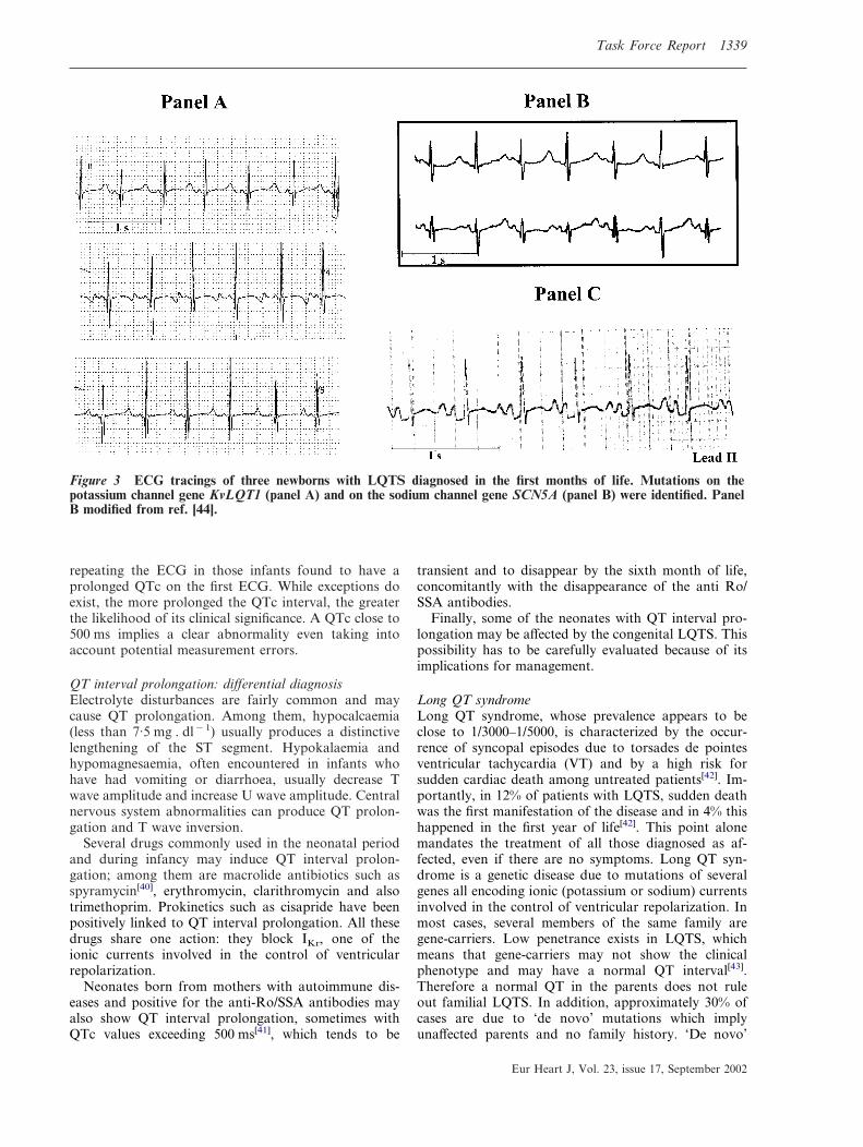
Figure 3 ECG tracings of three newborns with LQTS diagnosed in the first months of life. Mutations on thepotassium channel gene KvLQT1 (panel A) and on the sodium channel gene SCN5A (panel B) were identified. PanelB modified from ref. [44].
Eur Heart J, Vol. 23, issue 17, September 2002
1340 Task Force Report
LQTS mutations have been demonstrated in infantvictims of cardiac arrest and sudden death diagnosed asSudden Infant Death Syndrome[2,44,45]. Even thoughrelatively few LQTS patients have cardiac events duringthe first year of life, the vast majority become sympto-matic later on, either during childhood or adolescenceaccording to genetic subgroups[46]. Therefore treatmentmust continue. Beta-blockers are the first choice therapyin LQTS and are effective in preventing recurrences in80% of already symptomatic patients; different degreesof protection exist according to genetic subgroups[46]. Ifbeta-blockers are unable to prevent new cardiac events,additional drug therapy, left cardiac sympathetic dener-vation, pacemakers or the implantable cardioverterdefibrillator should be considered based on evidence,with due consideration for body size.
ECG tracings of newborns with LQTS are shownin Fig. 3.Work-up
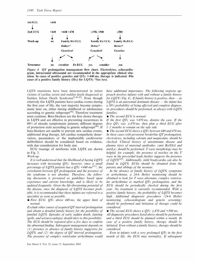
It is well understood that the likelihood of having LQTSincreases with increasing QTc; however, since a smallpercentage of LQTS patients has a QTc <440 ms[47], thecorrelation between QT prolongation and the presence ofthe syndrome is not absolute. Therefore, the follow-ing discussion is presented as guidelines based uponexperience and current knowledge, and is likely to beupdated frequently. Given the life-threatening potential ofthe disease, once the diagnosis of LQTS becomes prob-able, it is recommended that these infants are referred to aspecialist as soon as possible.� First ECG: QTc above 440 ms, the upper limit of
normal.Exclude other causes of acquired QT interval prolongationand obtain a detailed family history for the possibility offamilial LQTS. Episodes of early sudden death, faintingspells, and seizures-epilepsy should alert to this possibility.The ECG should be repeated after a few days to confirmthe abnormal finding. Subsequent management depends on(1) presence or absence of family history suggestive forLQTS, and (2) the degree of QT interval prolongation.The presence of complex ventricular arrhythmias would
Eur Heart J, Vol. 23, issue 17, September 2002
have additional importance. The following stepwise ap-proach involves infants with and without a family historyfor LQTS (Fig. 4). If family history is positive, then — asLQTS is an autosomal dominant disease — the infant hasa 50% probability of being affected and complete diagnos-tic procedures should be performed, as always with LQTSfamilies.� The second ECG is normal.If the first QTc was <470 ms, dismiss the case. If thefirst QTc was �470 ms, then plan a third ECG after1–2 months to remain on the safe side.� The second ECG shows a QTc between 440 and 470 ms.In these cases with persistent borderline QT prolongation,electrolytes, including calcium and magnesium, should bechecked. Clinical history of autoimmune disease andplasma titres of maternal antibodies (anti Ro/SSA andantiLa) should be performed. T wave morphology may behelpful; for example, the presence of notches on the Twave in the precordial leads further suggests the presenceof LQTS[48]. Additionally, mild bradycardia can also befound in LQTS. ECGs should be obtained from theparents and siblings of the neonate.
In the absence of family history of LQTS, symptomsor arrhythmias, a 24-h Holter monitoring should beobtained to look for T wave alternans, complex ventricu-lar arrhythmias or marked QTc prolongation, and theECG should be periodically checked during the firstyear. No treatment is currently recommended. With apositive family history, the probability of LQTS becomeshigh. Additional diagnostic procedures (24-h Holtermonitoring, echocardiogram and genetic screening)should be performed and initiation of therapy could beconsidered.� The second ECG shows a QTc �470 and <500 ms.All diagnostic procedures listed above should be performedand a third ECG should be planned within a month. Incase of a positive family history, therapy should beinitiated. Even without a family history, therapy should beconsidered.
Even in infants with a very prolonged QTc in the firstmonth of life, the ECG may normalize. If subsequent
Figure 4 QT prolongation management flow chart. Electrolytes, echocardio-gram, intracranial ultrasound are recommended in the appropriate clinical situ-ation. In cases of positive genetics and QTc >440 ms, therapy is indicated. #Incases of a positive family history (Hx) for LQTS; *See text.
Task Force Report 1341
ECGs and diagnostic procedures do not confirm thepresence of LQTS, it is logical to progressively withdrawtherapy and to return to periodic observations.� The second ECG shows a QTc �500 ms.Infants with a QTc �500 ms are very likely to be affectedby LQTS and to become symptomatic. All diagnosticprocedures listed above should be performed and theseinfants should be treated.
Highest risk. The presence of QTc close to 600 ms, or ofT wave alternans, or of 2:1 AV block secondary to majorQT prolongation, or of hearing loss, identify infants atextremely high risk[47,49].
ST segment elevationCauses. There are multiple causes of ST segment elev-ation in infancy. The most frequent is pericarditis. Lessfrequent causes of ST segment elevation with or withoutT wave abnormalities are hyperkalaemia, intracranialhaemorrhage, pneumothorax and pneumopericardium,subepicardial injury due to anomalous left coronaryartery or to Kawasaki disease with cardiac involvement.Whenever an anomalous left coronary artery or, morefrequently, Kawasaki disease produce an acute myocar-dial infarction, QT prolongation may also be present.
ST segment elevation with a RBBB pattern in theright precordial leads (V1–V2) is the typical finding of theBrugada syndrome, a genetic disorder associated with ahigh incidence of sudden cardiac death secondary toventricular fibrillation, in the absence of cardiac struc-tural abnormalities[50]. The ST segment elevation istypically downsloping or ‘coved’ and it is followed by anegative T wave, at variance with the early repolariz-
ation syndrome where ST segment elevation has anupward concavity, it is confined to mid-precordial leadsand is associated with a positive T wave. The diagnosisof the Brugada syndrome is made difficult by the inter-mittent nature of the ECG abnormalities, as 40% ofcases may normalize transiently. Rare cases of Brugadasyndrome have been reported during infancy[51,52].Work-up
Whenever the underlying cause has been identified, itshould be treated. If the Brugada syndrome is suspected,careful family history should be collected, 24-h Holtermonitoring obtained, and the patient should be referred toa specialist.
Atrial and ventricular arrhythmias
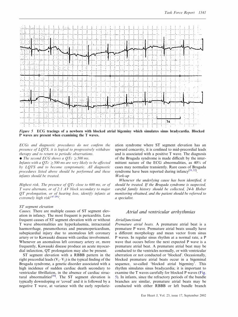
Atrial/junctionalPremature atrial beats. A premature atrial beat is apremature P wave. Premature atrial beats usually havea different morphology and mean vector from sinusP waves. In regular sinus rhythm at a normal rate, a Pwave that occurs before the next expected P wave is apremature atrial beat. A premature atrial beat may beconducted to the ventricles normally, or with ventricularaberration or not conducted or ‘blocked’. Occasionally,blocked premature atrial beats occur in a bigeminalsequence, so-called ‘blocked atrial bigeminy’. Thisrhythm simulates sinus bradycardia; it is important toexamine the T waves carefully for blocked P waves (Fig.5). In infants, since the refractory periods of the bundlebranches are similar, premature atrial beats may beconducted with either RBBB or left bundle branch
Figure 5 ECG tracings of a newborn with blocked atrial bigeminy which simulates sinus bradycardia. BlockedP waves are present when examining the T waves.
Eur Heart J, Vol. 23, issue 17, September 2002

1342 Task Force Report
aberration. It is relatively common in the same stripto see premature atrial beats conducted normally,aberrantly and blocked. It is relatively uncommon foran infant to have both premature atrial beats andpremature ventricular beats, although it does occur.Therefore, in an infant with premature P waves and wideQRS complexes on the same strip, a careful searchshould be made for a premature P wave preceding awide QRS, before making the diagnosis of both prema-ture atrial beats and premature ventricular beats on thesame strip.Work-up
In patients with frequent premature atrial beats, afollow-up ECG at 1 month may be performed. Relativelylong periods of blocked atrial bigeminy may simulate sinusbradycardia. The distinction is important since blockedatrial bigeminy is most often benign while severe sinusbradycardia may accompany systemic illness.
Supraventricular tachycardia. Supraventricular tachycar-dia (SVT) is a rapid regular tachyarrhythmia, whichresults from an abnormal mechanism originating proxi-mal to the bifurcation of the bundle of His and does nothave the morphology of atrial flutter. This definition ofSVT specifically excludes sinus tachycardia (Table 2)and premature wide QRS. The usual infant with SVThas an extremely regular R-R interval after the first10–20 beats, most often at rates greater than 230beats . min�1 and usually 260–300 per min. In 60% ofcases, the P waves are visible, but the P waves almostalways have a different morphology from sinus. In over90% of infants and children with SVT, the QRS complexis narrow and in only 3% is the QRS complex differentfrom the underlying sinus QRS. Therefore, the persistentaberration of SVT in infants is exceedingly rare, imply-ing that in the majority of infants with a QRS complexdifferent from sinus, the diagnosis is VT. In very rare
Eur Heart J, Vol. 23, issue 17, September 2002
cases of SVT in infants, there may be atrial tachycardiawith AV block or junctional tachycardia with AVdissociation.Work-up
It is important to document SVT with a 12-lead ECGbefore attempting conversion of the rhythm unless theinfant is critically ill. After sinus rhythm is achieved, theWPW pattern should be sought on a 12-lead ECG.Treatment to prevent further episodes of SVT in infancy isgenerally recommended. An echocardiogram is indicatedto determine ventricular function or the presence ofcongenital heart disease.
Atrial flutter. Atrial flutter is characterized by a rapid,regular form of atrial depolarization: the ‘flutter wave’.The picket fence morphology is similar to adults. How-ever, the flutter wave durations are generally 0·09 to0·18 s with atrial rates in infants between 300–500beats . min�1. In general, there is variable AV conduc-tion from 1:1 to 4:1 yielding an irregular ventricular rate.The QRS complex is usually the same as in sinus rhythmalthough there may be occasional aberrancy (Table 2).Due to the occasional association with WPW, thispattern should be specifically sought.
Other types of supraventricular arrhythmias such asatrial fibrillation or multifocal tachycardia are extremelyrare in the neonate.Work-up
Conversion to sinus rhythm should be attempted. Anechocardiogram is worthwhile to determine ventricular func-tion and the possible presence of congenital heart disease.
Table 2 Distinguishing tachyarrhythmias in infants
Sinus tachycardia SVT Atrial flutter VT
History Sepsis, fever, hypovolaemia,etc.
Usually otherwise normal Most have a normal heart Many with abnormalheart
Rate Almost always <230 b/min Most often 260–300 b/min
Atrial 300–500 b/min. Vent.1:1 to 4:1 conduction
200–500 b/min
R-R intervalvariation
Over several seconds may getfaster and slower
After first 10–20 beats,extremely regular
May have variable block(1:1, 2:1, 3:1) giving differentventricular rates
Slight variation overseveral beats
P wave axis Same as sinus almost alwaysvisible P waves
60% visible P waves,P waves do not looklike sinus P waves
Flutter waves (best seen inLII, LIII, aVF, V1)
May have sinus P wavescontinuing unrelated toVT (AV dissociation),retrograde P waves, orno visible P waves
QRS Almost always same asslower sinus rhythm
After first 10–20 beats,almost always same assinus
Usually same as sinus, mayhave occasional beatsdifferent from sinus
Different from sinus (notnecessarily ‘wide’)
SVT=Supraventricular tachycardia.VT=ventricular tachycardia.
Ventricular arrhythmiasPremature ventricular beats. A premature ventricularbeat is manifest on the surface ECG as a prematureabnormal QRS (not similar to the sinus QRS complex)that is not preceded by a premature P wave. It is

Task Force Report 1343
important to recognize that in infants, the QRS durationmay be normal — or slightly prolonged (i.e. less than0·08 s) — but if the complex has a different morphologyfrom the sinus, and is not preceded by a premature Pwave, the diagnosis is a premature ventricular beat.The relationship between morphology and the site oforigin is not exact enough to be able to predict whichventricle is causing the arrhythmia. It is not possible todistinguish premature ventricular beats from prematureatrial beats with aberrancy on the basis of QRSmorphology.Work-up
The QT interval should be measured carefully duringperiods of sinus rhythm (see section on repolariz-ation abnormalities). In complex ventricular arrhythmias,24-h Holter monitoring may be worthwhile. An echo-cardiogram may be performed to determine ventricularfunction or structural abnormalities. Occasionally ma-ternal drugs that cause ventricular arrhythmias may betransferred in utero or post-natally in breast milk.
Ventricular tachycardia. Ventricular tachycardia is aseries of three or more repetitive complexes that origi-nate from the ventricles. The complexes are thereforedifferent from the patient’s normal QRS; usually, theQRS duration is prolonged for the age of the patient(0·09 s or more in infants). It is therefore usually a ‘wideQRS’ tachycardia. However, infants may have a QRSduration in VT less than 0·09 s but clearly different fromthe sinus complex (Table 2). The specific morphology ofthe QRS is generally not helpful to distinguish VT fromsupraventricular tachycardia with aberration. However,SVT in infants with a different QRS beyond the first10–20 beats is rare, therefore, in this situation, a diag-nosis of VT should be strongly considered. In infants,the rate of VT may be from 200–500 beats . min�1.There may be a slight variation in the R-R interval overseveral beats. There may be sinus P waves continuingunrelated to VT (AV dissociation), retrograde P wavesor no visible P waves. There may also be fusion andcapture beats. The diagnosis of VT should be stronglyconsidered if the patient has premature ventricular beatsduring times of sinus rhythm with a similar morphologyto the tachyarrhythmia.Work-up
An underlying cardiac or central nervous systemabnormality may be found in infants with VT. The QTinterval should be carefully measured (see section onrepolarization abnormalities), 24-h Holter monitoring andechocardiogram should be obtained. Treatment is gener-ally indicated.
Accelerated ventricular rhythm. Accelerated ventricularrhythm is also known as ‘slow VT’ — generally the rateis <200 beats . min�1. It occurs at approximately thesame rate as the infant’s sinus rate, and the rhythms tendto alternate.Work-up
While these infants most often have a normal heart, awork-up similar to VT is indicated.
Conclusion
The main objective of the present document was toprovide cardiologists, who work with adults, with apractical approach to neonatal electrocardiography, andpaediatricians and neonatologists with a tool that shouldfacilitate medical interaction on cardiologic issues. Thereare important differences between neonatal and adultECGs. When a cardiologist examines the ECG of anapparently normal and healthy infant the focus has to beon distinguishing between patterns that should cause noalarm and those that require action or additional inves-tigations. To provide clues for this distinction is what themembers of the Task Force have attempted to do and,whenever possible or appropriate, steps in managementhave also been suggested. This document is not intendedto be all-inclusive or to substitute for textbooks onpaediatric and neonatal ECG.
The manuscript has been reviewed by the Members ofthe CPGPC: Werner Klein (Chair), Maria Angeles Alonso,Gianfranco Mazzotta, Carina Blomstrom Lundqvist. The authorsare grateful to Pinuccia De Tomasi, BS, for expert editorial supportin the preparation of the manuscript.
References
[1] Schwartz PJ, Stramba-Badiale M, Segantini A et al. Prolon-gation of the QT interval and the sudden infant deathsyndrome. N Engl J Med 1998; 338: 1709–14.
[2] Schwartz PJ, Priori SG, Bloise R et al. Molecular diagnosis ina child with sudden infant death syndrome. Lancet 2001; 358:1342–3.
[3] Davignon A, Rautaharju P, Boisselle E, Soumis F, MegelasM, Choquette A. Normal ECG standards for infants andchildren. Pediatr Cardiol 1979; 1: 123–52.
[4] Rijnbeek PR, Witsenburg M, Schrama E, Hess J, Kors JA.New normal limits for the paediatric electrocardiogram. EurHeart J 2001; 22: 702–11.
[5] Garson A Jr. Electrocardiography. In: Garson A Jr, BrickerJT, Fisher DJ, Neish SR, eds. The Science and Practice ofPaediatric Cardiology, 2nd edn. Baltimore, MD: Williams &Wilkins, 1998: 713–88.
[6] Tippel M. Interpretation of electrocardiograms in infantsand children. Images Paediatr Cardiol 1999; 1: 3–13 availableat: http://www.health.gov.mt/impaedcard/issue/issue1/ecg1/ect1.htm#top
[7] Garson A Jr. Clinically significant differences between the‘old’ analog and the ‘new’ digital electrocardiograms. AmHeart J 1987; 114 (1 Pt 1): 194–7.
[8] Rijnbeek PR, Kors JA, Witsenburg M. Minimum bandwidthrequirements for recording of paediatric electrocardiograms.Circulation 2001; 104: 3087–90.
[9] Yamamoto H, Miyahara H, Domae A. Is a higher samplingrate desirable in the computer processing of the paediatricelectrocardiogram? J Electrocardiol 1987; 20: 321–8.
[10] Macfarlane PW, Coleman EN, Pomphrey EO, McLaughlin S,Houston A, Aitchison T. Normal limits of the high-fidelitypaediatric ECG. Preliminary observations. J Electrocardiol1989; 22 (Suppl): 162–8.
[11] Bailey JJ, Berson AS, Garson A Jr et al. Recommendationsfor standardization and specifications in automated electro-cardiography: bandwidth and digital signal processing. Areport for health professionals by an ad hoc writing group ofthe Committee on Electrocardiography and Cardiac Electro-physiology of the Council on Clinical Cardiology, AmericanHeart Association. Circulation 1990; 81: 730–9.
Eur Heart J, Vol. 23, issue 17, September 2002

1344 Task Force Report
[12] Stramba-Badiale M, Lazzarotti M, Schwartz PJ. Develop-ment of cardiac innervation, ventricular fibrillation, andsudden infant death syndrome. Am J Physiol 1992; 263:H1514–22.
[13] Thomaidis C, Varlamis G, Karamperis S. Comparative studyof the electrocardiograms of healthy fullterm and prematurenewborns. Acta Paediatr Scand 1988; 77: 653–7.
[14] Stramba-Badiale M, Spagnolo D, Bosi G, Schwartz PJ. Aregender differences in QTc present at birth? MISNES Investi-gators. Multicenter Italian Study on Neonatal Electrocardiog-raphy and Sudden Infant Death Syndrome. Am J Cardiol1995; 75: 1277–8.
[15] Schwartz PJ, Montemerlo M, Facchini M et al. The QTinterval throughout the first 6 months of life: a prospectivestudy. Circulation 1982; 66: 496–501.
[16] Vincent GM. Heart rate of Romano–Ward syndromepatients. Am Heart J 1986; 112: 61–4.
[17] Hofbeck M, Ulmer H, Beinder E, Sieber E, Singer H. Prenatalfindings in patients with prolonged QT interval in the neonatalperiod. Heart 1997; 77: 198–204.
[18] Ho SY, Fagg N, Anderson RH, Cook A, Allan L. Dispositionof the atrioventricular conduction tissues in the heart withisomerism of the atrial appendages: its relation to congenitalcomplete heart block. J Am Coll Cardiol 1992; 20: 904–10.
[19] Brucato A, Frassi M, Franceschini F et al. Risk of congenitalcomplete heart block in newborns of mothers with anti-Ro/SSA antibodies detected by counterimmunoelectrophoresis: aprospective study of 100 women. Arthritis Rheum 2001; 44:1832–5.
[20] Buyon JP, Hiebert R, Copel J et al. Autoimmune-associatedcongenital heart block: long-term outcome of children andimmunogenetic study. J Am Coll Cardiol 1998; 31: 1658–66.
[21] Geggel RL, Tucker L, Szer I. Postnatal progression fromsecond to third degree heart block in neonatal lupus syn-drome. J Pediatr 1988; 113: 1049–52.
[22] Trippel DL, Parsons MK, Gillette PC. Infants with long-QTsyndrome and 2:1 atrio-ventricular block. Am Heart J 1995;130: 1130–4.
[23] Gorgels APM, Al Fadley F, Zaman L, Kantoch MJ, AlHalees Z. The long QT syndrome with impaired atrioventricu-lar conduction: a malignant variant in infants. J CardiovascElectrophysiol 1998; 9: 1225–32.
[24] Van Hare GF, Franz MR, Roge C, Scheinman MM. Persist-ent functional atrio-ventricular block in two patients withprolonged QT intervals: elucidation of the mechanism ofblock. PACE 1990; 13: 608–18.
[25] Lupoglazoff JM, Cheav T, Baroudi G et al. HomozygousSCN5A mutation in long QT syndrome with functionaltwo-to-one atrioventricular block. Circ Res 2001; 89: e16–e21.
[26] Villain E, Kachaner J, Le Bidois J et al. Bloc auricolo-ventriculaire partiel et allongement de QT chez quatre prema-tures recevant du diphemanil. Arch Fr Pediatr 1990; 47: 33–5.
[27] Southall DP, Johnson AM, Shinebourne EA, Johnston PG,Vulliamy DG. Frequency and outcome of disorders of cardiacrhythm and conduction in a population of newborn infants.Paediatrics 1981; 68: 58–66.
[28] Gnota JF, Samson RA. Left bundle branch block in in-fants with dilated cardiomyopathy conveys a poor prognosis.Cardiol Young 1999; 9: 55–7.
[29] Stephan E, de Meeus A, Bouvagnet P. Hereditary bundlebranch defect: right bundle branch blocks of different causeshave different morphologic characteristics. Am Heart J 1997;133: 249–56.
[30] Brink PA, Ferreira A, Moolman JC, Weymar HW, van derMerwe PL, Corfield VA. Gene for progressive familial heartblock type I maps to chromosome 19q13. Circulation 1995;91: 1633–40.
[31] Ewing LL. Intraventricular conduction disturbances in chil-dren. Prog Pediatr Cardiol 1994; 4: 11–19.
[32] Perry JC, Giuffre RM, Garson A. Clues to the electrocardio-graphic diagnosis of subtle Wolff–Parkinson–White syn-drome. J Pediatr 1990; 117: 871–5.
Eur Heart J, Vol. 23, issue 17, September 2002
[33] Sorbo MD, Buja GF, Miorelli M et al. The prevalence of theWolff–Parkinson–White syndrome in a population of 116 542young males. G Ital Cardiol 1995; 25: 681–7.
[34] Munger TM, Packer DL, Hammill SC et al. A populationstudy of the natural history of Wolff–Parkinson–White syn-drome in Olomsted County, Minnesota, 1953–1989. Circu-lation 1993; 87: 866–73.
[35] Paul T, Guccione P, Garson A Jr. Relation of syncope inyoung patients with Wolff–Parkinson–White syndrome torapid ventricular response during atrial fibrillation. Am JCardiol 1990; 65: 318–21.
[36] Deal BJ, Keane JF, Gillette PC, Garson A. Wolff–Parkinson–White syndrome and supraventricular tachycardia duringinfancy: management and follow-up. J Am Coll Cardiol 1985;5: 130–5.
[37] Perry JC, Garson A. Supraventricular tachycardia due toWolff–Parkinson–White syndrome in children: early dis-appearance and late recurrence. J Am Coll Cardiol 1990; 16:1215–20.
[38] Saul JP, Walsh EP, Triedman JK. Mechanisms and therapy ofcomplex arrhythmias in paediatric patients. J CardiovascElectrophysiol 1995; 6: 1129–48.
[39] Villain E, Bonnet D, Acar P, Aggoun Y, Sidi D, Kachaner J.Recommendations for the treatment of recurrent supra-ventricular tachycardia in infants. Arch Pediatr 1998; 5:133–8.
[40] Stramba-Badiale M, Nador F, Porta N et al. QT intervalprolongation and risk of life-threatening arrhythmias duringtoxoplasmosis prophylaxis with spiramycin in neonates. AmHeart J 1997; 133: 108–11.
[41] Cimaz R, Stramba-Badiale M, Brucato A, Catelli L, PanzeriP, Meroni PL. QT interval prolongation in asymptomaticanti-SSA/Ro-positive infants without congenital heart block.Arthritis Rheum 2000; 43: 1049–53.
[42] Schwartz PJ, Priori SG, Napolitano C. The long QT syn-drome. In: Zipes DP, Jalife J, eds. Cardiac electrophysiology:from cell to bedside, 3rd edn. Philadelphia: WB Saunders,2000: 597–615.
[43] Priori SG, Napolitano C, Schwartz PJ. Low penetrance in thelong QT syndrome. Clinical impact. Circulation 1999; 99:529–33.
[44] Schwartz PJ, Priori SG, Dumaine R et al. A molecular linkbetween the sudden infant death syndrome and the long-QTsyndrome. N Engl J Med 2000; 343: 262–7.
[45] Ackerman MJ, Siu BL, Sturner WQ et al. Postmortemmolecular analysis of SCN5A defects in sudden infant deathsyndrome. JAMA 2001; 286: 2264–9.
[46] Schwartz PJ, Priori SG, Spazzolini C et al. Genotype-phenotype correlation in the long-QT syndrome: gene-specifictriggers for life threatening arrhythmias. Circulation 2001;103: 89–95.
[47] Garson A Jr, Dick M 2nd, Fournier A et al. The long QTsyndrome in children. An international study of 287 patients.Circulation 1993; 87: 1866–72.
[48] Malfatto G, Beria G, Sala S, Bonazzi O, Schwartz PJ.Quantitative analysis of T wave abnormalities and theirprognostic implications in the idiopathic long QT syndrome.J Am Coll Cardiol 1994; 23: 296–301.
[49] Villain E, Levy M, Kachaner J, Garson A Jr. Prolonged QTinterval in neonates: benign, transient, or prolonged risk ofsudden death. Am Heart J 1992; 124: 194–7.
[50] Brugada J, Brugada R, Brugada P. Right bundle branch blockand ST-segment elevation in leads V1 through V3: a markerfor sudden death in patients without demonstrable structuralheart disease. Circulation 1998; 97: 457–60.
[51] Priori SG, Napolitano C, Giordano U, Collisani G, MemmiM. Brugada syndrome and sudden cardiac death in children.Lancet 2000; 355: 808–9.
[52] Suzuki H, Torigoe K, Numata O, Yazaki S. Infant case witha malignant form of Brugada Syndrome. J CardiovascElectrophysiol 2000; 11: 1277–80.