Guidelines for Blood Grouping & Antibody Screening in the ...
15
Australian & New Zealand Society of Blood Transfusion Ltd The Royal Australian and New Zealand College of Obstetricians and Gynaecologists _____________________________________________________________________________ 3rd Edition, March 2007 ____________________________________________________________________________________________________________ GUIDELINES FOR BLOOD GROUPING & ANTIBODY SCREENING IN THE ANTENATAL & PERINATAL SETTING The Royal Australian and New Zealand College of Obstetricians and Gynaecologists
Transcript of Guidelines for Blood Grouping & Antibody Screening in the ...
Australian & New Zealand Society of Blood Transfusion Ltd
The Royal Australian and New Zealand College of Obstetricians and Gynaecologists
_____________________________________________________________________________
3rd Edition, March 2007
____________________________________________________________________________________________________________
GUIDELINESFOR
BLOOD GROUPING & ANTIBODY SCREENINGIN THE
ANTENATAL & PERINATAL SETTING
The Royal Australianand New Zealand College of
Obstetricians and Gynaecologists
1
Copyright© by The Australian & New Zealand Society of Blood Transfusion Ltd.
Apart from any fair dealing for the use of private study, research, criticism, or review as permitted under theCopyright Act, no part of this book may be transmitted or reproduced in any form, electronic or mechanical, orby any information storage and retrieval system, without the written permission of the Publishers.
Published by:
Australian & New Zealand Society of Blood Transfusion Ltd.145 Macquarie StreetSydney NSW 2000AUSTRALIA
ISBN: 0 9577262 6 0
1ST Edition 19992nd Edition 2004
2
FOREWORD
The introduction of antenatal RhD-Ig immunoprophylaxis in Australia has raised a number of significant issues,some related to the timing of RhD-Ig injections but most significantly in laboratory testing and reporting. Therequirement for antenatal prophylaxis continues to be reviewed in New Zealand.
Current guidelines, in both countries, recommend that all RhD negative females should have red cell antibodyscreening performed at their initial antenatal visit and at least once between 28 and 36 weeks gestation.
Some recent Australian cases of significant adverse fetal outcomes have highlighted the importance of cliniciansensuring the correct timing of the RhD-Ig injection and providing information of such administration on thelaboratory request form, and interpretation by the laboratory of red cell antibody screening results in the context ofthe RhD-Ig antenatal prophylaxis program.
These cases have reportedly been attributed to a lack of clarity as to whether the presence of RhD antibodieswas due to active alloimmunisation or prophylaxis. Evidence suggests that in these cases, the RhD-Ig was givenprior to blood sample collection thus further complicating laboratory findings and reporting.
In light of these reported cases, ANZSBT in conjunction with the RhD-Ig Joint Consultative Committee (JCC)comprising representatives from all key stakeholders, including,
RANZCOG ARCBS NZBS RACGPRCPA ACMI CSL Limited
met to review the current issues these cases have highlighted and to review and suggest recommended changesto the previous guidelines, chiefly on testing and reporting.
Again these revised guidelines lay out a consensus approach to cover this area of practice however they may notcover all aspects and individual laboratories may have validated alternative protocols in place.
However AZNSBT again considers the principles of these guidelines can be useful throughout Australasia to thebenefit of patients, requesting medical officers and laboratory staff.
These guidelines are the considered opinion of the Council of the Australian and New Zealand Society ofBlood Transfusion. They are not intended as prescriptive statements but best practice guides.
All correspondence should be directed through the Secretariat of the Society.
Ken DavisPresident ANZSBTMarch 2007
3
INTRODUCTION
The objectives of immunohaematology testing in the antenatal and perinatal setting are essentially to minimisethe incidence and severity of Haemolytic Disease of the Newborn (HDN) by:
• identifying RhD negative females• identifying females with clinically significant alloantibodies to red cell antigens• assisting in the diagnosis and management of HDN both during pregnancy and following delivery
With the introduction of antenatal RhD-Ig immunoprophylaxis, RhD-Ig is usually administered during pregnancy toprevent alloimmunisation to RhD, and again at delivery. Tests can be performed to determine the dose required.
In females who are alloimmunised, the role of the testing laboratory is to determine antibody specificity and, whenpotentially clinically significant antibodies are present, to monitor antibody levels either by titration or quantitation[quantitation is currently limited to measurement of antibodies to the RhD and c antigens].
Where HDN is present, it is the role of the laboratory to provide the appropriate blood components for transfusionto the affected fetus or newborn infant. In cases where HDN is suspected but the maternal serum appears to lackantibodies of aetiological significance, or is unavailable, laboratories are required to exclude or identify animmunological basis for the infant’s clinical condition. This entails the testing for ABO incompatibility betweenmother and child, although on occasions, the maternal serum may be found to contain an antibody to a lowfrequency antigen, paternally derived.
Laboratory methodology has also changed with an increasing use of column technology. The sensitivity of thisnewer technology requires laboratories to reassess what constitutes a significant reaction score as compared tothe previously used tube technology.
These revised guidelines are designed to assist laboratories undertaking antenatal and perinatal testing as wellas providing recommendations on transfusion support for fetuses affected by haemolytic disease of the newborn(HDN).
Close communication between laboratories and clinicians will facilitate appropriate diagnosis and management.
All laboratories need to be mindful of the possible medico-legal implications of the report they generateand the information it provides. It is recommended that laboratories choose a more minimal reportapproach where no or minimal information is provided regarding RhD-Ig administration.
4
RECOMMENDED TESTING PROCEDURES DURING PREGNANCY AND AT DELIVERY
1 ROUTINE TESTING
1.1 First antenatal visitAll females should be typed for ABO and RhD as early as possible during each pregnancy, preferably at their firsttrimester visit.
ABO typing is done primarily to aid in patient identification and a record of the maternal ABO type can be usefulshould the newborn infant develop clinical signs and symptoms consistent with ABO HDN. The results should notconflict with historical records and any discrepancy must be fully investigated and resolved.
Controls shall be used to prevent false typing of RhD negative females as RhD positive. Testing for a weakexpression of RhD is not required, however, if performed should be done according to the test manufacturer’sinstructions. If a decision is made to include a test for weak D and the test is clearly positive the woman shouldbe regarded as RhD positive and treated as such. When such testing is undertaken care must be taken toensure that clinically significant D variants are not typed as RhD positive.
Antibody screening must be undertaken and in the event that the antibody screen is positive then further testingmust be performed to identify the presence of any clinically significant red cell antibodies.
Antiglobulin testing should be done to detect those antibodies with the potential to cross the placenta and causeHDN.
Methods such as LISS, PEG, column agglutination, and solid phase adherence used to detect unexpectedantibodies during pretransfusion testing may also be used for antenatal antibody screening. The use of enzymetreated red cells or polyspecific anti-human globulin is not recommended as both methods may promoteunwanted positive reactions, although they may be useful in special situations to assist antibody identification.
Detection of any antibody at the first antenatal visit is abnormal and should trigger further clinical assessment [thisshould include previous transfusion history and recent administration of RhD-Ig]. The clinical significance of theantibody should be assessed by reference to section 4.1.
1.2 Testing at 28 weeks gestationAll RhD negative females should have an antibody screen at 28 weeks. For RhD positive females the decision torepeat the antibody screen at 28 weeks is dictated by individual circumstances and the judgement of the clinician/ obstetrician
For RhD negative females receiving RhD-Ig, the blood sample must be collected prior to the injection beinggiven. If RhD-Ig has already been given for an earlier sensitising event, antibody screening must still beperformed and the date of administration of RhD-Ig must be clearly stated on the request form to assist withinterpretation of the result.
It is acceptable for Rh-DIg to be given immediately after the blood sample has been taken, before results areavailable. This is because the vast majority of RhD negative females will not be sensitised and this is the mostpractical approach for optimising patient care.
Summary - routine antenatal testing is designed to:• determine the blood group - in particular to identify RhD negative females who may require the
administration of prophylactic RhD-Ig both during pregnancy and after delivery• detect the presence of red cell antibodies in particular those that have potential for causing HDN• monitor the level [by titration or quantitation] of clinically significant antibodies or to detect any further
antibodies that may form during pregnancy• provide compatible blood for intrauterine or intrapartum transfusion when necessary
5
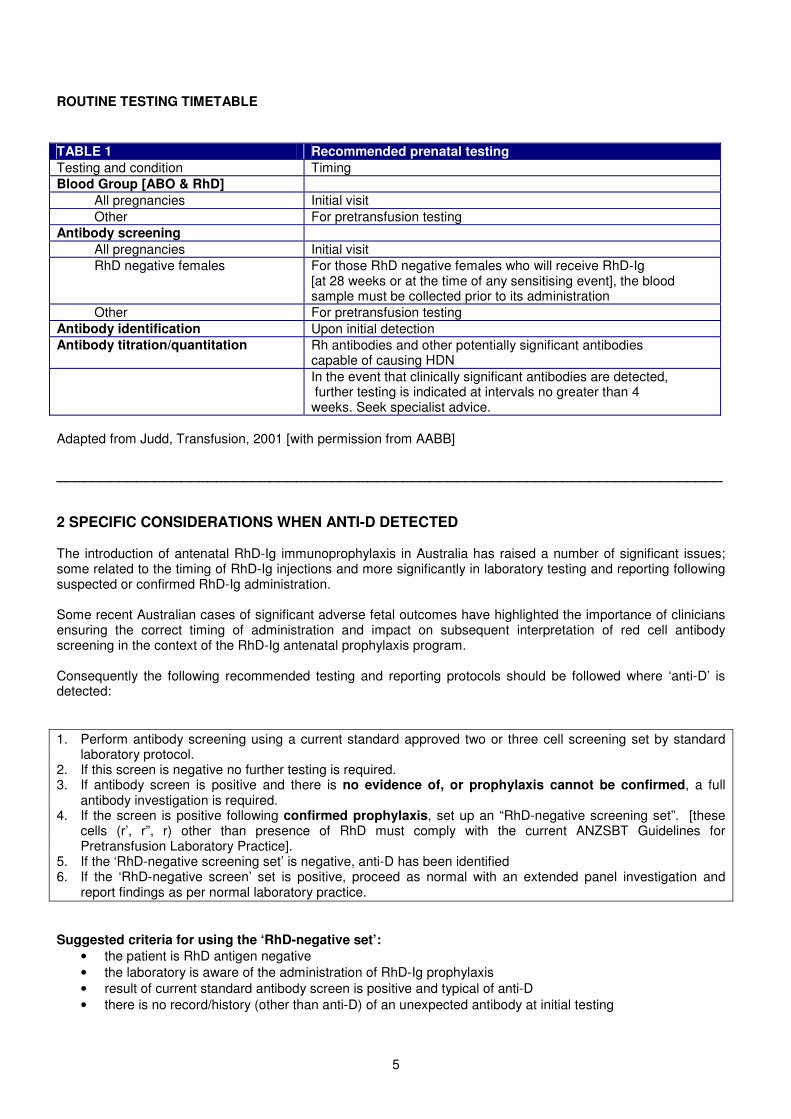
ROUTINE TESTING TIMETABLE
TABLE 1 Recommended prenatal testingTesting and condition TimingBlood Group [ABO & RhD]
All pregnancies Initial visitOther For pretransfusion testing
Antibody screeningAll pregnancies Initial visitRhD negative females For those RhD negative females who will receive RhD-Ig
[at 28 weeks or at the time of any sensitising event], the bloodsample must be collected prior to its administration
Other For pretransfusion testingAntibody identification Upon initial detectionAntibody titration/quantitation Rh antibodies and other potentially significant antibodies
capable of causing HDNIn the event that clinically significant antibodies are detected,further testing is indicated at intervals no greater than 4weeks. Seek specialist advice.
Adapted from Judd, Transfusion, 2001 [with permission from AABB]
___________________________________________________________________________
2 SPECIFIC CONSIDERATIONS WHEN ANTI-D DETECTED
The introduction of antenatal RhD-Ig immunoprophylaxis in Australia has raised a number of significant issues;some related to the timing of RhD-Ig injections and more significantly in laboratory testing and reporting followingsuspected or confirmed RhD-Ig administration.
Some recent Australian cases of significant adverse fetal outcomes have highlighted the importance of cliniciansensuring the correct timing of administration and impact on subsequent interpretation of red cell antibodyscreening in the context of the RhD-Ig antenatal prophylaxis program.
Consequently the following recommended testing and reporting protocols should be followed where ‘anti-D’ isdetected:
1. Perform antibody screening using a current standard approved two or three cell screening set by standardlaboratory protocol.
2. If this screen is negative no further testing is required.3. If antibody screen is positive and there is no evidence of, or prophylaxis cannot be confirmed, a full
antibody investigation is required.4. If the screen is positive following confirmed prophylaxis, set up an “RhD-negative screening set”. [these
cells (r’, r”, r) other than presence of RhD must comply with the current ANZSBT Guidelines forPretransfusion Laboratory Practice].
5. If the ‘RhD-negative screening set’ is negative, anti-D has been identified6. If the ‘RhD-negative screen’ set is positive, proceed as normal with an extended panel investigation and
report findings as per normal laboratory practice.
Suggested criteria for using the ‘RhD-negative set’:• the patient is RhD antigen negative• the laboratory is aware of the administration of RhD-Ig prophylaxis• result of current standard antibody screen is positive and typical of anti-D• there is no record/history (other than anti-D) of an unexpected antibody at initial testing
6
3.INTERPRETATION OF PRESENCE OF ANTI-D
3.1 For LABORATORY:(1) Any ‘anti-D’ with score 2 or >2 (0-4 grading scale) or 8 or >8 (0-12 grading scale) requires:
• antibody titration• referral for quantitation if appropriate and available• follow up antibody testing at 4 weeks, or earlier, if clinically indicated
(2) For an ‘anti-D’ with score <2 (0-4 grading scale) or <8 (0-12 grading scale):• where there is a confirmed history of RhD-Ig administration within the previous 6 weeks, report as ‘most
likely to be due to recent RhD-Ig administration’• where there is no confirmed history of RhD-Ig administration, or RhD-Ig given > 6 weeks ago: treat as
immune and investigate as in (1) above
3.2 For CLINICIANS to interpret results:The laboratory results should not be reviewed in isolation but should take into account the clinical historyincluding the presence or absence of potentially sensitising events and recent administration of RhD-Ig.
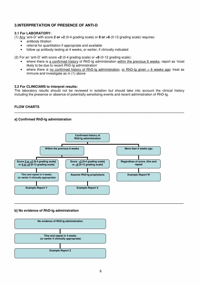
FLOW CHARTS__________________________________________________________________________________
a] Confirmed RhD-Ig administration
___________________________________________________________________________
b] No evidence of RhD-Ig administration
No evidence of RhD-Ig administration
Titre and repeat in 4 weeks(or earlier if clinically appropriate)
Example Report Z
Confirmed history ofRhD-Ig administration
Within the previous 6 weeks More than 6 weeks ago
Score 2 or >2 [0-4 grading scale]or 8 or >8 [0-12 grading scale]
Titre and repeat in 4 weeks(or earlier if clinically appropriate��
Score <2 [0-4 grading scale]or <8 [0-12 grading scale]
Assume RhD-Ig prophylaxis
Regardless of score, titre andrepeat
Example Report XExample Report Y
Example Report W
7
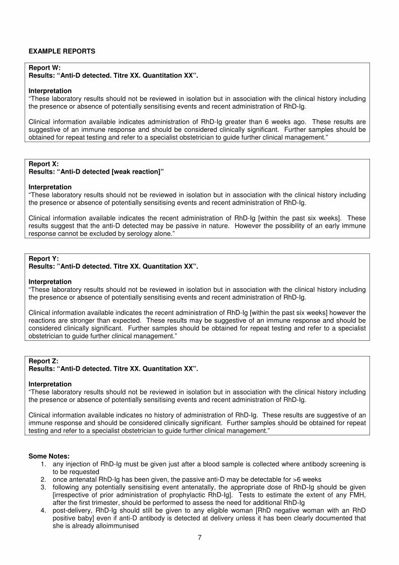
EXAMPLE REPORTS
Report W:Results: “Anti-D detected. Titre XX. Quantitation XX”.
Interpretation“These laboratory results should not be reviewed in isolation but in association with the clinical history includingthe presence or absence of potentially sensitising events and recent administration of RhD-Ig.
Clinical information available indicates administration of RhD-Ig greater than 6 weeks ago. These results aresuggestive of an immune response and should be considered clinically significant. Further samples should beobtained for repeat testing and refer to a specialist obstetrician to guide further clinical management.”
Report X:Results: “Anti-D detected [weak reaction]”
Interpretation“These laboratory results should not be reviewed in isolation but in association with the clinical history includingthe presence or absence of potentially sensitising events and recent administration of RhD-Ig.
Clinical information available indicates the recent administration of RhD-Ig [within the past six weeks]. Theseresults suggest that the anti-D detected may be passive in nature. However the possibility of an early immuneresponse cannot be excluded by serology alone.”
Report Y:Results: ”Anti-D detected. Titre XX. Quantitation XX”.
Interpretation“These laboratory results should not be reviewed in isolation but in association with the clinical history includingthe presence or absence of potentially sensitising events and recent administration of RhD-Ig.
Clinical information available indicates the recent administration of RhD-Ig [within the past six weeks] however thereactions are stronger than expected. These results may be suggestive of an immune response and should beconsidered clinically significant. Further samples should be obtained for repeat testing and refer to a specialistobstetrician to guide further clinical management.”
Report Z:Results: “Anti-D detected. Titre XX. Quantitation XX”.
Interpretation“These laboratory results should not be reviewed in isolation but in association with the clinical history includingthe presence or absence of potentially sensitising events and recent administration of RhD-Ig.
Clinical information available indicates no history of administration of RhD-Ig. These results are suggestive of animmune response and should be considered clinically significant. Further samples should be obtained for repeattesting and refer to a specialist obstetrician to guide further clinical management.”
Some Notes:1. any injection of RhD-Ig must be given just after a blood sample is collected where antibody screening is
to be requested2. once antenatal RhD-Ig has been given, the passive anti-D may be detectable for >6 weeks3. following any potentially sensitising event antenatally, the appropriate dose of RhD-Ig should be given
[irrespective of prior administration of prophylactic RhD-Ig]. Tests to estimate the extent of any FMH,after the first trimester, should be performed to assess the need for additional RhD-Ig
4. post-delivery, RhD-Ig should still be given to any eligible woman [RhD negative woman with an RhDpositive baby] even if anti-D antibody is detected at delivery unless it has been clearly documented thatshe is already alloimmunised
8
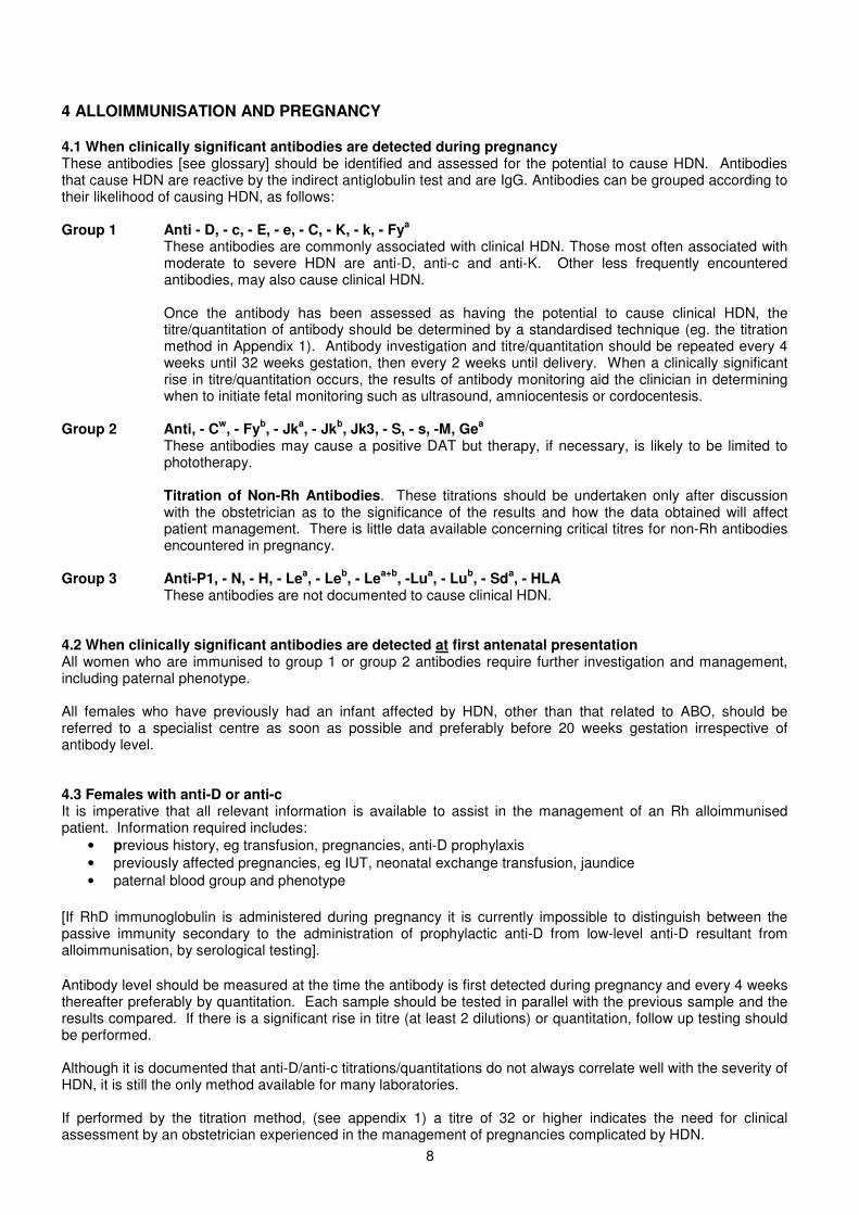
4 ALLOIMMUNISATION AND PREGNANCY
4.1 When clinically significant antibodies are detected during pregnancyThese antibodies [see glossary] should be identified and assessed for the potential to cause HDN. Antibodiesthat cause HDN are reactive by the indirect antiglobulin test and are IgG. Antibodies can be grouped according totheir likelihood of causing HDN, as follows:
Group 1 Anti - D, - c, - E, - e, - C, - K, - k, - Fya
These antibodies are commonly associated with clinical HDN. Those most often associated withmoderate to severe HDN are anti-D, anti-c and anti-K. Other less frequently encounteredantibodies, may also cause clinical HDN.
Once the antibody has been assessed as having the potential to cause clinical HDN, thetitre/quantitation of antibody should be determined by a standardised technique (eg. the titrationmethod in Appendix 1). Antibody investigation and titre/quantitation should be repeated every 4weeks until 32 weeks gestation, then every 2 weeks until delivery. When a clinically significantrise in titre/quantitation occurs, the results of antibody monitoring aid the clinician in determiningwhen to initiate fetal monitoring such as ultrasound, amniocentesis or cordocentesis.
Group 2 Anti, - Cw, - Fyb, - Jka, - Jkb, Jk3, - S, - s, -M, Gea
These antibodies may cause a positive DAT but therapy, if necessary, is likely to be limited tophototherapy.
Titration of Non-Rh Antibodies. These titrations should be undertaken only after discussionwith the obstetrician as to the significance of the results and how the data obtained will affectpatient management. There is little data available concerning critical titres for non-Rh antibodiesencountered in pregnancy.
Group 3 Anti-P1, - N, - H, - Lea, - Leb, - Lea+b, -Lua, - Lub, - Sda, - HLAThese antibodies are not documented to cause clinical HDN.
4.2 When clinically significant antibodies are detected at first antenatal presentationAll women who are immunised to group 1 or group 2 antibodies require further investigation and management,including paternal phenotype.
All females who have previously had an infant affected by HDN, other than that related to ABO, should bereferred to a specialist centre as soon as possible and preferably before 20 weeks gestation irrespective ofantibody level.
4.3 Females with anti-D or anti-cIt is imperative that all relevant information is available to assist in the management of an Rh alloimmunisedpatient. Information required includes:
• previous history, eg transfusion, pregnancies, anti-D prophylaxis• previously affected pregnancies, eg IUT, neonatal exchange transfusion, jaundice• paternal blood group and phenotype
[If RhD immunoglobulin is administered during pregnancy it is currently impossible to distinguish between thepassive immunity secondary to the administration of prophylactic anti-D from low-level anti-D resultant fromalloimmunisation, by serological testing].
Antibody level should be measured at the time the antibody is first detected during pregnancy and every 4 weeksthereafter preferably by quantitation. Each sample should be tested in parallel with the previous sample and theresults compared. If there is a significant rise in titre (at least 2 dilutions) or quantitation, follow up testing shouldbe performed.
Although it is documented that anti-D/anti-c titrations/quantitations do not always correlate well with the severity ofHDN, it is still the only method available for many laboratories.
If performed by the titration method, (see appendix 1) a titre of 32 or higher indicates the need for clinicalassessment by an obstetrician experienced in the management of pregnancies complicated by HDN.
9
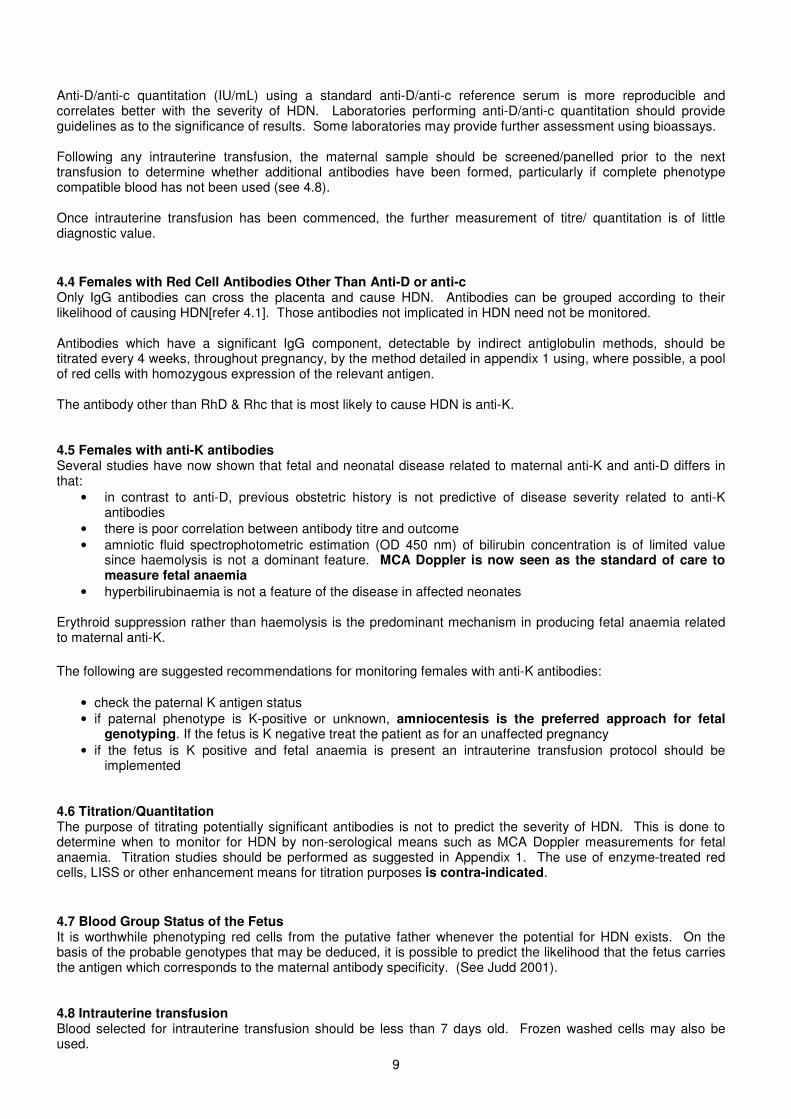
Anti-D/anti-c quantitation (IU/mL) using a standard anti-D/anti-c reference serum is more reproducible andcorrelates better with the severity of HDN. Laboratories performing anti-D/anti-c quantitation should provideguidelines as to the significance of results. Some laboratories may provide further assessment using bioassays.
Following any intrauterine transfusion, the maternal sample should be screened/panelled prior to the nexttransfusion to determine whether additional antibodies have been formed, particularly if complete phenotypecompatible blood has not been used (see 4.8).
Once intrauterine transfusion has been commenced, the further measurement of titre/ quantitation is of littlediagnostic value.
4.4 Females with Red Cell Antibodies Other Than Anti-D or anti-cOnly IgG antibodies can cross the placenta and cause HDN. Antibodies can be grouped according to theirlikelihood of causing HDN[refer 4.1]. Those antibodies not implicated in HDN need not be monitored.
Antibodies which have a significant IgG component, detectable by indirect antiglobulin methods, should betitrated every 4 weeks, throughout pregnancy, by the method detailed in appendix 1 using, where possible, a poolof red cells with homozygous expression of the relevant antigen.
The antibody other than RhD & Rhc that is most likely to cause HDN is anti-K.
4.5 Females with anti-K antibodiesSeveral studies have now shown that fetal and neonatal disease related to maternal anti-K and anti-D differs inthat:
• in contrast to anti-D, previous obstetric history is not predictive of disease severity related to anti-Kantibodies
• there is poor correlation between antibody titre and outcome• amniotic fluid spectrophotometric estimation (OD 450 nm) of bilirubin concentration is of limited value
since haemolysis is not a dominant feature. MCA Doppler is now seen as the standard of care tomeasure fetal anaemia
• hyperbilirubinaemia is not a feature of the disease in affected neonates
Erythroid suppression rather than haemolysis is the predominant mechanism in producing fetal anaemia relatedto maternal anti-K.
The following are suggested recommendations for monitoring females with anti-K antibodies:
• check the paternal K antigen status• if paternal phenotype is K-positive or unknown, amniocentesis is the preferred approach for fetal
genotyping. If the fetus is K negative treat the patient as for an unaffected pregnancy• if the fetus is K positive and fetal anaemia is present an intrauterine transfusion protocol should be
implemented
4.6 Titration/QuantitationThe purpose of titrating potentially significant antibodies is not to predict the severity of HDN. This is done todetermine when to monitor for HDN by non-serological means such as MCA Doppler measurements for fetalanaemia. Titration studies should be performed as suggested in Appendix 1. The use of enzyme-treated redcells, LISS or other enhancement means for titration purposes is contra-indicated.
4.7 Blood Group Status of the FetusIt is worthwhile phenotyping red cells from the putative father whenever the potential for HDN exists. On thebasis of the probable genotypes that may be deduced, it is possible to predict the likelihood that the fetus carriesthe antigen which corresponds to the maternal antibody specificity. (See Judd 2001).
4.8 Intrauterine transfusionBlood selected for intrauterine transfusion should be less than 7 days old. Frozen washed cells may also beused.
10
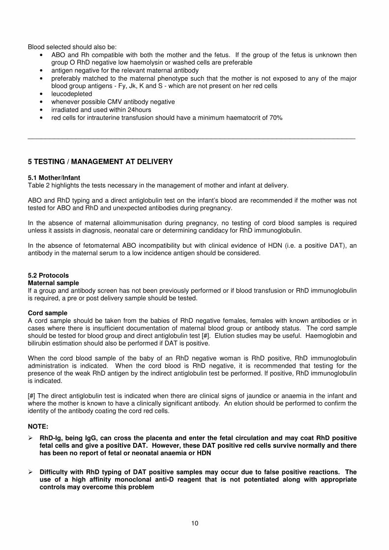
Blood selected should also be:• ABO and Rh compatible with both the mother and the fetus. If the group of the fetus is unknown then
group O RhD negative low haemolysin or washed cells are preferable• antigen negative for the relevant maternal antibody• preferably matched to the maternal phenotype such that the mother is not exposed to any of the major
blood group antigens - Fy, Jk, K and S - which are not present on her red cells• leucodepleted• whenever possible CMV antibody negative• irradiated and used within 24hours• red cells for intrauterine transfusion should have a minimum haematocrit of 70%
___________________________________________________________________________
5 TESTING / MANAGEMENT AT DELIVERY
5.1 Mother/InfantTable 2 highlights the tests necessary in the management of mother and infant at delivery.
ABO and RhD typing and a direct antiglobulin test on the infant’s blood are recommended if the mother was nottested for ABO and RhD and unexpected antibodies during pregnancy.
In the absence of maternal alloimmunisation during pregnancy, no testing of cord blood samples is requiredunless it assists in diagnosis, neonatal care or determining candidacy for RhD immunoglobulin.
In the absence of fetomaternal ABO incompatibility but with clinical evidence of HDN (i.e. a positive DAT), anantibody in the maternal serum to a low incidence antigen should be considered.
5.2 ProtocolsMaternal sampleIf a group and antibody screen has not been previously performed or if blood transfusion or RhD immunoglobulinis required, a pre or post delivery sample should be tested.
Cord sampleA cord sample should be taken from the babies of RhD negative females, females with known antibodies or incases where there is insufficient documentation of maternal blood group or antibody status. The cord sampleshould be tested for blood group and direct antiglobulin test [#]. Elution studies may be useful. Haemoglobin andbilirubin estimation should also be performed if DAT is positive.
When the cord blood sample of the baby of an RhD negative woman is RhD positive, RhD immunoglobulinadministration is indicated. When the cord blood is RhD negative, it is recommended that testing for thepresence of the weak RhD antigen by the indirect antiglobulin test be performed. If positive, RhD immunoglobulinis indicated.
[#] The direct antiglobulin test is indicated when there are clinical signs of jaundice or anaemia in the infant andwhere the mother is known to have a clinically significant antibody. An elution should be performed to confirm theidentity of the antibody coating the cord red cells.
NOTE:
� RhD-Ig, being IgG, can cross the placenta and enter the fetal circulation and may coat RhD positivefetal cells and give a positive DAT. However, these DAT positive red cells survive normally and therehas been no report of fetal or neonatal anaemia or HDN
� Difficulty with RhD typing of DAT positive samples may occur due to false positive reactions. Theuse of a high affinity monoclonal anti-D reagent that is not potentiated along with appropriatecontrols may overcome this problem
11
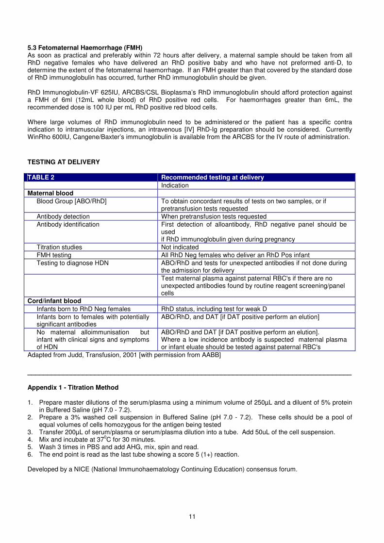
5.3 Fetomaternal Haemorrhage (FMH)As soon as practical and preferably within 72 hours after delivery, a maternal sample should be taken from allRhD negative females who have delivered an RhD positive baby and who have not preformed anti-D, todetermine the extent of the fetomaternal haemorrhage. If an FMH greater than that covered by the standard doseof RhD immunoglobulin has occurred, further RhD immunoglobulin should be given.
RhD Immunoglobulin-VF 625IU, ARCBS/CSL Bioplasma’s RhD immunoglobulin should afford protection againsta FMH of 6ml (12mL whole blood) of RhD positive red cells. For haemorrhages greater than 6mL, therecommended dose is 100 IU per mL RhD positive red blood cells.
Where large volumes of RhD immunoglobulin need to be administered or the patient has a specific contraindication to intramuscular injections, an intravenous [IV] RhD-Ig preparation should be considered. CurrentlyWinRho 600IU, Cangene/Baxter’s immunoglobulin is available from the ARCBS for the IV route of administration.
TESTING AT DELIVERY
TABLE 2 Recommended testing at deliveryIndication
Maternal bloodBlood Group [ABO/RhD] To obtain concordant results of tests on two samples, or if
pretransfusion tests requestedAntibody detection When pretransfusion tests requestedAntibody identification First detection of alloantibody, RhD negative panel should be
usedif RhD immunoglobulin given during pregnancy
Titration studies Not indicatedFMH testing All RhD Neg females who deliver an RhD Pos infantTesting to diagnose HDN ABO/RhD and tests for unexpected antibodies if not done during
the admission for deliveryTest maternal plasma against paternal RBC's if there are nounexpected antibodies found by routine reagent screening/panelcells
Cord/infant bloodInfants born to RhD Neg females RhD status, including test for weak DInfants born to females with potentiallysignificant antibodies
ABO/RhD, and DAT [if DAT positive perform an elution]
No maternal alloimmunisation butinfant with clinical signs and symptomsof HDN
ABO/RhD and DAT [if DAT positive perform an elution].Where a low incidence antibody is suspected maternal plasmaor infant eluate should be tested against paternal RBC's
Adapted from Judd, Transfusion, 2001 [with permission from AABB]
__________________________________________________________________________________
Appendix 1 - Titration Method
1. Prepare master dilutions of the serum/plasma using a minimum volume of 250µL and a diluent of 5% proteinin Buffered Saline (pH 7.0 - 7.2).
2. Prepare a 3% washed cell suspension in Buffered Saline (pH 7.0 - 7.2). These cells should be a pool ofequal volumes of cells homozygous for the antigen being tested
3. Transfer 200µL of serum/plasma or serum/plasma dilution into a tube. Add 50uL of the cell suspension.4. Mix and incubate at 370C for 30 minutes.5. Wash 3 times in PBS and add AHG, mix, spin and read.6. The end point is read as the last tube showing a score 5 (1+) reaction.
Developed by a NICE (National Immunohaematology Continuing Education) consensus forum.
1
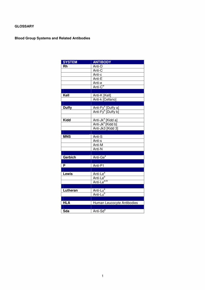
GLOSSARY
Blood Group Systems and Related Antibodies
SYSTEM ANTIBODYRh Anti-D
Anti-CAnti-cAnti-EAnti-eAnti-Cw
Kell Anti-K [Kell]Anti-k [Cellano]
Duffy Anti-Fya [Duffy a]Anti-Fyb [Duffy b]
Kidd Anti-Jka [Kidd a]Anti-Jkb [Kidd b]Anti-Jk3 [Kidd 3]
MNS Anti-SAnti-sAnti-MAnti-N
Gerbich Anti-Gea
P Anti-P1
Lewis Anti-Lea
Anti-Leb
Anti-Lea+b
Lutheran Anti-Lua
Anti-Lub
HLA Human Leucocyte Antibodies
Sda Anti-Sda
1
REFERENCES
ASBT “Guidelines for Blood Group and Antibody Screening During Pregnancy”, June 1999.
ANZSBT “Guidelines for Blood Grouping & Antibody Screening in the Antenatal & Perinatal Setting. 2nd Ed. 2004
AABB Technical Manual, 12-14th Edition.
Bowell P, Wainscoat JS Peto TEA and Gunson HH. Maternal anti-D concentrations and outcome in rhesushaemolytic disease of the newborn. British Medical Journal 1982; 285; 327-329.
Bowman J M, Pollock J M, Manning F A, Harman C R Menticoglou S. Maternal Kell Blood GroupAlloimmunisation. Obstetrics and Gynaecology 1992: 79:239 - 244.
Collins G. Obstetric immunohaematology in Australia. Australian Journal of Medical Science 1993; 14: 157-162.
Contreras M, Garner S and de Silva M. Prenatal testing to predict the severity of haemolytic disease of thefetus and newborn. Transfusion Medicine 1996; 480 - 484.
Duerbeck N and Sceds J. Rhesus immunisation in pregnancy. Obstetrics and Gynaecology Survey, Vol 48,12:801-810.
Gilbert GL, Hayes K, Hudson IL, James J. Prevention of transfusion-acquired cytomegalovirus infection ininfants by blood filtration to remove leucocytes. Lancet 1989; i: 1228-1231.
Green RE, Ford DS, Condon JA, Lowe VA. Basic Blood Grouping techniques and procedures. 2nd Ed.Melbourne: Victorian Immunohematology Discussion Group. 1992: 114.
Guidelines for blood grouping and red cell antibody testing during pregnancy. BCSH. Transfusion Medicine,1996; 6, 71-74
Hughes R, Craig J, Murphy W and Green I. Causes and clinical consequences of Rhesus (D) haemolyticdisease of the newborn. A study of a Scottish population, 1985-1990. British Journal of Obstetrics andGynaecology, 1994; Vol 101, 297 - 300.
Judd, W.J. (2001) Practice guidelines for prenatal and perinatal immunohaematology, revisited. Transfusion,41, 1450.
Judd W, Luban N, Ness P, Silberstein L, Stroup M and Widmann F. Prenatal and perinatalimmunohaematology: Recommendations for serologic management of the, newborn infant and obstetricpatient. Transfusion 1990; Vol 30, 2:175 - 183.
Klein H G & Anstee D J. Mollison’s Blood Transfusion in Clinical Medicine, 11th Ed. 2005. BlackwellPublishing.
2
Leggatt H M, Gibson J M, Barron, Reid MM. Anti-Kell in pregnancy. British Journal of Obstetrics andGynaecology 1991;98: 162-166.
Mitchell R, Bowell P, Letsky E, de Silva M, Whittle M. (1996). Guidelines for blood grouping and red cellantibody testing during pregnancy. Transfusion Medicine, 1996, 6, 71-74.
National Blood Authority “Guidelines on the prophylactic use of Rh D immunoglobulin (anti-D) in obstetrics”.June 2003.
NHMRC “Guidelines on the prophylactic use of RhD Immunoglobulin (Anti-D) in Obstetrics”, March 1999.
Vaughan J I, Warwick R, Letsky E, Nicolini U, Rodeck C H, Fisk N, Erythropoietic suppression in fetal anaemiabecause of Kell alloimmunisation. American Journal of Obstetrics and Gynaecology 1994; 171 (1) 247-251.
Vaughan J.I et al. Inhibition of erythroid progenitor cells by anti-Kell antibodies in fetal alloimmune anaemia.New England Journal of Medicine 1998, 338: 798-803.
Whittle M. Antenatal serology testing in pregnancy. British Journal of Obstetrics and Gynaecology, 1996; Vol103, 195-196.
Shulman, I.A., Calderon, C., Nelson, J.M., Nakayama, R. (1994) The routine use of Rh-negative reagent redcells for the identification of anti-D and the detection of non-D red cell antibodies. Transfusion, 34, 666-670.