Guest Molecule-Responsive Functional CaPhosphonates proton conductivity (JACS)
9
Guest Molecule-Responsive Functional Calcium Phosphonate Frameworks for Tuned Proton Conductivity Montse Bazaga-García, † Rosario M. P. Colodrero, † Maria Papadaki, ‡ Piotr Garczarek, § Jerzy Zoń , ∥ Pascual Olivera-Pastor, † Enrique R. Losilla, † Laura Leó n-Reina, ⊥ Miguel A. G. Aranda, †,# Duane Choquesillo-Lazarte, ∇ Konstantinos D. Demadis,* ,‡ and Aurelio Cabeza* ,† † Departamento de Química Inorga ́ nica, Universidad de Ma ́ laga, Campus Teatinos s/n, Ma ́ laga 29071, Spain ‡ Crystal Engineering, Growth and Design Laboratory, Department of Chemistry, University of Crete, Voutes Campus, Crete, GR-71003, Greece § Department of Medicinal Chemistry and Microbiology, Faculty of Chemistry and ∥ Department of Thermodynamics, Institute of Heat Engineering and Mechanics, Faculty of Mechanical and Power Engineering, Wroclaw University of Technology, 27 Wybrzeż e Wyspiańskiego, 50-370 Wroclaw, Poland ⊥ Servicios Centrales de Apoyo a la Investigació n, SCAI, Universidad de Ma ́ laga, Campus Teatinos s/n, 29071-Ma ́ laga, Spain # CELLS-ALBA Synchrotron, Carretera BP 1413, Km. 3.3, E-08290 Cerdanyola, Barcelona, Spain ∇ Laboratorio de Estudios Cristalogra ́ ficos, IACT-CSIC, Granada, Spain * S Supporting Information ABSTRACT: We report the synthesis, structural characterization, and functionality (framework interconversions together with proton conductivity) of an open-framework hybrid that combines Ca 2+ ions and the rigid polyfunctional ligand 5-(dihydroxyphosphoryl)isophthalic acid (PiPhtA). Ca 2 [(HO 3 PC 6 H 3 COOH) 2 ] 2 [(HO 3 PC 6 H 3 (COO) 2 H)(H 2 O) 2 ]·5H 2 O(Ca-PiPh- tA-I) is obtained by slow crystallization at ambient conditions from acidic (pH ≈ 3) aqueous solutions. It possesses a high water content (both Ca coordinated and in the lattice), and importantly, it exhibits water-filled 1D channels. At 75 °C, Ca-PiPhtA-I is partially dehydrated and exhibits a crystalline di ffraction pattern that can be indexed in a monoclinic cell with parameters close to the pristine phase. Rietveld refinement was carried out for the sample heated at 75 °C, Ca-PiPhtA-II, using synchrotron powder X-ray di ffraction data, which revealed the molecular formula Ca 2 [(HO 3 PC 6 H 3 COOH) 2 ] 2 [(HO 3 PC 6 H 3 (COO) 2 H)(H 2 O) 2 ]. All connectivity modes of the “parent” Ca-PiPhtA-I framework are retained in Ca-PiPhtA-II. Upon Ca-PiPhtA-I exposure to ammonia vapors (28% aqueous NH 3 ) a new derivative is obtained (Ca-PiPhtA-NH 3 ) containing 7 NH 3 and 16 H 2 O molecules according to elemental and thermal analyses. Ca-PiPhtA-NH 3 exhibits a complex X-ray di ffraction pattern with peaks at 15.3 and 13.0 Å that suggest partial breaking and transformation of the parent pillared structure. Although detailed structural identi fication of Ca-PiPhtA-NH 3 was not possible, due in part to nonequilibrium adsorption conditions and the lack of crystallinity, FT-IR spectra and DTA-TG analysis indicate profound structural changes compared to the pristine Ca-PiPhtA-I. At 98% RH and T = 24 °C, proton conductivity, σ, for Ca-PiPhtA-I is 5.7 × 10 −4 S· cm −1 . It increases to 1.3 × 10 −3 S·cm −1 upon activation by preheating the sample at 40 °C for 2 h followed by water equilibration at room temperature under controlled conditions. Ca-PiPhtA-NH 3 exhibits the highest proton conductivity, 6.6 × 10 −3 S·cm −1 , measured at 98% RH and T = 24 °C. Activation energies (E a ) for proton transfer in the above-mentioned frameworks range between 0.23 and 0.4 eV, typical of a Grothuss mechanism of proton conduction. These results underline the importance of internal H-bonding networks that, in turn, determine conductivity properties of hybrid materials. It is highlighted that new proton transfer pathways may be created by means of cavity “derivatization” with selected guest molecules resulting in improved proton conductivity. Received: January 13, 2014 Published: March 18, 2014 Article pubs.acs.org/JACS © 2014 American Chemical Society 5731 dx.doi.org/10.1021/ja500356z | J. Am. Chem. Soc. 2014, 136, 5731−5739
-
Upload
maria-papadaki -
Category
Documents
-
view
39 -
download
0
Transcript of Guest Molecule-Responsive Functional CaPhosphonates proton conductivity (JACS)
Guest Molecule-Responsive Functional Calcium PhosphonateFrameworks for Tuned Proton ConductivityMontse Bazaga-García,† Rosario M. P. Colodrero,† Maria Papadaki,‡ Piotr Garczarek,§ Jerzy Zon,∥
Pascual Olivera-Pastor,† Enrique R. Losilla,† Laura Leon-Reina,⊥ Miguel A. G. Aranda,†,#
Duane Choquesillo-Lazarte,∇ Konstantinos D. Demadis,*,‡ and Aurelio Cabeza*,†
†Departamento de Química Inorganica, Universidad de Malaga, Campus Teatinos s/n, Malaga 29071, Spain‡Crystal Engineering, Growth and Design Laboratory, Department of Chemistry, University of Crete, Voutes Campus, Crete, GR-71003, Greece§Department of Medicinal Chemistry and Microbiology, Faculty of Chemistry and ∥Department of Thermodynamics, Instituteof Heat Engineering and Mechanics, Faculty of Mechanical and Power Engineering, Wrocław University of Technology,27 Wybrzeze Wyspian skiego, 50-370 Wrocław, Poland⊥Servicios Centrales de Apoyo a la Investigacion, SCAI, Universidad de Malaga, Campus Teatinos s/n, 29071-Malaga, Spain#CELLS-ALBA Synchrotron, Carretera BP 1413, Km. 3.3, E-08290 Cerdanyola, Barcelona, Spain∇Laboratorio de Estudios Cristalograficos, IACT-CSIC, Granada, Spain
*S Supporting Information
ABSTRACT: We report the synthesis, structural characterization, and functionality (framework interconversions togetherwith proton conductivity) of an open-framework hybrid that combines Ca2+ ions and the rigid polyfunctional ligand5-(dihydroxyphosphoryl)isophthalic acid (PiPhtA). Ca2[(HO3PC6H3COOH)2]2[(HO3PC6H3(COO)2H)(H2O)2]·5H2O (Ca-PiPh-tA-I) is obtained by slow crystallization at ambient conditions from acidic (pH ≈ 3) aqueous solutions. It possesses a high water content(both Ca coordinated and in the lattice), and importantly, it exhibits water-filled 1D channels. At 75 °C, Ca-PiPhtA-I is partiallydehydrated and exhibits a crystalline diffraction pattern that can be indexed in a monoclinic cell with parameters close to the pristinephase. Rietveld refinement was carried out for the sample heated at 75 °C, Ca-PiPhtA-II, using synchrotron powder X-ray diffractiondata, which revealed the molecular formula Ca2[(HO3PC6H3COOH)2]2[(HO3PC6H3(COO)2H)(H2O)2]. All connectivity modes ofthe “parent” Ca-PiPhtA-I framework are retained in Ca-PiPhtA-II. Upon Ca-PiPhtA-I exposure to ammonia vapors (28% aqueousNH3) a new derivative is obtained (Ca-PiPhtA-NH3) containing 7 NH3 and 16 H2O molecules according to elemental and thermalanalyses. Ca-PiPhtA-NH3 exhibits a complex X-ray diffraction pattern with peaks at 15.3 and 13.0 Å that suggest partial breaking andtransformation of the parent pillared structure. Although detailed structural identification of Ca-PiPhtA-NH3 was not possible, due inpart to nonequilibrium adsorption conditions and the lack of crystallinity, FT-IR spectra and DTA-TG analysis indicate profoundstructural changes compared to the pristine Ca-PiPhtA-I. At 98% RH and T = 24 °C, proton conductivity, σ, for Ca-PiPhtA-I is 5.7 ×10⁻⁴ S·cm−1. It increases to 1.3 × 10⁻³ S·cm−1 upon activation by preheating the sample at 40 °C for 2 h followed by water equilibrationat room temperature under controlled conditions. Ca-PiPhtA-NH3 exhibits the highest proton conductivity, 6.6 × 10⁻³ S·cm−1,measured at 98% RH and T = 24 °C. Activation energies (Ea) for proton transfer in the above-mentioned frameworks range between0.23 and 0.4 eV, typical of a Grothuss mechanism of proton conduction. These results underline the importance of internal H-bondingnetworks that, in turn, determine conductivity properties of hybrid materials. It is highlighted that new proton transfer pathways may becreated by means of cavity “derivatization” with selected guest molecules resulting in improved proton conductivity.
Received: January 13, 2014Published: March 18, 2014
Article
pubs.acs.org/JACS
© 2014 American Chemical Society 5731 dx.doi.org/10.1021/ja500356z | J. Am. Chem. Soc. 2014, 136, 5731−5739

■ INTRODUCTION
Metal−organic framework (MOF) materials are now at theepicenter of research efforts of many groups around the globe.An obvious exegesis for the “explosion” in the field is two-fold:(a) new structural motifs are constantly being sought,1a and (b)functionality in these materials becomes increasingly impor-tant.1b−d More specifically, potential applications are inves-tigated in fields such as gas adsorption, storage and separation,2
magnetism,3 catalysis,4 photoluminescence, sensors,5 con-trolled release of pharmaceuticals, medicinal and dentalmaterials,6 chiral separations,7 and ion exchange,8 just tomention a few.A plethora of organic linkers/ligands have been widely utilized
to construct these hybridMOFmaterials, with the majority beingcarboxylate, imidazolate, or polypyridyl based. On the otherhand, multichelating phosphonate ligands are “alternative”organic linkers (to the widespread polycarboxylates, imidazoles,and various derivatives) and provide synthetic access to a numberof thermally and chemically stable MOFs.9
Recently, phosphonates,10 phosphocarboxylates,11 and phos-phonosulfonates12 that possess an aromatic entity (usually oneor more phenyl rings) have been exploited in synthetic efforts,yielding multidimensional (mostly 2D and 3D) platforms.Proton conductivity has long been investigated as a desirable
property in a variety of diverse materials, whether organic,13
inorganic,14 or hybrid.15 The field of MOFs and more specificallymetal phosphonates has provided an opportune arena forelegant efforts for accessing suitable and highly conductingmaterials.15f,16 Furthermore, postsynthetic modification1d of theinternal surfaces of MOFs offers opportunities for enhancingdesired properties of crystalline solids.16c The main requirementfor a MOF material to be proton conductive is the presence ofacidic protons within an appropriately structured metal−organicframework that facilitates charge transport. In the case ofmetal phosphonate materials the phosphonic acid group (orpartially protonated phosphonate) assumes the role of the acidicgroup. Such well-ordered structural motifs, with highly orderedphosphonate groups located in close proximity to lattice watermolecules, often present appropriate platforms that arecandidates for high proton conductivity.12 For instance, protonconductivity higher than 10−2 S cm−1 has been reportedfor PCMOF21/2,15f a mixed sulfonate−phosphonate MOF,and for derivatized MIL-101 with H2SO4 and/or H3PO4.
15g
Other notable examples are metal derivatives containing theN,N-amino-bis(methylenephosphonate) moiety, such asZrChDTMP,17 MgH6ODTMP,18 and LaH5DTMP,19 phases,which exhibit conductivity values from 1 × 10⁻⁴ to 8 × 10⁻³
S cm−1. The high flexibility imparted by the ligand is consideredto exert a key role in determining internal proton transferpathways.In the present work, we report the synthesis, structural
characterization, and proton conductivity of an open-frameworkhybrid that combines Ca2+ ions and the rigid polyfunctionalligand 5-(dihydroxyphosphoryl)isophthalic acid (PiPhtA). Thismaterial was obtained by slow crystallization at ambientconditions from acidic (pH ≈ 3) aqueous solutions. It possessesa high water content (both Ca coordinated and in the lattice),and importantly, it exhibits water-filled 1D channels. Bothcharacteristics are, in principle, considered to be remarkableattributes to evaluate the proton conductivity of this hybridcompound. The effects of the guest species (water and NH3) onthe conductivity properties will be highlighted.
■ EXPERIMENTAL SECTIONMaterials and Common Instrumentation. The starting
salt CaCl2·2H2O was from EM Science Merck and used as received.Deionized (DI) water from an in-house laboratory cation-exchangecolumn was used for all syntheses. Stock solutions of HCl and NaOHwere used for pH adjustments. The pH meter used was a wTw pH315isetup, equipped with a SeTix 41 electrode. Elemental analyses (C, H, N)were measured on a Perkin-Elmer 240 analyzer. Infrared spectra werecollected in a FTIR Nicolet 5DXC spectrometer. All spectra wererecorded in the 4000−400 cm−1 range at 4 cm−1 resolution, and 50 scanswere accumulated. The 1H, 13C{1H}, and 31P{1H} NMR (see below;operating frequency, solvent) spectra in solution were recorded on aBruker AvanceDRX300 instrument. Chemical shifts (δ) are given in ppm.
Syntheses. The ligand 5-(dihydroxyphosphoryl)isophthalic acid[5-(H2O3P)C6H3-1,3-(COOH)2] (PiPhtA, 1) was synthesized througha number of intermediates, shown in Figure 1, as follows.
5-Iodoisophthalic Acid (2). This compound was prepared fromcommercially available 5-aminoisophthalic acid according to a literatureprocedure.20 Specifically, 5-aminoisophthalic acid (5.00 g, 0.0276 mol)was dissolved in a mixture of concentrated hydrochloric acid (49 cm3)and water (49 cm3). This solution was cooled in an ice−water bath, anda solution of sodium nitrite (2.05 g, 0.0295 mol) in water (9.6 cm3) wasadded with such a rate that the temperature of the mixture did notexceed 4 °C. Next, a solution of potassium iodide (15.0 g, 0.0903mol) inwater (38 cm3) was slowly added. Evolution of nitrogen was observed,and the mixture turned a yellow-reddish color. Afterwards the mixturewas refluxed for 1 h. After cooling the precipitate was filtered off and airdried to yield 5-iodoisophthalic acid: 5.76 g (72%), mp 284−285 °C(lit. 288−289 °C20).
Dimethyl 5-Iodoisophthalate (3). 5-Iodoisophatlic acid (2, 14.83 g,0.050 mol) was heated under reflux with thionyl chloride (65 cm3,0.89 mol) for 3.5 h. Next, the excess of thionyl chloride was distilled off.Methanol (50 cm3) was added to the residue carefully due to anexothermic effect and evolution of hydrogen chloride, and the mixturewas heated to reflux for a few hours. A precipitate formed during theheating process and was filtered off and recrystallized from methanol toobtain dimethyl 5-iodoisophthalate (3): 13.32 g (82%), mp 103−105 °C(lit. 104 °C21).
Dimethyl 5-(Diisopropyloxyphosphoryl)isophthalate (4a). A50 cm3 round-bottom flask was charged with dimethyl 5-iodoisophthalate(3, 2.00 g, 6.25mmol), acetonitrile (20mL), diisopropyl phosphite (1.246 g,7.5 mmol), and diisopropylethylamine (1.05 g, 8.125 mmol). Theflask was flushed with argon for 15 min, after which palladium acetate(0.014 g, 0.0625 mmol) and 1,1′-bis(diphenylphosphine)ferrocene(dppf, 0.038 g, 0.0687 mmol) were added. The reaction mixture wasstirred and heated in an oil bath at 90 °C for 24 h under constant flow ofargon. After cooling to room temperature the mixture was evaporated todryness in vacuo, and the remaining residue was purified by column
Figure 1. Protocol for the synthesis of 5-(dihydroxyphosphoryl)-isophthalic acid (PiPhtA, 1): (i) NaNO3, HCl, KI; (ii) SOCl2, CH3OH;(iii) HP(O)(OR)2, (i-C3H7)2NC2H5, Pd(OOCCH3)2, dppf, CH3CN;(iv) aqueous HCl, H2O; (R = C2H5).
Journal of the American Chemical Society Article
dx.doi.org/10.1021/ja500356z | J. Am. Chem. Soc. 2014, 136, 5731−57395732

chromatography on silica gel using ethyl acetate as eluent to yielddimethyl 5-(diisopropyloxyphosphoryl)isophthalate (4a): 1.434 g(64%), Rf = 0.40 (silica gel on TLC-PET foil, ethyl acetate, UV light254 nm). 1HNMR (300MHz, CDCl3) δ: 8.82 (m, 1H, CH arom.), 8.63(dd, JHH = 1.68 Hz, JHP = 16.35 Hz, 2H, CH arom.), 4.74 (m, 2H, CH),3.96 (s, 3H, CH3), 1.39 (d, J
HH = 6.18Hz, CH3), 1.24 (d, JHH = 6.18Hz).
13C NMR (75 MHz, CDCl3) δ: 165.43, 136.64 (J = 10.94 Hz), 133.88,131.88 (J = 192.01 Hz), 130.97 (J = 14.94 Hz), 71.51 (J = 5.59 Hz),52.60, 24.02 (J = 3.81 Hz), 23.86 (J = 4.58 Hz). 31P{1H} NMR (121MHz, CDCl3) δ: 14.16 (s).Dimethyl 5-(Diethoxyphosphoryl)isophthalate (4b). Compound
4bwas synthesized using the same procedure as 4a, starting from 1.173 gof dimethyl 5-iodoisophthalate, although diethyl phosphite was used.Yield of the ester 4b: 42%, Rf = 0.30 (silica gel on TLC-PET foil, ethylacetate, UV light 254 nm). 1HNMR (300MHz, CDCl3) δ: 8.83 (m, 1H,CH arom.), 8.63 (dd, JHH = 1.68Hz, JHP = 13.32Hz, 2HCH arom.), 4.16(m, 4H, CH2), 3.96 (s, 6H, CH3), 1.34 (t, J = 7.06 Hz, 6H, CH3).31P{1H} NMR (121 MHz, CDCl3) δ: 16.33 (s).5-(Dihydroxyphosphoryl)isophthalic Acid (PiPhtA, 1). A 25 cm3
round-bottomflaskwas chargedwith dimethyl 5-(diisopropyloxyphosphoryl)-isophthalate (4a, 1.072 g, 3.00 mmol), water (4 cm3), and concentratedhydrochloric acid (4 cm3). The mixture was refluxed for 20 h. Aftercooling down to room temperature it was evaporated to dryness invacuo to yield the product 1 as a white solid: 0.734 g (99%). 1H NMR(300MHz, D2O) δ: 8.29 (d, J = 3.82 Hz, 1H, CH arom.), 8.21 (dd, JHP =13.36 Hz, JHH = 2.62 Hz, 2H, CH arom.);. 31P{1H} NMR (121 MHz,D2O) δ: 12.63 (s).Ca2[(HO3P−C6H3−COOH)2]2[(HO3P−C6H3−(COO)2H)−(H2O)2])·
5H2O (Ca-PiPhtA-I). A quantity of the ligand 5-(dihydroxyphosphoryl)-isophthalic acid (1, 0.061 g, 0.250 mmol) was dissolved in 10 cm3 ofDI water. A small quantity of a 0.1 M NaOH stock solution was addeddropwise until the ligand dissolved completely and a clear, colorlesssolution was obtained. A quantity of CaCl2·2H2O (0.037 g, 0.250mmol)was separately dissolved in 5 cm3 of DI water. The two solutions weresubsequently mixed, and the final solution pHwas adjusted to 3.00 usingthe 0.1 M NaOH stock solution. The transparent colorless solutionwas left undisturbed for 1 week, after which the product appeared as awhite crystalline material. It was isolated by filtration, washed with DIwater, and air dried. Yield: 92%. Anal. Calcd for Ca2[(HO3P−C6H3−COOH)2]2[(HO3P−C6H3−(COO)2H)(H2O)2]·5H2O, fw 940.56,C24H31Ca2O28P3: C, 30.81; H, 2.80. Found: C, 29.27; H, 3.36.Ca2[(HO3P−C6H3−COOH)2]2[(HO3P−C6H3−(COO)2H)−(H2O)2]
(Ca-PiPhtA-II). This compound was obtained by thermal treatment ofsolid Ca-PiPhtA-I at 75 °C for 2 h.Absorption of NH3 Vapors (Aqueous Solution). A 150 mg
amount of Ca-PiPhtA-I was placed in contact with vapors from acommercial solution of 28 wt % of NH3 in water for 2 h in a closedcontainer. Subsequently, the product was air dried for 15min and kept at98% relative humidity at 10 °C for 18 h following the same proceduredescribed below tomeasure the proton conductivity. The obtained solid,Ca2[(HO3P−C6H3−COOH)2]2[(HO3P−C6H3−(COO)2H)]-(NH3)7(H2O)16 (Ca-PiPhtA-NH3), has the following elementalanalysis. Anal. Calcd: C, 23.69; H, 5.38; N, 8.06. Found: C, 23.86; H,4.75; N, 7.65. TG -analysis: weight loss up to 400 °C, found 32.67%,calcd 33.36%.Thermal Analysis. Differential thermal analysis (DTA) and
thermogravimetric analysis (TGA) data were recorded on an SDT-Q600 analyzer from TA Instruments. The temperature varied fromroom temperature to 900 °C at a heating rate of 10 °C·min−1. Mea-surements were carried out on samples in open platinum crucibles undera flow of air.X-ray Crystallography. Laboratory X-ray powder diffraction
(XRPD) patterns were collected on a PANanalytical X’Pert Proautomated diffractometer. Powder patterns were recorded in Bragg−Brentano reflection configuration using a Ge(111) primary mono-chromator (Cu Kα1) and the X’Celerator detector. Thermodiffracto-metric studies for Ca-PiPhtA-I and Ca-PiPhtA-NH3 were carried outon samples loaded in an Anton Paar HTK 1200N chamber under staticair. Data were collected between room temperature and 240 °C. Allmeasurements were performed with a heating rate of 10 °C·min−1 and a
delay time of 5 min to ensure thermal stabilization. Data acquisition wascarried out between 4° and 40° (2θ) due to “less-than-ideal” crystallinityof the samples. A step size of 0.017° and an equivalent counting time of150 s step−1 were used.
Crystal Structure Determination. Suitable-size crystals ofCa-PiPhtA-I were kept under inert conditions and immersed inperfluoropolyether as protecting oil for manipulation. A suitable crystalwas mounted on MiTeGen Micromounts, and this sample was used fordata collection. Data were collected with a Bruker SMART APEXdiffractometer. Data were processed with APEX222 suite and correctedfor absorption using SADABS.23 The structure was solved by directmethods,24 which revealed the position of all non-hydrogen atoms.These atoms were refined on F2 by a full-matrix least-squares procedureusing anisotropic displacement parameters.24 All hydrogen atoms werelocated in difference Fourier maps, except those corresponding to C−Hgroups, which were placed geometrically and included as fixed con-tributions riding on attached atoms with isotropic thermal displacementparameters 1.2 (C−H) or 1.5 (O−H) times those of the respectivebonded atom.
Synchrotron X-ray powder diffraction data (SXRDP) forCa-PiPhtA-II were collected in Debye−Scherrer (transmission) mode using theX-ray powder diffraction station of ALBA, the Spanish SynchrotronRadiation Facility (Barcelona, Spain).25 The wavelength, 0.62020(1) Å,was selected with a double-crystal Si (111) monochromator anddetermined from a Si640d NIST standard (a = 5.43123 Å) mea-surement. The capillary was rotated during data collection to improvediffracting particle statistics. The detector system used was theMYTHEN, which allows fast collection of good-resolution patterns.The data acquisition time was ∼20 min per pattern to attain very goodsignal-to-noise ratio over the angular range 1−41° (2θ).
The X-ray powder pattern was autoindexed using the DICVOL06program.26 The crystal structure of Ca-PiPhtA-II compound wasdetermined by the Rietveld method27 using the structure ofCa-PiPhtA-I available from single-crystal data as starting model. Rietveld refinementwas carried out using the GSAS package28 with soft constraints tomaintain chemically reasonable geometries for the phosphonate,aromatic ring, and carboxylic groups. The soft constraints were asfollows: /PO3C tetrahedron/P−O, 1.53(1) Å; P−C, 1.80(1) Å; O···O,2.55(2) Å; O···C, 2.73(2) Å; /aryl/aromatic ring/C−C [1.40(1) Å],Cring···Cring [2.40(1) Å], Cring···Cring [2.78(1) Å], /COO group/, Caryl-Ccarb [1.50(1) Å], C−Ocarb [1.23(1) Å], Ocarb···Ocarb, 2.21(2) Å; Caryl···Ocarb, 2.36(2) Å. Two isotropic atomic displacement parameters wererefined, one for calcium and phosphorus atom and a second one forcarbon and oxygen atoms. Hydrogen atoms were not included in therefinements. Crystallographic data and CCDC reference codes are givenin Table 1.
Proton Conductivity Studies. Impedance data were collectedon cylindrical pellets (∼10 mm diameter; ∼0.9−1.1 mm thickness)obtained by pressing ∼0.15 g of sample at 500 MPa for 2 min betweenporous C electrodes (Sigracet, GDL 10 BB, no Pt). Impedance data werecollected using a HP4284A impedance analyzer over the frequencyrange from 20 Hz to 1 MHz with an applied voltage of 1 V. All mea-surements were electronically controlled by the winDETA package ofprograms.29
Electrical measurements were taken in a double-walled, temperature-controlled glass chamber with a gas inlet and outlet. The temperature ofthe chamber was controlled by a Julabo F32-MA refrigerated/heatingcirculator from 10 to 24 °C every 2 °C with a heating rate of0.4 °C·min−1 using EasyTEMP software. Samples were equilibrated for15 min after each step in temperature, measured in closest vicinity to thesample. The relative humidity (RH) was obtained by a continuous flow ofwater-saturated nitrogen at different temperatures through the cell. Pelletswere equilibrated at 98% RH for 18 h to ensure a fixed water content ofthe sample and stable conductivity values. RH is approximately constantin the measured temperature range from 10 to 24 °C.
■ RESULTS AND DISCUSSION
Synthesis. The product Ca-PiPhtA-I is formed at a relativelylow reaction pH (∼3.0). At this pH the carboxylic acid groups are
Journal of the American Chemical Society Article
dx.doi.org/10.1021/ja500356z | J. Am. Chem. Soc. 2014, 136, 5731−57395733

expected to be deprotonated, whereas the phosphonic acid groupis monodeprotonated. This would render the ligand trianionic,and a possible 1:1 adduct with Ca2+ would be anionic. However,based on elemental analyses and the structure solved, a solidwith a Ca:ligand ratio of 2:3 is instead obtained, indicating thatthe protonation state of the ligand is complex. As suggested bythe empirical formula, the ligand is found in different protonationstates, which are adjusted depending on the various modes ofcoordinating to Ca2+ (Figure 2).Structural Study. Ca-PiPhtA-I shows a complex pillared
layered structure with a unit cell containing two calcium atoms,three PiPhtA ligands, and seven water molecules (two are Cacoordinated and the remaining five are situated within thelattice). The coordination geometry of both calcium centers ispentagonal bipyramidal (see Figure 3).In this geometry, four of the equatorial positions are occupied
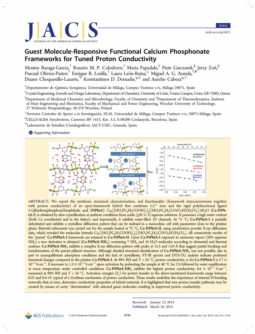
by oxygen atoms of three different phosphonate groups, one ofthem chelating the metal ion in a bidentate fashion. The fifthequatorial coordination position is occupied by an oxygen atomof a carboxylate group. All of these equatorial oxygen atomsbelong to two ligands, P2 and P3, which configure edge-sharingcalcium polyhedral chains and connect them into the layers,while leaving alternately arranged free carboxylic groups insidethe interlayer space (Figure 4). The axial positions of the calciumpolyhedron are occupied by a water molecule and a phosphonateor carboxylate oxygen of the third organic ligand, P1, which ispillaring adjacent layers.Within the layers, each phosphonate moiety uses only two
oxygen atoms to coordinate calcium in such a way that oneoxygen atom is bridging two metal centers, thus leaving a freePOH pointing to the interlayer space. One of the carboxylatemoieties of both dicarboxyphosphonate ligands, P2 and P3, is
coordinated to calcium atoms through one of the oxygens, whilethe other one is protonated. Besides, the second carboxylic groupof each these ligands is pointing toward the interlayer spaceand faced up to each other interacting strongly by a H-bond. Thisdisposition creates H-bond rings contributing to the stabilizationof the pillared layered structure. The third dicarboxyphosphonateligand, P1, is linking adjacent layers only by one phosphonateoxygen, by one side, and one carboxylate oxygen atom, from theother side. This connectivity mode leaves a POH and a freecarboxylic group inside the interlayer space (see Figure 4).The overall structure architecture reveals interlayer hydro-
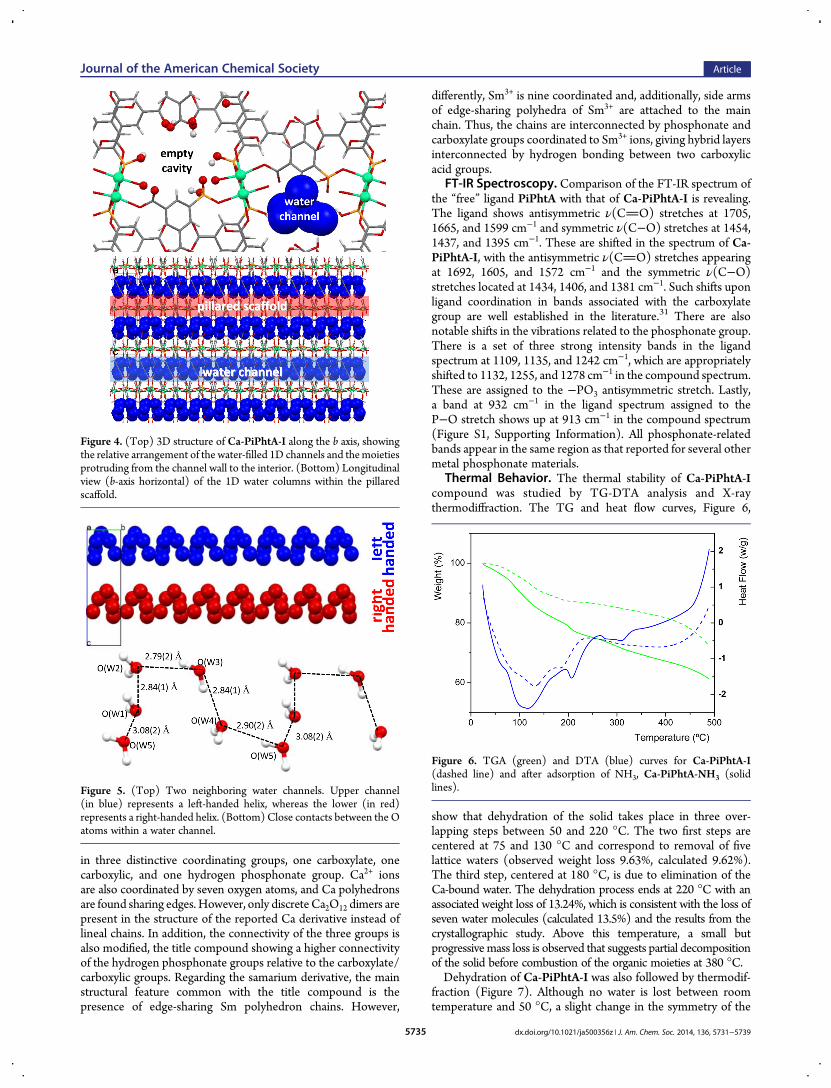
philic 1-D channels running parallel to the b axis, filled with fivelattice waters per unit cell forming zigzag chains inside thecavities (Figure 5).The parallel zigzag chains acquire a left-handed helix and a
right-handed helix along the c axis. The water-to-water interac-tions between these lattice waters create an extended network ofH bonds along the 1-D channels and therefore a potential protontransfer pathway, which may play an important role in the protonconductivity exhibited by theCa-PiPhtAmaterials (see Table S1,Supporting Information).The title compound shares some similarities with compounds
Ca2(H2O)[H(OOC)2C6H3PO3H]2 and [Sm2(H2O)4(H-(OOC)2C6H3PO3)2]·2H2O obtained by hydrothermal treat-ment.30 In the former, each ligand is 2-fold deprotonated resulting
Table 1. Crystallographic Data for Ca-PiPhtA-I andCa-PiPhtA-II
Ca-PiPhtA-I Ca-PiPhtA-II
chemical formula Ca2C24H31O28P3 Ca2C24H21O23P3
formula mass 940.56 850.49cryst syst orthorhombic monocliniccryst size (mm) color/shape
0.10 × 0.09 × 0.08 colorless/block
λ/Å 0.71073 0.62020(1)a/Å 23.112(4) 23.0652(7)b/Å 6.9534(13) 6.9731(1)c/Å 22.638(4) 22.4165(7)α/deg 90.0 90β/deg 90.0 91.540(4)γ/deg 90.0 90unit cell volume/Å3 3638.1(12) 3604.1(2)temp/°C −173 25space group Pca21 PaZ 4 4Rint 0.0832no. of independent reflns 4603 3455data (reflns)/restraints/params
6716/1/522 5500/299/357
R factor [I > 2σ(I)] R1a = 0.0715; wR2a = 0.1610R factor (all data) R1a = 0.1075; wR2a = 0.1834RWP 0.0654RP 0.0464GoF/RF 1.032 0.075CCDC number 968321 968322aR1(F) = Σ∥Fo| − |Fc∥/Σ|Fo|; wR2(F2) = [Σw(Fo2 − Fc
2)2/ΣF4]1/2.
Figure 2. Three deprotonation states of the ligand PiPhtA in thestructure of Ca-PiPhtA-I.
Figure 3. Coordination environment of the two Ca centers in thestructure of Ca-PiPhtA-I.
Journal of the American Chemical Society Article
dx.doi.org/10.1021/ja500356z | J. Am. Chem. Soc. 2014, 136, 5731−57395734
in three distinctive coordinating groups, one carboxylate, onecarboxylic, and one hydrogen phosphonate group. Ca2+ ionsare also coordinated by seven oxygen atoms, and Ca polyhedronsare found sharing edges. However, only discreteCa2O12 dimers arepresent in the structure of the reported Ca derivative instead oflineal chains. In addition, the connectivity of the three groups isalso modified, the title compound showing a higher connectivityof the hydrogen phosphonate groups relative to the carboxylate/carboxylic groups. Regarding the samarium derivative, the mainstructural feature common with the title compound is thepresence of edge-sharing Sm polyhedron chains. However,
differently, Sm3+ is nine coordinated and, additionally, side armsof edge-sharing polyhedra of Sm3+ are attached to the mainchain. Thus, the chains are interconnected by phosphonate andcarboxylate groups coordinated to Sm3+ ions, giving hybrid layersinterconnected by hydrogen bonding between two carboxylicacid groups.
FT-IR Spectroscopy. Comparison of the FT-IR spectrum ofthe “free” ligand PiPhtA with that of Ca-PiPhtA-I is revealing.The ligand shows antisymmetric ν(CO) stretches at 1705,1665, and 1599 cm−1 and symmetric ν(C−O) stretches at 1454,1437, and 1395 cm−1. These are shifted in the spectrum of Ca-PiPhtA-I, with the antisymmetric ν(CO) stretches appearingat 1692, 1605, and 1572 cm−1 and the symmetric ν(C−O)stretches located at 1434, 1406, and 1381 cm−1. Such shifts uponligand coordination in bands associated with the carboxylategroup are well established in the literature.31 There are alsonotable shifts in the vibrations related to the phosphonate group.There is a set of three strong intensity bands in the ligandspectrum at 1109, 1135, and 1242 cm−1, which are appropriatelyshifted to 1132, 1255, and 1278 cm−1 in the compound spectrum.These are assigned to the −PO3 antisymmetric stretch. Lastly,a band at 932 cm−1 in the ligand spectrum assigned to theP−O stretch shows up at 913 cm−1 in the compound spectrum(Figure S1, Supporting Information). All phosphonate-relatedbands appear in the same region as that reported for several othermetal phosphonate materials.
Thermal Behavior. The thermal stability of Ca-PiPhtA-Icompound was studied by TG-DTA analysis and X-raythermodiffraction. The TG and heat flow curves, Figure 6,
show that dehydration of the solid takes place in three over-lapping steps between 50 and 220 °C. The two first steps arecentered at 75 and 130 °C and correspond to removal of fivelattice waters (observed weight loss 9.63%, calculated 9.62%).The third step, centered at 180 °C, is due to elimination of theCa-bound water. The dehydration process ends at 220 °C with anassociated weight loss of 13.24%, which is consistent with the loss ofseven water molecules (calculated 13.5%) and the results from thecrystallographic study. Above this temperature, a small butprogressivemass loss is observed that suggests partial decompositionof the solid before combustion of the organic moieties at 380 °C.Dehydration of Ca-PiPhtA-I was also followed by thermodif-
fraction (Figure 7). Although no water is lost between roomtemperature and 50 °C, a slight change in the symmetry of the
Figure 4. (Top) 3D structure of Ca-PiPhtA-I along the b axis, showingthe relative arrangement of the water-filled 1D channels and themoietiesprotruding from the channel wall to the interior. (Bottom) Longitudinalview (b-axis horizontal) of the 1D water columns within the pillaredscaffold.
Figure 5. (Top) Two neighboring water channels. Upper channel(in blue) represents a left-handed helix, whereas the lower (in red)represents a right-handed helix. (Bottom) Close contacts between the Oatoms within a water channel.
Figure 6. TGA (green) and DTA (blue) curves for Ca-PiPhtA-I(dashed line) and after adsorption of NH3, Ca-PiPhtA-NH3 (solidlines).
Journal of the American Chemical Society Article
dx.doi.org/10.1021/ja500356z | J. Am. Chem. Soc. 2014, 136, 5731−57395735

compound is already apparent in this temperature range, asindicated by the splitting of some of the diffraction peaks. At75 °C, the sample is partially dehydrated and exhibits a diffractionpattern that can be indexed in a monoclinic cell with parametersvery close to the “as-synthesized sample”. This framework re-mains stable upon heating up to 135 °C, as confirmed by thecorresponding LeBail fits. The appearance of additionaldiffraction peaks in the range 135−220 °C, which cannot beindexed in the monoclinic cell, is indicative of further structuralchanges taking place concurrently with removal of thecoordinated water and a progressive loss of crystallinity.However, the crystallinity is restored when the sample preheatedat 220 °C is exposed to air or put in contact with a controlledhumidity atmosphere (saturated K2SO4 solution) at room tem-perature for 4 days (Figure 7). The resulting diffraction patternagrees well with a mixture of the orthorhombic and monoclinicphases. Thermogravimetric analysis also reveals that this sampleis fully rehydrated (13.39% of weight loss at 250 °C).Rietveld refinement was carried out for the sample heated at
75 °C,Ca-PiPhtA-II, in order to gain better insight on the structuralidentity of the monoclinic phase. For refinement, the crystalstructure of Ca-PiPhtA-I was used as a starting model afterreducing symmetry to the monoclinic space subgroup Pa. Atotal of 104 atoms in the asymmetric unit were refined usingsynchrotron powder X-ray diffraction data. The final Rietveldplot is given as Supporting Information (Figure S2). The watercontent for this sample was determined to be 9.70% bythermogravimetric analysis, at a time scale similar to that of thethermodiffractometric study. The weight loss agrees well withcomplete removal of the lattice water.Removal of lattice waters provokes small structural changes,
according to the refined unit cell parameters (see Table 1), whichare more significant along the pillaring direction (c axis). Theresulting monoclinic phase, Ca-PiPhtA-II, has 4 Ca atoms, 6ligands, and 4 water molecules in the asymmetric unit. Allconnectivity modes of the “parent” Ca-PiPhtA-I framework areretained inCa-PiPhtA-II. However, the higher complexity of theunit cell results in higher distortion of the calcium polyhedra andhence formation of an alternate interlayer region with slightlydifferent cavity dimensions (see Figure 8), the alteration of theaxial angles O−Ca−O along the c axis being the most significantstructural change in the monoclinic cell. Hence, new O5b−Ca1b−Ow6b = 153(3)° and O1b−Ca2−Ow76 = 153(3)° angles are ob-served, together with Oax−Ca−Oax angles close to 180°characteristic of the orthorhombic cell. A consequence of these
changes in the interlayer region of the monoclinic cell is theappearance of asymmetric H-bond rings between facing-upcarboxylic groups, with interaction distances −COO−H···O−C−OH− of 2.1 and 2.8 Å. The existence of different 1-Dchannels may affect the H-bond strength in the monoclinic cellupon rehydration.
Absorption of NH3 Vapors. Ca-PiPhtA-I was exposed tovapors from a concentrated aqueous ammonia solution (28%). Anew derivative was obtained (Ca-PiPhtA-NH3) containing 7NH3 and 16 H2O molecules according to elemental and thermalanalyses. This material exhibits a complex X-ray diffractionpattern with peaks at 15.3 and 13.0 Å that suggest partial breakingof the pillared structure (Figure S3, Supporting Information).This hypothesis is strongly supported by the inability of theoriginal narrow 1-D channels to accommodate the high number(23) of intercalated guest species. Although a detailed structuralanalysis ofCa-PiPhtA-NH3 has not yet been possible, due in partto the lack of crystallinity, inspection of the IR spectra and DTA-TG analysis indicate profound structural changes compared tothe pristineCa-PiPhtA-I. Hence, in addition to the characteristicIR bands corresponding to metal carboxyphosphonates observedfor Ca-PiPhtA-I and in agreement with the crystallographicdata,32 the IR spectrum of Ca-PiPhtA-NH3 (Figure S1,Supporting Information) shows a shift to lower wave numbersin the water stretching vibrations region, which is indicative ofthe existence of strong hydrogen bonds between intercalatedwater and ammonia guest molecules. In addition, the character-istic bands corresponding to the carboxylic31 (∼1700 cm−1) andhydrogen phosphonate33 (915−930 cm−1) groups are practicallyabsent in the vibrational spectrum of Ca-PiPhtA-NH3 as aconsequence of the reaction of these acidic groups with NH3. Inaddition, the DTA-TG curves of the ammonia derivativeCa-PiPhtA-NH3 (Figure 6) show a higher guest moleculecontent between room temperature and 400 °C. Importantly, adifferent weight loss profile is noted, revealing a newendothermic peak at 210 °C, indicative of strongly retainedguest species. Thermodiffraction of Ca-PiPhtA-NH3 shows aprogressive amorphization of the sample with removal of theguest species, although the peak at 15.3 Å (corresponding to thenew intercalated phase) persists up to 125 °C. This is a goodindication of the high stability of this phase.In the absence of precise structural data, we speculate that
Ca-PiPhtA-NH3 is a multiphase compound with peaks at 15.3and 13 Å representing the d(oo2) basal spacing of two new layeredphases originated by rupture of the interlayer phosphonate/Ca2+ or carboxylate/Ca2+ bonds (see Figure 9). The similarbond lengths for the distances Ca−Ocarboxylate [2.378(9) Å] and
Figure 7. Thermodiffractometric study for Ca-PiPhtA-I at selectedtemperatures.
Figure 8. b-axis view (a-axis horizontal) of the crystal structures for Ca-PiPhtA-I (left) and Ca-PiPhtA-II (right) showing the slightly differentconformation of the 1D channels along the c axis.
Journal of the American Chemical Society Article
dx.doi.org/10.1021/ja500356z | J. Am. Chem. Soc. 2014, 136, 5731−57395736

Ca−Ophosphonate [2.394(9) Å] may support both breakingmechanisms. Other phases at lower d spacing, if existing, aremore difficult to distinguish due to overlapping with otherreflections of the crystal. The reactivity of Ca-PiPhtA-I toward aconcentrated solution of ammonia is in clear contrast to thealmost inexistent adsorption by running a typical NH3-TPDexperiment on a sample previously dehydrated at 200 °C. Thissuggests that ammonia and water adsorption occurs through acooperative mechanism of hydrogen-bond interaction.Proton Conductivity Study. The Ca-PiPhtA-I framework
possesses 1D channels hosting zigzag chains of water moleculesthat simultaneously interact with the pendant−POH and−COOH groups. These structural features prompted us toundertake a study of its proton conductivity. Effective pathwaysfor proton transfer could be created through the H-bondinteractions among the acidic groups and both lattice andCa-bound water molecules. Thermogravimetric analysis revealedthat the starting samples exposed to 98% RHwere fully hydrated,as the measured weight losses were very close to the observedvalue of 13% at 250 °C for the as-synthesized materialCa-PiPhtA-I. All conductivity measurements were carried outafter equilibration for 18 h to ensure that the samples exhibit stableconductivity values and were fully reproducible after several cyclesof measurements (Figure S4, Supporting Information).The impedance plots, Figure 10, show similar features with
spikes inclined to the Z′ axis by ∼70°, indicating a partial-blocking electrode response that allows limited diffusion and,therefore, confirms the conducting species must be ionic, i.e.,H+ ions. The total pellet resistances, RT, were obtained from theintercept of the spikes and/or the arcs (low frequency end) onthe Z′ axis. At 98% RH and T = 24 °C, the conductivity values,σ, are 5.7 × 10⁻⁴ and 3.6 × 10⁻⁴ S·cm−1 for Ca-PiPhtA-I andCa-PiPhtA-II, respectively. These conductivities rise up to 1.3 ×10⁻³ S·cm−1 upon preheating the sample at 40 °C for 2 h andsubsequently water-equilibrated (Ca-PiPhtA-III). The X-raydiffraction pattern for Ca-PiPhtA-III is similar to that ofCa-PiPhtA-I, and its water content remains unchanged asconfirmed by thermal analysis. We attribute the increase inconductivity to a thermal activation mechanism implying smallbut effective structural and/or textural changes (surface effect)leading to an enhancement of the proton conduction properties.The conductivity value obtained for the Ca-PiPhtA-I sample
after thermal activation is of the same magnitude as for otherfunctionalized metal phosphonates (from 1.6 × 10⁻³ to 8 × 10⁻³
S·cm−1)18,19 and other MOF materials, some of them reaching10−2 S·cm−1.15g,f However, the proton conductivity ofCa-PiPhtA
materials is higher than those reported for other Ca-basedMOFs, the latter exhibiting values in the range from 10−5 to4 × 10−4 S·cm−1 under similar experimental conditions.34,35
As observed in Figure 11, the proton conductivity ofCa-PiPhtA-III remains practically constant up to ∼40 °C,
notwithstanding the relative humidity is progressively decreas-ing. Higher temperatures cause a profound drop in conductivity,most likely due to dehydration, according to the TG curve. Thisbehavior is characteristic of water-mediated proton conductorswhen a drastic reduction in proton carrier number occurs.15d
As the nature and size of the internal cavities may stronglyinfluence the proton conductivity behavior, it was decided tomeasure the conductivity of the NH3 derivative, which can hosta considerably high number of proton carriers and thus possessesimpressive swelling properties. As shown in Figure 10, Ca-PiPhtA-NH3 exhibits the highest proton conductivity in theselected temperature range. The maximum conductivity valuemeasured at 98% RH and T = 24 °C was 6.6 × 10⁻³ S·cm−1.These results underline the importance of internal H-bondingnetworks that, in turn, determine conductivity properties ofhybrid materials. Additionally, post-synthetic cavity “derivatization”
Figure 9. Schematic representation for the rupture of the 3D frameworkto give new 2D intercalated derivatives.
Figure 10. Plot of the complex impedance plane for Ca-PiPhtAcompounds at 24 °C and 98% RH.
Figure 11. Plot of log σ vs 1000/T for Ca-PiPhtA-III from 24 to 75 °C.
Journal of the American Chemical Society Article
dx.doi.org/10.1021/ja500356z | J. Am. Chem. Soc. 2014, 136, 5731−57395737

with selected guest molecules may enhance these properties bycreating new proton transfer pathways.The activation energies (Ea) for the proton transfer in
Ca-PiPhtA-I, Ca-PiPhtA-III, and Ca-PiPhtA-NH3 rangebetween 0.23 and 0.4 eV (Figure 12). Preheating the sample at
40 °C seems to induce small structural distortions favorable forproton transfer through a pathway of sites separated by a smallerenergy barrier.15f,16 The activation energy of Ca-PiPhtA-NH3although increased with respect to the two other Ca-PiPhtAcompounds is still within the range of values consistent with theGrotthuss H+ transfer mechanism.36
■ CONCLUSIONWe reported the synthesis, structural characterization, and someimportant properties (framework interconversions and protonconductivity) of an open-framework hybrid material containing1D channels filled with hydrogen-bonded water molecules,namely, Ca2[(HO3PC6H3COOH)2]2[(HO3PC6H3(COO)2H)-(H2O)2]·5H2O (Ca-PiPhtA-I). Its partial dehydration at 75 °Cproduces Ca-PiPhtA-II, which exhibits lower water contentbut otherwise retains the framework structural features of itsparent Ca-PiPhtA-I. Structure solution using synchrotronpowder X-ray diffraction data revealed its molecular formulaCa2[(HO3PC6H3COOH)2]2[(HO3PC6H3(COO)2H)(H2O)2].Upon Ca-PiPhtA-I exposure to ammonia vapors (28% aqueousNH3) a new derivative is obtained (Ca-PiPhtA-NH3) containing7 NH3 and 16 H2O molecules. The above-mentioned materialswere studied for proton conductivity. At 98% RH and T = 24 °C,proton conductivity for Ca-PiPhtA-I is 5.7 × 10⁻⁴ S·cm−1. Theproton conductivity properties of this phase can be enhanced upto 1.3 × 10⁻³ S·cm−1 by activation upon preheating the sample at40 °C for 2 h followed by water equilibration. Ca-PiPhtA-NH3exhibits the highest proton conductivity, 6.6 × 10⁻³ S·cm−1,measured at 98% RH and T = 24 °C. Activation energies (Ea) forproton transfer in the above-mentioned frameworks rangebetween 0.23 and 0.4 eV, typical of a Grothuss mechanism ofproton conduction. These results emphasize the significance ofinternal H-bonding networks and their profound effect onconductivity properties, at least in the case of Ca-PiPhtA-I.Finally, cavity “derivatization” with selected guest molecules may
be an important mechanism to create new proton transferpathways that improve the proton conductivity.
■ ASSOCIATED CONTENT*S Supporting InformationATR-IR spectra for Ca-PiPhtA-I and Ca-PiPhtA-NH3; Rietveldrefinement powder XRD profile of Ca-PiPhtA-II; powder X-raydiffraction patterns for (a) Ca-PiPhtA-I and (b) Ca-PiPhtA-NH3; plot of log σ vs time for Ca-PiPhtA-I and Ca-PiPhtA-IIIfor two cycles; table of bond lengths and H-bond distances forCa-PiPhtA-I; CIF files for Ca-PiPhtA-I and Ca-PiPhtA-II. Thismaterial is available free of charge via the Internet at http://pubs.acs.org.
■ AUTHOR INFORMATIONCorresponding [email protected]. (K.D.D.)[email protected] (A.C.)Author ContributionsThe manuscript was written through contributions of all authors.All authors have given approval to the final version of themanuscript.NotesThe authors declare no competing financial interest.
■ ACKNOWLEDGMENTSALBA synchrotron is thanked for providing synchrotronbeamtime at BL04-MSPD beamline. The project “Factoria deCristalizacion, CONSOLIDER INGENIO-2010” provided X-ray diffraction facilities for this work. The work at UMA wasfunded by a MAT2010-15175 research grant (Spain) which iscofunded by FEDER. The work at the UoC was supported bygrants from the Research Committee of the University of Crete,ELKE (KA 3517 and KA 3806). The work atWrocławUniversityof Technology was founded by The Faculty of Chemistry and theFaculty of Mechanical and Power Engineering.
■ REFERENCES(1) (a) Special thematic issue on Metal Organic Frameworks. Chem.Rev. 2012, 112 (2), 673−702. (b) Special thematic issue on HybridMaterials. Chem. Soc. Rev. 2011, 40 (2), 453−1152. (c) Themed issueon Metal−Organic Frameworks. Chem. Soc. Rev. 2009, 38, 1201−1508.(d) Wang, Z. Q.; Cohen, S. M. Chem. Soc. Rev. 2009, 38, 1315−1329.(2) (a) Jiang, H.-L.; Xu, Q. Chem. Commun. 2011, 47, 3351−3370.(b) Suh, M. P.; Park, H. J.; Prasad, T. K.; Lim, D.-W. Chem. Rev. 2012,112, 782−835. (c) Functional Metal-Organic Frameworks: GasStorage, Separation and Catalysis. In Topics in Current Chemistry;Schroder, M., Ed.; Springer: New York, 2010; Vol. 293.(3) (a) Mondal, S. S.; Bhunia, A.; Demeshko, S.; Kelling, A.; Schilde,U.; Janiakb, C.; Holdt, H.-J. CrystEngComm. 2014, 16, 39−42. (b) Lago,A. B.; Carballo, R.; Fabelo, O.; Fernandez-Hermida, N.; Lloretd, F.;Vazquez-Lopez, E. M. CrystEngComm. 2013, 15, 10550−10562.(4) (a) Yoon, M.; Srirambalaji, R.; Kim, K. Chem. Rev. 2012, 112,1196−1231. (b) Gu, X.; Su, H.Mater. Focus 2012, 1, 97−111. (c) Gu, Z.-Y.; Park, J.; Raiff, A.; Wei, Z.; Zhou, H.-C. ChemCatChem. 2013,DOI: 10.1002/cctc.201300493.(5) (a) Liu, D.; Lu, K.; Poon, C.; Lin, W. Inorg. Chem. 2014, 53, 1916−1924. (b) Kreno, L. E.; Leong, K.; Farha, O. K.; Allendorf, M.; VanDuyne, R. P.; Hupp, J. T. Chem. Rev. 2012, 112, 1105−1125. (c) Manos,M. J.; Moushi, E. E.; Papaefstathiou, G. S.; Tasiopoulos, A. J. Cryst.Growth Des. 2012, 12, 5471−5480.(6) (a) Devic, T.; Serre, C. In Ordered Porous Solids, Recent Advancesand Propects; Valtchev, V., Mintova, S., Tsapatsis, M., Eds.; Elsevier B.V.:New York, 2009; Chapter 4, pp 77−99. (b) Su, C.-Y.; Qin, C.; Wang, X.-L.;
Figure 12. Arrhenius plots vs T−1 from 10 to 24 °C.
Journal of the American Chemical Society Article
dx.doi.org/10.1021/ja500356z | J. Am. Chem. Soc. 2014, 136, 5731−57395738

Su, Z.-M. Expert Opin. Drug Delivery 2013, 10, 89−101. (c) Demadis, K.D.; Theodorou, I.; Paspalaki, M. Ind. Eng. Chem. Res. 2011, 50, 5873−5876. (d) Horcajada, P.; Serre, C.; Maurin, G.; Ramsahye, N. A.; Balas, F.;Vallet-Regí, M.; Sebban, M.; Taulelle, F.; Ferey, G. J. Am. Chem. Soc. 2008,130, 6774−6780.(7) (a) Nickerl, G.; Henschel, A.; Grunker, R.; Gedrich, K.; Kaskel, S.Chem. Ing. Tech. 2011, 83, 90−103. (b) Jiang, H.-L.; Xu, Q. Chem.Commun. 2011, 47, 3351−3370. (c) Dong, D.-P.; Li, J.; Sun, Z.; Zheng,X.; Chen, H.; Meng, L.; Zhu, Y.; Zhao, Y.; Zhang, J. Inorg. Chem.Commun. 2007, 10, 1109−1112.(8) Kim, M.; Cahill, J. F.; Fei, H.; Prather, K. A.; Cohen, S. M. J. Am.Chem. Soc. 2012, 134, 18082−18088.(9) (a) Prasad, T. K.; Hong, D. H.; Suh, M. P. Chem.Eur. J. 2010, 16,14043−14050. (b) Xue, D.-X.; Cairns, A. J.; Belmabkhout, Y.; Wojtas,L.; Liu, Y.; Alkordi, M. H.; Eddaoudi, M. J. Am. Chem. Soc. 2013, 135,7660−7667. (c) Eubank, J. F.; Mouttaki, H.; Cairns, A. J.; Belmabkhout,Y.; Wojtas, L.; Luebke, R.; Alkordi, M. H.; Eddaoudi, M. J. Am. Chem.Soc. 2011, 133, 14204−14207. (d) Eubank, J. F.; Wojtas, L.; Hight, M.R.; Bousquet, T.; Kravtsov, V. C.; Eddaoudi, M. J. Am. Chem. Soc. 2011,133, 17532−17535.(10) (a) Zhun, H.; Huang, J.; Bao, S.-S.; Ren, M.; Zheng, L.-M.CrystEngComm 2013, 15, 10316−10322. (b) He, G.; Li, Y.; Li, Z.; Nie,L.; Wu, H.; Yang, X.; Zhao, Y.; Jiang, Z. J. Power Sources 2014, 248, 951−961. (c) Białek, M. J.; Janczak, J.; Zon , J. CrystEngComm 2013, 15, 390−399.(11) (a) Deng, M.; Liu, X.; Zheng, Q.; Chen, Z.; Fang, C.; Yue, B.; He,H. CrystEngComm. 2013, 15, 7056−7061. (b) Schilling, L.-H.; Stock, N.Dalton Trans. 2014, 43, 414−422. (c) In Metal phosphonate chemistry:From synthesis to applications; Clearfield, A.;, Demadis, K. D., Eds.; TheRoyal Society of Chemistry: London, 2012; Chapter 4, pp 107−132.(12) (a) Zima, V.; Svoboda, J.; Melanova, K.; Benes, L.; Casciola, M.;Sganappa, M.; Brus, J.; Trchova, M. Solid State Ionics 2010, 181, 705−713. (b) Aberti, G.; Casciola, M.; Palombari, R.; Peraio, A. Solid StateIonics 1992, 58, 339−344. (c) Jang, M. Y.; Park, Y. S.; Yamazaki, Y.Electrochemistry 2003, 71, 691−694.(13) (a) Jimenez-García, L.; Kaltbeitzel, A.; Enkelmann, V.; Gutmann,J. S.; Klapper, M.; Mullen, K. Adv. Funct. Mater. 2011, 21, 2216−2224.(b) Yoon, M.; Suh, K.; Kim, H.; Kim, Y.; Selvapalam, N.; Kim, K. Angew.Chem. Int. Ed. 2011, 50, 7870−7873. (c) Jimenez-García, L.; Kaltbeitzel,A.; Pisula, W.; Gutmann, J. S.; Klapper, M.; Mullen, K. Angew. Chem. Int.Ed. 2009, 48, 9951−9953.(14) (a) Malavasi, L.; Fisher, C. A. J.; Islam, M. S. Chem. Soc. Rev. 2010,39, 4370−4387. (b) Zayas-Rey, M. J.; Santos-Gomez, L. D.; Marrero,D.; Leon-Reina, L.; Canales-Vazquez, J.; Aranda, M. A. G.; Losilla, E. R.Chem. Mater. 2013, 25, 448−456. (c) Dos Santos-Gomez, L.; Leon-Reina, L.; Porras-Vazquez, J. M.; Losilla, E. R.; Marrero-Lopez, D. SolidState Ionics 2013, 239, 1−7.(15) (a) Horike, S.; Umeyama, D.; Kitagawa, S. Acc. Chem. Res. 2013,46, 2376−2384. (b) Thanganathan, U.; Nogami, M. J. Solid StateElectrochem. 2014, 18, 97−104. (c) Colodrero, R. M. P.; Papathanasiou,K. E.; Stavgianoudaki, N.; Olivera-Pastor, P.; Losilla, E. R.; Aranda, M. A.G.; Leon-Reina, L.; Sanz, J.; Sobrados, I.; Choquesillo-Lazarte, D.;García-Ruiz, J. M.; Atienzar, P.; Rey, F.; Demadis, K. D.; Cabeza, A.Chem. Mater. 2012, 24, 3780−3792. (d) Yoon, M.; Suh, K.; Natarajan,S.; Kim, K. Angew. Chem., Int. Ed. 2013, 52, 2688−2700. (e) Sadakiyo,M.; Yamada, T.; Kitagawa, H. J. Am. Chem. Soc. 2009, 131, 9906−9907.(f) Kim, S.; Dawson, K. W.; Gelfand, B. S.; Taylor, J. M.; Shimizu, G. K.H. J. Am. Chem. Soc. 2013, 135, 963−966. (g) Ponomareva, V. G.;Kovalenko, K. A.; Chupakhin, A. P.; Dybtsev, D. N.; Shutova, E. S.;Fedin, V. P. J. Am. Chem. Soc. 2012, 134, 15640−15643.(16) (a) Shimizu, G. K. H.; Taylor, J. M.; Kim, S. Science 2013, 341,354−355. (b) Taylor, J. M.; Dawson, K. W.; Shimizu, G. K. H. J. Am.Chem. Soc. 2013, 135, 1193−1196. (c) Aubrey, M. L.; Ameloot, R.;Wiers, B. M.; Long, J. R. Energy Environ. Sci. 2014, 7, 667−671.(17) Costantino, F.; Donnadio, A.; Casciola, M. Inorg. Chem. 2012, 51,6992−7000.(18) Colodrero, R. M.P.; Olivera-Pastor, P.; Losilla, E. R.; Hernandez-Alonso, D.; Aranda, M. A. G.; Leon-Reina, L.; Rius, J.; Demadis, K. D.;
Moreau, B.; Villemin, D.; Palomino, M.; Rey, F.; Cabeza, A. Inorg. Chem.2012, 51, 7689−7698.(19) Colodrero, R. M. P.; Olivera-Pastor, P.; Losilla, E. R.; Aranda, M.A. G.; Leon-Reina, L.; Papadaki, M.; McKinlay, A. C.; Morris, R. E.;Demadis, K. D.; Cabeza, A. Dalton Trans. 2012, 41, 4045−4051.(20) Grahl, A. J. Chem. Abstr. 1895, 28, 84−90.(21) Boomgaarden, W.; Voegtle, F.; Nieger, M.; Hupfer, H. Chem.Eur. J. 1999, 5, 345−355.(22) Bruker,APEX2 Software, V2012.2; Bruker AXS Inc.:Madison,WI,2012.(23) Sheldrick, G. M. SADABS, Program for Empirical AbsorptionCorrection of Area Detector Data; University of Gottingen: Gottingen,Germany, 2012.(24) Sheldrick, G. M. Acta Crystallogr. 2008, A64, 112−122.(25) Knapp, M.; Peral, I.; Nikitina, L.; Quispe, M.; Ferrer, S. Z.Kristallogr. Proc. 2011, 1, 137−142.(26) Boultif, A.; Louer, D. J. Appl. Crystallogr. 2004, 37, 724−731.(27) Rietveld, H. M. J. Appl. Crystallogr. 1969, 2, 65−71.(28) (a) Toby, B. H. J. Appl. Crystallogr. 2001, 34, 210−213.(b) Larson, A. C.; von Dreele, R. B. General Structure Analysis System(GSAS), Los Alamos National Laboratory Report LAUR 86−748, LosAlamos, NM, USA; 2004.(29) winDETA; Novocontrol GmbH: Hundsangen, Germany, 1995.(30) Bauer, S.; Stock, N. J. Solid State Chem. 2007, 180, 3111−3120.(31) (a) Danilich, M. J.; Burton, D. J.; Marchant, R. E. Vibr. Spectrosc.1995, 9, 229−234. (b) Chaplais, G.; Le Bideau, J.; Leclercq, D.; Vioux, A.Chem. Mater. 2003, 15, 1950−1956. (c) Demadis, K. D.; Katarachia, S.D. Phosphorus Sulfur Silicon 2004, 179, 627−640. (d) Kim, C. S.; Lad, R.J.; Tripp, C. P. Sens. Actuators B: Chem. 2001, 76, 442−448.(32) (a) Gomez-Alcantara, M.M.; Aranda,M. A. G.; Olivera-Pastor, P.;Beran, P.; García-Munoz, J. L.; Cabeza, A.Dalton Trans. 2006, 577−585.(b) Gomez-Alcantara, M. M.; Cabeza, A.; Aranda, M. A. G.; Guagliardi,A.; Mao, J. G.; Clearfield, A. Solid State Sci. 2004, 6, 479−487.(33) Zenobi, M. C; Luengo, C. V.; Avena, M. J.; Rueda, E. H.Spectrochim. Acta A 2008, 70, 270−276.(34) Liang, X.; Zhang, F.; Feng, W.; Zou, Z.; Zhao, C.; Na, H.; Liu, C.;Suna, F.; Zhu, G. Chem. Sci. 2013, 4, 983−992.(35) Kundu, T.; Sahoo, C.; Banerjee, R. Chem. Commun. 2012, 48,4998−5000.(36) Colomban, P. Proton Conductors: Solids, Membranes and GelsMaterials and Devices. Chemistry of Solid State Materials; CambridgeUniversity Press: Cambridge, U.K., 1992; Vol. 2.
Journal of the American Chemical Society Article
dx.doi.org/10.1021/ja500356z | J. Am. Chem. Soc. 2014, 136, 5731−57395739