Growth of Human Breast Carcinomas in Nude Mice and...
7
[CANCER RESEARCH 40, 95-100, January 1980] 0008-5472/80/0040-0000$02.00 Growth of Human Breast Carcinomas in Nude Mice and Subsequent Establishment in Tissue Culture1 Barbara Rae-Venter@ and Lola M. Reid3 Department of Biology, University of California, San Diego, La Jolla, California 92093 slicing (9, 14), treatment with trypsin (13), and collagenase (1). A different approach was taken by Cailleau et a!. (3) who developed a number of cell lines from pleural effusions. Using this same technique of culturing cells from pleural effusions, Soule et a!. (19) developed a human breast carcinoma cell line that has been reported to have an estradiol receptor (2) and to produce a-lactalbumin (18). In this paper, we report on the establishment of 4 transplant able human breast tumors in the nude mouse and the devel opment of 3 cell strains from 2 of these transplantable tumors. MATERIALS AND METHODS Nude Mouse Colony. A colony of nude mice on the BALB/ c background was developed from breeding stock purchased from Bomholtsgaard, Ltd., Ry, Denmark. The colony of mice is maintained in isolation, and all food, water, cages, and bedding are sterilized. The cages are capped with sterile filter barriers (Maryland Plastics, Inc., New York, N. V.). The colony has been described in detail elsewhere (16). Breast tumor tissue from local hospitals was received in sterile F-i 2 medium supplemented with 10% human serum. A portion of each tumor was prepared for histological evaluation of tumor pathology, and a second portion was frozen for biochemical assays. The remaining tumor tissue was finely minced, rinsed for 5 mm in antibiotics (streptomycin and peni cillin), and injected with an 18-gauge needle s.c. into the flank of female nude mice. An estrogen pellet was implanted on the contralateral side[each pellet contained 25 mg of 17@-estradiol (Sigma Chemical Co., St Louis, Mo.)]. In preliminary studies, tumors were injected into mice with and without pellets; tumors routinely grew better in animals with pellets.4 It was not known if the nude mice produced enough estrogen to support those tumors which required or were growth responsive to estrogen; some 40 to 50% of human breast tumors are dependent upon ovarian hormones for growth (7). The size of the tumor was determined at biweekly intervals by caliper measurement of the s.c. tissue mass. The tumor was transplanted when it reached a size of approximately 1.5 cm. Tissue Culture. Some of the original tumor, when available, or the mouse-passaged tumor was cultured after the method originally outlined by Lasfargues and Ozzello (9). Following external sterilization of the tumor in 70% alcohol, the tissue was trimmed of fat and minced with sharp scissors. This permits the tumor cells to spill out into the medium. The following day, the supematant containing the spilled tumor cells was trans ferred to a second tissue culture dish. Any fibroblast contami nation or other cellular contaminants that were present in the tumors grown in the nude mice were of murine origin and were readily removed by treatment of the culture with rabbit anti 4L M. Reid, unpublished observation. ABSTRACT Thirty-two malignant human breast tumors were implanted s.c. in female nude mice. Seven tumors survived for two pas sages, and four were established into permanent transplantable tumor lines. The transplantable tumors have retained the his topathology of the original tumor throughout passaging in the nude mice. In addition, two of the transplantable tumors have low concentrations of estrogen receptor. Tissue culture of the original tumor specimens upon receipt resulted in epithelial outgrowth in 15 of 32 primary cultures. However, no permanent cell lines were established. Attempts to culture 23 tumors frozen with dimethyl sulfoxide upon receipt were unsuccessful. In contrast, establishment of cell strains was successful with tumor specimens cultured following pas sage in the nude mice; three cell strains were initiated from two of the transplantable tumors. INTRODUCTION Nude mice, mice that are unable to mount a T-ceIl-mediated immune response because they lack a thymus (6, 12), have been used as a convenient animal host for the growth of tumors, including those of human origin. Human tumors from typically soft tissues such as the colon and rectum have been grown relatively easily when injected s.c. into nude mice (14). Tumors that are particularly difficult to grow include hormone secreting and -dependent neoplasms. Attempts to transplant tumors originating in tissues such as the breast into the nude mouse have only recently met with success (1 1). In a previous paper, we reported on some of the variables that can affect the growth of human tumors in nude mice (15). The difficulty of growing human breast carcinomas in tissue culture is testified to by the dearth of established cell lines derived from this tissue. Lasfargues and Ozzello (9) were the first to establish a continuous cell line, BT-20. Dobrynin (4) reported on the development of a second cell line, CaMa. More recently, Bassin et a!. (1) and Plata et a!. (13) reported on the isolation of cell lines HBT-3 and HBT-39, and Trempe and Fogh (20) established line SK-Br-3. The above cultures were all derived by manipulation of the primary tumor. The methods used to free the tumor cells from surrounding stroma included I This work was supported In part by American Cancer Society Grant BC-60- R USPHS Grant CA 11683 to Nathan 0. Kaplan, and USPHS Grant CA I 88885- 02 to GordonSato. 2 Portions of the work reported here were taken from a dissertation submitted to the University of California, San Diego, in partial fulfillment of the requirements for the Ph.D. degree. To whom requests for reprints should be addressed. Present address: Department of Surgery. University of Texas Medical Branch, Galveston, Texas 77550. 3 Supported by NIH Postdoctoral Fellowship Award 5-F-22-AM00274. Present address: Department of Molecular Pharmacology, Albert Einstein College of Medicine,1300MorrIsParkAvenue,Bronx,N.V.10461. Received November 27, 1978; accepted November 11, 1979. 95 JANUARY1980 on May 26, 2019. © 1980 American Association for Cancer Research. cancerres.aacrjournals.org Downloaded from
Transcript of Growth of Human Breast Carcinomas in Nude Mice and...

[CANCER RESEARCH 40, 95-100, January 1980]0008-5472/80/0040-0000$02.00
Growth of Human Breast Carcinomas in Nude Mice and SubsequentEstablishment in Tissue Culture1
Barbara Rae-Venter@ and Lola M. Reid3
Department of Biology, University of California, San Diego, La Jolla, California 92093
slicing (9, 14), treatment with trypsin (13), and collagenase (1).A different approach was taken by Cailleau et a!. (3) whodeveloped a number of cell lines from pleural effusions. Usingthis same technique of culturing cells from pleural effusions,Soule et a!. (19) developed a human breast carcinoma cell linethat has been reported to have an estradiol receptor (2) and toproduce a-lactalbumin (18).
In this paper, we report on the establishment of 4 transplantable human breast tumors in the nude mouse and the development of 3 cell strains from 2 of these transplantable tumors.
MATERIALS AND METHODS
Nude Mouse Colony. A colonyof nude mice on the BALB/c background was developed from breeding stock purchasedfrom Bomholtsgaard, Ltd., Ry, Denmark. The colony of mice ismaintained in isolation, and all food, water, cages, and beddingare sterilized. The cages are capped with sterile filter barriers(Maryland Plastics, Inc., New York, N. V.). The colony has beendescribed in detail elsewhere (16).
Breast tumor tissue from local hospitals was received insterile F-i 2 medium supplemented with 10% human serum. Aportion of each tumor was prepared for histological evaluationof tumor pathology, and a second portion was frozen forbiochemical assays. The remaining tumor tissue was finelyminced, rinsed for 5 mm in antibiotics (streptomycin and penicillin), and injected with an 18-gauge needle s.c. into the flankof female nude mice. An estrogen pellet was implanted on thecontralateral side[each pellet contained 25 mg of 17@-estradiol(Sigma Chemical Co., St Louis, Mo.)]. In preliminary studies,tumors were injected into mice with and without pellets; tumorsroutinely grew better in animals with pellets.4 It was not knownif the nude mice produced enough estrogen to support thosetumors which required or were growth responsive to estrogen;some 40 to 50% of human breast tumors are dependent uponovarian hormones for growth (7).
The size of the tumor was determined at biweekly intervalsby caliper measurement of the s.c. tissue mass. The tumor wastransplanted when it reached a size of approximately 1.5 cm.
Tissue Culture. Some of the original tumor, when available,or the mouse-passaged tumor was cultured after the methodoriginally outlined by Lasfargues and Ozzello (9). Followingexternal sterilization of the tumor in 70% alcohol, the tissuewas trimmed of fat and minced with sharp scissors. This permitsthe tumor cells to spill out into the medium. The following day,the supematant containing the spilled tumor cells was transferred to a second tissue culture dish. Any fibroblast contamination or other cellular contaminants that were present in thetumors grown in the nude mice were of murine origin and werereadily removed by treatment of the culture with rabbit anti
4 L M. Reid, unpublished observation.
ABSTRACT
Thirty-two malignant human breast tumors were implanteds.c. in female nude mice. Seven tumors survived for two passages, and four were established into permanent transplantabletumor lines. The transplantable tumors have retained the histopathology of the original tumor throughout passaging in thenude mice. In addition, two of the transplantable tumors havelow concentrations of estrogen receptor.
Tissue culture of the original tumor specimens upon receiptresulted in epithelial outgrowth in 15 of 32 primary cultures.However, no permanent cell lines were established. Attemptsto culture 23 tumors frozen with dimethyl sulfoxide upon receiptwere unsuccessful. In contrast, establishment of cell strainswas successful with tumor specimens cultured following passage in the nude mice; three cell strains were initiated fromtwo of the transplantable tumors.
INTRODUCTION
Nude mice, mice that are unable to mount a T-ceIl-mediatedimmune response because they lack a thymus (6, 12), havebeen used as a convenient animal host for the growth oftumors, including those of human origin. Human tumors fromtypically soft tissues such as the colon and rectum have beengrown relatively easily when injected s.c. into nude mice (14).Tumors that are particularly difficult to grow include hormonesecreting and -dependent neoplasms. Attempts to transplanttumors originating in tissues such as the breast into the nudemouse have only recently met with success (11). In a previouspaper, we reported on some of the variables that can affect thegrowth of human tumors in nude mice (15).
The difficulty of growing human breast carcinomas in tissueculture is testified to by the dearth of established cell linesderived from this tissue. Lasfargues and Ozzello (9) were thefirst to establish a continuous cell line, BT-20. Dobrynin (4)reported on the development of a second cell line, CaMa. Morerecently, Bassin et a!. (1) and Plata et a!. (13) reported on theisolation of cell lines HBT-3 and HBT-39, and Trempe andFogh (20) established line SK-Br-3. The above cultures wereall derived by manipulation of the primary tumor. The methodsused to free the tumor cells from surrounding stroma included
I This work was supported In part by American Cancer Society Grant BC-60-
R USPHSGrant CA 11683 to Nathan 0. Kaplan, and USPHSGrant CA I 88885-02 to GordonSato.
2 Portions of the work reported here were taken from a dissertation submitted
to the University of California, San Diego, in partial fulfillment of the requirementsfor the Ph.D. degree. To whom requests for reprints should be addressed.Present address: Department of Surgery. University of Texas Medical Branch,Galveston, Texas 77550.
3 Supported by NIH Postdoctoral Fellowship Award 5-F-22-AM00274. Present
address: Department of Molecular Pharmacology, Albert Einstein College ofMedicine,1300MorrIsParkAvenue,Bronx,N.V. 10461.
Received November 27, 1978; accepted November 11, 1979.
95JANUARY1980
on May 26, 2019. © 1980 American Association for Cancer Research. cancerres.aacrjournals.org Downloaded from

Human breast tumor growth in nud@miceNo.
of tumors Surviv@1for 2 EstablIshedHost inoculated Failed to grow pass@ges(P4-P8)Female
32 22@ 4mouse
umor inoculation (25 mg
B. Rae-Venter and L. M. Reid
(L-929), and the rabbit was then bled by@ardiac puncture. Theanti-mouse serum was further tested on @eIllines A-9 (a mousefibroblastic tumorigenic cell line) and B-16 (mouse melanoma).it was used at a dilution of 1:100 and res@Jltedin 99% efficiencyof cell kill. At this dilution, the serum dic@not affect KB cells orHeLa cells (both of human origin).
RESULTS
Growth of Tumors in Nude Mice. 4 total of 107 breasttumors were received from local [email protected] these tumors, 52were benign and 55 were malignant. S nce previous studies(17) had shown that benign tumors will Iwill not develop into tumor lines, the berfor culture studies only. Of the 55 maligtotal of 32 had sufficient surgical matericulture studies and for inoculation intotumors inoculated grew at least temporar$in the mice. Seven of these tumors were2 passages in the mice, and 4 of these 7permanent transplantable tumor lines. Nnot receiving an estrogen pellet implant;did form transplantable lines, 2 (R-110initially put into mice without the estrogthese 2 tumors do have low amountsprotein and did not subsequently grow inpellets.4 These data are summarized in 1
Growth curves of 2 of the breast tumc@plantable tumor lines were developed areChart 1A, the breast adenocarcinoma dderwent degeneration for the first 50 dgrowing. It was transplanted to anotherin the first mouse. The growth of breast tin Chart 1B. This tumor reduced in matgrew temporarily for 10 days, regressed i3 weeks and then more rapidly until it vmonths. In subsequent passages in the Ization occurred within 20 days, and thtumor was approximately equal to that fithe first passage after the lag period. Theighth passage. Two other transplantabloped in the nude mice, R-111 and Rpatterns were observed (data not shown)now in passages 8 and 4, respectively.
Pathology of Tumors Grown in Nudewas isolated from an infiltrating intraductalremoved from a 55-year-old Caucasian wmetastasized to the axillary lymph nodreceived no previous clinical treatment.the original tumor and the tumor from pamice are shown in Fig. 1, A and B. Tumogrowth pattern is shown in Fig. 1B, was
mouse serum (see below for procedure) and rabbit complement(Grand Island Biological Co., Grand Island, N. V.).
Three to 4 weeks following initiation of the primary culture,colonies of epithelial-like cells had usually developed. Thesewere ‘‘cloned'â€by encircling the colonies with sterile silicondipped Penicylinders (Fisher Scientific, Pittsburgh, Pa.). Thecolony was then treated with AN5 for up to 30 mm. Dependingupon the size of the colony, the cells were then transferred toa 35- or 60-mm tissue culture dish. Large plaques of tumorcells developed, and these were again selectively removed toa new 60-mm dish. By the third passage, the majority of thefibroblasts had been removed. These were not true clones ofcells, but rather substrains, and therefore, they contained morethan one (morphological) cell type.
Pathology of Tumors Grown in the Nude Mice. Each mousewas autopsied following tumor removal and examined for evidence of a thymus, macroscopic evidence of metastases andstate of health, and microscopic evidence of metastases to thelymphoid tissues or of pathological changes in the lymphoidtissues. For the histological examinations, the tissue sampleswere fixed in buffered formaldehyde or Bouin's fixative, dehydrated with an ethanol series, and embedded in paraffin. Eight@mparaffin sections were cut and stained with hemotoxylin:
eosin.Demonstration of Estrogen Receptor Concentration. Es
trogen receptor content of the tumor was determined on afrozen section of the tumor removed from the nude mouseaccording to the method of Jensen et a!. (7). The tissue waspulverized using a Thermovac autopulverizer cooled in liquidnitrogen and homogenized using a Tekmar Tissuemisor. Anantiestrogen, CI-628 (a gift from Parke-Davis), was used todemonstrate binding specificity.
Elimination of Mouse Cells in Primary Cultures of theTumors. After 3 to 4 passages in nude mice, transplantablehuman tumors are composed of human tumor cells plus mousestroma and vascular components. LDH isoenzyme assays onthe transplantable human tumors removed from the mice mdicate a ratio of human to mouse components varying from 95%human:5% mouse to 85% human:i 5% mouse. The range inthe ratio seems to be correlated with the degree of vascularization of the tumor. Primary cultures of the transplantabletumors will, therefore, contain both human tumor cells plus themouse stroma and vascular cells. The mouse cells can beeliminated readily by treating the cultures with anti-mouseantiserum. The procedure used for elimination of murine cellsfrom cultures of human mouse-passaged tumors has beendescribed (i 6) and was performed as follows. Plates of primaryculures or cell suspensions ofthe tumor were rinsed with sterilephosphate-buffered saline and then treated with 1% antiserum(see below) plus complement for 1 hr at 37°.The cell suspension or the plates of cells were then rinsed with phosphatebuffered saline and placed again in regular medium.
Rabbit Anti-Mouse Serum. S-80 mouse melanoma cellswere homogenized in Freund's complete adjuvant and injecteds.d. into a New Zealand White rabbit. A booster shot of cellshomogenized in Freund's incomplete adjuvant was administered 5 weeks later. A week later, a small sample of serum wastested for anti-mouse activity on cultured mouse fibroblast cells
5 The abbreviations used are: ATV, trypsin (0.05%) and Versene (0.02%) in
Dulbecco's Salt Solution A: s.d., subdermally.
96
row in nude mice butgn tumors were usedant breast tumors, aI to be used for bothude mice. All of the
ly in the first passagetransplanted throughwereestablished intotumors took in micef the 4 tumors whichnd A-i 12) were notn implant. However,of estrogen-bindingice without estrogentble 1.rs from which transshown in Chart I . Insignated A-i 10 unys and then beganouse after 4 monthsmor A-i 12 is shownS in the first month,
jam, grew slowly forIS transplanted at 4
ude mice, vascularigrowth rate of the
md for the tumor in; tumor is now in its
tumors were devel49. Similar growththese 2 tumors are
Mice. Tumor A-I 10@arcinomasurgicallyman. The tumor hadS. The patient had
ight micrographs of@sage2 in the nude
@ A-i 12, for which a@oiatedfrom a scir
Table1
a Estrogen pellets were implanted at the time of I
estradiol per pellet).
CANCER @ESEARCHVOL. 40
on May 26, 2019. © 1980 American Association for Cancer Research. cancerres.aacrjournals.org Downloaded from

Human breast tumorgrowth in tissue cultureTime
toEpithelialThirdpas third pas
Totaloutgrowthsagesage(mos.)Cultured
immediately upon321 5102—3receiptCultured
after freezing8Cultured after passage in23 3b0031nude
mice
80 20
Human Breast Tumors in Nude Mice and Tissue Cu!ture
Table2:.@8 40
@ 4I-LL.0
@ 1.0LU
LU .6
LU>
a Tumorswere mincedIn culturemediumcontaining10% dlmethylsulfoxideand frozen.
b Two different passages of R-1 10 and one passage of R-1 12 were cultured.
selectively removed to a second tissue culture dish as described where the same growth pattern was again demonstrated. These clumps usually took between 1 and 2 weeks todevelop. If left undisturbed, they continued to grow until thespot looked as though a small piece of tissue were attached tothe dish.
Photomicrographs of the primary cultures of A-i I 0 and A-112 are shown in Fig. 2. It can be seen that, whereas A-i 10tends to grow in layers, A-i 12 exhibits a tendency to grow inmounds. This growth pattern was maintained in subsequentpassages.
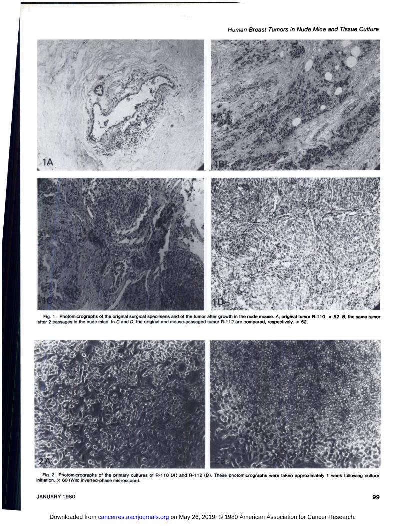
The cells were very resistant to treatment with AN; somewould detach after 15 mm at 37°,but 30 mm incubation wererequired to remove the majority of the cells in the colony. Thecolonies that were ‘‘cloned―by the selective removal of wholemounds of cells contained few fibroblast-like cells. However, itwas noticed that in some clones, the cells seemed to prefer toattach to fibroblast-like cells that were already spread out onthe growing surface of the dish. A series of photomicrographsshowing what appears to be selective attachment of tumor cellsto fibroblast-like cells is presented in Fig. 3. These cells arefrom passage 7 of R-112 cultures.
Estradiol Receptors. Frozen sectionsof the originaltumorswere not available for estradiol receptor analysis, but assessment of the tumors removed from the nude mice showedreceptor concentrations of 5.3 and 3.4 fmol/mg protein for A-110 and A-i 12, respectively. A-i 11 showed no specific estrogen binding; R-249 has not been tested. The receptor concentrations are relatively low; however, the mice had been implanted with estrogen pellets and the binding sites may alreadyhave been occupied and unavailable for binding with radioactive ligand. The percentage of mouse stromal cells (about i 0%for both A-i 10 and A-i I 2 according to LDH assays) may alsohave diluted to some extent the actual estrogen receptor values. Characterization of the cell strains in culture has not beencompleted. However, preliminary studies on the cultured tumorcells suggest that all 3 cell strains, A-i 1OA, A-i 1OB, and A-i i 2 require insulin and estradiol for growth.6 No attempt hasbeen made to culture A-i 11 and A-249.
DISCUSSION
The growth curves of the human breast tumor implants innude mice indicate 3 separate phases occurring in the initialpassage in the mice: (a) a period of degeneration of much ofthe tissue; (b) a period of survival of the tissue remaining; and(c) a period of growth of those cells that have somehow
6 B. Rae-Venter, unpublished observations.
40 80 20
DAYS
Chart 1. The growth of human breast tumor tissue in athymic nude mice. A,first passage of the tumor designated R-110: B, first passage of the tumordesignated R-112. The tumors were inoculated s.c. into the flank of a femalemouse. An estrogen pellet was implanted on the contralateral side.
rhous carcinoma. The original tumor and the tumor from passage 2 from the nude mice are shown in the light photomicrographs in Fig. 1, C and D. These 2 breast carcinomas wereoriginally characterized histopathologically as showing an infiltrating ductal pattern formed by polygonal tumor cells. Thecells had a moderate amount of cytoplasm and moderatelydyskaryotic nuclei. A moderate fibrous stromal response wasassociated with the nests of tumor cells. The first 2 passagesof A-i 10 in the nude mouse show very few tumor cells, withpredominantly fibrous stroma with foci of calcification in theimplantation sites. Not until the third passage did the tumorresume growing in a pattern similar to that of the primary tumor.The histological pattern of R-i 12 was maintained in all passages in the nude mouse as were those of A-i 11 and A-249.
In other organs, including spleen, liver, lymph nodes, andlung from the mice receiving transplantable tumors and incontrol mice not receiving tumor transplants, no significantdifferences were noted in histological appearance. In the micereceiving estrogen implants, no mammary tumors or pituitarytumors were observed. The animals lived a normal life span (upto 1 year or more). Skin keratinization was observed in a higherpercentage of the mice treated with estrogen than those notreceiving estrogen.4 No other effects which may be related toestrogen administration were noted.
Tissue Culture from Biopsy Material. Thirty-two of the malignant tumors were cultured immediately upon receipt fromthe hospitals; 23 were minced in growth medium containing10% dimethyl sulfoxide and frozen. Of the 32 tumors culturedimmediately, epithelial outgrowth was observed in 15, and from10 of these, cells were obtained in third passage. However,after periods ranging from 3 to 9 months, it was not possible toobtain enough cells for a fourth passage. Little mitotic activity
; was observed in these cultures. A variety of media, with and
without steroid hormones and insulin were tried. Some of thesecultures had exhibited exceptionally rapid growth in the primaryculture and even in the second passage. Those tumors thatwere cultured subsequent to freezing did not grow. A summaryof these results is presented in Table 2.
Culture of Mouse-passaged Tumors. Portions of tumors A-110 and A-i 12 that had been grown for at least 2 passages inthe nude mice were cultured as described in ‘‘MaterialsandMethods. ‘‘A very dense culture of epithelial-like cells growingin mounds developed within 1 week. These mounds were
97JANUARY1980
on May 26, 2019. © 1980 American Association for Cancer Research. cancerres.aacrjournals.org Downloaded from

B. Rae-Venter and L. M. Reid
â€â€˜adapted― to the conditions. This apparent adaptation process
in the initial passage is not fully understood at this time but isthe focus of a number of studies. One obvious factor is theability of the tissues to become vascularized. In the initialpassage, vascularization requires at least i month, whereas insubsequent passages, it may occur in 3 to 4 days. Disaggregation of the tissue enzymatically or mechanically prior toimplantation for the first passage did not reduce the 1-monthperiod necessary for vascularization in this initial passage orreduce the up-to-6-month “adaptationperiod' ‘necessary forsome tumors to initiate growth. Studies of biochemical markers(such as estrogen receptors) and histopathoiogy indicate thatthe tumors have remained differentiated throughout passaginginthenudemice.
Benign and malignant tumor specimens grew in the mice.However, for all the benign tumors and for a portion of themalignant ones, the tumor growth was temporary, lasting atmost through 3 passages in the mice or, in time, at most 1year. After cessation of growth, neither transplantation of thetissue nor culturing of it succeeded in reinitiating growth of thecells. The percentage of breast tumors that have resulted inpermanent lines in the mice is low (approximately 10%) incomparison to tumors from nonendocrine tissues. The causeof the cessation of growth of the tissue is unknown. It isperhaps a ‘‘senescence―of the cells or a depletion of anessential factor. Those tumors which were developed intotransplantable lines were all initiated in animals receiving animplantation of estrogen. However, it is not clear if this couldhave been related to a primary effect of the estrogen on thesetumors or perhaps a secondary effect of increasing the secretion of pituitary prolactin. It will be important to obtain a moreprecise determination of the endocrine factors that are requiredby these tumors to facilitate their survival and growth in thenude mice. These observations also hold true for a number ofother tumors that have been implanted in the nude mice (16).
The technique of using nude mice to aid in the establishmentof cultures of human tumor cells appears from the results ofothers (10) and from our own studies to be quite advantageous.Cultures of human breast tumors initiated directly from surgicalspecimens failed to thrive in culture past the third passage. Incontrast to these findings, when R-110 (passages 2 and 4) andA-i 12 (passage 2) were cultured subsequent to passage in thenude mice, outgrowth of epithelial cells and establishment ofcell strains occurred in all 3 attempts. The successful establishment of cultures from human tumors first grown in nudemice when these same tumors failed to survive in culturesderived directly from the surgical specimen is perhaps relatedto the adaptation-selection process discussed above. In someway, the adaption to growth in the nude mice contributes tothe ability of the cells to be cultured. Also possibly relevant tothis question is the observation that the tumor cells showed atendency to attach to areas of fibroblast proliferation and thatthe rate of growth of the epithelial cells decreased in thosecultures from which fibroblast-like cells had been eliminated.
This study was prompted by the need for human in viva andin vitro models for testing potential drugs for cancer chemotherapy (8, 21). The transplantable human tumor lines and theepithelial cell strains established from the tumor lines provideexcellent models for preliminary testing of new drugs (5). In
98
addition, they may be useful for furth@ stUdies Into the mechanism of steroid and peptide hormovie action on mammaryepithelium.
ACKNOWLEDGMENTS I
The authors would like to thank Professors N@tt1an0. Kaplan and GordonSato, In whose laboratories this work was done, @ortheir support and encouragement. The help of Drs. G. Niwayama and R. Williams with the pathology isgratefully acknowledged. We would also like to th$nk the personnel of the manySan Diego hospitals cooperating in these studies. @rhelrassistance In obtainingtumor tissue enabled these studies to be done.
REFERENCES
1. Bassin, R. H., Plate, E. J., Gerwln. B. I., [email protected]. F., Haapala, D. K., andChu, E. W. Continuous epithelloid cell line. P@T-3. from a human breastcarcinoma (36850). Proc. Soc. Exp. Bid. Med@141: 673—680,1972.
2. Brooks, S. C.. Locke, E. R., and Soule, H. 0. E$rogen receptor In a humancell line (MCF-7) from breast carcinoma. J. [email protected]., 248: 6251-6253,1973.
3. Cailleau, R., Young, A., Olive, M., and ReevesjW. J., Jr. Breast tumor celllines from pleural effusions. J. Nat). Cancer lns4, 53: 661-674, 1974.
4. Dobrynin, Y. V. Establishment and characteristI@sof cell strains from someepithellal tumors of human origin. J. Nat). Car@cerInst., 31: 1173-1196,1963.
5, Everse, J., Lappi, D. A., Beglau, Lee, C-I., and I@ap1an,N. 0. Investlgstlonsinto the relationship between structure and fu@lctionof dIphtheria toxin.Proc. Nat). Aced. Sd. U. S. A., 74: 472-476, 1q77.
6. Flanagan, S. P. “Nude,―a new hairless gene w1@hplelofropic effects In themouse. Genet. Res., 8: 295-309, 1966.
7, Jensen, E. v., DeSombre, E. R., and Jungbluft, P@W. Estrogen receptors Inhormone-responsive tissues and tumors. In: A. @.Weesler, 1. L. Dao, andS. Wood, Jr. (ode.), Endogenous Factors Influen$@lngHost Tumor Balance,PP. 15-30. ChIcago: The University Press, 1967@
8. Kaplan, N. Target-directed cancerchemotherapet@ticaIagents. In: J. Schutz,and I. Ahmad, (ads.), Cancer Enzymology, PP. 2@1-227. New Yor$: Acedemlc Press Inc., 1976. I
9, Lasfargues. E. Y., and Ozzello, L Cultivation of h@imanbreast carcinomas.J. Nat). CancerInst., 21: 1131—1147,1958.
10. Merenda. C., Sordat, B., Mach, J. P., and Carr@, S. Human endomstrialcarcinomas serially transplanted in nude mice and $tablished In continuouscell lines. Int. J. Cancer, 16: 559—570,1975.
11. Outzen, H. C., and Custer, R. P. Growth of hum@ normal and neoplastlcmammary tissues in the cleared mammary fat pa4Iof the nude mouse. J.NatI.Cancerlnst.,55:1461—1463,1975.
12. Pantelouris, E. M. Absence of thymus In a [email protected] (Load.),217:370—371,1968.
13. Plata, E. J., Aoki, T., Robertson, D. D., Chu, E. V,1,and Gerwln. B. I. Anestablished cultured cell line (HBT-39) from hum4n breast carcinoma. J.Nat). Cancer Inst., 50: 849-857, 1973.
14. Polvsen, C. 0., and Rygaard, J. Heterotransplantat@onof human adenocarcinomas of the colon and rectum in the mouse mut$@tnude. A study of nineconsecutive transplantations. Acta Pathol. Microbi$1. Scand. Sect. A, 79:159—169,1971. I
15. Reid, L, Holland, J., Jones, C., Wolf, B., Nlwayama,$., Williams, A., Kaplan,N.. and Sato, G. Some of the variables affectIng 114success of traneplantation of human tumors into the athumic nude [email protected]: D. Houchens andT. Ouejera, (eds.), Proceedings of the Symposium @nthe Use of AthymlC(Nude) Mice in Cancer Research. New York: Gustav @lsher,In press, 1979.
16. ReId, L C. M. and Shin, S-I. TransplantatIon of h@terologousendocrinetumor cells in nude mice. In: J. Fogh and B. Glova4ella (edo.), The NudeMouse In Experimental and ClInIcal Research, PP. 313-351 . New York:Academic Press, Inc., 1978.
17. ReId, L, Wolf, B., Niwayama, G., Kaplan, N. 0., and nato, G. DevelOpmentof transplantable human tumors in nude mice. Prod. Am. Aseoc. CancerRae.,18:161,1977.
18. Rose, H. N. and McGrath, C. M. a-Lactalbumln prod@cttonin human mammary carcinoma. Science, 190: 673-675, 1975.
19. Soule, H. D., Vazquez, J., Long, A., Albert, S., and Bi@ennan,J. Human cellline from a pleural effusion derived from a breast [email protected]. Nat).CancerInst.,51:1409—1419,1973. I
20. Trempe, G.. and Fogh. J. Variation in Characterlstlcslof human tumor celllines derived from similar tumors. In Vitro. 8: 433—435,1973.
21. Venter, B. R. Hexestrol-mustard: a new antlneoplastIc@agent with estrogenreceptor affinity. In: Dissertation submitted to the Un@ierettyof California,San Diego, PP.132-1 58, 1976 (DIssertation Abstracts@nternatIonaINo. 77-533). 1
CANCER RES RCH VOL. 40
on May 26, 2019. © 1980 American Association for Cancer Research. cancerres.aacrjournals.org Downloaded from
.@i@ ,:_@ -.@‘ ° @f,@ -
. / .@ ..@,@ @:.- —V •‘@?
. .@ .
@. I ,‘@ @;@
.‘ @, ,;.
.‘ @@:‘:
.. ‘I. •
I.' . ‘ . .. , .. .. @‘.@
,
.@@ . S : ‘ . : •@‘
k1( ,)qt,, ,@ ‘ . . . ‘.S.'.'-'@ . @,.‘ .-.,,.
-@ , ‘ .@ â€.- . . â€. @.
k)@@ I@@@@
@@ . @, ..‘. ‘, ,
.. ‘. - (.\ ‘F-..,•.@@@ .
. S
. ,—@@,. . ‘
;‘@ -‘4
@ :
. - ‘@@ \ ,@
A
..—
@‘
@..@1@::
:@ .s, @.@ :@ . @.-,@,
@ @‘% @: ‘@ :@@@
@ ._d,t@ @‘
4@ @.. ‘ ‘, @t@\'::4――. @—@‘:.@.,@_•_s'@ . ‘@
@,e ‘ @:@ :@‘@ ‘
Fig. 1. Photomicrographs of the original surgical specimens and of the tumor after growth in the nude mouse. A, original tumor R-110. x 52. B, the same tumorafter 2 passages in the nude mice. In c and D, the original and mouse-passaged tumor R-112 are compared, respectively. x 52.
‘@@rz:.@ I;‘;@d@ç..@ /@ c-.@,,@@@@ @,,@ 4*.r@@.c@1ì;@@ @/@‘@::@@ , @.
@@@@ .@ ¶@ ‘,@ . I @“,@@ _ ‘.
I-@@@ .@ •‘r1‘•‘â€â€˜ .@@r'@)'@ .@ .‘@ ‘. \ (V
@ @‘@‘@J.‘-. ,.., .@@ ..@ () . ? . ./@; , @;@ @,
@@ L4@
c@-i-@@:';.@“@ . . . /,@‘ _c4d@:. •:, @: .@
@ ,4—.@ “@:‘ :. , \ ;@ . I@@ .@ @r'@ . ‘ .
e@d@':@i;;/ .. ‘1'@@
@ :@‘.-r C@:..H i@@
Fig. 2. Photomlcrographs of the primary cultures of R-110 (A) and R-112 (B). These photomlcrographs were taken approximately 1 week following cultureinitiatIon. x 60 (Wild Inverted-phase microscope).
99
Human Breast Tumors in Nude Mice and Tissue Cu!ture
...:‘@‘.;, -,‘..@@ ;. :.@ .L_@I ‘. .@@ .
@ .@@@ ‘V
@ø;i::@ ‘@ i@ci_::1fA@ r@
‘ . . -@ ., s@. :.@@@ . •@@ @. . ‘@,t,; ‘,.@ @.‘:@ ,, .
@@ 1@'a
;@ -@
. . .. [email protected]..,,.. I..'. .-@ ‘.@.‘ ‘I @V, @:@-@‘•:@@@
,, .â€: . .
JANUARY1980
on May 26, 2019. © 1980 American Association for Cancer Research. cancerres.aacrjournals.org Downloaded from

@-.@ .@
@ .., S.,@@ ,,s5@@‘:‘‘,:@..@ .t_..•@'@#@
%.. S.'. S@ ,,@@@ ‘
. ‘. â€4' . , S@ ,‘@ @• S.:―@@ S@ :@ .5@@
‘@ ,,,, @•o@,,:i;@ 4. •@ ‘I@,#p.4.,,,•. @:-@@ . I‘ S.@@@ 5,,
.@ . -# .@S@@
0_i@ C h@@ .I,_'@
S@ ..@ .‘ ;@@ \.
. .,%,@@ ‘ S.k,'.@.@ •,•5@#•‘D@S •@,a@ @•@@ VI
c@ ‘,.@
S@@@@ 0@ e'•@@,@@@ :@ @‘@@ .@.@@ S
C,,,, ,@@@ I
B. Rae-Venter and L. M. Reid
@—. .:C'@ 0@: @1
.6
/.1
0‘C,
:
0@ ef.@A'@ ,.k
-,
S
U
CC,:@,,@ ,@3
0
C
0
,@:‘@@
&
00C.‘V.C
p.C.,
.@
a.
Fig. 3.area of the culture Is left undisturbed.
100 CANCER VOL. 40
on May 26, 2019. © 1980 American Association for Cancer Research. cancerres.aacrjournals.org Downloaded from

1980;40:95-100. Cancer Res Barbara Rae-Venter and Lola M. Reid Subsequent Establishment in Tissue CultureGrowth of Human Breast Carcinomas in Nude Mice and
Updated version
http://cancerres.aacrjournals.org/content/40/1/95
Access the most recent version of this article at:
E-mail alerts related to this article or journal.Sign up to receive free email-alerts
Subscriptions
Reprints and
To order reprints of this article or to subscribe to the journal, contact the AACR Publications
Permissions
Rightslink site. Click on "Request Permissions" which will take you to the Copyright Clearance Center's (CCC)
.http://cancerres.aacrjournals.org/content/40/1/95To request permission to re-use all or part of this article, use this link
on May 26, 2019. © 1980 American Association for Cancer Research. cancerres.aacrjournals.org Downloaded from