Grip Strength and Muscle Fatigue
6
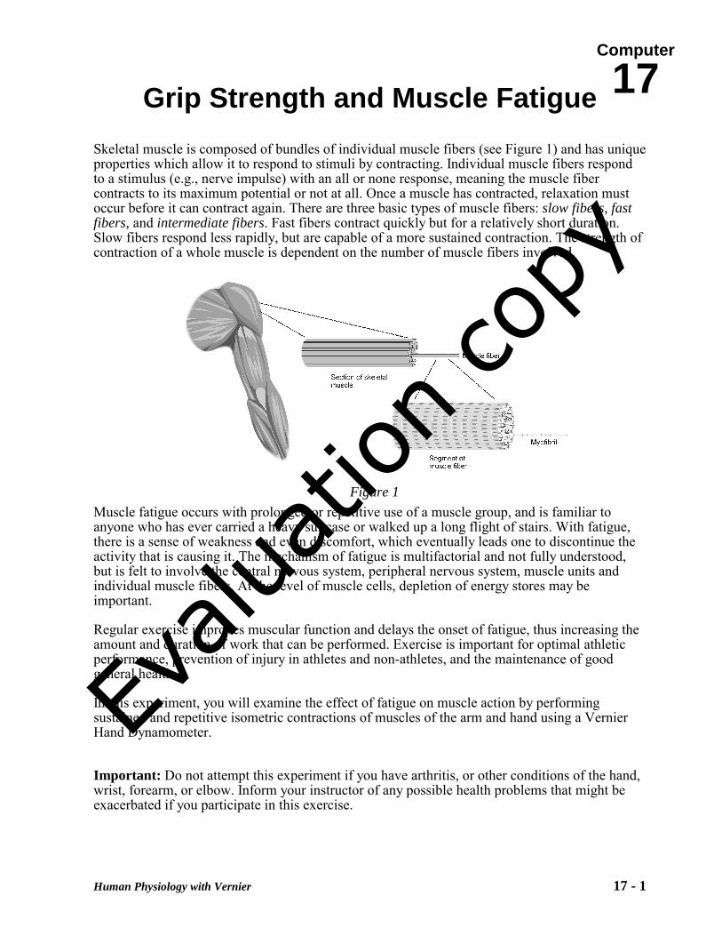
Computer 17 Human Physiology with Vernier 17 - 1 Grip Strength and Muscle Fatigue Skeletal muscle is composed of bundles of individual muscle fibers (see Figure 1) and has unique properties which allow it to respond to stimuli by contracting. Individual muscle fibers respond to a stimulus (e.g., nerve impulse) with an all or none response, meaning the muscle fiber contracts to its maximum potential or not at all. Once a muscle has contracted, relaxation must occur before it can contract again. There are three basic types of muscle fibers: slow fibers, fast fibers, and intermediate fibers. Fast fibers contract quickly but for a relatively short duration. Slow fibers respond less rapidly, but are capable of a more sustained contraction. The strength of contraction of a whole muscle is dependent on the number of muscle fibers involved. Muscle fatigue occurs with prolonged or repetitive use of a muscle group, and is familiar to anyone who has ever carried a heavy suitcase or walked up a long flight of stairs. With fatigue, there is a sense of weakness and even discomfort, which eventually leads one to discontinue the activity that is causing it. The mechanism of fatigue is multifactorial and not fully understood, but is felt to involve the central nervous system, peripheral nervous system, muscle units and individual muscle fibers. At the level of muscle cells, depletion of energy stores may be important. Regular exercise improves muscular function and delays the onset of fatigue, thus increasing the amount and duration of work that can be performed. Exercise is important for optimal athletic performance, prevention of injury in athletes and non-athletes, and the maintenance of good general health. In this experiment, you will examine the effect of fatigue on muscle action by performing sustained and repetitive isometric contractions of muscles of the arm and hand using a Vernier Hand Dynamometer. Important: Do not attempt this experiment if you have arthritis, or other conditions of the hand, wrist, forearm, or elbow. Inform your instructor of any possible health problems that might be exacerbated if you participate in this exercise. Figure 1 Evaluation copy
Transcript of Grip Strength and Muscle Fatigue
Computer
17
Human Physiology with Vernier 17 - 1
Grip Strength and Muscle Fatigue Skeletal muscle is composed of bundles of individual muscle fibers (see Figure 1) and has unique properties which allow it to respond to stimuli by contracting. Individual muscle fibers respond to a stimulus (e.g., nerve impulse) with an all or none response, meaning the muscle fiber contracts to its maximum potential or not at all. Once a muscle has contracted, relaxation must occur before it can contract again. There are three basic types of muscle fibers: slow fibers, fast fibers, and intermediate fibers. Fast fibers contract quickly but for a relatively short duration. Slow fibers respond less rapidly, but are capable of a more sustained contraction. The strength of contraction of a whole muscle is dependent on the number of muscle fibers involved.
Muscle fatigue occurs with prolonged or repetitive use of a muscle group, and is familiar to anyone who has ever carried a heavy suitcase or walked up a long flight of stairs. With fatigue, there is a sense of weakness and even discomfort, which eventually leads one to discontinue the activity that is causing it. The mechanism of fatigue is multifactorial and not fully understood, but is felt to involve the central nervous system, peripheral nervous system, muscle units and individual muscle fibers. At the level of muscle cells, depletion of energy stores may be important.
Regular exercise improves muscular function and delays the onset of fatigue, thus increasing the amount and duration of work that can be performed. Exercise is important for optimal athletic performance, prevention of injury in athletes and non-athletes, and the maintenance of good general health.
In this experiment, you will examine the effect of fatigue on muscle action by performing sustained and repetitive isometric contractions of muscles of the arm and hand using a Vernier Hand Dynamometer.
Important: Do not attempt this experiment if you have arthritis, or other conditions of the hand, wrist, forearm, or elbow. Inform your instructor of any possible health problems that might be exacerbated if you participate in this exercise.
Figure 1
Evalua
tion co
py
Computer 17
17 - 2 Human Physiology with Vernier
OBJECTIVES In this experiment, you will
• Obtain graphical representation of the force exerted by your hand while gripping. • Observe the change in hand strength during a continuous grip over time. • Observe the change in hand strength during rapid, repetitive gripping.
MATERIALS
computer Logger Pro Vernier computer interface Vernier Hand Dynamometer
PROCEDURE Select one person from your lab group to be the subject. Part I Muscle Strength with Continuous Grip 1. Connect the Hand Dynamometer to the Vernier computer interface. Open
the file “17a Grip Strength Fatigue” from the Human Physiology with Vernier folder.
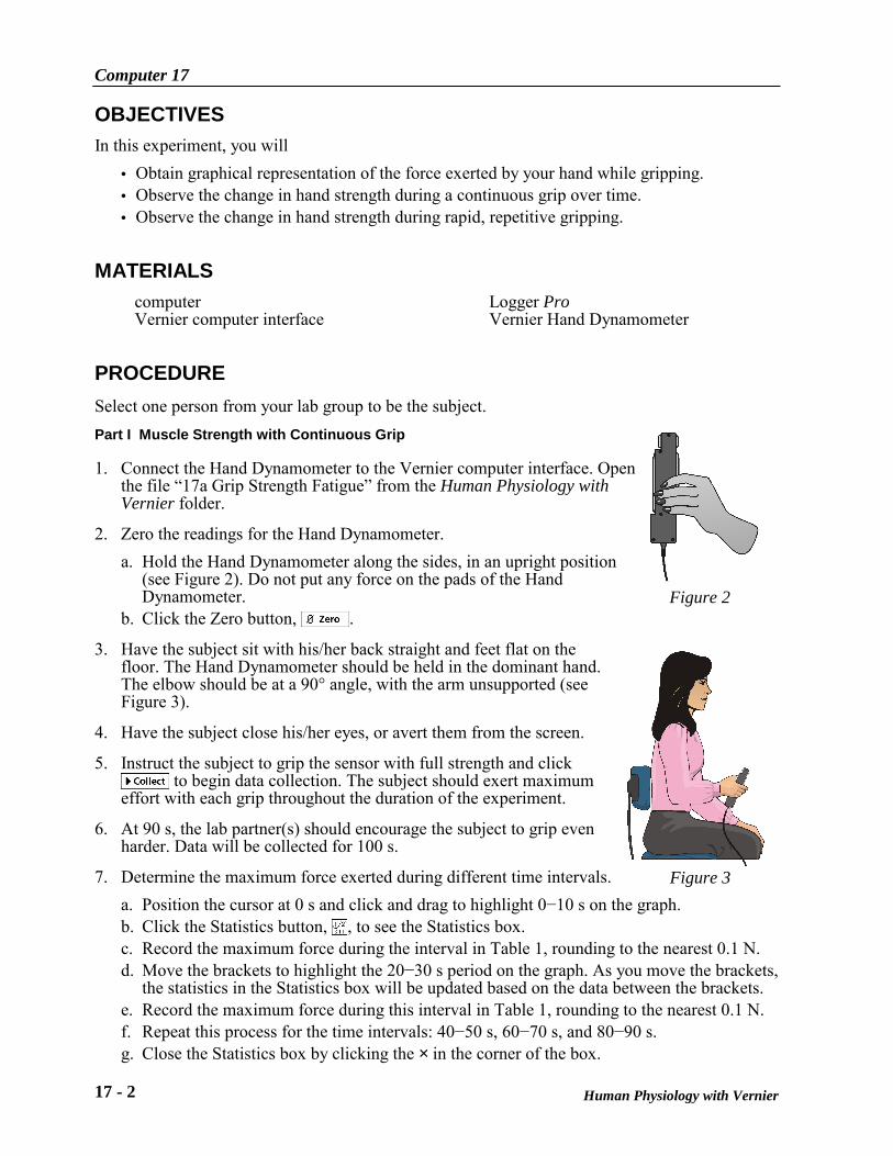
2. Zero the readings for the Hand Dynamometer. a. Hold the Hand Dynamometer along the sides, in an upright position
(see Figure 2). Do not put any force on the pads of the Hand Dynamometer.
b. Click the Zero button, . 3. Have the subject sit with his/her back straight and feet flat on the
floor. The Hand Dynamometer should be held in the dominant hand. The elbow should be at a 90° angle, with the arm unsupported (see Figure 3).
4. Have the subject close his/her eyes, or avert them from the screen.
5. Instruct the subject to grip the sensor with full strength and click to begin data collection. The subject should exert maximum
effort with each grip throughout the duration of the experiment.
6. At 90 s, the lab partner(s) should encourage the subject to grip even harder. Data will be collected for 100 s.
7. Determine the maximum force exerted during different time intervals. a. Position the cursor at 0 s and click and drag to highlight 0−10 s on the graph. b. Click the Statistics button, , to see the Statistics box. c. Record the maximum force during the interval in Table 1, rounding to the nearest 0.1 N. d. Move the brackets to highlight the 20−30 s period on the graph. As you move the brackets,
the statistics in the Statistics box will be updated based on the data between the brackets. e. Record the maximum force during this interval in Table 1, rounding to the nearest 0.1 N. f. Repeat this process for the time intervals: 40−50 s, 60−70 s, and 80−90 s. g. Close the Statistics box by clicking the × in the corner of the box.
Figure 3
Figure 2
Grip Strength and Muscle Fatigue
Human Physiology with Vernier 17 - 3
8. Calculate the difference between each maximum value and the next and record these values in Table 1.
9. Position the cursor at 0 s. Click and drag to highlight 0−90 s on the graph. Click the Linear fit button, , and record the slope (round to the nearest 0.01) in Table 3.
Part II Muscle Strength with Repetitive Grip
10. Open the file “17b Grip Strength Fatigue” from the Human Physiology with Vernier folder. 11. Have the subject sit with his/her back straight and feet flat on the floor. The Hand
Dynamometer should be held in the dominant hand. The elbow should be at a 90° angle, with the arm unsupported (see Figure 2).
12. Have the subject close his/her eyes, or avert them from the screen.
13. Zero the readings for the Hand Dynamometer. a. Hold the Hand Dynomometer along the sides, in an upright position. Do not put any force
on the gray pads of the Hand Dynamometer. b. Click the Zero button, .
14. Instruct the subject to rapidly grip and relax his/her grip on the sensor (approximately twice
per second). Click to begin data collection. The subject should exert maximum effort throughout the duration of the experiment.
15. At 90 s, the lab partner(s) should encourage the subject to grip even harder. Data will be collected for 100 s.
16. Determine the maximum force exerted during different time intervals. a. Position the cursor at 0 s and click and drag to highlight 0−10 s on the graph. b. Click the Statistics button, , and record the maximum force during that interval in
Table 2, rounding to the nearest 0.1 N. c. Move the brackets to highlight the 20–30 s period on the graph. As you move the brackets,
the statistics in the Statistics box will be updated based on the data between the brackets. d. Record the maximum force during this interval in Table 1, rounding to the nearest 0.1 N. e. Repeat this process for the time intervals: 40–50 s, 60–70 s, and 80–90 s and then close
the Statistics box by clicking the × in the corner of the box.
17. Calculate the difference between each maximum value and the next and record these values in Table 2.
18. Position the cursor at 0 s. Click and drag to highlight 0–90 s on the graph. Click the Linear fit button, , and record the slope in Table 3.
Computer 17
17 - 4 Human Physiology with Vernier
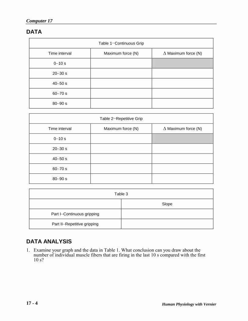
DATA
Table 1−Continuous Grip
Time interval Maximum force (N) ∆ Maximum force (N)
0–10 s
20–30 s
40–50 s
60–70 s
80–90 s
Table 2−Repetitive Grip
Time interval Maximum force (N) ∆ Maximum force (N)
0–10 s
20–30 s
40–50 s
60–70 s
80–90 s
Table 3
Slope
Part I–Continuous gripping
Part II–Repetitive gripping
DATA ANALYSIS 1. Examine your graph and the data in Table 1. What conclusion can you draw about the
number of individual muscle fibers that are firing in the last 10 s compared with the first 10 s?
Grip Strength and Muscle Fatigue
Human Physiology with Vernier 17 - 5
2. Is the change in number of muscle fibers that contract occurring at a constant rate?
3. Use your knowledge of fast, slow, and intermediate skeletal muscle fibers to hypothesize which fibers are contracting in the first, third, and final 10 s intervals.
4. How might you explain the subject’s response to coaching? This should be evident in the last 10 s of data for Parts I and II of the exercise. Discuss the possible involvement of the central nervous system, in addition to the muscle fibers.
5. Compare the slopes recorded in Table 3. Give a possible explanation for the difference, if any, in muscle fatigue rates seen in continuous versus repetitive gripping.

Vernier Lab Safety Instructions Disclaimer
THIS IS AN EVALUATION COPY OF THE VERNIER STUDENT LAB. This copy does not include:
Safety information Essential instructor background information Directions for preparing solutions Important tips for successfully doing these labs
The complete Human Physiology with Vernier lab manual includes 24 labs and essential teacher information. The full lab book is available for purchase at: http://www.vernier.com/cmat/hpa.html
Vernier Software & Technology
13979 S.W. Millikan Way • Beaverton, OR 97005-2886 Toll Free (888) 837-6437 • (503) 277-2299 • FAX (503) 277-2440
[email protected] • www.vernier.com

![B277 BIOLOGY OF MUSCLE The molecular motor of muscle Learning Outcome. Discuss muscle contraction [development, adaptation to training and fatigue]. Learning.](https://static.fdocuments.in/doc/165x107/56649ce65503460f949b3a0b/b277-biology-of-muscle-the-molecular-motor-of-muscle-learning-outcome-discuss.jpg)