Grasping Execution and Grasping Observation Activity of ... · Grasping Execution and Grasping...
14
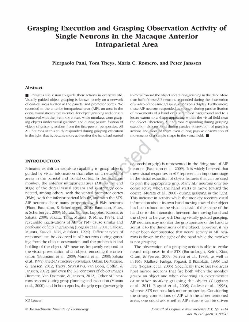
Uncorrected Proof Grasping Execution and Grasping Observation Activity of Single Neurons in the Macaque Anterior Intraparietal Area Pierpaolo Pani, Tom Theys, Maria C. Romero, and Peter Janssen Abstract ■ Primates use vision to guide their actions in everyday life. Visually guided object grasping is known to rely on a network of cortical areas located in the parietal and premotor cortex. We recorded in the anterior intraparietal area (AIP), an area in the dorsal visual stream that is critical for object grasping and densely connected with the premotor cortex, while monkeys were grasp- ing objects under visual guidance and during passive fixation of videos of grasping actions from the first-person perspective. All AIP neurons in this study responded during grasping execution in the light, that is, became more active after the hand had started to move toward the object and during grasping in the dark. More than half of these AIP neurons responded during the observation of a video of the same grasping actions on a display. Furthermore, these AIP neurons responded as strongly during passive fixation of movements of a hand on a scrambled background and to a lesser extent to a shape appearing within the visual field near the object. Therefore, AIP neurons responding during grasping execution also respond during passive observation of grasping actions and most of them even during passive observation of movements of a simple shape in the visual field. ■ INTRODUCTION Primates exhibit an exquisite capability to grasp objects guided by visual information that relies on a network of areas in the parietal and frontal cortex. In the macaque monkey, the anterior intraparietal area (AIP) is the end stage of the dorsal visual stream and is strongly con- nected, among others, with the ventral premotor cortex (PMv), with the inferior parietal lobule, and with the STS. AIP neurons share many properties with PMv neurons (Fluet, Baumann, & Scherberger, 2010; Baumann, Fluet, & Scherberger, 2009; Murata, Gallese, Luppino, Kaseda, & Sakata, 2000; Sakata, Taira, Murata, & Mine, 1995), and reversible inactivations of AIP or PMv cause similar and profound deficits in grasping (Fogassi et al., 2001; Gallese, Murata, Kaseda, Niki, & Sakata, 1994). Different types of responses can be observed in AIP neurons during grasp- ing, from the object presentation until the prehension and holding of the object. AIP neurons frequently respond to the visual presentation of an object, encoding the orien- tation (Baumann et al., 2009; Murata et al., 2000; Sakata et al., 1995), the 3-D structure (Srivastava, Orban, De Maziere, & Janssen, 2012; Theys, Srivastava, van Loon, Goffin, & Janssen, 2012), and even the 2-D contours of object images (Romero, Van Dromme, & Janssen, 2012). Other AIP neu- rons respond during grasp planning and execution (Murata et al., 2000), and in both epochs, the grip type (power grip or precision grip) is represented in the firing rate of AIP neurons (Baumann et al., 2009). It is widely believed that these visual responses in AIP represent an important stage in the visual extraction of object features that can be used to plan the appropriate grip. Many AIP neurons only be- come active when the hand starts to move toward the object (Murata et al., 2000) during grasping in the light. This increase in activity while the monkey receives visual information about its own hand moving toward the object has been related to the visual analysis of the shape of the hand or to the interaction between the moving hand and the object to be grasped: During visually guided grasping, AIP neurons may monitor the grip aperture of the hand to adjust it to the dimensions of the object. However, it has never been demonstrated that neural activity in AIP neu- rons is driven by the sight of the hand while the monkey is not grasping. The observation of a grasping action is able to evoke neural responses in the STS (Barraclough, Keith, Xiao, Oram, & Perrett, 2009; Perrett et al., 1989), as well as in PMv (Gallese, Fadiga, Fogassi, & Rizzolatti, 1996) and PFG (Fogassi et al., 2005). Specifically these last two areas host mirror neurons that fire both when the monkey grasps an object and when observing an experimenter or another monkey grasping the object (Caggiano et al., 2011; Fogassi et al., 2005; Gallese et al., 1996), whereas STS neurons lack motor properties. Considering the strong connections of AIP with the aforementioned areas, one could ask whether AIP neurons can be driven KU Leuven © Massachusetts Institute of Technology Journal of Cognitive Neuroscience X:Y, pp. 1–14 doi:10.1162/jocn_a_00647
Transcript of Grasping Execution and Grasping Observation Activity of ... · Grasping Execution and Grasping...
UncorrectedProof
Grasping Execution and Grasping Observation Activity ofSingle Neurons in the Macaque Anterior
Intraparietal Area
Pierpaolo Pani, Tom Theys, Maria C. Romero, and Peter Janssen
Abstract
■ Primates use vision to guide their actions in everyday life.Visually guided object grasping is known to rely on a networkof cortical areas located in the parietal and premotor cortex. Werecorded in the anterior intraparietal area (AIP), an area in thedorsal visual stream that is critical for object grasping and denselyconnected with the premotor cortex, while monkeys were grasp-ing objects under visual guidance and during passive fixation ofvideos of grasping actions from the first-person perspective. AllAIP neurons in this study responded during grasping executionin the light, that is, became more active after the hand had started
to move toward the object and during grasping in the dark. Morethan half of these AIP neurons responded during the observationof a video of the same grasping actions on a display. Furthermore,these AIP neurons responded as strongly during passive fixationof movements of a hand on a scrambled background and to alesser extent to a shape appearing within the visual field nearthe object. Therefore, AIP neurons responding during graspingexecution also respond during passive observation of graspingactions and most of them even during passive observation ofmovements of a simple shape in the visual field. ■
INTRODUCTION
Primates exhibit an exquisite capability to grasp objectsguided by visual information that relies on a network ofareas in the parietal and frontal cortex. In the macaquemonkey, the anterior intraparietal area (AIP) is the endstage of the dorsal visual stream and is strongly con-nected, among others, with the ventral premotor cortex(PMv), with the inferior parietal lobule, and with the STS.AIP neurons share many properties with PMv neurons(Fluet, Baumann, & Scherberger, 2010; Baumann, Fluet,& Scherberger, 2009; Murata, Gallese, Luppino, Kaseda, &Sakata, 2000; Sakata, Taira, Murata, & Mine, 1995), andreversible inactivations of AIP or PMv cause similar andprofound deficits in grasping (Fogassi et al., 2001; Gallese,Murata, Kaseda, Niki, & Sakata, 1994). Different types ofresponses can be observed in AIP neurons during grasp-ing, from the object presentation until the prehension andholding of the object. AIP neurons frequently respond tothe visual presentation of an object, encoding the orien-tation (Baumann et al., 2009; Murata et al., 2000; Sakataet al., 1995), the 3-D structure (Srivastava, Orban, DeMaziere,& Janssen, 2012; Theys, Srivastava, van Loon, Goffin, &Janssen, 2012), and even the 2-D contours of object images(Romero, Van Dromme, & Janssen, 2012). Other AIP neu-rons respond during grasp planning and execution (Murataet al., 2000), and in both epochs, the grip type (power grip
or precision grip) is represented in the firing rate of AIPneurons (Baumann et al., 2009). It is widely believed thatthese visual responses in AIP represent an important stagein the visual extraction of object features that can be usedto plan the appropriate grip. Many AIP neurons only be-come active when the hand starts to move toward theobject (Murata et al., 2000) during grasping in the light.This increase in activity while the monkey receives visualinformation about its own hand moving toward the objecthas been related to the visual analysis of the shape of thehand or to the interaction between the moving hand andthe object to be grasped: During visually guided grasping,AIP neurons may monitor the grip aperture of the hand toadjust it to the dimensions of the object. However, it hasnever been demonstrated that neural activity in AIP neu-rons is driven by the sight of the hand while the monkeyis not grasping.
The observation of a grasping action is able to evokeneural responses in the STS (Barraclough, Keith, Xiao,Oram, & Perrett, 2009; Perrett et al., 1989), as well asin PMv (Gallese, Fadiga, Fogassi, & Rizzolatti, 1996) andPFG (Fogassi et al., 2005). Specifically these last two areashost mirror neurons that fire both when the monkeygrasps an object and when observing an experimenteror another monkey grasping the object (Caggianoet al., 2011; Fogassi et al., 2005; Gallese et al., 1996),whereas STS neurons lack motor properties. Consideringthe strong connections of AIP with the aforementionedareas, one could ask whether AIP neurons can be drivenKU Leuven
© Massachusetts Institute of Technology Journal of Cognitive Neuroscience X:Y, pp. 1–14doi:10.1162/jocn_a_00647
UncorrectedProof
by the observation of a grasping action, and if so, whataspect of this visual stimulus is driving AIP activity. Notethat Fujii, Hihara, and Iriki (2008) observed activity dur-ing grasping execution and during grasping observationin the medial bank of the IPS, not in AIP. Furthermore,preliminary data reported in Murata and Ishida (2007)showed that PFG (and possibly AIP) neurons respondto videos of the experimenters actions.
In this study, we investigated whether AIP neuronsactive during grasping execution also respond when theanimal simply observes a grasping action. Specifically, wetested whether AIP neurons active during grasping exe-cution also respond during passive fixation of a videoof the same action, that is, show both activity duringaction execution and activity during action observation.Moreover, we asked whether the response during grasp-ing observation is related to the visual analysis of theshape of the hand approaching the object and whetherAIP activity can also be elicited with simple visual stimulimoving in the visual field. We observed that most AIPneurons active during grasping execution could be acti-vated by pure visual stimulation with a video of the samegrasping action. However, the majority of these neuronswere also activated by videos of an isolated hand on ascrambled background and even by a shape entering orappearing within the visual field.
METHODS
Subjects
Two adult male rhesus monkeys (MY, 9 kg and MD, 8 kg)served as subjects for the experiments. All experimentalprocedures, surgical techniques, and veterinary care wereperformed in accordance with the NIH Guide for Careand Use of Laboratory Animals and in accordance withthe European Communities Council Directive 2010/63/EUand were approved by the local ethical committee of theKU Leuven.
Surgery, Apparatus, and Recording Procedures
Under isofluorane anesthesia, an MRI-compatible headfixation post and recording chamber (Crist Instruments)were implanted using dental acrylic and ceramic screwsabove the left intraparietal sulcus in both monkeys.
During the experiments, the monkey was seated up-right in a chair with the head fixed, with the arm contra-lateral to the recorded hemisphere free while the otherarm was kept restrained in a comfortable position. Infront of the monkey, a custom-built, vertically rotatingcarousel was used to present the target objects used toperform the grasping task. Six different objects (a smallcylinder [15 × 15 (diameter) mm], and a small cube [side15 mm], a large cylinder [35 × 35 mm], a sphere [diam-eter 35 mm], a large cube [side 35 mm], and a cylinderwith a groove [cylinder 35 × 35 mm; groove dimensions:
35 × 7 × 5 mm]) were pseudorandomly presented oneat a time in the same position (28 cm viewing distance, atthe chest level, ∼20 cm reaching distance measured fromthe center of the hand rest position to the center of theobjects). The objects drove two different types of graspdepending on their dimensions: a pad-to-side grip (forsmall objects), which is a subtype of precision grip, anda finger-splayed wrap (Macfarlane & Graziano, 2009), cor-responding to a whole-hand grip. Both monkeys usedthe same grip types. The resting position of the hand,the start of the reach to grasp movement, and the liftingof the object were detected by fiber-optic cables. Thestart of the hand movement was detected as soon asthe palm of the hand was 0.3 cm above the resting plan,whereas lifting of the object was detected when the objectwas lifted for 0.5 cm in the vertical axis.Behind the carousel was located a display (20-in.monitor
equipped with ultrafast P46 phosphor, frame rate 120 Hz,Vision Research Graphics, experimental setup as in Theys,Pani, van Loon, Goffin, & Janssen, 2012) on which videosof grasping actions were presented at a viewing distance of40 cm (i.e., in extrapersonal space) during the passive fixa-tion of a small spot located in the center of the display.Movie onset and end were registered by a photodiodeattached to the lower right corner of the screen detectingthe onset of a bright square (occluded and not visible tothemonkey) appearing simultaneously with stimulus onsetand endpoint. For each of the six objects, movies wererecorded during training sessions by a Fujicom cameraand a laptop running Streampix software (500 frames persec, 640 × 480 resolution). The camera was positionedabove the monkeyʼs head and recorded the work spacethat the monkey could see from his position during thetask (see individual frames in Figure 1B). Thus, all moviesshowed grasping actions from the same perspective as themonkey (first-person perspective). One monkey (MY) wasrecorded while performing a visually guided grasping task.The movies were then edited and modified (Matlab) toproduce the sequence of events described in the tasks(see below).The horizontal and the vertical coordinates of the
right eye were monitored using an infrared-based cam-era system (EyeLink II; SR Research). Eye position sig-nals were sampled at 500 Hz, whereas spiking activityand photodiode pulses were sampled at 20 kHz on aDSP (C6000 series; Texas Instruments). Spikes were dis-criminated online using a dual time window discriminatoron the DSP and displayed using LabView and custom-builtsoftware.Neural activity of single units was recorded extracellu-
larly by means of “Micro Matrix” microdrives (ThomasRecording, Marburg, Germany) with integrated single-channel preamplifier. Electrodes were quartz-platinum/tungsten fibers (∼0.5–1 MÙ at 1 kHz; 80-μm diameter)referenced to their own guide tube. Neural signals wereamplified and filtered between 0.5 and 5 kHz for spikes.Spike discrimination was performed online using a dual
2 Journal of Cognitive Neuroscience Volume X, Number Y
UncorrectedProof
time window discriminator. That recordings were of singleunits was confirmed by offline analysis using the OfflineSpike sorter software (Plexon, Inc.).
Behavioral Tasks
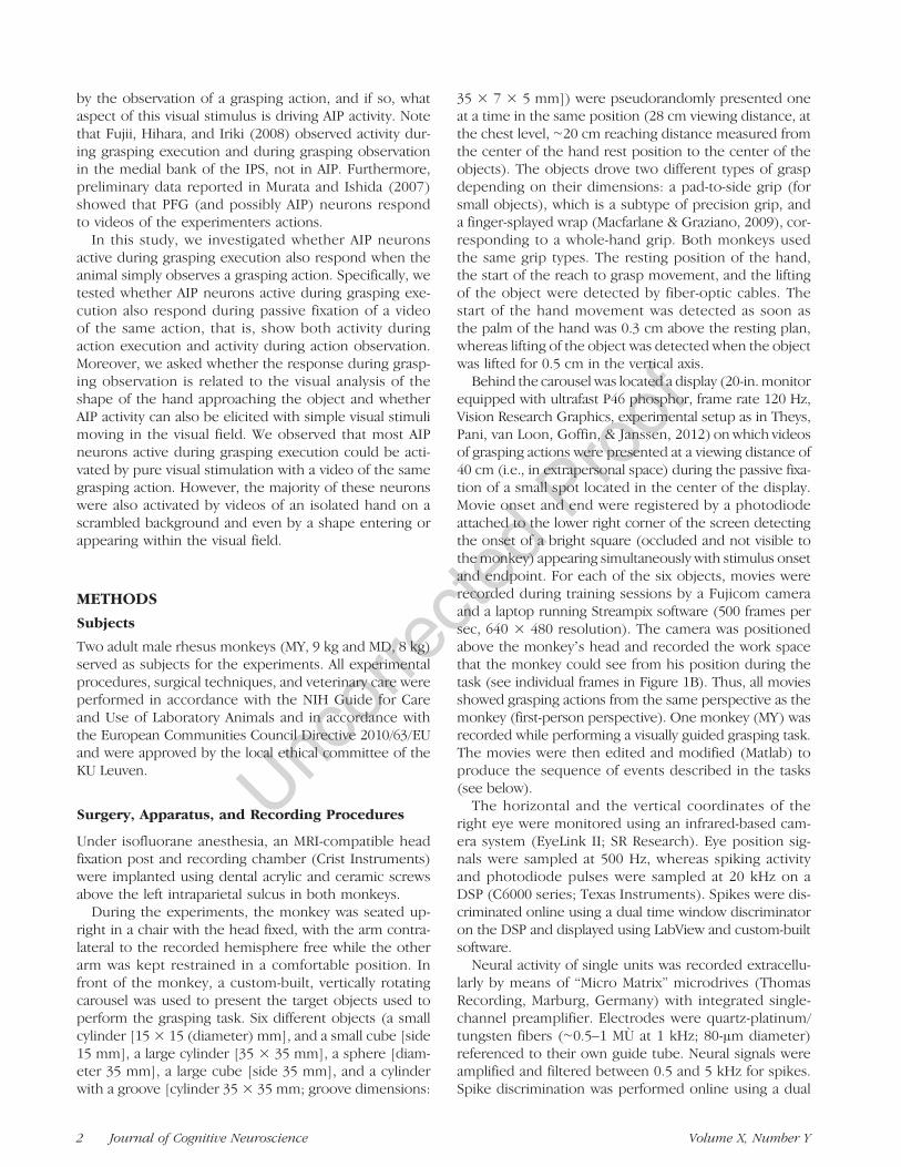
The monkeys were trained to perform various tasks (Fig-ure 1), which we classify for simplicity as Grasping tasks,requiring the interaction of the monkeys with real objects,and observation tasks, requiring the monkeys to fixate asmall spot on the monitor.
Grasping Tasks
Two versions of the grasping task were used (Figure 1A):a visually guided grasping task (VGG) and a memory-guided grasping task (MGG). In the VGG, the monkeyhad to place its right hand in a resting position in com-plete darkness for a variable time (intertrial interval,3000–5000 msec). During this time, the carousel rotatedto position the test object. An LED located near the bot-tom of the object was then illuminated, which the mon-key had to fixate (keeping the gaze inside a ±2.5-degreefixation window throughout the trial until the object waslifted). After a fixation time of 500 msec, the object wasilluminated by a lamp located above the object. After avariable delay (900–1100 msec) an auditory GO (graspingobservation) cue instructed the monkey to release the
rest position, reach, grasp, lift, and hold the object fora variable interval (holding time, 500–900 msec). Onlyat the end of a correctly completed trial was a drop ofjuice reward given. During this task, the time delay be-tween the start of the movement and lifting of the objectwas calculated (reaching–grasping time, RGT).
The MGG differed from the VGG task in that the objectwas illuminated for only 400 msec. After this time, thelight went off, and after a delay of 500–700 msec, an audi-tory GO cue instructed the monkey to grasp the objectin the dark (Figure 1, blue mark around the task).
Observation Tasks
During the observation tasks, the monkey sat in darknessin the same rest position as in the grasping tasks. All trialsstarted in the same way: After a variable time (intertrialinterval, 1000–4000 msec), a fixation spot (0.20 deg)appeared at eye level in the center of the display. Themonkey had to fixate it with the hand in the resting posi-tion and maintain his gaze around the fixation pointthroughout the trial inside a ±1.5 degree fixation window.After 500 msec, the stimulus (a movie) started. The fixa-tion point was in the same position for all tests and wassuperimposed onto the bottom of the object (as in thereal grasping task) appearing in the movies. There wasno sound in any of the videos tested. Three different testswere run in the observation task.
Figure 1. Methods. (A)Schematic illustration of thevisually guided grasping task(VGG). The red spot is thefixation point, which wasprojected onto the base ofthe object. The dotted linerepresents the period in thedark in the visually guidedgrasping task (MGG). (B)Schematic illustration of thestatic video frames of the movieof the real grasping actionpresented on the display inthe observation task. (C1)Schematic illustration of thestatic video frames of theBackground–Effector test. Wetested two effectors (isolatedhand and ellipse) and twobackgrounds (natural andscrambled). Both effectorsfollowed trajectories similarto the monkeyʼs hand in thevideo of the real grasping.(C2) Schematic illustration ofthe static video frames of theellipse test. The single ellipsecould move from the peripherytoward the object (red arrows)or away from the object, towardthe periphery (blue arrows).
Pani et al. 3
UncorrectedProof
Observation Task 1: Real Grasping Observation Test
After 700 msec (200 msec in 49% of the cells) from themovie onset, a monkey hand appeared in the right lowercorner of the movie (roughly corresponding to the start-ing position of the grasping task; Figure 1B, “Movementon”) and remained stationary for 800 msec (300 msec in49% of the cells). Then, the hand started moving (Move-ment on) and reached and grasped the object. In thistask, only one grasping action was presented, based onthe object to which the neuron gave the strongest re-sponse in the VGG task ( judged online). The distancebetween the lower right corner of the movie and thecenter of the object was 15 cm (21.2 deg). The averageestimated RGT for all the movies presented was 651.7 ±272.26 msec, which was longer than the RGT recordedduring the VGG of the same objects presented in themovies (494.2 ± 182.5) but nevertheless within the rangeof the RGTs performed (movies RGT = 0.86 z valuesreferred to the VGG RGT).
Observation Task 2: Effectors and Background Test
The sequence of events in this task was the same as inthe previous task (Figure 1B) and with the same timingregarding the onset of the effector and the start of themovement, but two different effectors and two differentbackgrounds were combined (Figure 1C1): The effectorswere either an isolated hand (top panels) or an ellipse(with a texture consisting of a scrambled version of thehand [major axis ∼95 mm, minor axis ∼43 mm], contrastequalized, bottom panels), and the background waseither the natural (right) or the scrambled background(a scrambled version of the natural background, left).For all four combinations, the same kinematic parameters(speed, trajectory) characterized the effector displace-ment in the visual field. The kinematic parameters werematched to the real grasping test: We recreated a pathfor reach-to-grasp for the finger-splayed wrap used onthe large objects and one for the pad-to-side grip usedfor the small objects. We did this because the reach-to-grasp approach movements were very similar for thedifferent objects in the two grip types and because no pre-shaping of the hand was present in this test. The durationof the RGT was ∼750 msec.
Observation Task 3: Ellipse Test
In this task, an ellipse appeared in one of six possiblepositions and moved along three different axes of motion:vertical, oblique, and horizontal (Figure 1C2). In threeconditions, the ellipse appeared in the periphery andmoved toward the object, and in the other three con-ditions, the ellipse appeared on the object and movedtoward one of the three peripheral positions (total tra-jectory lengths: vertical 8.5 cm, diagonal 11 cm, hori-zontal 7 cm, or 12.1, 15.7 and 10 deg, respectively).
The speed and trajectory of the movement was linear,and the time of the movement was always 500 msec.The background in this case was always the originalnatural background (hence with the object visible).The sequence of events of this task was the same as inthe previous task (Figure 1C).
Sequence of Tasks during the Recordings
During the experiment, the VGG task was used as a firsttask (Figure 1A) to isolate neurons. Up to six objectswere alternately presented. As soon as a clear task-modulated response (i.e., difference of activity comparedto the intertrial interval, irrespective of the precise epochof the task) for at least one of the conditions (objectspresented) was observed in the online histogram, thedata collection was started. Two different objects werepresented, one preferred (eliciting the highest responseas observed online) and one nonpreferred. To excludesomatosensory or proprioceptive responses, each neuronwas tested before data collection by the experimentergoing inside the setup and stimulating the hand, forearm,upper arm, and shoulder of the monkey with light anddeep touch and manipulating the joints of the fingers,the wrist, the elbow, and the shoulder (see Rozzi, Ferrari,Bonini, Rizzolatti, & Fogassi, 2008, for a similar procedure).Only neurons not responding to this kind of stimulationwere recorded. Roughly between 10 and 15 neuronsacross all recording sessions were excluded in this way.All neurons were also tested in the MGG task, and theseneurons were the topic of this study. This task was, de-pending on the session, presented just after the VGG taskor as the final test after all the observation tasks had beenpresented. Two objects were presented in this task (thepreferred object selected during the VGG task and thenonpreferred), and we selected only cells tested with aminimum of six trials (median number of trials: 10) percondition. The typical order of testing after the VGG testwas real grasping observation test (a video of the graspingaction with the same object as in the VGG test) or MGGtask and then effector and background test, ellipse test(minimumnumber of trials: 5,median number of trials: 10).
Data Analysis
To assess the involvement of a neuron in a task, we de-fined three epochs of interest for the VGG and MGGtasks: baseline, starting 400 msec before and ending atthe light onset; early grasping: starting 50 msec aftermonkey removed his hand from the resting position,until 50 msec before lifting; late grasping: starting 50 msecbefore lifting and ending 250 msec after the lifting started.Grasping execution activity was defined as a significantchange in spike rate in the early and/or late grasping phasecompared to the baseline epoch (before the light onset).In the analysis of the grasping observation task, similarepochs were defined: The baseline epoch started 400msec
4 Journal of Cognitive Neuroscience Volume X, Number Y
UncorrectedProof
before movement onset and lasted until movement onset,the early grasping epoch (comprising the approach tothe object and the closure of the hand around it) ran from50 msec after the hand started to move until the handwas closed around the object (duration of epochs depend-ing on the object grasped in the movie: min 350 msec,max 950 msec, median 550 msec), and the late graspingepoch started ∼100 msec before the start of the liftingand ended 200 msec later. There was no overlap betweenthe two epochs.For the effector-and-background tests, the early and
late grasping phases were also defined by the positionof the effector on the target object, mimicking the earlyand late phases of the VGG task. In this case, the epochdurations were both 400 msec, because the stimuli wereconstructed by video editing. A neuron was consideredellipse responsive if its firing rate was significantly modu-lated by the presence of the ellipse in the normal back-ground task compared to the baseline for at least one ofthe two epochs.In the ellipse test, we calculated a modulation index,
defined as (activity for movement toward the object −activity for movement away from the object)/(activity formovement toward the object + activity for movementaway from the object), doing this separately on the earlyphase (50–300 msec after effector onset, before the startof the movement onset) and the late phase (50-300 msecafter the effector reached the final position, i.e., after theend of the movement).All statistics were performed on the exact spike counts
in the defined epochs. To perform analysis on each cell,we used nonparametric tests (Kruskall–Wallis p < .05,Bonferroni corrected); to perform the analysis at thepopulation level, we used parametric tests on the averagefiring rate of the grasping epochs. For each trial, the netfiring rate was obtained by subtracting the baseline activ-ity from the epochs of interest. Normalization, when per-formed, was obtained by dividing the net firing rate ofeach cell by the maximum absolute firing rate of thesmoothed average firing rate in the grasping executionor grasping observation epoch (depending on the task).No inversion of the activity was used for population aver-ages. Spike density functions were obtained by using aGaussian half-kernel of 50 msec and were used for illus-tration purposes only.
RESULTS
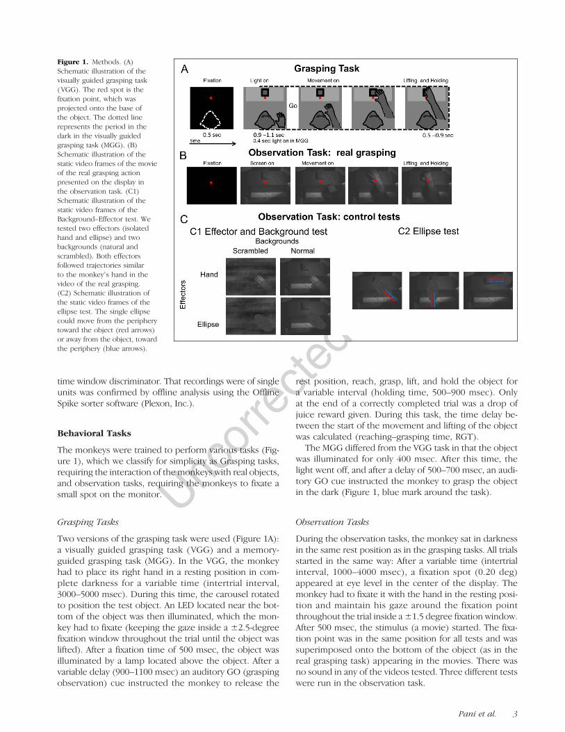
Figure 2A shows anatomical MRIs with glass capillariesinserted into grid positions that were used in this studyfor both monkeys. The depth measurements on themicrodrive and the pattern of gray to white matter tran-sitions indicated that all neurons were recorded in theanterior part (anterior–posterior range of recording posi-tions: 3 and 4 mm for MD and MY, respectively, centeredon Horsley–Clark coordinates 3 and 3.5 mm anterior and
14 and 15 mm lateral) of the lateral bank of the IPS (areaAIP). Consistent with previous studies (Baumann et al.,2009), all recordings were performed within 7 mm fromthe tip of the IPS. The recording area is indicated on thehorizontal sections (Figure 2B).
Using the VGG task, we selected online 128 neurons(n = 83 in MD, n = 45 in MY) that were significantlymodulated during the grasping phase, that is, showedgrasping execution-related activity. We use the term“grasping execution activity” simply to describe a mod-ulation in activity after the hand starts to move towardthe object. For this study, we considered only neuronsrecorded both in the VGG and MGG task showinggrasping execution activity in both tasks (104 neurons).All these AIP neurons were also tested during graspingobservation, in which we presented videos of the sameactions in the center of a display located in front of theanimal behind the carousel. The results were qualita-tively similar for the two animals and were thereforecombined.
Activity during Action Execution andAction Observation in AIP
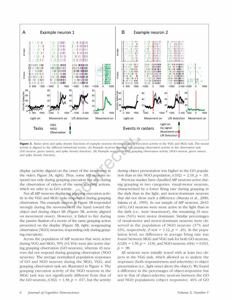
The example neuron in Figure 3A fired strongly whenthe animal grasped an object both in the light andin the dark (i.e., grasping execution activity in MGG andVGG task) but not to the onset of the light above theobject (Figure 3A, left). Interestingly, this neuron alsofired during the observation of a video of the same grasp-ing action (fist-person perspective) presented on the
Figure 2. Estimated recording positions in the AIP in monkeys D andY, indicated by the arrows on a structural MRI. (A) Coronal sections.(B) Horizontal sections.
Pani et al. 5
UncorrectedProof
display (activity aligned on the onset of the movement inthe video; Figure 3A, right). Thus, some AIP neurons re-spond not only during grasping execution but also duringthe observation of videos of the same grasping actions,which we refer to as GO activity.
Not all AIP neurons displaying grasping execution activ-ity in the VGG and MGG tasks responded during graspingobservation. The example neuron in Figure 3B respondedstrongly during the movement of the hand toward theobject and during object lift (Figure 3B, activity alignedon movement onset). However, it failed to fire duringthe passive fixation of a movie of the same grasping actionpresented on the display (Figure 3B, right; nongraspingobservation [NGO] neurons, responding only during grasp-ing execution).
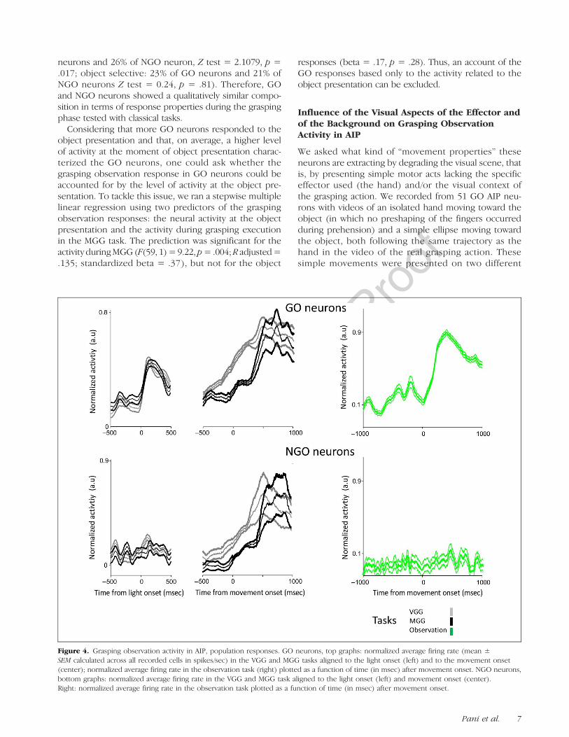
Across the population of AIP neurons that were activeduring VGG and MGG, 59% (61/104) were also active dur-ing grasping observation (GO neurons), whereas 43 neu-rons did not respond during grasping observation (NGOneurons). The average normalized population responsesof GO and NGO neurons during the MGG, VGG, andgrasping observation task are illustrated in Figure 4. Thegrasping execution activity of the NGO neurons in theMGG task was not significantly different from that ofthe GO neurons, t(102) = 1.38, p = .017, but the activity
during object presentation was higher in the GO popula-tion than in the NGO population, t(102) = 2.18, p = .03.Previous studies have classified AIP neurons active dur-
ing grasping in two categories: visual-motor neurons,characterized by a lower firing rate during grasping inthe dark than in the light, and motor-dominant neuronsthat did not show such a difference (Murata et al., 2000;Sakata et al., 1995). In our sample of AIP neurons, 28/61(46%) GO neurons were more active in the light than inthe dark (i.e., were visuomotor), the remaining 33 neu-rons (54%) were motor dominant. Similar percentagesof visual-motor and motor-dominant neurons were ob-served in the population of NGO neurons (37% and63%, respectively, Z test = 1.12, p = .26). At the popu-lation level, no difference in average firing rate wasfound between MGG and VGG task for both GO neurons,t(120) = 1.59, p= .1136, and NGO neurons, t(84) = 0.013,p = .98.All neurons were initially tested with at least two ob-
jects in the VGG task, which allowed us to analyze theresponses (both responsiveness and selectivity) to objectpresentation (i.e., light onset above the object). We founda difference in the percentages of object-responsive butnot in that of object-selective neurons between the GOand NGO populations (object responsive: 46% of GO
Figure 3. Raster plots and spike density functions of example neurons showing grasping execution activity in the VGG and MGG task. The neuralactivity is aligned to the different behavioral events. (A) Example neurons showing real grasping observation activity in the observation task(GO neuron, green rasters, and spike density function). (B) Example neurons lacking grasping observation activity (NGO neuron, green rasters,and spike density function).
6 Journal of Cognitive Neuroscience Volume X, Number Y
UncorrectedProof
neurons and 26% of NGO neuron, Z test = 2.1079, p =.017; object selective: 23% of GO neurons and 21% ofNGO neurons Z test = 0.24, p = .81). Therefore, GOand NGO neurons showed a qualitatively similar compo-sition in terms of response properties during the graspingphase tested with classical tasks.Considering that more GO neurons responded to the
object presentation and that, on average, a higher levelof activity at the moment of object presentation charac-terized the GO neurons, one could ask whether thegrasping observation response in GO neurons could beaccounted for by the level of activity at the object pre-sentation. To tackle this issue, we ran a stepwise multiplelinear regression using two predictors of the graspingobservation responses: the neural activity at the objectpresentation and the activity during grasping executionin the MGG task. The prediction was significant for theactivity duringMGG(F(59, 1)=9.22, p=.004;R adjusted=.135; standardized beta = .37), but not for the object
responses (beta = .17, p = .28). Thus, an account of theGO responses based only to the activity related to theobject presentation can be excluded.
Influence of the Visual Aspects of the Effector andof the Background on Grasping ObservationActivity in AIP
We asked what kind of “movement properties” theseneurons are extracting by degrading the visual scene, thatis, by presenting simple motor acts lacking the specificeffector used (the hand) and/or the visual context ofthe grasping action. We recorded from 51 GO AIP neu-rons with videos of an isolated hand moving toward theobject (in which no preshaping of the fingers occurredduring prehension) and a simple ellipse moving towardthe object, both following the same trajectory as thehand in the video of the real grasping action. Thesesimple movements were presented on two different
Figure 4. Grasping observation activity in AIP, population responses. GO neurons, top graphs: normalized average firing rate (mean ±SEM calculated across all recorded cells in spikes/sec) in the VGG and MGG tasks aligned to the light onset (left) and to the movement onset(center); normalized average firing rate in the observation task (right) plotted as a function of time (in msec) after movement onset. NGO neurons,bottom graphs: normalized average firing rate in the VGG and MGG task aligned to the light onset (left) and movement onset (center).Right: normalized average firing rate in the observation task plotted as a function of time (in msec) after movement onset.
Pani et al. 7
UncorrectedProof
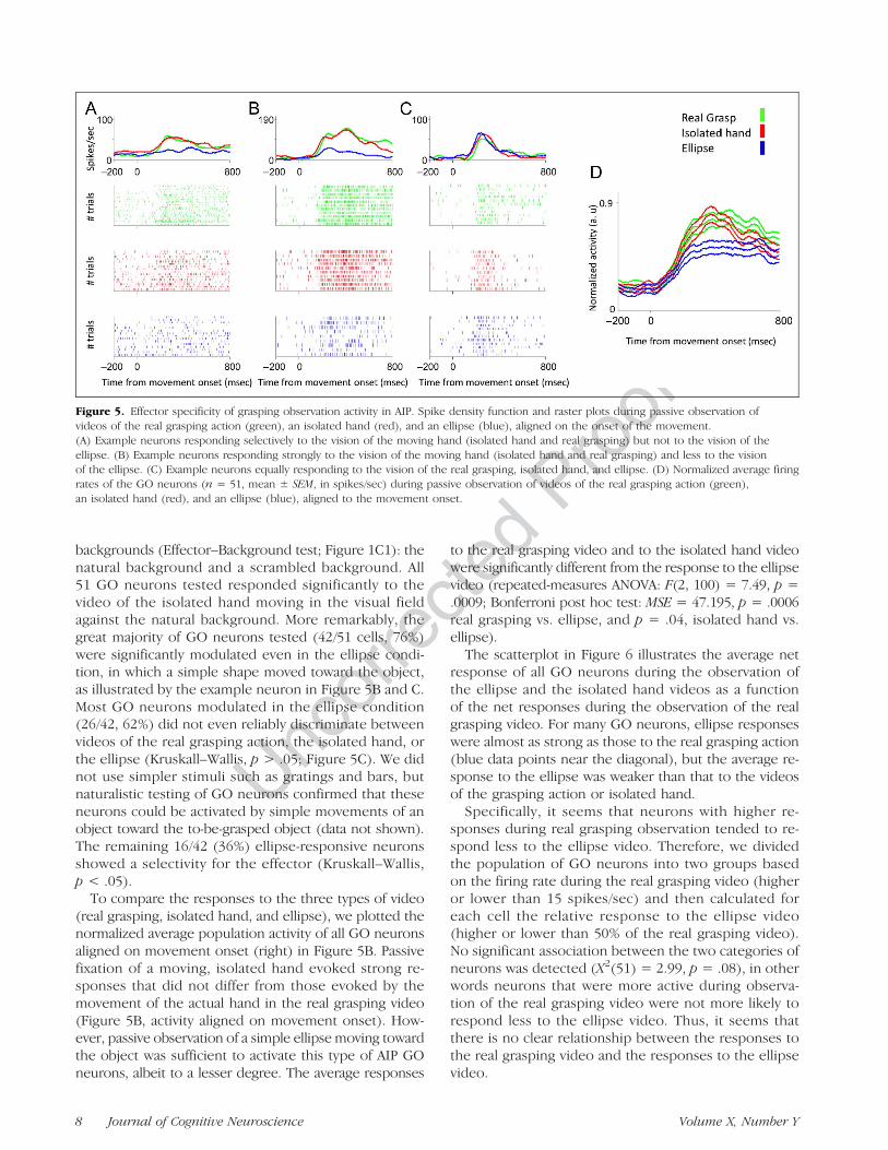
backgrounds (Effector–Background test; Figure 1C1): thenatural background and a scrambled background. All51 GO neurons tested responded significantly to thevideo of the isolated hand moving in the visual fieldagainst the natural background. More remarkably, thegreat majority of GO neurons tested (42/51 cells, 76%)were significantly modulated even in the ellipse condi-tion, in which a simple shape moved toward the object,as illustrated by the example neuron in Figure 5B and C.Most GO neurons modulated in the ellipse condition(26/42, 62%) did not even reliably discriminate betweenvideos of the real grasping action, the isolated hand, orthe ellipse (Kruskall–Wallis, p > .05; Figure 5C). We didnot use simpler stimuli such as gratings and bars, butnaturalistic testing of GO neurons confirmed that theseneurons could be activated by simple movements of anobject toward the to-be-grasped object (data not shown).The remaining 16/42 (36%) ellipse-responsive neuronsshowed a selectivity for the effector (Kruskall–Wallis,p < .05).
To compare the responses to the three types of video(real grasping, isolated hand, and ellipse), we plotted thenormalized average population activity of all GO neuronsaligned on movement onset (right) in Figure 5B. Passivefixation of a moving, isolated hand evoked strong re-sponses that did not differ from those evoked by themovement of the actual hand in the real grasping video(Figure 5B, activity aligned on movement onset). How-ever, passive observation of a simple ellipse moving towardthe object was sufficient to activate this type of AIP GOneurons, albeit to a lesser degree. The average responses
to the real grasping video and to the isolated hand videowere significantly different from the response to the ellipsevideo (repeated-measures ANOVA: F(2, 100) = 7.49, p =.0009; Bonferroni post hoc test: MSE = 47.195, p = .0006real grasping vs. ellipse, and p = .04, isolated hand vs.ellipse).The scatterplot in Figure 6 illustrates the average net
response of all GO neurons during the observation ofthe ellipse and the isolated hand videos as a functionof the net responses during the observation of the realgrasping video. For many GO neurons, ellipse responseswere almost as strong as those to the real grasping action(blue data points near the diagonal), but the average re-sponse to the ellipse was weaker than that to the videosof the grasping action or isolated hand.Specifically, it seems that neurons with higher re-
sponses during real grasping observation tended to re-spond less to the ellipse video. Therefore, we dividedthe population of GO neurons into two groups basedon the firing rate during the real grasping video (higheror lower than 15 spikes/sec) and then calculated foreach cell the relative response to the ellipse video(higher or lower than 50% of the real grasping video).No significant association between the two categories ofneurons was detected (X2(51) = 2.99, p = .08), in otherwords neurons that were more active during observa-tion of the real grasping video were not more likely torespond less to the ellipse video. Thus, it seems thatthere is no clear relationship between the responses tothe real grasping video and the responses to the ellipsevideo.
Figure 5. Effector specificity of grasping observation activity in AIP. Spike density function and raster plots during passive observation ofvideos of the real grasping action (green), an isolated hand (red), and an ellipse (blue), aligned on the onset of the movement.(A) Example neurons responding selectively to the vision of the moving hand (isolated hand and real grasping) but not to the vision of theellipse. (B) Example neurons responding strongly to the vision of the moving hand (isolated hand and real grasping) and less to the visionof the ellipse. (C) Example neurons equally responding to the vision of the real grasping, isolated hand, and ellipse. (D) Normalized average firingrates of the GO neurons (n = 51, mean ± SEM, in spikes/sec) during passive observation of videos of the real grasping action (green),an isolated hand (red), and an ellipse (blue), aligned to the movement onset.
8 Journal of Cognitive Neuroscience Volume X, Number Y

UncorrectedProof
At the population level, in the normal backgroundcondition only, we tested which of two predictors,the response to the isolated hand or the response tothe ellipse, could better predict the response to thereal grasping. By using a stepwise multiple linear re-gression, we found that the response to the isolatedalone was able to explain up the 80% of the varianceobserved in the real grasping (F(49, 1) = 211.3, p =.000, R adjusted = .808, standardized beta = .90;ellipse: beta = .029, p = .769). These data show thatoverall the response of the GO neurons can be driven—at least to some degree—by a simple shape movingin the visual field and that the movement of the fin-gers to grasp the object are not necessary to drive theresponses.To test whether the visual context in which the move-
ment was performed could affect the activity in GOneurons, we also presented videos of the isolated handand ellipse against a scrambled background (Figure 1C1).We found that the Background exerted a small effectacross the population of neurons considered: Specifi-cally, there was a slightly higher response to the normalcompared to the scrambled background (repeated-measures ANOVA, F(1, 50) = 4.11, p = .04791, partialeta squared = .07). This small effect was also confirmedat the single neuron level. In fact, most GO neurons(63%) were not significantly affected by the background.As illustrated in Figure 7, the average responses to theisolated hand and to the ellipse on the normal back-ground correlated strongly with the responses to thesame stimuli against a scrambled background (r(49) =.96, p < .000, for the isolated hand and r(49) = .88,for the ellipse, p < .000). Therefore, the visual context
of the observed action exerted a small influence on GOneurons.
Responses of GO Neurons to Ellipses Moving inDifferent Directions
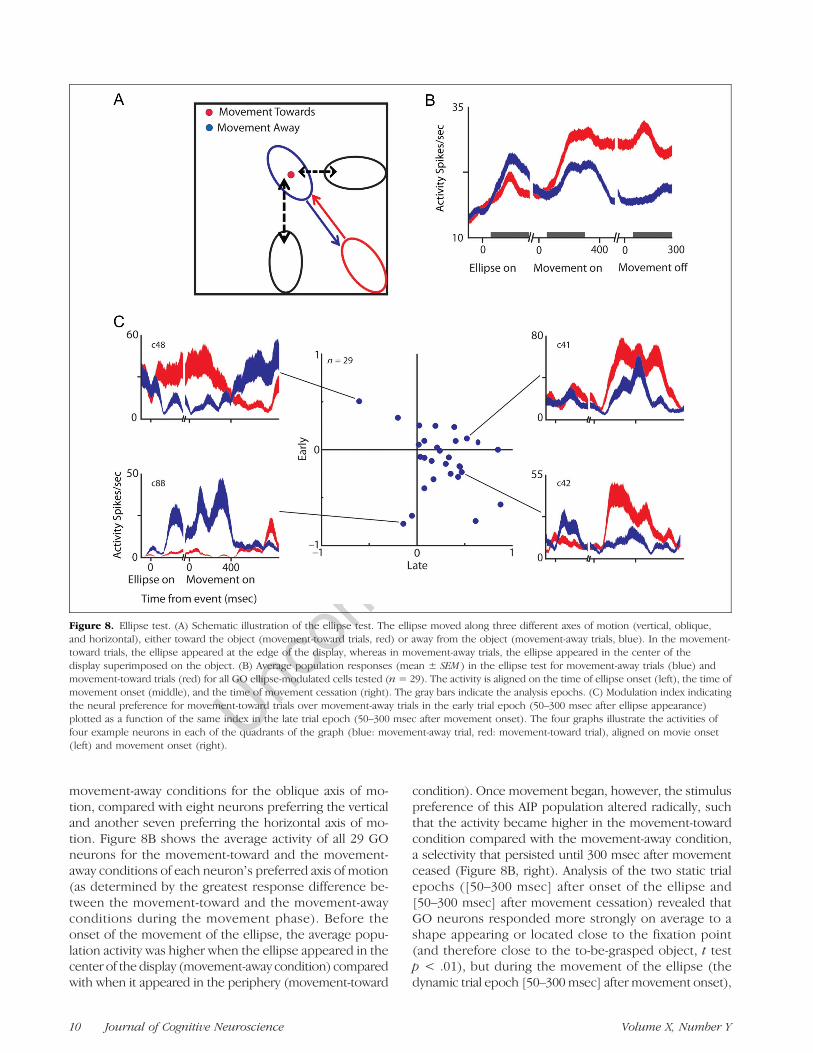
Taken together, the data presented so far demonstratethat for most GO neurons in AIP, the simple movementof a shape (ellipse) against a scrambled background wassufficient to evoke visual responses. The responses ofthese GO AIP neurons to the ellipse could representselectivity for the direction of motion (e.g., motion fromthe lower right corner to the center of the display) orcould arise by virtue of a spatial selectivity, that is, becaused by the ellipse entering a region in the visual field.To distinguish between these two possibilities, we tested29 GO ellipse-responsive neurons with videos in whichthe ellipse moved against the natural background alongthree different axes of motion (vertical, oblique, andhorizontal) and in two different directions for each axisof motion (movement toward and movement away fromthe object; Figure 8A) while the monkeys fixated a smallspot in the center of the display. In the movement-awayconditions, the ellipse appeared in the center of thedisplay (superimposed on the object and on the fixationpoint) and moved to the edge of the display, whereas inthe movement-toward conditions, the ellipse appeared inperipheral vision and moved toward the center of thedisplay.
Most GO neurons (n = 14/29, 48%) gave the greatestresponse differences between the movement-toward and
Figure 7. Effect of background on grasping observation activity inAIP. Average net responses (= response minus baseline activity) to theisolated hand (red) and ellipse (blue) on the scrambled backgroundplotted as a function of the average net responses to the isolatedhand and ellipse moving on the normal background for all GO neurons(n = 51).
Figure 6. Scatterplot of the average net responses (= response minusbaseline activity) to the isolated hand (red) and ellipse (blue)on the normal background as a function of the average net responsesto the real grasping observation for all GO neurons (n = 51).
Pani et al. 9
UncorrectedProof
movement-away conditions for the oblique axis of mo-tion, compared with eight neurons preferring the verticaland another seven preferring the horizontal axis of mo-tion. Figure 8B shows the average activity of all 29 GOneurons for the movement-toward and the movement-away conditions of each neuronʼs preferred axis of motion(as determined by the greatest response difference be-tween the movement-toward and the movement-awayconditions during the movement phase). Before theonset of the movement of the ellipse, the average popu-lation activity was higher when the ellipse appeared in thecenter of the display (movement-away condition) comparedwith when it appeared in the periphery (movement-toward
condition). Once movement began, however, the stimuluspreference of this AIP population altered radically, suchthat the activity became higher in the movement-towardcondition compared with the movement-away condition,a selectivity that persisted until 300 msec after movementceased (Figure 8B, right). Analysis of the two static trialepochs ([50–300 msec] after onset of the ellipse and[50–300 msec] after movement cessation) revealed thatGO neurons responded more strongly on average to ashape appearing or located close to the fixation point(and therefore close to the to-be-grasped object, t testp < .01), but during the movement of the ellipse (thedynamic trial epoch [50–300msec] after movement onset),
Figure 8. Ellipse test. (A) Schematic illustration of the ellipse test. The ellipse moved along three different axes of motion (vertical, oblique,and horizontal), either toward the object (movement-toward trials, red) or away from the object (movement-away trials, blue). In the movement-toward trials, the ellipse appeared at the edge of the display, whereas in movement-away trials, the ellipse appeared in the center of thedisplay superimposed on the object. (B) Average population responses (mean ± SEM ) in the ellipse test for movement-away trials (blue) andmovement-toward trials (red) for all GO ellipse-modulated cells tested (n = 29). The activity is aligned on the time of ellipse onset (left), the time ofmovement onset (middle), and the time of movement cessation (right). The gray bars indicate the analysis epochs. (C) Modulation index indicatingthe neural preference for movement-toward trials over movement-away trials in the early trial epoch (50–300 msec after ellipse appearance)plotted as a function of the same index in the late trial epoch (50–300 msec after movement onset). The four graphs illustrate the activities offour example neurons in each of the quadrants of the graph (blue: movement-away trial, red: movement-toward trial), aligned on movie onset(left) and movement onset (right).
10 Journal of Cognitive Neuroscience Volume X, Number Y
UncorrectedProof
the average population activity became stronger formovement-toward trials. The strong influence of the posi-tion where the ellipse appeared and the reversal of theneural preference (from the move-away condition to themove-toward condition) indicates that GO activity in AIPcannot be entirely explained by a selectivity for the direc-tion of motion. Instead, AIP neurons with grasping obser-vation responses appear to be influenced primarily byspatial position, where an object (or shape) is located inproximity of the to-be-grasped object that the monkeyis fixating, combined with a preference for movementstoward the object.We quantified themodulation of AIP neuronal responses
in the ellipse test using a modulation index (Methods)computed in the early epoch of the trial ([50–300 msecafter the appearance of the ellipse) and in the late epochof the trial ([50–300 msec] after movement ceased) forthe preferred axis of motion of each cell. Individual AIPneurons showed a variety of response patterns, but thearrangement most frequently observed consisted of aninitial preference for the ellipse appearing in the centerof the display followed by a later preference for movementtoward the center of the display (data points in the lowerright quadrant of Figure 8C). Although the neural selec-tivity for movement-toward versus movement-away trialswas more balanced in the early trial epoch before the startof the movement (62% preferring the movement-awaycondition), this preference clearly shifted during the latertrial epoch such that the great majority of AIP neurons(25/29, 86%) preferred movement-toward condition oncethe ellipse had started to move.The example neurons in Figure 8C illustrate that, for
most AIP neurons, the responses were driven by thespatial position of the ellipse. Only a very small minorityof GO neurons (see c88, bottom left) showed robustmotion-selective responses. Thus, the responses of GOneurons in AIP to a moving shape appeared to be basedon a spatial selectivity combined with a preference formovement toward the object or the center of gaze.Because we searched for responsive neurons using aVGG task in which the monkeysʼ hand moved from thelower right to the center of gaze, this preference formovement-toward trials may have been partly the resultof bias in the search test.
DISCUSSION
We investigated the responses of AIP neurons duringgrasping execution and grasping observation and foundthat most AIP neurons in this study fired during the exe-cution of an action in the dark and during observation ofa video of this same action, thus exhibiting mirror-likeproperties. These neurons were also active in a similarway during observation of movements of an isolatedhand on the screen and to a lesser extent during move-ments of an abstract shape on the screen.
The great majority of AIP neurons in this study showeda modulation in activity when the hand started to movetoward the object. Previous studies (Murata et al., 2000;Sakata et al., 1995; Taira, Mine, Georgopoulos, Murata, &Sakata, 1990) have labeled movement-related responsesas “motor” or “visuomotor” based on the persistence ofthe activity during grasping in the dark (in the absence ofvisual feedback). Here we showed that, although all AIPneurons in this study remained active during grasping inthe dark, a large fraction of these neurons could also beactivated by pure visual stimulation with a video of ahand (or another shape) moving toward the object thatis fixated. Therefore, visual information is sufficient—butnot necessary—to activate these AIP neurons. In contrast,other AIP neurons were also active during grasping in thedark but could not be activated by visual stimulation.Thus, AIP houses a variety of neurons that can be distin-guished by the presence or absence of responses to actionobservation.
The convergence of motor-related and observation-related responses on the same neuron is reminiscent oftwo types of activity in which a match between a motorcode and a visual stimulus corresponding to that motorcode occurs: Rehearsal activity (Cisek & Kalaska, 2004) andmirror activity (Gallese et al., 1996). Cisek and Kalaska(2004) found that neurons in dorsal premotor cortex re-spond during the execution of reaching movements inthe preferred direction, during the observation of cursormovements in the preferred direction, and even in theinstructed delay period before cursor movement begins,as if monkeys are mentally rehearsing the movementbefore it actually starts. Our study was not designed totest whether the GO responses in AIP also represent pre-dictive activity, making it difficult to sustain this hypothesis.Indirect evidence comes from the activity we measured inthe ellipse test. The 800-msec epoch before movementonset of the ellipse after it had appeared could be con-sidered as an instructed delay period because the directionof movement was entirely predictable based on the posi-tion where the ellipse appeared. We found very littleevidence for predictive activity in this epoch. Only twoneurons showed strong responses throughout the pre-movement epoch and during the movement epoch ofthe same condition (e.g., neuron c88 in Figure 8). In thetop right quadrant of Figure 8, all neurons are located closeto the main axes, indicating that these neurons respondedweakly before movement onset. Thus, the data of theellipse test suggest that at least the subpopulation ofellipse-responsive AIP neurons did not show predictivecoding of the ellipse movements; the close relation ofthe AIP activity to the onset of movement of the ellipse sug-gests that the GO responses in AIP were largely sensorial.
Although the responses of our AIP population appearedvisual during grasping observation, all neurons in thisstudy remained active during grasping in the dark, thatis, showed “motor” activity. Motor activity might appeardifficult to reconcile with the robust visual responses we
Pani et al. 11
UncorrectedProof
measured during grasping observation, unless the activ-ity during grasping in the dark represents an efferencecopy from premotor areas (Rizzolatti & Luppino, 2001).The possibility exists that the reafference of motor activ-ity is integrated with visual information in these neuronsduring grasping execution.
The necessity of the presence of a moving stimulusmakes the AIP neurons in this study more similar to mirrorneurons. Caggiano et al. (2010) showed that mirror neu-rons in PMv can exhibit both view-dependent (i.e., tunedfor a particular viewpoint such as first-person perspective)and view-independent responses to videos of actions.Thus, a specific subpopulation of mirror neurons respondsto videos similar to the one we used. Nevertheless, PMvand PFGmirror neurons also encode the goal of the action,responding differentially to, for example, grasping-for-eating compared with grasping-for-placing, or even topartially occluded actions (Bonini et al., 2010; Rizzolatti &Sinigaglia, 2010; Fogassi et al., 2005; Umiltà et al., 2001).Our data are consistent with a role for AIP in the mirrorneuron network (Nelissen et al., 2011; Rizzolatti & Sinigaglia,2010; Oztop & Arbib, 2002) but do not permit to decidewhether GO activity in AIP is in fact related to the mirrorneuron system or to action recognition in general. Futurestudies should test whether GO activity in AIP constitutesan early stage in which the mapping between visual andmotor properties occurs to provide input for cortical areasinvolved in the recognition of biological actions and/ormirror activity (Fleischer, Caggiano, Thier, & Giese, 2013).
Earlier areas in the dorsal visual stream such as thecaudal intraparietal area (Sakata, Taira, Kusunoki, Murata,& Tanaka, 1997) or the STS regions may share some ofthe responses we observed in AIP (Vangeneugden et al.,2011; Barraclough et al., 2009; Perrett et al., 1989), butnot the motor activity in the dark. AIP is connected withthe F5a sector in PMv and to PFG (Gerbella, Belmalih,Borra, Rozzi, & Luppino, 2011; Belmalih et al., 2009; Borraet al., 2008), and these latter areas are both connected withthe F5c sector in PMv. Furthermore TMS in humans hasshown that AIP and PMv causally interact during objectgrasping (Davare, Rothwell, & Lemon, 2010), and it hasbeen shown that AIP and F5a neurons share very similarvisual selectivities for 3-D shape and grasping activity(Theys, Pani, van Loon, Goffin, & Janssen, 2013; Theys,Pani, et al., 2012; Theys, Srivastava, et al., 2012). Interest-ingly, videos of a grasping isolated hand activate themacaque AIP and F5a—even in the absence of an object—but not F5c (Nelissen et al., 2011; Nelissen, Luppino,Vanduffel, Rizzolatti, & Orban, 2005). Targeted inactivationexperiments combined with recordings in PMv shouldclarify to what extent neurons in PMv and PFG dependupon input from AIP.
Area AIP has been implicated in online visual controlduring grasping (Tunik, Frey, & Grafton, 2005; Rizzolatti& Luppino, 2001; Murata et al., 2000). Specifically thenonobject type visual neurons may be involved in visualfeedback for the adjustment of the grip around the object
(Murata et al., 2000). Could the GO activity in AIP berelated to the online visual control of the hand con-figuration to match it with the dimensions of the object?Several of the findings in our study are relevant for thishypothesis: (1) the control experiment with the isolatedhand demonstrated that the dynamics of the observedgrasping movement (extension of the fingers followedby flexion) rarely affected the AIP responses, (2) mostAIP neurons also responded to the moving ellipse (i.e.,hand preshaping and detailed aspects of the hand werenot necessary), and (3) the videos with the scrambledbackground (no object present) evoked responses simi-lar to those with a natural background.Although we cannot be sure that the monkeys were
actually using visual information to online correct theirgrasping actions, the AIP responses during action obser-vation could be related to the monitoring of the effectorposition and axis of movement during grasping based onvisual information, possibly in retinotopic coordinates. Insupport of this idea is the observation that these neuronsmostly responded to an ellipse entering the central visualfield (close to the fixation point). Consistent with thishypothesis, Lehmann and Scherberger (2009) recentlyshowed robust reach position signals in area AIP thatwere mainly encoded in retinotopic coordinates. Further-more, TMS of the posterior parietal cortex in humans(Reichenbach, Bresciani, Peer, Bülthoff, & Thielscher,2011; Chib, Krutky, Lynch, &Mussa-Ivaldi, 2009; Desmurgetet al., 1999) interferes with the online control of visuallyguided reaches. It should be acknowledged that we didnot test different positions of the object and the fixationpoint, and the grasping movements made by our animalswere highly overtrained and probably did not require pre-cise online visual control. Thus, in the absence of conclusivedata, the online control hypothesis remains to be tested infuture studies.Our population of GO neurons in AIP also showed a
clear preference for movement toward the center of gazeover movement toward the periphery. This bias couldhave been at least partially a consequence of a selectionbias, because all our neurons showed movement-relatedactivity during visually guided grasping, where the animalcan see its own hand moving from the rest position inperipheral vision toward the object at the center of gaze.The bias for movement toward the center of gaze mayrepresent a first stage in the encoding of goal-directedmotor acts. Motter, Steinmetz, Duffy, and Mountcastle(1987) also showed opponently organized directionalpreferences (toward or away from the center of gaze)in neighboring area PG, and MT/ V5 neurons show astrong bias for diagonal motion in the lower contralateralquadrant (Maunsell & Van Essen, 1987).To conclude, we found neurons in AIP that match a
motor code with a visual stimulus corresponding to thatmotor code. This match is mostly driven by the vision ofa hand mimicking the kinematic characteristics of thereal action, but for some of the neurons a simple shape
12 Journal of Cognitive Neuroscience Volume X, Number Y
UncorrectedProof
sharing kinematic parameters with the moving hand isable to evoke a response. The combination of motortasks and detailed visual testing of single neurons willbe critical in future studies investigating the neural cir-cuitry underlying object grasping in the primate brain.
Acknowledgments
This study was supported by Geconcerteerde Onderzoeksacties(GOA 2010/19), Fonds voor Wetenschappelijk OnderzoekVlaanderen (G.0495.05, G.0713.09), Programmafinanciering(PFV/10/008), Interuniversity Attraction Poles 7/11, and ERC-StG-260607. We thank Sara De Pril, Piet Kayenbergh, GerritMeulemans, Stijn Verstraeten, Peter Parker, Marc Depaep,WouterDepuydt, and Inez Puttemans for assistance and Steve Raiguel forcomments on the manuscript.
Reprint requests should be sent to Peter Janssen, Laboratorium voorNeuro- en Psychofysiologie, Leuven Medical School, Herestraat 49,bus 1021B-3000 Leuven, Belgium, or via e-mail: [email protected].
REFERENCES
Barraclough, N. E., Keith, R. H., Xiao, D., Oram, M. W., &Perrett, D. I. (2009). Visual adaptation to goal-directed handactions. Journal of Cognitive Neuroscience, 21, 1806–1820.
Baumann, M. A., Fluet, M. C., & Scherberger, H. (2009).Context-specific grasp movement representation in themacaque anterior intraparietal area. Journal of Neuroscience,29, 6436–6448.
Belmalih, A., Borra, E., Contini, M., Gerbella, M., Rozzi, S., &Luppino, G. (2009). Multimodal architectonic subdivision ofthe rostral part (area F5) of the macaque ventral premotorcortex. Journal of Computational Neurology, 512, 183–217.
Bonini, L., Rozzi, S., Serventi, F. U., Simone, L., Ferrari, P. F., &Fogassi, L. (2010). Ventral premotor and inferior parietalcortices make distinct contribution to action organization andintention understanding. Cerebral Cortex, 6, 1372–1378.
Borra, E., Belmalih, A., Calzavara, R., Gerbella, M., Murata, A.,Rozzi, S., et al. (2008). Cortical connections of the macaqueanterior intraparietal (AIP) area. Cerebral Cortex, 18,1094–1111.
Caggiano, V., Fogassi, L., Rizzolatti, G., Pomper, J. K., Thier, P.,Giese, M. A., et al. (2011). View-based encoding of actions inmirror neurons of area f5 in macaque premotor cortex.Current Biology, 21, 144–148.
Chib, V. S., Krutky, M. A., Lynch, K. M., & Mussa-Ivaldi, F. A.(2009). The separate neural control of hand movementsand contact forces. Journal of Neuroscience, 29, 3939–3947.
Cisek, P., & Kalaska, J. F. K. (2004). Neural correlates ofmental rehearsal in dorsal premotor cortex. Nature, 431,993–996.
Davare, M., Rothwell, J. C., & Lemon, R. N. (2010). Causalconnectivity between the human anterior intraparietal areaand premotor cortex during grasp. Current Biology, 20,176–181.
Desmurget, M., Epstein, C. M., Turner, R. S., Prablanc, C.,Alexander, G. E., & Grafton, S. T. (1999). Role of theposterior parietal cortex in updating reaching movementsto a visual target. Nature Neuroscience, 2, 563–567.
Fleischer, F., Caggiano, V., Thier, P., & Giese, M. A. (2013).Physiologically inspired model for visual recognition oftransitive hand actions. Journal of Neuroscience, 33,6563–6580.
Fluet, M. C., Baumann, M. A., & Scherberger, H. (2010).Context-specific grasp movement representation in macaqueventral premotor cortex. Journal of Neuroscience, 30,15175–15184.
Fogassi, L., Ferrari, P. F., Gesierich, B., Rozzi, S., Chersi, F., &Rizzolatti, G. (2005). Parietal lobe: From action organizationto intention understanding. Science, 308, 662–667.
Fogassi, L., Gallese, V., Buccino, G., Craighero, L., Fadiga, L., &Rizzolatti, G. (2001). Cortical mechanism for the visualguidance of hand grasping movements in the monkey: Areversible inactivation study. Brain, 124, 571–586.
Fujii, N., Hihara, S., & Iriki, A. (2008). Social cognition inpremotor and parietal cortex. Social Neuroscience, 3,250–260.
Gallese, V., Fadiga, L., Fogassi, L., & Rizzolatti, G. (1996).Action recognition in the premotor cortex. Brain, 119,593–609.
Gallese, V., Murata, A., Kaseda, M., Niki, N., & Sakata, H. (1994).Deficit of hand preshaping after muscimol injection inmonkey parietal cortex. NeuroReport, 5, 1525–1529.
Gerbella, M., Belmalih, A., Borra, E., Rozzi, S., & Luppino, G.(2011). Cortical connections of the anterior (F5a) subdivisionof the macaque ventral premotor area F5. Brain Structureand Function, 216, 43–65.
Lehmann, S. J., & Scherberger, H. (2009). Reach and gazerepresentations in macaque parietal and premotor grasp areas.Journal of Neuroscience, 33, 7038–7049.
Macfarlane, N. B., & Graziano, M. S. (2009). Diversity of gripin Macaca mulatta. Experimental Brain Research, 197,255–268.
Maunsell, J. H., & Van Essen, D. C. (1987). Topographicorganization of the middle temporal visual area in themacaque monkey: Representational biases and therelationship to callosal connections and myeloarchitectonicboundaries. Journal of Comparative Neurology, 266,535–555.
Motter, B. C., Steinmetz, M. A., Duffy, C. J., & Mountcastle, V. B.(1987). Functional properties of parietal visual neurons:Mechanisms of directionality along a single axis. Journal ofNeuroscience, 7, 154–176.
Murata, A., Gallese, V., Luppino, G., Kaseda, M., & Sakata, H.(2000). Selectivity for the shape, size, and orientation ofobjects for grasping in neurons of monkey parietal area AIP.Journal of Neurophysiology, 83, 2580–2601.
Murata, A., & Ishida, H. (2007). Representation of bodily self inthe multimodal parieto-premotor network. In S. Funahashi(Ed.), Representation and brain (pp. 151–176). Japan:Springer.
Nelissen, K., Borra, E., Gerbella, M., Rozzi, S., Luppino, G.,Vanduffel, W., et al. (2011). Action observation circuits inthe macaque monkey cortex. Journal of Neuroscience, 31,3743–3756.
Nelissen, K., Luppino, G., Vanduffel, W., Rizzolatti, G., &Orban, G. A. (2005). Observing others: Multiple actionrepresentation in the frontal lobe. Science, 310, 332–336.
Oztop, E., & Arbib, M. A. (2002). Schema design andimplementation of the grasp-related mirror neuron system.Biological Cybernetics, 87, 116–140.
Perrett, D. I., Harries, M. H., Bevan, R., Thomas, S., Benson, P. J.,Mistlin, A. J., et al. (1989). Frameworks of analysis for theneural representation of animate objects and actions.Journal of Experimental Biology, 146, 87–113.
Reichenbach, A., Bresciani, J. P., Peer, A., Bülthoff, H. H., &Thielscher, A. (2011). Contributions of the PPC to onlinecontrol of visually guided reaching movements assessedwith fMRI-guided TMS. Cerebral Cortex, 21, 1602–1612.
Rizzolatti, G., & Luppino, G. (2001). The cortical motor system.Neuron, 31, 889–901.
Pani et al. 13
UncorrectedProof
Rizzolatti, G., & Sinigaglia, C. (2010). The functional role ofthe parieto-frontal mirror circuit: Interpretations andmisinterpretations.Nature Reviews Neuroscience, 11, 264–274.
Romero, M. C., Van Dromme, I., & Janssen, P. (2012). Responsesto two-dimensional shapes in themacaque anterior intraparietalarea. European Journal of Neuroscience, 36, 2324–2334.
Rozzi, S., Ferrari, P. F., Bonini, L., Rizzolatti, G., & Fogassi, L.(2008). Functional organization of inferior parietal lobuleconvexity in the macaque monkey: Electrophysiologicalcharacterization of motor, sensory and mirror responses andtheir correlation with cytoarchitectonic areas. EuropeanJournal of Neuroscience, 28, 1569–1588.
Sakata, H., Taira, M., Kusunoki, M., Murata, A., & Tanaka, Y.(1997). The TINS lecture. The parietal association cortex indepth perception and visual control of hand action. Trendsin Neuroscience, 20, 350–357.
Sakata, H., Taira, M., Murata, A., & Mine, S. (1995). Neuralmechanisms of visual guidance of hand action in the parietalcortex of the monkey. Cerebral Cortex, 5, 429–438.
Srivastava, S., Orban, G. A., De Maziere, P. A., & Janssen, P.(2012). A distinct representation of three-dimensional shapein macaque anterior intraparietal area: Fast, metric, andcoarse. Journal of Neuroscience, 29, 10613–10626.
Taira, M., Mine, S., Georgopoulos, A. P., Murata, A., & Sakata, H.(1990). Parietal cortex neurons of the monkey related to the
visual guidance of hand movement. Experimental BrainResearch, 83, 29–36.
Theys, T., Pani, P., van Loon, J., Goffin, J., & Janssen, P. (2012).Selectivity for three-dimensional shape and grasping-relatedactivity in the macaque ventral premotor cortex. Journal ofNeuroscience, 32, 12038–12050.
Theys, T., Pani, P., van Loon, J., Goffin, J., & Janssen, P. (2013).Three-dimensional shape coding in grasping circuits: Acomparison between the anterior intraparietal area andventral premotor area F5a. Journal of CognitiveNeuroscience, 25, 352–364.
Theys, T., Srivastava, S., van Loon, J., Goffin, J., & Janssen, P.(2012). Selectivity for three-dimensional shape and contoursin the anterior intraparietal area. Journal of Neurophysiology,107, 995–1008.
Tunik, E., Frey, S. H., & Grafton, S. T. (2005). Virtual lesions ofthe anterior intraparietal area disrupt goal-dependent on-lineadjustments of grasp. Nature Neuroscience, 8, 505–511.
Umiltà, M. A., Kohler, E., Gallese, V., Fogassi, L., Fadiga, L.,Keysers, C., et al. (2001). I know what you are doing: Aneurophysiological study. Neuron, 31, 155–165.
Vangeneugden, J., De Maziere, P. A., Van Hulle, M. M., Jaeggli,T., Van Gool, L., & Vogels, R. (2011). Distinct mechanisms forcoding of visual actions in macaque temporal cortex. Journalof Neuroscience, 31, 385–401.
14 Journal of Cognitive Neuroscience Volume X, Number Y