Graphical and statistical analyses of the oculocardiac ...
11
RESEARCH ARTICLE Graphical and statistical analyses of the oculocardiac reflex during a non-invasive intracranial pressure measurement Yasin Hamarat 1 , Laimonas Bartusis 1 *, Mantas Deimantavicius 1 , Lina Siaudvytyte 2 , Ingrida Januleviciene 2 , Arminas Ragauskas 1 , Eric M. Bershad 3 , Javier Fandino 4 , Jenny Kienzler 4 , Elke Remonda 4 , Vaidas Matijosaitis 5 , Daiva Rastenyte 5 , Kestutis Petrikonis 5 , Kristina Berskiene 6 , Rolandas Zakelis 1 1 Health Telematics Science Institute, Kaunas University of Technology, Kaunas, Lithuania, 2 Eye Clinic, Lithuanian University of Health Sciences, Kaunas, Lithuania, 3 Department of Neurology, Baylor College of Medicine, Houston, Texas, United States of America, 4 Department of Neurosurgery, Kantonsspital Aarau, Aarau, Switzerland, 5 Department of Neurology, Lithuanian University of Health Sciences, Kaunas, Lithuania, 6 Sports Institute, Lithuanian University of Health Sciences, Kaunas, Lithuania * [email protected] Abstract Purpose This study aimed to examine the incidence of the oculocardiac reflex during a non-invasive intracranial pressure measurement when gradual external pressure was applied to the orbital tissues and eye. Methods Patients (n = 101) and healthy volunteers (n = 56) aged 20–75 years who underwent a non- invasive intracranial pressure measurement were included in this retrospective oculocardiac reflex analysis. Prespecified thresholds greater than a 10% or 20% decrease in the heart rate from baseline were used to determine the incidence of the oculocardiac reflex. Results None of the subjects had a greater than 20% decrease in heart rate from baseline. Four sub- jects had a greater than 10% decrease in heart rate from baseline, representing 0.9% of the total pressure steps. Three of these subjects were healthy volunteers, and one was a glau- coma patient. Conclusion The incidence of the oculocardiac reflex during a non-invasive intracranial pressure mea- surement procedure was very low and not associated with any clinically relevant effects. PLOS ONE | https://doi.org/10.1371/journal.pone.0196155 April 19, 2018 1 / 11 a1111111111 a1111111111 a1111111111 a1111111111 a1111111111 OPEN ACCESS Citation: Hamarat Y, Bartusis L, Deimantavicius M, Siaudvytyte L, Januleviciene I, Ragauskas A, et al. (2018) Graphical and statistical analyses of the oculocardiac reflex during a non-invasive intracranial pressure measurement. PLoS ONE 13 (4): e0196155. https://doi.org/10.1371/journal. pone.0196155 Editor: Ted S Acott, Oregon Health and Science University, UNITED STATES Received: October 30, 2017 Accepted: April 6, 2018 Published: April 19, 2018 Copyright: © 2018 Hamarat et al. This is an open access article distributed under the terms of the Creative Commons Attribution License, which permits unrestricted use, distribution, and reproduction in any medium, provided the original author and source are credited. Data Availability Statement: Data are available from the Health Telematics Science Institutional Server: ftp://158.129.13.155/array1/Public/OCR_ PLOS_ONE/ and Open Science Framework: https:// osf.io/9w4uc/. Funding: Professor Arminas Ragauskas is an inventor of patented the non-invasive ICP measurement method and a shareholder of Vittamed Neuroscience (Waltham, MA, USA). AR, LB, MD and RZ have received financial support
Transcript of Graphical and statistical analyses of the oculocardiac ...

RESEARCH ARTICLE
Graphical and statistical analyses of the
oculocardiac reflex during a non-invasive
intracranial pressure measurement
Yasin Hamarat1, Laimonas Bartusis1*, Mantas Deimantavicius1, Lina Siaudvytyte2,
Ingrida Januleviciene2, Arminas Ragauskas1, Eric M. Bershad3, Javier Fandino4,
Jenny Kienzler4, Elke Remonda4, Vaidas Matijosaitis5, Daiva Rastenyte5,
Kestutis Petrikonis5, Kristina Berskiene6, Rolandas Zakelis1
1 Health Telematics Science Institute, Kaunas University of Technology, Kaunas, Lithuania, 2 Eye Clinic,
Lithuanian University of Health Sciences, Kaunas, Lithuania, 3 Department of Neurology, Baylor College of
Medicine, Houston, Texas, United States of America, 4 Department of Neurosurgery, Kantonsspital Aarau,
Aarau, Switzerland, 5 Department of Neurology, Lithuanian University of Health Sciences, Kaunas, Lithuania,
6 Sports Institute, Lithuanian University of Health Sciences, Kaunas, Lithuania
Abstract
Purpose
This study aimed to examine the incidence of the oculocardiac reflex during a non-invasive
intracranial pressure measurement when gradual external pressure was applied to the
orbital tissues and eye.
Methods
Patients (n = 101) and healthy volunteers (n = 56) aged 20–75 years who underwent a non-
invasive intracranial pressure measurement were included in this retrospective oculocardiac
reflex analysis. Prespecified thresholds greater than a 10% or 20% decrease in the heart
rate from baseline were used to determine the incidence of the oculocardiac reflex.
Results
None of the subjects had a greater than 20% decrease in heart rate from baseline. Four sub-
jects had a greater than 10% decrease in heart rate from baseline, representing 0.9% of the
total pressure steps. Three of these subjects were healthy volunteers, and one was a glau-
coma patient.
Conclusion
The incidence of the oculocardiac reflex during a non-invasive intracranial pressure mea-
surement procedure was very low and not associated with any clinically relevant effects.
PLOS ONE | https://doi.org/10.1371/journal.pone.0196155 April 19, 2018 1 / 11
a1111111111
a1111111111
a1111111111
a1111111111
a1111111111
OPENACCESS
Citation: Hamarat Y, Bartusis L, Deimantavicius M,
Siaudvytyte L, Januleviciene I, Ragauskas A, et al.
(2018) Graphical and statistical analyses of the
oculocardiac reflex during a non-invasive
intracranial pressure measurement. PLoS ONE 13
(4): e0196155. https://doi.org/10.1371/journal.
pone.0196155
Editor: Ted S Acott, Oregon Health and Science
University, UNITED STATES
Received: October 30, 2017
Accepted: April 6, 2018
Published: April 19, 2018
Copyright: © 2018 Hamarat et al. This is an open
access article distributed under the terms of the
Creative Commons Attribution License, which
permits unrestricted use, distribution, and
reproduction in any medium, provided the original
author and source are credited.
Data Availability Statement: Data are available
from the Health Telematics Science Institutional
Server: ftp://158.129.13.155/array1/Public/OCR_
PLOS_ONE/ and Open Science Framework: https://
osf.io/9w4uc/.
Funding: Professor Arminas Ragauskas is an
inventor of patented the non-invasive ICP
measurement method and a shareholder of
Vittamed Neuroscience (Waltham, MA, USA). AR,
LB, MD and RZ have received financial support
Introduction
The oculocardiac reflex (OCR), also known as the Aschner reflex, is a cardiac phenomenon that
is triggered by physical stimulation of the eye [1]. This phenomenon was first described in 1908
independently by Aschner and Dagnini. Presently, the OCR is considered to be a subtype of a
well-known brainstem reflex, the trigeminocardiac reflex [2]. The physiological response of the
heart caused by the OCR is characterized by bradycardia or arrhythmia, which may even lead to
cardiac arrest [1,3,4]. The OCR can be evoked by pressure on the ocular globe, traction on the
extrinsic muscles of the eye, intraorbital injections, hematomas, acute glaucoma, and/or stretch-
ing of the eyelid muscles [5]. The stimulus signal is primarily conducted along the ophthalmic
division of the trigeminal nerve (V1), which then connects to the ciliary ganglion and visceral
motor nucleus of the vagus nerve. The vagus nerve, which is the longest cranial and major para-
sympathetic nerve, supplies parasympathetic fibers to all major organs, including the heart [6].
The criteria for the evoked OCR is a threshold of decrease in the mean heart rate (MHR) relative
to the baseline heart rate (BHR) recorded before physical stimulation of the eye. Various studies
have defined thresholds for the evoked OCR [7]. Vrabec et al. (1987) [8] and Eustis et al. (1992)
[9] defined a 10% decrease, while others have used a 20% decrease in BHR to define the evoked
OCR [10–12]. Finally, Yu & Wang (1991) defined the threshold for the evoked OCR as a
decrease of 10 beats per minute compared with baseline [13].
A non-invasive measurement of the absolute value of the intracranial pressure (ICP) has
been developed in the Health Telematics Science Institute of Kaunas University of Technology
in Lithuania [14]. This method is based on the principles of a non-invasive arterial blood pres-
sure measurement and uses transcranial Doppler (TCD) ultrasonography to assess pulse
waves of the ophthalmic artery (OA) during a gradual externally applied pressure (Pe) over a
closed eyelid that is transmitted to the eye and orbital (peri-ocular) tissues. The accuracy, pre-
cision, and diagnostic reliability of this non-invasive ICP measurement technique have previ-
ously been reported [15–17]; however, the incidence of the evoked OCR, which may have
important clinical consequences, was not reported.
The aim of this retrospective study was to analyze the presence of the evoked OCR during
the non-invasive measurement of ICP when step-wise increases in Pe are applied to the orbital
tissues, including the eye.
Materials and methods
Equipment and measurement technique
Vittamed 205 (Kaunas, Lithuania), which contains a two-depth TCD with a 2-MHz ultrasonic
transducer, was used to collect TCD data during non-invasive ICP measurements. Three ver-
sions of the device (Vittamed 205) have been clinically validated since 2009.
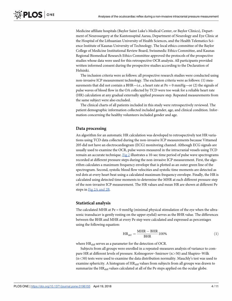
The non-invasive ICP absolute value measurement method uses the intracranial and extra-
cranial parts of the OA [18,19] as a scale to detect the pressure balance between ICP and the
gradual externally applied pressure [Pe(t)]. A schematic representation of this non-invasive
measurement technique is depicted in Fig 1. Air fills a toroidal-shaped soft plastic cuff installed
into the head frame together with an ultrasonic transducer to transmit pressure to orbital tis-
sues. The Pe transmits to the non-compressible orbital tissues and thus, exerts a transmural
force on the extracranial OA, but not the intracranial OA, due to segmentation by the dura
mater (Fig 1A). A pressure controller automatically increases Pe from 0 to 48 mmHg (maxi-
mum) with pressure steps selected by the operator (Fig 1B). The flow velocity pulsations of the
OA are continuously monitored by two-depth TCD in the intracranial and extracranial parts
of the OA.
Analyses of the oculocardiac reflex during a non-invasive intracranial pressure measurement
PLOS ONE | https://doi.org/10.1371/journal.pone.0196155 April 19, 2018 2 / 11
from Vittamed Neuroscience (Waltham, MA, USA).
AR, RZ, VM, DR and KP have received financial
support from European Commission’s Seventh
Framework Programme project ‘BrainSafe’ (grant
no. 232545). LB, AR, JF, JK, ER, VM and RZ have
received financial support from Lithuanian-Swiss
Programe project ‘BrainCare’ (grant no. CH-3SMM-
01/06). LB, LS and IJ have received financial
support from European Social Fund under the
Global Grant measure (grant no. VP1-3.1-SMM-
07-K-03-080). EB has received financial support
from National Space Biomedical Research Institute
via NASA NCC9-58, and Center for Space
Medicine, Baylor College of Medicine. No funding
bodies had any role in study design, data collection
and analysis, decision to publish, or preparation of
the manuscript.
Competing interests: Professor Arminas
Ragauskas is an inventor of patented the non-
invasive ICP measurement method and a
shareholder of Vittamed Neuroscience (Waltham,
MA, USA). Arminas Ragauskas, Laimonas
Bartusis, Mantas Deimantavicius and Rolandas
Zakelis have received financial support from
Vittamed Neuroscience (Waltham, MA, USA).
Clinical studies were funded by the: European
Commission’s Seventh Framework Programme
project ‘BrainSafe’ (grant no. 232545), Lithuanian-
Swiss Programe project ‘BrainCare’ (grant no. CH-
3SMM01/06), European Social Fund under the
Global Grant measure (grant no. VP1-3.1-SMM-
07-K-03-080), National Space Biomedical
Research Institute via NASA NCC9-58, and Center
for Space Medicine, Baylor College of Medicine.
This does not alter our adherence to PLOS ONE
policies on sharing data and materials.

Subjects and settings
Neurological and ophthalmological (glaucoma) patients, as well as healthy volunteers, under-
went a non-invasive ICP measurement in the setting of several prospective research studies
from October 2009 to December 2016 at the Texas Medical Center at Baylor College of
Fig 1. Schematic representation of the non-invasive intracranial pressure (ICP) measurement equipment Vittamed 205. (A) Relevant orbit and brain anatomy in
contact with the ICP measurement device. (B) Block diagram of the system control unit. ICA—internal carotid artery; IOA—intracranial part of the ophthalmic artery;
EOA—extracranial part of the ophthalmic artery; TCD—transcranial Doppler; Pe—external pressure applied to the ocular globe.
https://doi.org/10.1371/journal.pone.0196155.g001
Analyses of the oculocardiac reflex during a non-invasive intracranial pressure measurement
PLOS ONE | https://doi.org/10.1371/journal.pone.0196155 April 19, 2018 3 / 11
Medicine affiliate hospitals (Baylor Saint Luke’s Medical Center, or Baylor Clinics), Depart-
ment of Neurosurgery at the Kantonsspital Aarau, Department of Neurology and Eye Clinic at
the Hospital of the Lithuanian University of Health Sciences, and the Health Telematics Sci-
ence Institute of Kaunas University of Technology. The local ethics committee of the Baylor
College of Medicine Institutional Review Board, Swissmedic Ethics Committee, and Kaunas
Regional Biomedical Research Ethics Committee approved the protocols of the prospective
studies whose data were used for this retrospective OCR analysis. All participants provided
written informed consent during the prospective studies according to the Declaration of
Helsinki.
The inclusion criteria were as follows: all prospective research studies were conducted using
non-invasive ICP measurement technology. The exclusion criteria were as follows: (1) mea-
surements that did not contain a BHR—i.e., a heart rate at Pe = 0 mmHg—or (2) the signals of
pulse waves of blood flow in the OA collected by TCD were too weak for a reliable heart rate
(HR) calculation at any gradual externally applied pressure step. Repeated measurements from
the same subject were also excluded.
The clinical charts of all patients included in this study were retrospectively reviewed. The
patient demographic information collected included gender, age, and clinical condition. Infor-
mation concerning the healthy volunteers included gender and age.
Data processing
An algorithm for an automatic HR calculation was developed to retrospectively test HR varia-
tions using TCD data collected during the non-invasive ICP measurements because Vittamed
205 did not have an electrocardiogram (ECG) monitoring channel. Although ECG signals are
usually used to examine the OCR, pulse waves measured in the intracranial vessels using TCD
remain an accurate technique. Fig 2 illustrates a 10-sec time period of pulse wave spectrograms
recorded at different pressure steps during the non-invasive ICP measurement. First, the algo-
rithm calculates a maximum frequency envelope that is plotted as an outer green line of the
spectrogram. Second, systolic blood flow velocities and systolic time moments are detected as
red dots at every heart beat using a calculated maximum frequency envelope. Finally, the HR is
calculated using detected time moments to determine the MHR at each different pressure step
of the non-invasive ICP measurement. The HR values and mean HR are shown at different Pe
steps in Fig 2A and 2B.
Statistical analysis
The calculated MHR at Pe = 0 mmHg (minimal physical stimulation of the eye when the ultra-
sonic transducer is gently resting on the upper eyelid) serves as the BHR value. The differences
between the BHR and MHR at every Pe step were calculated and expressed as percentages
using the following equation:
HRdiff ¼MHR � BHR
BHR100% ð1Þ
where HRdiff serves as a parameter for the detection of OCR.
Subjects from all groups were enrolled in a repeated-measures analysis of variance to com-
pare HR at different levels of pressure. Kolmogorov–Smirnov (n>50) and Shapiro–Wilk
(n<50) tests were used to examine the data distribution normality. Mauchly’s test was used to
examine sphericity. A histogram of HRdiff values from subjects from all groups was drawn to
summarize the HRdiff values calculated at all of the Pe steps applied on the ocular globe.
Analyses of the oculocardiac reflex during a non-invasive intracranial pressure measurement
PLOS ONE | https://doi.org/10.1371/journal.pone.0196155 April 19, 2018 4 / 11

Statistical analysis was performed using IBM SPSS Statistics software (version 23.0; IBM
Corporation, Armonk, NY, USA). The level of significance was defined as p<0.05.
Results
One hundred fifty-seven subjects were included in the OCR analysis. Repeated measurements
on the same subject were excluded to eliminate the correlated data points. The mean age
(±SD) was 47.6 (±14.4) years (range: 20–75 years), and the male:female ratio was 57:100. The
demographic data of the subjects divided into four groups are presented in Table 1.
A typical HR variation of a single subject recorded during the non-invasive ICP measure-
ment is depicted graphically (Fig 3).
The maximum external pressure of 48 mmHg and 4-mmHg pressure increase per pressure
step are not standard setups for non-invasive ICP measurements. Therefore, the setup can be
changed according to the clinical study protocol.
The number of performed measurements using different protocol parameters are presented
according to the subject type in all groups in Table 2. The transition time of the pressure
increase between two successive pressure steps affects the time period of each pressure step.
The time period was approximately 30 sec for each pressure step of the non-invasive ICP
Fig 2. Example spectrograms of pulse waves of blood flow in the ophthalmic artery collected by a transcranial
Doppler using different external pressure steps. (A) 0 mmHg; (B) 48 mmHg. HR—heart rate.
https://doi.org/10.1371/journal.pone.0196155.g002
Table 1. Demographic data of the included subjects in this retrospective study.
Subject group No. of subjects Age
mean±SD (range), years
Gender, female, %
TBI patients 8 53.3±10.2 (37–69) 50.0
Neurologic patients 10 41.0±12.3 (21–60) 60.0
Glaucoma patients 83 53.4±12.5 (24–75) 72.3
Healthy volunteers 56 39.4±13.2 (20–72) 53.6
TBI, traumatic brain injury; SD, standard deviation.
https://doi.org/10.1371/journal.pone.0196155.t001
Analyses of the oculocardiac reflex during a non-invasive intracranial pressure measurement
PLOS ONE | https://doi.org/10.1371/journal.pone.0196155 April 19, 2018 5 / 11

measurements; however, a time period of 6 min. per pressure step was used for the 10 healthy
volunteers out of 157 subjects according to a different protocol for the non-invasive ICP mea-
surement to determine the number of TCD pulse waves needed per pressure step to reliably
measure ICP.
The MHR was calculated separately for pressure steps of each measurement. Repeated-mea-
sures analysis of variance was performed to compare the MHR at different levels of pressure.
The MHR data matched the normality assumptions according to the Kolmogorov–Smirnov or
Shapiro–Wilk test at each different Pe step (the results are presented in Table 3).
Fig 3. Typical heart rate variation. (A) A heart rate variation in a healthy volunteer. (B) A pressure increase of 4
mmHg per pressure Pe(t) step (time period of approximately 30 sec each) was used on the ocular globe from 0 mmHg
to 48 mmHg.
https://doi.org/10.1371/journal.pone.0196155.g003
Table 2. Protocol parameters for the non-invasive intracranial pressure measurement according to group.
Subject group Pressure steps, mmHg Time period, sec. Body position No. of measurements
TBI patients 0; 4; 8; 12; 16; 20 30 HUT 20 deg 7
0; 4; 8; 12 30 HUT 20 deg 1
Neurologic patients 0; 4; 8; 12; 16; 20; 24 30 LD 6
0; 4; 12; 16; 20; 24 30 LD 2
0; 4; 8; 12; 16; 20; 24; 28 30 LD 1
0; 4; 8; 12; 16; 20; 24; 28; 32; 36 30 LD 1
Glaucoma patients 0; 4; 8; 12; 16; 20 30 Supine 83
Healthy volunteers 0; 4; 8; 12; 16; 20 30 Supine 38
0; 4; 8; 12; 16; 20; 24; 28; 32; 36; 40; 44; 48 30 HDT 30 deg 3
0; 4; 8; 12; 16; 20; 24; 28; 32; 36; 40; 44; 48 30 Supine 5
0; 8; 12; 16; 24; 32; 40; 48 360 Supine 8
0; 8; 12; 16; 24; 32 360 Supine 1
0; 4; 8; 12; 16; 24; 32; 40; 48 360 Supine 1
TBI, traumatic brain injury; HUT, head up tilt; HDT, head down tilt; Time period, duration of one pressure step; LD, lateral decubitus.
https://doi.org/10.1371/journal.pone.0196155.t002
Analyses of the oculocardiac reflex during a non-invasive intracranial pressure measurement
PLOS ONE | https://doi.org/10.1371/journal.pone.0196155 April 19, 2018 6 / 11

Mauchly’s test indicated that the assumption of sphericity was violated (p = 0.047). There-
fore, the degrees of freedom were corrected using Greenhouse Geisser estimates of sphericity
(ε = 0.23). The results showed that no significant effect on the MHR [F(2.71, 18.95) = 0.644;
p = 0.581] at different levels of pressure.
The differences between the BHR and MHR were calculated and expressed as percentages
for the detection of the OCR. A histogram of the HRdiff values calculated at every Pe step for
subjects from all groups is presented in Fig 4. Obtained HRdiff values with a minus sign repre-
sent a decrease in the MHR compared with the BHR, while a plus sign represents an increase
in the MHR compared with the BHR.
Table 3. Results of the heart rate and tests of data normality at pressure steps from 0 mmHg to 48 mmHg.
Pe, mmHg Mean±SD 95% CI of the mean Med Min Max K-S test value df p value Skewness (SE) Kurtosis (SE)
0 64.8±8.9 63.4–66.2 64.3 42.4 89.4 0.047 157 0.200 0.25(0.19) -0.17(0.38)
4 63.8±8.8 62.4–65.3 63.2 41.9 90.1 0.057 148 0.200 0.49(0.20) 0.15(0.39)
8 64.5±8.8 63.1–65.9 63.9 42.3 89.6 0.054 155 0.200 0.34(0.19) -0.17(0.39)
12 64.5±8.8 63.1–65.8 63.6 42.6 91.0 0.066 157 0.096 0.40(0.19) -0.07(0.39)
16 64.6±8.8 63.2–66.0 63.9 41.8 90.0 0.056 156 0.200 0.26(0.19) 0.04(0.39)
20 64.5±9.1 63.0–66.0 63.5 41.4 89.2 0.070 146 0.078 0.40(0.20) -0.21(0.39)
24 63.6±9.6 59.8–67.3 63.4 40.9 87.8 0.973� 28 0.662 0.11(0.44) 0.56(0.86)
28 66.7±9.5 58.7–74.8 66.6 53.4 86.3 0.936� 10 0.508 0.42(0.68) -0.96(1.30)
32 66.5±8.1 62.6–70.3 69.6 53.1 80.0 0.913� 19 0.083 -0.43(0.52) -0.94(1.01)
36 65.3±9.3 58.2–72.4 64.6 54.1 79.8 0.920� 9 0.389 0.19(0.72) -1.54(0.94)
40 66.0±8.2 61.8–70.2 69.3 51.7 80.5 0.917� 17 0.131 -0.37(0.55) -0.79(0.93)
44 62.5±9.1 54.9–70.2 59.1 53.6 74.1 0.878� 8 0.051 0.45(0.75) -2.12(1.48)
48 66.3±8.1 62.1–70.5 67.6 51.9 77.3 0.942� 17 0.340 -0.38(0.55) -1.09(1.06)
Pe, external pressure applied to the ocular globe; SD, standard deviation; CI, confidence interval; Med, median; K-S test, Kolmogorov-Smirnov test; df, degrees of
freedom; SE, standard error.
�Shapiro–Wilk test.
https://doi.org/10.1371/journal.pone.0196155.t003
Fig 4. Histogram of HRdiff (parameter for the detection of the oculocardiac reflex) for subjects from all groups
and 870 external pressure steps applied during all of the non-invasive intracranial pressure measurements.
https://doi.org/10.1371/journal.pone.0196155.g004
Analyses of the oculocardiac reflex during a non-invasive intracranial pressure measurement
PLOS ONE | https://doi.org/10.1371/journal.pone.0196155 April 19, 2018 7 / 11

The histogram of HRdiff shows a 20% decrease in the MHR compared to the BHR, consider-
ing that a firm threshold of the evoked OCR was not reached in any of the 870 pressure steps.
A 10% decrease in the MHR, also considered to be a threshold of the evoked OCR, was
observed during 8 pressure steps in 4 subjects, representing 0.9% of the total used pressure
steps. Among the 4 subjects, 3 were healthy volunteers and one was a glaucoma patient.
Discussion
Although the OCR was described decades ago, the underlying mechanism has not yet been
fully explored [20]. Some studies have focused on understanding the physiological parameters,
molecular mechanisms and hemodynamic changes that occur during the OCR [2,21,22]. In
this paper, we investigated the prevalence of the OCR using Vittamed 205. In this retrospective
study that included healthy individuals and patients with various conditions, a gradual external
pressure applied to the ocular globe during the non-invasive ICP measurement did not result
in an OCR when using the 20% decrease in the HR criterion and rarely when using the 10%
decreased HR criterion.
The threshold for defining the evoked OCR remains debatable. Some studies define OCR
based on changes in the HR from baseline—a 10% decrease [8,9] or 20% decrease [10–12].
Another study defined a threshold for the evoked OCR as a decrease of 10 beats per minute
[13]. A 10% decrease in the MHR compared to the BHR was exceeded at 8 pressure steps (in 3
healthy volunteers and 1 glaucoma patient). The HR variations in these 4 subjects are depicted
graphically in Fig 5 together with the gradual pressure steps applied on the ocular globe. It was
observed that the HR did not rapidly decrease in association with the increasing external pres-
sure applied on the ocular globe.
The MHR during non-invasive ICP measurements might be influenced by the normal HR
variability, and changes in the MHR might not necessarily be related to the OCR. According
to Umetani et al. (1998), the HR variability is related to age, and they found that the HR vari-
ability ranges from 8.97% (for healthy subjects aged 30 to 49 years) to 13.70% (for healthy sub-
jects aged 80 to 99 years old) [23]. Corrales et al. (2012) showed that, in an active population,
the HR variability ranges from 12.96% (for the active men group) to 13.55% (for the active
women group) [24]. Nunan et al. (2010) reviewed thirty studies and found that, in healthy
adults, the HR variability was 10% [25].
Several important limitations of our study must be mentioned. First, the incidence of the
OCR is age dependent. The evoked OCR is more common in children [26]; therefore, most
clinical studies of the OCR are reported during strabismus surgery in children [3,5,27,28].
Non-invasive ICP measurements have been conducted only in adult subjects to date; therefore,
we cannot assume that a similar procedure could produce the OCR in children. The mean age
of all subjects included in this retrospective analysis was 47.6 years (range: 20–75 years). How-
ever, this non-invasive ICP measurement device is intended to be used mostly for adult glau-
coma patients, not children or anesthetized patients. Next, we did not implement ECG
monitoring in our study participants given that clinically significant changes in the HR and/or
arrhythmias were not expected in this population; therefore, it is possible that the measure-
ment procedure could have produced non-detectable cardiac arrhythmias that did not trans-
mitting pulse waves to the intracranial vessels that were being measured. However, this is
unlikely as a skipped beat or arrhythmia would still be detected as an irregularity of the pulse
waves or as an interval between pulse waves as measured in the intracranial vessels.
It is likely that the low magnitude of the tractional force on the extraocular muscles or the
level of applied pressure on the ocular globe was the reason for not observing the OCR in our
study. Previous studies also have found that to be important factor in producing the OCR.
Analyses of the oculocardiac reflex during a non-invasive intracranial pressure measurement
PLOS ONE | https://doi.org/10.1371/journal.pone.0196155 April 19, 2018 8 / 11

Blanc et al. (1983) and Vrabec et. al. (1987) reported the incidence of the OCR using the force
of acute or slow gradual traction and reached a peak at not less than 150 grams (maximum =
300 grams) in extraocular muscles [5,8]. They found that slow gradual traction often failed to
evoke the OCR. However, an acute force of traction reaching up to 300 grams evoked the OCR
quite often (86.7%). The force of traction using a 4–0 suture silk loop, as described by Blanc
et al., (1983) produced a pressure of hundreds of mmHg to evoke the OCR [5]. Therefore, the
lack of an evoked OCR in this retrospective analysis may be explained by the slow increase in
pressure (4 mmHg pressure increase per pressure step each 30 sec) on the ocular globe and the
low level of the maximum applied pressure (48 mmHg).
It is even speculated that OCR responses in humans might not occur in resting humans but
only become evident during stress (such as diving or operations) when oxygen requirements
are increased [21]. Subjects did not experienced this type of stress during the non-invasive ICP
measurement using Vittamed 205.
Fig 5. Heart rate (HR) variation at every external pressure step (Pe) applied on the ocular globe in the case of a 10% decrease in the MHR compared to the BHR.
(A) Healthy subject. (B) Glaucoma patient. (C) Healthy subject. (D) Healthy subject.
https://doi.org/10.1371/journal.pone.0196155.g005
Analyses of the oculocardiac reflex during a non-invasive intracranial pressure measurement
PLOS ONE | https://doi.org/10.1371/journal.pone.0196155 April 19, 2018 9 / 11

Conclusions
In this retrospective study, we observed a very low incidence of the OCR during a non-invasive
ICP measurement when the gradual external pressure was increased from 0 mmHg to 48
mmHg on the ocular globe in adult neurological, ophthalmological (glaucoma) patients and
healthy volunteers. Further studies are needed to exclude a clinically significant OCR in chil-
dren or patients with underlying cardiovascular disease.
Acknowledgments
The authors would like to express gratitude to all of the subjects whose data were used in this
retrospective study.
Author Contributions
Conceptualization: Arminas Ragauskas.
Data curation: Yasin Hamarat, Laimonas Bartusis.
Formal analysis: Kristina Berskiene.
Funding acquisition: Ingrida Januleviciene, Arminas Ragauskas, Javier Fandino, Daiva Raste-
nyte, Kestutis Petrikonis.
Investigation: Yasin Hamarat, Laimonas Bartusis, Lina Siaudvytyte, Eric M. Bershad, Jenny
Kienzler, Elke Remonda, Vaidas Matijosaitis, Rolandas Zakelis.
Methodology: Yasin Hamarat, Laimonas Bartusis.
Software: Laimonas Bartusis, Mantas Deimantavicius.
Supervision: Arminas Ragauskas.
Visualization: Yasin Hamarat, Laimonas Bartusis.
Writing – original draft: Yasin Hamarat, Laimonas Bartusis.
Writing – review & editing: Yasin Hamarat, Laimonas Bartusis, Ingrida Januleviciene, Armi-
nas Ragauskas, Eric M. Bershad, Javier Fandino.
References1. Ohashi T, Kase M, Yokoi M. Quantitative analysis of the oculocardiac reflex by traction on human extra-
ocular muscle. Invest Ophthalmol Vis Sci. 1986; 27(7): 1160–1164. PMID: 3721794
2. Sandu N, Cornelius J, Filis A, Nothen C, Rasper J, Kulinsky VI, et al. Cerebral hemodynamic changes
during the trigeminocardiac reflex: description of a new animal model protocol. ScientificWorldJournal.
2010; 10: 1416–1423. https://doi.org/10.1100/tsw.2010.136 PMID: 20661534
3. Paton JF, Boscan P, Pickering AE, Nalivaiko E. The yin and yang of cardiac autonomic control: vago-
sympathetic interactions revisited. Brain Res Brain Res Rev. 2005; 49(3): 555–565. https://doi.org/10.
1016/j.brainresrev.2005.02.005 PMID: 16269319
4. Yi C, Jee D. Influence of the anaesthetic depth on the inhibition of the oculocardiac reflex during sevo-
flurane anaesthesia for paediatric strabismus surgery. Br J Anaesth. 2008; 101(2): 234–238. https://doi.
org/10.1093/bja/aen129 PMID: 18524784
5. Blanc VF, Hardy JF, Milot J, Jacob JL. The oculocardiac reflex: a graphic and statistical analysis in
infants and children. Can Anaesth Soc J. 1983; 30(4): 360–369. PMID: 6871777
6. Lang S, Lanigan DT, van der Wal M. Trigeminocardiac reflexes: maxillary and mandibular variants of
the oculocardiac reflex. Can J Anaesth. 1991; 38(6): 757–760. https://doi.org/10.1007/BF03008454
PMID: 1914059
7. Seshubabu G. The oculocardiac reflex in cataract surgery in the elderly. Br Ophthalmol. 1998; 82(5):
589.
Analyses of the oculocardiac reflex during a non-invasive intracranial pressure measurement
PLOS ONE | https://doi.org/10.1371/journal.pone.0196155 April 19, 2018 10 / 11

8. Vrabec MP, Preslan MW, Kushner BJ. Oculocardiac reflex during manipulation of adjustable sutures
after strabismus surgery. Am J Ophthalmol. 1987; 104(1): 61–63. PMID: 3300354
9. Eustis HS, Eiswirth CC, Smith DR. Vagal responses to adjustable sutures in strabismus correction. Am
J Ophthalmol. 1992; 114(3): 307–310. PMID: 1524119
10. Karhunen U, Cozanitis DA, Brander P. The oculocardiac reflex in adults. A dose response study of gly-
copyrrolate and atropine. Anaesthesia. 1984; 39(6): 524–528. PMID: 6742383
11. Lai YH, Hsu HT, Wang HZ, Cheng KI, Wu KY. The oculocardiac reflex during strabismus surgery: its
relationship to preoperative clinical eye findings and subsequent postoperative emesis. J AAPOS.
2014; 18(2): 151–155. https://doi.org/10.1016/j.jaapos.2013.11.024 PMID: 24698612
12. Karaman T, Demir S, Dogru S, Şahin A, Tapar H, Karaman S, et al. The effect of anesthesia depth on
the oculocardiac reflex in strabismus surgery. J Clin Monit Comput. 2016; 30(6): 889–893. https://doi.
org/10.1007/s10877-015-9789-1 PMID: 26438656
13. Yu XM, Wang LH. The oculocardiac reflex during ocular operation under various anesthesia. Zhonghua
Yan Ke Za Zhi. 1991; 27(1): 34–36. PMID: 2060403
14. Ragauskas A, Daubaris G, Dziugys A, Azelis V, Gedrimas V. Innovative non-invasive method for abso-
lute intracranial pressure measurement without calibration. Acta Neurochir Suppl. 2005; 95: 357–361.
PMID: 16463881
15. Ragauskas A, Matijosaitis V, Zakelis R, Petrikonis K, Rastenyte D, Piper I, et al. Clinical assessment of
noninvasive intracranial pressure absolute value measurement method. Neurology. 2012; 78(21):
1684–1691. https://doi.org/10.1212/WNL.0b013e3182574f50 PMID: 22573638
16. Ragauskas A, Bartusis L, Piper I, Zakelis R, Matijosaitis V, Petrikonis K, et al. Improved diagnostic
value of a TCD-based non-invasive ICP measurement method compared with the sonographic ONSD
method for detecting elevated intracranial pressure. Neurol Res. 2014; 36(7): 607–614. https://doi.org/
10.1179/1743132813Y.0000000308 PMID: 24620972
17. Bershad EM, Anand A, DeSantis SM, Yang M, Tang RA, Calvillo E, et al. Clinical Validation of a Tran-
scranial Doppler-Based Noninvasive Intracranial Pressure Meter: A Prospective Cross-Sectional
Study. World Neurosurg. 2016; 89: 647–653.e1. https://doi.org/10.1016/j.wneu.2015.11.102 PMID:
26724629
18. Hayreh SS, Dass R. The ophthalmic artery: i. Origin and intra-cranial and intra-canalicular course. Br J
Ophthalmol. 1962; 46(2): 65–98. PMID: 18170762
19. Hayreh SS. Orbital vascular anatomy. Eye (Lond). 2006; 20(10): 1130–1144.
20. Schaller B, Cornelius JF, Prabhakar H, Koerbel A, Gnanalingham K, Sandu N, et al. The trigemino-car-
diac reflex: an update of the current knowledge. J Neurosurg Anesthesiol. 2009; 21(3): 187–195.
https://doi.org/10.1097/ANA.0b013e3181a2bf22 PMID: 19542994
21. Sandu N, Spiriev T, Lemaitre F, Filis A, Schaller B, Trigemino-Cardiac-Reflex-Examination-Group (T.C.
R.E.G). New molecular knowledge towards the trigemino-cardiac reflex as a cerebral oxygen-conserv-
ing reflex. ScientificWorldJournal. 2010; 10: 811–817. https://doi.org/10.1100/tsw.2010.71 PMID:
20454763
22. Schaller BJ, Sandu N, Cornelius JF, Filis A, Perez-Pinzon MA, Trigemino-Cardiac-Reflex-Examination-
Group (T.C.R.E.G). Oxygen-conserving implications of the trigemino-cardiac reflex in the brain: the
molecular basis of neuroprotection? Mol Med. 2009; 15(5–6): 125–126. https://doi.org/10.2119/
molmed.2009.00013 PMID: 19287512
23. Umetani K, Singer DH, McCraty R, Atkinson M. Twenty-four hour time domain heart rate variability and
heart rate: relations to age and gender over nine decades. J Am Coll Cardiol. 1998; 31(3): 593–601.
PMID: 9502641
24. Corrales M, Torres B, Esquivel A, Salazar M, Naranjo Orellana J. Normal values of heart rate variability
at rest in a young, healthy and active Mexican population. Health 2012; 4(7): 377–385.
25. Nunan D, Sandercock GR, Brodie DA. A quantitative systematic review of normal values for short-term
heart rate variability in healthy adults. Pacing Clin Electrophysiol. 2010; 33(11): 1407–1417. https://doi.
org/10.1111/j.1540-8159.2010.02841.x PMID: 20663071
26. Bhargava D, Thomas S, Chakravorty N, Dutt A. Trigeminocardiac Reflex: A Reappraisal with Rele-
vance to Maxillofacial Surgery. J Maxillofac Oral Surg. 2014; 13(4): 373–377. https://doi.org/10.1007/
s12663-013-0541-4 PMID: 26224999
27. Choi SH, Lee SJ, Kim SH, Kim JH, Kwon HH, Shin YS, et al. Single bolus of intravenous ketamine for
anesthetic induction decreases oculocardiac reflex in children undergoing strabismus surgery. Acta
Anaesthesiol Scand. 2007; 51(6): 759–762. https://doi.org/10.1111/j.1399-6576.2007.01329.x PMID:
17488312
28. Zhang K, Gu E, Lu J. Setting up a heart rate alarm limit to decrease oculocardiac reflex during strabis-
mus surgery in children. Front Med China. 2008; 2(3): 295–297.
Analyses of the oculocardiac reflex during a non-invasive intracranial pressure measurement
PLOS ONE | https://doi.org/10.1371/journal.pone.0196155 April 19, 2018 11 / 11