Gram positive Bacilli Gram positive...
56
Gram positive Bacilli BIOLOGY Gram positive Bacilli
Transcript of Gram positive Bacilli Gram positive...

Gram positive Bacilli
BIOLOGY
Gram positive Bacilli

Aya Mohammad
Aya Emad el deen
Aya Nabeel
Aya Abdulkareem
Aya Hafith
Aya raid


Spore forming
1. Bacillus
2. Clostridium
Non spore forming
1.Corynebacterium
2.Listeria
3.Lactobacillus
Bacilli / branching
filaments
1.Actinomyces
2.Nocardia

Genus Bacillus
Genus Clostridium
5

Gram-positive, endospore-formingSpores seen after
several days of incubation, but not
typically in fresh clinical specimens motile rods Large (0.5 x 1.2 to 2.5 x 10) um Mostly saprophytic Aerobic or facultatively anaerobic catalase positive (mostly)
6

Bacillus spp. are ubiquitous
• Soil, water, and airborne dust, the primary source is
the soil
Most are saprophytic and are isolated as
contaminants,
Bacillus anthracis is a major pathogen
• Others are opportunists

Spores are produced when the bacteria get stressed (i.e. drying, temp.)
Heat shock (heat to 56o) will induce spore formation
On gram stain, appear as clear areas within the bacterial cell
Spores aid in the survival of the bacteria

Bacillus anthracis• Agent of anthrax, a disease in livestock
• Humans acquire infection by contamination of wound
or ingestion or inhalation of spores
Bacillus cereus• transmission through traumatic introduction, contaminated
medical equipment, or ingestion of contaminated food
• Causes food poisoning, frequently from left-over rice
• An opportunist
Bacillus subtilis• Common laboratory contaminant

Large, block-shaped rodsCentral spores that develop under all
conditions except in the living bodyVirulence factors – polypeptide capsule
(antiphagocytic) and exotoxins (that mediate tissue destruction)
3 types of anthrax:• cutaneous – spores enter through skin• pulmonary –inhalation of spores• gastrointestinal – ingested spores
10

B. anthracis in a gram stain from a
cutaneous lesion

Cutaneous anthrax

13

http://textbookofbacteriology.net/Anthrax.html
Bacillus sp. on Gram stain

http://textbookofbacteriology.net/Anthrax.html
Left: B. cereus Right: B. anthracis

Large, motile, saprophytic bacillus Heat resistant spores Pre formed heat and acid stable toxin (Emetic
syndrome) Heat labile enterotoxin (Diarrhoeal disease)
Infections in the immunosuppressed hosts
• Opportunistic infections of the eye
• Meningitis, bacteremia ,septicemia, and
osteomyelitis
• diarrheal and emetic food poisoning

Food poisoning (can be cultured from stool or vomitus)
• Diarrheal syndrome
Associated with meat, poultry, and soups
Incubation period of 8 to16 hours
Fever uncommon
Resolves within 24 hours
• Emetic form
Associated with fried rice
Abdominal cramps and vomiting
Incubation period of 1 to 5 hours
Resolves in 9 hours


Gram-positive, spore-forming rods Anaerobic and catalase negative 120 species Oval or spherical spores produced only under
anaerobic conditions, Diameter of the spore is larger than the cell resemble a spindle
Synthesize organic acids, alcohols, and exotoxins
Cause wound infections, tissue infections, and food intoxications
19

Clostridium perfringens most frequent
clostridia involved in soft tissue and wound
infections - myonecrosis
Spores found in soil, human skin, intestine, and
vagina
Predisposing factors – surgical incisions,
compound fractures, diabetic ulcers, septic
abortions, puncture wounds, gunshot wounds
20

21

Clostridium tetani
Common resident of soil and GI tracts of animals
- Agricultural workers and gardeners and
are more prone because the spores are
present in the soil.
- At birth under unhygienic conditions baby’s
can get – tetanus neonatorum.
Causes tetanus or lockjaw, a neuromuscular disease
Most commonly among geriatric patients and IV
drug abusers; neonates in developing countries
22

Spores usually enter through accidental puncture wounds, burns, umbilical stumps, frostbite, and crushed body parts.
Anaerobic environment is ideal for vegetative cells to grow and release toxin.
Drum stick appearance
Motile with peritrichous flagella
Obligatory anaerobes Tetanospasmin – neurotoxin causes paralysis by
binding to motor nerve endings; blocking the release of neurotransmitter for muscular contraction inhibition; muscles contract uncontrollably
Death most often due to paralysis of respiratory muscles
23

GABA
GLYCINE

25


Clostridium botulinum – rare but severe
intoxication usually from home canned food
Clostridium perfringens – mild intestinal
illness; second most common form of food
poisoning worldwide
27

Botulism – intoxication associated with
inadequate food preservation
Clostridium botulinum – spore-forming
anaerobe; commonly inhabits soil and water
Infant botulism – caused by ingested spores
that germinate and release toxin; flaccid
paralysis
Wound botulism – spores enter wound and
cause food poisoning symptoms
28

Spores are present on food when gathered and processed.
If reliable temperature and pressure are not achieved air will be evacuated but spores will remain.
Anaerobic conditions favor spore germination and vegetative growth.
Potent toxin, botulin, is released.Toxin is carried to neuromuscular junctions
and blocks the release of acetylcholine, necessary for muscle contraction to occur.
Double or blurred vision, difficulty swallowing, neuromuscular symptoms
29

30

Medically important:
Listeria monocytogenes
31

Non-spore-forming Gram-positive Ranging from coccobacilli to long
filaments1-4 flagellaNo capsulesResistant to cold, heat, salt, pH extremes
and bileVirulence attributed to ability to replicate
in the cytoplasm of cells after inducing phagocytosis; avoids humoral immune system
32

a. Can be normal GI flora, found in the environment, as well as animals
b. transmission through ingestion of contaminated food (meat and dairy); also mother to fetus in colonized moms
Primary reservoir is soil and water; animal intestines
Can contaminate foods and grow during
refrigeration
Listeriosis - most cases associated with dairy
products, poultry, and meat
Often mild or subclinical in normal adults
Immunocompromised patients, fetuses and neonates;
affects brain and meninges
• 20% death rate33

http://www.geocities.com/CapeCanaveral/3504/gallery.htm
Listeria sp. on Gram stain


Medically important genera:
Corynebacterium
Proprionibacterium
Mycobacterium
Actinomyces
Nocardia
36

Pleomorphic; stain unevenly
20 genera; Corynebacterium,
Mycobacterium, and Nocardia greatest
clinical significance
All produce catalase, possess mycolic
acids, and a unique peptidoglycan.
37

Gram-positive irregular bacilli
Acid-fast staining
Strict aerobes
Produce catalase
Possess mycolic acids and a unique type of
peptidoglycan
Do not form capsules, flagella or spores
Grow slowly
38

Tubercle bacillus
Produces no exotoxins or enzymes that
contribute to infectiousness
Virulence factors - contain complex waxes
and cord factor that prevent destruction by
lysosomes or macrophages
39

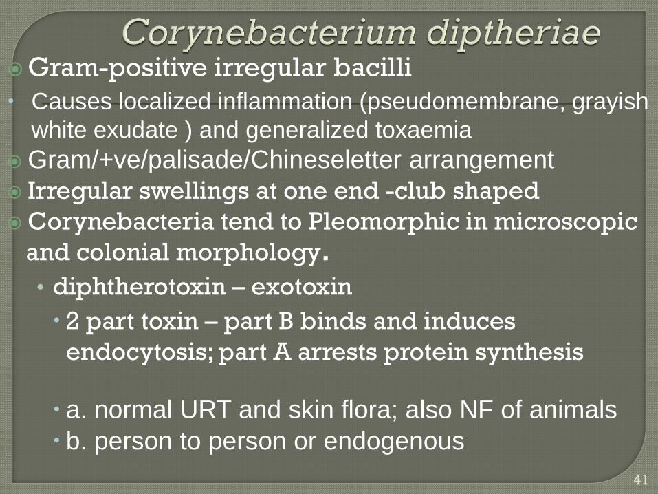
Gram-positive irregular bacilli
Causes localized inflammation (pseudomembrane, grayish
white exudate ) and generalized toxaemia
Gram/+ve/palisade/Chineseletter arrangement
Irregular swellings at one end -club shaped
Corynebacteria tend to Pleomorphic in microscopic
and colonial morphology.
• diphtherotoxin – exotoxin
2 part toxin – part B binds and induces
endocytosis; part A arrests protein synthesis
a. normal URT and skin flora; also NF of animals
b. person to person or endogenous
41

Stained Corynebacterium cells. The "barred" appearance is due to the
presence of polyphosphate inclusions called metachromatic granules.
Note also the characteristic "Chinese-letter" arrangement of cells.

• diphtherotoxin – exotoxin
2 part toxin – part B binds and induces endocytosis; part A arrests protein synthesis
a. normal URT and skin flora; also NF of animals b. person to person or endogenous
a. C. diptheriae i. diphtheria toxin: exotoxin that destroys cells Respiratory diphth: pseudomembrane exudates; respir compromise; toxin affects organs Cutaneous: non-healing ulcers
b. C. jeikeium i. resistance to multiple antibiotics ii. septicemia, wound infections

Reservoir of healthy carriers; potential for diphtheria is always present
Most cases occur in non-immunized children living in crowded, unsanitary conditions.
Acquired via respiratory droplets from carriers or actively infected individuals
2 stages of disease: 1. Local infection –upper respiratory tract
inflammation – sore throat, nausea, vomiting, swollen lymph nodes;
pseudomembrane formation can cause asphyxiation
2. Diptherotoxin production and toxemia • target organs primarily heart and nerves
44

Faucial
Laryngeal
Nasal
Conjunctival
Vulvovaginal
Otitic
Cutaneous around the mouth and the
nose

46


Genera Actinomyces & Nocardia are nonmotile filamentous bacteria related to mycobacteria.Branching, filamentous, gram-positive rodsSome are partially acid-fastDifficult to recognize clinically and difficult to isolate
Nocardia, Streptomyces, Rhodococcus, Tsukamurella May cause chronic infection of skin and soft tissues Actinomyces israelii – responsible for diseases of the
oral cavity, thoracic or intestines - actinomycoses Nocardia brasiliensis causes pulmonary disease
similar to TB.
48

Branched, strictly aerobic bacillus Environmental saprophytes (exogenous infection) Lightly acid-fast Uncommon causes of opportunistic pulmonary
disease Causes primary post-traumatic or post-inoculation
lung disease
a. World-wide inhabitants of soil and water, responsible for decomposition of plant material
b. Can be colonizers or cause infection following traumatic inoculation or inhalation
c. Intracellular pathogens, prevent destruction in phagocytes, tropism for neural tissue

a. Immune competent: skin infections
i. Mycetoma
ii. Lymphocutaneous
iii. Skin abscesses or cellulitis
b. Immune compromised: pulmonary and disseminated
i. pulmonary disease is non-specific, so risk factors add it to differential
ii. organism can spread hematogenously from primary site, resulting in brain and/or skin lesions (dissemination = poor prognosis)


http://home.primus.com.au/royellis/nocast.html
Gram stain of Nocardia


Has branching filaments
Facultative anaerobes
Normal flora of oral cavity
Causes ‘Actinomycosis’ characterized by
multiple abscess and granuloma
formation
Tissue destruction, fibrosis and sinus
formation

ACTINOMYCOSIS

Thank you