Differential expression of the homeobox gene Hox-1.3 in F9 ...
Development 112, 669-678 (1991)Printed in Great Britain © The Company of Biologists Limited 1991
Review Article 669
Gradient fields and homeobox genes
EDDY M. DE ROBERTIS, ELAINE A. MORITA and KEN W. Y. CHO
Department of Biological Chemistry, University of California, Los Angeles, CA 90024-1737, USA
Summary
We review here old experiments that defined theexistence of morphogenetic gradient fields in vertebrateembryos. The rather abstract idea of cell fields of organ-forming potential has become less popular amongmodern developmental and molecular biologists. Resultsobtained with antibodies directed against homeodomainproteins suggest that gradient fields may indeed be
visualized at the level of individual regulatory moleculesin vertebrate embryos.
Key words: homeobox, gradients, embryonic fields,Xenopus laevis, zebrafish embryos, limb development,feather development.
Introduction
The purpose of this essay is to increase awarenessamong modern developmental biologists of the oldconcepts of morphogenetic gradient fields. Fig. 1 showsa 1934 view of the amphibian neurula. Experimentalembryology revealed the existence of fields of organ-forming potential at these very early stages, whichprecede any signs of overt differentiation. By trans-planting newt embryo fragments into heterotopicpositions, fields of cells that were able to give rise, atlater stages, to various organs such as forelimb,hindlimb, tail, balancers and gills were identified. Cellsin these 'morphogenetic fields' have the interestingproperty that they can 'regulate', i.e. produce a normalstructure, after a number of surgical manipulations. Forexample, if part of the field is excised, or if a fragmentof uncommitted tissue is introduced within it, a normalstructure is still formed. If the field is divided intomultiple fragments, multiple copies of the wholestructure ensue. Entire books relate these observations(Huxley and de Beer, 1934; Weiss, 1939; Child, 1941).In particular, "The Elements of Experimental Embry-ology' by Huxley and de Beer contains a lode ofinformation and is highly recommended.
In the 1940s embryonic fields gradually lost centerstage. Perhaps it was because they were considered tobe the result of abstract, almost metaphysical, morpho-genetic forces that could' only be revealed aftertransplantation. The same set of properties can also beexplained by graded positional information models,which have attracted wide attention (Wolpert, 1969,1989). Another important advance in the analysis of cellfields undergoing differentiation was the proposal of thepolar coordinate model (French et at. 1976) in which acircular and a radial set of positional information can
explain, without invoking gradients, the regulationevents observed after manipulation of a field.
The advent of new molecular markers has now madeit possible to follow visually fields of embryonic cellsthat give rise to organs in later development. It may beuseful to review some of the properties of embryonicfields in this light.
Fields in the embryo
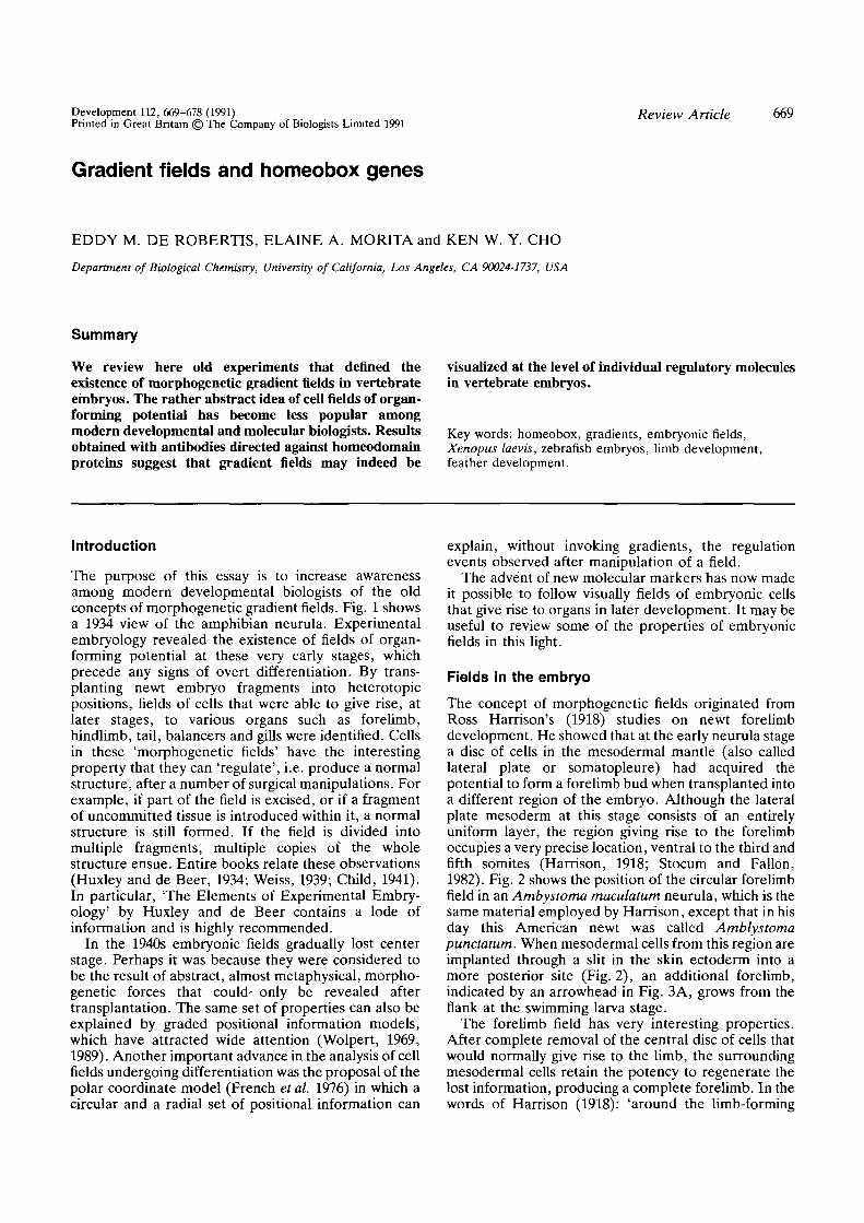
The concept of morphogenetic fields originated fromRoss Harrison's (1918) studies on newt forelimbdevelopment. He showed that at the early neurula stagea disc of cells in the mesodermal mantle (also calledlateral plate or somatopleure) had acquired thepotential to form a forelimb bud when transplanted intoa different region of the embryo. Although the lateralplate mesoderm at this stage consists of an entirelyuniform layer, the region giving rise to the forelimboccupies a very precise location, ventral to the third andfifth somites (Harrison, 1918; Stocum and Fallon,1982). Fig. 2 shows the position of the circular forelimbfield in an Ambystoma maculatum neurula, which is thesame material employed by Harrison, except that in hisday this American newt was called Amblystomapunctatum. When mesodermal cells from this region areimplanted through a slit in the skin ectoderm into amore posterior site (Fig. 2), an additional forelimb,indicated by an arrowhead in Fig. 3A, grows from theflank at the swimming larva stage.
The forelimb field has very interesting properties.After complete removal of the central disc of cells thatwould normally give rise to the limb, the surroundingmesodermal cells retain the potency to regenerate thelost information, producing a complete forelimb. In thewords of Harrison (1918): 'around the limb-forming

670 E. M. De Robertis, E. A. Morita and K. W. Y. Cho
EarFUU
/L«n*\FUld
/Bal«ne«r\FUld
/Hnrt fGfll (Fonlimb\Fl«U \Fleld 17UU.
fHlndlinbIFleld
Fig. 1. Diagram of an amphibian neurula showing thelocalization of the main morphogenetic fields discovered byexperimental analysis. Reproduced from Huxley and deBeer, 1934, with permission of Cambridge University Press.
cells there is thus a zone of tissue which has the power,in gradually diminishing intensity towards the periph-ery, to form a limb vicariously'. If a limb field is cut intotwo halves and transplanted elsewhere, two perfectlimbs are obtained. Conversely, a single -limb can beobtained from two half-fields grafted in the correctorientation. Harrison called this a 'self-differentiatingequipotential system', in which each part can give riseto any part. The term 'field' was coined later to accountfor these properties, first by Spemann (1921) to describethe region of organizer activity present on the dorsalside of the amphibian gastrula, and then by Paul Weiss
Fig. 2. Harrison's limb field transplantation experiment. Aphotograph of an Ambystoma maculatum neurula is shown.At this stage the potency to form forelimb is located in acircular region of the lateral plate mesoderm just ventraland posterior to the pronephros. If this mesoderm issurgically removed and inserted through a slit in theectoderm into a more posterior region (arrow), an extraforelimb will grow at the site of the transplant (shown inFig. 3A). Photo courtesy of Christopher Wright.
Fig. 3. Forelimb and pectoral fin fields in newt and fishembryos. (A) A. maculatum tadpole resulting from a limbfield transplantation at the neurula stage (Fig. 2); note thesupernumerary forelimb bud indicated by the arrowhead.The balancer (bal.) and external gills are indicated.(B-D) Zebrafish embryos stained with anti-XJHbox 1antibodies (photos courtesy of Anders Molven). (B) Lowmagnification view of a 19 h embryo, the arrow indicates acircular area of the lateral plate mesodermal mantle inwhich nuclei contain XlHbox 1 antigen. (C) High powermagnification of B, showing that the cells that will give riseto the future pectoral fin express XlHbox 1 antigen.(D) Section of a pectoral fin bud of a 48 h zebrafishembryo, XlHbox 1 protein is expressed in the anterior andproximal mesoderm as well as in ectodermal skin; thisstaining pattern is very similar to that found in thetetrapod forelimb.
to explain observations concerning regeneration andthe formation of organ rudiments (reviewed by Weiss,1939). Detailed transplantation studies showed that themaximal limb-forming potency is located in theanterodorsal region of the forelimb field and graduallydecreases away from this point (Swett, 1923). This ledto the view that each field consisted of a gradient oforgan-forming potential, i.e. a 'gradient-field' (Huxleyand de Beer, 1934).
A recent example of the power of regulation of thelimb field was provided by tadpoles from a particularpond in Northern California which had supernumerarylegs (Sessions and Ruth, 1990). Both tree frog (Hyla)and salamander tadpoles were afflicted. It was shownthat the malformations were due to parasitic flatworms(trematodes) which burrowed into the developing limbbuds, subdividing them into multiple regions. In somecases a single limb bud gave rise to five well-formed legs(Sessions and Ruth, 1990).
Gradient fields in the adult
The existence of gradient fields can also be revealed inadult organisms which are capable of regeneration. Weshall consider here only two cases: the section ofplanarians in half and the consequence of nervedeflections in adult newts.
The experiments with planarians are discussed herebecause they provide evidence for positional infor-mation of a graded nature along the main body axis. Ifplanarians are cut transversely, as is well known, thefront end will regenerate a tail and the hind piece ahead. This is also true if two different animals aresectioned along the closely located planes indicated as aand b in Fig. 4. The cells located between a and b willproliferate and in one case will form a head and in theother a tail. Because the cells that proliferate in bothcases are essentially the same (Fig. 4), the formation ofhead or tail does not depend on the type of cell presentin the wound, but rather on their relationship to theanteroposterior (A-P) axis of the rest of the embryo.This differs somewhat from the limb field transplan-tation experiments described earlier, in which the cells
bal
gills
3A
•rfiH
3B
3D

9A
la*
f
9B
9CT •
Sc
No
9D

Gradient fields and homeobox genes 671
Fig. 9. Gradients of homeodomain protein expression.Anterior is always to the left and posterior to the right.(A) Formation of a gradient of XlHbox 1 protein duringfeather development in a day 8 chicken embryo.Development proceeds from left to right. Initially a patchof mesodermal cells starts expressing the homeodomainprotein before any morphological changes are detectable.As the feather bud begins to grow, XlHbox 1 antigen losesits homogeneous staining of the feather field and forms agradient of nuclear staining in mesoderm, with a maximumin the anterior and proximal region of the bud. Theectoderm is stained uniformly. (B) Expression of Hox 4.4protein in developing feather buds. Development proceedsfrom left to right. Hox 4.4 initially is present in allmesodermic nuclei, but at later stages becomes localizedpreferentially in the posterior and distal regions of thedeveloping feather bud. The ectoderm is negative.(C) Sagittal section of a 25 h zebrafish embryo showing arow of Rohon-Beard sensory neurons in the dorsal spinalcord (large nuclei indicated by arrowheads). The cells areseparated and arranged in a row. Note that XlHbox 1staining starts abruptly and gradually decreases in theposterior direction until it fades entirely. Sc, spinal cord;No, notochord. (D) Developing chicken wing budimmunostained with XlHbox 1 antibodies. Mesodermalexpression is maximal in the anterior and proximal region.(E) Contralateral wing bud of the same embryo shown inthe previous panel, into which a Dowex bead containingretinoic acid was implanted 18 h previously. Note that theintensity and area of XlHbox 1 expression in mesoderm isgreatly increased by local treatment with retinoic acid. Thisplate shows color photographs of observations reported infull elsewhere: Chuong et al. 1990 (A and B), Molven et al.1990 (C), and Tickle, 1990 (D and E).
still formed limb after being placed in an ectopicposition. The planarian body, however, shares withother fields the capacity to regulate after surgicalmanipulation.
The rate and completeness of regeneration inPlanaria depends on the A-P level of the cut,suggesting the existence of an axial gradient ofregeneration potential (Child, 1915, 1941). The per-centage of animals able to regenerate a head decreasesin a graded way as the transection is carried out inprogressively more posterior regions (reviewed bySlack, 1987). Furthermore, this axial gradient behaveslike a field, because, if for example deep cuts areintroduced into the head region, as shown in Fig. 5,multiple heads are formed. Unfortunately one of themain methods used to study potential gradients inPlanaria was that of 'differential susceptibility' (Child,1941). This would involve, for example, exposing intactor regenerating planarians to various amounts of KCN,strychnine or ethanol, in order to determine whichregion stopped regenerating or died first (usually thehead was more sensitive). These findings were in-terpreted in terms of 'physiological gradients ofmetabolic activity' (Child, 1941) and may have con-tributed to the loss of interest in gradient fields bymodern biologists.
Embryonic fields persist in adult urodeles. Theirexistence can be shown experimentally in Triturus
Fig. 4. Regeneration in Planaria. The same set of cells(located between section planes a and b, indicated inblack) can regenerate either a head or a tail (shaded area)after cutting the animal in half. The experiment shows thatcells are not predetermined to form head or tail, but rathercan sense their relationship to the A-P body axis field. Inthe animal in the center a new pharynx is regenerated at adistance under the influence of the head regenerate.Drawing based on a paradox discussed by Huxley and deBeer, 1934.
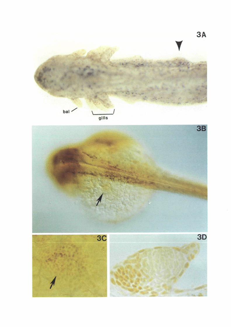
cristatus by deflecting nerves from their normal courseso that they end in the dermis instead. If a sciatic nerveis deflected to a region close to the base of the leg, asupernumerary hindlimb is induced (Guyenot andSchotte, 1926). (In the newt, hindlimbs can berecognized because they have five digits while forelimbshave only four). However, if the nerve is introducedinto the tail region, an extra tail is induced. If the sciaticnerve is deviated into the dorsal crest, a supplementarydorsal crest is induced. Similarly, if the brachial nerve isdeflected close to the arm or shoulder, a supernumeraryforelimb (with four digits) is induced, as shown inFig. 6. If the brachial nerve terminus is placed close tothe dorsal midline an additional dorsal crest is inducedinstead, and if placed at a distance from the forelimb
Fig. 5. Regulation in thehead field of a Planarian.The anterior end receiveda number of deep cuts;the animal produced tenheads. Reproduced fromHuxley and de Beer, 1934,with permission ofCambridge UniversityPress.
672 E. M. De Robertis, E. A. Morita and K. W. Y. Cho
Fig. 6. Nerve deflections in the adult newt can stimulatethe growth of new structures. If the brachial nerve ofTriturus cristatus is cut and deflected so that it now endsclose to the base of the forelimb (1), a supernumeraryforelimb is induced. If the nerve is placed in the dermis ofthe dorsal crest (2), an additional dorsal crest is formed. Ifthe nerve is deflected at a distance of the limb (3), it exitsthe forelimb field and is unable to induce growth of extrastructures. After experiments performed by Guyenot et al.1948.
and dorsal crest - outside of their respective fields - itdoes not induce any growth (see Fig. 6, Guyenot, 1927;Guyenot et al. 1948).
In these experiments, the nerve itself is thought tohave a non-specific trophic action, its effect dependingon the type of mesoderm that it stimulates. The latentpotentialities of the mesoderm can only be revealed byexperimental manipulation. The conclusion from thesestudies is that newts contain, even as adults, a forelimbfield, a hindlimb field, a dorsal crest field, and a tail field(Guyenot, 1927).
Homeobox genes in vertebratesOur interest in gradient fields started with theobservation that a homeodomain protein, XlHbox 1,was expressed as an A-P gradient in the forelimb ofseveral tetrapods (Oliver et al. 1988a). XlHbox 1 wasthe first gene isolated from vertebrate DNA by virtue ofits homology to the Drosophila homeobox (Carrasco etal. 1984). In time we obtained antibody probes thatdetected the protein products of this Xenopus laevisgene (Oliver et al. 19886). Importantly, the antibodiesalso reacted with the homologous protein in a numberof other species such as mouse, chick and zebrafish,affording a molecular glimpse into the comparativeembryology of vertebrates.
There are about 40 homeobox genes of the Antenna-pedia-type in the genomes of the mouse and most othervertebrates (reviewed by Wright et al. 1989a; DeRobertis et al. 1990; Kessel and Grass, 1990). Theyencode transcription factors which are expressed inspecific A-P regions of the embryo. The genes arelocated in four clusters of about 10 genes each, withgenes located at the 5' end of the complexes expressedin posterior regions of the embryo and those in more 3'
positions expressed in progressively more anteriorregions (Gaunt et al. 1988; Graham et al. 1989;Wilkinson et al. 1989). This genomic organization isstrikingly similar to that of Drosophila homeotic geneclusters (Lewis, 1978; Gehring, 1987), suggesting thatthis gene arrangement arose in a common ancestor.Because mammals did not evolve from insects, thesecommon ancestors must go back at least to organismssuch as flatworms. It would be interesting to knowwhether primitive metazoans such as rotifers, whichhave well-defined A-P polarity, possess homeoboxgene complexes.
The function of at least some vertebrate homeoboxgenes is to specify cell identity along the A-P axis. Bothloss-of-function (obtained by microinjection of anti-bodies into Xenopus embryos, Wright et al. 19896) andgain-of-function (obtained by overexpression in trans-genic mice, Kessel et al. 1990) phenotypes suggest thatthe vertebrate genes have similar functions to theirDrosophila homeotic counterparts. Furthermore, over-expression of mouse and human homeobox genes intransgenic fruit flies leads to homeotic transformationsof cell fate (Malicki et al. 1990; McGinnis et al. 1990).
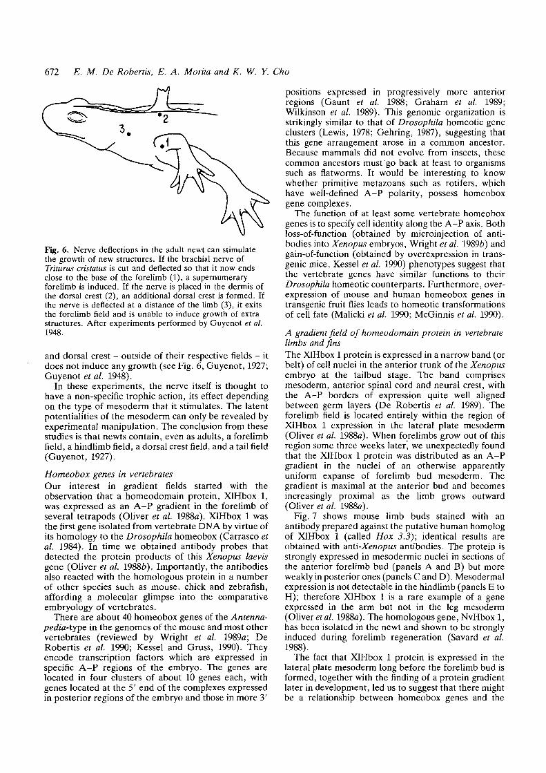
A gradient field of homeodomain protein in vertebratelimbs and finsThe XlHbox 1 protein is expressed in a narrow band (orbelt) of cell nuclei in the anterior trunk of the Xenopusembryo at the tailbud stage. The band comprisesmesoderm, anterior spinal cord and neural crest, withthe A-P borders of expression quite well alignedbetween germ layers (De Robertis et al. 1989). Theforelimb field is located entirely within the region ofXlHbox 1 expression in the lateral plate mesoderm(Oliver et al. 1988a). When forelimbs grow out of thisregion some three weeks later, we unexpectedly foundthat the XlHbox 1 protein was distributed as an A-Pgradient in the nuclei of an otherwise apparentlyuniform expanse of forelimb bud mesoderm. Thegradient is maximal at the anterior bud and becomesincreasingly proximal as the limb grows outward(Oliver et al. 1988a).
Fig. 7 shows mouse limb buds stained with anantibody prepared against the putative human homologof XlHbox 1 (called Hox 3.3); identical results areobtained with anti-Xenopus antibodies. The protein isstrongly expressed in mesodermic nuclei in sections ofthe anterior forelimb bud (panels A and B) but moreweakly in posterior ones (panels C and D). Mesodermalexpression is not detectable in the hindlimb (panels E toH); therefore XlHbox 1 is a rare example of a geneexpressed in the arm but not in the leg mesoderm(Oliver et al. 1988a). The homologous gene, NvHbox 1,has been isolated in the newt and shown to be stronglyinduced during forelimb regeneration (Savard et al.1988).
The fact that XlHbox 1 protein is expressed in thelateral plate mesoderm long before the forelimb bud isformed, together with the finding of a protein gradientlater in development, led us to suggest that there mightbe a relationship between homeobox genes and the

Gradient fields and homeobox genes 673
cfli d
Fig. 7. A gradient of XlHbox 1 antigen is present in nuclei of the anterior mesoderm of developing mouse forelimbs(panels a through d) but is absent in hindlimbs (panels e through h). The ectoderm is stained in both fore- and hindlimbs.Transverse sections of a day 10 mouse embryo are shown. D, dorsal; V, ventral; Di, distal; Pr, proximal; Ant., anterior;Post., posterior. Reproduced, with permission, from Oliver et al. Cell 55, 1017-1024 (1988).
gradient fields described by experimental embryologists(Oliver et al. 1988a). The best indication that this mightbe the case came from studies on the development ofthe fish pectoral fin bud (Molven et al. 1990). The fishpectoral fin, which is the evolutionary precursor of thetetrapod forelimb, develops by proliferation of a groupof cells of the lateral plate mesoderm, as demonstratedinitially in the salmon embryo (Harrison, 1895).
The zebrafish embryo is better material than Xeno-pus for these studies because the pectoral fin bud formsearly on in embryogenesis, rather than three weekslater as part of the metamorphosis process in the case ofthe tadpole forelimb. In addition, in the early fishembryo, the lateral plate mesoderm extends as a thinhomogenous cell layer that surrounds the yolk, greatlyfacilitating its study. Figs 3B and 3C show that in the19 h embryo a circular region of XlHbox 1-positivenuclei can be distinguished in the lateral platemesoderm (indicated by an arrow). This is about 10 hbefore the pectoral fin bud itself is morphologicallyrecognizable. This circular patch of cells can befollowed throughout development and corresponds tothe pectoral fin region (Molven et al. 1990). In 25 h
embryos staining becomes stronger in the anteriorregion and cells start to proliferate; by 48 h a well-developed finbud is present. At the latter stage, asshown in Fig. 3D, expression is maximal in the anteriorand proximal fin bud. This pattern of expression is verysimilar to what one would find in, say, frog, chicken ormouse forelimb buds. In addition to pointing to aconservation of basic developmental mechanisms dur-ing vertebrate evolution, the zebrafish study suggeststhat expression of a homeobox gene can demarcate amorphogenetic field (Molven et al. 1990).
Many other homeobox genes are also expressedduring limb development (Eichele, 1989). An antibodyagainst the Hox 4.4 gene of human origin detects aprotein gradient that has the opposite polarity to that ofXlHbox 1 in the forelimb (Oliver et al. 1989). The Hox4.4 gradient is maximal in the distal and posteriorregion of Xenopus, mouse, and chick fore- andhindlimb buds. Although direct proof is still lacking,the opposing gradients of Hox 4.4 and XlHbox 1proteins could be involved in specifying positionalvalues in developing limbs.
Local application of retinoic acid to the anterior
674 E. M. De Robertis, E. A. Morita and K. W. Y. Cho
region of chick wing buds leads to changes inmorphogenesis, and it has been proposed that A-Ppositional information in the limb bud is provided by adiffusible gradient of retinoic acid (reviewed byEichele, 1989). Recent experiments suggest that reti-noic acid applied to the anterior limb bud does notestablish a new retinoic acid gradient throughout thebud, but rather changes the fate of nearby cells(Wanecke/a/. 1991; Noji etal. 1991). Thus the existenceof a diffusible morphogen gradient has been chal-lenged. The gradients of homeodomain proteins,although not yet proven to affect morphogenesis,strongly argue that graded positional information ofsome sort must exist in limb buds. Thus it may beworthwhile to study the positional signalling systemsthat set up the gradients of homeoproteins in limbdevelopment.
Duboule and colleagues have shown that followingexpression of Hox 4.4, the three genes located 5' to it inthe chromosome (recently renamed Hox 4.5, 4.6 and4.7, Duboule et al. 1990) are sequentially activated atthe tip of the limb as it grows longer (Dolle et al. 1989).Thus, the timing of expression of these genes followsthe order that they occupy in the gene complex. Hox 4genes are also expressed along the main body A-P axis,where they are deployed in the same order (4.4 moreanterior than 4.5, 4.5 more anterior than 4.6, and soon).
An unexpected conclusion from the studies onhomeodomain protein gradients in limbs is that thesame set of genes utilized during development of themain body A-P axis are also brought into play duringlimb growth.
Limb fields in the fruit fly
The early Drosophila embryo, which has such tremen-dous advantages for genetic studies on development,does not lend itself easily to transplantation studies ofthe type that are possible, say, in Amphibia. It istherefore not surprising that embryonic field conter-parts have not been found. However, experiments onregeneration of cockroach legs strongly argue that, atleast at later stages in life, insect legs do have theregulatory properties of cell fields (Bohn, 1974; Frenchet al. 1976). Recent studies on the gene Distallesssuggest that fly embryos indeed have fields of cellsinvolved in the formation of appendages.
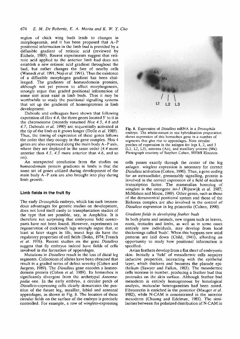
Mutations in Distalless result in the loss of distal legsegments. Collections of alleles have been obtained thatresult in a graded series of defect severity (Cohen andJurgens, 1989). The Distalless gene encodes a homeo-domain protein (Cohen et al. 1989). Its homeobox issignificantly divergent from the archetypal Antenna-pedia one. In the early embryo, a circular patch ofDu/a/tors-expressing cells clearly demarcates the pos-ition of the future leg, maxillar, labial and antennalappendages, as shown in Fig. 8. The location of thesecircular fields on the surface of the embryo is preciselycontrolled. For example, a row of wmg/ew-expressing
Fig. 8. Expression of Distalless mRNA in a Drosophilaembryo. The whole-mount in situ hybridization preparationshows expression of this homeobox gene in a number ofsegments that give rise to appendages. Note circularpatches of expression in the anlagen for legs 1,2, and 3(LI, L2, L3), antenna (An), and maxillary process (Mx).Photograph courtesy of Stephen Cohen, HHMI Houston.
cells passes exactly through the center of the leganlagen. wingless expression is necessary for correctDistalless activation (Cohen, 1990). Thus, a gene codingfor an extracellular, presumably signalling, protein isinvolved in the correct expression of a field of nucleartranscription factor. The mammalian homolog ofwingless is the oncogene int-1 (Rijsewijk et al. 1987;McMahon and Moon, 1989). Other genes, such as thoseof the dorsoventral positional system and those of theBithorax complex are also involved in the control ofDistalless expression in leg primordia (Cohen, 1990).
Gradient fields in developing feather budsIn both plants and animals, new organs such as leaves,roots, tentacles and limbs, as well as in some casesentirely new individuals, may develop from localthickenings called 'buds'. When this happens new axialpatterns are laid down (Child, 1941), affording anopportunity to study how positional information isspecified.
Avian feathers develop from a flat sheet of embryonicskin. Initially a 'field' of mesodermic cells acquiresinductive properties, interacting with the epitheliallayer, which thickens and becomes the placode epi-thelium (Sawyer and Fallon, 1983). The mesodermiccells increase in number, producing a feather bud thatprotrudes on the skin surface. Although feather budmesoderm is entirely homogeneous by histologicalanalysis, molecular heterogeneities had been noted.Fibronectin is enriched in the posterior (Mauger et al.1982), while N-CAM is concentrated in the anteriormesoderm (Chuong and Edelman, 1985). The simi-larities between the polarized distribution of N-CAM in
Gradient fields and homeobox genes 675
feather buds and the gradient of XlHbox 1 in forelimbbuds provided the initial impulse for analyzing theexpression of homeodomain proteins in developingfeathers.
Fig. 9A shows the expression of XlHbox 1 antigen infeather buds at various stages of development. The firstsign of feather formation is the appearance of patches ofXlHbox 1-positive cells in the dermis or feather field.Nuclei over the entire field express XlHbox 1 protein,even as a visible bud begins to form. As the buds growstaining is lost in the posterior and XlHbox 1 adopts agraded distribution, with maximal expression in theanterior and proximal region (Chuong et al. 1990).Detailed analysis showed that N-CAM expressionoccupies a smaller region of the anterior feather budthan that of the XlHbox 1 gradient. Fig. 9B shows thatHox 4.4 is expressed uniformly and very strongly in theinitial field and early feather buds. As developmentproceeds, Hox 4.4 protein becomes polarized to theposterior and distal mesoderm (Chuong et al. 1990).This complementary distribution of the two homeo-domain proteins is very reminiscent of the gradientsthey adopt during the development of an entirelydifferent structure, the forelimb bud (Oliver et al.1989).
Gradients of homeobox expression along themain body axis
Most in situ hybridization studies on the expression ofmouse homeobox genes in mid-gestation embryos showa graded distribution of mRNA in the spinal cord. Themaximum corresponds to the anterior border ofexpression, with levels usually diminishing posteriorly(e.g. Breier et al. 1988). While this is true for totalmRNA levels, it has been difficult to extend this to thesingle cell level. In the zebrafish, there is a populationof large sensory neurons called Rohon-Beard neurons.Their function is to process the sensory input fromswimming movements and can be easily recognizedbecause of their large size and dorsal position in thespinal cord. Rohon-Beard neurons express XlHbox 1antigen, and display an interesting pattern (Molven etal. 1990). Very anterior ones are devoid of the antigenbut, as shown in Fig. 9C, they abruptly start expressingXlHbox 1 within the anterior spinal cord and exhibit agraded decrease in intensity in the posterior directionuntil expression fades entirely. Thus, although allRohon-Beard neurons serve the same function, theydifferentially express XlHbox 1 according to theirposition along the A-P axis. It is as if the XlHbox 1gene in these cells were able to sense a gradient ofpositional information present along the body axiswhich affects its level of expression.
A gradient of expression in the opposite direction ofthe body axis exists for another homeobox gene,Xhox3, during early Xenopus development (Ruiz iAltaba and Melton, 1989a). This gene, which has ahomeobox related to Drosophila evenskipped, is ex-pressed very early in development, with maximal
mRNA levels in the posterior end of mid-neurulaembryos. Antibody staining has shown that thisgradient is established in embryonic mesoderm and thatit fades in the anterior direction (Ruiz i Altaba et al.1991). Both loss- and gain-of-function experimentssupport the view that this gene plays an important rolein A-P axis formation (Ruiz i Altaba and Melton,19896; Ruiz i Altaba et al. 1991).
Thus the main body axis of the vertebrate seems tohave A-P gradients of positional information duringthe course of embryogenesis, which are able to activatehomeobox genes.
Spemann's organizer field
A field of organization potential is present on the dorsalside of the early amphibian gastrula (Spemann, 1921;Holtfreter and Hamburger, 1955; Wakahara, 1989;Stewart and Gerhart, 1990). It determines the extent towhich cells will invaginate through the blastopore lipand consequently the extent of the future A-P bodyaxis. This dorsal organizer region exhibits many of theproperties of a morphogenetic field: if one organizer isdivided into several fragments each will lead to theformation of a new body axis after transplantation, partof the organizer field can be removed and a well-proportioned axial system can still be formed, uncom-mitted embryonic cells grafted into the dorsal lip canbecome part of the organizer, two organizer fields canbe fused to form a single axial system (Spemann, 1938;Holtfreter and Hamburger, 1955).
The generation of the organizer field has been thesubject of intense investigation. It can be traced back tofertilization, which elicits a rotation movement of theegg cortex (reviewed by Gerhart et al. 1989). The futuredorsal side forms usually on the opposite side of thesperm entry point, where the cortical rotation bringslarge yolk platelets and animal pole cytoplasm intoclose contact. By the 32-cell stage, the two most dorsaland vegetal blastomeres acquire the potential to induceother cells. This 'Nieuwkoop center' (Gerhart et al.1989) is thought to release growth factors of the TGF-/?family, inducing 'Spemann's organizer' (or, in otherwords, anterodorsal mesoderm) activity in overlyingcells (Smith et al. 1989; Thomsen et al. 1990). If the sizeof the organizer is decreased by surgical removal at thelate blastula stage, tadpoles with a graded series ofanterior axial defects are obtained (Stewart andGerhart, 1990).
The organizer is the morphogenetic field mostamenable to molecular analysis, at least in amphibians.While interesting patterns of antibody stainings suggesta correlation between homeodomain proteins andgradient fields, there is no direct evidence as yet thatthis has a causal effect in morphogenesis. In the case ofthe organizer it is known that growth factors areinvolved in its generation. It is also known that growthfactors can activate the expression of certain homeoboxgenes in Xenopus (Rosa, 1989; Ruiz i Altaba andMelton, 1989c; Cho and De Robertis, 1990). The recent

676 E. M. De Robertis, E. A. Morita and K. W. Y. Cho
findings that overexpression of homeodomain proteinscan confer axis-inducing properties to Xenopus embry-onic cells (Cho et al. 1991) and that organizer-specifichomeobox genes are expressed in Xenopus gastrulae(Blumberg et al. 1991) encourage us to think thatprogress in this area will be forthcoming.
Conclusions and prospects
One of the main challenges for the future will beunderstanding what sets up a circular field or a gradientof nuclear protein in an otherwise homogeneouspopulation of mesenchymal cells. Unravelling the cell-to-cell signalling mechanisms that achieve this shouldshed light on the nature of positional information.
One obvious candidate is retinoic acid, which hasprofound effects on limb development (Eichele, 1989)and on the expression of homeobox gene complexes(Simeone et al. 1990). Implantation of a bead contain-ing retinoic acid into developing chick wing buds greatlyexpands the gradient of XlHbox 1 expression, as shownin Figs 9D and 9E. This is accompanied by malforma-tions in which excess anterior shoulder structures(which are the normal fate of XlHbox 1-expressingcells) are formed at the expense of the rest of the limbbud (Oliver et al. 1990). In the case of Hox 4 complex,implantation of a retinoic acid bead in the anterior ofthe chick wing bud induces the sequential activation ofhomeobox genes. This leads to a mirror imageduplication of Hox 4 expression which correlates verywell with the digit duplications caused by retinoic acid(Izpisua-Belmonte et al. 1991; Nohno et al. 1991).
Many other molecules could be involved as well.N-CAM, peptide growth factors, and the extracellularprotein wingless • (int-1 in vertebrates) have beenmentioned already. Many orphan nuclear receptors, forwhich the ligands are as yet unknown, have beenisolated (Evans, 1988). Cell surface molecules involvedin the activation of nuclear regulatory proteins inDrosophila (such as notch, sevenless and bride-of-sevenless, see Banerjee and Zipursky, 1990) either have(Coffman et al. 1990) or can be presumed to havevertebrate homologs. About 50 mutations are known toaffect limb development in the mouse; perhaps some ofthem affect this intercellular signalling system. Trans-genic mice expressing gradients of reporter genes fusedto homeobox gene promoters would facilitate thisanalysis.
In this essay, we have dealt mostly with the XlHbox 1gene. There are about 40 different homeobox genes ofthe Antennapedia-type in vertebrates, and there is noreason to think that their expression patterns will be lessrich or informative. What made XlHbox 1 special wasthe early availability of antibodies that cross-reactedwith the homologous genes in a wide spectrum ofvertebrates.
During embryogenesis XlHbox 1 is expressed inseveral regions of very different developmental poten-tial. First, it subdivides the body axis into a band ofhomeodomain protein expression in the anterior trunk
region which spans the mesoderm, CNS and neuralcrest. In some cases (Fig. 9C), it can be seen togradually decrease towards the posterior-end. Second,it is expressed in the region of the lateral platemesoderm that will give rise to the forelimb and, as alimb bud develops, it forms an A-P gradient ofexpression in mesodermal nuclei. Finally, in the case offeather development, circular patches of expression inthe dermis are followed by bud growth and a new A-Pgradient in each feather bud. Thus, the morphogeneticgradient fields defined by experimental embryology(Harrison, 1918; Huxley and de Beer, 1934) seem tohave a molecular substratum that can be followedvisually with antibody markers. It seems that theproblem of setting up pattern in the vertebrate isresolved within fields of cells. Gradients of homeo-domain proteins are utilized again and again duringembryogenesis, perhaps to provide A-P polarity, inthese cell fields undergoing pattern formation.
We have discussed correlations between gradients ofexpression of homeodomain proteins and the behaviorof morphogenetic fields defined by transplantationexperiments. We have emphasized that there is nodirect evidence linking the two in a causal way atpresent. The purpose of this essay was to stimulatethought and research in gradient fields, an area ofdevelopment ripe for molecular studies.
We are indebted to several colleagues that made importantcontributions to the XlHbox 1 localization studies reviewedhere: G. Oliver (now at the University of Uruguay), C.V.E.Wright (now at Vanderbilt University), A. Molven (Univer-sity of Bergen, Norway), C.B. Kimmel (University ofOregon), CM. Chuong (University of Southern California),and C. Tickle (University College, London). We are gratefulto Stephen Cohen (HHMI, Houston) for Fig. 8, and toChristof Niehrs and Dennis Bittner for suggestions. Particularthanks to Phil De Vellis, a high school student, who recoveredthis essay from the depths of a floppy disk after the entire textwas accidentally erased. Work in our laboratory is supportedby the N.I.H. (HD 21502-06). K.W.Y.C. was supported bythe UCLA Human Genetics Postdoctoral Training Program(GM 08243) and E.A.M. by Training Program GM 07104.
References
BANERJEE, U. AND ZIPURSKY, S. L. (1989). The role of cell-cellinteraction in the development of the Drosophila visual system.Neuron 4, 177-187.
BLUMBERG, B., WRIGHT, C. V. E., D E ROBERTIS, E. M. AND CHO,
K. W. Y. (1991). Organizer-specific homeobox genes inXenopus laevis embryos. Science (in press).
BOHN, H. (1974). Extent and properties of the regeneration fieldin the larval legs of cockroaches (Leucophaea maderae).J. Embryol. exp. Morph. 31, 557-572.
BRETER, G., DRESSLER, G. R. AND GRUSS, P. (1988). Primary
structure and developmental expression pattern of Hox 3.1, amember of the murine Hox 3 homeobox gene cluster. EMBO J.7, 1329-1336.
CARRASCO, A. E., MCGINNIS, W., GEHRING, W. J. AND D E
ROBERTIS, E. M. (1984). Cloning of an X. laevis gene expressedduring early embryogenesis coding for a peptide regionhomologous to Drosophila homeotic genes. Cell 37, 409-414.
CHILD, C. M. (1915). Individuality in Organisms, University ofChicago Press.
Gradient fields and homeobox genes 611
CHILD, C. M. (1941). Patterns and Problems of Development272-303, Chicago: University of Chicago Press.
CHO, K. W. Y. AND DE ROBERTIS, E. M. (1990). Differentialactivation of Xenopus homeobox genes by mesodenn inducinggrowth factors and retinoic acid. Genes Dev. 4, 1910-1916.
CHO, K. W. Y., MORTTA, E. A., WRIGHT, C. V. E. AND DEROBERTO, E. M. (1991). Overexpression of a homeodomainprotein confers organizer activity to uncommitted Xenopusembryonic cells. Cell 65, 1-20.
CHUONG, C. M. AND EDELMAN, G. M. (1985). Expression of celladhesion molecules in embryonic induction. I. Morphogenesis ofnestling feathers. J. Cell Biol. 101, 1009-1026.
CHUONG, C. M., OLIVER, G., TING, S. A., JEGALIAN, B. G., CHEN,H. M. AND DE ROBERTO, E. M. (1990). Gradients ofhomeoproteins in developing feather buds. Development 110,1021-1030.
COFFMAN, C , HARRIS, W. AND KINTNER, C. (1990). Xotch, theXenopus homolog of Drosophila Notch. Science 249, 1438-1441.
COHEN, S. M. (1990). Specification of limb development in theDrosophila embryo by positional cues from segmentation genes.Nature 343, 173-177.
COHEN, S. M., BRONNER, G., KUTTNER, F., JURGENS, G. ANDJACKLE, H. (1989). Distal-less encodes a homeodomain proteinrequired for limb development in Drosophila. Nature 338,432-434.
COHEN, S. M. AND JURGENS, G. (1989). Proximal-distal patternformation in Drosophila: cell autonomous requirement forDistal-less gene activity in limb development. EMBO J. 8,2045-2055.
DE ROBERTO, E. M., OLIVER, G. AND WRIGHT, C. V. E. (1989).Determination of axial polarity in the vertebrate embryo:homeodomain proteins and homeogenetic induction. Cell 57,189-191.
DE ROBERTO, E. M., OLIVER, G. AND WRIGHT, C. V. E. (1990).Homeobox genes and the vertebrate body plan. ScientificAmerican 263, July, pp. 46-52.
DOLLE, P., IZPISUA-BELMONTE, J. C , FALKENSTEIN, H., RENUCCI,A. AND DUBOLTLE, D. (1989). Coordinate expression of themurine Hox-5 complex homeobox-containing genes during limbpattern formation. Nature 342, 767-772.
DUBOULE, D., BONCTNELU, E., DE ROBERTO, E. M.,FEATHERSTONE, M., LONAI, P., OLIVER, G. AND RUDDLE, F.(1990). An update of mouse and human HOX genenomenclature. Genomics 7, 458-459.
EICHELE, G. (1989). Rerinoids and vertebrate limb patternformation. Trends Genet. 8, 246—250.
EVANS, R. M. (1988). The steroid and thyroid hormone receptorsuperfamily. Science 240, 889-895.
FRENCH, V., BRYANT, P. J. AND BRYANT, S. V. (1976). Patternregulation in epimorphic fields. Science 193, 969-981.
GAUNT, S. J., SHARPE, P. T. AND DUBOULE, D. (1988). Spatiallyrestricted domains of homeogene transcripts in mouse embryos:relation to a segmented body plan. Development 104Supplement, 169-179.
GEHRTNG, W. J. (1987). Homeo boxes in the study ofdevelopment. Science 236, 1245-1252.
GERHART, J., DANIIXHIK, M., DONIACH, T., ROBERTS, S.,ROWNING, B. AND STEWART, R. (1989). Cortical rotation of theXenopus egg: consequences for the anteroposterior pattern ofembryonic dorsal development. Development 107 Supplement,37-51.
GRAHAM, A., PAPOLOPULU, N. AND KRUMLAUF, R. (1989). Themurine and Drosophila homeobox gene complexes havecommon features of organization and expression.' Cell 57,367-378.
GUYENOT, E. (1927). Le probleme morphogenetique dans laregeneration des Urodeles: determination et potentialites desregenerats. Rev. Suisse de Zool. 34, 127-154.
GUYENOT, E., DINICHERT-FAVASGER, J. AND GALLAND, M. (1948).L'exploration du territoire de la patte anterieure du Triton.Revue Suisse de Zoologie 00, 1-120.
GUYENOT, E. AND SCHOTTE, O. (1926). Demonstration del'existence de territoires spccifiques de regeneration par le
methode de la deviation des troncs nerveux. C. r. hebd. Soc.Biol. 94, 1050.
HARRISON, R. G. (1895). Die Entwicklung der unpaaren undpaarigen Flossen der Teleostier. Arch. f. mikr. Anat. 46,500-578.
HARRISON, R. G. (1918). Experiments on the development of thefore-limb of Amblystoma, a self-differentiating equipotentialsystem. J. exp. Zool. 25, 413-461.
HOLTFRETER, J. AND HAMBURGER, V. (1955). Amphibians, InAnalysis of Development, (ed. B. H. Willier, P. Weiss and V.Hamburger), pp. 230-296, W. B. Saunders Company,Philadelphia and London.
HUXLEY, J. S. AND DE BEER, G. R. (1934). The Elements ofExperimental Embryology, Cambridge University Press.
IZPISUA-BELMONTE, J. C , TICKLE, C , DOLLE, P., WOLPERT, L.AND DUBOULE, D. (1991). Expression of the homeobox Hox-4genes and the specification of position in chick wingdevelopment. Nature 350, 585-589.
KESSEL, M., BALLING, R. AND GRUSS, P. (1990). Variations ofcervical vertebrae after expression of a Hox-1.1 transgene inmice. Cell 61, 301-308.
KESSEL, M. AND GRUSS, P. (1990). Murine developmental controlgenes. Science 249, 374-379.
LEWIS, E. B. (1978). A gene complex controlling segmentation inDrosophUa. Nature 276, 565.
MAUCKI, J., SCHUGHART, K. AND MCGINNIS, W. (1990). MouseHox-2.2 specifies thoracic segmental identity in Drosophilaembryos and larvae. Cell 63, 961-967.
MAUGER, A., DEMARCHEZ, M., HERBAGE, D., GRIMAUD, J. A.,DRUGUET, M., HARTMANN, D. AND SENGEL, P. (1982).Immunofluorescent localization of collagen type I and III, andof fibronectin during feather morphogenesis in the chickenembryo. Devi Biol. 94, 93-105.
MCGINNIS, N., KUZIORA, M. A. AND MCGINNIS, W. (1990).Human Hox-4.2 and Drosophila deformed encode similarregulatory specificities in Drosophila embryos and larvae. Cell63, 969-976.
MCMAHON, A. P. AND MOON, R. T. (1989). mt-1 - a proto-oncogene involved in cell signalling. Development 107Supplement, 161-167.
MOLVEN, A., WRIGHT, C. V. E., BREMILLER, R., DE ROBERTO, E.M. AND KTMMEL, C. B. (1990). Expression of a homeobox geneproduct in normaJ and mutant zebrafish embryos: evolution ofthe tetrapod body plan. Development 109, 279-288.
NOHNO, T., Non, S., KOYAMA, E., OHYAMA, K., MOYOKAI, F.,KUROIWA, A., SATTO, T. AND TANIGUCHI, S. (1991). Involvementof the Chox-4 chicken homeobox genes in determination ofanteroposterior axial polarity during limb development. Cell 64,1197-1205.
NOJI, S., NOHO, T., KOYAMA, E., MUTO, K., OHYAMA, K., AOH,Y., TAMURA, K., OHSUGI, K., IDE, H., TANIGUCHI, S. ANDSAITO, T. (1991). Retinoic acid induces polarizing activity but isunlikely to be a morphogen in the chick limb bud. Nature 350,83-86.
OLIVER, G., SIDELL, N., FISKE, W., HEINZMANN, C , MOHANDAS,T., SPARKES, R. S. AND DE ROBERTO, E. M. (1989).Complementary homeo protein gradients in developing limbbuds. Genes Dev. 3, 641-650.
OLIVER, G., WRIGHT, C. V. E., HARDWICKE, J. AND DE ROBERTIS,E. M. (1988a). A gradient of homeodomain protein indeveloping forelimbs of Xenopus and mouse embryos. Cell 55,1017-1024.
OLIVER, G., WRIGHT, C. V. E., HARDWICKE, J. AND DE ROBERTIS,E. M. (19886). Differential antero-posterior expression of twoproteins encoded by a homeobox gene in Xenopus and mouseembryos. EMBO J. 7, 3199-3209.
RlJSEWUK, F., SCHUERMANN, M., WAGENAAR, E . , PARREN, P.,
WEIGEL, D. AND NUSSE, R. (1987). The Drosophila homolog ofthe mouse mammary oncogene mt-1 is identical to the segmentpolarity gene wingless. Cell 50, 649-657.
ROSA, F. M. (1989). Mix.l, a homeobox mRNA inducible bymesoderm inducers, is expressed mostly in the presumptiveendoderm cells of Xenopus embryos. Cell 57, 965-974.
Ruiz I ALTABA, A., CHOI, T. AND MELTON, D. A. (1991).

678 E. M. De Robertis, E. A. Morita and K. W. Y. Cho
Blocking the function of the Xenopus homeobox gene Xhox 3suggests its requirement for normal posterior development.Submitted.
Ruiz I ALTABA, A. AND MELTON, D. A. (1989a). Bimodal andgraded expression of the Xenopus homeobox gene Xhoxh duringembryonic development. Development 106, 173-183.
Ruiz I ALTABA, A. AND MELTON, D. A. (19896). Involvement ofthe Xenopus homeobox gene Xhox3 in pattern formation alongthe antenor-posterior axis. Cell 57, 317-326.
Ruiz I ALTABA, A. AND MELTON, D. A. (1989c). Interactionbetween peptide growth factors and homeobox genes in theestablishment of antero-posterior polarity in frog embryos.Nature 341, 33-38.
SAVARD, P., GATES, P. B. AND BROCKES, J. P. (1988). Position-dependent expression of a homeobox gene transcript in relationto amphibian limb regeneration. EMBO J. 7, 4275-4282.
SAWYER, R. H. AND FALLON, J. F. (eds) (1983).Epithelial-Mesenchymal Interactions in Development, pp.147-161, Praeger Publishers, New York.
SESSIONS, S. K. AND RUTH, S. B. (1990). Explanation for naturallyoccurring supernumerary limbs in Amphibians. /. exp. Zool.254, 38-47.
SlMEONE, A . , ACAMPORA, P . , ARCIONI, L . , ANDREWS, P . W . ,BONCINELLI, E. AND MAVILIO, F. (1990). Sequential activation ofHox 2 homeobox genes by retinoic acid in human embryonalcarcinoma cells. Nature 346, 763-766.
SLACK, J. M. W. (1987). Morphogenetic gradients - past andpresent. Trends Biochem. Set. 12, 200-204.
SMITH, J. C , COOKE, J., GREEN, J. B. A., HOWES, G. AND SYMES,K. (1989) Inducing factors and the control of mesodermalpattern in Xenopus laevis. Development 107 Supplement,149-159.
SPEMANN, H. (1921). Die Erzeugung trierischer Chimaeren durchheteroplastische embryonale Transplantation zwischen Tntoncristatus und taeniatus. Roux' Arch. Ent. mech. 48, 533-570.
SPEMANN, H. (1938). Embryonic Development and Induction.(Yale University press, New Haven).
STEWART, R. M. AND GERHART, J. C. (1990). The anterior extentof dorsal development of the Xenopus embryonic axis depends
on the quantity of organizer in the late blastula. Development109, 363-372.
STOCUM, D. L. AND FALLON, J. F. (1982). Control of patternformation in urodele limb ontogeny: A review and a hypothesis./. Embryol. exp. Morph. 69, 7-36.
SWETT, F. H. (1923). The prospective significance of the cellscontained in the four quadrants of the primitive limb disc ofAmblystoma. J. exp. Zool. 37, 207-220.
THOMSEN, G., WOOLF, T., WHITMAN, M., SOKOL, S., VAUGHAN, J.,VALE, W. AND MELTON, D. A. (1990). Activins are expressedearly in Xenopus embryogenesis and can induce axial mesodermand anterior structures. Cell 63, 485-493.
TICKLE, C. (1991). Retinoic acid and chick limb development.Development 1991 Supplement 1, 113-121.
WAKAHARA, M. (1989). Specification and establishment of dorsal-ventral polarity in eggs and embryos of Xenopus laevis.Develop. Growth and Differ. 31, 197-207.
WANEK, N., GARDINER, D. M., MUNEOKA, K. AND BRYANT, S.(1991). Conversion by retinoic acid of anterior cells into ZPAcells in the chick wing bud. Nature 350, 81-83.
WEISS, P. (1939). Principles of Development, Holt and Co., NewYork.
WILKINSON, D. G., BHATT, S., COOK, M., BONCINELLI, E. ANDKRUMLAUF, R. (1989). Segmental expression of Hox-2homeobox-containing genes in the developing mouse hindbrain.Nature 341, 405-409.
WOLPERT, L. (1969). Positional information and the spatial patternof cellular differentiation. J theor. Biol. 25, 1-47.
WOLPERT, L. (1989). Positional information revisited. Development107 Supplement, 3-12.
WRIGHT, C. V. E., CHO, K. W. Y., OLIVER, G. AND DE ROBERTIS,E. M. (1989a). Vertebrate homeodomain proteins: families ofregion-specific transcription factors. Trends Biochem. Set. 14,52-56.
WRIGHT, C. V. E., CHO, K. W. Y., HARDWICKE, J., COLLINS, R.H. AND DE ROBERTIS, E. M. (19896). Interference with functionof a homeobox gene in Xenopus embryos producesmalformations of the anterior spinal cord. Cell 59, 81-93.
(Accepted 28 March 1991)