Gossypol Modification of Ala-1 of Secreted Phospholipase A 2 : A Probe...
12
Gossypol Modification of Ala-1 of Secreted Phospholipase A 2 : A Probe for the Kinetic Effects of Sulfate Glycoconjugates ² Bao-Zhu Yu, ‡ Joseph Rogers, ‡ Girish Ranadive, ‡ Sharon Baker, § David C. Wilton, § Rafael Apitz-Castro, | and Mahendra Kumar Jain* ,‡ Department of Chemistry and Biochemistry, UniVersity of Delaware, Newark, Delaware 19716, Department of Biochemistry, UniVersity of Southampton, Bassett Crescent East, Southampton SO167PX, U.K., and Thrombosis Laboratory, IVIC, Caracas, Venezuela ReceiVed December 4, 1996; ReVised Manuscript ReceiVed June 19, 1997 X ABSTRACT: Gossypol is shown to covalently modify secreted phospholipase A 2 (PLA2) in the aqueous phase, but not at the interface. A rapid initial noncovalent binding of gossypol is followed by a slow covalent modification of the R-amino group of Ala-1 by stoichiometric amounts of gossypol. The rate of modification increases in the presence of calcium, but occupancy of the substrate binding site does not alter the rate. Pancreatic PLA2 is modified at the R-amino group of the N terminus to form a Schiff base, which can be stabilized by reduction with borohydride. Residual activity of the gossypol-modified PLA2 from several different sources is about 10%, indicative of impaired catalytic turnover. The half- time for the inactivation is about 10 min, and it is more than 100-fold longer for PLA2 at the interface. Gossypol promotes binding of PLA2 to the interface, and the binding of PLA2 to the interface promotes only the noncovalent binding of gossypol, but not the covalent modification. Gossypol, in conjunction with spectroscopic and kinetic protocols, is used to characterize the kinetic effects of sulfated glycoconjugates, heparin and artery wall proteoglycans, with human inflammatory and pancreatic PLA2. 1 The conjugates do not interfere with the binding of PLA2 to the interface or with the catalytic cycle at the interface. The conjugates do not influence the kinetics of modification of PLA2 by gossypol in the aqueous phase, and the enzyme at the interface is not modified in the presence of the conjugates. The conjugates bind to PLA2 at the interface with only a modest effect on the interfacial catalytic turnover without dislodging the bound enzyme. Complex kinetic effects induced by the conjugates are shown to be due to sequestration of PLA2 in the aqueous phase as a high-molecular mass complex, which dissociates with added NaCl. The enzymes of 14 kDa phospholipase A 2 (PLA2) family are secreted from pancreas, venoms, and inflammatory cells in response to a variety of stimuli. For example, elevated levels of hiPLA2 in the circulation are associated with inflammatory conditions (Vadas, 1993), including pancreatitis (Nevalainnen & Gronroos, 1994), and certain surgical procedures involving heparin administration such as car- diopulmonary bypass (Nakamura et al., 1995). Metabolic roles and biochemical functions of interfacial enzymes in general, and of PLA2 in particular (Gelb et al., 1995), are difficult to evaluate in terms of the underlying kinetic mechanisms because observed activities of interfacial en- zymes are modulated by factors not directly associated with the catalytic turnover cycle (Verheij et al., 1981; Jain & Berg, 1989). To understand possible mechanisms through which the activity of PLA2s is controlled and regulated, and to identify possible kinetic artifacts, we (Jain et al., 1995) have developed protocols and controls to characterize interfacial kinetics as A key feature of this minimal kinetic scheme for interfacial catalysis is the fact that the fraction of the enzyme bound to the interface (E to E* equilibrium) determines the effective rate of catalytic turnover (Berg et al., 1991), because changes in the E to E* equilibrium change the residence time of the enzyme at the interface (Jain & Berg, 1989). In addition, a slower rate of substrate replenishment on the enzyme- containing interface, relative to the rate-limiting chemical step, also leads to anomalous kinetic effects with mixed micelles (Jain et al., 1993a). If the exchange equilibria are constrained, it is possible to obtain kinetic results useful for unequivocal mechanistic interpretation (Jain et al., 1995). For example, if substrate and products do not exchange and if the binding of enzyme to the interface is of high affinity, the E to E* step becomes a pre-steady state step for the interfacial catalytic turnover in the highly processive scooting mode, where only the substrate on the enzyme-containing ² This work was supported by the NIH (GM29703 to M.K.J.). * To whom correspondence should be addressed. ‡ University of Delaware. § University of Southampton. | IVIC. X Abstract published in AdVance ACS Abstracts, September 15, 1997. 1 Abbreviations: AMPA, peramidinated PLA2 from bovine pancreas; deoxy-LPC, 1-hexadecylpropanediol-3-phosphocholine; DCnPC, 1,2- diacylglycero-sn-3-phosphocholine; DC8PM, 1,2-dioctanoylglycero-sn- 3-phosphomethanol; DMPM, 1,2-dimyristoylglycero-sn-3-phospho- methanol; DTPM, 1,2-ditetradecylglycero-sn-3-phosphomethanol; DNS- DTPE, N-dansylated 1,2-ditetradecylglycero-rac-3-phosphoethanolamine; g-PLA2, pig pancreatic PLA2 modified with gossypol and then reduced with borohydride; 2H-GPC, 2-hexadecylglycerol-rac-3-phosphocholine; hiPLA2, phospholipase A2 secreted by human inflammatory cells; HPC, hexadecyl-1-phosphocholine; hsPG-I and -II, heparan sulfate-conjugated proteoglycans from human aorta; i-face, face of PLA2 that interacts with the interface during catalysis; iso-ppPLA2, natural isozyme of ppPLA2; MJ33, 1-hexadecyl-3-(trifluoroethyl)glycero-rac-2-phospho- methanol; MJ72, 1-octyl-3-(trifluoroethyl)glycero-sn-2-phosphometha- nol; PLA2, secreted phospholipase A2; ppPLA2, phospholipase A2 from pig pancreas; PNBr, p-nitrophenacyl bromide (2-bromo-4′-nitro- acetophenone). E a E* + S a E*S f E* + P (1) 12400 Biochemistry 1997, 36, 12400-12411 S0006-2960(96)02972-8 CCC: $14.00 © 1997 American Chemical Society
-
Upload
mahendra-kumar -
Category
Documents
-
view
212 -
download
0
Transcript of Gossypol Modification of Ala-1 of Secreted Phospholipase A 2 : A Probe...

Gossypol Modification of Ala-1 of Secreted Phospholipase A2: A Probe for theKinetic Effects of Sulfate Glycoconjugates†
Bao-Zhu Yu,‡ Joseph Rogers,‡ Girish Ranadive,‡ Sharon Baker,§ David C. Wilton,§ Rafael Apitz-Castro,| andMahendra Kumar Jain*,‡
Department of Chemistry and Biochemistry, UniVersity of Delaware, Newark, Delaware 19716, Department of Biochemistry,UniVersity of Southampton, Bassett Crescent East, Southampton SO167PX, U.K., and Thrombosis Laboratory, IVIC,
Caracas, Venezuela
ReceiVed December 4, 1996; ReVised Manuscript ReceiVed June 19, 1997X
ABSTRACT: Gossypol is shown to covalently modify secreted phospholipase A2 (PLA2) in the aqueousphase, but not at the interface. A rapid initial noncovalent binding of gossypol is followed by a slowcovalent modification of theR-amino group of Ala-1 by stoichiometric amounts of gossypol. The rate ofmodification increases in the presence of calcium, but occupancy of the substrate binding site does notalter the rate. Pancreatic PLA2 is modified at theR-amino group of the N terminus to form a Schiffbase, which can be stabilized by reduction with borohydride. Residual activity of the gossypol-modifiedPLA2 from several different sources is about 10%, indicative of impaired catalytic turnover. The half-time for the inactivation is about 10 min, and it is more than 100-fold longer for PLA2 at the interface.Gossypol promotes binding of PLA2 to the interface, and the binding of PLA2 to the interface promotesonly the noncovalent binding of gossypol, but not the covalent modification. Gossypol, in conjunctionwith spectroscopic and kinetic protocols, is used to characterize the kinetic effects of sulfatedglycoconjugates, heparin and artery wall proteoglycans, with human inflammatory and pancreatic PLA2.1
The conjugates do not interfere with the binding of PLA2 to the interface or with the catalytic cycle atthe interface. The conjugates do not influence the kinetics of modification of PLA2 by gossypol in theaqueous phase, and the enzyme at the interface is not modified in the presence of the conjugates. Theconjugates bind to PLA2 at the interface with only a modest effect on the interfacial catalytic turnoverwithout dislodging the bound enzyme. Complex kinetic effects induced by the conjugates are shown tobe due to sequestration of PLA2 in the aqueous phase as a high-molecular mass complex, which dissociateswith added NaCl.
The enzymes of 14 kDa phospholipase A2 (PLA2) familyare secreted from pancreas, venoms, and inflammatory cellsin response to a variety of stimuli. For example, elevatedlevels of hiPLA2 in the circulation are associated withinflammatory conditions (Vadas, 1993), including pancreatitis(Nevalainnen & Gronroos, 1994), and certain surgicalprocedures involving heparin administration such as car-diopulmonary bypass (Nakamura et al., 1995). Metabolicroles and biochemical functions of interfacial enzymes in
general, and of PLA2 in particular (Gelb et al., 1995), aredifficult to evaluate in terms of the underlying kineticmechanisms because observed activities of interfacial en-zymes are modulated by factors not directly associated withthe catalytic turnover cycle (Verheij et al., 1981; Jain & Berg,1989). To understand possible mechanisms through whichthe activity of PLA2s is controlled and regulated, and toidentify possible kinetic artifacts, we (Jain et al., 1995) havedeveloped protocols and controls to characterize interfacialkinetics as
A key feature of this minimal kinetic scheme for interfacialcatalysis is the fact that the fraction of the enzyme bound tothe interface (E to E* equilibrium) determines the effectiverate of catalytic turnover (Berg et al., 1991), because changesin the E to E* equilibrium change the residence time of theenzyme at the interface (Jain & Berg, 1989). In addition, aslower rate of substrate replenishment on the enzyme-containing interface, relative to the rate-limiting chemicalstep, also leads to anomalous kinetic effects with mixedmicelles (Jain et al., 1993a). If the exchange equilibria areconstrained, it is possible to obtain kinetic results useful forunequivocal mechanistic interpretation (Jain et al., 1995).For example, if substrate and products do not exchange andif the binding of enzyme to the interface is of high affinity,the E to E* step becomes a pre-steady state step for theinterfacial catalytic turnover in the highly processive scootingmode, where only the substrate on the enzyme-containing
† This work was supported by the NIH (GM29703 to M.K.J.).* To whom correspondence should be addressed.‡ University of Delaware.§ University of Southampton.| IVIC.X Abstract published inAdVance ACS Abstracts,September 15, 1997.1 Abbreviations: AMPA, peramidinated PLA2 from bovine pancreas;
deoxy-LPC, 1-hexadecylpropanediol-3-phosphocholine; DCnPC, 1,2-diacylglycero-sn-3-phosphocholine; DC8PM, 1,2-dioctanoylglycero-sn-3-phosphomethanol; DMPM, 1,2-dimyristoylglycero-sn-3-phospho-methanol; DTPM, 1,2-ditetradecylglycero-sn-3-phosphomethanol; DNS-DTPE,N-dansylated 1,2-ditetradecylglycero-rac-3-phosphoethanolamine;g-PLA2, pig pancreatic PLA2 modified with gossypol and then reducedwith borohydride; 2H-GPC, 2-hexadecylglycerol-rac-3-phosphocholine;hiPLA2, phospholipase A2 secreted by human inflammatory cells; HPC,hexadecyl-1-phosphocholine; hsPG-I and -II, heparan sulfate-conjugatedproteoglycans from human aorta; i-face, face of PLA2 that interactswith the interface during catalysis; iso-ppPLA2, natural isozyme ofppPLA2; MJ33, 1-hexadecyl-3-(trifluoroethyl)glycero-rac-2-phospho-methanol; MJ72, 1-octyl-3-(trifluoroethyl)glycero-sn-2-phosphometha-nol; PLA2, secreted phospholipase A2; ppPLA2, phospholipase A2 frompig pancreas; PNBr,p-nitrophenacyl bromide (2-bromo-4′-nitro-acetophenone).
Ea E* + Sa E*Sf E* + P (1)
12400 Biochemistry1997,36, 12400-12411
S0006-2960(96)02972-8 CCC: $14.00 © 1997 American Chemical Society
vesicles is hydrolyzed during the course of the reactionprogress. This permits determination of the primary rate andequilibrium constants for the catalytic turnover (Berg et al.,1991; Jain et al., 1995), useful for interpretation of observedkinetics (Jain & Berg, 1989; Gelb et al., 1995). In this paper,we use such analysis to resolve complex kinetic effects ofgossypol and sulfated glycoconjugates on PLA2.(a) First (Figures 1-5 and Tables 1 and 2), we show that
a stoichiometric amount of gossypol inactivates PLA2 in theaqueous phase by covalent modification of theR-aminogroup at the N terminus. The modification of PLA2 (E) bygossypol (G) is a two-step process where the initial rapidnoncovalent binding to form E‚G is followed by Schiff base(EdG) formation:
The Schiff base reduced with borohydride gives a stablemodified enzyme with about 5% residual activity. SincePLA2 at the interface is not inactivated, gossypol is a usefulchemical probe for monitoring binding of the enzyme to theinterface.(b) In the rest of this paper, we develop a kinetic basis
for the apparent inhibition of PLA2-catalyzed hydrolysisunder certain conditions by sulfated glycoconjugates. Theconjugates do not significantly change the kinetics ofinactivation by gossypol. Complex effects of heparin andconjugates on the PLA2 kinetics are rationalized in termsof the following equilibria:
Binding of heparin to PLA2 at the interface to form E*Hhas only a modest effect on the intrinsic interfacial catalyticturnover (last step). On the other hand, PLA2 preincubatedwith heparin does not show a significant turnover. The originof this apparent inhibition is due to the aggregation tendencyof the heparin-enzyme complex in aqueous phase, EH, toform a stable high-molecular mass complex, EHn, whichmakes PLA2 virtually inaccessible for interfacial catalyticturnover.
EXPERIMENTAL PROCEDURES
Recombinant hiPLA2 expressed in a Chinese hamsterovary cell line was kindly provided by J. Browning (Biogen,Cambridge). The V3W mutant of hiPLA2 was constructedand characterized as described (Othman et al., 1996). AMPAand semisynthetic Ala-1-Gly AMPA, the peramidinatedderivatives of PLA2, were provided by A. Slotboom (Utre-cht). ppPLA2 (Jain et al., 1991b), pro-ppPLA2 and iso-ppPLA2 (Jain et al., 1988), DMPM (Jain et al., 1986), MJ33(Jain et al., 1991b), 2H-GPC (Jain et al., 1991a), DNS-DTPE(Jain & Vaz, 1987), DC8PM, DC8PM-ether, and DC8PC-ether (Jain et al., 1986; Rogers et al., 1992, 1996) wereprepared as described. DCnPC was from Avanti.Chondroitin sulfate A (Sigma C7571), 4% heparin-
agarose (type 1), and three grades of heparin from pigmucosa were obtained from Sigma: 1-A (H3393), un-bleached crude (H5515), and low-molecular mass (LMW,H3400). A sample of higher-molecular mass (HMW)heparin was kindly provided by J. Harmony. Several batchesof hsPG-I and hsPG-II from human aorta, prepared asdescribed by Hurt-Camejo et al. (1990), were kindly providedby F. Lopez (IVIC, Caracas). Typically, the hexuronatecontent of hsPG-I was about 30 wt %, and that of hsPG-IIwas 60 wt %. The concentrations are given on the proteinmass basis. Qualitatively, the behavior of heparin and hsPGswas comparable in assays of the type shown in Figure 7 andTable 4; however, most of the studies were carried out withhsPG-I and LMW-heparin (H3400 from Sigma). Note thatboth of these preparations are microscopically heterogeneous.Heparins are sulfated oligosaccharides secreted by certaininflammatory cells. On the other hand, arterial wall hsPGscontain somewhat different sulfated oligosaccharide heparanrepeat units conjugated to peptide chains. The peptidogly-cans are solubilized during isolation; however, they tend toform insoluble aggregates on repeated lyophilization orfreezing-thawing cycles.
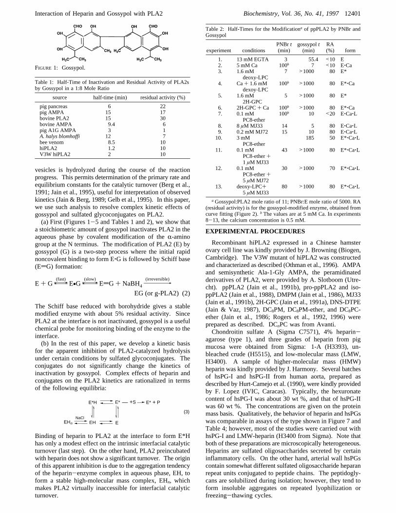
FIGURE 1: Gossypol.
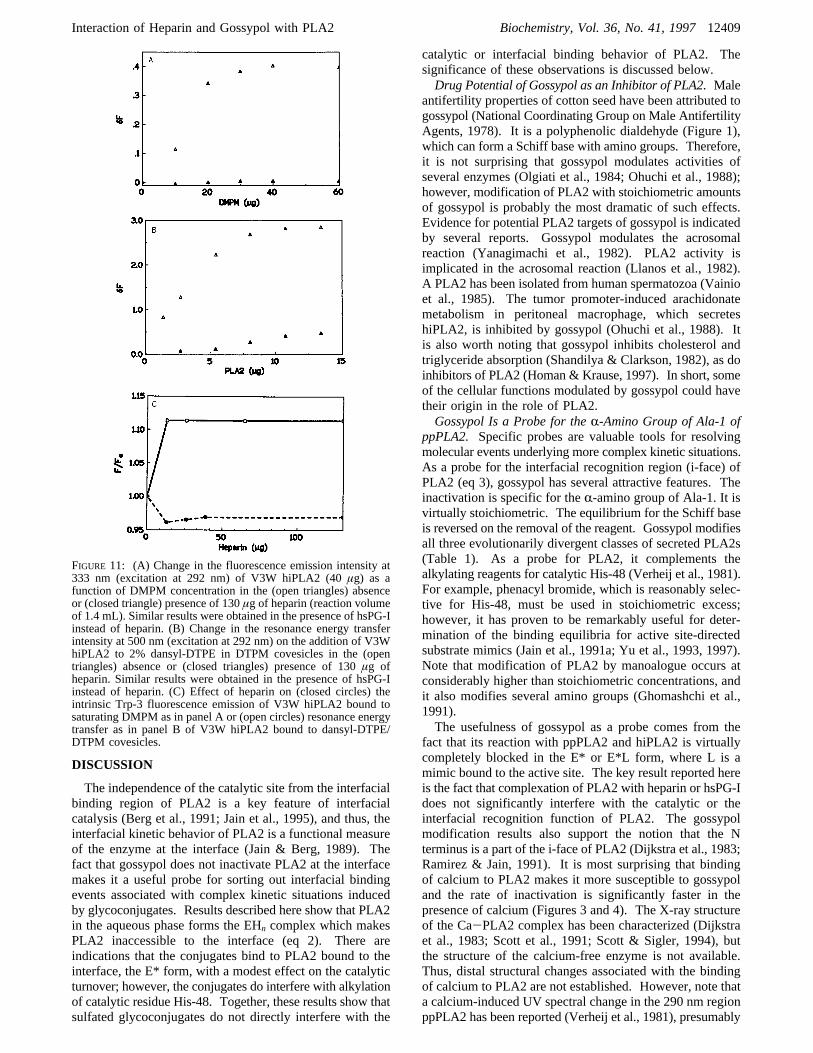
Table 1: Half-Time of Inactivation and Residual Activity of PLA2sby Gossypol in a 1:8 Mole Ratio
source half-time (min) residual activity (%)
pig pancreas 6 22pig AMPA 15 17bovine PLA2 15 30bovine AMPA 9.4 6pig A1G AMPA 3 1A. halys blomhoffi 12 7bee venom 8.5 10hiPLA2 1.2 10V3W hiPLA2 2 10
E+ G {\}(fast)
E•G {\}(slow)
EdG+ NaBH498(irreversible)
EG (or g-PLA2) (2)
NaClEHn EH E
E*H E*
(3)
E* + P+S
Table 2: Half-Times for the Modificationa of ppPLA2 by PNBr andGossypol
experiment conditionsPNBr t(min)
gossypolt(min)
RA(%) form
1. 13 mM EGTA 3 55.4 <10 E2. 5 mM Ca 100b 7 <10 E‚Ca3. 1.6 mM
deoxy-LPC7 >1000 80 E*
4. Ca+ 1.6 mMdexoy-LPC
100b >1000 80 E*‚Ca
5. 1.6 mM2H-GPC
5 >1000 80 E*
6. 2H-GPC+ Ca 100b >1000 80 E*‚Ca7. 0.1 mM
PC8-ether100b 10 <20 E‚Ca‚L
8. 8µM MJ33 14 5 80 E‚Ca‚L9. 0.2 mM MJ72 15 10 80 E‚Ca‚L10. 3 mM
PC8-ether185 50 E*‚Ca‚L
11. 0.1 mMPC8-ether+1 µM MJ33
43 >1000 80 E*‚Ca‚L
12. 0.1 mMPC8-ether+5 µM MJ72
30 >1000 70 E*‚Ca‚L
13. deoxy-LPC+5 µM MJ33
80 >1000 80 E*‚Ca‚L
aGossypol:PLA2 mole ratio of 11; PNBr:E mole ratio of 5000. RA(residual activity) is for the gossypol-modified enzyme, obtained fromcurve fitting (Figure 2).b The values are at 5 mM Ca. In experiments8-13, the calcium concentration is 0.5 mM.
Interaction of Heparin and Gossypol with PLA2 Biochemistry, Vol. 36, No. 41, 199712401

Phospholipid dispersions were prepared in water atconcentrations of 10-30 mg/mL, and the stock solution wasdiluted in an appropriate medium. Kinetic measurementsby pH-stat titration (Radiometer or Metrohm) were carriedout in 4 mL of 1 mM NaCl and 0.5 mM CaCl2 at pH 8.0 inpolypropylene cups (Jain et al., 1986). The kinetic basisand experimental protocols for interfacial catalysis in thehighly processive scooting mode by PLA2 (Berg et al., 1991;Jain et al., 1995) have been described for the reactionprogress on DMPM vesicles by the pH-stat method (Jain etal., 1986; Berg et al., 1991), kinetic characterization ofcompetitive inhibition (Jain et al., 1991b, 1995), determi-nation of the equilibrium dissociation constants of ligandbound to PLA2 at the interface by the protection fromalkylation of the active site histidine by ligand (Jain et al.,1991a), and measurement of residual PLA2 activity in thecovalent modification experiments with fluorescent substrate(Yu et al., 1993). Binding of PLA2 to the interface wasmonitored as a change in the intrinsic fluorescence emissionfrom Trp-3 of PLA2 (Jain et al., 1982, 1986; Ramirez &Jain, 1991) or as the energy transfer to a dansyl fluorophorelocalized in the interface (Jain & Vaz, 1987). Fluorescencemeasurements were carried out on a SLM-Aminco AB2apparatus with slit widths of 4 nm each. UV-visibleabsorbance measurements were carried out on an HP8452diode array spectrophotometer. Unless indicated otherwise,all spectroscopic measurements were carried out at pH 8.0in 10 mM Tris-HCl at 23-24 °C. Specific conditions usedfor these measurements are given in the figure legends. Onthe basis of standard deviations, uncertainty in primaryexperimental data is 10%, and 30% in the derived parameters.Modification of PLA2 by Gossypol. The time course of
covalent modification of PLA2 by gossypol was monitoredin an incubation mixture containing varying mole ratios ofgossypol to PLA2 (typically 0.2-0.4µM enzyme) in 0.1 M
cacodylate buffer with 0.1 M NaCl at pH 7.3 and 23°C.For certain studies, the reaction mixture also containedcalcium, deoxy-LPC, and competitive inhibitors of PLA2.The progress of inactivation by gossypol or PNBr wasmonitored by measuring the residual PLA2 activity with afluorescent substrate (Yu et al., 1993). The half-time forinactivation was obtained with a nonlinear fit of the residualactivity to a single- or double-exponential decay. Spectralchanges in the initial phase of the inactivation of PLA2 bygossypol were similar to those observed with the reactionproducts of gossypol with polylysine or phenylalanine.Characterization of g-PLA2, the Peramidinated ppPLA2
CoValently Modified by Gossypol. g-PLA2 was preparedby incubating 2 mg of bovine AMPA with a stoichiometricamount of gossypol (1:1.2 mole ratio) in 0.4 mL of 10 mMHEPES and 0.5 mM CaCl2 at pH 8.0 and 23°C. After 3 h,the mixture was cooled in an ice bath and treated with a100-fold molar excess of sodium borohydride with stirringfor 1 h. The mixture was dialyzed against the same bufferand then concentrated by ultrafiltration (Novacell with a 1Kcutoff filter). It was purified by HPLC on a Vydec-C4column in a 0 to 85%acetonitrile linear gradient (30 min at1 mL/min) in 0.1% trifluoroacetic acid in water. Themodified enzyme, g-PLA2, eluted at 35.9 min, compared to29.9 min for the unmodified enzyme. When the reactionmixture without reduction with borohydride was applied tothe HPLC column, the time at which AMPA eluted wasunchanged due to reversal of the Schiff’s base formed inthe first step (eq 2). Control runs without gossypol showedthat borohydride reduction and related manipulations hadlittle effect on the elution profile or activity of AMPA.The homogeneity of g-PLA2 obtained from AMPA was
further checked by FPLC on an anionic MONO-S columnwhere it eluted in 6 min (compared to 15 min for AMPA)in a 0 to 0.8 MNaCl gradient in 10 mM acetate buffer atpH 5.0. The molar extinction coefficient of g-PLA2 was85 680 M-1 cm-1 at 260 nm;E1% ) 69 at 260 nm andE1%) 29.6 at 280 nm, compared to theE1% value of 13 at 280nm for AMPA. The stability of g-PLA2 during dialysis andHPLC suggests that after reduction a stable linkage is formedbetween a gossypol molecule and a PLA2 molecule. Thecatalytic activity of g-PLA2 was 2-10% of the activity ofAMPA in the various assays (Jain et al., 1991a, 1995; Yu etal., 1993); however, g-PLA2 was not kinetically characterizedfurther.Gel Filtration Studies. HPLC measurements were carried
out on ISCO or Rainin hardware with appropriate columns.The TSK-250 gel permeation column gave very reproducibleresults. TSK-4000 and GPC-100 columns also gave com-parable results under certain conditions, but could not beused under the whole range of conditions of interest.Although the matrix of the TSK columns is anionic andshows ion exchange properties, this was not a seriousproblem for monitoring the molecular sieving characteristicsbecause heparins are anionic. Retention times for PLA2 andheparin on the TSK-250 column were monitored as absor-bance (at 220 nm) and fluorescence (excitation at 280 nmwith emission at 345 nm). All samples of heparins showeda weak UV absorbance with no detectable fluorescence,whereas PLA2, other than hiPLA2, gave the absorbance andfluorescence signals confirmed by the activity assay. Typi-cally, the column was equilibrated at room temperature with0.5 mM CaCl2 and 5 mM KH2PO4 buffer at pH 7.0. Elutionwas carried out in the same buffer after injection of up to
Table 3: Characteristic Equilibrium Parametersa for the Complexesof V3W hiPLA2
parameterV3WhiPLA2 ppPLA2 iso-PLA2
I50(HMW-heparin) (µg) 3 (0.7) 9 (0.75) 810 (0.72)I50(NaCl) (mM) 350 20 25I50(3K-heparin) (µg) 3.2 (0.75) 13 (0.73) 800 (0.73)I50(NaCl) (mM) 330 25 30KSV (M-1)E alone 11.3 6.5 10.9E+ heparin 2.6 3.75 9.4E+ hsPG-I 3.2 4.4 7.8E+ DTPM (E*L) 0.7 0.3 0.4
a I50(heparin) is the concentration of heparin (micrograms per 1.4mL) for a 50% change in the fluorescence obtained as in Figure 10A.I50(NaCl) is the concentration of NaCl (millimolar) for a 50% increasein the fluorescence as in Figure 10B.KSV is the Stern-Volmer constantfor quenching acrylamide.
Table 4: Elution Timesa (Minutes) for Heparin on a TSK-250HPLC Column
no added salt with 0.48 M NaCl
heparin alone with ppPLA2b alone with ppPLA2b
unbleached 5.0 5.0 7.4/11.15 10.31-A 5.14 5.2 7.26 10.3LMW 5.7 5.4 9.9 10.4HMW 4.74 5.1 8.65 10.3
a Elution was carried out with a flow rate of 1 mL/min with 0.5mM CaCl2 in 5 mM phosphate buffer at pH 7.0.b ppPLA2 (complex)elution times.
12402 Biochemistry, Vol. 36, No. 41, 1997 Yu et al.

20 µg of PLA2 preincubated with the indicated amount ofheparin in 20µL of buffer. The column was calibrated withseveral proteins at an elution rate of 1 mL/min with theuncertainty in the retention times of less than 0.1 min: 2000kDa blue dextran (4.64 min), 440 kDa ferritin (6.2 min),158 kDa aldolase (5.2 min), 66 kDa bovine serum albumin(7.88 min), 18.4 kDa lactoglobulin (9 min), 13.9 kDappPLA2 and other PLA2s (10.3 min), and 12.4 kDacytochromec (11.2 min). Comparable studies could not becarried out with hsPGs because they show a strong fluores-cence, tend to aggregate, and have a broad distribution ofmolecular species in the 10-100 kDa range.
RESULTS
As outlined in the introductory section, this sectiondescribes two sets of studies. First, we show that inactivationof ppPLA2 by gossypol is due to covalent modification ofthe R-amino group of Ala-1 (eq 2). The fact that themodification is not seen with PLA2 bound to the interfaceis consistent with the consensus that the N terminus is a partof the face of PLA2 in contact with the substrate interfaceduring the catalytic turnover (Jain & Maliwal, 1993), whichwe call the i-face (Ramirez & Jain, 1991). In the secondpart, we use gossypol as a probe, in conjunction with otherprotocols, to show that sulfated glycoconjugates bind to asite on PLA2 that is not the i-face or the catalytic site.Kinetics of InactiVation of ppPLA2 by Gossypol. As
shown in Figure 2, the loss of activity of ppPLA2 incubatedwith gossypol is time-dependent. About 70% of the inac-tivation of the native enzyme is adequately described by asingle-exponential decay, and the balance can be fitted to asecond exponential, suggesting that additional groups aremodified at a somewhat slower rate in the presence of excessgossypol (see below). These results suggest that modificationof one or more amino groups may be responsible for thereduced PLA2 activity. As shown in Figure 2, a rapid single-exponential decay completely describes the inactivation ofbovine AMPA in which only the N terminus amino groupis free. As shown in Figure 3A for ppPLA2, the half-timefor inactivation does not change when G:E> 2, and theresidual activity of the modified enzyme was less than 10%.Comparable results were obtained with bovine AMPA.
The rate of inactivation is more rapid in the presence ofcalcium (Figures 2 and 3B). The apparent affinity for theeffect of calcium on the inactivation half-time is about 0.06mM (Figure 3B), compared to the dissociation constant of0.35 mM for the enzyme in the aqueous phase or 0.25 mMat the interface (Jain et al., 1991a; Yu et al., 1993). Theseresults show that the affinity for calcium increases 5-7-foldin the presence of gossypol. The converse is also true; i.e.,enhanced binding of gossypol to PLA2 is seen in thepresence of calcium. For example, gossypol quenches theTrp-3 fluorescence emission intensity of ppPLA2 im-mediately after mixing, and the intensity does not changeafter that. According to the two-step mechanism (eq 2),instantaneous quenching of Trp-3 by gossypol is due to thenoncovalent binding, which precedes slower covalent inac-tivation (Figure 2). The emission intensity after the rapidnoncovalent binding of gossypol depends on its concentration(Figure 4). A high affinity for the noncovalent binding ofgossypol to PLA2 is indicated by the fact that the gossypolconcentration used for these measurements is comparableto that of PLA2. Also the apparent affinity depends on thepresence of calcium. For example, in a reaction mixturecontaining 2.6µM ppPLA2, 50% quenching is seen at about4 µM gossypol in the presence of calcium, and at about 18µM in EGTA. Note that the quenching efficiency ofgossypol is virtually the same for the ECa form as it is forthe E*Ca‚P form (Figure 4). Similar results were obtainedwith DTPM, the nonhydrolyzable ether analog of thesubstrate. Note that E*CaL forms of PLA2 are not co-valently modified by gossypol, although both bind gossypoland are modified in the aqueous phase.
FIGURE2: Time course for the inactivation of ppPLA2 by gossypolin the presence of 0.5 mM (open circles) or absence (closed circles)of calcium. The incubation mixture (30µL) contained 100 mMcacodylate buffer at pH 7.3 and 23°C with 0.2µM ppPLA2 and1.6 µM gossypol. The dashed line is a single-exponential fit, andthe full line is a double-exponential fit for the data in the presenceof calcium. Values of the inactivation half-times reported in thispaper are those obtained with a single-exponential fit and aretherefore approximate. Note that a single-exponential fit is adequatewith AMPA (results not shown; however, see the text for adiscussion).
FIGURE3: (A) Dependence of the residual activity after inactivationof ppPLA2 or (inset) of the corresponding half-times as a functionof the gossypol:ppPLA2 (G:E) mole ratio in the presence of 0.5mM calcium (open circles) or in the absence of calcium (closedcircles). (B) Dependence of the half-time for inactivation of ppPLA2by gossypol (G:E) 11) as a function of the calcium concentration.Other conditions were as in Figure 2.
Interaction of Heparin and Gossypol with PLA2 Biochemistry, Vol. 36, No. 41, 199712403

Inactivation of the amino group at the N terminus ofppPLA2 by gossypol is a two-step process (eq 2) in whichthe initial rapid noncovalent binding is followed by Schiffbase formation with the amino group of the N terminus witha half-time of about 5 min in the presence of calcium (Tables1 and 2). Results in Figures 2-4 show that only the E orECa form of the enzyme in the aqueous phase is covalentlymodified by gossypol, and the E* or E*L forms with orwithout calcium (where L) P, S, or I) are not modified bygossypol. Note that modification of additional amino groups,with a half-time of>20 min, may occur at high G:E ratios,which we have not characterized. The rapid noncovalentbinding is indicated by the observation that the kinetics ofmodification are not limited by the concentration of freegossypol to the enzyme, as well as by the rapid quenching.The fact that the slower Schiff base formation step isfacilitated by calcium suggests that a significant change atthe N terminus is induced by calcium, which enhances theapparent affinity of gossypol. This is an intriguing observa-tion, not only because the kinetics of gossypol modificationare not influenced by the occupancy of the active site of theenzyme in the aqueous phase but also because, as also shownbelow, inactivation by gossypol is completely blocked if theenzyme is bound to the interface. These results suggest acomplex interplay of catalytic and interfacial binding eventsthrough calcium, as also suggested by detailed kineticanalysis (Yu et al., 1993; Jain et al., 1993b) and site-directedmutagenesis (Huang et al., 1996).InactiVation of ppPLA2 by Gossypol Occurs through Schiff
Base Formation withR-NH2 of Ala-1. Evidence thatgossypol interacts with the amino group at the N terminusand forms a Schiff base, EdG in eq 2, is based on thefollowing observations. (a) Peramidinated PLA2, AMPA,which has no other free amino group except theR-amino ofAla-1, is inactivated by a single time-dependent process(Figure 2). (b) AMPA is maximally inactivated by less than2 equiv of gossypol (cf. Figure 3A). (c) The reaction productof ppPLA2 or AMPA with gossypol is stabilized afterreduction with borohydride (Experimental Procedures). (d)The single free amino group in AMPA, which is modifiedby dansyl chloride, is not free in AMPA treated withgossypol. (e) The time-dependent loss of activity of ppPLA2is not seen with gossypol pretreated with borohydride. (f)Localization of the gossypol moiety in the vicinity of theTrp-3 residue of ppPLA2 is also indicated by the fact thatthe emission intensity at 333 nm from the binding and
modification of ppPLA2 is reduced by 50% on modificationby gossypol, and the intensity does not change appreciablyon the denaturation of the enzyme under reducing conditionsin dodecyl sulfate micelles or in an 8 M urea solution. (g)In addition, modification of ppPLA2 or AMPA by gossypolis accompanied by spectroscopic changes consistent with theformation of a Schiff base. The time-dependent increase inthe absorbance at 400 nm is seen during the incubation ofAMPA with gossypol, and the half-time for the absorbancechange was the same as that measured by the assay of theresidual activity (cf. Figure 2). The value of the molarextinction coefficient for EdG at 400 nm, compared withthat of a reference standard prepared with phenylalanine,suggested that only one gossypol moiety is incorporated per14 000 Da of ppPLA2 (AMPA). The absorption maximumat 400 nm disappears on reduction of EdG with borohydride,and the product is stable under HPLC conditions (Experi-mental Procedures). Although dialdehydes can form mor-pholino derivatives with a single amino group (King &Colman, 1983), it is unlikely with gossypol due to theconstraints of the ring size and geometry.Gossypol Does Not InactiVate PLA2 at the Interface. As
shown in Figure 5A, the time course of modification ofppPLA2 by gossypol changes dramatically in the presenceof deoxy-LPC, a neutral diluent (ND). Recall that ppPLA2binds to aqueous dispersions of deoxy-LPC, and the activesite of the bound enzyme remains unoccupied in the absenceof added mimics (Jain et al., 1991b). This permits dissectionof two sequential equilibria:
FIGURE4: Relative fluorescence emission intensity of ppPLA2 (2.6µM) immediately (5 s) after the addition of gossypol. The reactionmixture contained 10 mM Tris at pH 8.0 (dots) in the presence of1 mM EGTA (E form) or (open circles) 0.5 mM CaCl2 (ECa form)alone or with (closed triangles) 0.4 mM DMPM (E*CaP form).
FIGURE5: (A) Time course for inactivation by gossypol of ppPLA2by gossypol in the (from the top down) E*I form (E+ 0.4 mMdeoxy-LPC+ 8 µM MJ33; closed circles), E* form (E+ deoxy-LPC; open circles), and EL form (E+ MJ33; open triangles). (B)Residual activity (circles) and inactivation half-times (triangles) forppPLA2 by gossypol (G:E) 11) as a function of deoxy-LPCconcentration. In both cases, other conditions were as in Figure 2with 0.5 mM Ca.
E+ ND a E* + L a E*L (4)
12404 Biochemistry, Vol. 36, No. 41, 1997 Yu et al.

At the ND interface, PLA2 is present only in the E* form,and both E and E* forms are alkylated by PNBr (Jain et al.,1991b). If a mimic (L) S, P, or I) is also present in theinterface, the equilibrium shifts toward the E*L form, whichis not alkylated by PNBr.Results in Figure 5A show that only the E form is modified
by gossypol and that E* and E*L forms are not. Resultssummarized in Figure 5B show that the inactivation timeand the residual activity increase with increasing concentra-tions of deoxy-LPC, and virtually complete protection is seenat saturating concentrations above 0.15 mM. The initial steepdependence of the residual activity on deoxy-LPC concentra-tion extrapolated to the maximum residual activity suggeststhat, at subsaturating concentrations of deoxy-LPC, PLA2forms a stoichiometric complex with the diluent and onlyexcess enzyme remains free to be modified by gossypol.These results are apparently surprising, yet consistent witheq 4. Our earlier results showed that the affinity of PLA2for deoxy-LPC in the absence of an active site-directed mimicis rather poor (Jain et al., 1991a,b, 1995), and the dissociationconstant for ppPLA2 from the interface decreases by 40-60-fold, from 1.6 to 0.02 mM, in the presence of an activesite-directed substrate mimic (Jain et al., 1993b). Gelpermeation studies on TSK-4000 also showed that the PLA2/ND mixture eluted as a high-molecular mass species onlyin the presence of gossypol or MJ33. In short, gelpermeation studies and the inactivation kinetics studiessuggest that the binding behavior of the E‚G complex ofppPLA2 for the interface is comparable to that of the ELcomplex; however, results described next show that, unlikeMJ33, gossypol does not bind to the active site of PLA2.Results in Figure 5 and Table 2 show that the E* or E*L
form of PLA2 is not modified by gossypol, while virtuallyno protection is seen in the EL form. Results in Table 2also show that catalytic His-48 in the E or E* form is readilyalkylated by PNBr, but the E‚Ca, E*Ca, E*CaL, and ECaLforms are not alkylated [see Yu et al. (1993)]. Results inrows 1-6 show that gossypol modifies the E and ECa formsbut not the E* or E*Ca forms. On the other hand, the half-time for the alkylation of His-48 is virtually the same in theE or E* form, and it is at least 50-fold higher for the ECa,ECaL, E*Ca, and E*CaL forms (Jain et al., 1991a; Yu etal., 1993). Protection from gossypol is not observed in theECaL form for mimics (rows 7-9, Table 2) such as DC8-PC, MJ33, or MJ72 present as solitary monomers in theincubation mixture below their critical micelle concentration.As expected, the half-time for alkylation by PNBr wasmodified under these conditions due to the occupancy of theactive site. Particularly striking are the results in rows 10-12, where the half-time for inactivation by gossypol increasessignificantly, even though both DC8PC-ether and inhibitorsare present below their cmc, which provides an independentconfirmation for the formation of E*L, also seen withspectroscopic and kinetic methods (Rogers et al., 1992,1996).To recapitulate, E or EL forms of ppPLA2 are inactivated
by gossypol. E* and E*L forms are not, even thoughgossypol binds to these forms. Since gossypol modifies theR-amino group of Ala-1, it may be concluded that in E*and E*L forms the amino group becomes inaccessible forthe modification. Although the kinetics of modificationdepend on the presence of calcium, the independence of thegossypol modification from events of the catalytic site isshown by the observation that the kinetics are not influenced
by the occupancy of the active site, as in the EL form of theenzyme in the aqueous phase. Also, the gossypol-modifiedenzyme retains 2-10% of the residual activity. Next, weuse gossypol to probe the basis for the kinetic effects ofsulfated glycoconjugates and to resolve certain mechanisticalternatives, implied in eq 3, that can lower the observedrate of hydrolysis by secreted PLA2 under certain conditions.Polyanionic Sulfated Glycoconjugates Reduce the Rate of
Hydrolysis of DC7PC Micelles by PLA2.The rationale fora detailed analysis of the effect of heparin and relatedconjugates on PLA2-catalyzed hydrolysis comes from avariety of observations. PLA2s retained on an immobilizedheparin-agarose column are eluted with 0.1-1 M NaCl(Diccianni et al., 1990, 1991a). A role for electrostaticinteractions during interfacial catalysis is indicated by thefact that anionic charge at the substrate interface promotesbinding and hydrolysis by PLA2 (Jain et al., 1982; Ramirez& Jain, 1991). Thus, lower rates in the presence of heparinare attributed not only to reduced binding of PLA2 to theinterface (Peers et al., 1987) but also to competitive inhibition(Dua & Cho, 1994) and allosteric modulation via the Nterminus (Diccianni et al., 1991b). Indeed, there are severalpossible ways in which the rate of interfacial catalysis canbe reversibly lowered (Jain & Jahagirdar, 1985; Gelb et al.,1994), and traditional assays used for earlier studies do notdistinguish such possibilities. The mechanistic basis (eq 3)of the kinetic effects of heparin and proteoglycans, hsPG-Iand -II from human aorta, is resolved below.The rate of hydrolysis of micellar DC7PC by hiPLA2
decreases in the presence of several sulfated glycoconjugates(Figure 6A). An about 95% loss of activity is seen withhsPG-I and -II, and in both cases, a 50% reduction in therate is seen at about 15µg in 4 mL of reaction mixture. Thepercents of reduction in the observed rate with 5µg of hsPG-Ifor the hydrolysis of DC7PC by PLA2 from the followingsources are as follows: hiPLA2 (25%), ppPLA2 (20%),venoms of bee (34%),Naja melanoleucaDE2 (33%), theAgkistrodon halis blomhoffiiacidic (55%) and basic enzymes(10%),Hemachatus hemachatus(10%),Vipra rusellii (11%),Cortalus adamenteus(22%), andNotachus scrutatum(11%).These results show that the hydrolysis by all the threeevolutionarily divergent classes of PLA2 is influenced byhsPG-I.The concentration dependence of HMW-heparin on the
rate of hydrolysis of DC7PC shows two steps (Figure 6A);about 40% of the activity is lost at 20µg of HMW-heparin,and then a less pronounced effect is seen at higher concen-trations. All commercially available heparin showed only amodest effect on the ppPLA2 or hiPLA2, comparable to thatseen with chondroitin sulfate (Figure 6A); i.e., the initialsharp decrease is not seen. In fact, a modest increase in therate was seen at low concentrations of some preparations ofheparin [see also Sartipy et al. (1996) and a personalcommunication of M. H. Gelb]. While it is tempting tointerpret the lower rates as a case of simple “competitiveinhibition” (Dua & Cho, 1994), detailed analysis of thekinetic effects developed below shows that the underlyingprocess is far more complex. In terms of eq 3, the apparentloss of activity is due to the formation of a complex of PLA2with the conjugate in the aqueous phase, EH and EHn, whichcan bind only an exchangeable substrate, such as DC7PC,to support hydrolysis. An interfacial complex, E*H, ispossibly formed with heparin added to the enzyme at theinterface; however, it is catalytically functional. As shown
Interaction of Heparin and Gossypol with PLA2 Biochemistry, Vol. 36, No. 41, 199712405

below, a dramatic loss of activity and other anomalous kineticeffects are associated with the formation of a high-molecularmass complex in the aqueous phase, EHn, which does notinteract with the substrate interface.hsPG-I Has a Modest Effect on Interfacial Catalysis by
ppPLA2 in the Scooting Mode.The rate of hydrolysis ofanionic DC8PM is lower in the presence of hsPG-I, and theeffect is less pronounced in the presence of 0.5 M NaCl(Figure 6B). Not only is the effect of the conjugate and saltcomparable to that seen with DC7PC micelles, but the orderof addition of components had little effect with micelles ofDC7PC or DC8PM (not shown). In accord with eq 3, activityis determined by the fraction of enzyme in E* or E*H forms,whereas the loss of activity is putatively due to the enzymesequestered as EHn.Results with short chain substrates do not distinguish
between the kinetic effects due to perturbed binding of theenzyme to the interface, lower intrinsic activity of the E*Hform, or trapping of the enzyme in the EHn form. Thisanalysis is possible with the scooting mode kinetics, wherethe order of addition of the components also becomes critical.For example, ppPLA2 remains bound to DMPM vesiclesduring the course of the reaction progress in the scootingmode (Jain et al., 1986, 1995). Thus, an apparent first-orderreaction progress (Figure 7, curve a) results because thereaction ceases as the substrate on the enzyme-containingvesicle surface is exhausted. hsPG-I has a small butsignificant effect on the first-order course of the reaction
progress by ppPLA2 (Figure 7, curves a and b). The half-time increases modestly without a significant effect on theextent of hydrolysis; i.e., hsPG-I does not promote thedesorption of bound ppPLA2, nor does it promote fusion orlipid mixing between vesicles. If this were not the case,additional hydrolysis would be seen under these conditions[e.g., see Jain et al. (1991c)]. In short, hsPG-I has only amodest effect on the interfacial catalytic turnover at theinterface; i.e., the activity of E*H is only modestly lower.Also, heparin did not show a significant effect on the scootingmode kinetics.A dramatic effect of the order of addition of hsPG-I on
the hydrolysis of DMPM vesicles is shown in Figure 7(curves c and d). Although comparable results were obtainedwith ppPLA2, for comparative purposes, these studies werecarried out with the V3W mutant of hiPLA2, which is alsoused later for the fluorescence binding measurements. Forexample, if 2.5µg of hsPG-I is added to the reaction mixturebefore the enzyme, virtually no hydrolysis occurs. Incontrast, only a modest decrease in the rate is seen if hsPG-Iis added after the initiation of the reaction, that is when theenzyme is already bound to the interface. Results with adifferent sequence of additions (not described here) werealways such that little hydrolysis is seen if the enzyme comesin contact with hsPG-I before it comes in contact with thestable DMPM interface; i.e., DMPM vesicles are notaccessible to the EHn complex in the aqueous phase. Thus,a large decrease in the observed rate is expected underconditions where the EHn complex is preformed in theaqueous phase. This will have a significant effect on therate if the enzyme returns to the aqueous phase during thecourse of reaction progress, as is the case with hiPLA2(Bayburt et al., 1993) but not with its V3W mutant whichbinds more tightly to the interface (Othman et al., 1993).The stability of EHn complexes of sulfated glycoconjugatesdepends on the nature of the enzyme and the salt concentra-tion (see below).Sulfated Glycoconjugates Do Not Influence the Binding
of Substrate Mimics to the ActiVe Site of PLA2. The half-time for alkylation of catalytic His-48 of PLA2 is verysensitive to the occupancy of the catalytic site (Jain et al.,1991a; Bayburt et al., 1993). Not only is the effect of heparinon the intrinsic catalytic turnover in the interface modest atbest, as in the preceding subsection, but independentmeasurements also showed thatKCa, KCa*, and KP* values
FIGURE 6: (A) Residual activity of hiPLA2 (36 pmol) on 3.1 mMDC7PC in the presence of increasing amounts of (open circles)hsPG-II, (closed circles) HMW heparin, or (triangles) chondroitinsulfate A. (B) Residual activity of ppPLA2 (3 pmol) on 0.3 mMDC8PM as a function of hsPG-II with 1 mM (open circles) or 500mM NaCl (closed circles). In both cases, reaction progress wasmonitored by pH-stat titration at pH 8.0 and 24°C in 4 mL of 0.5mM CaCl2 and 1 or 500 mM NaCl. Typically, the reaction wasinitiated by the addition of the enzyme; however, the order ofaddition of the components showed no significant difference in thesetwo assays.
FIGURE 7: Reaction progress curves for the hydrolysis of 0.24 mMDMPM as sonicated vesicles in 4 mL of 0.5 mM CaCl2 and 1 mMNaCl at pH 8.0 and 24°C. The reaction was initiated with 14 pmolof ppPLA2 for curves a and b or 36 pmol of V3W hiPLA2 forcurves c and d. The reaction mixture for curves b and d alsocontained 2.5µg of hsPG-I. Note that in the scooting mode theextent of hydrolysis (curve a versus c) depends on the amount ofenzyme.
12406 Biochemistry, Vol. 36, No. 41, 1997 Yu et al.

for ppPLA2 or hiPLA2 did not change in the presence ofheparin or hsPG-I. Also, calcium-dependentKI* values forcompetitive inhibitors, MJ33 with ppPLA2 (Jain et al.,1991b) or HK40 with hiPLA2 (Bayburt et al., 1993), arenot altered in the presence of the conjugates. These resultsshow that the binding of conjugates must occur at a site otherthan the catalytic site, that the conjugate does not influencethe binding to the catalytic site, and that the conjugate mustnot influence the binding to the catalytic site.Heparin Does Not Influence the Accessibility of the
R-Amino Group. hsPG-I or heparin has little effect on therate of inactivation by gossypol. For example, ppPLA2bound to deoxy-LPC is not inactivated by gossypol in thepresence or absence of hsPG-I (Figure 8A). The time courseof inactivation of the E form is not significantly influencedin the presence of heparin or hsPG-I. Similar results wereobtained with hiPLA2 (Figure 8B). These results show thathsPG-I has only a modest effect on the time course ofinactivation of ppPLA2 or hiPLA2 in the aqueous phase.The fact that the E* form of both of these enzymes is notinactivated in the presence of hsPG-I shows that the boundenzyme does not leave the interface in the presence of theglycoconjugate. The conclusion, from the kinetic resultsdescribed in this and the preceding sections, that theconjugates bind to a site other than the catalytic site or thei-face of PLA2 is also supported by spectroscopic results.Spectroscopic EVidence for the Binding of Heparin to the
E Form. Results in Figure 9A show that heparin quenches
the fluorescence emission from Trp-3 of the V3W mutantof hiPLA2, and that there is little effect of heparin in thepresence of 0.8 M NaCl. Qualitatively similar results wereobtained with ppPLA2 and isoPLA2 (Table 3). Althoughthe heparin-induced decrease in the Trp-3 emission was about30% in all cases, the concentration of heparin required forhalf of the change, theI50(heparin) values in Table 3, isappreciably different for the three PLA2s. Although thestructural basis for the quenching is not known, the resultsdescribed below suggest that it is due to the formation ofEHn aggregates. For example, the effect of the NaClconcentration on the fluorescence emission of V3W hiPLA2with or without heparin, shown in Figure 9B, clearly showsthat the effect of heparin on the environment of Trp-3 iscompletely reversed by NaCl.I50(NaCl) values (Table 3),the salt concentrations required for a 50% decrease of theeffect at the saturating heparin concentration, suggest thatthe EHn complex of V3W hiPLA2 is appreciably more stable.The accessibility of Trp-3 in the complex of PLA2 with
heparin was monitored by fluorescence quenching by acryl-amide, a water-soluble quencher. The Stern-Volmer plots,from which the KSV values were obtained, suggest acollisional quenching mechanism. Stern-Volmer constants(KSV) in Table 3 suggest that Trp-3 in the E form can beaccessed from the aqueous phase (Jain & Maliwal, 1993).The KSV value decreases significantly in the presence ofheparin or hsPG-I, and the decrease is lost at 0.5 M NaCl
FIGURE 8: (A) Time course of inactivation by gossypol of the E*form of ppPLA2 in deoxy-LPC (closed circles) without or (opencircles) with 10µg of hsPG-I. The time course for the inactivationof the E form in the aqueous phase in the presence of (opentriangles) 130µg of heparin or (closed triangles) 10µg of hsPG-Ior (diamonds) without any additive. (B) Time course of inactivationof hiPLA2 by gossypol in the presence of 10µg of hsPG-I for(open circles) the E or (closed circles) the E* form in the presenceof 5 mM hexadecylphosphocholine (a neutral diluent for hiPLA2).The enzyme:gossypol mole ratio for these measurements was 11.Other conditions were as in Figure 2.
FIGURE 9: (A) Change in the fluorescence emission intensity ofV3W hiPLA2 at 345 nm (excitation at 280 nm) as a function ofheparin (reaction volume of 1.4 mL) in 10 mM Tris at pH 8.0:(closed circles) high-molecular mass heparin or (open circles)LMW-heparin and (squares) LMW-heparin in the presence of 0.8M NaCl. (B) Salt concentration dependence of the change in thefluorescence emission from V3W hiPLA2 (square) alone or in thepresence of 130µg per 1.4 mL (open circles) high-molecular massheparin or (closed circles) LMW-heparin.
Interaction of Heparin and Gossypol with PLA2 Biochemistry, Vol. 36, No. 41, 199712407

(not shown), which is consistent with the salt-induceddissociation of the complex. Note that the accessibility ofTrp-3 in the EHn form is lower compared to that of the Eform; however, the shielding is far more pronounced in theE*L form on DTPM.Gel Permeation BehaVior of EHn Complexes. Complex-
ation of ppPLA2 with heparin was characterized by gelpermeation through a TSK-250 column (Tables 4 and 5),which adequately separated 14 kDa PLA2 from largercomplexes (Figure 10). The proportion of enzyme elutingas a high-molecular mass complex increases with the ratioof heparin to ppPLA2. The elution profile also shifts towardthe higher-molecular mass range, suggesting that a range ofcomplexes are formed. As summarized in Table 4, at theflow rate of 1 mL/min, all four samples of heparin eluted at5 min (compared to the void volume of 3.9 min). On theother hand, in the presence of 0.48 M NaCl, heparins eluteas lower-molecular mass species with elution times of 7-10min. Results in Table 4 also show that at low salt ppPLA2eluted with the aggregated heparin peak, but at 0.48 M NaClheparin and PLA2, peaks correspond to the lower-molecularmass species. The elution behavior of the four heparinpreparations as complexes with ppPLA2 was comparablewith significant quantitative differences. The effect of saltconcentrations on the elution profile indicated that theproportion of ppPLA2 that eluted with the higher-molecularmass complex decreases with [NaCl]. We did not investigatethese complexes in detail, not only because heparins and
hsPG are microscopically heterogeneous but also becausethe tryptophan fluorescence from the protein component ofhsPG, and a lack of tryptophan in the synovial PLA2, makeit more difficult to do the kind of analysis which have beencarried out with heparins and ppPLA2. At least qualitatively,these results clearly show that the heparin-ppPLA2 com-plexes exist as large molecular mass species, the EHn formin the absence of NaCl.
Comparable elution behavior is seen with all the fivePLA2s (Table 5), indicating that they form a stable complexwith heparin which dissociates with added NaCl. Thissimilarity, despite significant differences in their interfacebinding behavior, suggests that the primary effect of salt ison the stability of EH and EHn complexes. Note that pro-ppPLA2 with seven extra amino acid residues at the Nterminus behaves just like ppPLA2, which also rules out arole for Ala-1 in heparin binding.
Heparin Binds to PLA2 at the Interface. Measured as anincrease in the fluorescence emission from Trp-3, the high-affinity binding of V3W hiPLA2 to anionic vesicles (Figure11A) is similar to that seen with pancreatic enzymes (Jain& Vaz, 1987). Also as shown elsewhere (Othman et al.,1996), compared to the wild type, the V3W mutant has asomewhat higher affinity for the interface, as is the case withthe W3 mutants of pancreatic PLA2 (Liu et al., 1995). Onlythe magnitude of the change depends on the source of theenzyme; however, a linear increase with the bulk lipidconcentration shows that apparentKd, the dissociationconstant for the bound enzyme, is much smaller than 50µM.The initial region of this curve extrapolated to the maximumchange suggests that each enzyme molecule binds to about25 molecules of DMPM at the interface.
It is particularly striking that the fluorescence change,associated with the binding of PLA2 to the interface, is notseen on the addition of DMPM if heparin or hsPG-I is presentin the reaction mixture (Figure 11A). Similar behavior(Figure 11B) was seen if the binding of V3W hiPLA2 tothe interface was monitored by resonance energy transferfrom Trp-3 to the dansyl fluorophore, DNS-DTPE, at theinterface (Figure 11B). The increase in the energy transferintensity at 500 nm (excitation at 292 nm) is seen withincreasing enzyme concentrations, and a 3-fold increase isseen at saturating levels of the enzyme where the vesiclesurface is completely covered by the enzyme. In thepresence of heparin in the reaction mixture, the change inthe resonance energy transfer intensity is considerably smallerand the increase is seen only at higher enzyme concentra-tions, where only the enzyme not bound to heparin isavailable for the binding to the interface. Results in panelsA and B of Figure 11 show that the enzyme bound to heparin(as the EHn complex) cannot bind, nor does it dissociate forthe binding of free enzyme, to DTPM vesicles. Note thataddition of heparin or hsPG-I to the E*L (DMPM) form onvesicles does not cause a significant desorption of the boundenzyme. If the enzyme was desorbed, a decrease in theemission from Trp-3 or the energy transfer signal will beseen. In fact, as shown in Figure 11C, addition of heparincauses a small (12%) but significant increase in the energytransfer intensity and a small (4%) decrease in the Trp-3emission. hsPG-I also showed a similar behavior (resultsnot shown). Collectively, spectroscopic results confirm thekinetic results and show that the conjugates bind to PLA2at the interface without dislodging it.
Table 5: Elution Timesa (Minutes) for PLA2s from a TSK-250HPLC Column
alone with heparin (1:15)
PLA2 no NaCl 0.48 M NaCl no NaCl 0.48 M NaCl
ppPLA2 10.2 10.3 5.4 10.5iso-ppPLA2 9.9 9.9 5.6 9.9pro-ppPLA2 9.9 9.9 5.7 9.9bovine PLA2 10.3 10.3 5.6 10.3V3W hiPLA2 9.6 11.2 4.2 11.2
aConditions as in the footnote to Table 4 with details in ExperimentalProcedures.
FIGURE10: HPLC elution profiles from a TSK-250 gel permeationcolumn monitored as the tryptophan fluorescence signal (a) for 8µg of ppPLA2 alone, (b) with 0.12 mg of 3 kDa heparin with 10mM NaCl, or (c) with 0.48 M NaCl in the elution buffer. Otherconditions are given in Experimental Procedures. The flow ratewas 1 mL/min.
12408 Biochemistry, Vol. 36, No. 41, 1997 Yu et al.
DISCUSSION
The independence of the catalytic site from the interfacialbinding region of PLA2 is a key feature of interfacialcatalysis (Berg et al., 1991; Jain et al., 1995), and thus, theinterfacial kinetic behavior of PLA2 is a functional measureof the enzyme at the interface (Jain & Berg, 1989). Thefact that gossypol does not inactivate PLA2 at the interfacemakes it a useful probe for sorting out interfacial bindingevents associated with complex kinetic situations inducedby glycoconjugates. Results described here show that PLA2in the aqueous phase forms the EHn complex which makesPLA2 inaccessible to the interface (eq 2). There areindications that the conjugates bind to PLA2 bound to theinterface, the E* form, with a modest effect on the catalyticturnover; however, the conjugates do interfere with alkylationof catalytic residue His-48. Together, these results show thatsulfated glycoconjugates do not directly interfere with the
catalytic or interfacial binding behavior of PLA2. Thesignificance of these observations is discussed below.Drug Potential of Gossypol as an Inhibitor of PLA2. Male
antifertility properties of cotton seed have been attributed togossypol (National Coordinating Group on Male AntifertilityAgents, 1978). It is a polyphenolic dialdehyde (Figure 1),which can form a Schiff base with amino groups. Therefore,it is not surprising that gossypol modulates activities ofseveral enzymes (Olgiati et al., 1984; Ohuchi et al., 1988);however, modification of PLA2 with stoichiometric amountsof gossypol is probably the most dramatic of such effects.Evidence for potential PLA2 targets of gossypol is indicatedby several reports. Gossypol modulates the acrosomalreaction (Yanagimachi et al., 1982). PLA2 activity isimplicated in the acrosomal reaction (Llanos et al., 1982).A PLA2 has been isolated from human spermatozoa (Vainioet al., 1985). The tumor promoter-induced arachidonatemetabolism in peritoneal macrophage, which secreteshiPLA2, is inhibited by gossypol (Ohuchi et al., 1988). Itis also worth noting that gossypol inhibits cholesterol andtriglyceride absorption (Shandilya & Clarkson, 1982), as doinhibitors of PLA2 (Homan & Krause, 1997). In short, someof the cellular functions modulated by gossypol could havetheir origin in the role of PLA2.Gossypol Is a Probe for theR-Amino Group of Ala-1 of
ppPLA2. Specific probes are valuable tools for resolvingmolecular events underlying more complex kinetic situations.As a probe for the interfacial recognition region (i-face) ofPLA2 (eq 3), gossypol has several attractive features. Theinactivation is specific for theR-amino group of Ala-1. It isvirtually stoichiometric. The equilibrium for the Schiff baseis reversed on the removal of the reagent. Gossypol modifiesall three evolutionarily divergent classes of secreted PLA2s(Table 1). As a probe for PLA2, it complements thealkylating reagents for catalytic His-48 (Verheij et al., 1981).For example, phenacyl bromide, which is reasonably selec-tive for His-48, must be used in stoichiometric excess;however, it has proven to be remarkably useful for deter-mination of the binding equilibria for active site-directedsubstrate mimics (Jain et al., 1991a; Yu et al., 1993, 1997).Note that modification of PLA2 by manoalogue occurs atconsiderably higher than stoichiometric concentrations, andit also modifies several amino groups (Ghomashchi et al.,1991).The usefulness of gossypol as a probe comes from the
fact that its reaction with ppPLA2 and hiPLA2 is virtuallycompletely blocked in the E* or E*L form, where L is amimic bound to the active site. The key result reported hereis the fact that complexation of PLA2 with heparin or hsPG-Idoes not significantly interfere with the catalytic or theinterfacial recognition function of PLA2. The gossypolmodification results also support the notion that the Nterminus is a part of the i-face of PLA2 (Dijkstra et al., 1983;Ramirez & Jain, 1991). It is most surprising that bindingof calcium to PLA2 makes it more susceptible to gossypoland the rate of inactivation is significantly faster in thepresence of calcium (Figures 3 and 4). The X-ray structureof the Ca-PLA2 complex has been characterized (Dijkstraet al., 1983; Scott et al., 1991; Scott & Sigler, 1994), butthe structure of the calcium-free enzyme is not available.Thus, distal structural changes associated with the bindingof calcium to PLA2 are not established. However, note thata calcium-induced UV spectral change in the 290 nm regionppPLA2 has been reported (Verheij et al., 1981), presumably
FIGURE 11: (A) Change in the fluorescence emission intensity at333 nm (excitation at 292 nm) of V3W hiPLA2 (40µg) as afunction of DMPM concentration in the (open triangles) absenceor (closed triangle) presence of 130µg of heparin (reaction volumeof 1.4 mL). Similar results were obtained in the presence of hsPG-Iinstead of heparin. (B) Change in the resonance energy transferintensity at 500 nm (excitation at 292 nm) on the addition of V3WhiPLA2 to 2% dansyl-DTPE in DTPM covesicles in the (opentriangles) absence or (closed triangles) presence of 130µg ofheparin. Similar results were obtained in the presence of hsPG-Iinstead of heparin. (C) Effect of heparin on (closed circles) theintrinsic Trp-3 fluorescence emission of V3W hiPLA2 bound tosaturating DMPM as in panel A or (open circles) resonance energytransfer as in panel B of V3W hiPLA2 bound to dansyl-DTPE/DTPM covesicles.
Interaction of Heparin and Gossypol with PLA2 Biochemistry, Vol. 36, No. 41, 199712409

due to the perturbation of Trp-3. Our results are alsoconsistent with the observations that calcium is not requiredfor the binding of PLA2 to the interface, that the binding ofa ligand to the active site is not required for the binding ofPLA2 to the interface, and that under certain conditionspremicellar aggregates of PLA2 are formed with monomericamphiphiles (Table 2).Sequestering of PLA2 by Heparin. A key result reported
here is the fact that secreted PLA2 forms a high-molecularmass complex (EHn) with glycoconjugates, and that PLA2sequestered in these complexes is not readily accessible tothe substrate in the bilayer form. The fact that both thecatalytic site and the i-face of PLA2 in EHn complex arefree and not perturbed is also suggested by the kinetic andprotection studies. Evidence for an apparently high affinityof heparin binding to PLA2 comes from conditions thatpromote formation of EHn. Although it is not a focus ofthe present study, formation of a stable high-molecular masscomplex of PLA2 and heparin (Figure 11, Tables 4 and 5)must involve multiple interactions between several PLA2 andheparin molecules. It is however clear that the EHn complexsequesters PLA2 and makes it inaccessible for the hydrolysisof nonexchangeable substrate on vesicles.At this stage, one can only speculate about whether there
is a heparin binding site on PLA2, which for the presentdiscussion is called the h-site. Immunological evidence foran h-site on PLA2 has been reported (Murakami et al., 1991).Our results show that heparin interacts with PLA2 at theinterface with little perturbation of the interfacial catalyticcycle, which indicates that the h-site is distinct from theactive site with His-48, or from the i-face of PLA2 with Ala-1. The conjugates bind to E, E*, and E*L forms of PLA2,and that with hsPGs the E*H complex (Figures 6, 7, and11C, Tables 4 and 5) of hiPLA2 has a modestly (50%) lowerrate of interfacial catalytic turnover without desorption ofthe enzyme from the interface. In terms of eq 3, the primaryevidence for the interactions involving the h-site comes fromthe E*H or EH species, and not from the EHn complex.Although the EHn complex is very stable in the absence ofsalt, the salt dependence shows that relatively weak ionicinteractions are at work in these complexes. Since the saltconcentration dependence is different for PLA2s fromdifferent sources, cationic groups on PLA2 exposed to theaqueous phase in the E* form of PLA2 must be involved inthe putative h-site.The structural rationale for electrostatic heparin-PLA2
interaction comes from the fact that heparin is a polyanioniclinear copolymer of glycosamine and uronic acid withnumerous sulfate ester linkages. hsPGs contain a charac-teristic core protein conjugated with sulfated glycosami-noglycan chains attached to a serine-glycine-X segment(Jackson et al., 1991). In addition to the possibility ofbonding through multiple sites, complex structural andfunctional issues regarding the role of these conjugates aredifficult to resolve because most glycoconjugate preparationsare heterogeneous with respect to the peptides, sugars chains,and the degree of N and O derivatization of sugars bysulfates.Possible Physiological Role for PLA2 Complexes of
Heparin Conjugates.Sulfated glycoconjugates bind to awide variety of proteins. This fact provides a strong rationalefor the interaction of several plasma proteins with the hsPGtype of molecules present on the subendothelial matrix. Inanalogy with the heparin binding region RLTRKRGLK or
SVKAETKKQKHRH of apoB-100, it has been suggestedthat a cationic segment, such as KRLEKR (53-58) of PLA2,could be a part of the h-site on hiPLA2 (Dua & Cho, 1994;Kinkaid & Wilton, 1995). It appears that the role of specificresidues in stabilizing heparin binding is rather modest, asexpected for weak electrostatic interactions. For example,mutation of cationic residues in the heparin binding site ofantithrombin results in only a modest (<3-fold) decrease inthe binding (Meagher et al., 1996).
hsPGs are major components of the subendothelial layersof the arterial walls (Camejo et al., 1993). The observationthat secreted PLA2 form the EHn type of complex supportsthe view that the role of hsPG is to sequester circulatinghydrolytic enzymes (Bosman et al., 1988), including lipo-protein lipase (Hultin et al., 1992; Lookene et al., 1996),hiPLA2 (Sartipy et al., 1996), and serum lipoproteins (Hurt-Camejo et al., 1990). Beyond the scavenger role, morespecific functions for such interactions have also beendescribed (Ross, 1993; Cheng et al., 1993; Suga et al., 1993),and the function of the peptidoglycan from the extracellularmatrix of artery vessel wall is still being debated (Sartipy etal., 1996). Much of the discussion is now focused on theconsequences of the view that in subendothelial matrix theproteoglycans retain and sequester cationic proteins fromplasma, and therefore increase their residence time. Forexample, depending on the relative affinities of hiPLA2 forlow-density lipoproteins and peptidoglycan, and the saltconcentration in the extracellular matrix, increasing theresidence time of hiPLA2 will lead to an increased productionof bioactive lipids which trigger undesirable cell responsesin the artery wall, including degeneration of macrophage intofoam cell.
On the basis of our results, it can now be said that hiPLA2bound to proteoglycans at the subendothelial surface wouldbe catalytically functional with their active site exposed tothe aqueous environment. Of course, the interfacial catalyticfunction of the bound enzyme will be expressed only if thesubstrate can access the i-face of the bound enzyme. In thiscontext, the fact that LDL is also sequestered in theproteoglycan matrix deserves serious attention (Camejo etal., 1993, 1996). It is also relevant to point out that the 44kDa calcium-independent PLA2 (PAF-hydrolase) present inplasma is neither inhibited nor sequestered by proteoglycans(R. Apitz-Castro and M. K. Jain, to be published).
To recapitulate, sulfated glycoconjugates sequester secreted14 kDa PLA2 in a high-molecular mass stable complex, andsuch a complex could sequester the circulating PLA2. Inthe complex, catalytic site and interface recognition regionsof PLA2 are exposed to the aqueous environment. Seques-tration of PLA2 can have a significant role in regulating theformation of bioactive lipids produced by lipolysis, andchanges in the proteoglycan matrix (related to age or disease)could result in the loss of this regulatory role.
ACKNOWLEDGMENT
We thank Dr. G. Camejo (Astra Hassle, Goteborg,Sweden) and Dr. Eva Hurt Camejo (Sahlgrenska UniversityHospital, Goteborg, Sweden) for bringing to our attentionthe problem of PLA2 interaction with heparin conjugatesand for many useful suggestions. We also thank ProfessorMichael Gelb (University of Washington, Seattle) for com-ments on the draft manuscript.
12410 Biochemistry, Vol. 36, No. 41, 1997 Yu et al.
REFERENCES
Bayburt, T., Yu, B. Z., Lin, H., Browning, J., Jain, M. K., & Gelb,M. H. (1993)Biochemistry 32, 573-582.
Berg, O. G., Yu, B. Z., Rogers, J., & Jain, M. K. (1991)Biochemistry 30, 7283-7297.
Bosman, M. S., Gulick, T., Riley, D. J. S., Spilburg, C. A., & Lange,L. G. (1988)Proc. Natl. Acad. Sci. U.S.A. 85, 7438-7442.
Camejo, G., Fager, G., Rosengren, B., Hurt-Camejo, E., & Bondjers,G. (1993)J. Biol. Chem. 268,14131-14137.
Cheng, M. Y., Lees, A. M., & Lees, R. S. (1993)Biochemistry 32,8518-8524.
Diccianni, M. B., Mistry, M. J., Hug, K., & Harmony, J. A. K.(1990)Biochim. Biophys. Acta 1046, 242-248.
Diccianni, M. B., McLean, L. R., Sturat, W. D., Mistry, M. J., Gil,C. M., & Harmony, J. A. K. (1991a)Biochim. Biophys. Acta1082, 85-93.
Diccianni, M. B., Lilly-Stauderman, M., Mclean, L. R., Balasubra-maniam, A., & Harmony, J. A. K. (1991b)Biochemistry 30,9090-9097.
Dijkstra, B. W., Renetseder, R., Kalk, K. H., Hol, W. G. J., &Drenth, J. (1983)J. Mol. Biol. 147, 97-123.
Dua, R., & Cho, W. (1994)Eur. J. Biochem. 221, 481-490.Gelb, M. H., Jain, M. K., & Berg, O. G. (1994)FASEB J. 8, 916-924.
Gelb, M. H., Jain, M. K., Hanel, A. M., & Berg, O. G. (1995)Annu. ReV. Biochem. 64, 653-688.
Ghomaschchi, F., Yu, B. Z., Mihelich, E., Jain, M. K., & Gelb, M.H. (1991)Biochemistry 30, 9559-9569.
Homan, R., & Krause, B. R. (1997)Curr. Pharm. Des. 3, 29-44.Huang, B., Yu, B. Z., Rogers, J., Byeon, I. J., Sekar, K., Chen, X.,Sundaralingam, M., Tsai, T., & Jain, M. K. (1996)Biochemistry35, 12164-12174.
Hultin, M., Bengtsson-Olivecrona, G., & Olivecrona, T. (1992)Biochim. Biophys. Acta 1125, 97-103.
Hurt-Camejo, E., Camejo, G., Rosengren, B., Lopez, F., Wiklund,O., & Bondjers, G. (1990)J. Lipid Res. 31, 1387-1398.
Jackson, R. L., Busch, S. J., & Cardin, A. D. (1991)Physiol. ReV.71, 481-539.
Jain, M. K., & Jahagirdar, D. V. (1985)Biochim. Biophys. Acta814, 319-326.
Jain, M. K., & Vaz, W. L. C. (1987)Biochim. Biophys. Acta 905,1-8.
Jain, M. K., & Berg, O. G. (1989)Biochim. Biophys. Acta 1002,127-156.
Jain, M. K., & Maliwal, B. P. (1993)Biochemistry 32, 11838-11846.
Jain, M. K., Egmond, M. R., Verheij, H. M., Apitz-Castro, R. J.,Dijkman, R., & de Haas, G. H. (1982)Biochim. Biophys. Acta688, 341-348.
Jain, M. K., Rogers, J., Jahagirdar, D. V., Marecek, J. F., & RamirezF. (1986)Biochim. Biophys. Acta 860, 435-447.
Jain, M. K., Rogers, J., & de Haas, G. H. (1988)Biochim. Biophys.Acta 940, 51-62.
Jain, M. K., Yu, B. Z., Rogers, J., Ranadive, G. N., & Berg, O. G.(1991a)Biochemistry 30, 7306-7317.
Jain, M. K., Tao, W., Rogers, J., Arenson, C., Eibl, H., & Yu, B.Z. (1991b)Biochemistry 30, 10256-10268.
Jain, M. K., Rogers, J., Berg, O., & Gelb, M. H. (1991c)Biochemistry 30, 7340-7348.
Jain, M. K., Rogers, J., Hendrickson, H. S., & Berg, O. G. (1993a)Biochemistry 32, 8360-8367.
Jain, M. K., Yu, B. Z., & Berg, O. G. (1993b)Biochemistry 32,11319-11329.
Jain, M. K., Gelb, M. H., Rogers, J., & Berg, O. G. (1995)MethodsEnzymol. 249, 567-614.
King, M. M., & Colman, R. F. (1983)Biochemistry 22, 1656-1665.
Kinkaid, A. R., & Wilton, D. C. (1995)Biochem. J. 308, 507-512.
Liu, X., Zhu, H., Huang, B., Rogers, J., Yu, B. Z., Kumar, A.,Jain, M. K., Sundaralingam, M., & Tsai, M. D. (1995)Biochem-istry 34, 7322-7334.
Llanos, M. N., Lui, C. W., & Huang, T. F. (1982)DeV., GrowthDiffer. 24, 305-310.
Lookene, A., Chevreuil, O., Ostergaard, P., & Olivecrona, G. (1996)Biochemistry 35, 12155-12163.
Meagher, J. L., Huntington, J. A., Fan, B., & Gettins, P. G. W.(1996)J. Biol. Chem. 46, 29353-29358.
Murakami, M., Takayama, K., Umeda, M., Kudo, I., & Inoue, K.(1991)Methods Enzymol. 197, 223-233.
Nakamura, H., Kim, D. K., Philbin, D. M., Peterson, M. B., Debros,F., Koski, G., & Bonventre, J. V. (1995)J. Clin. InVest. 95,1062-1070.
National Coordinating Group on Male Antifertility Agents (1978)Chin. Med. J. (Beijing, Engl. Ed.) 4, 417-428.
Nevalainen, T. J., & Gronroos, J. M. (1994) inEsterase, Lipasesand Phospholipase(Mackness, M. I., & Clerc, M., Eds.) pp 253-261, Plenum, New York.
Ohuchi, K., Watanabe, M., Hirasawa, N., Tsurufuji, S., Ozeki, T.,& Fujiki, H. (1988) Biochim. Biophys. Acta 971, 85-91.
Olgiati, K. I., Toscano, D. G., Atkins, W. A., & Toscano, W. A.(1984)Arch. Biochem. Biophys. 231, 411-415.
Othman, R., Baker, S., Li, Y., Worrall, A. F., & Wilton, D. C.(1996)Biochim. Biophys. Acta 1303, 92-102.
Peers, S. H., Taylor, R. D., & Flowers, R. J. (1987)Biochem.Pharmacol. 26, 4287-4291.
Ramirez, F., & Jain, M. K. (1991)Proteins 9, 229-239.Rogers, J., Yu, B. Z., & Jain, M. K. (1992)Biochemistry 31, 6056-6062.
Rogers, J., Yu, B. Z., Serves, S. V., Tsivgoulis, G. M., Sotiropoulos,D. N., Ioannou, P. V., & Jain, M. K. (1996)Biochemistry 35,9375-9384.
Ross, R. (1993)Nature 362, 801-809.Sartipy, P., Johansen, B., Camejo, G., Rosengren, B., Bondjers,G., & Camejo, E. H. (1996)J. Biol. Chem. 271, 26307-26314.
Scott, D. L., & Sigler, P. B. (1994)AdV. Protein Chem. 45, 53-88.
Scott, D. L., White, S. P., Otwinowski, Z., Yuan, W., Gelb, M. H.,& Sigler, P. B. (1990)Science 250, 1541-1546.
Shandilya, L. N., & Clarkson, T. B. (1982)Lipid 17, 285-290.Suga, H., Murakami, M., Kudo, I., & Inoue, K. (1993)Eur. J.Biochem. 218, 807-813.
Vadas, P., Browning, J., Edelson, J., & Pruzanski, W. (1993)J.Lipid Mediators 8, 1-30.
Vainio, P., Thuren, T., Wichman, K., Luukkainen, T., & Kinnunen,P. K. J. (1985)Biochim. Biophys. Acta 814, 405-408.
Verheij, H. M., Slotboom, A. J., & de Haas, G. H. (1981)ReV.Physiol. Biochem. Pharmacol. 91, 91-203.
Yanagimachi, R. (1982)Genet. Res. 5, 323-344.Yu, B. Z., Berg, O. G., & Jain, M. K. (1993)Biochemistry 32,6485-6492.
Yu, B. Z., Ghomashchi, F., Cajal, Y., Annand, R., Berg, O. G.,Gelb, M. H., & Jain, M. K. (1997)Biochemistry 36, 3870-3881.
BI962972I
Interaction of Heparin and Gossypol with PLA2 Biochemistry, Vol. 36, No. 41, 199712411