Goel, Harish; Amity University junctions Draft · Journal: Applied Physiology, ... Amity...
35
Draft Radioprotective potential of Lagenaria siceraria extract against radiation induced gastrointestinal injury Journal: Applied Physiology, Nutrition, and Metabolism Manuscript ID apnm-2016-0136.R2 Manuscript Type: Article Date Submitted by the Author: 12-Jun-2016 Complete List of Authors: Sharma, Dhara; University of Central Florida; Amity University Goel, Harish; Amity University Chauhan, Sonal; Amity University Keyword: Radiation-protection, Cucurbits, Lagenaria Siceraria, Gut villi, Tight junctions https://mc06.manuscriptcentral.com/apnm-pubs Applied Physiology, Nutrition, and Metabolism
Transcript of Goel, Harish; Amity University junctions Draft · Journal: Applied Physiology, ... Amity...
Draft
Radioprotective potential of Lagenaria siceraria
extract against radiation induced gastrointestinal injury
Journal: Applied Physiology, Nutrition, and Metabolism
Manuscript ID apnm-2016-0136.R2
Manuscript Type: Article
Date Submitted by the Author: 12-Jun-2016
Complete List of Authors: Sharma, Dhara; University of Central Florida; Amity University Goel, Harish; Amity University Chauhan, Sonal; Amity University
Keyword: Radiation-protection, Cucurbits, Lagenaria Siceraria, Gut villi, Tight junctions
https://mc06.manuscriptcentral.com/apnm-pubs
Applied Physiology, Nutrition, and Metabolism
Draft
1
Title page
Radioprotective potential of Lagenaria siceraria extract against radiation induced
gastrointestinal injury
Short title: Radio-modifying effect of Lagenaria
Authors names:
1. Dhara Sharma
Amity Center for Radiation Biology
Amity University, Noida-125
Uttar Pradesh (INDIA)
Email: [email protected]
2. Harish Chandra Goel
Amity Center for Radiation Biology
Amity University, Noida-125
Uttar Pradesh (INDIA)
Email: [email protected]
3. Sonal Chauhan
Amity Center for Radiation Biology
Amity University, Noida-125
Uttar Pradesh (INDIA)
Email: [email protected]
Address for correspondence:
Harish Chandra Goel
Amity Center for Radiation Biology
Amity University, Noida-125
Uttar Pradesh (INDIA)
Email: [email protected]
Page 1 of 34
https://mc06.manuscriptcentral.com/apnm-pubs
Applied Physiology, Nutrition, and Metabolism
Draft
2
Radiation-modifying effect of Lagenaria siceraria extract in-vitro and in-vivo
Abstract:
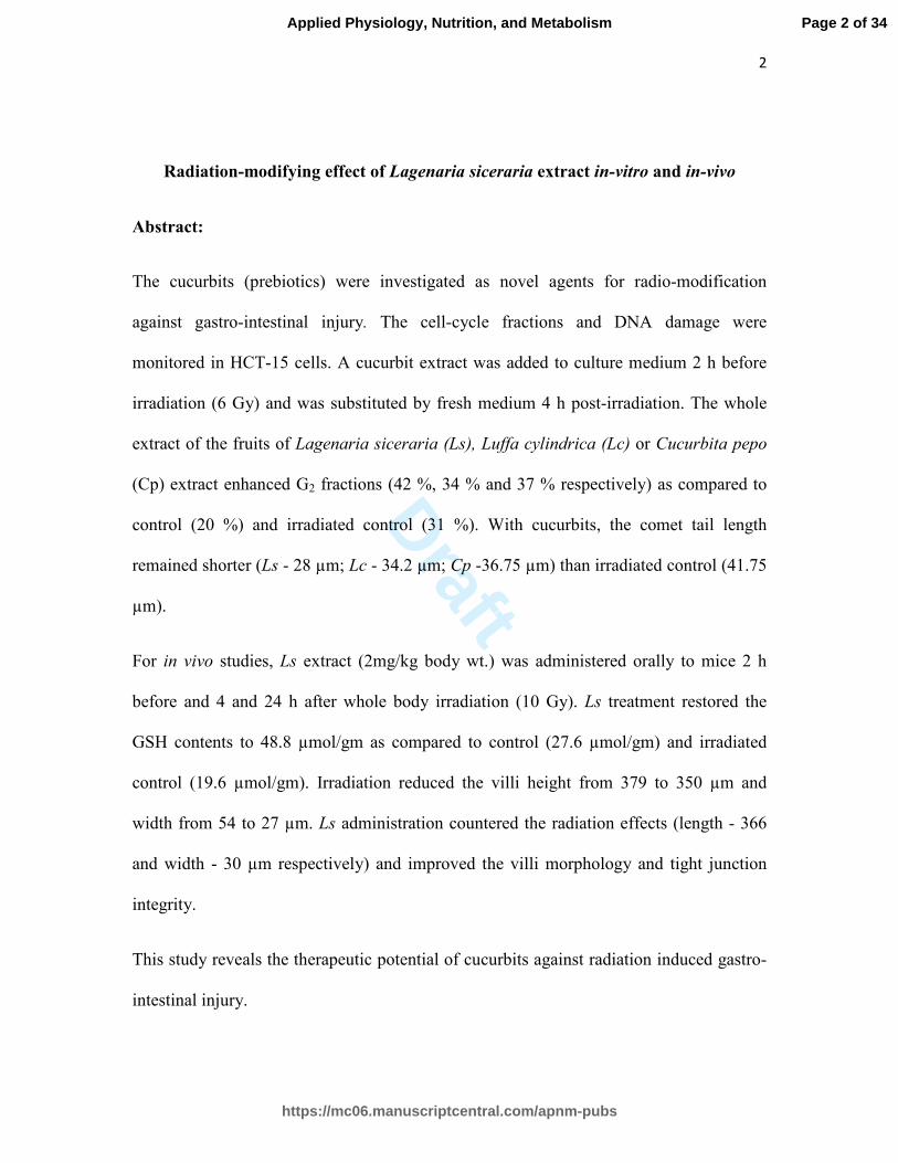
The cucurbits (prebiotics) were investigated as novel agents for radio-modification
against gastro-intestinal injury. The cell-cycle fractions and DNA damage were
monitored in HCT-15 cells. A cucurbit extract was added to culture medium 2 h before
irradiation (6 Gy) and was substituted by fresh medium 4 h post-irradiation. The whole
extract of the fruits of Lagenaria siceraria (Ls), Luffa cylindrica (Lc) or Cucurbita pepo
(Cp) extract enhanced G2 fractions (42 %, 34 % and 37 % respectively) as compared to
control (20 %) and irradiated control (31 %). With cucurbits, the comet tail length
remained shorter (Ls - 28 µm; Lc - 34.2 µm; Cp -36.75 µm) than irradiated control (41.75
µm).
For in vivo studies, Ls extract (2mg/kg body wt.) was administered orally to mice 2 h
before and 4 and 24 h after whole body irradiation (10 Gy). Ls treatment restored the
GSH contents to 48.8 µmol/gm as compared to control (27.6 µmol/gm) and irradiated
control (19.6 µmol/gm). Irradiation reduced the villi height from 379 to 350 µm and
width from 54 to 27 µm. Ls administration countered the radiation effects (length - 366
and width - 30 µm respectively) and improved the villi morphology and tight junction
integrity.
This study reveals the therapeutic potential of cucurbits against radiation induced gastro-
intestinal injury.
Page 2 of 34
https://mc06.manuscriptcentral.com/apnm-pubs
Applied Physiology, Nutrition, and Metabolism
Draft
3
Keywords: Radiation-protection, Cucurbits, Lagenaria Siceraria, Gut villi, Tight
junctions
Introduction:
Whole body irradiation received during planned or unplanned situations leads to different
kinds of syndromes depending on the radiation dose delivered. Radiation dose (3-7 Gy)
leads to hemopoietic (HP) syndrome (Suman et al. 2012) and death may occur within 30-
60 days. Radiation doses, more than 6 Gy (6-20 Gy) can lead to the damage to
gastrointestinal (GI) system and may lead death within few weeks (Donnelly et al. 2010).
For HP syndrome, therapeutics like blood transfusion, stem cell transplantation offer
reasonable success (Nagayama et al. 2002; Weisdorf et al. 2006). For restoration and
repair of HP system, a large number of chemicals like amifostine and gamma-tocotrienol
have been synthesized and investigated (Trajkovic et al. 2007; Ghosh et al. 2009). The
toxicity and limited therapeutic gain achieved by these agents against HP syndrome yet
remains unacceptable clinically. Radiation damage to GI system is however still managed
in palliative manner only and no effective treatment of GI syndrome is available till date
(Takemura et al. 2014). The development of new agents of synthetic or natural origin has
remained elusive in this context and the attention of researchers has been hardly
noticeable. Under the present study, we have attempted to investigate the natural dietary
cucurbits, against GI syndrome.
Etiology of GI syndrome depends mainly on the damage highly proliferative stem cells,
the crypts of Lieberkuhn, located at villar bases (Brown 2008). The damaged crypt cells
lag in replenishment of the damaged epithelial cells of villi causing shortening of villi
height. The denudating villi may be responsible for pathological manifestations like poor
Page 3 of 34
https://mc06.manuscriptcentral.com/apnm-pubs
Applied Physiology, Nutrition, and Metabolism
Draft
4
absorption of nutrients (Kau et al. 2011), loss of fluids and electrolytes, perturbation in
barrier function (Turner 2009) and microbial infections (Williams et al. 2015; Marchesi
et al. 2016).
The synthetic radio-modifying agents target the specific molecular pathways yet are often
antagonistic to many co-existing metabolic pathways. Therefore, scientists and clinicians
have devised the combinational philosophy which envisages the combining of several
agents to achieve better therapeutic gain. The toxicity of even combination modalities has
still remained high and thus warranted the use of natural agents in this connection. In fact
each plant extract contains hundreds of bioactive molecules which act synergistically in
multiple directions. Some of the components of the plant extract may yield direct
therapeutic effects while others may concurrently accelerate and reinforce the recovery
process and still other may overcome adverse toxic reactions.
Some cucurbits like Lagenaria siceraria (Ls), Luffa cylindrica (Lc) and Cucurbita
pepo (Cp) have been evaluated for their radio-protective activities. These cucurbits have
bioactive molecules as alkaloids, flavonoids, steroids, saponins and glycosides (Irshad et
al. 2010). In the previous studies done at our laboratory, these cucurbits displayed
significant antioxidant, antimicrobial and anti-inflammatory activities (Sharma et al.
2012; Rawat et al. 2014). The radiation damage is since mainly mediated by generation
of free radicals and may lead to the development of several inflammatory and infectious
diseases (Arora et al. 2005). Therefore, biological activities (antioxidant, antimicrobial
and anti-inflammatory) of these cucurbits were considered important for the
radioprotection and it warranted investigations on the radio-modifying efficacy in a
holistic manner.
Page 4 of 34
https://mc06.manuscriptcentral.com/apnm-pubs
Applied Physiology, Nutrition, and Metabolism
Draft
5
Material and Methods:
Chemicals:
Dulbecco’s modified eagle medium (DMEM), Minimum essential medium (MEM), Fetal
bovine serum (FBS), Trypsin- EDTA solution, 2,5-diphenyltetrazoliumbromide (MTT),
were procured from M/s Himedia (India). NaCl, EDTA, Triton X-100 and Tris were
procured from M/s Merck, Mumbai (India). Hematoxylin and Eosin were procured from
M/S Fisher Scientific, Mumbai. Propidium iodide and Osmium tetroxide were procured
from M/s Sigma Aldrich (USA).
Preparation of Extracts:
Fresh fruits of the cucurbits namely bottle gourd (Ls), sponge gourd (Lc) and pumpkin
(Cp) procured from the local market were thoroughly washed with sterile distilled water
several times and 100 g of each plant material was homogenized separately in 100 ml
solvent (absolute alcohol and triple distilled water; 50:50, v/v). After 24 h, the
homogenate was filtered through a fine strainer having a spread of muslin cloth and
thereafter through membrane filter of 0.22 µ. It was further concentrated using rota-
vapor. Filtered whole extract of cucurbit fruits was stored at 4°C in air-tight bottles.
Cell Culture:
The human carcinoma cells (HCT-15) procured from National Centre for Cell Sciences,
Pune, were cultured in DMEM containing 10% FBS and penicillin/streptomycin (100
µg/mL) at 370C in an incubator (CO2 conc.- 5%). One million cells were inoculated in 5
Page 5 of 34
https://mc06.manuscriptcentral.com/apnm-pubs
Applied Physiology, Nutrition, and Metabolism
Draft
6
ml medium contained in a petri-dish having 20 ml capacity. On reaching about 70%
confluency, cells were washed with PBS and were trypsinized and sub-cultured.
Animals:
Swiss albino Strain ‘A’ male mice (6-8 weeks old) weighing about 22 ± 3 g were
maintained under controlled laboratory environment (~25 ± 2°C, photoperiod-12 h). Mice
were given standard animal feed (Lipton, India) and tap water ad libitum. Animals for
these experiments were used according to the guidelines of animal ethics committee of
Amity University, Noida and INMAS, New Delhi.
Irradiation:
The Teletherapy cobalt machine (Bhabhataron II) at ‘Institute of Nuclear Medicine and
Allied Sciences’ was obtained from Board of Radiation and Isotope Technology (BRIT),
Mumbai (India). The dose rate in the irradiation chamber was ~1.9 to 1.86 Gy/minute
during the course of these investigations.
For in vitro studies, HCT-15 cells were cultured in petri-dishes each containing 5 ml
culture medium. Each dish was exposed to gamma irradiation (6 Gy) individually. For in
vivo studies, whole body irradiation (10 Gy) was delivered to each mouse individually.
Experimental design:
In vitro:
The cells were divided into 3 experimental groups each containing 4 petri dishes:
a) Control group: cells given no treatment
b) Radiation alone group: cells received a dose of 6 Gy
Page 6 of 34
https://mc06.manuscriptcentral.com/apnm-pubs
Applied Physiology, Nutrition, and Metabolism
Draft
7
c) Cucurbits + radiation group: 500 µL of cucurbit extract (Ls, Lc or Cp) was added
to the cell cultures 2 h before irradiation and fresh medium without the extract
was provided at 4 h post-irradiation period.
24 h after the radiation exposure, cells were processed for different experimental
parameters.
In vivo:
Three groups of swiss albino strain ‘A’ male mice, each containing 6 animals, were
randomly selected for these experiments. The animals were grouped as under:
i) Control: mice receiving no treatment
ii) Irradiated group: Each mouse received 10 Gy whole body gamma irradiation only
iii) Ls + radiated group: Each mouse received Ls extract at the rate of 2 mg/kg body wt.
2 h before, and 4 and 24 h after irradiation (10 Gy).
On 4th
post-irradiation day, the jejunum part of each mouse was taken out and processed
for histological study.
For dose mortality response curves, four sets of animals each having 12 mice were
selected to see the effect of Ls extract on the survival of mice exposed to gamma
irradiation (10 Gy). The animals were grouped as under:
a) Control group
b) Radiation group (10 Gy)
c) Ls treated group (Ls extract administered orally at 0 h, 6 h & 26 h)
Page 7 of 34
https://mc06.manuscriptcentral.com/apnm-pubs
Applied Physiology, Nutrition, and Metabolism
Draft
8
d) Ls + radiation group (Ls extract administered 2 h before irradiation and 4 and 24
h after irradiation)
All the animals of each experimental group were kept under controlled environment and
were observed for mortality up to 30 days.
Cell cycle analysis:
Cells were processed for cell cycle analysis by following the method described by
Pozarowski and Darzynkiewicz (2004). After 24 h of radiation exposure, cells were
trypsinized and centrifuged at 2000 g for 10 min. Cell pellet was washed three times and
re-suspended in 0.5 ml PBS. Fixation was completed by adding 1.2 mL of 70% cold
ethanol for 2 h. The fixed cells were washed with PBS and centrifuged at 2000 ×g for
10 min. After suspending cells in 0.3 mL PBS, DNAase free RNAse (50 mg/mL) was
added and incubated for 1 h. After adding 2 µL of propidium iodide (10 mg/mL in PBS),
cells were incubated at 4°C for 30 min. DNA contents were analyzed for cell cycle using
flow cytometer (Becton and Dickinson) with an excitation wavelength of 488 nm and
emission at 670 nm.
Comet assay:
For this, the method described by Singh et al. (1988) was adopted with slight
modifications. 24 after the radiation exposure, 100 µL of singled cells suspension was
added to 500 µL of 0.8% agarose (Low melting point: 30-35º C) in phosphate-buffered
saline (PBS) which is put on a glass slide pre-coated with 1 % agarose having normal-
melting point (50- 60º C). Each slide was covered with a cover slip and kept on ice for 5
min. The slides were immersed in ice-cold alkaline lysing solution [2.5 M NaCl, 100 mM
Page 8 of 34
https://mc06.manuscriptcentral.com/apnm-pubs
Applied Physiology, Nutrition, and Metabolism
Draft
9
Tris, 100 mM ethylene diamine tetra acetic acid (EDTA), 1 % sodium lauroyl sarcosine
sodium salt, 1% Triton X-100 and final pH was adjusted to 10 using 1 N NaOH solution]
for at least 1 h at 4° C. The slides were then washed 4-5 times with ice cold Milli Q water
and were kept into ice cold buffer (0.2 N NaOH and 200 mM EDTA ) for 30 min in the
dark at 4°C to unwind DNA. Now slides were incubated for 20 min in ice-cold
electrophoresis solution (200 mM NaOH, 500 mM EDTA, pH-13.1), followed by
electrophoresis at 25 V (1.25 V/cm) for 25 min. After electrophoresis, the slides were
washed and dehydrated with 70 % ice cold ethanol for 5 min and were air dried
thereafter. The slides were stained with propidium iodide (50 µg/mL PBS) and kept in
dark for 10 min. 50 cells were scored from each slide at a magnification of 400X using a
Olympus fluorescence microscope employing excitation at λ 488 nm and emission barrier
at λ 515 nm. Quantification of DNA damage was measured microscopically and
compared with control slides.
GSH contents:
The glutathione level in the jejunum was determined following the method described by
Verma et al. (2011). Briefly, jejunum homogenate was added to 20 % trichloro acetic
acid and was centrifuged to collect the supernatant. The supernatant was mixed with 0.3
M Na2HPO4 and 5-5, dithiobis-2-nitrobenzoic acid (DTNB) reagent, and allowed to stand
for 10 min at the room temperature. The absorbance was taken against blank at 412 nm
using a UV-VIS Systronics spectrophotometer.
Page 9 of 34
https://mc06.manuscriptcentral.com/apnm-pubs
Applied Physiology, Nutrition, and Metabolism
Draft
10
Histological study:
About 2 cm long pieces of jejunum were taken out using surgical procedure, from a
mouse immediately after cervical dislocation and fixed in 10% neutral formalin (pH 7.0–
7.6) for 24 hours. The tissue was processed for dehydration and paraffin block making
following standard procedure. Microtomy was done to get 5 µ sections which were
processed for haematoxylin and eosin (H&E) staining following the method described by
Khojasteh et al. (2009) Morphology of the villi was studied under a light microscope. 10
villi per section were assessed and mean value was calculated. Villus morphology,
height, width and area were measured at the magnification of 40 × 10 X and compared
with the control.
Transmission electron microscopy (TEM)
For Tem, Few pieces of jejunem from each experimental mouse were collected
immediately after cervical dislocation and were fixed in 2% glutaraldehyde and postfixed
in 1% osmium tetroxide. The tissue was stained in 1% uranyl acetate and embedded in
Epon following the method described by Soderholm et al. (2002). The staining of
sections was done by lead citrate for study under TEM at 80 kV. To evaluate changes in
tight junction integrity, the junctional regions of four randomly selected villi were
examined in each group.
Statistical Analysis
All the data are presented as mean ± SE and student’s t test were applied for determining
the statistical significance between different groups.
Page 10 of 34
https://mc06.manuscriptcentral.com/apnm-pubs
Applied Physiology, Nutrition, and Metabolism
Draft
11
Results:
Cell cycle analysis:
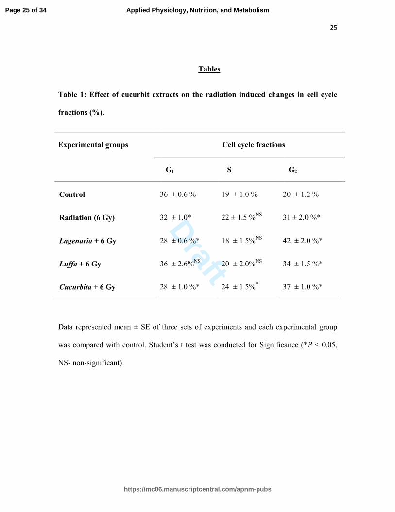
Effect of different cucurbits on the modulation of radiation induced changes in the
different phases of cell cycle has been depicted in Table 1. The control group had 36 ±
0.6 % cells in G1 phase and gamma irradiation (6 Gy) decreased G0/G1 phase cell
population to about 32 ± 1.0 %. The extracts of Ls and Cp also decreased the number of
cells in G1 phase further to about 28 % whereas Lc extract did not decrease the G1
fraction. In the S phase, cells were not influenced significantly by the Ls and Lc
treatment but Cp distinctly increased the S phase population to about 24 ± 1.5 as
compared to control (19 ± 1.0). In the control group, G2 fraction was about 20 ± 1.2 %
which increased to about 31 ± 2.0 % in radiation-alone group. In Ls, Lc and Cp treated
groups, cell population in G2 phase increased significantly (P < 0.05) to about 42 ± 2.0,
34 ± 1.5 and 37 ± 1.0 % respectively.
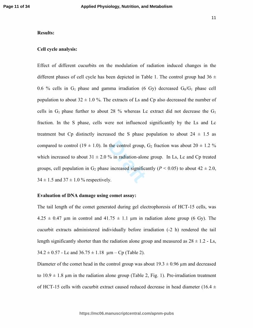
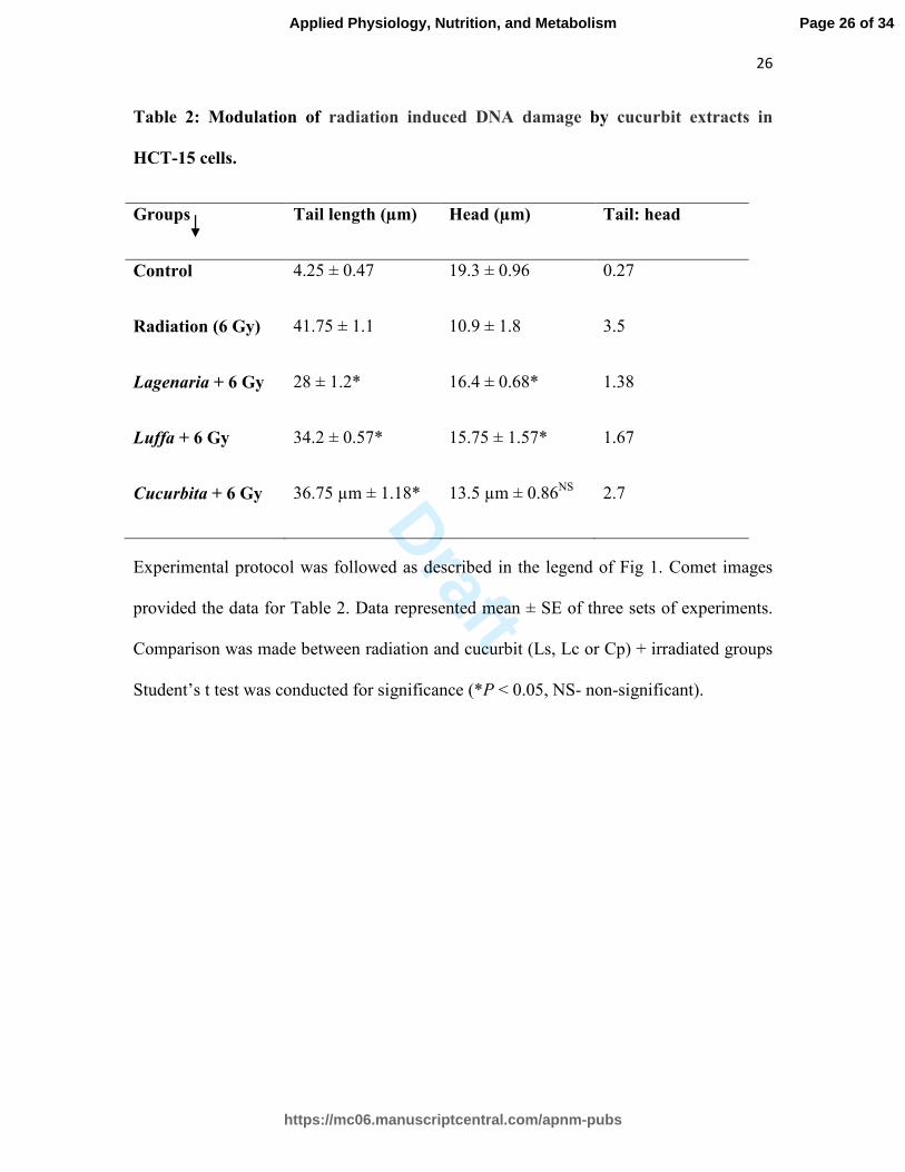
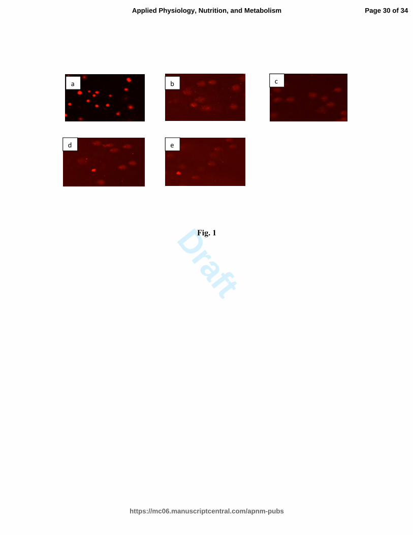
Evaluation of DNA damage using comet assay:
The tail length of the comet generated during gel electrophoresis of HCT-15 cells, was
4.25 ± 0.47 µm in control and 41.75 ± 1.1 µm in radiation alone group (6 Gy). The
cucurbit extracts administered individually before irradiation (-2 h) rendered the tail
length significantly shorter than the radiation alone group and measured as 28 ± 1.2 - Ls,
34.2 ± 0.57 - Lc and 36.75 ± 1.18 µm – Cp (Table 2).
Diameter of the comet head in the control group was about 19.3 ± 0.96 µm and decreased
to 10.9 ± 1.8 µm in the radiation alone group (Table 2, Fig. 1). Pre-irradiation treatment
of HCT-15 cells with cucurbit extract caused reduced decrease in head diameter (16.4 ±
Page 11 of 34
https://mc06.manuscriptcentral.com/apnm-pubs
Applied Physiology, Nutrition, and Metabolism
Draft
12
0.68- Ls, 15.75 ± 1.57- Lc and 13.5 ± 0.86 µm- Cp. The ratio of head diameter to tail
length was 0.27 in control and 3.5 in radiation alone group. In Ls, Lc and Cp treated
groups this ratio remained as 1.38, 1.67 and 2.7 respectively.
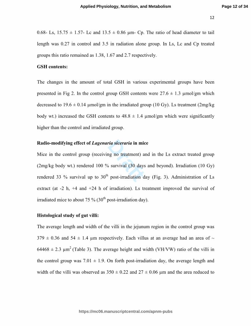
GSH contents:
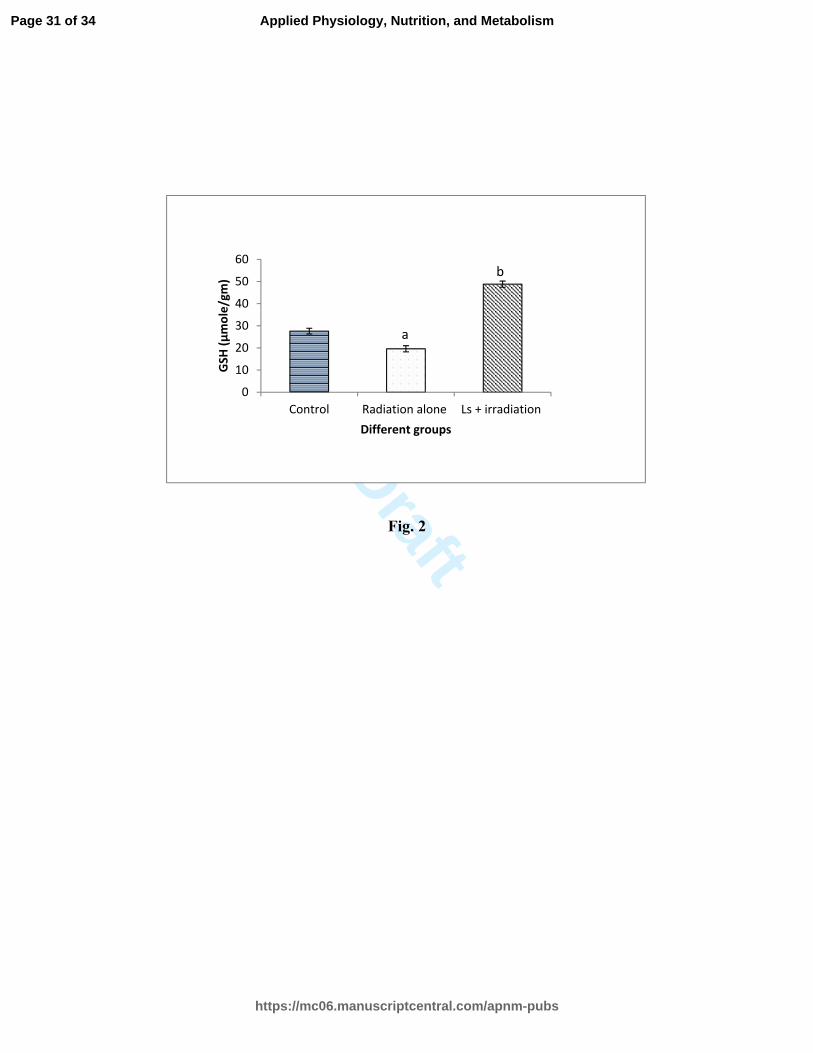
The changes in the amount of total GSH in various experimental groups have been
presented in Fig 2. In the control group GSH contents were 27.6 ± 1.3 µmol/gm which
decreased to 19.6 ± 0.14 µmol/gm in the irradiated group (10 Gy). Ls treatment (2mg/kg
body wt.) increased the GSH contents to 48.8 ± 1.4 µmol/gm which were significantly
higher than the control and irradiated group.
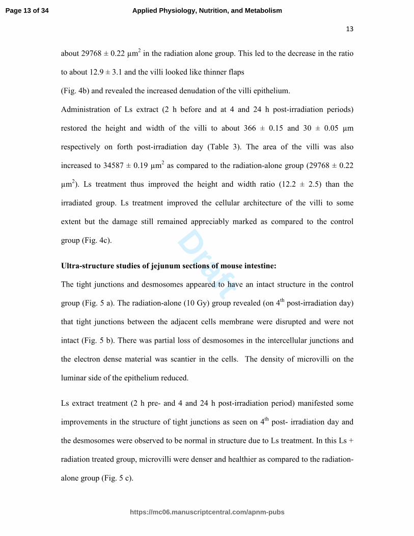
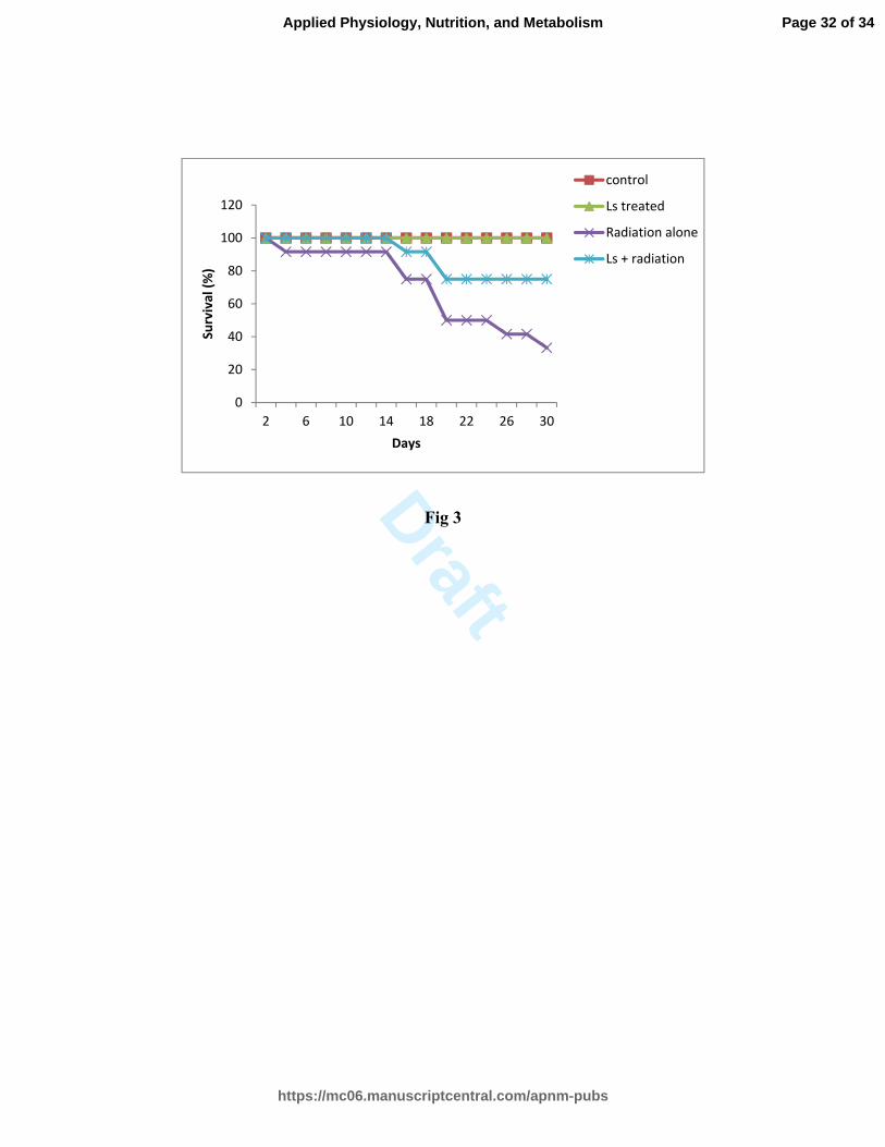
Radio-modifying effect of Lagenaria siceraria in mice
Mice in the control group (receiving no treatment) and in the Ls extract treated group
(2mg/kg body wt.) rendered 100 % survival (30 days and beyond). Irradiation (10 Gy)
rendered 33 % survival up to 30th
post-irradiation day (Fig. 3). Administration of Ls
extract (at -2 h, +4 and +24 h of irradiation). Ls treatment improved the survival of
irradiated mice to about 75 % (30th
post-irradiation day).
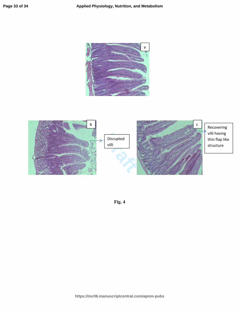
Histological study of gut villi:
The average length and width of the villi in the jejunum region in the control group was
379 ± 0.36 and 54 ± 1.4 µm respectively. Each villus at an average had an area of ~
64468 ± 2.3 µm2
(Table 3). The average height and width (VH/VW) ratio of the villi in
the control group was 7.01 ± 1.9. On forth post-irradiation day, the average length and
width of the villi was observed as 350 ± 0.22 and 27 ± 0.06 µm and the area reduced to
Page 12 of 34
https://mc06.manuscriptcentral.com/apnm-pubs
Applied Physiology, Nutrition, and Metabolism
Draft
13
about 29768 ± 0.22 µm2 in the radiation alone group. This led to the decrease in the ratio
to about 12.9 ± 3.1 and the villi looked like thinner flaps
(Fig. 4b) and revealed the increased denudation of the villi epithelium.
Administration of Ls extract (2 h before and at 4 and 24 h post-irradiation periods)
restored the height and width of the villi to about 366 ± 0.15 and 30 ± 0.05 µm
respectively on forth post-irradiation day (Table 3). The area of the villi was also
increased to 34587 ± 0.19 µm2 as compared to the radiation-alone group (29768 ± 0.22
µm2). Ls treatment thus improved the height and width ratio (12.2 ± 2.5) than the
irradiated group. Ls treatment improved the cellular architecture of the villi to some
extent but the damage still remained appreciably marked as compared to the control
group (Fig. 4c).
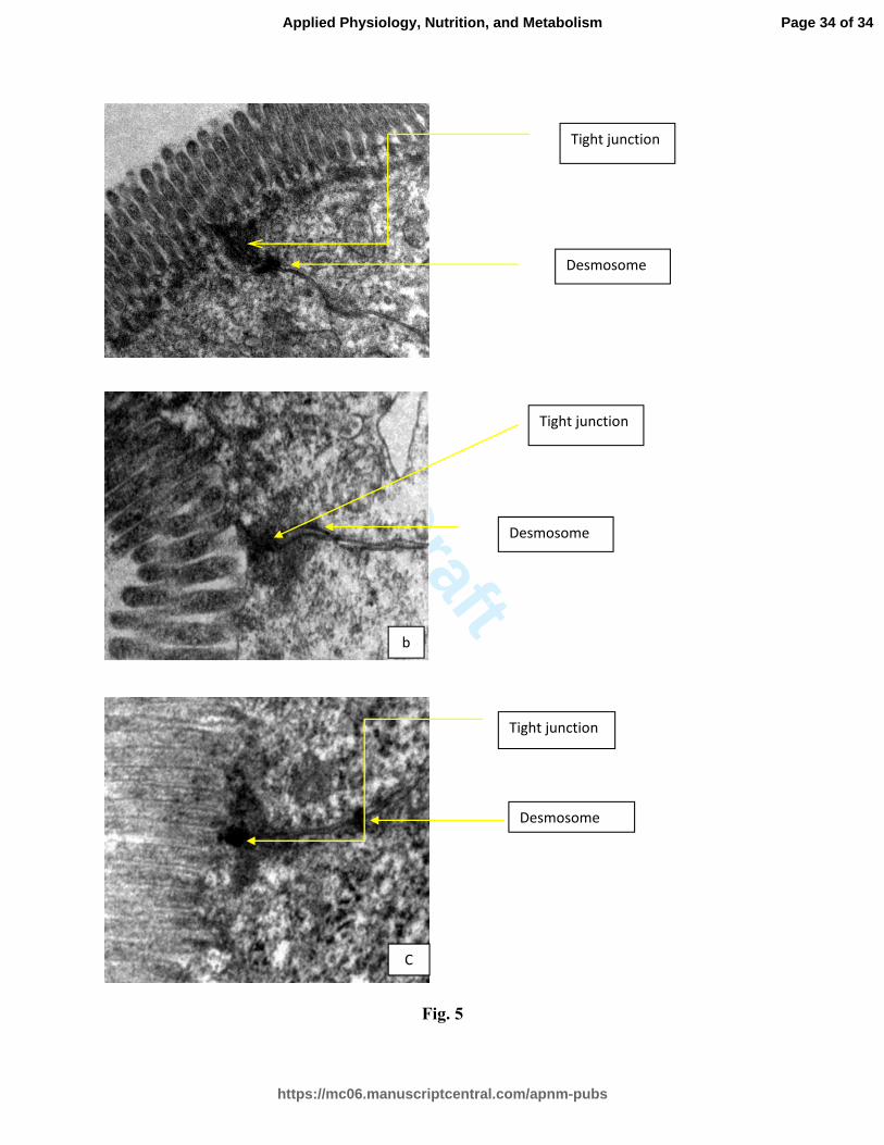
Ultra-structure studies of jejunum sections of mouse intestine:
The tight junctions and desmosomes appeared to have an intact structure in the control
group (Fig. 5 a). The radiation-alone (10 Gy) group revealed (on 4th
post-irradiation day)
that tight junctions between the adjacent cells membrane were disrupted and were not
intact (Fig. 5 b). There was partial loss of desmosomes in the intercellular junctions and
the electron dense material was scantier in the cells. The density of microvilli on the
luminar side of the epithelium reduced.
Ls extract treatment (2 h pre- and 4 and 24 h post-irradiation period) manifested some
improvements in the structure of tight junctions as seen on 4th
post- irradiation day and
the desmosomes were observed to be normal in structure due to Ls treatment. In this Ls +
radiation treated group, microvilli were denser and healthier as compared to the radiation-
alone group (Fig. 5 c).
Page 13 of 34
https://mc06.manuscriptcentral.com/apnm-pubs
Applied Physiology, Nutrition, and Metabolism
Draft
14
Discussion:
Radiation induced GI syndrome involves serious disturbances in luminal environment,
cells and tissues of GI tract and the enteric microbiota. In our previous studies, dietary
cucurbit fruits (Ls, Lc and Cp) have been shown to act as prebiotics which foster the
interaction among enteric microbes (probiotics), diet, cells, tissues and secretions of the
GI tract manifesting antimicrobial, antioxidant and anti-inflammatory activity on one
hand (Sharma et al. 2012; Rawat et al. 2014) and augment the cellular repair and
recovery and enhance cell proliferation to replenish apoptotic and necrotic cells.
Oxidative and inflammatory manifestations display parallel situations with respect to the
etiology of radiation damage and many acute and chronic ailments of metabolic and
oncogenic nature. It was therefore considered rational to investigate the role of cucurbits
in the recovery of radiation damage. The radio-protective attributes of cucurbits have not
been documented as yet. Therefore, present study was undertaken to explore radio-
modifying potential of some dietary cucurbits in-vitro and in-vivo.
Cell cycle: The DNA repair and replenishment of damaged and dead cells require
augmentation of cell proliferation and G2 fraction. Radiation induced G2 blockage of cells
has been widely reported in the literature and this blockage is to ensure repair and
recovery of damage to the cells before allowing them to progress to M-phase (Vucic et al.
2006). Addition of the cucurbit extracts to the irradiated cells has enhanced the
accumulation of cells at G2 check- point as compared to radiation-alone treated group
(Table 1). This accumulation is considered necessary to repair the DNA damage
maximally which was induced by radiation exposure.
Page 14 of 34
https://mc06.manuscriptcentral.com/apnm-pubs
Applied Physiology, Nutrition, and Metabolism
Draft
15
The cucurbit extracts contain hundreds of molecules which remain active in various
directions. In our previous studies, cucurbitacin, a characteristic molecule of the family
cucurbitaceae, revealed the anti-proliferative effect against HCT-15 cells. Cucurbitacin
has also been studied by others and has been reported to enhance accumulation of cells in
G2 phase (Duangmano et al. 2012). The mechanism involving G2 blockage may be the
consequence of reduced formation of spindle microtubules (Dutta and Gupta 2014).
Several cell cycle regulators like p53, p21, CDC1, CDC2 have also been reported to
affect the cell cycle (Kim et al. 2004). Therefore, it would be interesting to investigate
further the role of cucurbits on the expression of various cell cycle regulators.
Comet assay relies on the DNA fragmentation generated by a genotoxic agent. The
smaller fragments migrate faster than bigger jump of DNA during gel-electrophoresis and
create a typical comet-shaped pattern. In fact, there are several parameters exploited for
the evaluation of DNA damage and comet length is considered a reliable parameter in
this connection. The irradiated cells revealed a longer tail as compared to un-irradiated
control indicating large amount of fragmentation of DNA. Each of the three cucurbits
investigated here has been found to reduce the tail length significantly. The Ls extract
was more effective in reducing the comet tail length than Lc and Cp. The antioxidant
action of these cucurbits could be responsible for such a decrease in tail length because a
significant magnitude of damage induced by ionizing radiation results from the action of
free radicals (ROS) such as hydroxyl radicals, hydrogen peroxide, superoxide anions etc.
The findings of comet assay, where Ls was more effective in reducing the DNA damage
followed by Lc and Cp corroborate results of antioxidant activity (Ls > Lc > Cp) of these
cucurbits reported earlier (Sharma et al. 2012).
Page 15 of 34
https://mc06.manuscriptcentral.com/apnm-pubs
Applied Physiology, Nutrition, and Metabolism
Draft
16
Villi (jejunum): The epithelial cells of villi follow a definite pattern of migration and
maturation and are constantly replaced by the new cells produced by the stem cells, the
crypts of Lieberkuhn. The cells from the crypts migrate towards the tip of villi reaching
the top within about 72 h in mice and are thereafter denudated in the gut lumen. The
radiation induced effects on gastrointestinal system have been mostly studied by many
workers in jejunum (Bing et al. 2014; Zhao et al. 2014) because of very active crypts of
Lieberkuhn and larger villi in jejunum than in the colon. For reason of comparison we
have also studied this region.
The colon which harbors about 1014
microbes metabolizes the fiber like inulin
(polysaccharides) into short chain fatty acids and also produces many nutrients like
vitamin K, B12, riboflavin etc. These molecules may substantially influence the radio-
recovery and therefore, studies in the colon tissue deserve further investigations.
Radiation exposure has been reported to induce apoptosis in crypt cells (Matsuu-
Matsuyama et al. 2006) and also affects the proliferation of crypt cells adversely. The
slow proliferation of crypts cells decreased the replenishment of villi epithelial cells
leading to shortening of the height and width of the villi and subsequently the absorptive
area of the villus. These changes decreased absorption of the nutrients and essential
molecules and this may adversely influence the activity of proliferating and differentiated
cells.
Treatment with Ls extract (3 times: 2h pre- and 4 and 24 h post-irradiation) increased the
height, width and area of the villi in comparison to radiation alone group (Table 3 and
also rendered some improvement in the morphology of the villi (Fig 4c). The exact mode
Page 16 of 34
https://mc06.manuscriptcentral.com/apnm-pubs
Applied Physiology, Nutrition, and Metabolism
Draft
17
of action of radio-protection of Ls on gut villi is not known. Our studies on in-vitro
system (Sharma et al. 2012; Rawat et al. 2012) have demonstrated the antioxidant activity
of Ls extract. The endogenous antioxidants like glutathione (GSH), superoxide dismutase
may also help in the mitigation of radiation induced free radicals. Ls extract has been
shown to up-regulate the production of GSH (Fig. 2). However, these mechanisms alone
may not be sufficient to explain the extent of radio-protection. Indeed several other
mechanisms may also be concomitantly acting to achieve radio-protection. The Ls extract
contains a number of bioactive molecules like flavonoids, alkaloids, saponins etc, which
may contribute as important biomolecules for the repair of radiation induced damage.
Flavonoids have been demonstrated to act as anti- carcinogenic agents (Seelinger et al.
2008) and thus help in the repair of radiation induced damage and thus provide radio-
protection. Radiation has been reported to activate several inflammatory pathways like
NF-kB and COX-II (Chung et al. 2010). Ls has already been reported for its strong anti-
inflammatory activity (Rawat et al. 2012). However, Ls acting as a radio-protector may
have many more aspects like gut-neuronal network which need to be investigated further.
Tight junction: The epithelial cells of the villi are held together by complex structures
the ‘tight junctions’. The membranes of the two juxta-positioned cells join together and
form a dynamic barrier for the microbes. The integrity of tight junction is dependent on
the composition and organization of tight junction proteins (Shen et al. 2008) which are
composed of trans-membrane proteins (occludin, claudin and junctional adhesion
molecule) and cytoplasmic proteins (ZO-1, ZO-2, ZO-3 and cingulin). TJs open and close
all the times in response to a variety of stimuli like dietary state, hormonal and neuronal
signals, inflammatory mediators and mast cell products, in a regulated manner. However,
Page 17 of 34
https://mc06.manuscriptcentral.com/apnm-pubs
Applied Physiology, Nutrition, and Metabolism
Draft
18
dissociation of the protein complex and/or down-regulation of proteins may disrupt the
tight junctions (Peerapen and Thongboonkerd 2013) and may lead to the widening of the
paracellular passage. The pathogens at this stage may breach the gut epithelium to enter
the lamina propria and the blood circulation (Guttman and Finlay 2009). This may lead to
immunogenic reactions, onset of infections and several disorders subsequently.
Therefore, dietary agents were investigated for managing diarrhea, leaky junctions and
the microbial invasion.
Whole body gamma irradiation of 10 Gy disrupted the integrity of tight junctions of
adjacent cells (Fig. 5 b). The higher incidence of microbial pathogenicity after whole
body lethal irradiation (hemopoietic and gastro-intestinal syndrome) and after exposure
of abdomino-pelvic region during radio-therapy (Macnaughton 2000), may also be
explained accordingly.
Present study displayed that radiation induced disruptions in tight junctions were
recovered by Ls treatment (Fig. 5c). It indicates that Ls extract has some molecules which
are very important for regulation of tight junction proteins. In our previous studies, large
amount of flavonoids have been found in the Ls. Flavonoids present in the Ls extract
(Gangwal et al. 2010) have been reported to enhance the barrier function through
regulation of the TJ protein claudin-4 (Amasheh et al. 2008). Therefore, previous studies
done in our laboratory has been giving a direction towards the molecular mechanism of
Ls extract with respect to epithelial barrier function. Further investigations are necessary
to understand the mechanism of recovery by Ls treatment.
Page 18 of 34
https://mc06.manuscriptcentral.com/apnm-pubs
Applied Physiology, Nutrition, and Metabolism
Draft
19
Conclusion:
Application of dietary cucurbits for protection against lethal doses of radiation causing GI
syndrome has been experimentally demonstrated both in vitro and in vivo. Many aspects
of the mechanism of radio-protection have yet to be understood in more details before
cucurbits could be exploited for the development of radio-protective agents.
Acknowledgement:
The authors sincerely acknowledge Life Science Research Board, Defense Research &
Development Organisation, Ministry of Defence, New Delhi for financial support. The
authors are also thankful to Director, INMAS, Delhi-54 for permitting to use necessary
research facilities for conducting this study. The authors also thank Amity University
authorities for providing facilities to carry out this work.
Disclosure:
The authors of this research article declare that there is no conflict of interests regarding
the publication of this manuscript.
Page 19 of 34
https://mc06.manuscriptcentral.com/apnm-pubs
Applied Physiology, Nutrition, and Metabolism
Draft
20
References:
Amasheh, M., Schlichter, S., Amasheh, S., Mankertz, J., Zeitz, M., et al. 2008.
Quercetin enhances epithelial barrier function and increases claudin-4 expression in
Caco-2 cells. J. Nutr. 138: 1067-1073.
Arora, R., Gupta, D., Chawla, R., Sagar, R., Sharma, A., Kumar, R., et al. 2005.
Radioprotection by Plant Products: Present Status and Future Prospects. Phytother.
Res. 19: 1–22.
Bing, S.J., Kim, M.J., Ahn, G., Im, J., Kim, D.S., Ha, D., et al. 2014. Acidic
polysaccharide of Panax ginseng regulates the mitochondria/caspase-dependent
apoptotic pathway in radiation-induced damage to the jejunum in mice. Acta.
Histochemica. 116: 514-21.
Brown, M. 2008. Controversy Section: What causes the radiation GI syndrome?
2008. Int. J. Radiat. Oncol. Biol. Phys. 70: 799–800.
Chung, S.W., Kim, J.M., Kim, D.H., Kim, J.Y., Lee, E.K., Anton, S., et al. 2010.
Molecular delineation of gamma-ray-induced NF-kappaB activation and pro-
inflammatory genes in SMP30 knockout mice. Rad. Res. 173: 629-34.
Donnelly, EH., Nemhauser, JB., Smith, JM., Kazzi, ZN., Farfan, EB., Chang, AS., et
al. 2010. Acute radiation syndrome: assessment and management. South. Med. J. 103:
541-546.
Duangmano, S., Sae-lim, P., Suksamrarn, A., Patmasiriwat, P., and Domann, F.E.
2012. Cucurbitacin B causes increased radiation sensitivity of human breast cancer
Page 20 of 34
https://mc06.manuscriptcentral.com/apnm-pubs
Applied Physiology, Nutrition, and Metabolism
Draft
21
cells via G2/M cell cycle arrest. J. Oncol. 2012; Article ID 601682, 8 pages,
doi:10.1155/2012/601682.
Dutta, S., and Gupta, M.L. 2014. Alleviation of radiation-induced genomic damage
in human peripheral blood lymphocytes by active principles of Podophyllum
hexandrum: an in vitro study using chromosomal and CBMN assay. Mutagenesis.
29: 139-147.
Gangwal, A., Parmar, S.K., and Sheth, N.R. 2010. Triterpenoid, flavonoids and
sterols from Lagenaria siceraria fruits. Scholars. Res. Lib. 2: 307-317.
Ghosh, S.P., Kulkarni, S., Hieber, K., Toles, R., Romanyukha, L., Kao, T.C., et al.
2009. Gamma-tocotrienol, a tocol antioxidant as a potent radioprotector. Int. J. Rad.
Biol. 85: 598–606.
Guttman, J.A., and Finlay, B.B. 2009. Tight junctions as targets of infectious agents.
Biochemica. Et. Biophysica. Acta. 1788: 832-841.
Irshad, M., Ahmad, I., Goel, H.C., and Rizvi, M.M.A. 2010. Phytochemical screening
and high performance TLC analysis of some cucurbits. Res. J. Phytochem. 4: 242-
247.
Kau, A.L., Ahern, P.P., Griffin, N.W., Goodman, A.L., and Gordon, J.I. 2011. Human
nutrition, the gut microbiome and the immune system. Nature. 474: 327-36.
Khojasteh, S.M.B., Sheikhzadeh, F., Mohammadnejad, D., and Azami, A. 2009.
Histological, histochemical and ultrastructural study of the intestine of rainbow trout
(Oncorhynchus mykiss). World. Appl. Sci. J. 6: 1525-1531.
Page 21 of 34
https://mc06.manuscriptcentral.com/apnm-pubs
Applied Physiology, Nutrition, and Metabolism
Draft
22
Kim, H.S., Cho, H.J., Cho, H.J., Park, S.J., Park, K.W., Chae, I.H., et al. The essential
role of p21 in radiation-induced cell cycle arrest of vascular smooth muscle cell. J.
Mol. Cell. Cardiol. 37: 871-80.
Marchesi, J.R., Adams, D.H., Fava, F., Hermes, G.D., Hirschfield, G.M., Hold, G., et
al. 2016. The gut microbiota and host health: a new clinical frontier. Gut. 65:330-9.
Macnaughton. 2000. Review article: new insights into the pathogenesis of radiation-
induces intestinal dysfunction. Aliment. Pharmacol. Ther. 14: 523-528.
Matsuu-Matsuyama, M., Shichijo, K., Okaichi, K., Ishii, K., Wen, C.Y., Fukuda,
E., et al. 2006. Sucralfate protects intestinal epithelial cells from radiation-induced
apoptosis in rats. J. Rad. Res. 47: 1-8.
Nagayama, H., Ooi, J., Tomonari, A., Iseki, T., Tojo, A., Tani, K., et al. 2002. Severe
immune dysfunction after lethal neutron irradiation in a JCO nuclear facility accident
victim. Int. J. Hematol. 76: 157-164.
Peerapen, P., and Thongboonkerd, V. 2013. p38 MAPK mediates calcium oxalate
crystal-induced tight junction disruption in distal renal tubular epithelial cells. Scientific. Rep.
3:1041. doi: 10.1038/srep01041.
Pozarowski, P., and Darzynkiewicz, Z. 2004. Analysis of cell cycle by flow
cytometry. Methods Mol. Biol. 281:301-11.
Rawat, I., Sharma, D., and Goel, H.C. 2012. Effect of some cucurbits on the
population dynamics of Lactobacillus rhamnosus and its interaction with Escherichia
coli. World. J. Med. Pharm. Biol. 2: 11-19.
Page 22 of 34
https://mc06.manuscriptcentral.com/apnm-pubs
Applied Physiology, Nutrition, and Metabolism
Draft
23
Rawat, I., Sharma, D., and Goel, H.C. 2014. Antioxidant and anti-inflammatory
potential of some dietary cucurbits. Oxid. Antioxid. Med. Sci. 3: 65-72.
Seelinger, G., Merfort, I., Wolfle, U., and Schempp, C.M. 2008. Anti-carcinogenic
effects of the flavonoid luteolin. Molecules. 13: 2628-51.
Sharma, D., Rawat, I., and Goel, H.C. 2012. Antioxidant and prebiotic potential of
some dietary cucurbits. Res. J. Med. Plants. 6: 500-510.
Shen, L., Weber, C.R., and Turner, J.R. 2008. The tight junction protein complex
undergoes rapid and continuous molecular remodeling at steady state. The. J. Cell.
Biol. 181: 683-95.
Singh, N.P., McCoy, M.T., Tice, R.R., and Schneider, E.L. 1988. A simple technique
for quantification of low levels of DNA damage in individual cells. Exp. Cell. Res. 5:
184-491.
Soderholm, J.D., Olaison, G., Peterson, K.H., Franzen, L.E., Lindmark. T., Wiren,
M., et al. 2002. Augmented increase in tight junction permeability by luminal stimuli
in the non-inflamed ileum of Crohn's disease. Gut. 50: 307–313.
Suman S., Maniar, M., Fornace, Jr, AJ., and Datta K. 2010. Administration of ON
01,210.Na after exposure to ionizing radiation protects bone marrow cells by
attenuating DNA damage response. Radiat. Oncol. 7: 6.
Takemura, N., Kawasaki, T., Kunisawa, J., Sato, S., Lamichhane, A., Kobiyama, K.,
et al. 2014. Blockade of TLR3 protects mice from lethal radiation-induced
gastrointestinal syndrome. Nat. Commun. DOI: 10.1038/ncomms4492
Page 23 of 34
https://mc06.manuscriptcentral.com/apnm-pubs
Applied Physiology, Nutrition, and Metabolism
Draft
24
Trajkovic, S., Dobric, S., Jacevic, V., Dragojevic-Simic, V., Milovanovic, Z., and
Dordevic, A. 2007. Tissue-protective effects of fullerenol C60(OH)24 and amifostine
in irradiated rats. Colloids. Surf B: Biointerfaces, 58: 39–43.
Turner, J.R. 2009. Intestinal mucosal barrier function in health and disease. Nat.
Rev. Immunol. 9: 799-809.
Verma, P., Jahan, S., Kim, T.H., and Goyal, P.K. 2011. Management of radiation
injuries by Panax ginseng extract. J. Ginseng. Res. 35: 261-271.
Vucic, V., Isenovic, E.R., Adzic, M., Ruzdijic, S., and Radojcic, M.B. 2006. Effects
of gamma-radiation on cell growth, cycle arrest, death, and superoxide dismutase
expression by DU 145 human prostate cancer cells. Braz. J. Med. Biol. Res. 39: 227-
36.
Williams, JM., Duckworth, CA., Burkitt, MD., Watson, AJM., Campbell,
BJ., and Pritchard, DM. 2015. Epithelial cell shedding and barrier Function. A
matter of life and death at the small intestinal villus tip. Vet. Pathol. 52: 445–455.
Weisdorf, D., Chao, N., Waselenko, J.K., Dainiak, N., Armitage, J.O., McNiece, I., et
al. 2006. Acute radiation injury: contingency planning for triage, supportive care, and
transplantation. Biol. Blood. Marrow. Transplant. 12: 672–682.
Zhao, X., Yang, H., Jiang, G., Ni, M., Deng, Y., Cai, J., et al. 2014. Simvastatin
attenuates radiation-induced tissue damage in mice. J. Rad. Res. 55: 257-264.
Page 24 of 34
https://mc06.manuscriptcentral.com/apnm-pubs
Applied Physiology, Nutrition, and Metabolism
Draft
25
Tables
Table 1: Effect of cucurbit extracts on the radiation induced changes in cell cycle
fractions (%).
Experimental groups Cell cycle fractions
G1 S G2
Control 36 ± 0.6 % 19 ± 1.0 % 20 ± 1.2 %
Radiation (6 Gy) 32 ± 1.0* 22 ± 1.5 %NS
31 ± 2.0 %*
Lagenaria + 6 Gy 28 ± 0.6 %* 18 ± 1.5%NS
42 ± 2.0 %*
Luffa + 6 Gy 36 ± 2.6%NS
20 ± 2.0%NS
34 ± 1.5 %*
Cucurbita + 6 Gy 28 ± 1.0 %* 24 ± 1.5%* 37 ± 1.0 %*
Data represented mean ± SE of three sets of experiments and each experimental group
was compared with control. Student’s t test was conducted for Significance (*P < 0.05,
NS- non-significant)
Page 25 of 34
https://mc06.manuscriptcentral.com/apnm-pubs
Applied Physiology, Nutrition, and Metabolism
Draft
26
Table 2: Modulation of radiation induced DNA damage by cucurbit extracts in
HCT-15 cells.
Groups Tail length (µm) Head (µm) Tail: head
Control 4.25 ± 0.47 19.3 ± 0.96 0.27
Radiation (6 Gy) 41.75 ± 1.1 10.9 ± 1.8 3.5
Lagenaria + 6 Gy 28 ± 1.2* 16.4 ± 0.68* 1.38
Luffa + 6 Gy 34.2 ± 0.57* 15.75 ± 1.57* 1.67
Cucurbita + 6 Gy 36.75 µm ± 1.18* 13.5 µm ± 0.86NS
2.7
Experimental protocol was followed as described in the legend of Fig 1. Comet images
provided the data for Table 2. Data represented mean ± SE of three sets of experiments.
Comparison was made between radiation and cucurbit (Ls, Lc or Cp) + irradiated groups
Student’s t test was conducted for significance (*P < 0.05, NS- non-significant).
Page 26 of 34
https://mc06.manuscriptcentral.com/apnm-pubs
Applied Physiology, Nutrition, and Metabolism
Draft
27
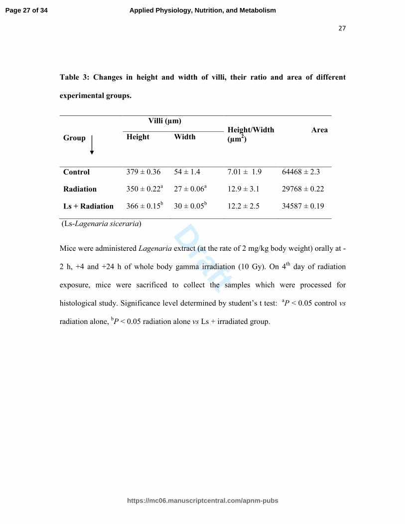
Table 3: Changes in height and width of villi, their ratio and area of different
experimental groups.
Group
Villi (µm)
Height/Width Area
(µm2) Height
Width
Control 379 ± 0.36 54 ± 1.4 7.01 ± 1.9 64468 ± 2.3
Radiation 350 ± 0.22a 27 ± 0.06
a 12.9 ± 3.1 29768 ± 0.22
Ls + Radiation 366 ± 0.15b 30 ± 0.05
b 12.2 ± 2.5 34587 ± 0.19
(Ls-Lagenaria siceraria)
Mice were administered Lagenaria extract (at the rate of 2 mg/kg body weight) orally at -
2 h, +4 and +24 h of whole body gamma irradiation (10 Gy). On 4th
day of radiation
exposure, mice were sacrificed to collect the samples which were processed for
histological study. Significance level determined by student’s t test: aP < 0.05 control vs
radiation alone, bP < 0.05 radiation alone vs Ls + irradiated group.
Page 27 of 34
https://mc06.manuscriptcentral.com/apnm-pubs
Applied Physiology, Nutrition, and Metabolism
Draft
28
Figure legends
Fig. 1: Images of the comets, stained with propidium iodide, observed through
florescent microscopy.
Cucurbit extracts (500 µL) were added to HCT-15 cells (1 million) in culture 2 h prior to
irradiation. 4 h after irradiation, the culture medium having cucurbit extract was replaced
by fresh medium without cucurbit extract. After 24 h of irradiation, cells were processed
for comet assay.
(a) Control (b) Radiation- 6 Gy (c) Ls + radiation (d) Lc + radiation (e) Cp + radiation.
Fig. 2: Effect of Lagenaria extract on the radiation induced changes in GSH level in
the mice intestine.
Mice were administered Lagenaria extract (at the rate of 2 mg/kg body weight) orally at
various periods (-2 h, +4 and +24 h of irradiation-10 Gy). On 4th
day of irradiation mice
were sacrificed to operate out jejunum sections and GSH contents were measured and
have been expressed as µmol/gm intestinal tissue. Data in each group represented mean ±
SE of three sets of experiments. Significance level determined by student’s t test: aP <
0.05 control vs radiation alone, bP < 0.05 radiation alone vs Ls + irradiated group.
Fig 3: Effect of Lagenaria extract on the survival of gamma irradiated mice.
(Ls- Lagenaria siceraria)
Page 28 of 34
https://mc06.manuscriptcentral.com/apnm-pubs
Applied Physiology, Nutrition, and Metabolism
Draft
29
Mice were observed for survival till 30 post-irradiation days. Data represented mean ± SE
of three independent experiments carried out with 12 animals/group.
Different groups: (a) Control group - without any treatment), (b) Ls treated group - Ls
extract administered at 0 h, 6 h & 26 h, (c) Radiation group (10 Gy), (d) Ls + radiation
group- Ls extract administered at -2 h and +4 and + 24 h of irradiation
Fig. 4: Images of Hematoxylene & Eosin stained sections of jejunum of mice
procured from different groups.
(a) Control showing healthy villi (b) Irradiation group showing disrupted villi (c) Ls +
irradiated group showing some improvement in the architecture of villi
Experimental protocol was followed as described in Table 3. Data of Table 3 was
computed on the basis of observations received through these images.
Fig. 5: Electron micrograph of the jejunum sections of mice of different groups.
Experimental protocol was followed as described in Table 3. To study the tight junctions,
jejunum sections were processed for ‘Transmission electron microscopy’ on 4th
day of
irradiation.
(a) Control showing normal structure of tight junction
(b) Radiation treated group showing disrupted tight junction and desmosome
(c) Ls treated group showing some improvement in the structure of tight junction and
desmosome
Page 29 of 34
https://mc06.manuscriptcentral.com/apnm-pubs
Applied Physiology, Nutrition, and Metabolism
Draft
Fig. 1
ed
cba
Page 30 of 34
https://mc06.manuscriptcentral.com/apnm-pubs
Applied Physiology, Nutrition, and Metabolism
Draft
Fig. 2
0
10
20
30
40
50
60
Control Radiation alone Ls + irradiation
GSH
(µmole/gm)
Different groups
a
b
Page 31 of 34
https://mc06.manuscriptcentral.com/apnm-pubs
Applied Physiology, Nutrition, and Metabolism
Draft
Fig 3
0
20
40
60
80
100
120
2 6 10 14 18 22 26 30
Survival (%)
Days
control
Ls treated
Radiation alone
Ls + radiation
Page 32 of 34
https://mc06.manuscriptcentral.com/apnm-pubs
Applied Physiology, Nutrition, and Metabolism
Draft
Fig. 4
b
Disrupted
villi
Recovering
villi having
thin flap like
structure
c
a
Page 33 of 34
https://mc06.manuscriptcentral.com/apnm-pubs
Applied Physiology, Nutrition, and Metabolism
Draft
Fig. 5
b
Tight junction
C
Desmosome
Tight junction
Desmosome
Tight junction
Desmosome
Page 34 of 34
https://mc06.manuscriptcentral.com/apnm-pubs
Applied Physiology, Nutrition, and Metabolism