GLYCOGENOLYSIS AND GLYCOGENdm5migu4zj3pb.cloudfront.net/manuscripts/101000/101088/JCI3910108… ·...
9
POSTMORTEM HEPATIC GLYCOGENOLYSIS IN HYPERINSULINISM AND GLYCOGEN DISEASE' By H. P. G. SECKEL (From the Department of Pediatrics, University of Chicago, Chicago) (Received for publication May 22, 1939) The fundamental pathology of von Gierke's gly- cogen disease (1) lies in the inhibition of in vivo and postmortem glycogenolysis of the liver and other organs. In the discussion of the pathogene- sis of this disorder, hyperinsulinism has been time and again suggested since Wilder and his collabo- rators (2) described their famous case of pan- creatogenic hyperinsulinism with severe spontane- ous hypoglycemia and a very high liver-glycogen content. They hinted at a similar situation in the equally famous case of Parnas and Wagner (3). This case, concerning a 9-year-old girl with hepa- tomegaly, hypoglycemia, ketonuria, and retardation of growth, is now almost universally recognized as the first case of glycogen disease ever described in the literature before the final elucidation of the dis- order by von Gierke in 1929 (1) (cf. the early case of Worster-Drought (4a), and especially that of Snapper and van Creveld (5)). It has been no- ticed, however, that in Wilder's case of carcinoma of the Langerhans islets with metastases to the liver, postmortem hepatic glycogenolysis was not determined and, as a matter of fact, has never yet been determined in the numerous cases of hyperinsulinism reported in the literature. It is the purpose of this communication to fill this gap by examining postmortem hepatic glycogenolysis in 2 cases of spontaneous hypoglycemia, one of which is almost certainly due to " pancreatogenic" hyperinsulinism, while the other will be tentatively classified as " neurogenic " hyperinsulinism. CASE REPORTS 2 Case 1. Albert R., 36 years, Number 195034. Ad- mitted to the Department of Neuro-Surgery, University of Chicago, on March 21, 1938. The patient had unexplained attacks of complete un- consciousness for quite some time which were revealed 'This work was aided by a grant from the Douglas Smith Foundation for Medical Research. 2 For more elaborate findings see later publication by N. B. Friedman and W. Sweet to whom the writer is greatly indebted for communicating the data. to be typical seizures of spontaneous hypoglycemia. Blood sugar on fasting and during attacks was between 16 and 23 mgm. per cent; no glycosuria, no ketonuria. The liver was found to be grossly enlarged and nodular to palpation. The hypoglycemic seizures were imme- diately stopped or prevented by intravenous glucose in- fusions, but not by adrenalin injections. The daily glu- cose requirement by mouth and parenterally was about 600 grams. Under this regime, further hypoglycemic seizures were prevented; the body weight increased from 57.1 to 64.3 kgm. within three months in spite of the underlying carcinoma. Alimentary glucose tolerance test: high rise of blood sugar falling off very early to hypo- glycemic levels. Adrenalin test (1 mgm. subcutaneously): non-fasting blood sugar 73 mgm. per cent, after 20 min- utes 98 mgm. per cent, after 45 minutes 65 mgm. per cent; slight vasomotor response. There was no ab- normal sensitivity to insulin. The blood cholesterol was normal. Clinical diagnosis. Spontaneous hypoglycemia, prob- ably due to a pancreatic tumor. An exploratory laparot- omy was done on April 15th. Innumerable white meta- static nodules were seen all over the liver. Follow- ing the ether anesthesia, the glucose requirement was considerably reduced for several days. During 24 hours preceding death, 580 grams of glucose were administered parenterally, the last dosage about 2 hours before death. Exitus on June 22, 1:20 p.m. Autopsy (started 15 minutes after death). Carcinoma of the pancreatic islets with nodular metastases to the liver and other viscera. Histologically, there was no abnormal glycogen storage outside the liver and muscles. An insulin assay of the liver nodules was unsatisfactory. Case 2. William M., 56 years, Number 206024. Ad- mitted to the Department of Medicine, University of Chicago, on September 23, 1938. The patient had supposedly uremic attacks with com- plete unconsciousness for about 8 weeks which were rec- ognized as typical seizures of spontaneous hypoglycemia. Blood sugar on fasting and during attacks was between 26 and 34 mgm. per cent, occasionally higher (72 mgm. per cent); no glycosuria, no ketonuria. The hypogly- cemic seizures were stopped and prevented by administra- tion of large amounts of glucose by mouth and paren- terally. Alimentary glucose tolerance test: High rise of blood sugar for about an hour falling off to around 30 mgm. per cent after Zl4 hours. Adrenalin tests (1 mgm. subcutaneously): fasting blood sugar 62 mgm. per cent, after 5, 15, and 60 minutes 58, 60 and 65 mgm. per cent respectively; 3 mgm. intravenously: non-fasting blood sugar 76 mgm. per cent, after 5, 10, and 40 minutes 84, 723
Transcript of GLYCOGENOLYSIS AND GLYCOGENdm5migu4zj3pb.cloudfront.net/manuscripts/101000/101088/JCI3910108… ·...

POSTMORTEMHEPATIC GLYCOGENOLYSISIN HYPERINSULINISMANDGLYCOGENDISEASE'
By H. P. G. SECKEL(From the Department of Pediatrics, University of Chicago, Chicago)
(Received for publication May 22, 1939)
The fundamental pathology of von Gierke's gly-cogen disease (1) lies in the inhibition of in vivoand postmortem glycogenolysis of the liver andother organs. In the discussion of the pathogene-sis of this disorder, hyperinsulinism has been timeand again suggested since Wilder and his collabo-rators (2) described their famous case of pan-creatogenic hyperinsulinism with severe spontane-ous hypoglycemia and a very high liver-glycogencontent. They hinted at a similar situation in theequally famous case of Parnas and Wagner (3).This case, concerning a 9-year-old girl with hepa-tomegaly, hypoglycemia, ketonuria, and retardationof growth, is now almost universally recognized asthe first case of glycogen disease ever described inthe literature before the final elucidation of the dis-order by von Gierke in 1929 (1) (cf. the early caseof Worster-Drought (4a), and especially that ofSnapper and van Creveld (5)). It has been no-ticed, however, that in Wilder's case of carcinomaof the Langerhans islets with metastases to theliver, postmortem hepatic glycogenolysis was notdetermined and, as a matter of fact, has neveryet been determined in the numerous cases ofhyperinsulinism reported in the literature. It isthe purpose of this communication to fill this gapby examining postmortem hepatic glycogenolysisin 2 cases of spontaneous hypoglycemia, one ofwhich is almost certainly due to " pancreatogenic"hyperinsulinism, while the other will be tentativelyclassified as " neurogenic " hyperinsulinism.
CASE REPORTS2
Case 1. Albert R., 36 years, Number 195034. Ad-mitted to the Department of Neuro-Surgery, Universityof Chicago, on March 21, 1938.
The patient had unexplained attacks of complete un-consciousness for quite some time which were revealed
'This work was aided by a grant from the DouglasSmith Foundation for Medical Research.
2 For more elaborate findings see later publication byN. B. Friedman and W. Sweet to whom the writer isgreatly indebted for communicating the data.
to be typical seizures of spontaneous hypoglycemia.Blood sugar on fasting and during attacks was between16 and 23 mgm. per cent; no glycosuria, no ketonuria.The liver was found to be grossly enlarged and nodularto palpation. The hypoglycemic seizures were imme-diately stopped or prevented by intravenous glucose in-fusions, but not by adrenalin injections. The daily glu-cose requirement by mouth and parenterally was about600 grams. Under this regime, further hypoglycemicseizures were prevented; the body weight increased from57.1 to 64.3 kgm. within three months in spite of theunderlying carcinoma. Alimentary glucose tolerance test:high rise of blood sugar falling off very early to hypo-glycemic levels. Adrenalin test (1 mgm. subcutaneously):non-fasting blood sugar 73 mgm. per cent, after 20 min-utes 98 mgm. per cent, after 45 minutes 65 mgm. percent; slight vasomotor response. There was no ab-normal sensitivity to insulin. The blood cholesterol wasnormal.
Clinical diagnosis. Spontaneous hypoglycemia, prob-ably due to a pancreatic tumor. An exploratory laparot-omy was done on April 15th. Innumerable white meta-static nodules were seen all over the liver. Follow-ing the ether anesthesia, the glucose requirement wasconsiderably reduced for several days. During 24 hourspreceding death, 580 grams of glucose were administeredparenterally, the last dosage about 2 hours before death.Exitus on June 22, 1:20 p.m.
Autopsy (started 15 minutes after death). Carcinomaof the pancreatic islets with nodular metastases to theliver and other viscera. Histologically, there was noabnormal glycogen storage outside the liver and muscles.An insulin assay of the liver nodules was unsatisfactory.
Case 2. William M., 56 years, Number 206024. Ad-mitted to the Department of Medicine, University ofChicago, on September 23, 1938.
The patient had supposedly uremic attacks with com-plete unconsciousness for about 8 weeks which were rec-ognized as typical seizures of spontaneous hypoglycemia.Blood sugar on fasting and during attacks was between26 and 34 mgm. per cent, occasionally higher (72 mgm.per cent); no glycosuria, no ketonuria. The hypogly-cemic seizures were stopped and prevented by administra-tion of large amounts of glucose by mouth and paren-terally. Alimentary glucose tolerance test: High rise ofblood sugar for about an hour falling off to around 30mgm. per cent after Zl4 hours. Adrenalin tests (1 mgm.subcutaneously): fasting blood sugar 62 mgm. per cent,after 5, 15, and 60 minutes 58, 60 and 65 mgm. per centrespectively; 3 mgm. intravenously: non-fasting bloodsugar 76 mgm. per cent, after 5, 10, and 40 minutes 84,
723

H. P. G. SECKEL
88, and 78 mgm. per cent, respectively; intensive vasom-otor response. Three mgm. adrenalin intravenously didnot arouse the patient from hypoglycemic coma. Bloodcholesterol was 185 mgm. per cent. The liver was notenlarged to palpation. On x-ray examination a markedelevation of the right leaf of the diaphragm with corre-sponding compression atelectasis of the right lung wasfound.
Clinical diagnosis. Spontaneous hypoglycemia, presum-ably from a pancreatic adenoma. On October 17th, anexploratory laparotomy was done under spinal and etheranesthesia. No pancreatic tumor was discovered; theliver appeared to be normal; a biopsy was taken forchemical analysis. Postoperatively, 4 times 75 grams ofglucose were administered parenterally without arousingthe patient from unconsciousness. Exitus next morningat 8:15.
Autopsy (started at 10 a.m.). Massive fibroma onright top of the liver bulging the medial aspect of theright diaphragm; pancreas normal; no metastases any-where. No autopsy of the brain was made. Histologi-cally, no glycogen was stored except in liver and muscles.An insulin assay of the fibroma did not reveal any insulin.
Chemical analyses
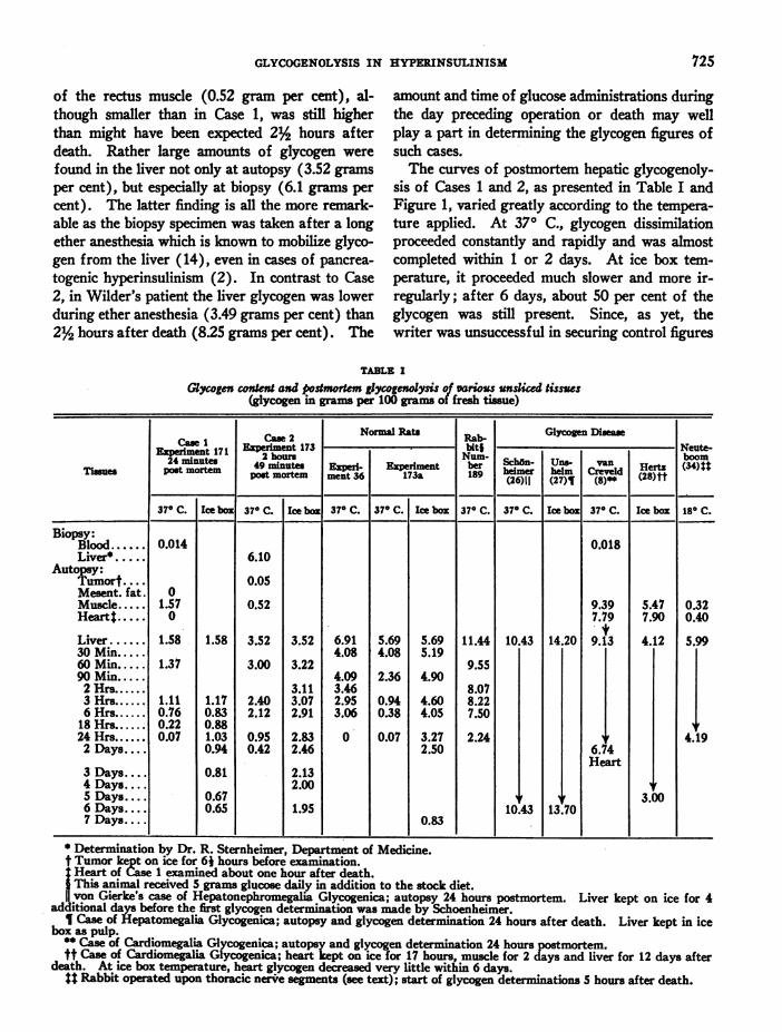
Glycogen was determined in various organs withPflueger's method as modified by Good, Kramer andSomogyi (6). The final estimation of glucose was madewith Somogyi's modification (7) of Shaffer and Hart-mann's method (factor 0.927). Blood glycogen wasestimated with van Creveld's method (8a). All figuresare based on duplicate determinations. Small pieces oftissue were put into dry ice immediately after the organshad been received at autopsy, ie., 24 minutes after deathin case 1, and 2 hours and 40 minutes after death incase 2. In the animal experiments performed for con-trol purposes, tissue pieces were put into dry ice 2 to 3minutes after death. In both the human and animalexperiments, unsliced pieces of one and the same liverlobe were preserved in a wet chamber, either at 37° C.with toluene added to prevent bacterial growth, or atice box temperature with no preservative added. Theglycogen content was determined at various intervals inthe liver tissue in order to obtain time curves of post-mortem hepatic glycogenolysis. In Table I, the figuresobserved in the 2 clinical cases of hyperinsulinism arelisted in conjunction with corresponding determinationsin a few cases of glycogen disease reported in the litera-ture as well as in normal rats and a rabbit. Liver glyco-genolysis curves of the 3 categories are presented inFigures 1 and 2.
In both clinical cases of hyperinsulinism sliced liverspecimens were suspended for 2 hours at 37° C. in aphosphate-buffered salt solution at pH 7.4 (for techniquesee Seckel (9a)). The following glycogen figures wereobserved in the liver slices: Case 1: At start (60 min-utes after the liver was received): 1.37 grams per cent;after 60 minutes 0.65 gram per cent; after 120 minutes0.53 gram per cent. By adding 1 per cent conjugated
bile salts to the suspension solution, as has been doneearlier with rat liver slices (Seckel, 9a), the 60 minutesglycogenolysis was increased to 0.396 gram per cent; 0.1per cent bile salt and 4 units of insulin per cc. of solutionproved to be ineffective. Case 2: At start (60 minutesafter the liver was received) 3.0 grams per cent; after60 minutes 2.81 grams per cent; after 120 minutes 1.85grams per cent.
Finally, the proportion of lyo- and desmo-glycogen wasdetermined in the two human livers by repeated extrac-tions with boiling water according to Willstitter andRohdewald's method (10). Liver pieces kept in the icebox for 6 days were used for this purpose. In Case 1,20.7 per cent of the total liver glycogen was found tobe desmo-glycogen and 79.3 per cent to be lyo-glycogen.In Case 2, the corresponding figures were 14.4 and 85.6per cent. In a normal rat liver, 18 and 82 per cent werefound respectively (Experiment Number 129). In freshgeese livers, Willstatter and Rohdewald found 10 to 13and 87 to 90 per cent, respectively.
COMMENT
In Case 1, the blood glycogen figure was withinnormal limits (average for normal children 12.9mgm. per cent (8a)). No glycogen was found inthe mesenteric fat. In Sch6nen's (11) experi-ments in young dogs fattened on " glycogen mast,"the fat contained up to 8.8 grams per cent glyco-gen. Probably the patient's heart was glycogen-free because of the delay in chemical examination.The glycogen content of the rectus abdominis mus-cle (1.57 grams per cent) seems to be unusuallyhigh. In view of the large amount of glucoseadministered to the patient, the liver-glycogen con-tent of 1.58 grams per cent appears to be ratherlow. However, it is still considerably above theindirect estimations of human liver glycogen at themoment of a " chronic death " as carried out byPopper and Wozasek (12a) and Burghard andPaffrath (13) (average of 0.65 gram per cent for"total carbohydrates"; for "glycogen" subtract0.23 gram per cent). Furthermore, this patient'sliver was filled with histologically glycogen-freemetastatic nodules and was also invaded with tinycell nests throughout those seemingly " normal "portions which were used for chemical examina-tion. This not only accounts for the compara-tively low glycogen content of the organ but alsofor the gross aberrations of individual figures inthe curve of hepatic glycogenolysis (Table I, icebox temperature, 6- and 18-hour samples).
In Case 2, the liver fibroma was almost glycogenfree on chemical analysis. The glycogen content
724
GLYCOGENOLYSISIN HYPERINSULINISM7
of the rectus muscle (0.52 gram per cent), al-
though smaller than in Case 1, was still higherthan might have been expected 2% hours afterdeath. Rather large amounts of glycogen were
found in the liver not only at autopsy (3.52 grams
per cent), but especially at biopsy (6.1 grams per
cent). The latter finding is all the more remark-able as the biopsy specimen was taken after a longether anesthesia which is known to mobilize glyco-gen from the liver (14), even in cases of pancrea-
togenic hyperinsulinism (2). In contrast to Case2, in Wilder's patient the liver glycogen was lowerduring ether anesthesia (3.49 grams per cent) than2%hours after death (8.25 grams per cent). The
amount and time of glucose administrations duringthe day preceding operation or death may wellplay a part in determining the glycogen figures ofsuch cases.
The curves of postmortem hepatic glycogenoly-sis of Cases 1 and 2, as presented in Table I andFigure 1, varied greatly according to the tempera-ture applied. At 370 C., glycogen dissimilationproceeded constantly and rapidly and was almostcompleted within or 2 days. At ice box tem-perature, it proceeded much slower and more ir-regularly; after 6 days, about 50 per cent of theglycogen was still present. Since, as yet, thewriter was unsuccessful in securing control figures
TABLE I
Glycogen cont and postmorkm glycogenolysis of various unsliced tissues(glycogen in grams per 100 grams of fresh tissue)
] Casle2 l Normal Rats Rab Glycogen DiseasExerant 17 Exeimn 173 -bltf __ -___ Neute-
24n tr 2 hrsNum_ bmnmMginutes' 49 minutes Exei- Experiment ber ScIISfnl an Het (34)StTisues post mortem post mortem ment 36 173a 189 hei r helm Creveld (28)tt(26)11 (27)¶1 (8)* 28f
37° C. Ice box 370 C. Ice boz 370 C. 370 C. Ice box 370 C. 370 C. Ice box 370 C. Ice bo 180 C.
Biopsy:Blood . 0.014 0.018Liver 6.10
Autopsy:Tumort.... 0.05Mesent. fat. 0Muscle. 1.57 0.52 9.39 5.47 0.32HeartI 0 7.79 7.90 0.40
Liver. 1.58 1.58 3.52 3.52 6.91 5.69 5.69 11.44 10.43 14.20 9.13 4.12 5.9930 Min 4.08 4.08 5.1960 Min. 1.37 3.00 3.22 9.5590 Min 4.09 2.36 4.90
2 Hrs 3.11 3.46 8.073 H
.1.11 1.17 2.40 3.07 2.95 0.94 4.60 8.22
6 Hrs.l...0.76 0.83 2.12 2.91 3.06 0.38 4.05 7.5018 Hrs ....0.22 0.8824 Hrs. 0.07 1.03 0.95 2.83 0 0.07 3.27 2.24 4 4.19
2 Days.... 0.94 0.42 2.46 2.50 6.74Heart
3 Days.... 0.81 2.134 Days.... 2.005 Days.... 0.67 3.006 Days.... 0.65 1.95 10.43 13.707 Day..... 0.83
* Determination by Dr. R. Sternheimer, Department of Medicine.t Tumor kept on ice for 6} hours before exation.::Heart of Case I examined about one hour after death.This animal received 5 grams glucose daily in addition to the stock diet.von Gierke's case of Hepatonephromegalia Glycogenica; autopsy 24 hours postmortem. Liver kept on ice for 4
ad itional days bore the first glycogen determination was made by Schoenheimer.¶ Case of Hepatomegalia Glycogenica; autopsy and glycogen determination 24 hours after death. Liver kept in ice
box as pulp.Case of Cardiomegalia Glycogenica; autopsy and glycogen determination 24 hours postmortem.
tt Case of Cardiomegalia Glycogenica; heart Icept on ice for 17 hours, muscle for 2 days and liver for 12 days afterdeath. At ice box temperature, heart glycogen decreased very little within 6 days.
fl Rabbit operated upon thoracic nerve segments (see text); start of glycogen determinations 5 hours after death.
725

H. P. G. SECKEL
FIG. 1. TIME CURVESOF POSTMORTEM-GLYCOGENOLYSIS OF VAmous DISEASED TISSUES(cf. TABLE I)
Intact tissue at 370 C. .- - -. Intact tissue at ice box temperature.Pulped tissue at ice box temperature. 1 = Liver of Case 1, pancreatogenic hyper-
insulinism. 2 = Liver of Case 2, " neurogenic " hyperinsulinism (dotted line between biopsy andautopsy). P = Liver of Popper and Wozasek's insulin-treated infant with diarrhea (slicedtissue, phosphate buffer, pH 6.9). S = Liver of Schoenheimer-von Gierke's case of glycogendisease. U= Liver of Unshelm's case of glycogen disease. C= Heart of van Creveld's caseof glycogen disease. H, H' = Heart and liver, respectively, of Hertz' case of glycogen disease.N= Liver of Neuteboom's operated rabbit (see text).
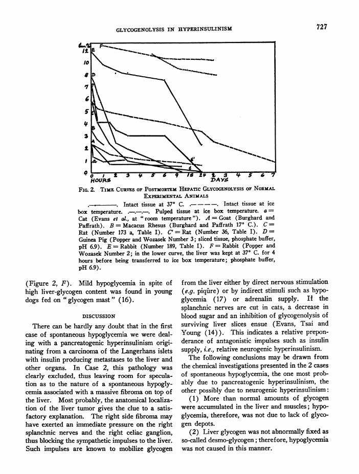
of normal human livers obtained 2% hours afterdeath and no such figures have been published inthe literature, we entirely depend upon the higherexperimental animals for normal controls of post-mortem hepatic glycogenolysis. Two experimentscarried out in normal rats and one in a glucose-fattened rabbit are recorded in Table I and Figure2. Moreover, experiments made in cats (14), ina goat and a macacus rhesus (13), and in guineapigs and rabbits (12b) are presented in Figure2.8 In comparison with these animal experiments,postmortem glycogenolysis in human hyperinsu-linism, as observed at 370 C., proceeded at a simi-lar rate in Case 1 and apparently somewhat slowerin Case 2; at ice box temperature it held a middlecourse between the various animals' curves.
By mechanical trauma, such as slicing of the tis-sue, postmortem glycogenolysis was accelerated in
8 Phosphate ions increase hepatic glycogenolysis (Fig-ure 2, D, F).
the 2 clinical cases. This corresponds to similarobservations in animal experiments, including thewriter's own. The same is true of the increase inCase 1 of postmortem hepatic glycogenolysis byadding highly concentrated bile salts to the sus-pended liver slices. The proportion of lyo- anddesmo-glycogen in the 2 human livers was alsoapproximately the same as in animal controls.
As is shown in the experiment made in theglucose-fattened rabbit (Table I, Figure 2), highliver-glycogen content does not in itself inhibitpostmortem glycogenolysis. This was demon-strated earlier by Kimmelstiel (15) in a dog puton " glycogen mast " whose liver glycogen disap-peared completely at ice box temperature within5 days after death. Less active, though distinctglycogenolysis was found by Popper and Wozasek(12b) in rabbits' livers rich in glycogen whichwere placed in the ice box either directly afterdeath or following a 4-hour incubation at 370 C.
726
727GLYCOGENOLYSISIN HYPERINSULINISM
FIG. 2. TIME CURVESOF POSTMORTEmHEPATIC GLYCOGENOLYSISOF NORMALEXPIMENTALANIMALS
Intact tissue at 370 C. .- -- . Intact tissue at icebox temperature. .-.-.-. Pulped tissue at ice box temperature. a =
Cat (Evans et al., at " room temperature "). A = Goat (Burghard andPaffrath). B = Macacus Rhesus (Burghard and Paffrath 170 C.). C=
Rat (Number 173 a, Table I). C' = Rat (Number 36, Table I). D=
Guinea Pig (Popper and Wozasek Number 3; sliced tissue, phosphate buffer,pH 6.9). E = Rabbit (Number 189, Table I). F = Rabbit (Popper andWozasek Number 2; in the lower curve, the liver was kept at 37° C. for 4hours before being transferred to ice box temperature; phosphate buffer,pH 6.9).
(Figure 2, F). Mild hypoglycemia in spite ofhigh liver-glycogen content was found in young
dogs fed on " glycogen mast " (16).
DISCUSSION
There can be hardly any doubt that in the firstcase of spontaneous hypoglycemia we were deal-ing with a pancreatogenic hyperinsulinism origi-nating from a carcinoma of the Langerhans isletswith insulin producing metastases to the liver andother organs. In Case 2, this pathology was
clearly excluded, thus leaving room for specula-tion as to the nature of a spontaneous hypogly-cemia associated with a massive fibroma on top ofthe liver. Most probably, the anatomical localiza-tion of the liver tumor gives the clue to a satis-factory explanation. The right side fibroma may
have exerted an immediate pressure on the rightsplanchnic nerves and the right celiac ganglion,thus blocking the sympathetic impulses to the liver.Such impulses are known to mobilize glycogen
from the liver either by direct nervous stimulation(e.g. piquire) or by indirect stimuli such as hypo-glycemia (17) or adrenalin supply. It thesplanchnic nerves are cut in cats, a decrease inblood sugar and an inhibition of glycogenolysis ofsurviving liver slices ensue (Evans, Tsai andYoung (14)). This indicates a relative prepon-derance of antagonistic impulses such as insulinsupply, i.e., relative neurogenic hyperinsulinism.
The following conclusions may be drawn fromthe chemical investigations presented in the 2 casesof spontaneous hypoglycemia, the one most prob-ably due to pancreatogenic hyperinsulinism, theother possibly due to neurogenic hyperinsulinism:
(1) More than normal amounts of glycogenwere accumulated in the liver and muscles; hypo-glycemia, therefore, was not due to lack of glyco-gen depots.
(2) Liver glycogen was not abnormally fixed asso-called desmo-glycogen; therefore, hypoglycemiawas not caused in this manner.
H. P. G. SECKEL
(3) High liver-glycogen content does not leadby itself to such an inhibition of hepatic glyco-genolysis as to produce severe seizures of spon-taneous hypoglycemia.
(4) Postmortem hepatic glycogenolysis wasproceeding at an almost normal or only slightlyretarded rate; the glycogenolytic enzyme wastherefore active on the livers' own glycogen.
(5) Reactions of postmortem hepatic glyco-genolysis to varying temperature, mechanical insultand high bile-salt concentration were also normal.
(6) Clinically, nevertheless, liver and muscleglycogen depots were mobilized with difficultythrough the normal stimuli such as severe hypo-glycemia, adrenalin injections and, in Case 2, pos-sibly also ether.
These observations made in 2 cases of hyper-insulinism are in good agreement with what isknown of insulin physiology. According to thisknowledge, one essential action of insulin on theliving tissue, especially liver and muscle, consistsof an inhibition of the glycogenolytic enzyme re-sulting in accumulation and fixation of glycogen.In animal experiments, various insulin doses, whenadded in vitro, were able to inhibit postmortemglycogenolysis of normal liver slices (9b) andmuscle extracts (18). In the postmortem speci-mens of the 2 patients' livers such inhibition waspresent only to a limited degree. Otherwise,in human pathology, only modest inhibitions ofpostmortem hepatic glycogenolysis have also beenobserved in insulin-treated cases of infantile ali-mentary intoxication (Figure 1, P), of diabetesmellitus, and schizophrenia (12b, c). In diabeticchildren injected with insulin for several monthsor years, a syndrome of enlarged abdomen, hepa-tomegaly, obesity, and retardation of growth has-been described by Mauriac (19) and other Frenchwriters (8b).
If the clinical and chemical pathology of spon-taneous hypoglycemia due to pancreatogenic hy-perinsulinism is compared with von Gierke's gly-cogen disease, there are a number of striking simi-larities to be observed in the two pictures. Severespontaneous hypoglycemia, glycogen accumulationin liver and muscles, and lack of blood sugar re-sponse to adrenalin injection are the chief amongthem. A minor common feature seems to be thestimulation of hepatic glycogenolysis by high bile-
salt concentrations such as has been demonstratedin titro in Case 1 (cf. Seckel, 9a) and may alsooccur in vivo in glycogen disease when jaundicecoincides with the disorder (20, 21, 22), or bileacids are given by mouth to such children (23).
On the other hand, there are outstanding dif-ferences between the two diseases. First of all,liver glycogen accumulation is more spectacularin glycogen disease than in hyperinsulinism lead-ing, as it does, to a tumorous enlargement of theorgan. Furthermore, organs other than the liverare often involved in glycogen disease, e.g. heartand kidneys. However, accumulation of gly-cogen as such is by no means pathognomonic ofglycogen disease. Similar or even larger amountsof available glycogen may be stored in the liver-to a far lesser degree in other organs-as aresult of simple dietary measures (18 to 22.4grams per cent in dogs (11, 16, 24); cf. rabbits,Figure 2) or continuous intravenous glucose in-fusions (upper limit in dogs 22 grams per cent(25)). The fundamental pathology of glycogendisease is rather the almost complete inhibition ofpostmortem glycogenolysis of the organs affected.For a week or longer, practically no glycogendisappears from the organs, both at 37° C. in anintact state (8, 26) and at ice box temperaturein an intact as well as a pulped state (27, 28,Table 1, Figure 1). Only in Hertz' (28) caseof a young baby, heart and liver showed a ratheractive postmortem glycogenolysis when pulpedand suspended in a phosphate buffer at pH 6.9and 370 C. for 1 or 2 days. The active post-mortem glycogenolysis in the intact liver andmuscles of Karlstroem's (29) baby (Case 1) in-dicates, along with an incomplete chemical picture,the presence of either an undeveloped stage or
an atypical variety of glycogen disease (cf. Karl-stroem's Case 2). In typical, fully-developedcases of glycogen disease, under natural circum-stances, the glycogenolytic enzyme does not act
after death on the tissues' own glycogen. Thisfundamental characteristic of glycogen diseasehas been shown to be missing in pancreatogenichyperinsulinism. Here, certainly, we are dealingwith the main distinguishing feature between thetwo diseases. Other specific symptoms of gly-cogen disease are the absence or rareness of hypo-glycemic seizures, high insulin sensitivity, constant
728

GLYCOGENOLYSISIN HYPERINSULINISM
fasting ketonuria, high blood glycogen, hyper-cholesterinemia, and increased basal metabolism(30b).
From the observations presented in this paperwe arrive at the conclusion that pancreatogenic(as well as therapeutic) hyperinsulinism is notidentical with typical glycogen disease and, con-sequently, that typical glycogen disease cannotpossibly originate from pancreatogenic hyperin-sulinism alone.
This does not mean to say that a certain degreeof pancreatogenic hyperinsulinism in conjunctionwith other endocrine disturbances could not playa part in the pathogenesis of glycogen disease.There are, for instance, 3 autopsy reports in theliterature concerning partial enlargement andabundance of the Langerhans islets in vonGierke's Disease ((15, 31), and Krakower's Case 1(32) ). Familial relations to diabetes mellitus arementioned in 3 cases ((21), Harnapp's Case 2(30a), Ellis' Case 1 (33)). Furthermore, theopinion has been offered that the insulin-treatedcases of puerile diabetes resulting in hepato-megaly, obesity, and retardation of growth mayrepresent a transition from diabetes into sec-ondary glycogen disease (34). Conversely, Par-nas and Wagner's (3) case of glycogen dis-ease turned into diabetes mellitus at the age of 16.On the other hand, the patient of Worster-Drought's (4b) appeared to be almost completelycured after puberty. These puberty changesoccurring in glycogen disease, put together withthe clinical and pathological reports of insular,thyrogenic, hypophyseal and adrenal 4 disturb-ances, seem to point to a pluriglandular disbalancewith an overfunction of the insulin apparatus asto the most probable pathogenesis of glycogendisease.
Finally, the question arises whether glycogendisease may not be an example of neurogenichyperinsulinism. This opinion has lately beenadvanced by Neuteboom (35). He distinguished2 "pure types of primary hepatomegalia glyco-genica." The first type is represented by typicalglycogen disease of the von Gierke-van Crevelddescription, consisting of a " hypofunction of the
'In in tvtro experiments with cortical extract, thewriter recently found a marked inhibition (up to 80 percent) of the glycogenolysis of surviving rat liver slices.(To be published in " Endocrinology," 1939.)
injured nerve tissue" (sympathetic pathways)with consequent " relative hyperinsulinism" and" insufficiency of the contrainsular system." Forthis type, no satisfactory experimental evidencewas provided by Neuteboom. The observationsmade in Case 2 of this report seem to furnisharguments against the supposed identity of gly-cogen disease with this type of neurogenic hyper-insulinism. In sharp contrast to the first type,the second type of so-called glycogen disease con-sists of a "hyperfunction of the injured nervetissue " with a consequent " real reactive hyper-insulinism." For this type, Neuteboom hasoffered clinical and experimental examples. Clin-ically, he presented a boy of 13 who early in lifehad twice suffered undefined injuries. Later, theliver became moderately enlarged. On the merebasis of a histological examination of a liverbiopsy the diagnosis of glycogen disease was made(no glycogenolysis test!). Experimentally, spinalinjuries were induced on a young rabbit by in-troducing platinum plates between the left I. andII. and the right V. and VI. thoracical segments.During life, the rabbit presented a picture in manyrespects similar to that of the boy; the liver andheart were enlarged and the blood glycogen washigh. However, such fundamental symptoms ashypoglycemia, ketonuria, hypercholesterinemia,missing adrenalin response, and insulin sensitivitywere absent or reversed in both the boy and therabbit. After death, the animal's liver and heartwere 30 and 40 per cent oversize, respectively.Liver glycogen examined 5 hours after death was5.99 grams per cent (control rabbit 2.25); theheart was not rich in glycogen. Postmortemhepatic glycogenolysis, estimated only from 5 to26 hours after death, was about as active as in ourpatient, Case 2, with possible neurogenic hyper-insulinism (Table I, Figure 1). Most interestingas these observations may be, they apparently donot justify the identification of either type ofneurogenic hyperinsulinism with typical vonGierke's glycogen disease.
SUMMARY
In 2 adult cases of spontaneous hypoglycemia,the one probably due to pancreatogenic hyper-insulinism (Case 1: carcinoma of the Langerhansislets with liver metastases), the other possibly due
729

H. P. G. SECKEL
to neurogenic hyperinsulinism (Case 2: massivefibroma on right top of the liver), there has beendemonstrated a comparatively high liver andmuscle glycogen content and an approximatelynormal or only slightly decreased postmortemhepatic glycogenolysis.
Since typical cases of glycogen disease arecharacterized by an abundance of glycogen ac-cumulated in the liver and other organs and analmost complete inhibition of postmortem glyco-genolysis in those organs, neither form of hyper-insulinism is identical with typical glycogen diseaseand, consequently, typical glycogen disease cannotoriginate from either form of hyperinsulinism.
BIBLIOGRAPHY
1. von Gierke, E., Hepato-Nephromegalia Glycogenica(Glykogenspeicherkrankheit der Leber und Nieren).Beitr. z. path. Anat. u. z. allg. Path., 1929, 82, 497.
2. Wilder, R. M., Allan, F. N., Power, M. H., andRobertson, H. E., Carcinoma of islands of pan-creas. Hyperinsulinism and hypoglycemia. J. A.M. A. 1927, 89, 348.
3. Parnas, J. K., and Wagner, R., (a) Beobachtungeniiber Zuckerneubildung. Biochem. Ztschr., 1922,127, 55; (b) Ueber eine eigenartige St6rung desKohlerhydratstoffwechsels und ihre Beziehungenzum Diabetes mellitus. Ztschr. f. d. ges. exper.Med., 1921, 25, 361.
4. Worster-Drought, C., (a) Case of enlarged liverwith persistent acetonuria and diaceturia. Proc.Roy. Soc. Med., Sect. Dis. Child., 1923, 16, 56;(b) Hepatomegaly with persistent ketonuria.
Ibid., 1935, 28, 829.5. Snapper, I., and van Creveld, S., Un cas d'hypogly-
cemie avec acetonemie chez un enfant. Bull. etmem. Soc. med. d. h6p. de Paris, 1928, 52, 1315.
6. Good, C. A., Kramer, H., and Somogyi, M., Deter-mination of glycogen. J. Biol. Chem., 1933, 100,485.
7. Somogyi, M., Sugar determination. J. Biol. Chem.,1926, 70, 599.
8. van Creveld, S., (a) Investigations on glycogen dis-ease. Arch. Dis. Childhood, 1934, 9, 9; (b) Gly-cogen disease. Medicine, 1939, 18, 1.
9. Seckel, H. P. G., (a) The influence of various physio-logical substances on the glycogenolysis of surviv-ing rat liver: methods; influence of the bile salts.Endocrinology, 1938, 23, 751; (b) Idem: Influenceof insulin added in vitro. Ibid., 1938, 23, 760.
10. Willstatter, R., and Rohdewald, M., Ueber den Zu-stand des Glykogens in der Leber, im Muskel undin Leukocyten (zur Kenntnis der Proteinbindungphysiologisch wichtiger Stoffe). Ztschr. f. physiol.Chem., 1934, 225, 103.
11. Sch6nen, H., Untersuchungen iiber den Einfluss derArt und Menge der Nahrung auf die Organzusam-mensetzung und das Stoffwechselgeschehen in ver-schiedenen Altersstuf en. Arch. f. d. ges. Physiol.,1932, 230, 179.
12. Popper, H., and Wozasek, O., (a) Zur Kenntnis desGlykogengehaltes der Leichenleber. Wien. Med.Wchnschr., 1929, 79, 456 ; (b) Idem. Ztschr. f. d.ges. exper. Med., 1932, 83, 682; (c) Ueber Di-astasehemmung in der Leber bei t6dlich verlauf-ender Insulin-Hypoglykamie. Virchows Arch. f.path. Anat., 1933, 288, 673.
13. Burghard, E., and Paffrath, H., Untersuchungen uberden Glykogengehalt der Leber; kritische Unter-suchungen fiber die Methodik der Glycogen- undKohlehydratbestimmung der Leber. Ztschr. f. Kin-derh., 1927, 45, 68.
14. Evans, C. L., Tsai, C., and Young, F. G., Behaviourof liver glycogen in experimental animals; meth-ods: effect of ether and amytal. J. Physiol., 1931,73, 67.
15. Kimmelstiel, P., Ueber Glykogenose. Beitr. z. path.Anat. u. z. allg. Path., 1933, 91, 1.
16. Junkersdorf, P., Glykogenspeicherung und Glykogen-speicherungskrankheit. Klin. Wchnschr., 1933, 12,899.
17. Macleod, J. J. R., The Fuel of Life. Princeton Uni-versity Press, Princeton, 1928, p. 50.
18. Lehmann, H., Action of insulin in cell-free extracts.Nature, 1938, 141, 690.
19. Mauriac, P., Hepatomegalies de l'enfance avec trou-bles de la croissance et du metabolisme des glucides.Paris Med., 1934, 2, 525.
20. Warner, E. C., Case of hepatomegaly due to vonGierke's disease. Lancet, 1933, 1, 1070.
21. Sundal, A., Glycogenosis (von Gierke's Krankheit).Acta Paediat., 1936, 19, 80.
22. Anderson, P. *M., Glycogen accumulation disease.Med. J. Austral., 1935, 22 (i), 362.
23. Linneweh, F., Zur Pathogenese der Glykogenkrank-heit. Monatsschr. f. Kinderh., 1937, 70, 238.
24. Sch6ndorff, B., Ueber den Maximalwerth des Ge-sammtglykogengehalts von Hunden. Arch. f. d.ges. Physiol., 1903, 99, 191.
25. Butsch, W. L., Glucose tolerance and glycogen stor-age capacity of dog. Am. J. Physiol., 1934, 108,639.
26. Sch6nheimer, R., Ueber eine eigenartige Storung desKohlehydratstoffwechsels. Ztschr. f. physiol.Chem., 1929, 182, 148.
27. Unshelm, E., Die Glykogenkrankheit (Zugleich einBeitrag zur Frage des hepatogenen Infantilismus).Jahrb. f. Kinderh., 1932, 137, 257.
28. Hertz, W., Untersuchungen iiber den vitalen undpostmortalen Kohlehydratstoffwechsel bei Glyko-genose und gestorter Schilddriisentitigkeit. Ztschr.f. Kinderh., 1936, 58, 259.
29. Karlstroem, F., Glycogenosis. Acta Paediat., 1938,20, 497.
730
GLYCOGENOLYSISIN HYPERINSULINISM
30. Harnapp, G. 0., (a) Zur Klinik der Hepatomegalienmit Kohlehydratstoffwechselst6rungen. I. Glyko-genspeicherungskrankheit. Monatsschr. f. Kin-derh., 1936, 66, 169; (b) Idem. III. Differential-diagnose und Pathogenese der Glykogenspeiche-rungskrankheit. Ibid., 1936, 66, 194.
31. Esser, M., and Scheidegger, S., Glykogenkrankheit.Beobachtung eines Falles. Schweiz. Med. Wchn-schr., 1937, 18, 970.
32. Krakower, C., The lipoid factor in glycogen storagedisease. J. Pediat., 1936, 9, 728.
33. Ellis, R. W. B., Extreme hepatomegaly in an infant.Proc. Roy. Soc. Med., 1933, 27, 118.
34. Gjuric, A., cited by Creveld (8b).35. Neuteboom, J. J., (a) Bijdrage tot de kennis der
hepatomegalia glycogenica. W. D. Meinema, The-sis, Utrecht, Holland, 1937. (b) Zur Kenntnis derGlykogenkrankheit. Klin. Wchnschr., 1938, 17,1437.
731