
Glycan–protein cross-linking mass spectrometry reveals ...
11
Glycan–protein cross-linking mass spectrometry reveals sialic acid-mediated protein networks on cell surfaces † Yixuan Xie, a Siyu Chen, a Qiongyu Li, a Ying Sheng, b Michael Russelle Alvarez, d Joeriggo Reyes, e Gege Xu, a Kemal Solakyildirim af and Carlito B. Lebrilla * ac A cross-linking method is developed to elucidate glycan-mediated interactions between membrane proteins through sialic acids. The method provides information on previously unknown extensive glycomic interactions on cell membranes. The vast majority of membrane proteins are glycosylated with complicated glycan structures attached to the polypeptide backbone. Glycan–protein interactions are fundamental elements in many cellular events. Although significant advances have been made to identify protein–protein interactions in living cells, only modest advances have been made on glycan–protein interactions. Mechanistic elucidation of glycan–protein interactions has thus far remained elusive. Therefore, we developed a cross-linking mass spectrometry (XL-MS) workflow to directly identify glycan–protein interactions on the cell membrane using liquid chromatography-mass spectrometry (LC- MS). This method involved incorporating azido groups on cell surface glycans through biosynthetic pathways, followed by treatment of cell cultures with a synthesized reagent, N-hydroxysuccinimide (NHS)–cyclooctyne, which allowed the cross-linking of the sialic acid azides on glycans with primary amines on polypeptide backbones. The coupled peptide–glycan–peptide pairs after cross-linking were identified using the latest techniques in glycoproteomic and glycomic analyses and bioinformatics software. With this approach, information on the site of glycosylation, the glycoform, the source protein, and the target protein of the cross-linked pair were obtained. Glycoprotein–protein interactions involving unique glycoforms on the PNT2 cell surface were identified using the optimized and validated method. We built the GPX network of the PNT2 cell line and further investigated the biological roles of different glycan structures within protein complexes. Furthermore, we were able to build glycoprotein– protein complex models for previously unexplored interactions. The method will advance our future understanding of the roles of glycans in protein complexes on the cell surface. 1. Introduction Cell surfaces are covered with a matrix of glycans on a scaffold of proteins and lipids. In particular, sialic acids at the terminus of glycans creates a highly interactive environment that governs many cellular functions such as cell–cell signaling, cell adhe- sions, and immunological recognition. 1–3 However, the nature of such weak and transient interactions impedes the systematic identication of the interacting complexes within living cells or tissues using existing analytical methods. In recent years, mass spectrometry (MS)-based methods including proximity labeling, hydrogen–deuterium exchange (HDX), affinity purication combined with mass spectrometry (AP-MS), and cross-linking mass spectrometry (XL-MS) have been extensively developed, which provide effective tools for characterizing protein–protein interactions. 4–6 Recently, Zhang et al. combined HDX with XL- MS, and characterized interleukin-7 and its a-receptors with largely enhanced spatial resolution. 7 However, these studies have rarely focused on characterizing glycoprotein–protein interactions due to the limitations in glycan analysis, despite the fact that glycans are vital mediators of many intercellular and extracellular interactions. 8 The inherent problems in the analysis are mostly due to the low abundance of glycoproteins and the large diversity of gly- coforms that further complicates the analysis. With the devel- opment of proteomic and glycoproteomic techniques, the methods that characterize protein complexes and networks on a Department of Chemistry, University of California, Davis, California, USA b Department of Chemistry, Biochemistry, Molecular, Cellular and Developmental Biology Graduate Group, University of California, Davis, California, USA c Department of Biochemistry, University of California, Davis, California, USA d Institute of Chemistry, University of the Philippines, Los Ba˜ nos, Laguna, Philippines e Marine Science Institute, University of the Philippines, Diliman, Quezon City, Philippines f Department of Chemistry, Erzincan Binali Yildirim University, Erzincan, Turkey. E-mail: [email protected] † Electronic supplementary information (ESI) available. See DOI: 10.1039/d1sc00814e Cite this: Chem. Sci. , 2021, 12, 8767 All publication charges for this article have been paid for by the Royal Society of Chemistry Received 8th February 2021 Accepted 17th May 2021 DOI: 10.1039/d1sc00814e rsc.li/chemical-science © 2021 The Author(s). Published by the Royal Society of Chemistry Chem. Sci., 2021, 12, 8767–8777 | 8767 Chemical Science EDGE ARTICLE Open Access Article. Published on 18 May 2021. Downloaded on 2/27/2022 11:43:44 PM. This article is licensed under a Creative Commons Attribution 3.0 Unported Licence. View Article Online View Journal | View Issue
Transcript of Glycan–protein cross-linking mass spectrometry reveals ...

ChemicalScience
EDGE ARTICLE
Ope
n A
cces
s A
rtic
le. P
ublis
hed
on 1
8 M
ay 2
021.
Dow
nloa
ded
on 2
/27/
2022
11:
43:4
4 PM
. T
his
artic
le is
lice
nsed
und
er a
Cre
ativ
e C
omm
ons
Attr
ibut
ion
3.0
Unp
orte
d L
icen
ce.
View Article OnlineView Journal | View Issue
Glycan–protein c
aDepartment of Chemistry, University of CalbDepartment of Chemistry, Biochemistry,
Biology Graduate Group, University of CalifcDepartment of Biochemistry, University of CdInstitute of Chemistry, University of the PheMarine Science Institute, University of t
PhilippinesfDepartment of Chemistry, Erzincan Binal
E-mail: [email protected]
† Electronic supplementary informa10.1039/d1sc00814e
Cite this: Chem. Sci., 2021, 12, 8767
All publication charges for this articlehave been paid for by the Royal Societyof Chemistry
Received 8th February 2021Accepted 17th May 2021
DOI: 10.1039/d1sc00814e
rsc.li/chemical-science
© 2021 The Author(s). Published by
ross-linking mass spectrometryreveals sialic acid-mediated protein networks oncell surfaces †
Yixuan Xie, a Siyu Chen,a Qiongyu Li, a Ying Sheng,b Michael Russelle Alvarez,d
Joeriggo Reyes,e Gege Xu, a Kemal Solakyildirimaf and Carlito B. Lebrilla*ac
A cross-linking method is developed to elucidate glycan-mediated interactions between membrane
proteins through sialic acids. The method provides information on previously unknown extensive
glycomic interactions on cell membranes. The vast majority of membrane proteins are glycosylated with
complicated glycan structures attached to the polypeptide backbone. Glycan–protein interactions are
fundamental elements in many cellular events. Although significant advances have been made to identify
protein–protein interactions in living cells, only modest advances have been made on glycan–protein
interactions. Mechanistic elucidation of glycan–protein interactions has thus far remained elusive.
Therefore, we developed a cross-linking mass spectrometry (XL-MS) workflow to directly identify
glycan–protein interactions on the cell membrane using liquid chromatography-mass spectrometry (LC-
MS). This method involved incorporating azido groups on cell surface glycans through biosynthetic
pathways, followed by treatment of cell cultures with a synthesized reagent, N-hydroxysuccinimide
(NHS)–cyclooctyne, which allowed the cross-linking of the sialic acid azides on glycans with primary
amines on polypeptide backbones. The coupled peptide–glycan–peptide pairs after cross-linking were
identified using the latest techniques in glycoproteomic and glycomic analyses and bioinformatics
software. With this approach, information on the site of glycosylation, the glycoform, the source protein,
and the target protein of the cross-linked pair were obtained. Glycoprotein–protein interactions
involving unique glycoforms on the PNT2 cell surface were identified using the optimized and validated
method. We built the GPX network of the PNT2 cell line and further investigated the biological roles of
different glycan structures within protein complexes. Furthermore, we were able to build glycoprotein–
protein complex models for previously unexplored interactions. The method will advance our future
understanding of the roles of glycans in protein complexes on the cell surface.
1. Introduction
Cell surfaces are covered with a matrix of glycans on a scaffoldof proteins and lipids. In particular, sialic acids at the terminusof glycans creates a highly interactive environment that governsmany cellular functions such as cell–cell signaling, cell adhe-sions, and immunological recognition.1–3 However, the natureof such weak and transient interactions impedes the systematic
ifornia, Davis, California, USA
Molecular, Cellular and Developmental
ornia, Davis, California, USA
alifornia, Davis, California, USA
ilippines, Los Banos, Laguna, Philippines
he Philippines, Diliman, Quezon City,
i Yildirim University, Erzincan, Turkey.
tion (ESI) available. See DOI:
the Royal Society of Chemistry
identication of the interacting complexes within living cells ortissues using existing analytical methods. In recent years, massspectrometry (MS)-basedmethods including proximity labeling,hydrogen–deuterium exchange (HDX), affinity puricationcombined with mass spectrometry (AP-MS), and cross-linkingmass spectrometry (XL-MS) have been extensively developed,which provide effective tools for characterizing protein–proteininteractions.4–6 Recently, Zhang et al. combined HDX with XL-MS, and characterized interleukin-7 and its a-receptors withlargely enhanced spatial resolution.7 However, these studieshave rarely focused on characterizing glycoprotein–proteininteractions due to the limitations in glycan analysis, despitethe fact that glycans are vital mediators of many intercellularand extracellular interactions.8
The inherent problems in the analysis are mostly due to thelow abundance of glycoproteins and the large diversity of gly-coforms that further complicates the analysis. With the devel-opment of proteomic and glycoproteomic techniques, themethods that characterize protein complexes and networks on
Chem. Sci., 2021, 12, 8767–8777 | 8767

Chemical Science Edge Article
Ope
n A
cces
s A
rtic
le. P
ublis
hed
on 1
8 M
ay 2
021.
Dow
nloa
ded
on 2
/27/
2022
11:
43:4
4 PM
. T
his
artic
le is
lice
nsed
und
er a
Cre
ativ
e C
omm
ons
Attr
ibut
ion
3.0
Unp
orte
d L
icen
ce.
View Article Online
cell membranes involving glycan–protein interactions havebeen explored. Paulson, Kohler, and their respective colleagueshave developed diazo-bearing sialic acid reporters to capture cis-and trans-interactions of CD22.9–11 They used the metaboliclabeling method to incorporate diazo groups into sialic acids.Proteins that associate with sialic acids can then be captured byproducing reactive carbenes through ultraviolet irradiation.However, only isolated binding-pairs can be characterizedbecause the method relied on purication by gel electropho-resis. To explore proteome-wide interactions, we have devel-oped the protein oxidation of sialic acid environments (POSE)method to identify potential sialic acid-associating proteins insitu.12 Frei et al. employed a trifunctional chemoproteomiccross-linker and created a prominent ligand-based receptor-capture (LRC) technology to identify glycan-binding proteins.13
The targets of vaccinia viruses on the human cell surface havebeen successfully illustrated by this method. The same groupfurther improved the method termed HATRIC-LRC, whichsuccessfully identied the receptors of endothelial growthfactor receptor (EGFR) antibody and Holo-transferrin (TRFE)from cells.14 However, glycan information within the interac-tions could not be obtained, and the glycan-mediated proteinnetworks on the cell surface were absent. Moreover, the methodinvolves the use of periodate, which can potentially disrupt theinteraction of native glycans and proteins.
Herein, we developed a cross-linking method to characterizeglycan–peptide cross-linked (GPX) products coupled in cross-linking reactions, which provided direct information on thesialic acid-mediated protein networks on cell membranes(Fig. 1). In our method, N-azidoacetylmannosamine (ManNAz)was metabolically incorporated into cell surface sialic acid-containing glycoproteins through the de novo biosyntheticpathway. This bioorthogonal reporter was further conjugatedwith the cyclooctyne functional group by the addition ofa synthesized N-hydroxysuccinimide–cyclooctyne (NHS–cyclo-octyne) reagent. An NHS ester-activated carboxylic acid reagentwas further added to react with primary amines on the lysineside chains of nearby proteins. Aer the glycan–protein cross-linking step, the cells were lysed and tryptic digested through
Fig. 1 Proposed method for producing and implementing glycan–proteincorporation of the azido groups into cell surface glycoproteins as Siagroups and primary amines. (c) The cross-linked glycopeptide–peptideSCX enrichment.
8768 | Chem. Sci., 2021, 12, 8767–8777
a previously reported workow.15 The glycan–peptide cross-linked (GPX) pairs were puried using the reverse-phasecolumn and the strong cation exchange (SCX) cartridge andanalyzed by reversed phase liquid chromatography coupledwith a high-resolution Orbitrap mass spectrometer. EnrichedGPX pairs were fragmented using high energy collision-induceddissociation (HCD) for identication through a modied pro-teomics workow. Although the NHS–cyclooctyne cross-linker isnot MS-cleavable, the glycans in the GPX pairs can be frag-mented during tandem MS/MS, which makes the pairs behaveas MS-cleavable cross-linked peptide pairs with different glycancompositions. The complexity derived from glycan heteroge-neity daunted the direct identication using the search so-ware. To address this problem, we obtained site-specicglycopeptide information from the specic cell line throughglycoproteomic analysis using LC-MS. In this way, glycanscontaining SiaNAz were mapped with protein site informationallowing us to narrow down the glycopeptides, thereby largelyavoiding misidentication from searching a much largercombinatorial space of peptides. By integrating the glycomicand glycoproteomic results with available cross-linking soware(MeroX), we could unambiguously identify the GPX products.16
The characterization of the GPX products coupled in cross-linking reactions provided direct information regarding thesialic acid-mediated protein networks on cell membranes. Wefurther illuminated the glycan–protein networks present on thePNT2 cell membrane which mediate the function of cells.
2. Methods2.1 ManNAz treatment in PNT2 cells
Human immortalized normal prostatic epithelial PNT2 cellswere obtained from ATCC and grown in Roswell Park MemorialInstitute (RPMI) 1640 medium, while supplemented with 10%(v/v) fetal bovine serum (FBS), 100 U ml�1 penicillin and 100 mgml�1 streptomycin. The cells were grown at 37 �C in a humidi-ed incubator with 5% CO2. At 60% cell conuency, the cellswere treated with 100 mM ManNAz in cell culture media and letto stand in the incubator for 72 hours.
in cross-linking on the cell membrane of cell lines. (a) The metabolicNAz. (b) The cross-linking reaction of NHS–cyclooctyne to the azidoproduct was obtained with subsequent cell lysis, tryptic digestion, and
© 2021 The Author(s). Published by the Royal Society of Chemistry

Edge Article Chemical Science
Ope
n A
cces
s A
rtic
le. P
ublis
hed
on 1
8 M
ay 2
021.
Dow
nloa
ded
on 2
/27/
2022
11:
43:4
4 PM
. T
his
artic
le is
lice
nsed
und
er a
Cre
ativ
e C
omm
ons
Attr
ibut
ion
3.0
Unp
orte
d L
icen
ce.
View Article Online
2.2 Cross-linking of glycan–protein interaction on the cellsurface
For the cross-linking reaction, 1 � 107 cells were treated witha 100 mM synthesized cross-linker in PBS for 60 minutes at 37�C. The reaction was quenched with 0.1 M tris-buffer for 5minutes at 37 �C and washed with PBS three times. The cellswere harvested with scrapers and resuspended in a homogeni-zation buffer containing a protease inhibitor cocktail (EMDMillipore, CA), 0.25 M sucrose, and 20 mM HEPES–KOH (pH7.4). The cells were then lysed at 4 �C using a probe sonicator(Qsonica, CT) with alternating on and off pulses of 5 and 10seconds, respectively.
2.3 Tryptic digestion of proteins
The lysed cell pellets were reconstituted with 60 mL of 8 M ureaand sonicated for 15 minutes for denaturing. 2 mL of 50 mMdithiothreitol (DTT) solution was then added to the samplesfollowed by incubation at 55 �C for 50 minutes. Thereaer, 4 mLof 450 mM iodoacetamide (IAA) was added to alkylate the freethiol groups of the denatured proteins for 25 minutes at roomtemperature in the dark. Aer diluting the urea concentrationand adjusting the pH of the samples by adding 420 mL of 50 mMammonium bicarbonate (NH4HCO3) solution, 3 mg of trypsinwas added to the mixture, and protein digestion was conductedat 37 �C for 18 hours. The resulting peptides were desalted bysolid-phase extraction with C18 cartridges containing 500 mg ofmaterials. The cross-linked samples were further enrichedusing SCX cartridges based on previous procedures. Thesamples were dried in vacuo using miVac (SP Scientic, PA).
2.4 Analysis of glycan–protein cross-linked (GPX) products
The GPX samples were reconstituted in H2O and characterizedusing the UltiMateTM WPS-3000RS nanoLC system coupledwith an Orbitrap Fusion Lumos (Thermo Fisher Scientic). A 1mL solution of each sample was injected, and the analytes wereseparated on an AcclaimTM PepMapTM 100 C18 LC Column (3mm, 0.075 mm � 500 mm, Thermo Fisher Scientic) at a owrate of 300 nL min�1. Water containing 0.1% formic acid and80% acetonitrile, and water containing 0.1% formic acid wereused as solvents A and B, respectively. The LC gradient was 0–5min, 4–4% (B); 5–130min, 4–35% (B); 130–150min, 35–50% (B);150–153 min, 50–100% (B); 153–168 min, 100–100% (B); 168–170 min, 100–4% (B); and 170–180 min, 4–4% (B). MS spectrawere collected within a mass range of m/z 300–1800 in thepositive ionization mode. The ltered top ten precursor ionswere subjected to fragmentation through 30 � 3% higher-energy C-trap dissociation (HCD) with nitrogen gas, and theprecursor ions with the same m/z were excluded within thefollowing 45 seconds.
2.5 Search of GPX pairs using the MeroX soware
The raw spectra were converted to .mgf les and searched withthe MeroX soware (v.2.0.1.4) using a modied human proteinFASTA database acquired fromUniProt. The database generatedfrom the glycoproteomic results was used to assign N-glycosites.
© 2021 The Author(s). Published by the Royal Society of Chemistry
The identied N-glycosites were annotated as glycosylatedasparagine (“J” amino acid), which has the same composition asthe nonglycosylated asparagine. The number of missed cleav-ages was restricted to 2. The precursor mass tolerance waslimited to 20 ppm, and the fragmentation mass tolerance waslimited to 0.1 Da. Carbamidomethylation at cysteine wasassigned as the xed modication, and the oxidation atmethionine was selected as the variable modication. Thecommon mass offsets of the glycopeptide, such as 203.08 Daand 406.16 Da corresponding to the single- and double-HexNAc-modied peptides, were added as the information of fragmentmodications. Our initial analysis used the quadratic modeinstead of the RISE and RISEUP modes because the requiredpeptide + cross-linker fragments of the two peptides were eitherlacking or highly variable. The identied cross-linked peptideswith high condence were ltered through a minimum peptidescore of 80. The overall score was not evaluated as the scoringsystem of MeroX was based on PPX and was not suitable for GPXproducts.
2.6 Glycomic and glycoproteomic analyses of cell surfaceglycoproteins
The detailed procedures were reported previously.15 Briey, theN-glycans were released from cell membrane using PNGase Faer overnight incubation at 37 �C. The released N-glycans werepuried using porous graphitic carbon (PGC) SPE plates, theglycan samples were analyzed with an Agilent 6520 AccurateMass Q-TOF LC/MS equipped with a PGC nano-chip (Agilent,CA), and the results were extracted with the MassHunter Qual-itative Analysis B08 soware (Agilent, CA). For glycoproteomicanalysis, glycopeptides aer trypsin digestion were enriched bysolid-phase extraction using iSPE®-HILIC cartridges (NestGroup, MA). The enriched glycopeptides were characterizedusing a UltiMate™ WPS-3000RS nanoLC system coupled withan Orbitrap Fusion Lumos (ThermoFisher Scientic), and an in-house human N-glycan database was applied to the raw resultsusing the Byonic soware (Protein Metrics, CA).
2.7 Protein docking
From our cross-linking experiment, the Hex(5)HexNAc(4)Fuc(1)-Sia(1) and Hex(5)HexNAc(4)Fuc(1)Sia(2) glycans on ITGB1 Asn97were found to cross-link with RAP1B Lys104. To corroborate thecross-linking results, modeling studies were conducted.Specically, DisVis was used to calculate distance restraints incross-linked proteins for specic glycan sizes (10–30 A). Putativeprotein complexes were then generated using the HADDOCKsoware. Out of 66 structures, the complex with the highestHADDOCK score was subsequently glycosylated using theCHARMM-GUI glycan modeler.
3. Results3.1 Method validation
The crosslinker was synthesized as described in the ESI.† It wasdesigned to take advantage of bioorthogonal methods thatproduce activated glycans on the cell membrane so that click
Chem. Sci., 2021, 12, 8767–8777 | 8769

Chemical Science Edge Article
Ope
n A
cces
s A
rtic
le. P
ublis
hed
on 1
8 M
ay 2
021.
Dow
nloa
ded
on 2
/27/
2022
11:
43:4
4 PM
. T
his
artic
le is
lice
nsed
und
er a
Cre
ativ
e C
omm
ons
Attr
ibut
ion
3.0
Unp
orte
d L
icen
ce.
View Article Online
chemistry can be applied on one end and a reaction with freeamines on the other. To evaluate rst the reactivity of the clickchemistry in situ, cells were fed ManNAz to produce SiaNAz onthe cell membrane. We then treated cells with just the cyclo-octyne carboxylic acid (no NHS terminus) to act as a mocklinker. The N-glycans released from the cell membrane aer theclick reaction of the mock linker were monitored using tandemMS to observe the extent of reaction and to obtain structuralinformation on the reacted glycan (Fig. S1†). These experimentswere performed repeatedly and the sialylated glycans (withSiaNAz) were reacted toward 70% completion in the 1 hourreaction period. The NHS ester-amine reaction was character-ized in an earlier study, and the reaction was found to reachnearly complete (>95%) within 1 hour at 37 �C.17 The copper-freeclick and NHS ester–amine trials were useful as they indicatedthat they were biocompatible under physiological conditions
Fig. 2 Overview of the method optimization. (a) The schematic diagramthe identified GPX pairs from the experiment with cross-linker treatmenfragmentations in HCD, as shown in the spectrum of an inter-linked pGACLLPK(204)IETMR.
8770 | Chem. Sci., 2021, 12, 8767–8777
and both exhibited high efficiencies and specicities within thesame reaction period.
We then optimized the workow with experiments usinga selected target protein. In these experiments, bovine serumalbumin (BSA) was modied with the cross-linker to yieldproteins with pendant cyclooctyne (Fig. 2a). The modiedprotein was then introduced to the cell culture to allow thecyclooctyne to react with SiaNAz-containing glycans on the cellmembrane. Tryptic digestion was then performed on the celllysates to yield the GPX products containing a glycopeptidefrom a cell membrane glycoprotein and a BSA peptide. Theresulting pairs were analyzed by LC-MS, and the generated datawere used to evaluate various cross-linking soware that arecurrently available.
The best suited informatic tools for identifying peptide–peptide cross-links (PPX) with LC-MS included pLink, XlinkX,
represents the modified BSA cross-linking concept. (b) Comparison oft and control (without treatment). (c) Cross-linked peptides generateeptide MFLLVGAPKAN(74)TTQPGIVEGGQ-Hex(5)HexNAc(4)Fuc(1)Sia(1)-
© 2021 The Author(s). Published by the Royal Society of Chemistry

Edge Article Chemical Science
Ope
n A
cces
s A
rtic
le. P
ublis
hed
on 1
8 M
ay 2
021.
Dow
nloa
ded
on 2
/27/
2022
11:
43:4
4 PM
. T
his
artic
le is
lice
nsed
und
er a
Cre
ativ
e C
omm
ons
Attr
ibut
ion
3.0
Unp
orte
d L
icen
ce.
View Article Online
andMeroX.16,18,19 Based on our evaluation, MeroX was found themost effective for identifying GPX products. Using MeroX, thefalse-discovery rate (FDR) was decreased to less than 5% whileltering out peptide scores less than 80 (Fig. 2b). To conrm thereproducibility of the method, BSA-cross-linked samples wereanalyzed in triplicates with more than 60% overlap of identiedGPX pairs obtained (not shown).
We also optimized the collision conditions in the higher-energy C-trap dissociation (HCD) for fragmenting GPX pairs.The HCDmethod was then sufficient for fragmenting GPX pairsand yielded high quality information for condent identica-tion. For example, the MS/MS spectrum of a GPX product shownin Fig. 2c readily identied the glycopeptides as the membraneglycoprotein ITGAV (integrin alpha V) cross-linked to a peptideof BSA. Other representative spectra are shown in Fig. S2.† Theglycopeptide–peptide cross-link did not behave like the morecommon peptide–peptide cross-link. The former, due to itssignicantly higher mass, required more energy to fragment.Controlling the HCD energy without strong attenuation of thesignal proved difficult. Therefore, the tandem MS appearedover-fragmented compared to the peptide–peptide cross-linkspectrum in some cases, but were nonetheless sufficient toidentify the sequences. It was noted that the presence of thecross-linking reaction on lysine or arginine resulted in a missedcleavage site. We also examined other factors including theincorporation of ManNAz, the efficacy of cross-linking reactionon cell lines, and the enrichment of GPX products to validatethe method further. Details of these experiments are providedin the ESI.†
3.2 Cross-linking on the cell membrane
Cross-linking reactions were performed in vivo using PNT2cells. This cell line was selected because the surface glycome ishighly sialylated and represented normal prostate epitheliumcells. More than 300 unique glycoprotein–protein pairs werefound in the chromatogram (Fig. S3†). The number of uniqueglycopeptide–peptide combinations was far greater withnumbers corresponding to approximately 500. We observedthat the number of identied GPX pairs was associated with therelative abundance of each glycoform. The bi-antennary mon-osialylated monofucosylated glycan, Hex(5)HexNAc(4)Fuc(1)Sia(1),was the most abundant glycan on the PNT2 cell membrane, andindeed many of the identied GPX pairs were cross-linkedthrough this glycan. The less abundant glycan Hex(5)-HexNAc(4)Sia(1) only produced limited interactions. Based onthe data, over 40 glycoproteins were identied as the sourceproteins in the GPX experiments. This represents a large frac-tion of the over 70 sialylated glycoproteins annotated in theproteomic analysis using the UniProt database.
The target proteins were presumed to be some type of sialicacid-binding proteins. Indeed, functional analysis of the targetproteins yielded binding proteins, with both anionic andcationic binding functions. The proteins with anion bindingfunctions were consistent with glycan-mediated interactionsand the anionic nature of sialic acids, while the proteins withcation binding functions were mostly associated with an alkali
© 2021 The Author(s). Published by the Royal Society of Chemistry
or alkaline earth metal and act like typical C-type lectins.Comparison of the target proteins to sialic acid-bindingproteins identied earlier through oxidative proximity labelingexperiments yielded a nearly 50% overlap in the total proteinsidentied (Fig. S4†).12 Additionally, a greater than 70% overlapwas observed when comparing the protein complexes identiedhere and in the interaction that was previously identied, whichconrmed the consistency of the results and suggested sialicacids may mediate these interactions. Meanwhile, the other30% of the proteins not found as sialic acid binding proteins inpublished databases should be assigned as potential glycan-binding proteins (Fig. S5†).
Interaction maps were constructed using Cytoscape andvisualized by grouping the source glycoproteins (Fig. 3a) andtheir protein targets (Fig. 3b). From the interaction maps, wenoted the emergence of hub proteins that interacted throughglycans with several other proteins simultaneously. The hubswere either glycoproteins that radiated their glycans towardmany other proteins (outward hubs), or proteins that interactedwith many glycoproteins through the other proteins' glycosyla-tion (inward hub). The presence of these hubs suggested thatkey proteins control or are controlled by other proteins throughthe sialic acid-mediated interaction network.
Outward hubs were glycoproteins associated with manytarget proteins through their sialylated glycans. Sialic acidslikely play a role in the function of these glycoproteins. Integ-rins, especially ITB1 (integrin beta-1) and ITA3 (integrin alpha-3), were noted to be large outward hubs. Integrins are known toparticipate in many biological functions including binding,signaling, and cell adhesions.20 Other sialylated glycoproteinsincluding AMPN (aminopeptidase N), CD166, L1CAM (neuralcell adhesion molecule L1), LAMP1, and LAMP2 (lysosome-associated membrane glycoprotein 1 and 2) were also noted tobe prominent outward hubs. The biological functions of sourceproteins and target proteins were further analyzed using theSTRING soware. As shown in Fig. S6,† nearly 50% of the sourceproteins were involved in cell adhesion, while only 12% of thetarget proteins were found with similar functions, suggestingthe unique role of sialic acid in cell adhesion and migration.This result demonstrated hub glycoproteins likely play majorroles in cellular attachments. Indeed, the sialylated CD166protein, a dominant outward hub, is known to be involved ininhibiting monocyte cell migration.21 Additionally, the twoLAMP proteins were reported to mediate cell–cell adhesionthrough sialic acids.22,23
The non-glycosylated protein AHNAK (also known as des-moyokin) was found to be an inward hub and a common targetof many glycoproteins. This very large protein has been shownto be important with a multitude of roles including as a struc-tural and signaling protein on cell membranes.24 It was previ-ously reported that the downregulation of AHNAK wasaccompanied with high tumor potential.25 Proteins includingFLNB (lamin-B), and TLN1 (talin-1) were also found to beinward hubs. FLNB and TLN1 were reported to interact withintegrins, and the inhibition of sialylation was reported todecrease their association.26
Chem. Sci., 2021, 12, 8767–8777 | 8771

Fig. 3 The protein interaction networks revealed from the PNT2 cell line. (a) The network was constructed based on the glycoproteins (red)interacting with other proteins through their glycans. The size of the circle is proportional to the number of proteins involved in the interaction.(b) A network corresponding to a protein that interacted with glycoproteins through other protein glycans. The glycan linkage was annotatedbased on Hex(n)HexNAc(n)Fuc(n)Sia(n) and colored as in the legend.
Chemical Science Edge Article
Ope
n A
cces
s A
rtic
le. P
ublis
hed
on 1
8 M
ay 2
021.
Dow
nloa
ded
on 2
/27/
2022
11:
43:4
4 PM
. T
his
artic
le is
lice
nsed
und
er a
Cre
ativ
e C
omm
ons
Attr
ibut
ion
3.0
Unp
orte
d L
icen
ce.
View Article Online
Interestingly, glycosylated proteins were also found asinward hubs. For example, EGFR was a dominant outwardconnected protein such as integrins; however, it was also theglycan target of several integrins, suggesting that the interac-tions between these proteins are extensively mediated byglycans. We compared all the source proteins and targetproteins with the lipid ra database,27 and over 80% and 60% ofthe source and target proteins can be found from the lipid ras,respectively (not shown). This result is consistent with theprevious observation from confocal microscopy and supports
8772 | Chem. Sci., 2021, 12, 8767–8777
the evidence that glycoproteins are oen associated with thelipid ra microdomains on the cell membrane.12 Overall, ourresults from the network integration analysis demonstrated theemergence of hub proteins that appear to play a central role inthe sialic acid-mediated interactome.
3.3 Glycan structures can modulate protein–proteininteractions
With the glycan–protein interaction networks mapped, wefurther investigated fundamental questions regarding the role
© 2021 The Author(s). Published by the Royal Society of Chemistry

Edge Article Chemical Science
Ope
n A
cces
s A
rtic
le. P
ublis
hed
on 1
8 M
ay 2
021.
Dow
nloa
ded
on 2
/27/
2022
11:
43:4
4 PM
. T
his
artic
le is
lice
nsed
und
er a
Cre
ativ
e C
omm
ons
Attr
ibut
ion
3.0
Unp
orte
d L
icen
ce.
View Article Online
of specic glycan types in their selection of protein targets. Werst examined the role of fucosylation by noting the proteintarget with and without fucosylation of the glycan. The rstfucose on N-glycan is generally located at the base of the chi-tobiose core, thus mono-fucosylated N-glycan would most likelybe core fucosylated.28 The comparison of a non-fucosylated anda core fucosylated glycan with regard to their target proteinsshould provide indications of the role of core fucosylation. Weused pairs with similar glycan compositions that differed onlyin fucosylation. For example, Hex(5)HexNAc(4)Fuc(1)Sia(1) had thesame glycan composition as Hex(5)HexNAc(4)Sia(1) with theexception of a single fucose. The two structures had abundancesof the same degree of magnitude, with Hex(5)HexNAc(4)Sia(1)having 45% of the abundance of Hex(5)HexNAc(4)Fuc(1)Sia(1).However, the number of proteins that interacted with Hex(5)-HexNAc(4)Fuc(1)Sia(1) was nearly ten times greater than that forHex(5)HexNAc(4)Sia(1). Further investigation of the proteininteractions of the two glycan compositions showed that thenumber of intra- (within the same protein) versus inter-(different protein) protein interactions differed for the twocompositions (Fig. S7†). The structure with core fucosylation(Hex(5)HexNAc(4)Fuc(1)Sia(1)) primarily interacted with otherproteins, with inter-protein interactions representing nearly100% of the GPX product compared to 80% of the intra-proteinproduct. However, the non-core fucosylated structures (Hex(5)-HexNAc(4)Sia(1)) produced as much as 25% intra-protein inter-actions compared to nearly zero for the core fucosylatedstructures. Therefore, it appeared that core fucosylation is likelyused to enhance inter-protein interactions, while the non-fucosylated structures can enhance intra-protein interactions.
We also investigated the effect of the degree of sialylationand fucosylation on protein association using the same baseglycan structures with different amounts of sialylation andfucosylation. For example, Hex(5)HexNAc(4)Sia(1) is a bianten-nary monosialylated structure, while Hex(5)HexNAc(4)Sia(2) is thesame base glycan composition with an additional sialic acid.For the two glycans, only a small fraction (4 out of 22) of theirtarget proteins overlap, specically ITA2 (integrin alpha-2),FLNB (lamin-B), PALLD (palladin), and K22E (keratin, type IIcytoskeletal 2 epidermal). However, the same glycans whenobserved over replicates generally yielded over 60% similarities.
We further investigated the potential biological signicanceof increasing (or decreasing) the extent of sialylation. Forexample, 65% (11 out of 17) of the proteins that interacted withdi-sialylated glycan (Hex(5)HexNAc(4)Sia(2)) having cell surfacereceptor signaling function, while only 30% (10 out of 29) ofmono-sialylated glycan (Hex(5)HexNAc(4)Sia(1)) targeted proteinswith similar functions. This result suggested that the change ofglycoprotein targets might be the requirement for some cellularprocesses. Indeed, the activity of some cell signaling proteinswas previously found depending on the degree of cell surfacesialic acid.29,30 Similarly, we examined the role of multiplefucosylation on the selection of target proteins by examiningglycan compositions with the same base structures and varyingamounts of fucosylation. The rst fucose is generally on the core(with exceptions), while the second and third are in theantennae. The effect of multiple fucosylation was more difficult
© 2021 The Author(s). Published by the Royal Society of Chemistry
to establish; however, we noted that glycans with only corefucose and glycans with antennary fucose had different prefer-ences for selecting targets. The proteins CTNA2 and CTNB1were previously found to interact with a(1–2)-linked antennaryfucose specically.31 Indeed, CTNA2 (catenin alpha-2) andCTNB1 (catenin beta-1) were only captured by glycans withantennary fucose (Hex(5)HexNAc(4)Fuc(2)Sia(1)), but not byglycans with only core fucose (Hex(5)HexNAc(4)Fuc(1)Sia(1)).
Further utility of this method was to further probe the role ofglycans in protein complexes on the cell membrane. Based onthe GPX data, we identied protein complexes that are known tointeract from previous literature and can now be characterizedby their glycan–protein interactions. We used the GPX results tomap known protein–protein interactions on the cell membranewhile inserting glycans into their respective interactions. Forexample, the Hex(5)HexNAc(4)Fuc(1)Sia(1) and Hex(5)HexNAc(4)-Fuc(1)Sia(2) glycans on ITGB1 Asn97 were found to cross-linkwith the RAP1B protein at Lys104. ITGA5 (integrin alpha-5) hasbeen previously shown to interact with ITGB1 (integrin beta-1)using X-ray crystallography.32 From the STRING database,RAP1B was predicted to interact with ITGB1 although the natureof this interaction is not well dened. We employed DisVis tocorroborate the cross-linking results using distance restraintsbetween the glycosylation site (Asn97) and the cross-linking site(RAP1B-Lys104) calculated with specic glycan lengths (10–30A).33 Putative ITGAV-ITGB1-RAP1B complexes were then gener-ated using the HADDOCK soware (Fig. S8†).34 Out of 66structures, the complex with the highest HADDOCK score wassubsequently glycosylated using the CHARMM-GUI glycanmodeler generating two complexes each glycosylated withHex(5)HexNAc(4)Fuc(1)Sia(1) and Hex(5)HexNAc(4)Fuc(1)Sia(2)(Fig. 4a). To show how the ITGB1-Asn97 glycans (Hex(5)-HexNAc(4)Fuc(1)Sia(1) and Hex(5)HexNAc(4)Fuc(1)Sia(2)) mayinteract with RAP1B, we mapped the residue interactions inChimera aer applying contact parameters (VDW overlap of�0.4 A).35 The interaction model of RAP1B to the integrincomplex suggested that the RAP1Bmolecule was rich in clustersof charged residues (Lys and Arg) that attracted sialic acids onthe ITGB1, which can increase their binding affinities. Theglobal structural similarity was quantied by using root-mean-square deviation (RMSD), and we observed that the ITGAV-ITGB1-RAP1B complex does not show signicant conforma-tional changes between the two glycoforms Hex(5)HexNAc(4)-Fuc(1)Sia(1) and Hex(5)HexNAc(4)Fuc(1)Sia(2) (Ca RMSD ¼ 0.004 A,1195 atoms). This observation was consistent with the previousmolecular dynamics studies where the minimal differences inprotein conformation and dynamics between glycosylated anddeglycosylated proteins were noted.34 Meanwhile, we observeddifferences in residue contacts between Hex(5)HexNAc(4)Fuc(1)-Sia(1) and Hex(5)HexNAc(4)Fuc(1)Sia(2), with the latter havingfewer interactions (Fig. 4b) potentially contributing to thedifference in biological activity. Overall, these GPX resultsoffered unprecedented views of the interactions betweenproteins that are mediated by specic glycan types on cellmembranes.
Finally, we identied a potential functional relevance of cellsurface sialic acid-mediated protein networks. While it is not yet
Chem. Sci., 2021, 12, 8767–8777 | 8773
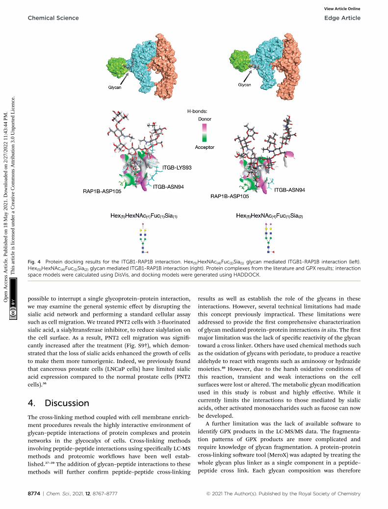
Fig. 4 Protein docking results for the ITGB1-RAP1B interaction. Hex(5)HexNAc(4)Fuc(1)Sia(1) glycan mediated ITGB1-RAP1B interaction (left).Hex(5)HexNAc(4)Fuc(1)Sia(2) glycan mediated ITGB1-RAP1B interaction (right). Protein complexes from the literature and GPX results; interactionspace models were calculated using DisVis, and docking models were generated using HADDOCK.
Chemical Science Edge Article
Ope
n A
cces
s A
rtic
le. P
ublis
hed
on 1
8 M
ay 2
021.
Dow
nloa
ded
on 2
/27/
2022
11:
43:4
4 PM
. T
his
artic
le is
lice
nsed
und
er a
Cre
ativ
e C
omm
ons
Attr
ibut
ion
3.0
Unp
orte
d L
icen
ce.
View Article Online
possible to interrupt a single glycoprotein–protein interaction,we may examine the general systemic effect by disrupting thesialic acid network and performing a standard cellular assaysuch as cell migration. We treated PNT2 cells with 3-uorinatedsialic acid, a sialyltransferase inhibitor, to reduce sialylation onthe cell surface. As a result, PNT2 cell migration was signi-cantly increased aer the treatment (Fig. S9†), which demon-strated that the loss of sialic acids enhanced the growth of cellsto make them more tumorigenic. Indeed, we previously foundthat cancerous prostate cells (LNCaP cells) have limited sialicacid expression compared to the normal prostate cells (PNT2cells).36
4. Discussion
The cross-linking method coupled with cell membrane enrich-ment procedures reveals the highly interactive environment ofglycan–peptide interactions of protein complexes and proteinnetworks in the glycocalyx of cells. Cross-linking methodsinvolving peptide–peptide interactions using specically LC-MSmethods and proteomic workows have been well estab-lished.37–39 The addition of glycan–peptide interactions to thesemethods will further conrm peptide–peptide cross-linking
8774 | Chem. Sci., 2021, 12, 8767–8777
results as well as establish the role of the glycans in theseinteractions. However, several technical limitations had madethis concept previously impractical. These limitations wereaddressed to provide the rst comprehensive characterizationof glycan mediated protein–protein interactions in situ. The rstmajor limitation was the lack of specic reactivity of the glycantoward a cross linker. Others have used chemical methods suchas the oxidation of glycans with periodate, to produce a reactivealdehyde to react with reagents such as aminooxy or hydrazidemoieties.40 However, due to the harsh oxidative conditions ofthis reaction, transient and weak interactions on the cellsurfaces were lost or altered. The metabolic glycan modicationused in this study is robust and highly effective. While itcurrently limits the interactions to those mediated by sialicacids, other activated monosaccharides such as fucose can nowbe developed.
A further limitation was the lack of available soware toidentify GPX products in the LC-MS/MS data. The fragmenta-tion patterns of GPX products are more complicated andrequire knowledge of glycan fragmentation. A protein–proteincross-linking soware tool (MeroX) was adapted by treating thewhole glycan plus linker as a single component in a peptide–peptide cross link. Each glycan composition was therefore
© 2021 The Author(s). Published by the Royal Society of Chemistry

Edge Article Chemical Science
Ope
n A
cces
s A
rtic
le. P
ublis
hed
on 1
8 M
ay 2
021.
Dow
nloa
ded
on 2
/27/
2022
11:
43:4
4 PM
. T
his
artic
le is
lice
nsed
und
er a
Cre
ativ
e C
omm
ons
Attr
ibut
ion
3.0
Unp
orte
d L
icen
ce.
View Article Online
a different cross-linker and was searched individually. Forexample, a bi-antennary monosialylated glycan (Hex(5)-HexNAc(4)Sia(1)) with composition and associated mass wasdeemed a different cross-linker from the bi-antennary glycanwith a fucose (Hex(5)HexNAc(4)Fuc(1)Sia(1)). We generally exam-ined the most abundant SiaNAzylated glycoforms expressed onthe PNT2 cell membrane. This corresponded to nine glycoformsthat were individually annotated using the MeroX soware.
The proteins identied by GPXs were all mediated by sialicacids. Thus, the majority of the target proteins were likely sialic-acid binding proteins. Indeed, a comparison of the targetproteins in this method and sialic acid binding proteins iden-tied on the cell membrane of the same cell line by proximityoxidative labeling showed at least a 50% overlap between thetwo methods.12 However, although sialic acids were the basis ofthe cross-linking reaction, the interactions could have beendriven by other glycan features present as part of the totalstructure. Thus, a terminal galactose or terminal fucose couldhave been the mediator of the glycan–peptide interaction whilethe sialic acid was merely an observer that produced the cross-link. The GPX between proteins, mediated by the glycans, couldtherefore be potentially structurally broader involving othertypes of (non-sialylated) glycan–peptide interactions. On theother hand, interactions among glycans with no sialic acid werenot represented here. However, cross-linking of activated resi-dues such as those based on fucose and N-acetylglucosaminescould be performed in the future using the methods developedfor sialic acid.
GPX has revealed the potential roles of specic glycoformsand provided additional clues regarding the large heterogeneityoen associated with glycan structures. For example, fucose isa key monosaccharide residue involved in many biologicalprocesses, most notably recognition. However, the role ofmultiple fucosylation ranging from zero to greater than four onan N-glycan is a mystery. We can at least understand thedifference between no fucose and one. The rst fucose isgenerally found as a (1,6)-link on the chitobiose core. Thecomplete loss of core fucosylation is generally lethal tohumans.41 We found that adding a core fucose increased transprotein interactions. Thus, core fucosylation is potentiallynecessary to increase trans protein interaction, while no fucosecan limit these interactions to favor cis-protein interactions.Further analysis of other proteins and other cell lines maysimilarly reveal the role of multiple fucosylation and multiplesialylation.
Sialic acids themselves are similarly important mono-saccharides and are oen found in cell membranes. The loss ofsialic acid is not lethal as some cell lines are devoid of sialic acidin their N-glycans; however, altering the overall amount of sialicacids or sialylated glycans have been correlated with diseasessuch as cancer.42 Indeed, a decrease in sialic acid glycosylationhas been associated with cancer progression. In the cell linesstudied here, suppressing sialylation of glycans increased thetumorigenic potential of the cell. Increasing the number ofsialic acids likely increases the strength of the glycan–peptideinteraction. The most striking feature of the GPX results is thehighly interactive networks of membrane proteins mediated by
© 2021 The Author(s). Published by the Royal Society of Chemistry
sialic acids and the rise of specic protein hubs. The outwardhubs are glycoproteins that simultaneously interact with a largenumber of proteins through their glycans. Thus, integrin beta-1(ITB1) and its partner integrin subunit alpha 3 are major hubproteins providing glycans that interact with the largest numberof proteins (49 and 42 distinct proteins, respectively). As theseproteins play important roles as receptors and in signaling,their high interactivity is consistent with these roles. However,proteins such as AMPN, which are known primarily to aid in thedigestion of peptides from proteins as part of the gastric andpancreatic processes appear to similarly be highly interactive.AMPN may be an important protein with other functions thatare yet to be revealed. Alternatively, the GPXmay simply identifythe peptides that were cross-linked in the process of beingdigested. Nonetheless, these interactive maps may eventuallyreveal the roles of the hubs in cells and the central roles theyplay in cellular functions.
5. Conclusion
Glycan–protein cross-linking adds a new dimension to protein–protein interactions. Traditional protein cross-linking methodsreveal polypeptide–polypeptide interactions. GPX can identifyglycan-mediated interactions that further localize the contactbetween proteins. This lls an important gap at a fortuitousperiod when glycomic methods are advancing signicantly,while the essential contributions of glycosylation in physiolog-ical events such as immune regulation and cancer developmentare being explored. Glycan–protein cross-linking provides vastinformation that can be discovered, and it opens the doortowards an exploration of pivotal functions of glycans inmediating fundamental cellular and molecular processes innative physiological conditions. The glycocalyx is indeeda highly interactive environment that can now be characterizedwith regard to the extent and nature of the interaction throughglycan–protein cross-linking methods.
Data availability
Raw mass spectrometry data are freely available and can befound on the MassIVE repository (DOI: 10.25345/C5VV5S,MSV000087442).
Author contributions
Y. X., S. C., Q. L., Y. S., and G. X. performed the experiments. Y.X., S. C., Q. L., G. X., and C. B. L. designed the study. Y. X., S. C.,M. R. A., J. R., and K. S. analyzed data. Y. X., S. C., and C. B. L.wrote the manuscript. C. B. L. supervised the study. All authorsreviewed, edited and approved the manuscript prior tosubmission.
Conflicts of interest
There are no conicts with this report.
Chem. Sci., 2021, 12, 8767–8777 | 8775

Chemical Science Edge Article
Ope
n A
cces
s A
rtic
le. P
ublis
hed
on 1
8 M
ay 2
021.
Dow
nloa
ded
on 2
/27/
2022
11:
43:4
4 PM
. T
his
artic
le is
lice
nsed
und
er a
Cre
ativ
e C
omm
ons
Attr
ibut
ion
3.0
Unp
orte
d L
icen
ce.
View Article Online
Acknowledgements
Research reported in this report was supported by GeneralMedicine of the National Institutes of Health under the awardnumbers RO1GM049077 and RO1AG062240.
References
1 J. D. Marth and P. K. Grewal, Mammalian glycosylation inimmunity, Nat. Rev. Immunol., 2008, 8(11), 874–887.
2 M. J. Kailemia, D. Park and C. B. Lebrilla, Glycans andglycoproteins as specic biomarkers for cancer, Anal.Bioanal. Chem., 2017, 409(2), 395–410.
3 L. R. Ruhaak, G. Xu, Q. Li, E. Goonatilleke and C. B. Lebrilla,Mass Spectrometry Approaches to Glycomic andGlycoproteomic Analyses, Chem. Rev., 2018, 118(17), 7886–7930.
4 X. R. Liu, M. M. Zhang and M. L. Gross, Mass Spectrometry-Based Protein Footprinting for Higher-Order StructureAnalysis: Fundamentals and Applications, Chem. Rev.,2020, 120(10), 4355–4454.
5 C. Yu and L. Huang, Cross-Linking Mass Spectrometry: AnEmerging Technology for Interactomics and StructuralBiology, Anal. Chem., 2018, 90(1), 144–165.
6 E. L. Huttlin, L. Ting, R. J. Bruckner, F. Gebreab, M. P. Gygi,J. Szpyt, S. Tam, G. Zarraga, G. Colby, K. Baltier, R. Dong,V. Guarani, L. P. Vaites, A. Ordureau, R. Rad,B. K. Erickson, M. Wuhr, J. Chick, B. Zhai,D. Kolippakkam, J. Mintseris, R. A. Obar, T. Harris,S. Artavanis-Tsakonas, M. E. Sowa, P. De Camilli,J. A. Paulo, J. W. Harper and S. P. Gygi, The BioPlexNetwork: A Systematic Exploration of the HumanInteractome, Cell, 2015, 162(2), 425–440.
7 M. M. Zhang, R. Y. C. Huang, B. R. Beno, E. G. Deyanova,J. Li, G. Chen and M. L. Gross, Epitope and ParatopeMapping of PD-1/Nivolumab by Mass Spectrometry-BasedHydrogen–Deuterium Exchange, Cross-linking, andMolecular Docking, Anal. Chem., 2020, 92(13), 9086–9094.
8 M. Cohen, Notable Aspects of Glycan-Protein Interactions,Biomolecules, 2015, 5(3), 2056–2072.
9 Y. Tanaka and J. J. Kohler, Photoactivatable CrosslinkingSugars for Capturing Glycoprotein Interactions, J. Am.Chem. Soc., 2008, 130(11), 3278–3279.
10 T. N. C. Ramya, E. Weerapana, L. Liao, Y. Zeng, H. Tateno,L. Liao, J. R. Yates, B. F. Cravatt and J. C. Paulson, In Situtrans Ligands of CD22 Identied by Glycan-ProteinPhotocross-linking-enabled Proteomics, Mol. Cell.Proteomics, 2010, 9(6), 1339.
11 S. Han, B. E. Collins, P. Bengtson and J. C. Paulson,Homomultimeric complexes of CD22 in B cells revealed byprotein-glycan cross-linking, Nat. Chem. Biol., 2005, 1(2),93–97.
12 Q. Li, Y. Xie, G. Xu and C. B. Lebrilla, Identication ofpotential sialic acid binding proteins on cell membranesby proximity chemical labeling, Chem. Sci., 2019, 10(24),6199–6209.
8776 | Chem. Sci., 2021, 12, 8767–8777
13 A. P. Frei, O.-Y. Jeon, S. Kilcher, H. Moest, L. M. Henning,C. Jost, A. Pluckthun, J. Mercer, R. Aebersold,E. M. Carreira and B. Wollscheid, Direct identication ofligand-receptor interactions on living cells and tissues,Nat. Biotechnol., 2012, 30(10), 997–1001.
14 N. Sobotzki, M. A. Schafroth, A. Rudnicka, A. Koetemann,F. Marty, S. Goetze, Y. Yamauchi, E. M. Carreira andB. Wollscheid, HATRIC-based identication of receptorsfor orphan ligands, Nat. Commun., 2018, 9(1), 1519.
15 Q. Li, Y. Xie, M. Wong, M. Barboza and C. B. Lebrilla,Comprehensive structural glycomic characterization of theglycocalyxes of cells and tissues, Nat. Protoc., 2020, 15(8),2668–2704.
16 C. Iacobucci, M. Gotze, C. H. Ihling, C. Piotrowski, C. Arlt,M. Schafer, C. Hage, R. Schmidt and A. Sinz, A cross-linking/mass spectrometry workow based on MS-cleavable cross-linkers and the MeroX soware forstudying protein structures and protein–proteininteractions, Nat. Protoc., 2018, 13(12), 2864–2889.
17 S. Madler, C. Bich, D. Touboul and R. Zenobi, Chemicalcross-linking with NHS esters: a systematic study on aminoacid reactivities, J. Mass Spectrom., 2009, 44(5), 694–706.
18 Z.-L. Chen, J.-M. Meng, Y. Cao, J.-L. Yin, R.-Q. Fang,S.-B. Fan, C. Liu, W.-F. Zeng, Y.-H. Ding, D. Tan, L. Wu,W.-J. Zhou, H. Chi, R.-X. Sun, M.-Q. Dong and S.-M. He, Ahigh-speed search engine pLink 2 with systematicevaluation for proteome-scale identication of cross-linkedpeptides, Nat. Commun., 2019, 10(1), 3404.
19 F. Liu, P. Lossl, R. Scheltema, R. Viner and A. J. R. Heck,Optimized fragmentation schemes and data analysisstrategies for proteome-wide cross-link identication, Nat.Commun., 2017, 8(1), 15473.
20 J. Z. Kechagia, J. Ivaska and P. Roca-Cusachs, Integrins asbiomechanical sensors of the microenvironment, Nat. Rev.Mol. Cell Biol., 2019, 20(8), 457–473.
21 A. Masedunskas, J. A. King, F. Tan, R. Cochran, T. Stevens,D. Sviridov and S. F. Ofori-Acquah, Activated leukocyte celladhesion molecule is a component of the endothelialjunction involved in transendothelial monocyte migration,FEBS Lett., 2006, 580(11), 2637–2645.
22 V. Saraan, M. Jadot, J.-M. Foidart, J.-J. Letesson, F. Van denBrule, V. Castronovo, R. Wattiaux and S. Wattiaux-DeConinck, Expression of Lamp-1 and Lamp-2 and theirinteractions with galectin-3 in human tumor cells, Int. J.Cancer, 1998, 75(1), 105–111.
23 J. Li, M. S. Deffieu, P. L. Lee, P. Saha and S. R. Pfeffer,Glycosylation inhibition reduces cholesterol accumulationin NPC1 protein-decient cells, Proc. Natl. Acad. Sci. U. S.A., 2015, 112(48), 14876.
24 T. A. Davis, B. Loos and A. M. Engelbrecht, AHNAK: Thegiant jack of all trades, Cell. Signalling, 2014, 26(12), 2683–2693.
25 B. Chen, J. Wang, D. Dai, Q. Zhou, X. Guo, Z. Tian, X. Huang,L. Yang, H. Tang and X. Xie, AHNAK suppresses tumourproliferation and invasion by targeting multiple pathwaysin triple-negative breast cancer, J. Exp. Clin. Cancer Res.,2017, 36(1), 65.
© 2021 The Author(s). Published by the Royal Society of Chemistry

Edge Article Chemical Science
Ope
n A
cces
s A
rtic
le. P
ublis
hed
on 1
8 M
ay 2
021.
Dow
nloa
ded
on 2
/27/
2022
11:
43:4
4 PM
. T
his
artic
le is
lice
nsed
und
er a
Cre
ativ
e C
omm
ons
Attr
ibut
ion
3.0
Unp
orte
d L
icen
ce.
View Article Online
26 F. M. Shaikh, E. C. Seales, W. C. Clem, K. M. Hennessy,Y. Zhuo and S. L. Bellis, Tumor cell migration andinvasion are regulated by expression of variant integringlycoforms, Exp. Cell Res., 2008, 314(16), 2941–2950.
27 A. Mohamed, A. D. Shah, D. Chen and M. M. Hill, RaProtV2: understanding membrane microdomain functionthrough lipid ra proteomes, Nucleic Acids Res., 2019,47(D1), D459–D463.
28 M. Takahashi, Y. Kuroki, K. Ohtsubo and N. Taniguchi, Corefucose and bisecting GlcNAc, the direct modiers of the N-glycan core: their functions and target proteins, Carbohydr.Res., 2009, 344(12), 1387–1390.
29 V. I. Otto, T. Schurpf, G. Folkers and R. D. Cummings,Sialylated Complex-type N-Glycans Enhance the SignalingActivity of Soluble Intercellular Adhesion Molecule-1 inMouse Astrocytes, J. Biol. Chem., 2004, 279(34), 35201–35209.
30 H.-Y. Yen, Y.-C. Liu, N.-Y. Chen, C.-F. Tsai, Y.-T. Wang,Y.-J. Chen, T.-L. Hsu, P.-C. Yang and C.-H. Wong, Effect ofsialylation on EGFR phosphorylation and resistance totyrosine kinase inhibition, Proc. Natl. Acad. Sci. U. S. A.,2015, 112(22), 6955.
31 H. E. Murrey, S. B. Ficarro, C. Krishnamurthy, S. E. Domino,E. C. Peters and L. C. Hsieh-Wilson, Identication of thePlasticity-Relevant Fucose-a(1�2)-Galactose Proteome fromthe Mouse Olfactory Bulb, Biochemistry, 2009, 48(30), 7261–7270.
32 M. Nagae, S. Re, E. Mihara, T. Nogi, Y. Sugita and J. Takagi,Crystal structure of a5b1 integrin ectodomain: Atomicdetails of the bronectin receptor, J. Cell Biol., 2012,197(1), 131–140.
33 G. C. P. van Zundert and A. M. J. J. Bonvin, DisVis:quantifying and visualizing accessible interaction space ofdistance-restrained biomolecular complexes,Bioinformatics, 2015, 31(19), 3222–3224.
© 2021 The Author(s). Published by the Royal Society of Chemistry
34 G. C. P. van Zundert, J. P. G. L. M. Rodrigues, M. Trellet,C. Schmitz, P. L. Kastritis, E. Karaca, A. S. J. Melquiond,M. van Dijk, S. J. de Vries and A. M. J. J. Bonvin, TheHADDOCK2.2 Web Server: User-Friendly IntegrativeModeling of Biomolecular Complexes, J. Mol. Biol., 2016,428(4), 720–725.
35 E. F. Pettersen, T. D. Goddard, C. C. Huang, G. S. Couch,D. M. Greenblatt, E. C. Meng and T. E. Ferrin, UCSFChimera—A visualization system for exploratory researchand analysis, J. Comput. Chem., 2004, 25(13), 1605–1612.
36 Y. Xie, Y. Sheng, Q. Li, S. Ju, J. Reyes and C. B. Lebrilla,Determination of the glycoprotein specicity of lectins oncell membranes through oxidative proteomics, Chem. Sci.,2020, 11, 9501–9512.
37 J. Mintseris and S. P. Gygi, High-density chemical cross-linking for modeling protein interactions, Proc. Natl. Acad.Sci. U. S. A., 2020, 117(1), 93.
38 M. Gotze, C. Iacobucci, C. H. Ihling and A. Sinz, A SimpleCross-Linking/Mass Spectrometry Workow for StudyingSystem-wide Protein Interactions, Anal. Chem., 2019,91(15), 10236–10244.
39 J. D. Chavez, C. F. Lee, A. Caudal, A. Keller, R. Tian andJ. E. Bruce, Chemical Crosslinking Mass SpectrometryAnalysis of Protein Conformations and Supercomplexes inHeart Tissue, Cell Syst., 2018, 6(1), 136–141.e5.
40 S. H. Rouhanifard, L. U. Nordstrøm, T. Zheng and P. Wu,Chemical probing of glycans in cells and organisms, Chem.Soc. Rev., 2013, 42(10), 4284–4296.
41 M. Schneider, E. Al-Shareffi and R. S. Haltiwanger, Biologicalfunctions of fucose in mammals, Glycobiology, 2017, 27(7),601–618.
42 A. Varki, Sialic acids in human health and disease, TrendsMol. Med., 2008, 14(8), 351–360.
Chem. Sci., 2021, 12, 8767–8777 | 8777


