
Glutamate Receptor-Mediated Restoration of Experience ...
14
Glutamate Receptor-Mediated Restoration of Experience-Dependent Place Field Expansion Plasticity in Aged Rats Sara N. Burke and Andrew P. Maurer Evelyn F. McKnight Brain Institute and Arizona Research Laboratories Division of Neural Systems, Memory and Aging Zhiyong Yang Arizona Research Laboratories Division of Neural Systems, Memory and Aging Zaneta Navratilova Evelyn F. McKnight Brain Institute and Arizona Research Laboratories Division of Neural Systems, Memory and Aging Carol A. Barnes Evelyn F. McKnight Brain Institute, Arizona Research Laboratories Division of Neural Systems, Memory and Aging, and University of Arizona Place fields of hippocampal pyramidal cells expand asymmetrically when adult rats repeatedly follow the same route. This behaviorally induced expression of neuronal plasticity uses an NMDA R -dependent, LTP-like mechanism and could be used by hippocampal networks to store information. Aged spatial memory-impaired rats exhibit defective experience-dependent place field expansion plasticity. One possible explanation for this aged-associated deficit is alterations in glutamatergic function. In fact, both NMDA R - and AMPA R -mediated field excitatory postsynaptic potentials in CA1 decrease with aging. The current study investigated whether modulation of either AMPA or NDMA receptor activity could restore this experience-dependent plasticity by prolonging AMPA R activity with the ampakine CX516 and modulating the NMDA R with the noncompetitive antagonist memantine. The spatial firing characteristics of multiple CA1 pyramidal cells were monitored under both treatment conditions as aged rats repeatedly traversed a circular track. Compared to the saline baseline condition, acute administration of memantine, but not CX516, reinstated experience-dependent place field expansion. Taken together, these data suggest that pharmacological manipulation of the NMDA R can improve the function of hippocampal networks critical to optimal cognition in aging. Keywords: aging, CA1, hippocampus, place cell, theta phase precession Normal aging is associated with a decline in cognitive function that can be explained by alterations in the brain structures that support these processes. The hippocampus is one region that is vulnerable to the aging process. Because spatial learning and memory require an intact hippocampus (Jarrard, 1993; Morris, Garrud, Rawlins, & O’Keefe, 1982; Sutherland, Kolb, & Wh- ishaw, 1982), it is not surprising that aged humans (Newman & Kasznaik, 2000; Wilkniss, Jones, Korol, Gold, & Manning, 1997) and other animals show deficits on tasks designed to test spatial navigation (Bach et al., 1999; Barnes, 1979; Gallagher & Rapp, 1997; Lai, Moss, Killiany, Rosene, & Herndon, 1995; Markowska et al., 1989; Rapp, Kansky, & Roberts, 1997). Therefore, the study of age-related disruptions in neuronal information processing in the hippocampus, and how normal function could be restored pharmacologically, is of considerable interest. In adult rats, neuronal recordings from the hippocampus reveal that when a rat explores an environment, pyramidal (O’Keefe & Dostrovsky, 1971) and granule (Jung & McNaughton, 1993) cells show patterned neural activity that is highly correlated with a rat’s position in space (i.e., the cells are active in distinct locations, or exhibit “place fields”). All hippocampal principal cells appear capable of expressing this form of selective activity, but in a typical experimental environment, only a fraction of cells show place fields (approximately 30%–50% of dorsal CA1 pyramidal cells; e.g., Gothard, Skaggs, Moore, & McNaughton, 1996; Muller & Kubie, 1987; M. A. Wilson & McNaughton, 1993). Although aged rats are impaired on many tasks that require an intact hippocampus, basic firing characteristics of rat CA1 pyra- midal cells such as spike amplitude, spike width, mean and max- imum firing rates, and interspike interval distributions do not Sara N. Burke, Andrew P. Maurer, and Zaneta Navratilova, Evelyn F. McKnight Brain Institute, and Arizona Research Laboratories Division of Neural Systems, Memory and Aging; Zhiyong Yang, Arizona Research Laboratories Division of Neural Systems, Memory and Aging; Carol A. Barnes, Evelyn F. McKnight Brain Institute, Arizona Research Laborato- ries Division of Neural Systems, Memory and Aging, and Departments of Psychology and Neurology, University of Arizona. We thank Stephen Cowen and Peter Lipa for assistance with the anal- ysis; Bruce L. McNaughton for helpful discussions; Gary L. Wenk for lending his pharmacology expertise; and Jie Wang, Jeri Meltzer, Frank Houston, and Kim Bohne for their technical support. In addition, we extend our deep thanks to Michelle Carroll and Luann Snyder for their adminis- trative assistance. This research was supported by McKnight Brain Research Foundation Grants AG012609 and AG010546 and the University of Arizona Under- graduate Biology Research Program. Correspondence concerning this article should be addressed to Carol A. Barnes, Evelyn F. McKnight Brain Institute, Life Sciences North Building, Room 384, University of Arizona, Tucson, AZ 85724. E-mail: [email protected] Behavioral Neuroscience Copyright 2008 by the American Psychological Association 2008, Vol. 122, No. 3, 535–548 0735-7044/08/$12.00 DOI: 10.1037/0735-7044.122.3.535 535
Transcript of Glutamate Receptor-Mediated Restoration of Experience ...

Glutamate Receptor-Mediated Restoration of Experience-Dependent PlaceField Expansion Plasticity in Aged Rats
Sara N. Burke and Andrew P. MaurerEvelyn F. McKnight Brain Institute and Arizona Research
Laboratories Division of Neural Systems, Memory and Aging
Zhiyong YangArizona Research Laboratories Division of Neural Systems,
Memory and Aging
Zaneta NavratilovaEvelyn F. McKnight Brain Institute and Arizona Research
Laboratories Division of Neural Systems, Memory and Aging
Carol A. BarnesEvelyn F. McKnight Brain Institute, Arizona Research
Laboratories Division of Neural Systems, Memory and Aging,and University of Arizona
Place fields of hippocampal pyramidal cells expand asymmetrically when adult rats repeatedly follow thesame route. This behaviorally induced expression of neuronal plasticity uses an NMDAR-dependent,LTP-like mechanism and could be used by hippocampal networks to store information. Aged spatialmemory-impaired rats exhibit defective experience-dependent place field expansion plasticity. Onepossible explanation for this aged-associated deficit is alterations in glutamatergic function. In fact, bothNMDAR- and AMPAR-mediated field excitatory postsynaptic potentials in CA1 decrease with aging. Thecurrent study investigated whether modulation of either AMPA or NDMA receptor activity could restorethis experience-dependent plasticity by prolonging AMPAR activity with the ampakine CX516 andmodulating the NMDAR with the noncompetitive antagonist memantine. The spatial firing characteristicsof multiple CA1 pyramidal cells were monitored under both treatment conditions as aged rats repeatedlytraversed a circular track. Compared to the saline baseline condition, acute administration of memantine,but not CX516, reinstated experience-dependent place field expansion. Taken together, these data suggestthat pharmacological manipulation of the NMDAR can improve the function of hippocampal networkscritical to optimal cognition in aging.
Keywords: aging, CA1, hippocampus, place cell, theta phase precession
Normal aging is associated with a decline in cognitive functionthat can be explained by alterations in the brain structures thatsupport these processes. The hippocampus is one region that isvulnerable to the aging process. Because spatial learning andmemory require an intact hippocampus (Jarrard, 1993; Morris,
Garrud, Rawlins, & O’Keefe, 1982; Sutherland, Kolb, & Wh-ishaw, 1982), it is not surprising that aged humans (Newman &Kasznaik, 2000; Wilkniss, Jones, Korol, Gold, & Manning, 1997)and other animals show deficits on tasks designed to test spatialnavigation (Bach et al., 1999; Barnes, 1979; Gallagher & Rapp,1997; Lai, Moss, Killiany, Rosene, & Herndon, 1995; Markowskaet al., 1989; Rapp, Kansky, & Roberts, 1997). Therefore, the studyof age-related disruptions in neuronal information processing inthe hippocampus, and how normal function could be restoredpharmacologically, is of considerable interest.
In adult rats, neuronal recordings from the hippocampus revealthat when a rat explores an environment, pyramidal (O’Keefe &Dostrovsky, 1971) and granule (Jung & McNaughton, 1993) cellsshow patterned neural activity that is highly correlated with a rat’sposition in space (i.e., the cells are active in distinct locations, orexhibit “place fields”). All hippocampal principal cells appearcapable of expressing this form of selective activity, but in atypical experimental environment, only a fraction of cells showplace fields (approximately 30%–50% of dorsal CA1 pyramidalcells; e.g., Gothard, Skaggs, Moore, & McNaughton, 1996; Muller& Kubie, 1987; M. A. Wilson & McNaughton, 1993).
Although aged rats are impaired on many tasks that require anintact hippocampus, basic firing characteristics of rat CA1 pyra-midal cells such as spike amplitude, spike width, mean and max-imum firing rates, and interspike interval distributions do not
Sara N. Burke, Andrew P. Maurer, and Zaneta Navratilova, Evelyn F.McKnight Brain Institute, and Arizona Research Laboratories Division ofNeural Systems, Memory and Aging; Zhiyong Yang, Arizona ResearchLaboratories Division of Neural Systems, Memory and Aging; Carol A.Barnes, Evelyn F. McKnight Brain Institute, Arizona Research Laborato-ries Division of Neural Systems, Memory and Aging, and Departments ofPsychology and Neurology, University of Arizona.
We thank Stephen Cowen and Peter Lipa for assistance with the anal-ysis; Bruce L. McNaughton for helpful discussions; Gary L. Wenk forlending his pharmacology expertise; and Jie Wang, Jeri Meltzer, FrankHouston, and Kim Bohne for their technical support. In addition, we extendour deep thanks to Michelle Carroll and Luann Snyder for their adminis-trative assistance.
This research was supported by McKnight Brain Research FoundationGrants AG012609 and AG010546 and the University of Arizona Under-graduate Biology Research Program.
Correspondence concerning this article should be addressed to Carol A.Barnes, Evelyn F. McKnight Brain Institute, Life Sciences North Building,Room 384, University of Arizona, Tucson, AZ 85724. E-mail:[email protected]
Behavioral Neuroscience Copyright 2008 by the American Psychological Association2008, Vol. 122, No. 3, 535–548 0735-7044/08/$12.00 DOI: 10.1037/0735-7044.122.3.535
535

change appreciably in advanced age (e.g., Barnes, McNaughton, &O’Keefe, 1983; Barnes, Suster, Shen, & McNaughton, 1997;Markus, Barnes, McNaughton, Gladden, & Skaggs, 1994; Mizu-mori, Lavoie, & Kalyani, 1996; Oler & Markus, 2000; Shen,Barnes, McNaughton, Skaggs, & Weaver, 1997). Moreover, on theinitial pass through a place field or during a task in which the rat’strajectory is not restricted (i.e., random foraging), the place fieldsof CA1 pyramidal cells in aged rats are just as specific as those ofyoung rats (e.g., Barnes et al., 1997; Markus et al., 1994; Mizumoriet al., 1996; Oler & Markus, 2000; Shen et al., 1997; Tanila,Shapiro, Gallagher, & Eichenbaum, 1997). In contrast, the dy-namic properties of place fields that require functional plasticityare altered in advanced age.
In young rats, CA1 pyramidal cells exhibit place fields thatexpand asymmetrically during repeated route following. Thisexperience-dependent backward expansion can be measured inseveral different ways, including a shift in the center of mass ofplace fields in the direction opposite to the rat’s trajectory (Lee,Rao, & Knierim, 2004; Mehta, Barnes, & McNaughton, 1997;Shen et al., 1997), changes in place field characteristics such asskewness (Mehta, Quirk, & Wilson, 2000), and an increase in thenumber of spikes fired within a place field (Ekstrom, Meltzer,McNaughton, & Barnes, 2001). The asymmetric expansion isconsistent with neural network models dating back to Hebb’s(1949) concept of the “phase sequence” of cell assemblies, whichhave suggested that an associative, temporally asymmetric synap-tic plasticity mechanism could serve to encode sequences or epi-sodes of experience (Hebb, 1949). The formation of such “phasesequences” allows future predictions of neural activity to be madeon the basis of current brain states and provides a means to encodelearned routes and goal locations necessary for spatial learning andmemory (Abbott & Blum, 1996; Blum & Abbott, 1996).
Interestingly, the behaviorally induced expansion of CA1 placefields resets after only a day’s absence from a familiar environ-ment (Lee et al., 2004; Mehta et al., 1997, 2000), and it isNMDAR-dependent (Ekstrom et al., 2001). The dependence ofplace field expansion on the NDMAR suggests that the phenom-enon uses an LTP-like mechanism (Ekstrom et al., 2001), and it ispresumed that uncorrelated neural activity during the interveninghours may enable the behaviorally induced increase in synapticstrength to either decay or actively depotentiate (Mehta et al.,1997, 2000).
Consistent with data showing age-related deficits in experimen-tally induced LTP (for review, see Burke & Barnes, 2006; Rosen-zweig & Barnes, 2003), the magnitude of behaviorally inducedplace field expansion and the resulting changes in the character-istics of place fields significantly decrease in aged rats (Shen et al.,1997). Because aged rats do not show robust place field expansion,it is possible that pharmacological agents that increase the induc-tion or maintenance of LTP might act to reinstate this experience-dependent plasticity phenomenon. Two agents that have beenshown to enhance in vivo LTP are ampakines and memantine.
Ampakines act as positive modulators of the glutamate AMPAreceptor to enhance and prolong AMPAR-mediated responses(e.g., Arai, Kessler, Ambros-Ingerson, et al., 1996a; Arai, Kessler,Rogers, & Lynch, 1996). One chemical in the ampakine family,CX516, is of special interest, because of its efficiency and safety(Arai, Xia, Rogers, Lynch, & Kessler, 2002). CX516 reaches thebrain approximately 5 min after intraperitoneal administration.
Peak concentration is reached at 10 min, after which CX516 ismetabolized rapidly, with a half-life in blood of approximately15–20 min (Hampson, Rogers, Lynch, & Deadwyler, 1998).CX516 has been shown to facilitate the induction of in vivo LTPin young rats when a suboptimal stimulus protocol is applied (Araiet al., 2002) and improves memory performance of young rats inseveral tasks (e.g., Granger et al., 1993; Larson et al., 1995). It hasalso been shown that CX516 facilitates old rats’ working memoryperformance in a radial maze (Davis et al., 1997) and improvesperformance of old humans on a delayed recall of nonsense syl-lables task (Lynch et al., 1997).
In contrast, memantine is a low-affinity, noncompetitive NMDAreceptor antagonist that blocks the open pore of the glutamatereceptor (Parsons et al., 1995). It is currently used therapeuticallyto reduce the cognitive impairments associated with Alzheimer’sdisease (Rogawski & Wenk, 2003). In human clinical trials, me-mantine has been shown to improve cognitive function and globalstatus significantly over a 24-week period in patients with mild tomoderate dementia (Peskind et al., 2006). In animals, memantinehas been shown to improve spatial learning in a transgenic mousemodel of Alzheimer’s disease (Minkeviciene, Banerjee, & Tanila,2004) and to increase adult rats’ retention of the memory for thelocation of the hidden escape platform on the spatial version of theMorris swim task (Barnes, Danysz, & Parsons, 1996). It is possiblethat the cognitive-enhancing properties of memantine result froman improvement in plasticity mechanisms, which are disrupted bypathology or the normal aging process. In support of this idea,memantine has been shown to increase the maintenance of exper-imentally induced LTP in adult rats (Barnes et al., 1996).
Although CX516 has been shown to enhance LTP induction andmemantine has been shown to improve LTP maintenance, neitheragent has been used to enhance behaviorally induced plasticity.The current experiments were designed to test whether the ampa-kine CX516 or memantine could reinstate experience-dependentplasticity of CA1 place fields in aged rats with known spatialmemory impairments.
Method
Subjects and Behavioral Training
Electrophysiological studies were conducted on 12 Fisher-344male rats between 24 and 30 months old. Seven were treated withCX516 (Experiment 1) and 5 were treated with memantine (Ex-periment 2). The rats were housed individually and maintained ona 12:12 light–dark cycle. Before rats were implanted with thehyperdrive recording device, all rats were screened for spatialmemory impairments and normal vision using the Morris swimtask (Morris, 1984). All animals were tested over 4 days with 6spatial trials on each day. Animals were then screened for visualability with 2 days of cued visual trials (6 trials/day) in which theescape platform was above the surface of the water but the positionof the platform changed between each trial. This procedure hasbeen described in detail previously (e.g., Barnes et al., 1996; Shen& Barnes, 1996). Rats’ performance on the swim task was ana-lyzed offline with in-house software (WMAZE, M. Williams).Because different release locations and differences in swimmingvelocity produce variability in the latency to reach the escapeplatform, a corrected integrated path length (CIPL) was calculated
536 BURKE, MAURER, YANG, NAVRATILOVA, AND BARNES

to ensure comparability of the rats’ performance across differentrelease locations (Gallagher, Burwell, & Burchinal, 1993).
During electrophysiological recordings, the animals were fooddeprived to about 85% of their ad libitum weight and trained to runon a circular track (286 cm in circumference for Experiment 1 and382 cm in circumference for Experiment 2) for food reinforce-ment. Rats ran unidirectionally in the counterclockwise direction,and all electrophysiological recordings took place during the darkphase of the rats’ light–dark cycle. Food was given at one positionon the track after the completion of each single lap, and all ratswere required to run approximately 40 laps or 30 min for eachrecording session, whichever criterion was reached first. Eachtrack running epoch was flanked by 30–40 min in which the ratrested in the same room in a pot off of the track. Thus, the activityof CA1 neurons was monitored during an initial rest session(before behavior), during track running, and during a rest sessionafter behavior. Data from the rest periods were used to assess firingstability across the entire recording session (data not shown).
Surgical Procedures
Surgery was conducted according to National Institutes ofHealth guidelines for rodents and protocols approved by the Uni-versity of Arizona IACUC. Prior to surgery, the rats were admin-istered bicillin (30,000 units im in each hind limb) to combatinfection. The rats were implanted, under isofluorane or Nembutalanesthesia, with an array of 14 separately moveable tetrode re-cording probes using a “hyperdrive” manipulator device. Thisdevice, implantation methods, and the parallel recording techniquehave been described in detail elsewhere (Gothard et al., 1996).Briefly, each hyperdrive consisted of 14 drive screws coupled bya nut to a guide cannula. Twelve guides of these cannulas con-tained tetrodes (McNaughton, O’Keefe, & Barnes, 1983b; Recce& O’Keefe, 1989), four-channel electrodes constructed by twistingtogether four strands of insulated 13-�m nichrome wire (H. P.Reid, Inc., Neptune, NJ). Two additional tetrodes with their indi-vidual wires shorted together served as an indifferent reference andan electroencephalogram (EEG) recording probe. A full turn of thescrew advanced the tetrode 318 �m. For all 12 rats, recordingswere made from the dorsal CA1 region (3.8 posterior, 2.0 lateral tobregma) and tetrode hippocampal location was verified histologi-cally. The implant was cemented in place with dental acrylicanchored by small screws. Immediately after surgery, all tetrodeswere lowered approximately 1 mm into the cortex, and rats wereorally administered 26 mg of acetaminophen (Children’s TylenolElixir, McNeil, PA) for analgesia. They also received 2.7 mg/mlacetaminophen in the drinking water for 1–3 days after surgery andoral ampicillin (Bicillin, Wyeth Laboratories, Madison, NJ) on a10 days on/10 days off regimen for the duration of the experiment.
Neurophysiology
The tetrodes were lowered after surgery into the hippocampus,allowed to stabilize for several days just above the CA1 hippocam-pal subregion, and then gradually advanced into the CA1 stratumpyramidale. The neutral reference electrode was located in or nearthe corpus callosum. The EEG probe was used to record theta fieldactivity from the vicinity of the hippocampal fissure. The fourchannels of each tetrode were attached to a 50-channel unity-gain
headstage (Neuralynx, Inc., Tucson, AZ). A multiwire cable con-nected the headstage to digitally programmable amplifiers (Neu-ralynx, Inc.). The spike signals were amplified by a factor of1,000–5,000, bandpass-filtered between 600 Hz and 6 kHz, andtransmitted to the Cheetah Data Acquisition system (Neuralynx,Inc.). Signals were digitized at 32 kHz, and events that reached apredetermined threshold were recorded for a duration of 1 ms.Spikes were sorted offline on the basis of the amplitude andprincipal components from the four tetrode channels by means ofa semiautomatic clustering algorithm (KlustaKwik, author: K. D.Harris, Rutgers–Newark). The resulting classification was cor-rected and refined manually with custom-written software(MClust, author: A. D. Redish, University of Minnesota; updatedby S. L. Cowen and D. R. Euston, University of Arizona), resultingin a spike-train time series for each of the well-isolated cells. Noattempt was made to match cells from one daily session to the next.Therefore, the numbers of recorded cells reported does not takeinto account possible recordings from the same cells on consecu-tive days; however, because the electrode positions were adjustedfrom 1 day to the next, recordings from the same cell over dayswere probably relatively infrequent. Putative pyramidal neuronswere identified by means of the standard parameters of firing rate,burstiness, and spike waveform (Ranck, 1973). The EEG wasrecorded from a separate probe that was positioned approximately0.5 mm below the CA1 pyramidal layer, near the hippocampalfissure. This is an optimal location for recording the prominent6–10 Hz oscillation that occurs during active exploration, referredto as theta activity (Vanderwolf, 1969). The theta signal wasbandpass-filtered between 1 and 300 Hz and sampled at a fre-quency of 2.4 kHz.
Several diodes were mounted on the headstage to allow positiontracking. The position of the diode array was detected by a TVcamera placed directly above the experimental apparatus and re-corded with a sampling frequency of 60 Hz. The sampling reso-lution was such that a pixel was approximately 0.3 cm.
Drug Preparation and Injection
For Experiment 1, in which the effect of ampakines onexperience-dependent plasticity was measured, CX516 (CortexPharmaceuticals, Irvine, CA) was dissolved in saline (0.9%) tomake a 35 mg/ml solution. The injection was given intraperitone-ally 10 min before the rats were placed onto the circular track torun. Each rat was administered saline or 35 mg/kg of CX516 onalternate days. This dose was selected because it has been shownthat doses of 30–35 mg of CX516 enhance short-term memory inrats performing the delayed nonmatch to sample task (Hampson etal., 1998) and facilitate old rats’ working memory performance ina radial maze (Davis et al., 1997). In Expermiment 2, memantinewas dissolved in saline to make a 15 mg/ml solution. Ten minutesafter the beginning of rest 1 and 20 min prior to track running, eachrat was injected subcutaneously with either 5 mg/kg, 10 mg/kg, 15mg/kg of memantine or 0.1 cc of saline control. The three doses ofmemantine and the saline control were given on consecutive daysin a pseudorandomized order, such that no drug dose or the salinecontrol was given on more than 2 consecutive days. In bothExperiments 1 and 2, injections were made while the rats wereattached to the head stage and spike and EEG data were beingcollected. Multiple doses of memantine were used because it has
537EXPERIENCE-DEPENDENT PLASTICITY IN AGED RATS

been shown that 5 mg/kg of memantine enhances spatial memoryin adult rats (Zoladz et al., 2006) and 10 mg/kg of memantineincreases recognition memory (Pitsikas & Sakellaridis, 2007). Inaddition, higher doses of memantine have been shown to reversecognitive dysfunction from entorhinal cortex lesions (Zajacz-kowski, Quack, & Danysz, 1996).
Analyses and Statistics
Place field diagrams were constructed by plotting the circulartrajectories of the animals on a linearized, one-dimensional scale,using a linear interpolation (Maurer, Vanrhoads, Sutherland, Lipa,& McNaughton, 2005). Place field location and size were deter-mined using the criterion that no place field could extend beyonda single cycle of theta phase precession (Maurer, Cowen, Burke,Barnes, & McNaughton, 2006). It is well documented that as a ratpasses through a CA1 neuron’s place field, the spike timing showsa systematic shift such that the spikes occur at successively earlierphases of the theta cycle until there is a complete cycle of phaseadvancement (O’Keefe & Recce, 1993; Skaggs, McNaughton,Wilson, & Barnes, 1996).
By using the phase precession definition, the start location of afield was defined as the location at which spikes fired late in thetaphase (approximately 360°) and began a cycle of precession. Theend location of the place field was determined to be the location ofspikes with the largest phase angle. This was accomplished bycreating a two-dimensional plot of occupancy normalized position(x-axis) versus phase of spikes (y-axis), as well as occupancy-normalized position versus phase density plots (Figure 1A). Toconstruct the theta phase of firing versus position plots, we as-signed each spike a nominal theta phase. Precisely, the phaseassigned to a spike at time t was 360° (t – t0)/(t1 – t0), where t0 andt1 are the times of the preceding and following peaks of the filteredreference EEG signal (Skaggs et al., 1996). The phase is always anumber between 0 and 360.
Place field boundaries were manually drawn using the thetaphase precession information by an experimenter who was blind tothe treatment condition of the recording session (see Figure 1A).Place fields that overlap with a food dish often show an abrupt haltin theta phase precession and can show considerably less than 360°of precession. For this reason, place fields that overlapped with afood dish were excluded. In addition, if a place field had two ormore clear cycles of precession that overlapped spatially (seeMaurer et al., 2006), it was excluded from the analysis. Further-more, neurons that did not fire for at least three quarters of the lapsor did not fire within the first four laps were eliminated from theanalysis. Based on these exclusion criteria, 10.8% of place fieldswere excluded from Experiment 1 (24 place fields) and 9.6% ofplace fields were excluded from Experiment 2 (98 place fields).Figure 1B shows the firing rate by position (top panel) and indi-vidual lap raster plot for a representative place field used in thecurrent analysis.
After the place field boundaries were determined, the number ofin-field spikes, place field size, and normalized center of mass shiftwere calculated for each place field by lap. Normalized center ofmass shift was calculated by finding the mean position of all spikeswithin the theta phase precession boundary and then subtractingthe center of mass of the first lap from the subsequent laps.Averages for each rat were then calculated so that all statistical
tests were conducted using the numbers of rats rather than thenumber of individual place fields. This was done so that statisticalpower was not inflated by the large number of degrees of freedomfrom the high number of cells recorded and so that rats in whichmore cells were recorded did not contribute disproportionately tothe analysis. Finally, paired-samples t tests (Experiment 1) orrepeated measures factorial analyses of variance (ANOVAs) andsimple contrasts, or Tukey’s honestly significant difference (HSD)post hoc analyses (Experiment 2) were performed using SPSSsoftware (SPSS Inc., Chicago) to assess possible drug effects.
For drug conditions that resulted in a significant difference inrunning speed (see Results), the effect of the drug on firing rate andrunning speed was examined further. To calculate the effect of drugcondition on the relationship between firing rate and running speed,we converted the position of the rat’s head in two dimensions, derivedfrom the video tracker data, to angular coordinates and projected ontoa one-dimensional line corresponding to the center of the track. Speedwas calculated by numerically differentiating the one-dimensional
Figure 1. (A) The theta phase (y-axis) by position (x-axis) plots used todetermine place field location and extent. The upper panel shows the placefield boundaries (vertical black lines) using the definition of a place field as thedistance traversed for the cell to precess no more than 360° with respect to thetheta rhythm. Each dot represents a spike, and the red dots are spikes that wereincluded in the place field. The bottom panel is the raw data presented as aphase by position density plot using a hanning window of 7 cm by 70°. Redareas indicate a higher density of spikes, and the dark areas indicate that nospikes were fired in those positions. Note that two cycles of theta phaseprecession are plotted for both panels. (B) Raster plots of the place field from(A) showing a CA1 pyramidal cell recorded in an aged rat injected with 10mg/kg of memantine. The rat was running from right to left. The top panelshows the firing rate histogram, and the bottom panel shows spikes by lap.
538 BURKE, MAURER, YANG, NAVRATILOVA, AND BARNES

position data, and for each recording session, speed occupancy dis-tributions were computed as the number of speed samples per 6 cm/sbin, with 0 speeds in the first bin. For each recorded neuron, wecomputed the number of spikes that occurred within each speedinterval. The firing rate versus speed function was computed from theratios of these two distributions. Because the animal’s average run-ning speed varied systematically with position on the track, theindividual tuning functions for pyramidal cells do not accuratelyreflect the hypothetical true firing rate versus speed function, becausefiring rate is a function of both speed and location. For example, a cellwith a place field in a region of low average speed would appear tohave a bias to fire at slower speeds; however, the average of the rateversus speed distributions over all cells should accurately reflect theensemble firing rate versus speed function. This is the desired rela-tionship and is used for the present analysis.
Results
Morris Swim Task
The mean performance of the aged rats on the spatial version ofthe Morris swim task was significantly worse than the performanceof 10 representative adult rats. Every aged rat was able to learn thevisual version of the task, however. Figure 2A shows the meanCIPL (Gallagher et al., 1993) for the 12 aged rats used in thisanalysis, compared with the CIPL of 10 young rats (9 months) thatwere tested on the Morris swim task at the same time or withinseveral weeks of the testing of the aged rats. All animals weretested over 4 days with 6 spatial trials on each day. As reportedpreviously (e.g., Barnes et al., 1997), the old rats had a signifi-cantly longer CIPL, compared with the young rats, on both Days3 and 4, F(7, 68) � 10.78, p � .001 (ANOVA), for both compar-isons. In fact, all but 1 aged rat had a CIPL that was greater than2 standard deviations from the mean CIPL score of the young ratson Day 4, not allowing for a separation of these animals intoage-impaired and age-unimpaired groups. The old rats, however,did show a modest but significant improvement in finding thehidden platform between Day 1 and Day 4, F(1, 11) � 6.34, p �.05 (repeated measures ANOVA). This improvement did not sig-nificantly differ between the animals that were administered me-mantine and the animals that were given CX516, F(1, 11) � 0.16,p � .7.
To test whether the spatial impairments of the aged rats weredue to visual problems, the aged rats also performed the visualversion of the Morris swim task in which the platform was raisedabove the surface of the water for 2 days and 6 visual trials wereperformed on each day. Figure 2B shows the CIPL for the visualtrials of the Morris swim task for the trials on Day 1 and Day 2 forthe old rats and control young animals. The aged rats showed asignificant decrease in the CIPL on the 2nd day compared with the1st day, t(9) � 4.03, p � .01 (paired-samples t test). In addition,the performance of the old rats used in the current experiments wasnot significantly different from young animals on Day 2 of thevisual trials ( p � .68, Tukey’s HSD). These data indicate that agedrats used for electrophysiological recordings were able to use thevisual cues to swim to the platform.
Finally, the aged rats used in Experiment 1 and the aged rats usedin Experiment 2 showed similar degrees of spatial impairment. Figure2C shows the mean CIPL values for the six aged rats used in
Experiment 1 compared with the five aged rats used in Experiment 2.Performance between the two groups of rats was not significantlydifferent, F(7, 28) � 0.5, p � .8, suggesting that the two groups of ratsshowed similar amounts of hippocampal dysfunction.
Experiment 1: CX516
Single-unit firing characteristics and running speed. The ac-tivity of 524 pyramidal cells in the CA1 region of seven aged ratswas recorded in this experiment. A summary of the database fromwhich the following results are derived is given in Table 1.
Figure 2. (A) The mean and standard error of the mean (SEM) for thecorrected integrated path length (CIPL) of the 12 old rats used in thecurrent study (diagonal bars) and 10 young rats (clear bars) on the spatialversion of the Morris swim task. The old rats were significantly impairedat learning the location of the hidden platform. (B) The CIPL of the young(clear bars) and old (diagonal bars) rats for the visual version of the watermaze in which the escape platform was no longer hidden below the surfaceof the water. The CIPL was significantly shorter on Day 2, indicating thatthe old rats were able to use the visual cues to find the platform. Inaddition, on Day 2, the mean behavioral score did not differ significantlybetween the young and aged rats. Error bars indicate � 1 SEM. (C) Themean and SEM for CIPL values of the aged rats separated by Experiment1 (light gray) versus Experiment 2 (dark gray). The aged rats in Experi-ments 1 and 2 show similar degrees of spatial impairment.
539EXPERIENCE-DEPENDENT PLASTICITY IN AGED RATS

Table 2 shows the effect of CX516 on the single-unit firingcharacteristics of neurons with place fields and the rats’ runningspeed compared with the saline control. There was no significantdifference between the running speed, firing rate, or in-field firingrate when rats were injected with CX516 versus saline.
CX516 and experience-dependent place field expansion plastic-ity. To assess the effects of CX516 on the place field character-istics over time, we identified place fields on the basis of theneuron’s spike timing relative to the theta rhythm, as described inthe Method section. Once place fields were identified, the effect ofmultiple traversals through a cell’s place field was assessed bymeasuring the number of spikes that occurred in a single placefield for laps 1–20 individually for all of the drug conditions. Thismeasure of experience-dependent place field change was used, inaddition to other measures, because it is less dependent on runningspeed. Specifically, the relationship between running speed andfiring rate tends to keep the number of spikes in a place fieldconstant even when the animal’s speed changes. Moreover, it hasbeen shown previously that, as the place field expands, the numberof spikes also increases (Ekstrom et al., 2001). Firing rate by lapwas not used because firing rate is proportional to running speedand the significantly faster running speeds on the 1st lap wouldconfound these results.
Figure 3A shows the mean number of spikes within a place fieldfor CX516 and the saline control for the 1st through the 20th laps.Overall, for the seven rats, the mean number of spikes that oc-curred during a single pass through a place field was not signifi-cantly different between the CX516 and the saline control condi-tions, t(6) � 0.35, p � .74 (paired-samples t test). Figure 3B showsthe difference in the number of in-field spikes between lap 1 andlap 20 for the saline and CX516 conditions. The number of spikesdid not increase between lap 1 and lap 20 for either the salinecontrol or CX516 treatment, F(1, 10) � 1.14, p � .31 (repeated
measures ANOVA). Moreover, in both cases the increase in spikenumber was not significantly different from zero.
In addition to spike number, the measures of place field size andcenter of mass shift were also used to assess whether CX516 couldrestore experience-dependent place field expansion plasticity.Place field boundaries were determined as described in the Methodsection (see Figure 1B), and then place field size for each lap wasmeasured by calculating the difference between the location of thefirst spike and the last spike within the theta phase precessionboundary. Figure 4A shows the mean size of the place fields forsaline and CX516 across laps 1 through 20. In contrast to theanalysis of spike number by lap, there was a significant increase inplace field size between the 1st and the 20th lap, F(1, 10) � 6.74,p � .05 (repeated measures ANOVA), but the interaction betweenlap (1 vs. 20) and drug condition (saline vs. CX516) was not
Figure 3. (A) The mean and standard error of the mean (SEM) of numberof spikes averaged for each rat across laps 1 through 20 for the salinecontrol (black) and 35 mg/kg of the ampakine CX516 (gray). The numberof spikes was similar between the saline and the CX516 conditions. (B)The difference in the mean number of spikes for lap 20 minus lap 1 forsaline (black) and CX516 (gray). The increase in the number of spikesbetween lap 1 and lap 20 was similar between the ampakine and the salineconditions and not significantly different from zero for both saline andCX516. Error bars indicate � 1 SEM.
Table 1Summary of the Database Used in Experiment 1
Rat
Saline CX516 35 mg/kg
Cells Place fields Cells Place fields
6978 72 23 36 136981 31 16 56 207009 14 8 30 117091 41 9 42 147096 45 15 16 87097 17 6 12 67144 47 18 65 30
Total 267 95 257 102
Table 2The Effect of CX516 on the Single-Unit Firing Characteristics and Running Speed
Variable SalineCX516
35 mg/kg t(33) p
Mean firing rate for all behavior (Hz) 0.64 � 0.14 0.61 � 0.06 0.24 .81Mean in-field firing rate (Hz) 23.69 � 3.41 21.41 � 2.34 0.56 .58Mean running speed (cm/s) 8.10 � 0.32 9.05 � 0.48 1.64 .11
Note. Data are the means � 1 SEM.
540 BURKE, MAURER, YANG, NAVRATILOVA, AND BARNES

significant, F(1, 10) � 0.20, p � .67 (repeated measuresANOVA). This suggests that CX516 did not increase place fieldsize beyond that observed in the saline control condition. Figure4B shows the mean change in the size of place fields between laps1 and 20 for both drug conditions. Overall, the mean size of placefields was similar between saline and CX516 conditions, t(5) �0.42, p � .69 (paired-samples t test).
As a final measure of place field expansion, normalized centerof mass shift was calculated by finding the mean position of allspikes within the theta phase precession boundary and then sub-tracting the center of mass of the 1st lap from the subsequent laps.Figure 5A shows the normalized center of mass shift across laps 1through 20. There was no significant effect of drug condition oncenter of mass shift in these old rats, t(5) � 2.0, p � .1 (paired-samples t test). Figure 5B shows that the mean center of mass shiftfor saline and CX516 conditions was similar.
Experiment 2: Memantine
Single-unit firing characteristics and running speed. The ac-tivity of 1995 pyramidal cells in the CA1 region of five aged ratswas recorded. A summary of the database from which the follow-ing results are derived is given in Table 3.
On different days, the same rats were injected with saline, or 5mg/kg, 10 mg/kg, or 15 mg/kg of memantine. Table 4 shows theeffects of the three different doses of memantine on the single-unitfiring characteristics of neurons with place fields and the rats’running speed. Overall, the effect of drug condition on firing rate
during track running was significant, F(3, 44) � 6.00, p � .01(ANOVA). The 10 mg/kg and 15 mg/kg doses of memantinesignificantly increased the overall mean firing rate of pyramidalcells during the entire behavioral epoch compared with the salinecontrol condition ( p � .01 for both comparisons; simple contrast),whereas the 5 mg/kg dose had no effect. None of the doses ofmemantine, however, had a significant effect on the mean in-fieldfiring rate, F(3, 44) � 1.59, p � .21 (ANOVA). Memantine alsosignificantly increased the mean running speed of the aged rats,F(3, 43) � 4.13, p � .05 (ANOVA) but only at the 15 mg/kg dose( p � .05), whereas the 5 mg/kg ( p � .97) and 10 mg/kg ( p � .98)doses had no significant effect on running speed. This is consistentwith a previous report, which showed that systemic injections ofhigh doses of memantine can increase locomotor activity (Bubser,Keseberg, Notz, & Schmidt, 1992).
Because the firing rate of hippocampal neurons has been shownto be proportionally related to an animal’s running speed (Czurko,Hirase, Csicsvari, & Buzsaki, 1999; Maurer et al., 2005; Mc-Naughton, Barnes, & O’Keefe, 1983a; Shen et al., 1997), therelationship between running speed on the track and firing rate wasinvestigated. Figure 6A shows the effects of memantine on themean running speed for the first 20 laps. For all drug conditionsand the saline control, rats had the fastest running speeds on thefirst lap and then slowed their running speed during subsequentlaps, F(19,860) � 2.33, p � .01 (ANOVA, deviation contrast).This is distinct from the observation in young rats that runningspeed is similar over 20 laps after saline injection but runningspeed increases over laps 1–10 after administration of the NMDAreceptor antagonist CPP (Ekstrom et al., 2001). As shown previ-
Figure 4. (A) The effects of ampakines on average place field size acrosslaps 1 through 20 for the saline control (black) and 35 mg/kg of CX516(gray). Both conditions showed a similar increase in place field size. (B)The average change in place field size for the saline control (black) and 35mg/kg of CX516 (gray) between lap 1 and lap 20. Error bars indicate � 1standard error of the mean (SEM).
Figure 5. (A) The mean and standard error of the mean (SEM) fornormalized center of mass (COM) shift of place fields from laps 1 to 20 forthe saline control (black) and 35 mg/kg of CX516 (gray). (B) The averagenormalized COM shift over all laps for the saline control (black) andCX516 (gray). The COM shift was similar between the two drug and thecontrol conditions. Error bars indicate � 1 SEM.
541EXPERIENCE-DEPENDENT PLASTICITY IN AGED RATS

ously in young (Czurko et al., 1999; Maurer et al., 2005; Mc-Naughton et al., 1983a) and aged rats (Shen et al., 1997), there wasan overall effect of running speed on pyramidal cell firing rate.Figure 6B shows the firing rate by running speed for all drugconditions. Similar to the previous observations, there is an ap-proximate linear increase in firing rate with increasing runningspeeds up to 35 cm/s (Czurko et al., 1999; Maurer et al., 2005;Shen et al., 1997). This effect was consistent across all drugconditions, F(8, 10288) � 21.84, p � .001 (ANOVA). Moreover,there was not a significant interaction between running speed anddrug condition, F(24, 10288) � 1.46, p � .07 (ANOVA), indicat-ing that firing rate was modulated by running speed similarly forthe control condition and the three doses of memantine. At runningspeeds faster than 35 cm/s, there was increasing variability in thefiring rate, which is likely due to the small sample size of fasterrunning speeds in the aged animals.
Memantine and experience-dependent place field expansionplasticity. To assess the effects of the three doses of memantineon the place field characteristics over time, we identified placefields on the basis of the neuron’s spike timing relative to the thetarhythm as in Experiment 1 (see Method section). Again, threemeasures of place field expansion plasticity were used: spikenumber, place field size, and center of mass.
Figure 7A shows the mean number of spikes within a place fieldfor the three doses of memantine and the saline control for laps1–20. There was a significant effect of drug condition on the meannumber of within field spikes over all 20 laps, F(3, 376) � 8.94,p � .001 (ANOVA), with both the 5 mg/kg and 10 mg/kg doses ofmemantine leading to an increase in the number of in-field spikescompared with the saline control ( p � .05, for all comparisons;simple contrast). Figure 7B shows the difference in the number ofspikes between lap 1 and lap 20 for all four drug conditions. Therewas a significant effect of drug condition on the increase in spikes
between lap 1 and lap 20, F(3, 9) � 25.46, p � .01 (repeatedmeasures ANOVA), with both the 5 mg/kg and the 10 mg/kg dosesresulting in a significant increase in spikes compared with thesaline control ( p � .05 for both comparisons; simple contrast).
A linear regression indicated that there was a significant in-crease in spikes over the first 20 laps, for the 5 mg/kg dose ofmemantine, slope � 0.64 � 0.12 spikes/lap, F(1, 98) � 27.98, p �.001; the 10 mg/kg dose, slope � 0.46 � 0.12 spikes/lap, F(1,98) � 13.85, p � .001; and the 15 mg/kg dose, slope � 0.40 �0.11 spikes/lap, F(1, 98) � 14.63, p � .001. The r coefficient wasnot significant for the control condition, indicating that the spikenumber did not continue to increase linearly over the first 20 lapswhen memantine was not administered. In fact, Figure 7A showsthat the largest increase in spike number for the control conditionwas between the first and the second lap. In contrast, the effect ofmemantine on spike number in the aged rats is similar to what hasbeen observed in young rats with normal place field expansionplasticity (Ekstrom et al., 2001).
In addition to spike number, the measures of place field size andcenter of mass shift were used to assess the effects of memantineon experience-dependent place field expansion plasticity. Placefield size was measured as described in Experiment 1 (see Figure1B). Figure 8A shows the mean size of the place fields for eachdrug condition across laps 1 through 20. When place field size wasanalyzed across all laps, there was a significant effect of drugcondition, F(3, 376) � 14.40, p � .001 (repeated measuresANOVA), with all doses of memantine resulting in significantlylarger place fields when compared with the saline control condi-tion ( p � .001 for all comparisons; simple contrast). Figure 8Bshows the change in the mean size of place fields between lap 1and lap 20 for all drug conditions. There was a significant effect ofdrug condition on the increase in place field size between laps 1and 20, F(3, 9) � 7.78, p � .05 (repeated measures ANOVA),
Table 3Summary of the Database Used in Experiment 2
Rat
Saline 5 mg/kg Memantine 10 mg/kg Memantine 15 mg/kg Memantine
Cells Place fields Cells Place fields Cells Place fields Cells Place fields
8092 68 30 50 17 67 22 NA NA8116 92 41 88 48 82 46 60 358117 134 69 195 83 215 102 191 1088118 82 31 123 59 68 28 109 398237 115 58 108 40 68 30 80 40Total 491 229 564 247 500 228 440 222
Table 4The Effect of Memantine on the Single-Unit Firing Characteristics and Running Speed
Variable Saline5 mg/kg
Memantine10 mg/kg
Memantine15 mg/kg
Memantine F(3, 44) p
Mean firing rate for allbehavior (Hz) 1.04 � 0.07 1.05 � 0.06 1.34* � 0.08 1.32* � 0.04 6.00 �.01
Mean in-field firing rate (Hz) 18.41 � 0.96 17.60 � 1.48 18.62 � 1.96 22.27 � 2.00 1.59 .21Mean running speed (cm/s) 4.12 � 0.68 3.59 � 0.54 4.63 � 0.74 7.33* � 0.46 4.13 �.05
* The mean is significantly different from the saline control condition at p � .05.Data are the means � 1 SEM.
542 BURKE, MAURER, YANG, NAVRATILOVA, AND BARNES

with the 5 mg/kg and the 10 mg/kg doses of memantine resultingin larger place fields compared with the saline control condition( p � .05 for all comparisons; simple contrast).
To estimate over how many laps the size of place fieldscontinued to expand, we fit the relationship between lap numberand place field size with a third-order polynomial and wecalculated the lap at which the first derivative was equal to zerofor each dose of memantine. This analysis revealed that for the5 mg/kg dose of memantine, dynamic place field expansionoccurred until lap 10. For the 10 mg/kg and 15 mg/kg doses, theincrease in place field size stopped at lap 9. This is similar towhat has been observed in young rats, where place fieldsdynamically expand with experience over approximately 10laps (estimate from Mehta et al., 1997).
Figure 9A shows the normalized center of mass shift for eachlap through lap 20. Across laps 1 through 20, there was a signif-icant effect of drug condition on mean center of mass shift, F(3,237) � 58.69, p � .001 (repeated measures ANOVA), with alldoses of memantine leading to a significant increase in the centerof mass shift ( p � .05 for all comparisons; simple contrast). Figure10B shows the mean center of mass shift for all drug conditions.
Because of the observation that memantine appeared to re-instate place field expansion in aged rats, we conducted a final
Figure 6. (A) The mean running speed by lap for laps 1 through 20 forthe saline control (black), 5 mg/kg (red), 10 mg/kg (yellow), and 15 mg/kg(blue) doses of memantine. For all four conditions, the rats ran fastest onthe 1st lap and then speeds were consistent for the subsequent 19 laps. (B)The relationship between running speed and firing rate for all doses ofmemantine and the saline control. Firing rate increased similarly for allconditions for speeds up to 35 cm/s. Error bars indicate � 1 standard errorof the mean (SEM).
Figure 7. (A) The mean and standard error of the mean (SEM) fornumber of spikes across laps 1 through 20. (B) The difference in the meannumber of spikes within place fields between lap 1 and lap 20 for the salinecontrol (black), 5 mg/kg (red), 10 mg/kg (yellow), and 15 mg/kg (blue)doses of memantine on lap 1 and lap 20. For all four conditions, there wasa significant increase in spike number, but this increase was largest for the5 mg/kg and 10 mg/kg doses of memantine. Error bars indicate � 1 SEM.
Figure 8. (A) The effects of memantine on place field size across laps 1through 20 for the saline control (black), 5 mg/kg (red), 10 mg/kg (yellow),and 15 mg/kg (blue) doses of memantine. All four conditions showed anincrease in place field size, but the increase was larger for all memantineconditions. (B) The average increase in the size of place fields between lap 1and lap 20 for the saline control (blue), 5 mg/kg (red), 10 mg/kg (green), and15 mg/kg (yellow) doses of memantine. Place fields were significantly largerwhen the rats had been given any dose of memantine compared with the salinecontrol. Error bars indicate � 1 standard error of the mean (SEM).
543EXPERIENCE-DEPENDENT PLASTICITY IN AGED RATS
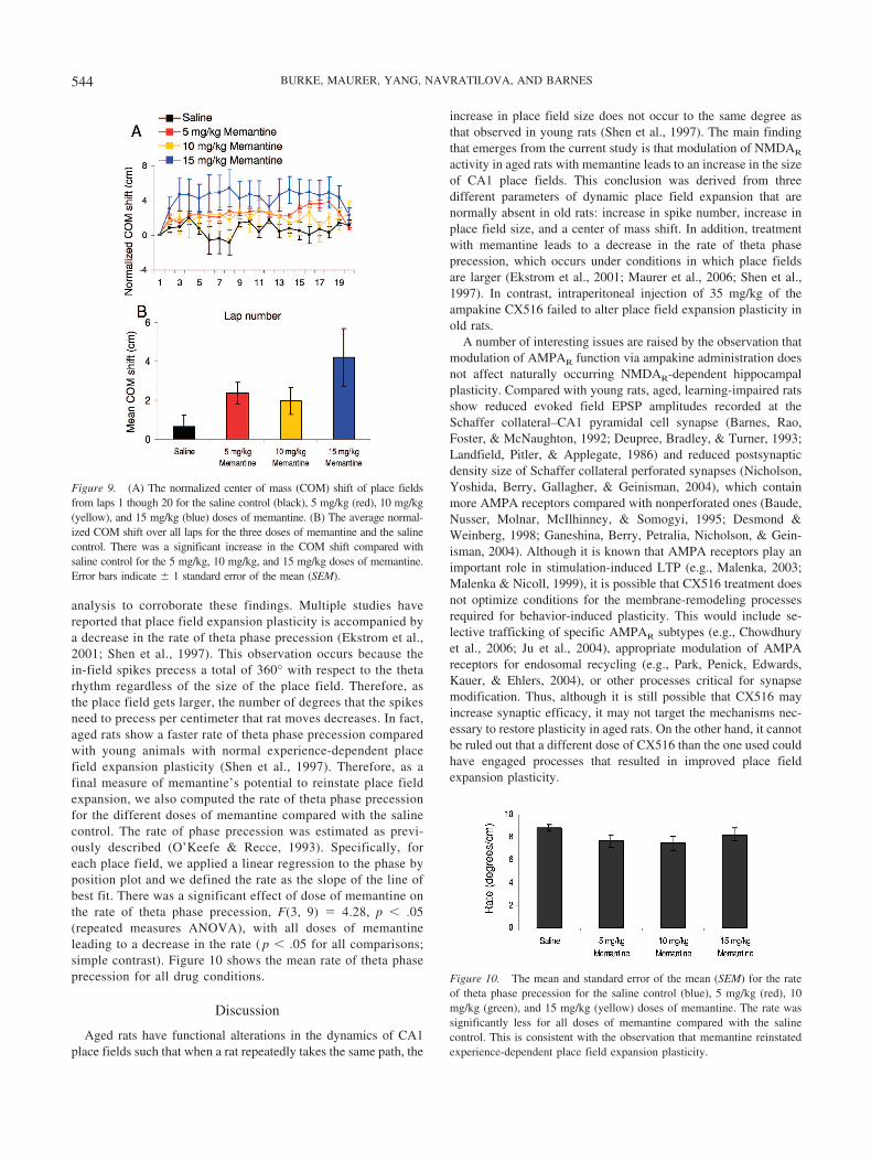
analysis to corroborate these findings. Multiple studies havereported that place field expansion plasticity is accompanied bya decrease in the rate of theta phase precession (Ekstrom et al.,2001; Shen et al., 1997). This observation occurs because thein-field spikes precess a total of 360° with respect to the thetarhythm regardless of the size of the place field. Therefore, asthe place field gets larger, the number of degrees that the spikesneed to precess per centimeter that rat moves decreases. In fact,aged rats show a faster rate of theta phase precession comparedwith young animals with normal experience-dependent placefield expansion plasticity (Shen et al., 1997). Therefore, as afinal measure of memantine’s potential to reinstate place fieldexpansion, we also computed the rate of theta phase precessionfor the different doses of memantine compared with the salinecontrol. The rate of phase precession was estimated as previ-ously described (O’Keefe & Recce, 1993). Specifically, foreach place field, we applied a linear regression to the phase byposition plot and we defined the rate as the slope of the line ofbest fit. There was a significant effect of dose of memantine onthe rate of theta phase precession, F(3, 9) � 4.28, p � .05(repeated measures ANOVA), with all doses of memantineleading to a decrease in the rate ( p � .05 for all comparisons;simple contrast). Figure 10 shows the mean rate of theta phaseprecession for all drug conditions.
Discussion
Aged rats have functional alterations in the dynamics of CA1place fields such that when a rat repeatedly takes the same path, the
increase in place field size does not occur to the same degree asthat observed in young rats (Shen et al., 1997). The main findingthat emerges from the current study is that modulation of NMDAR
activity in aged rats with memantine leads to an increase in the sizeof CA1 place fields. This conclusion was derived from threedifferent parameters of dynamic place field expansion that arenormally absent in old rats: increase in spike number, increase inplace field size, and a center of mass shift. In addition, treatmentwith memantine leads to a decrease in the rate of theta phaseprecession, which occurs under conditions in which place fieldsare larger (Ekstrom et al., 2001; Maurer et al., 2006; Shen et al.,1997). In contrast, intraperitoneal injection of 35 mg/kg of theampakine CX516 failed to alter place field expansion plasticity inold rats.
A number of interesting issues are raised by the observation thatmodulation of AMPAR function via ampakine administration doesnot affect naturally occurring NMDAR-dependent hippocampalplasticity. Compared with young rats, aged, learning-impaired ratsshow reduced evoked field EPSP amplitudes recorded at theSchaffer collateral–CA1 pyramidal cell synapse (Barnes, Rao,Foster, & McNaughton, 1992; Deupree, Bradley, & Turner, 1993;Landfield, Pitler, & Applegate, 1986) and reduced postsynapticdensity size of Schaffer collateral perforated synapses (Nicholson,Yoshida, Berry, Gallagher, & Geinisman, 2004), which containmore AMPA receptors compared with nonperforated ones (Baude,Nusser, Molnar, McIlhinney, & Somogyi, 1995; Desmond &Weinberg, 1998; Ganeshina, Berry, Petralia, Nicholson, & Gein-isman, 2004). Although it is known that AMPA receptors play animportant role in stimulation-induced LTP (e.g., Malenka, 2003;Malenka & Nicoll, 1999), it is possible that CX516 treatment doesnot optimize conditions for the membrane-remodeling processesrequired for behavior-induced plasticity. This would include se-lective trafficking of specific AMPAR subtypes (e.g., Chowdhuryet al., 2006; Ju et al., 2004), appropriate modulation of AMPAreceptors for endosomal recycling (e.g., Park, Penick, Edwards,Kauer, & Ehlers, 2004), or other processes critical for synapsemodification. Thus, although it is still possible that CX516 mayincrease synaptic efficacy, it may not target the mechanisms nec-essary to restore plasticity in aged rats. On the other hand, it cannotbe ruled out that a different dose of CX516 than the one used couldhave engaged processes that resulted in improved place fieldexpansion plasticity.
Figure 9. (A) The normalized center of mass (COM) shift of place fieldsfrom laps 1 though 20 for the saline control (black), 5 mg/kg (red), 10 mg/kg(yellow), and 15 mg/kg (blue) doses of memantine. (B) The average normal-ized COM shift over all laps for the three doses of memantine and the salinecontrol. There was a significant increase in the COM shift compared withsaline control for the 5 mg/kg, 10 mg/kg, and 15 mg/kg doses of memantine.Error bars indicate � 1 standard error of the mean (SEM).
Figure 10. The mean and standard error of the mean (SEM) for the rateof theta phase precession for the saline control (blue), 5 mg/kg (red), 10mg/kg (green), and 15 mg/kg (yellow) doses of memantine. The rate wassignificantly less for all doses of memantine compared with the salinecontrol. This is consistent with the observation that memantine reinstatedexperience-dependent place field expansion plasticity.
544 BURKE, MAURER, YANG, NAVRATILOVA, AND BARNES

Efficacy of the Doses of Memantine and CX516
In the current study, all three doses of memantine led to someincrease in place field size compared with the saline controlcondition. This is not surprising considering that injections of 5mg/kg and 7.5 mg/kg of memantine have been shown to im-prove spatial memory (Zoladz et al., 2006). Moreover, injec-tions of 10 mg/kg and 20 mg/kg of memantine improve recog-nition memory in adult rats (Pitsikas & Sakellaridis, 2007), and20 mg/kg of memantine has been shown to facilitate spontane-ous object recognition in aged rats (Pieta Dias et al., 2007).These data suggest that memantine may be effective at improv-ing behaviorally induced plasticity at doses ranging from 5mg/kg to 20 mg/kg.
In contrast, at a dose of 35 mg/kg, CX516 had no effect on placefield expansion plasticity. Although CX516 did not alter placefield dynamics, it is likely that the drug did affect AMPAR-mediated synaptic transmission in the aged animals at this dose.There are several reasons for this assertion. The range of thebehaviorally effective dose of CX516 is 12–50 mg/kg (UrsulaStaubli, personal communication, 2001), and a midlevel dose (35mg/kg) was used in the present study. In addition, 30 mg/kgCX516 has been shown to affect spatial working memory perfor-mance in 19-month-old Long–Evens rats (Davis et al., 1997).Furthermore, AMPAR-binding sites in the CA1 region do notdecline with age (Wenk & Barnes, 2000), nor do the numbers ofsynaptic contacts decline in the stratum radiatum (Geinisman et al.,2004). Although there is a reduction (approximately 50%) inAMPA binding in the dentate gyrus, this occurs between the age of3 and 12 months and is stable thereafter (Wenk & Barnes, 2000).Thus, it is unlikely that a lack of AMPAR target sites can explainthese results. More important, intraperitoneal injection of 35mg/kg CX516 has been shown to alter the performance of old ratsof the same age and strain in the radial-arm maze task, suggestingthat the drug is active centrally at this dose (Yang, Houston, &Barnes, 2003).
Possible Mechanism for Memantine’s Reinstatement ofExperience-Dependent Place Field Expansion Plasticity inAged Rats
Given that the NMDA antagonist CPP blocks place field expan-sion, it seems somewhat paradoxical that a different NMDAR
antagonist could facilitate plasticity. In contrast to other NMDAreceptor antagonists, however, memantine has fast offset kineticsand relatively strong functional voltage dependency because of itspositive charge. This allows memantine to rapidly leave theNMDA channel on transient physiological activation by glutamate(for review, see Parsons, Stoffler, & Danysz, 2007). Thus, me-mantine may act to increase signal-to-noise ratios under conditionsin which normal synaptic transmission has been disrupted. In oldanimals, this may be particularly important as it has been observedthat aged animals show disrupted Ca2� regulation in CA1 of thehippocampus, which has been associated with age-related impair-ments in plasticity (e.g., Foster & Norris, 1997; Landfield, 1988).Moreover, this Ca2� dysregulation and the related plasticity def-icits in old animals could be responsible for the decline in behav-iorally induced place field expansion plasticity.
In young animals, rapid behaviorally induced potentiation offeedforward synapses from CA3 to CA1 has been hypothesized to
mediate the experience-dependent expansion of CA1 place fields(Mehta et al., 2000). During the first pass through a place field, adorsal CA1 neuron may inherit its place-specific firing from neu-rons in layer III of the medial entorhinal cortex (Brun et al., 2002),and input to the CA1 neuron from CA3 would be relativelysymmetric. After repeated traversals, in which both directionallyselective CA3 and CA1 neurons are activated, a spike-timingdependent plasticity mechanism might be expected to strengthensynapses from those CA3 neurons that had a place field just beforethat of a CA1 neuron (Lee et al., 2004; Mehta et al., 2000). Thetheoretically predicted and empirically verified result is a back-ward, asymmetric shift of CA1 place fields, resulting in largerplace fields, a center of mass shift, and an increase in spikenumber.
If behaviorally induced plasticity of the CA3 to CA1 Schaffercollateral synapses is the primary mechanism for place field ex-pansion, then it may not be surprising that aged rats fail to showthis phenomenon under normal conditions given that a number ofaged-associated changes occur at this synapse. For example, agedCA1 pyramidal cells have increased Ca2� conductances due to ahigher density of L-type Ca2� channels (Thibault & Landfield,1996). As stated previously, this may lead to disruptions in Ca2�
homeostasis (for review, see Toescu, Verkhratsky, & Landfield,2004) that ultimately contribute to age-related plasticity deficits(Foster & Norris, 1997; Landfield, 1988). In addition, at the CA3to CA1 Schaffer collateral synapse, aged rats show impairments inboth the induction and maintenance of experimentally inducedLTP (for review, see Burke & Barnes, 2006; Rosenzweig &Barnes, 2003). Also, the Schaffer collateral synapses of aged ratsare more susceptible to LTD (Norris, Korol, & Foster, 1996) andto the reversal of LTP (Foster & Norris, 1997). Thus, an overallshift in the synaptic modification window of CA3 to CA1 Schaffercollateral synapses in aged rats could account for the decline inexperience-dependent place field expansion plasticity. This declinemay then be reinstated via a modification of the Ca2� influxthrough the NMDAR by memantine.
The increase in experience-dependent place field expansionplasticity by memantine administration may also be related tochanges within CA3 neurons. In addition to the observed age-related effects on the Schaffer collateral synapse, a number ofage-associated changes occur within the CA3 network (for review,see I. A. Wilson, Gallagher, Eichenbaum, & Tanila, 2006), includ-ing an increase in place field firing rate and rigidity (I. A. Wilson,Gallagher, Eichenbaum, & Tanila, 2005). Although the placefields of young CA3 pyramidal neurons do exhibit expansion (Leeet al., 2004), this phenomenon has not been investigated in agedanimals. Thus, it is possible that memantine affects the firingpatterns of aged CA3 neurons, and this may lead to restoration ofplasticity. Alternatively, memantine may affect both cell types,resulting in restored behaviorally induced plasticity.
An additional mechanism for the observed improvement inplace field expansion plasticity involves the affinity of memantinefor the �7* nicotinic acetylcholine receptor (nAChR). The �7*nAChR is present on hippocampal neurons and is also a Ca2�-conducting receptor. In cultured hippocampal neurons, memantinehas been shown to reduce Ca2� conductance through the �7*nAChR (Aracava, Pereira, Maelicke, & Albuquerque, 2005). Thus,memantine could also act to restore normal plasticity mechanisms
545EXPERIENCE-DEPENDENT PLASTICITY IN AGED RATS

in aged neurons by reducing the amount of Ca2� that enters theneurons through both the NMDA and the �7* nACh receptors.
Memantine also led to an increase in running speed but only atthe highest dose (15 mg/kg), and both the medium and high dosesled to an increase in firing rate. It is unlikely that memantine’sreinstatement of behaviorally induced place field expansion is dueto its effect on running speed or firing rate. First, measures ofexperience-dependent place field expansion were used that arerobust against changes in running speed, such as the number ofspikes within a place field and the center of mass shift. Moreover,there was evidence of place field expansion reinstatement at boththe low and medium doses, and these doses had no effect on theanimal’s running speed. In addition, the low dose of memantinealso led to a reinstatement of place expansion but did not affectoverall firing rate. Finally, the effects of the medium and highdoses of memantine on firing rate occurred only at running speedsfaster than 35 cm/s (see Figure 6B), and the aged rats did not oftenrun at speeds this fast.
Dynamic Place Field Expansion: Reinstatement in OldRats Compared With Young Rats
In young rats, repeated route-following behavior leads to a20%–40% increase in place size between the 1st pass through aplace field and the 20th pass (estimated from Ekstrom et al., 2001;Shen et al., 1997). In the dorsal CA1 subregion of the hippocam-pus, this translates to approximately a 6–7 cm increase in placefield size (estimated from Ekstrom et al., 2001; Shen et al., 1997).In the current study, between lap 1 and lap 20, the low, medium,and high doses of memantine resulted in a 6.6 cm (26%), 6.2 cm(24%), and 5.7 cm (22%) increase in place field size, respectively.Thus, the magnitude of experience-dependent place field expan-sion plasticity is at least comparable between young rats and agedrats that have been administered the noncompetitive NMDA re-ceptor antagonist memantine.
The current findings suggest a means to therapeutically treatnormal age-associated neurobiological changes in plasticity mech-anisms. By adjusting calcium influx via the NMDA receptor,memantine may alter the probability that synaptic weights of agedrats are adjusted over a dynamic range more similar to that ofyoung animals.
References
Abbott, L. F., & Blum, K. I. (1996). Functional significance of long-termpotentiation for sequence learning and prediction. Cerebral Cortex, 6,406–416.
Aracava, Y., Pereira, E. F., Maelicke, A., & Albuquerque, E. X. (2005).Memantine blocks alpha7* nicotinic acetylcholine receptors more po-tently than n-methyl-D-aspartate receptors in rat hippocampal neurons.Journal of Pharmacology and Experimental Therapeutics, 312, 1195–1205.
Arai, A., Kessler, M., Ambros-Ingerson, J., Quan, A., Yigiter, E., Rogers,G., et al. (1996). Effects of a centrally active benzoylpyrrolidine drug onAMPA receptor kinetics. Neuroscience, 75, 573–585.
Arai, A., Kessler, M., Rogers, G., & Lynch, G. (1996). Effects of amemory-enhancing drug on DL-alpha-amino-3-hydroxy-5-methyl-4-isoxazolepropionic acid receptor currents and synaptic transmission inhippocampus. Journal of Pharmacology and Experimental Therapeu-tics, 278, 627–638.
Arai, A. C., Xia, Y. F., Rogers, G., Lynch, G., & Kessler, M. (2002).Benzamide-type AMPA receptor modulators form two subfamilies withdistinct modes of action. Journal of Pharmacology and ExperimentalTherapeutics, 303, 1075–1085.
Bach, M. E., Barad, M., Son, H., Zhuo, M., Lu, Y. F., Shih, R., et al.(1999). Age-related defects in spatial memory are correlated with defectsin the late phase of hippocampal long-term potentiation in vitro and areattenuated by drugs that enhance the cAMP signaling pathway. Proceed-ings of the National Academy of Sciences of the United States ofAmerica, 96, 5280–5285.
Barnes, C. A. (1979). Memory deficits associated with senescence: Aneurophysiological and behavioral study in the rat. Journal of Compar-ative and Physiological Psychology, 93, 74–104.
Barnes, C. A., Danysz, W., & Parsons, C. G. (1996). Effects of theuncompetitive NMDA receptor antagonist memantine on hippocampallong-term potentiation, short-term exploratory modulation and spatialmemory in awake, freely moving rats. European Journal of Neuro-science, 8, 565–571.
Barnes, C. A., McNaughton, B. L., & O’Keefe, J. (1983). Loss of placespecificity in hippocampal complex spike cells of senescent rat. Neuro-biology of Aging, 4, 113–119.
Barnes, C. A., Rao, G., Foster, T. C., & McNaughton, B. L. (1992).Region-specific age effects on AMPA sensitivity: Electrophysiologicalevidence for loss of synaptic contacts in hippocampal field CA1. Hip-pocampus, 2, 457–468.
Barnes, C. A., Suster, M. S., Shen, J., & McNaughton, B. L. (1997, July17). Multistability of cognitive maps in the hippocampus of old rats.Nature, 388, 272–275.
Baude, A., Nusser, Z., Molnar, E., McIlhinney, R. A., & Somogyi, P.(1995). High-resolution immunogold localization of AMPA type gluta-mate receptor subunits at synaptic and non-synaptic sites in rat hip-pocampus. Neuroscience, 69, 1031–1055.
Blum, K. I., & Abbott, L. F. (1996). A model of spatial map formation inthe hippocampus of the rat. Neural Computation, 8, 85–93.
Brun, V. H., Otnass, M. K., Molden, S., Steffenach, H. A., Witter, M. P.,Moser, M. B., & Moser, E. I. (2002, June 21). Place cells and placerecognition maintained by direct entorhinal-hippocampal circuitry. Sci-ence, 296, 2243–2246.
Bubser, M., Keseberg, U., Notz, P. K., & Schmidt, W. J. (1992). Differ-ential behavioural and neurochemical effects of competitive and non-competitive NMDA receptor antagonists in rats. European Journal ofPharmacology, 229, 75–82.
Burke, S. N., & Barnes, C. A. (2006). Neural plasticity in the ageing brain.Nature Reviews Neuroscience, 7, 30–40.
Chowdhury, S., Shepherd, J. D., Okuno, H., Lyford, G., Petralia, R. S.,Plath, N., et al. (2006). Arc/Arg3.1 interacts with the endocytic machin-ery to regulate AMPA receptor trafficking. Neuron, 52, 445–459.
Czurko, A., Hirase, H., Csicsvari, J., & Buzsaki, G. (1999). Sustainedactivation of hippocampal pyramidal cells by ”space clamping” in arunning wheel. European Journal of Neuroscience, 11, 344–352.
Davis, C. M., Moskovitz, B., Nguyen, M. A., Tran, B. B., Arai, A., Lynch,G., & Granger, R. (1997). A profile of the behavioral changes producedby facilitation of AMPA-type glutamate receptors, Psychopharmacology(Berl), 133, 161–167.
Desmond, N. L., & Weinberg, R. J. (1998). Enhanced expression of AMPAreceptor protein at perforated axospinous synapses. NeuroReport, 9,857–860.
Deupree, D. L., Bradley, J., & Turner, D. A. (1993). Age-related alterationsin potentiation in the CA1 region in F344 rats. Neurobiology of Aging,14, 249–258.
Ekstrom, A. D., Meltzer, J., McNaughton, B. L., & Barnes, C. A. (2001).NMDA receptor antagonism blocks experience-dependent expansion ofhippocampal “place fields.” Neuron, 31, 631–638.
Foster, T. C., & Norris, C. M. (1997). Age-associated changes in Ca(2�)-
546 BURKE, MAURER, YANG, NAVRATILOVA, AND BARNES

dependent processes: Relation to hippocampal synaptic plasticity. Hip-pocampus, 7, 602–612.
Gallagher, M., Burwell, R., & Burchinal, M. (1993). Severity of spatiallearning impairment in aging: Development of a learning index forperformance in the Morris water maze. Behavioral Neuroscience, 107,618–626.
Gallagher, M., & Rapp, P. R. (1997). The use of animal models to study theeffects of aging on cognition. Annual Review of Psychology, 48, 339–370.
Ganeshina, O., Berry, R. W., Petralia, R. S., Nicholson, D. A., & Geinis-man, Y. (2004). Differences in the expression of AMPA and NMDAreceptors between axospinous perforated and nonperforated synapses arerelated to the configuration and size of postsynaptic densities. Journal ofComparative Neurology, 468, 86–95.
Geinisman, Y., Ganeshina, O., Yoshida, R., Berry, R. W., Disterhoft, J. F.,& Gallagher, M. (2004). Aging, spatial learning, and total synapsenumber in the rat CA1 stratum radiatum, Neurobiology of Aging, 25,407–416.
Gothard, K. M., Skaggs, W. E., Moore, K. M., & McNaughton, B. L.(1996). Binding of hippocampal CA1 neural activity to multiple refer-ence frames in a landmark-based navigation task. Journal of Neuro-science, 16, 823–835.
Granger, R., Staubli, U., Davis, M., Perez, Y., Nilsson, L., Rogers, G. A.,et al. (1993). A drug that facilitates glutamatergic transmission reducesexploratory activity and improves performance in a learning-dependenttask. Synapse, 15, 326–329.
Hampson, R. E., Rogers, G., Lynch, G., & Deadwyler, S. A. (1998).Facilitative effects of the ampakine CX516 on short-term memory inrats: Enhancement of delayed-nonmatch-to-sample performance. Jour-nal of Neuroscience, 18, 2740–2747.
Hebb, D. (1949). The organization of behavior: A neurophysiologicaltheory. New York: Wiley.
Jarrard, L. E. (1993). On the role of the hippocampus in learning andmemory in the rat. Behavioral and Neural Biology, 60, 9–26.
Ju, W., Morishita, W., Tsui, J., Gaietta, G., Deerinck, T. J., Adams, S. R.,et al. (2004). Activity-dependent regulation of dendritic synthesis andtrafficking of AMPA receptors. Nature Neuroscience, 7, 244–253.
Jung, M. W., & McNaughton, B. L. (1993). Spatial selectivity of unitactivity in the hippocampal granular layer. Hippocampus, 3, 165–182.
Lai, Z. C., Moss, M. B., Killiany, R. J., Rosene, D. L., & Herndon, J. G.(1995). Executive system dysfunction in the aged monkey: Spatial andobject reversal learning. Neurobiology of Aging, 16, 947–954.
Landfield, P. W. (1988). Hippocampal neurobiological mechanisms ofage-related memory dysfunction. Neurobiology of Aging, 9, 571–579.
Landfield, P. W., Pitler, T. A., & Applegate, M. D. (1986). The effects ofhigh Mg2�-to-Ca2� ratios on frequency potentiation in hippocampalslices of young and aged rats. Journal of Neurophysiology, 56, 797–811.
Larson, J., Lieu, T., Petchpradub, V., LeDuc, B., Ngo, H., Rogers, G. A.,et al. (1995). Facilitation of olfactory learning by a modulator of AMPAreceptors. Journal of Neuroscience, 15, 8023–8030.
Lee, I., Rao, G., & Knierim, J. J. (2004). A double dissociation betweenhippocampal subfields: Differential time course of CA3 and CA1 placecells for processing changed environments. Neuron, 42, 803–815.
Lynch, G., Granger, R., Ambros-Ingerson, J., Davis, C. M., Kessler, M., &Schehr, R. (1997). Evidence that a positive modulator of AMPA-typeglutamate receptors improves delayed recall in aged humans. Experi-mental Neurology, 145, 89–92.
Malenka, R. C. (2003). Synaptic plasticity and AMPA receptor trafficking.Annals of the New York Academy of Sciences, 1003, 1–11.
Malenka, R. C., & Nicoll, R. A. (1999, September 17). Long-term poten-tiation—A decade of progress? Science, 285, 1870–1874.
Markowska, A. L., Stone, W. S., Ingram, D. K., Reynolds, J., Gold, P. E.,Conti, L. H., et al. (1989). Individual differences in aging: Behavioraland neurobiological correlates. Neurobiology of Aging, 10, 31–43.
Markus, E. J., Barnes, C. A., McNaughton, B. L., Gladden, V. L., &Skaggs, W. E. (1994). Spatial information content and reliability ofhippocampal CA1 neurons: Effects of visual input. Hippocampus, 4,410–421.
Maurer, A. P., Cowen, S. L., Burke, S. N., Barnes, C. A., & McNaughton,B. L. (2006). Organization of hippocampal cell assemblies based ontheta phase precession. Hippocampus, 16, 785–794.
Maurer, A. P., Vanrhoads, S. R., Sutherland, G. R., Lipa, P., & McNaugh-ton, B. L. (2005). Self-motion and the origin of differential spatialscaling along the septo-temporal axis of the hippocampus. Hippocam-pus, 15, 841–852.
McNaughton, B. L., Barnes, C. A., & O’Keefe, J. (1983a). The contribu-tions of position, direction, and velocity to single unit activity in thehippocampus of freely moving rats. Experimental Brain Research, 52,41–49.
McNaughton, B. L., O’Keefe, J., & Barnes, C. A. (1983b). The stereotrode:A new technique for simultaneous isolation of several single units in thecentral nervous system from multiple unit records. Journal of Neuro-science Methods, 8, 391–397.
Mehta, M. R., Barnes, C. A., & McNaughton, B. L. (1997). Experience-dependent, asymmetric expansion of hippocampal place fields. Proceed-ings of the National Academy of Sciences of the United States ofAmerica, 94, 8918–8921.
Mehta, M. R., Quirk, M. C., & Wilson, M. A. (2000). Experience-dependent asymmetric shape of hippocampal receptive fields. Neuron,25, 707–715.
Minkeviciene, R., Banerjee, P., & Tanila, H. (2004). Memantine improvesspatial learning in a transgenic mouse model of Alzheimer’s disease.Journal of Pharmacology and Experimental Therapeutics, 311, 677–682.
Mizumori, S. J., Lavoie, A. M., & Kalyani, A. (1996). Redistribution ofspatial representation in the hippocampus of aged rats performing aspatial memory task. Behavioral Neuroscience, 110, 1006–1016.
Morris, R. (1984). Developments of a water-maze procedure for studyingspatial learning in the rat. Journal of Neuroscience Methods, 11, 47–60.
Morris, R. G., Garrud, P., Rawlins, J. N., & O’Keefe, J. (1982, June 24).Place navigation impaired in rats with hippocampal lesions. Nature, 297,681–683.
Muller, R. U., & Kubie, J. L. (1987). The effects of changes in theenvironment on the spatial firing of hippocampal complex-spike cells.Journal of Neuroscience, 7, 1951–1968.
Newman, M., & Kasznaik, A. (2000). Spatial memory and aging: Perfor-mance on a human analog of the Morris water maze. Aging, Neuropsy-chology and Cognition, 7, 86–93.
Nicholson, D. A., Yoshida, R., Berry, R. W., Gallagher, M., & Geinisman,Y. (2004). Reduction in size of perforated postsynaptic densities inhippocampal axospinous synapses and age-related spatial learning im-pairments. Journal of Neuroscience, 24, 7648–7653.
Norris, C. M., Korol, D. L., & Foster, T. C. (1996). Increased susceptibilityto induction of long-term depression and long-term potentiation reversalduring aging. Journal of Neuroscience, 16, 5382–5392.
O’Keefe, J., & Dostrovsky, J. (1971). The hippocampus as a spatial map.Preliminary evidence from unit activity in the freely moving rat. BrainResearch, 34, 171–175.
O’Keefe, J., & Recce, M. L. (1993). Phase relationship between hippocam-pal place units and the EEG theta rhythm. Hippocampus, 3, 317–330.
Oler, J. A., & Markus, E. J. (2000). Age-related deficits in the ability toencode contextual change: A place cell analysis. Hippocampus, 10,338–350.
Park, M., Penick, E. C., Edwards, J. G., Kauer, J. A., & Ehlers, M. D.(2004, September 24). Recycling endosomes supply AMPA receptorsfor LTP. Science, 305, 1972–1975.
Parsons, C. G., Quack, G., Bresink, I., Baran, L., Przegalinski, E., Kos-towski, W., et al. (1995). Comparison of the potency, kinetics and
547EXPERIENCE-DEPENDENT PLASTICITY IN AGED RATS

voltage-dependency of a series of uncompetitive NMDA receptor an-tagonists in vitro with anticonvulsive and motor impairment activity invivo. Neuropharmacology, 34, 1239–1258.
Parsons, C. G., Stoffler, A., & Danysz, W. (2007). Memantine: A NMDAreceptor antagonist that improves memory by restoration of homeostasisin the glutamatergic system—Too little activation is bad, too much iseven worse. Neuropharmacology, 53, 699–723.
Peskind, E. R., Potkin, S. G., Pomara, N., Ott, B. R., Graham, S. M., Olin,J. T., et al. (2006). Memantine treatment in mild to moderate Alzheimerdisease: A 24-week randomized, controlled trial. American Journal ofGeriatric Psychiatry, 14, 704–715.
Pieta Dias, C., Martins de Lima, M. N., Presti-Torres, J., Dornelles, A.,Garcia, V. A., Siciliani Scalco, F., et al. (2007). Memantine reducesoxidative damage and enhances long-term recognition memory in agedrats. Neuroscience, 146, 1719–1725.
Pitsikas, N., & Sakellaridis, N. (2007). Memantine and recognition mem-ory: Possible facilitation of its behavioral effects by the nitric oxide(NO) donor molsidomine. European Journal of Pharmacology, 571,174–179.
Ranck, J. B., Jr. (1973). Studies on single neurons in dorsal hippocampalformation and septum in unrestrained rats: I. Behavioral correlates andfiring repertoires. Experimental Neurology, 41, 461–531.
Rapp, P. R., Kansky, M. T., & Roberts, J. A. (1997). Impaired spatialinformation processing in aged monkeys with preserved recognitionmemory. NeuroReport, 8, 1923–1928.
Recce, M. L., & O’Keefe, J. (1989). The tetrode: An improved techniquefor multiunit extracellular recording. Society for Neuroscience Abstracts,15, 1250.
Rogawski, M. A., & Wenk, G. L. (2003). The neuropharmacological basisfor the use of memantine in the treatment of Alzheimer’s disease. CNSDrug Review, 9, 275–308.
Rosenzweig, E. S., & Barnes, C. A. (2003). Impact of aging on hippocam-pal function: Plasticity, network dynamics, and cognition. Progress inNeurobiology, 69, 143–179.
Shen, J., & Barnes, C. A. (1996). Age-related decrease in cholinergicsynaptic transmission in three hippocampal subfields. Neurobiology ofAging, 17, 439–451.
Shen, J., Barnes, C. A., McNaughton, B. L., Skaggs, W. E., & Weaver,K. L. (1997). The effect of aging on experience-dependent plasticity ofhippocampal place cells. Journal of Neuroscience, 17, 6769–6782.
Skaggs, W. E., McNaughton, B. L., Wilson, M. A., & Barnes, C. A. (1996).Theta phase precession in hippocampal neuronal populations and thecompression of temporal sequences. Hippocampus, 6, 149–172.
Sutherland, R. J., Kolb, B., & Whishaw, I. Q. (1982). Spatial mapping:
Definitive disruption by hippocampal or medial frontal cortical damagein the rat. Neuroscience Letters, 31, 271–276.
Tanila, H., Shapiro, M., Gallagher, M., & Eichenbaum, H. (1997). Brainaging: Changes in the nature of information coding by the hippocampus.Journal of Neuroscience, 17, 5155–5166.
Thibault, O., & Landfield, P. W. (1996, May 17). Increase in single L-typecalcium channels in hippocampal neurons during aging. Science, 272,1017–1020.
Toescu, E. C., Verkhratsky, A., & Landfield, P. W. (2004). Ca2� regula-tion and gene expression in normal brain aging. Trends in Neuro-sciences, 27, 614–620.
Vanderwolf, C. H. (1969). Hippocampal electrical activity and voluntarymovement in the rat. Electroencephalography and Clinical Neurophys-iology, 26, 407–418.
Wenk, G. L., & Barnes, C. A. (2000). Regional changes in the hippocampaldensity of AMPA and NMDA receptors across the lifespan of the rat.Brain Research, 885, 1–5.
Wilkniss, S. M., Jones, M. G., Korol, D. L., Gold, P. E., & Manning, C. A.(1997). Age-related differences in an ecologically based study of routelearning. Psychology and Aging, 12, 372–375.
Wilson, I. A., Gallagher, M., Eichenbaum, H., & Tanila, H. (2005).Age-associated alterations in place cells are subregion specific. Journalof Neuroscience, 25, 6877–6886.
Wilson, I. A., Gallagher, M., Eichenbaum, H., & Tanila, H. (2006).Neurocognitive aging: Prior memories hinder new hippocampal encod-ing. Trends in Neurosciences, 29, 662–670.
Wilson, M. A., & McNaughton, B. L. (1993, August 20). Dynamics of thehippocampal ensemble code for space. Science, 261, 1055–1058.
Yang, Z., Houston, F. P., & Barnes, C. A. (2003, November). Effects ofampakines on spatial learning and memory in aged rats. Paper presentedat the Society for Neuroscience, New Orleans.
Zajaczkowski, W., Quack, G., & Danysz, W. (1996). Infusion of (�)-MK-801 and memantine—Contrasting effects on radial maze learningin rats with entorhinal cortex lesion. European Journal of Pharmacol-ogy, 296, 239–246.
Zoladz, P. R., Campbell, A. M., Park, C. R., Schaefer, D., Danysz, W., &Diamond, D. M. (2006). Enhancement of long-term spatial memory inadult rats by the noncompetitive NMDA receptor antagonists, meman-tine and neramexane. Pharmacology, Biochemistry and Behavior, 85,298–306.
Received November 19, 2007Revision received January 8, 2008
Accepted January 24, 2008 �
548 BURKE, MAURER, YANG, NAVRATILOVA, AND BARNES


