
GLP-1 Active Kit - Meso Scale/media/files/product inserts/v... · 2020-03-17 · GLP-1R is...
29
FOR RESEARCH USE ONLY. NOT FOR USE IN DIAGNOSTIC OR THERAPEUTIC PROCEDURES. 17379-v1-2007Mar GLP-1 Active Kit V-PLEX ® V-PLEX Plus GLP-1 Active K1503OD K1503OG
Transcript of GLP-1 Active Kit - Meso Scale/media/files/product inserts/v... · 2020-03-17 · GLP-1R is...
FOR RESEARCH USE ONLY. NOT FOR USE IN DIAGNOSTIC OR THERAPEUTIC PROCEDURES.
17379-v1-2007Mar
GLP-1 Active Kit
V-PLEX® V-PLEX Plus
GLP-1 Active K1503OD K1503OG
18192-v3-2020Feb | 2
MSD Metabolic Assays
GLP-1 Active Kit For use with human, NHP, mouse, rat, and canine plasma and cell culture supernatants.
This package insert must be read in its entirety before using this product.
FOR RESEARCH USE ONLY.
NOT FOR USE IN DIAGNOSTIC PROCEDURES.
MESO SCALE DISCOVERY® A division of Meso Scale Diagnostics, LLC. 1601 Research Blvd. Rockville, MD 20850 USA www.mesoscale.com
MESO SCALE DISCOVERY, MESO SCALE DIAGNOSTICS, MSD, mesoscale.com, www.mesoscale.com, methodicalmind.com, www.methodicalmind.com, DISCOVERY WORKBENCH, MESO, MesoSphere, Methodical Mind, MSD GOLD, MULTI-ARRAY, MULTI-SPOT, QuickPlex, ProductLink, SECTOR, SECTOR PR, SECTOR HTS, SULFO-TAG, TeamLink, TrueSensitivity, TURBO-BOOST, TURBO-TAG, N-PLEX, R-PLEX, S-PLEX, T-PLEX, U-PLEX, V-PLEX, MSD (design), MSD (luminous design), Methodical Mind (design), 96 WELL SMALL-SPOT (design), 96 WELL 1-, 4-, 7-, 9-, & 10-SPOT (designs), 384 WELL 1- & 4-SPOT (designs), N-PLEX (design), R-PLEX (design), S-PLEX (design), T-PLEX (design), U-PLEX (design), V-PLEX (design), It’s All About U, SPOT THE DIFFERENCE, The Biomarker Company, and The Methodical Mind Experience are trademarks and/or service marks owned by or licensed to Meso Scale Diagnostics, LLC. All other trademarks and service marks are the property of their respective owners.
©2018, 2020 Meso Scale Diagnostics, LLC. All rights reserved.
18192-v3-2020Feb | 3
Table of Contents Introduction ..................................................................................................................................................... 4 Principle of the Assay ....................................................................................................................................... 6 Kit Components ................................................................................................................................................ 7 Additional Materials and Equipment .................................................................................................................... 8 Optional Materials and Equipment ....................................................................................................................... 8 Safety ............................................................................................................................................................. 8 Best Practices .................................................................................................................................................. 9 Reagent Preparation ....................................................................................................................................... 10 Protocol ........................................................................................................................................................ 13 Validation ...................................................................................................................................................... 14 Analysis of Results ......................................................................................................................................... 16 Typical Data .................................................................................................................................................. 16 Sensitivity ...................................................................................................................................................... 17 Precision ....................................................................................................................................................... 18 Dilution Linearity ............................................................................................................................................ 19 Spike Recovery .............................................................................................................................................. 20 Specificity ..................................................................................................................................................... 21 Stability......................................................................................................................................................... 22 Calibration ..................................................................................................................................................... 22 Tested Samples ............................................................................................................................................. 23 Assay Components ......................................................................................................................................... 25 References .................................................................................................................................................... 25 Appendix A .................................................................................................................................................... 26 Summary Protocol .......................................................................................................................................... 27 Catalog Numbers ............................................................................................................................................ 28 Plate Diagram ................................................................................................................................................ 29
Contact Information MSD Customer Service Phone: 1-240-314-2795 Fax: 1-301-990-2776 Email: [email protected]
MSD Scientific Support Phone: 1-240-314-2798 Fax: 1-240-632-2219 attn: Scientific Support Email: [email protected]
18192-v3-2020Feb | 4
Introduction MSD offers V-PLEX assays for customers who require unsurpassed performance and quality. V-PLEX products are developed under
rigorous design control and are analytically validated according to fit-for-purpose principles in accordance with MSD’s Quality
Management System. They offer exceptional sensitivity, simple protocols, reproducible results, and lot-to-lot consistency. In
addition to the analytical validation, robustness of the assay protocol is assessed during development along with the stability of the
assay components and kits. V-PLEX assays are available in both single-assay and multiplex formats.
The V-PLEX assay menu is organized by panels. Grouping the assays into panels by species, analytical compatibility, clinical range,
and expected use, ensures optimal and consistent performance from each assay while still providing the benefits and efficiencies
of multiplexing. V-PLEX panels are provided in MSD’s MULTI-SPOT® 96-well plate format. The composition of each panel and the
location of each assay (i.e., its spot within the well) are maintained from lot to lot. Most individual V-PLEX assays are provided on
MSD’s single-spot, 96-well plates.
Glucagon like peptide-1 (GLP-1) is a peptide hormone that belongs to the incretin (enhances the meal-stimulated release of
insulin) family. It is synthesized by the enteroendocrine L-cells in the gut (ileum / colon) and neurons of the brain stem /
hypothalamus. GLP-1 is formed by the post-translational processing of proglucagon (160 amino acids) by prohormone convertase
1/3 (PC-1/3). The circulating bioactive forms GLP-1 (7–36) amide (MW – 3.29 kD) and GLP-1 (7–37) (MW – 3.35 kD) are further
generated from GLP-1(1–37). The primary amino acid sequence of GLP-1 is conserved among the mammalian species, i.e. human,
mouse, rat, monkey, canine, etc.1 Inter-species differences with respect to the dominant molecular forms of GLP-1 have been
documented.5,9 In humans and mice, the majority of the active circulating form is the 7-36 amide.2
Both of these active forms of GLP-1 are rapidly degraded by dipeptidyl peptidase-4 (DPP-IV) to their corresponding inactive forms
GLP-1 (9–36) amide and GLP-1 (9–37) following their release from the intestine (half-life 1-5 minutes).1-5 This makes it critical that
blood samples are collected in the presence of a DPP-IV inhibitor as well as broad spectrum protease inhibitors such as aprotinin
when evaluating the active forms of GLP-1.6-8 More recently, neutral endopeptidase NEP 24.11 (also known as neprilysin, CD10,
and CALA antigen) has been identified to be involved in GLP-1 degradation. The major degradation product derived from NEP 24.11
activity is a nonapeptide GLP-1 (28–36) amide. NEP 24.11 is predominantly a membrane-associated enzyme that is expressed in
the central nervous system, hepatocytes, bile canaliculi, and other organs10,11.
Actions of GLP-1 are mediated by a specific receptor (GLP-1R) which belongs to the G protein coupled receptor (GPCR) family.1-2
GLP-1R is expressed in the pancreas, stomach, GI tract, brain, heart, kidney, and lung. However, the expression of the GLP-1R in
other tissues such as liver, muscle, and fat cells is controversial.12
GLP-1 plays an important role in glucose homeostasis via several mechanisms. Plasma concentration of active GLP-1 rises quickly
following ingestion of food (carbohydrates, fat, and protein all stimulate GLP-1 secretion). In the pancreas, GLP-1 stimulates the
secretion of insulin by β cells and suppresses secretion of glucagon by α cells in a glucose-dependent manner. In addition to the
“incretin effect,” it protects β-cell function, i.e. it stimulates proliferation and inhibits apoptosis of pancreatic β cells.1-3 Actions of
GLP-1 also lead to delayed gastric and small bowel emptying, resulting in delayed nutrient absorption. Sustained GLP-1R signaling
reduces appetite, leading to reduced food intake and weight loss.1-3
GLP-1 is also produced in the CNS, and peripheral GLP-1 has been shown to cross the blood brain barrier. In the brain, GLP-1R is
expressed in regions that control glucose homeostasis, gut motility, food intake, and cardiovascular function. It has been reported
that GLP-1R signaling exerts neuroprotective and neurotrophic effects, with possibilities for the treatment of neurodegenerative
diseases such as Alzheimer’s disease.3
18192-v3-2020Feb | 5
Protective effects of GLP-1R agonists on the myocardium and the vascular endothelium have been demonstrated. Recent studies
indicate that inflammatory stimuli such as the presence of LPS increase GLP-1 secretion and GLP-1, in turn, modulates
inflammation in multiple sites, including the heart and blood vessels. Preliminary results of several cardiovascular studies suggest
that some GLP-1R agonists significantly reduce the risk of major cardiovascular complications.13-14 Benefits of GLP-1 therapy in
rodent models of diabetic nephropathy and acute kidney injury have been demonstrated. Protection is thought to be mediated by
inhibition of renal inflammation and oxidative stress.15 The therapeutic potential of the GLP-1 pathway is thus not limited to type 2
diabetes mellitus (T2DM) and is now being explored in neurodegenerative, cardiovascular, and renal diseases.13,15,16-18
Although the inactive GLP-1 (9–36) amide form exhibits very weak incretin effect through the GLP-1R, it suppresses hepatic glucose
production, exerts cardioprotective actions, and reduces oxidative stress in vascular tissues. These insulin-like effects of GLP-1 (9–
36) amide suggest that it has a role in the modulation of mitochondrial functions by mechanisms independent of the GLP-1 receptor.
A dual receptor hypothesis for GLP-1 has thus been proposed. Following removal of the two amino acids by DPP-IV, the GLP-1 (9–
36) amide is translocated to the cytoplasm via a receptor where it is further degraded by endopeptidases into smaller peptides of
5–7 amino acids. These smaller peptides then modulate metabolic processes (oxidative phosphorylation involving fatty acid and
glucose metabolism, energy expenditure) and apoptotic pathways within the mitochondria10,11.

18192-v3-2020Feb | 6
Principle of the Assay MSD metabolic assays provide a rapid and convenient method for measuring the levels of protein targets within a single, small-
volume sample. The V-PLEX GLP-1 Active assay is a single analyte sandwich immunoassay. MSD provides a Small Spot plate pre-
coated with the capture antibody (Figure 1). The user sequentially adds the calibrator / sample and a solution containing the
detection antibody conjugated with electrochemiluminescent (ECL) labels (MSD SULFO-TAG™) over the course of two incubation
periods separated by wash steps. The active form of GLP-1, (7–36) amide, in the calibrator or sample binds to the capture antibody
immobilized on the working electrode surface and the detection antibody completes the sandwich. The user then adds the MSD
read buffer that creates the appropriate chemical environment for the ECL reaction and loads the plate into an MSD instrument
where a voltage applied to the plate electrodes causes the captured labels to emit light. The instrument measures the intensity of
emitted light to provide a quantitative measure of analytes in the sample. V-PLEX assay kits have been validated according to the
principles outlined in “Fit-for-Purpose Method Development and Validation for Successful Biomarker Measurement” by J. W. Lee,
et al.19
Figure 1. Small Spot plate diagram showing placement of analyte capture antibodies.
Figure 2. Schematic of the antibody recognition sites for the V-PLEX GLP-1 Active assay on GLP-1 protein amino acids.
18192-v3-2020Feb | 7
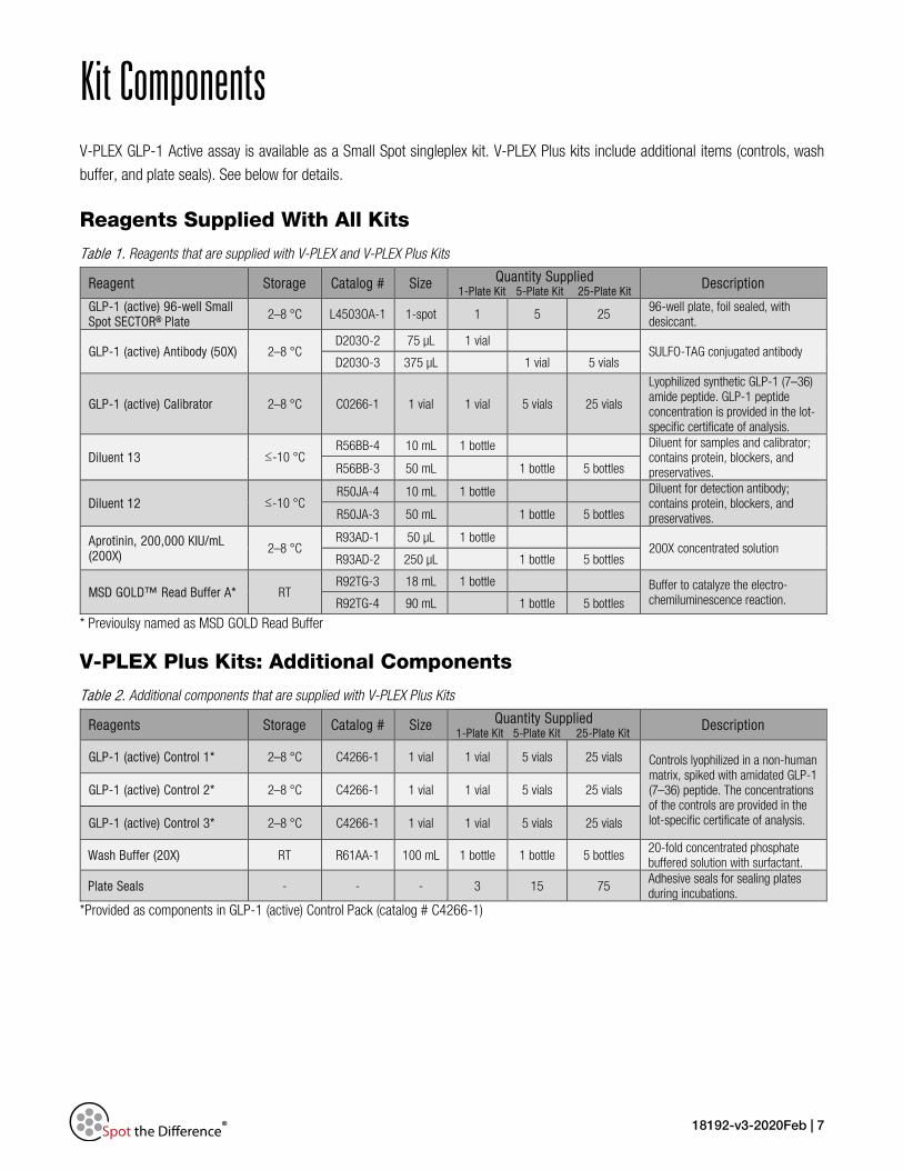
Kit Components V-PLEX GLP-1 Active assay is available as a Small Spot singleplex kit. V-PLEX Plus kits include additional items (controls, wash
buffer, and plate seals). See below for details.
Reagents Supplied With All Kits
Table 1. Reagents that are supplied with V-PLEX and V-PLEX Plus Kits
Reagent Storage Catalog # Size Quantity Supplied 1-Plate Kit 5-Plate Kit 25-Plate Kit Description
GLP-1 (active) 96-well Small Spot SECTOR® Plate 2–8 °C L4503OA-1 1-spot 1 5 25
96-well plate, foil sealed, with desiccant.
GLP-1 (active) Antibody (50X) 2–8 °C D203O-2 75 µL 1 vial
SULFO-TAG conjugated antibody D203O-3 375 µL 1 vial 5 vials
GLP-1 (active) Calibrator 2–8 °C C0266-1 1 vial 1 vial 5 vials 25 vials
Lyophilized synthetic GLP-1 (7–36) amide peptide. GLP-1 peptide concentration is provided in the lot-specific certificate of analysis.
Diluent 13 ≤-10 °C R56BB-4 10 mL 1 bottle Diluent for samples and calibrator;
contains protein, blockers, and preservatives. R56BB-3 50 mL 1 bottle 5 bottles
Diluent 12 ≤-10 °C R50JA-4 10 mL 1 bottle Diluent for detection antibody;
contains protein, blockers, and preservatives. R50JA-3 50 mL 1 bottle 5 bottles
Aprotinin, 200,000 KIU/mL (200X)
2–8 °C R93AD-1 50 µL 1 bottle
200X concentrated solution R93AD-2 250 µL 1 bottle 5 bottles
MSD GOLD™ Read Buffer A* RT R92TG-3 18 mL 1 bottle Buffer to catalyze the electro-
chemiluminescence reaction. R92TG-4 90 mL 1 bottle 5 bottles
* Previoulsy named as MSD GOLD Read Buffer
V-PLEX Plus Kits: Additional Components
Table 2. Additional components that are supplied with V-PLEX Plus Kits
Reagents Storage Catalog # Size Quantity Supplied 1-Plate Kit 5-Plate Kit 25-Plate Kit Description
GLP-1 (active) Control 1* 2–8 °C C4266-1 1 vial 1 vial 5 vials 25 vials Controls lyophilized in a non-human matrix, spiked with amidated GLP-1 (7–36) peptide. The concentrations of the controls are provided in the lot-specific certificate of analysis.
GLP-1 (active) Control 2* 2–8 °C C4266-1 1 vial 1 vial 5 vials 25 vials
GLP-1 (active) Control 3* 2–8 °C C4266-1 1 vial 1 vial 5 vials 25 vials
Wash Buffer (20X) RT R61AA-1 100 mL 1 bottle 1 bottle 5 bottles 20-fold concentrated phosphate buffered solution with surfactant.
Plate Seals - - - 3 15 75 Adhesive seals for sealing plates during incubations.
*Provided as components in GLP-1 (active) Control Pack (catalog # C4266-1)
18192-v3-2020Feb | 8
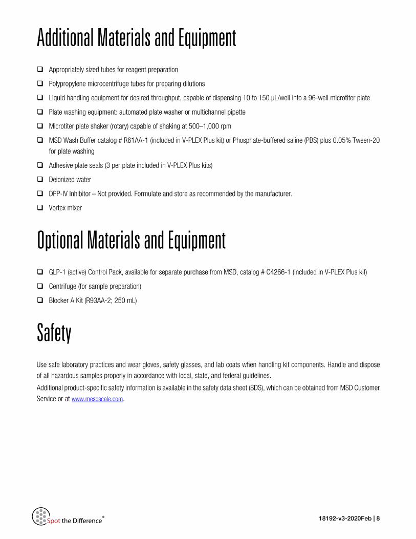
Additional Materials and Equipment Appropriately sized tubes for reagent preparation
Polypropylene microcentrifuge tubes for preparing dilutions
Liquid handling equipment for desired throughput, capable of dispensing 10 to 150 µL/well into a 96-well microtiter plate
Plate washing equipment: automated plate washer or multichannel pipette
Microtiter plate shaker (rotary) capable of shaking at 500–1,000 rpm
MSD Wash Buffer catalog # R61AA-1 (included in V-PLEX Plus kit) or Phosphate-buffered saline (PBS) plus 0.05% Tween-20
for plate washing
Adhesive plate seals (3 per plate included in V-PLEX Plus kits)
Deionized water
DPP-IV Inhibitor – Not provided. Formulate and store as recommended by the manufacturer.
Vortex mixer
Optional Materials and Equipment GLP-1 (active) Control Pack, available for separate purchase from MSD, catalog # C4266-1 (included in V-PLEX Plus kit)
Centrifuge (for sample preparation)
Blocker A Kit (R93AA-2; 250 mL)
Safety Use safe laboratory practices and wear gloves, safety glasses, and lab coats when handling kit components. Handle and dispose
of all hazardous samples properly in accordance with local, state, and federal guidelines.
Additional product-specific safety information is available in the safety data sheet (SDS), which can be obtained from MSD Customer
Service or at www.mesoscale.com.

18192-v3-2020Feb | 9
Best Practices • Mixing and substituting reagents from different sources or different kit lots is not recommended. Lot information is provided
in the lot-specific COA.
• Assay incubation steps should be performed between 20-26 °C to achieve the most consistent signals between runs.
• Bring frozen diluent to room temperature in a 24 °C water bath. Thaw other reagents on wet ice and use as directed
without delay.
• Prepare calibrators, samples, and controls in polypropylene microcentrifuge tubes; use a fresh pipette tip for each dilution;
vortex after each dilution before proceeding.
• Avoid prolonged exposure of detection antibody (stock or diluted) to light. During the antibody incubation step, plates do
not need to be shielded from light except for direct sunlight.
• Avoid bubbles in wells at all pipetting steps. Bubbles may lead to variable results; bubbles introduced when adding MSD
GOLD Read Buffer A may interfere with signal detection.
• Do not touch the pipette tip on the bottom of the wells when pipetting into the MSD plate.
• Use reverse pipetting when necessary to avoid introduction of bubbles. For empty wells, pipette to the bottom corner.
• Plate shaking should be vigorous, with a rotary motion between 500 and 1,000 rpm. Binding reactions may reach
equilibrium sooner if you use shaking at the middle of this range (~700 rpm) or above.
• When using an automated plate washer, rotate the plate 180 degrees between wash steps to improve assay precision.
• Gently tap the plate on a paper towel to remove residual fluid after washing.
• MSD GOLD Read Buffer A should be at room temperature when added to the plate.
• Keep time intervals consistent between adding read buffer and reading the plate to improve inter-plate precision. Unless
otherwise directed, read plate as soon as practical after adding read buffer.
• Do not shake the plate after adding MSD GOLD Read Buffer A.
• If an incubation step needs to be extended, avoid letting the plate dry out by keeping sample or detection antibody solution
in the plate.
• Remove the plate seals prior to reading the plate.
• If assay results are above the top of the calibration curve, dilute the samples and repeat the assay.
• When running a partial plate, seal the unused sectors (see sector map in instrument and software manuals) to avoid
contaminating unused wells. Remove all seals before reading. Partially used plates may be sealed and stored up to
30 days at 2–8 °C in the original foil pouch with desiccant. You may adjust volumes proportionally when preparing
reagents.
18192-v3-2020Feb | 10
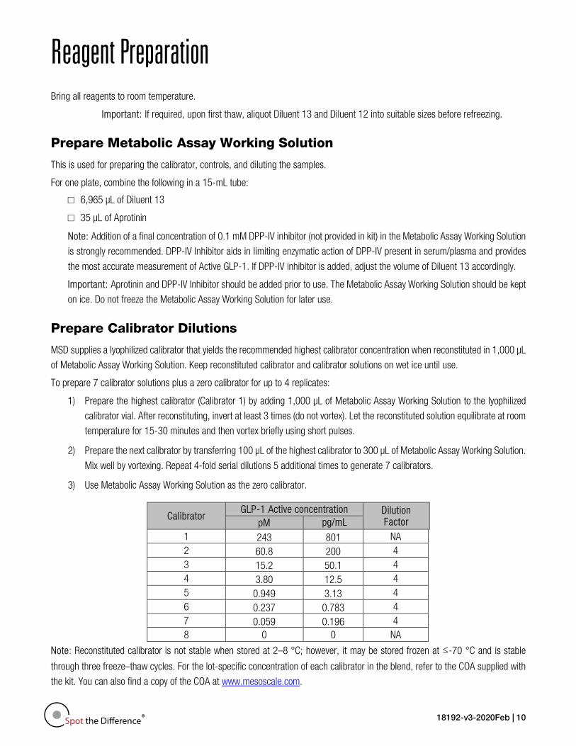
Reagent Preparation Bring all reagents to room temperature.
Important: If required, upon first thaw, aliquot Diluent 13 and Diluent 12 into suitable sizes before refreezing.
Prepare Metabolic Assay Working Solution
This is used for preparing the calibrator, controls, and diluting the samples.
For one plate, combine the following in a 15-mL tube:
□ 6,965 µL of Diluent 13
□ 35 µL of Aprotinin
Note: Addition of a final concentration of 0.1 mM DPP-IV inhibitor (not provided in kit) in the Metabolic Assay Working Solution
is strongly recommended. DPP-IV Inhibitor aids in limiting enzymatic action of DPP-IV present in serum/plasma and provides
the most accurate measurement of Active GLP-1. If DPP-IV inhibitor is added, adjust the volume of Diluent 13 accordingly.
Important: Aprotinin and DPP-IV Inhibitor should be added prior to use. The Metabolic Assay Working Solution should be kept
on ice. Do not freeze the Metabolic Assay Working Solution for later use.
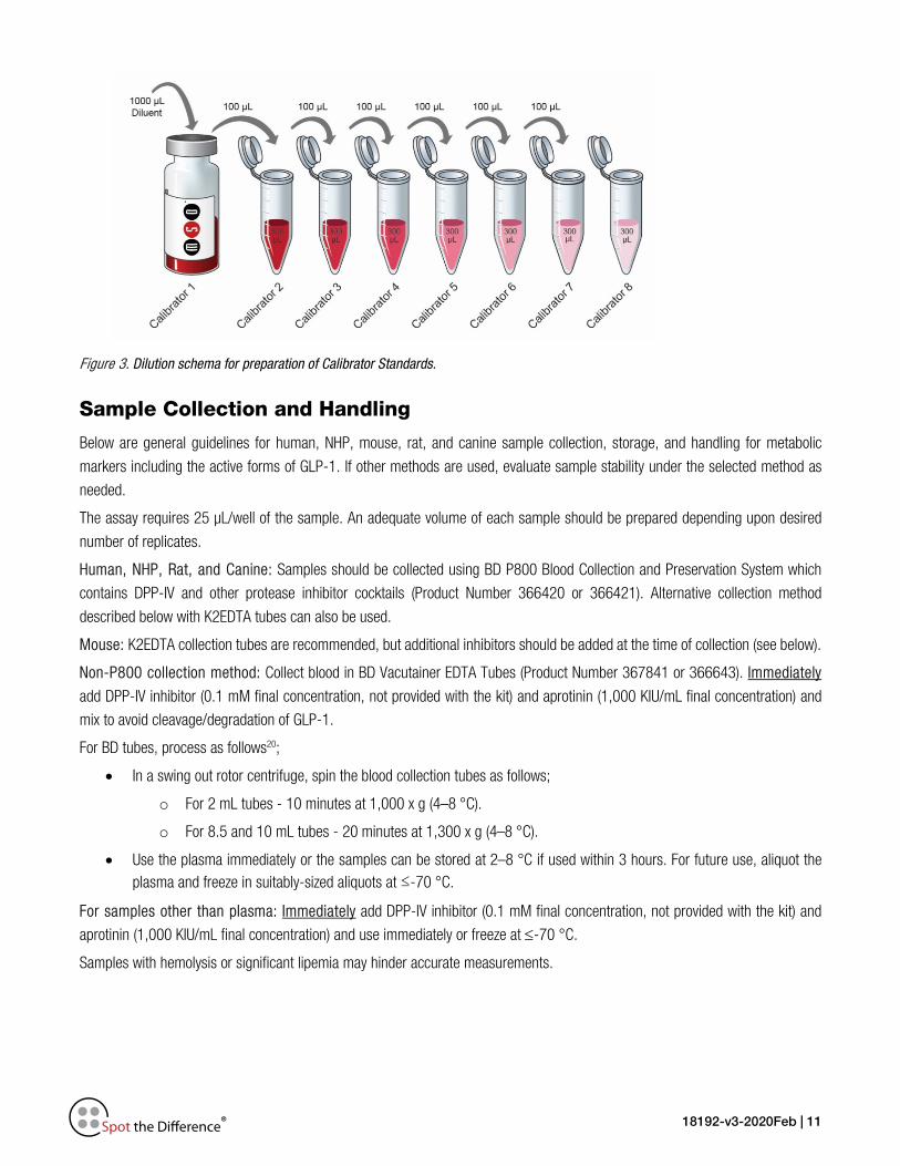
Prepare Calibrator Dilutions
MSD supplies a lyophilized calibrator that yields the recommended highest calibrator concentration when reconstituted in 1,000 µL
of Metabolic Assay Working Solution. Keep reconstituted calibrator and calibrator solutions on wet ice until use.
To prepare 7 calibrator solutions plus a zero calibrator for up to 4 replicates:
1) Prepare the highest calibrator (Calibrator 1) by adding 1,000 µL of Metabolic Assay Working Solution to the lyophilized
calibrator vial. After reconstituting, invert at least 3 times (do not vortex). Let the reconstituted solution equilibrate at room
temperature for 15-30 minutes and then vortex briefly using short pulses.
2) Prepare the next calibrator by transferring 100 µL of the highest calibrator to 300 µL of Metabolic Assay Working Solution.
Mix well by vortexing. Repeat 4-fold serial dilutions 5 additional times to generate 7 calibrators.
3) Use Metabolic Assay Working Solution as the zero calibrator.
Calibrator GLP-1 Active concentration Dilution
Factor pM pg/mL 1 243 801 NA 2 60.8 200 4 3 15.2 50.1 4 4 3.80 12.5 4 5 0.949 3.13 4 6 0.237 0.783 4 7 0.059 0.196 4 8 0 0 NA
Note: Reconstituted calibrator is not stable when stored at 2–8 °C; however, it may be stored frozen at ≤-70 °C and is stable
through three freeze–thaw cycles. For the lot-specific concentration of each calibrator in the blend, refer to the COA supplied with
the kit. You can also find a copy of the COA at www.mesoscale.com.
18192-v3-2020Feb | 11
Figure 3. Dilution schema for preparation of Calibrator Standards.
Sample Collection and Handling
Below are general guidelines for human, NHP, mouse, rat, and canine sample collection, storage, and handling for metabolic
markers including the active forms of GLP-1. If other methods are used, evaluate sample stability under the selected method as
needed.
The assay requires 25 µL/well of the sample. An adequate volume of each sample should be prepared depending upon desired
number of replicates.
Human, NHP, Rat, and Canine: Samples should be collected using BD P800 Blood Collection and Preservation System which
contains DPP-IV and other protease inhibitor cocktails (Product Number 366420 or 366421). Alternative collection method
described below with K2EDTA tubes can also be used.
Mouse: K2EDTA collection tubes are recommended, but additional inhibitors should be added at the time of collection (see below).
Non-P800 collection method: Collect blood in BD Vacutainer EDTA Tubes (Product Number 367841 or 366643). Immediately
add DPP-IV inhibitor (0.1 mM final concentration, not provided with the kit) and aprotinin (1,000 KIU/mL final concentration) and
mix to avoid cleavage/degradation of GLP-1.
For BD tubes, process as follows20;
• In a swing out rotor centrifuge, spin the blood collection tubes as follows;
o For 2 mL tubes - 10 minutes at 1,000 x g (4–8 °C).
o For 8.5 and 10 mL tubes - 20 minutes at 1,300 x g (4–8 °C).
• Use the plasma immediately or the samples can be stored at 2–8 °C if used within 3 hours. For future use, aliquot the plasma and freeze in suitably-sized aliquots at ≤-70 °C.
For samples other than plasma: Immediately add DPP-IV inhibitor (0.1 mM final concentration, not provided with the kit) and
aprotinin (1,000 KIU/mL final concentration) and use immediately or freeze at ≤-70 °C.
Samples with hemolysis or significant lipemia may hinder accurate measurements.
18192-v3-2020Feb | 12
Repeated freezing and thawing of samples is not recommended. After thawing, centrifuge samples at 2,000g for 3 minutes to
remove particulates prior to using in the assay. If the samples are clear and no particulates are visible, you may not need to
centrifuge. Hold on wet ice or 4–8 °C until processed and used in the assay.
Dilute Samples
Dilute samples with Metabolic Assay Working Solution. For plasma from human and all other species MSD recommends a minimum
2-fold dilution. For example, when running samples in duplicate, add 75 µL of sample to 75 µL of Metabolic Assay Working Solution.
We recommend running at least two replicates per sample. You may conserve sample volume by using a higher dilution when
possible. Tissue culture supernatants may require additional dilution based on stimulation and analyte concentrations in the sample.
Additional diluent can be purchased at www.mesoscale.com.
Prepare Controls
Three levels of single-analyte lyophilized controls are available for separate purchase from MSD in the GLP-1 (active) Control Pack,
catalog # C4266-1. (Controls are included only in V-PLEX Plus kits.)
Reconstitute the lyophilized controls in 250 µL of Metabolic Assay Working Solution. Do not invert or vortex the vials . Wait for a
minimum of 15-30 minutes at room temperature before diluting controls 2-fold in Metabolic Assay Working Solution. Vortex briefly using short pulses. Reconstituted controls may be stored frozen at ≤-70 °C and are stable through three freeze–thaw cycles. For
the lot-specific concentration of each analyte in the control pack, refer to the supplied COA. You can also find a copy of the COA at
www.mesoscale.com.
Prepare Detection Antibody Solution
MSD provides the detection antibody as a 50X stock solution. The working solution is 1X. Prepare the detection antibody solution
immediately prior to use. For one plate, add 60 µL of the supplied detection antibody to 2,940 µL of Diluent 12.
Prepare Wash Buffer
MSD provides 100 mL of Wash Buffer as a 20X stock solution in the V-PLEX Plus kit. Dilute the stock solution to 1X before use.
PBS + 0.05% Tween-20 can be used instead.
For one plate, combine:
15 mL of MSD Wash Buffer (20X)
285 mL of deionized water
1X MSD Wash Buffer can be stored at room temperature for up to two weeks.
Read Buffer
MSD provides MSD GOLD Read Buffer A ready for use. Do not dilute.
Important: Unlike Read Buffer T, which is provided at a 4X concentration, MSD GOLD Read Buffer A is provided at the working
concentration of the assay. Dilution of MSD GOLD Read Buffer A may compromise the results of this assay.
Prepare MSD Plate
MSD plates are pre-coated with capture antibodies (Figure 1) and exposed to a proprietary stabilizing treatment to ensure the
integrity and stability of the immobilized antibodies. Pre-wash plates before use as recommended on the assay protocol.
18192-v3-2020Feb | 13
Protocol Note: Follow Reagent Preparation before beginning this assay protocol.
STEP 1: Wash and Add Sample
Wash the plate 3 times with 150 µL/well of 1X MSD Wash Buffer.
Prepare the Metabolic Assay Working Solution
Add 50 µL of prepared samples, calibrators or controls per well. Seal the plate with an adhesive plate seal
and incubate at room temperature with shaking for 2 hours.
Note: Washing the plate prior to sample addition is an optional step that may provide greater uniformity of results
for certain assays. Analytical parameters, including limits of quantification, recovery of controls, and sample
quantification, are not affected by washing the plate prior to sample addition.
Blocking step: A pre-assay, blocking step may be incorporated if consistency with older protocols is required.
Prepare Blocker A according to the instructions provided in the Blocker A Kit. Dispense 150 µL of Blocker A
solution into each well. Seal the plate with an adhesive plate seal and incubate for 1 hour with shaking at room
temperature, then proceed to step 1 above.
STEP 2: Wash and Add Detection Antibody Solution
Wash the plate 3 times with at least 150 µL/well of 1X MSD Wash Buffer.
Add 25 µL of detection antibody solution to each well. Seal the plate with an adhesive plate seal and incubate
at room temperature with shaking for 2 hours.
STEP 3: Wash and Read
Wash the plate 3 times with at least 150 µL/well of 1X MSD Wash Buffer.
Add 150 µL of MSD GOLD Read Buffer A* to each well. Analyze the plate on an MSD instrument. Incubation
in Read Buffer is not required before reading the plate.
*Important: Unlike Read Buffer T which is provided at a 4X concentration, MSD GOLD Read Buffer A should
not be diluted. Dilution of MSD GOLD Read Buffer A may compromise the results of this assay.
Alternate Protocols
The suggestions below may be useful as alternate protocols; however, not all were tested using multiple kit lots.
• Alternate Protocol 1, Extended Sample Incubation: Incubating samples overnight at 2–8 °C may improve sensitivity
for some assays. See Appendix A.
• Alternate Protocol 2, Reduced Wash: You may simplify the protocol by eliminating one of the wash steps. After
incubating diluted sample, calibrator, or control, add detection antibody solution to the plate without decanting or washing
the plate. See Appendix A for assay performance using this protocol.
• Alternate Protocol 3, Dilute-in-Plate: To limit sample handling, you may dilute samples and controls in the plate. For
2-fold dilution, add 25 µL of assay diluent to each sample/control well, and then add 25 µL of neat control or sample.
Calibrators should not be diluted in the plate; add 50 µL of each calibrator directly into empty wells. Tests conducted
according to this alternate protocol produced results that were similar to the recommended protocol (data not shown).
18192-v3-2020Feb | 14
Validation MSD’s V-PLEX products are analytically validated following fit-for-purpose principles18 and MSD design control procedures. V-PLEX
assay components go through an extensive critical reagents program to ensure that the reagents are controlled and well
characterized. Prior to the release of each V-PLEX panel, at least three independent kit lots are produced. Using results from
multiple runs (typically greater than 50) and multiple operators, these lots are used to establish production specifications for
sensitivity, specificity, accuracy, and precision. The COA provided with each kit outlines the kit release specifications for sensitivity,
specificity, accuracy, and precision.
Dynamic Range
Calibration curve concentrations for the assay are optimized for a maximum dynamic range while maintaining enough
calibration points near the bottom of the curve to ensure a proper fit for accurate quantification of samples that require
high sensitivity.
Sensitivity
The lower limit of detection (LLOD) is a calculated concentration corresponding to the average signal 2.5 standard
deviations above the background (zero calibrator). The LLOD is calculated using results from multiple plates for each lot,
and the median and range of calculated LLODs for a representative kit lot are reported in this product insert. The upper
limit of quantification (ULOQ) and lower limit of quantification (LLOQ) are established for each lot by measuring multiple
levels near the expected LLOQ and ULOQ levels. The final LLOQ and ULOQ specifications for the product are established
after assessment of all validation lots.
Accuracy and Precision
Accuracy and precision are evaluated by measuring calibrators and matrix-based validation samples or controls across
multiple runs and multiple lots. For most assays, the results of control measurements fall within 20% of the expected
concentration for each run. Precision is reported as the coefficient of variation (CV). Intra-run CVs are typically below 7%,
and inter-run CVs are typically below 25%. Rigorous management of inter-lot reagent consistency and calibrator
production results in typical inter-lot CVs below 10%. Validation lots are compared using controls and at least 30 samples
in various sample matrices. Samples are well correlated with an inter-lot bias typically below 10%.
Matrix Effects and Samples
Matrix effects from plasma, and cell culture media are measured as part of development and validation. Dilution linearity
and spike recovery studies are performed on individual samples to assess variability of results due to matrix effects. The
sample dilution suggested in the protocol gives an appropriate dilution factor for all assays in the multiplex. In addition to
the matrices listed above, Peripheral blood mononuclear cells (PBMCs), and/or cell lines that have been stimulated to
generate elevated levels of analytes are tested.
Specificity
The specificity of both capture and detection antibodies is measured during assay development. For the GLP-1 (active)
antibody, specificity was assessed by testing closely related forms of GLP-1, products of the proglucagon protein and
other related cytokines.
18192-v3-2020Feb | 15
Assay Robustness and Stability
The robustness of the assay protocol is assessed by examining the boundaries of the selected incubation times and
evaluating the stability of assay components during the experiment and the stability of reconstituted lyophilized
components during storage. Assay component (calibrator, antibody, control) stability was assessed through freeze–thaw
testing and accelerated stability studies. The validation program includes a real-time stability study with scheduled
performance evaluations of complete kits for up to 54 months from date of manufacture.
Representative data from the verification and validation studies are presented in the following sections. The calibration curve and
measured limits of detection for each lot can be found in the lot-specific COA that is included with each kit and available for
download at www.mesoscale.com.

18192-v3-2020Feb | 16
Analysis of Results The calibration curves used to calculate analyte concentrations were established by fitting the signals from the calibrators to a
4-parameter logistic (or sigmoidal dose-response) model with a 1/Y2 weighting. The weighting function provides a better fit of data
over a wide dynamic range, particularly at the low end of the calibration curve. Analyte concentrations were determined from the
ECL signals by back-fitting to the calibration curve. These assays have a wide dynamic range (>3 logs), which allows accurate
quantification of samples without the need for multiple dilutions or repeated testing. The calculations to establish calibration curves
and determine concentrations were carried out using the MSD DISCOVERY WORKBENCH® analysis software.
Best quantification of unknown samples will be achieved by generating a calibration curve for each plate using a minimum of two
replicates at each calibrator level.
Typical Data Data from the V-PLEX GLP-1 Active Kit were collected over one week of testing by five operators (20 runs, 60 plates in total).
Calibration curve accuracy and precision were assessed for two kit lots. Representative data from one lot are presented below.
Appendix A compares data from alternate protocols 1, 2, and 3 to the reference protocol.
Figure 4. Typical calibration curve for the V-PLEX GLP-1 Active Kit.

18192-v3-2020Feb | 17
Sensitivity The LLOD is a calculated concentration corresponding to the signal 2.5 standard deviations above the background (zero calibrator).
The LLOD shown below was calculated based on three runs from three kit lots.
The ULOQ is the highest concentration at which the CV of the calculated concentration is <20% and the recovery of each analyte
is within 80% to 120% of the known value.
The LLOQ is the lowest concentration at which the CV of the calculated concentration is <20% and the recovery of each analyte is
within 80% to 120% of the known value.
The quantitative range of the assay lies between the LLOQ and ULOQ.
The LLOQ and ULOQ are verified for each kit lot and the results are provided in the lot-specific COA that is included with each kit
and available at www.mesoscale.com.
Table 3. LLOD, LLOQ, and ULOQ for the V-PLEX GLP-1 Active Kit
Median LLOD (pM)
LLOD Range (pM)
LLOQ (pM)
ULOQ (pM)
GLP-1 (active) 0.020 0.010–0.020 0.300 120
18192-v3-2020Feb | 18
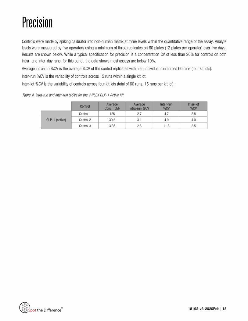
Precision Controls were made by spiking calibrator into non-human matrix at three levels within the quantitative range of the assay. Analyte
levels were measured by five operators using a minimum of three replicates on 60 plates (12 plates per operator) over five days.
Results are shown below. While a typical specification for precision is a concentration CV of less than 20% for controls on both
intra- and inter-day runs, for this panel, the data shows most assays are below 10%.
Average intra-run %CV is the average %CV of the control replicates within an individual run across 60 runs (four kit lots).
Inter-run %CV is the variability of controls across 15 runs within a single kit lot.
Inter-lot %CV is the variability of controls across four kit lots (total of 60 runs, 15 runs per kit lot).
Table 4. Intra-run and Inter-run %CVs for the V-PLEX GLP-1 Active Kit
Control Average Conc. (pM)
Average Intra-run %CV
Inter-run %CV
Inter-lot %CV
GLP-1 (active)
Control 1 126 2.7 4.7 2.8
Control 2 30.5 3.1 4.9 4.0
Control 3 3.35 2.8 11.8 2.5

18192-v3-2020Feb | 19
Dilution Linearity To assess linearity, normal human plasma (collected in P800 tubes) from a commercial source as well as cell culture supernatants
were spiked with GLP-1 (active) Calibrator and diluted 2-fold, 4-fold, 8-fold, and 16-fold, before testing. Percent recovery at each
dilution level was normalized to the 2-fold dilution-adjusted concentration. The average percent recovery is based on samples
within the quantitative range of the assay.
% 𝑅𝑅𝑅𝑅𝑅𝑅𝑅𝑅𝑅𝑅𝑅𝑅𝑅𝑅𝑅𝑅 =𝑚𝑚𝑅𝑅𝑚𝑚𝑚𝑚𝑚𝑚𝑅𝑅𝑅𝑅𝑚𝑚 𝑅𝑅𝑅𝑅𝑐𝑐𝑅𝑅𝑅𝑅𝑐𝑐𝑐𝑐𝑅𝑅𝑚𝑚𝑐𝑐𝑐𝑐𝑅𝑅𝑐𝑐𝑅𝑅𝑒𝑒𝑒𝑒𝑅𝑅𝑅𝑅𝑐𝑐𝑅𝑅𝑚𝑚 𝑅𝑅𝑅𝑅𝑐𝑐𝑅𝑅𝑅𝑅𝑐𝑐𝑐𝑐𝑅𝑅𝑚𝑚𝑐𝑐𝑐𝑐𝑅𝑅𝑐𝑐
∗ 100
Table 5. GLP-1 (7-36) amide percent recovery at various dilutions in P800 EDTA plasma, and Cell Culture Supernatant samples. Data from human, mouse, rat, canine, NHP are shown in the following tables. Mouse samples were tested using EDTA plasma spiked with aprotinin & the DPP-IV inhibitor (P800 tubes were not used for mouse samples).
Sample Type Fold
Dilution Average % Recovery
% Recovery Range
Human P800 EDTA plasma
(N=18)
4 90 79–99
8 94 85–101
16 95 87–100
NHP P800 EDTA plasma
(N = 10)
4 101 91–110
8 101 90–113
16 102 83–118
Mouse Citrate plasma
(N = 10)
4 104 95–114
8 104 90–115
16 104 88–122
Mouse K2 EDTA plasma
(N = 10)
4 98 91–105
8 100 90–107
16 100 91–111
Rat P800 EDTA plasma
(N = 10)
4 100 77–113
8 95 70–115
16 89 75–107
Sample Type Fold
Dilution Average % Recovery
% Recovery Range
Canine P800 EDTA plasma
(N = 10)
4 95 89–100
8 95 86–101
16 95 89–107
Krebs Ringer
4 99 NA
8 100 NA
16 85 NA
RPMI (10% FBS, 1%
Pen-Strep, 2mM Glu)
4 92 NA
8 90 NA
16 92 NA
DMEM (10% FBS, 1%
Pen-Strep)
4 86 NA
8 85 NA
16 85 NA
DMEM (2.5% FBS,
15% HS)
4 97 NA
8 100 NA
16 93 NA
18192-v3-2020Feb | 20
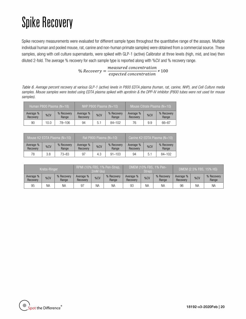
Spike Recovery Spike recovery measurements were evaluated for different sample types throughout the quantitative range of the assays. Multiple
individual human and pooled mouse, rat, canine and non-human primate samples) were obtained from a commercial source. These
samples, along with cell culture supernatants, were spiked with GLP-1 (active) Calibrator at three levels (high, mid, and low) then
diluted 2-fold. The average % recovery for each sample type is reported along with %CV and % recovery range.
% 𝑅𝑅𝑅𝑅𝑅𝑅𝑅𝑅𝑅𝑅𝑅𝑅𝑅𝑅𝑅𝑅 =𝑚𝑚𝑅𝑅𝑚𝑚𝑚𝑚𝑚𝑚𝑅𝑅𝑅𝑅𝑚𝑚 𝑅𝑅𝑅𝑅𝑐𝑐𝑅𝑅𝑅𝑅𝑐𝑐𝑐𝑐𝑅𝑅𝑚𝑚𝑐𝑐𝑐𝑐𝑅𝑅𝑐𝑐𝑅𝑅𝑒𝑒𝑒𝑒𝑅𝑅𝑅𝑅𝑐𝑐𝑅𝑅𝑚𝑚 𝑅𝑅𝑅𝑅𝑐𝑐𝑅𝑅𝑅𝑅𝑐𝑐𝑐𝑐𝑅𝑅𝑚𝑚𝑐𝑐𝑐𝑐𝑅𝑅𝑐𝑐
∗ 100
Table 6. Average percent recovery at various GLP-1 (active) levels in P800 EDTA plasma (human, rat, canine, NHP), and Cell Culture media samples. Mouse samples were tested using EDTA plasma spiked with aprotinin & the DPP-IV inhibitor (P800 tubes were not used for mouse samples).
Human P800 Plasma (N=18) NHP P800 Plasma (N=10) Mouse Citrate Plasma (N=10)
Average % Recovery
%CV % Recovery
Range Average % Recovery
%CV % Recovery
Range Average % Recovery
%CV % Recovery
Range
90 10.0 78–106 94 5.1 84–102 76 9.9 66–87
Mouse K2 EDTA Plasma (N=10) Rat P800 Plasma (N=10) Canine K2 EDTA Plasma (N=10)
Average % Recovery
%CV % Recovery
Range Average % Recovery
%CV % Recovery
Range Average % Recovery
%CV % Recovery
Range
78 3.8 73–83 97 4.3 91–103 94 5.1 84–102
Krebs-Ringer RPMI (10% FBS, 1% Pen-Strep, 2mM Glu)
DMEM (10% FBS, 1% Pen-Strep)
DMEM (2.5% FBS, 15% HS)
Average % Recovery %CV
% Recovery Range
Average % Recovery %CV
% Recovery Range
Average % Recovery %CV
% Recovery Range
Average % Recovery %CV
% Recovery Range
95 NA NA 97 NA NA 93 NA NA 96 NA NA
18192-v3-2020Feb | 21
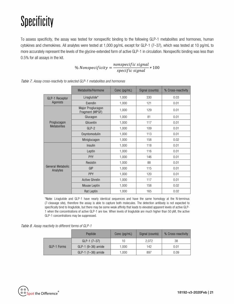
Specificity To assess specificity, the assay was tested for nonspecific binding to the following GLP-1 metabolites and hormones, human
cytokines and chemokines. All analytes were tested at 1,000 pg/mL except for GLP-1 (7–37), which was tested at 10 pg/mL to
more accurately represent the levels of the glycine-extended form of active GLP-1 in circulation. Nonspecific binding was less than
0.5% for all assays in the kit.
% 𝑁𝑁𝑅𝑅𝑐𝑐𝑚𝑚𝑒𝑒𝑅𝑅𝑅𝑅𝑐𝑐𝑁𝑁𝑐𝑐𝑅𝑅𝑐𝑐𝑐𝑐𝑅𝑅 =𝑐𝑐𝑅𝑅𝑐𝑐𝑚𝑚𝑒𝑒𝑅𝑅𝑅𝑅𝑐𝑐𝑁𝑁𝑐𝑐𝑅𝑅 𝑚𝑚𝑐𝑐𝑠𝑠𝑐𝑐𝑚𝑚𝑠𝑠𝑚𝑚𝑒𝑒𝑅𝑅𝑅𝑅𝑐𝑐𝑁𝑁𝑐𝑐𝑅𝑅 𝑚𝑚𝑐𝑐𝑠𝑠𝑐𝑐𝑚𝑚𝑠𝑠
∗ 100
Table 7. Assay cross-reactivity to selected GLP-1 metabolites and hormones
Metabolite/Hormone Conc (pg/mL) Signal (counts) % Cross-reactivity
GLP-1 Receptor Agonists
Liraglutide* 1,000 330 0.03
Exendin 1,000 121 0.01
Proglucagon Metabolites
Major Proglucagon Fragment (MPGF) 1,000 129 0.01
Glucagon 1,000 81 0.01
Glicentin 1,000 117 0.01
GLP-2 1,000 109 0.01
Oxyntomodulin 1,000 113 0.01
Miniglucagon 1,000 158 0.02
General Metabolic Analytes
Insulin 1,000 118 0.01
Leptin 1,000 116 0.01
PYY 1,000 146 0.01
Resistin 1,000 88 0.01
GIP 1,000 115 0.01
PPY 1,000 120 0.01
Active Ghrelin 1,000 117 0.01
Mouse Leptin 1,000 158 0.02
Rat Leptin 1,000 165 0.02
*Note: Liraglutide and GLP-1 have nearly identical sequences and have the same homology at the N-terminus (7-cleavage site), therefore the assay is able to capture both molecules. The detection antibody is not expected to specifically bind to liraglutide, but there may be some weak affinity that leads to elevated apparent levels of active GLP-1 when the concentrations of active GLP-1 are low. When levels of liraglutide are much higher than 50 pM, the active GLP-1 concentrations may be suppressed.
Table 8. Assay reactivity to different forms of GLP-1
Peptide Conc (pg/mL) Signal (counts) % Cross-reactivity
GLP-1 Forms
GLP-1 (7–37) 10 2,072 38
GLP-1 (9–36) amide 1,000 142 0.01
GLP-1 (1–36) amide 1,000 897 0.09
18192-v3-2020Feb | 22
Stability The reconstituted calibrator, reconstituted controls, and diluents were tested for freeze–thaw stability. Results (not shown)
demonstrated that reconstituted calibrator, reconstituted controls, and diluents can go through three freeze–thaw cycles without
significantly affecting the performance of the assay. Reconstituted calibrator and controls must be stored frozen at ≤-70 °C. Partially
used MSD plates may be sealed and stored up to 30 days at 2–8 °C in the original foil pouch with desiccant. Results from control
measurements changed by ≤30% after partially used plates were stored for 30 days. The validation study includes a real-time
stability study with scheduled performance evaluations of complete kits for up to 54 months from date of manufacture.
Calibration There is no GLP-1 reference standard available from NIBSC or other equivalent body. The peptide calibrator concentrations are
calibrated against a reference calibrator generated at MSD that was assigned using three independent lots of amino acid-analyzed
active GLP-1 peptide to provide a highly accurate measurement of active GLP-1.
18192-v3-2020Feb | 23
Tested Samples Normal Samples
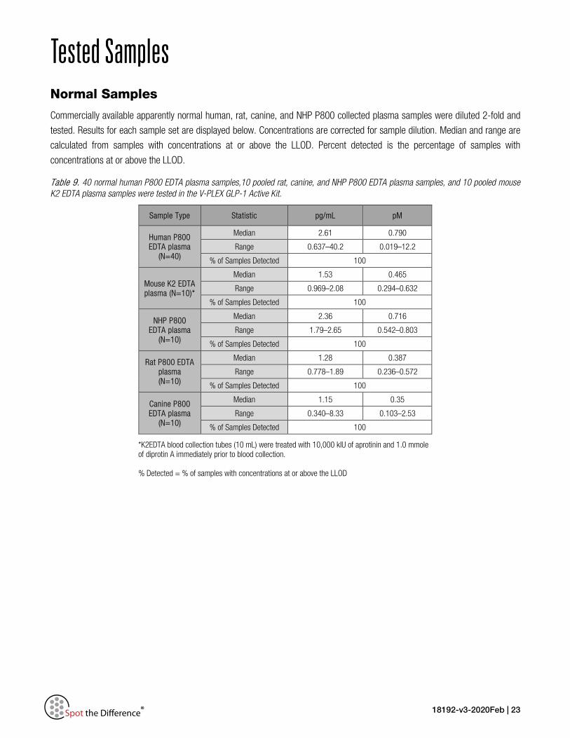
Commercially available apparently normal human, rat, canine, and NHP P800 collected plasma samples were diluted 2-fold and
tested. Results for each sample set are displayed below. Concentrations are corrected for sample dilution. Median and range are
calculated from samples with concentrations at or above the LLOD. Percent detected is the percentage of samples with
concentrations at or above the LLOD.
Table 9. 40 normal human P800 EDTA plasma samples,10 pooled rat, canine, and NHP P800 EDTA plasma samples, and 10 pooled mouse K2 EDTA plasma samples were tested in the V-PLEX GLP-1 Active Kit.
Sample Type Statistic pg/mL pM
Human P800 EDTA plasma
(N=40)
Median 2.61 0.790
Range 0.637–40.2 0.019–12.2
% of Samples Detected 100
Mouse K2 EDTA plasma (N=10)*
Median 1.53 0.465
Range 0.969–2.08 0.294–0.632
% of Samples Detected 100
NHP P800 EDTA plasma
(N=10)
Median 2.36 0.716
Range 1.79–2.65 0.542–0.803
% of Samples Detected 100
Rat P800 EDTA plasma (N=10)
Median 1.28 0.387
Range 0.778–1.89 0.236–0.572
% of Samples Detected 100
Canine P800 EDTA plasma
(N=10)
Median 1.15 0.35
Range 0.340–8.33 0.103–2.53
% of Samples Detected 100
*K2EDTA blood collection tubes (10 mL) were treated with 10,000 kIU of aprotinin and 1.0 mmole of diprotin A immediately prior to blood collection. % Detected = % of samples with concentrations at or above the LLOD

18192-v3-2020Feb | 24
In vitro Stimulated Cells and Post-Meal Plasma Samples
Cell culture supernatants from In vitro Stimulated NCI-H716 Cells:
NCI-H716 cells (Human Epithelium (cecum) colorectal adenocarcinoma) were treated with various stimulants, including
progesterone (1 µM), forskolin (10 µM), insulin (2 µM), and sucrose (200 mM) for a given period of time. These cell culture
supernatants were tested for the presence of active GLP-1 (7–36) amide. Active GLP-1 was expressed at levels higher than the
detection limit of the assay, so they were diluted 4 fold prior to the assay. The dilution-adjusted concentrations (pM) for each
stimulation condition are displayed below.
Figure 5. Effect of cell stimulation on active GLP-1 production as measured in the V-PLEX GLP-1 Active Kit.
Post-meal changes in circulating GLP-1:
Post-meal samples from apparently healthy individuals were tested for the active GLP-1 levels. Samples were collected from
different individuals at different time points. The dilution-adjusted concentrations (pM) for each sample are displayed below.
Figure 6. Post-meal levels of active GLP-1 in individuals (black circles), measured using the V-PLEX GLP-1 Active Kit. Dotted line indicates the in-sample LLOQ, which is two times the in-well LLOQ.
18192-v3-2020Feb | 25
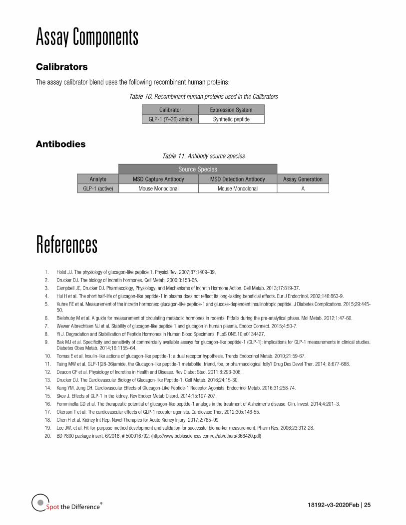
Assay Components Calibrators
The assay calibrator blend uses the following recombinant human proteins:
Table 10. Recombinant human proteins used in the Calibrators
Calibrator Expression System
GLP-1 (7–36) amide Synthetic peptide
Antibodies Table 11. Antibody source species
Source Species
Analyte MSD Capture Antibody MSD Detection Antibody Assay Generation
GLP-1 (active) Mouse Monoclonal Mouse Monoclonal A
References 1. Holst JJ. The physiology of glucagon-like peptide 1. Physiol Rev. 2007;87:1409–39.
2. Drucker DJ. The biology of incretin hormones. Cell Metab. 2006;3:153-65.
3. Campbell JE, Drucker DJ. Pharmacology, Physiology, and Mechanisms of Incretin Hormone Action. Cell Metab. 2013;17:819-37.
4. Hui H et al. The short half-life of glucagon-like peptide-1 in plasma does not reflect its long-lasting beneficial effects. Eur J Endocrinol. 2002;146:863-9.
5. Kuhre RE et al. Measurement of the incretin hormones: glucagon-like peptide-1 and glucose-dependent insulinotropic peptide. J Diabetes Complications. 2015;29:445-50.
6. Bielohuby M et al. A guide for measurement of circulating metabolic hormones in rodents: Pitfalls during the pre-analytical phase. Mol Metab. 2012;1:47-60.
7. Wewer Albrechtsen NJ et al. Stability of glucagon-like peptide 1 and glucagon in human plasma. Endocr Connect. 2015;4:50-7.
8. Yi J. Degradation and Stabilization of Peptide Hormones in Human Blood Specimens. PLoS ONE.10;e0134427.
9. Bak MJ et al. Specificity and sensitivity of commercially available assays for glucagon-like peptide-1 (GLP-1): implications for GLP-1 measurements in clinical studies. Diabetes Obes Metab. 2014;16:1155–64.
10. Tomas E et al. Insulin-like actions of glucagon-like peptide-1: a dual receptor hypothesis. Trends Endocrinol Metab. 2010;21:59-67.
11. Taing MW et al. GLP-1(28-36)amide, the Glucagon-like peptide-1 metabolite: friend, foe, or pharmacological folly? Drug Des Devel Ther. 2014; 8:677-688.
12. Deacon CF et al. Physiology of Incretins in Health and Disease. Rev Diabet Stud. 2011;8:293-306.
13. Drucker DJ. The Cardiovascular Biology of Glucagon-like Peptide-1. Cell Metab. 2016;24:15-30.
14. Kang YM, Jung CH. Cardiovascular Effects of Glucagon-Like Peptide-1 Receptor Agonists. Endocrinol Metab. 2016;31:258-74.
15. Skov J. Effects of GLP-1 in the kidney. Rev Endocr Metab Disord. 2014;15:197-207.
16. Femminella GD et al. The therapeutic potential of glucagon-like peptide-1 analogs in the treatment of Alzheimer’s disease. Clin. Invest. 2014;4:201–3.
17. Okerson T et al. The cardiovascular effects of GLP-1 receptor agonists. Cardiovasc Ther. 2012;30:e146-55.
18. Chen H et al. Kidney Int Rep. Novel Therapies for Acute Kidney Injury. 2017;2:785–99.
19. Lee JW, et al. Fit-for-purpose method development and validation for successful biomarker measurement. Pharm Res. 2006;23:312-28.
20. BD P800 package insert, 6/2016, # 500016792. (http://www.bdbiosciences.com/ds/ab/others/366420.pdf)

18192-v3-2020Feb | 26
Appendix A Calibration curves below illustrate the relative sensitivity for each assay under Alternate Protocols: Reference Protocol (2-hour
sample incubation/2 wash steps, blue curve), Alternate Protocol 1 (overnight [O/N] sample incubation, orange curve), Alternate
Protocol 2 (single wash, red curve), and Alternate Protocol 3 (in-well dilution, grey curve). Details of the alternate protocols are
found on page 13.
Figure 7. Calibrator curves generated by the indicated protocols. Table shows the hillslope of each curve and the estimated lower limit of detection (LOD) for the assay when performed using each protocol. Each control (high, mid, and low) was tested under each alternate protocol. The plot below demonstrates for each protocol the
recovery of measured concentrations of the controls relative to the expected concentrations, which shows equivalent performance
among all the protocols tested.
Figure 8. Recovery of measured concentrations of the controls resulting from each protocol relative to the expected concentrations.
Hill Slope LOD
Reference Curve 1.12 0.021
Single Wash 1.14 0.022
In Well 1.11 0.037
O/N Incubation 1.12 0.017
18192-v3-2020Feb | 27
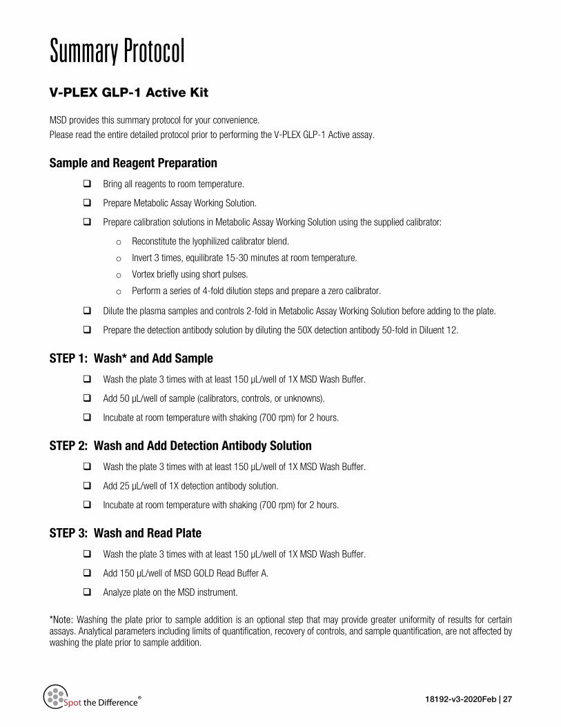
Summary Protocol V-PLEX GLP-1 Active Kit
MSD provides this summary protocol for your convenience.
Please read the entire detailed protocol prior to performing the V-PLEX GLP-1 Active assay.
Sample and Reagent Preparation
Bring all reagents to room temperature.
Prepare Metabolic Assay Working Solution.
Prepare calibration solutions in Metabolic Assay Working Solution using the supplied calibrator:
o Reconstitute the lyophilized calibrator blend.
o Invert 3 times, equilibrate 15-30 minutes at room temperature.
o Vortex briefly using short pulses.
o Perform a series of 4-fold dilution steps and prepare a zero calibrator.
Dilute the plasma samples and controls 2-fold in Metabolic Assay Working Solution before adding to the plate.
Prepare the detection antibody solution by diluting the 50X detection antibody 50-fold in Diluent 12.
STEP 1: Wash* and Add Sample
Wash the plate 3 times with at least 150 µL/well of 1X MSD Wash Buffer.
Add 50 µL/well of sample (calibrators, controls, or unknowns).
Incubate at room temperature with shaking (700 rpm) for 2 hours.
STEP 2: Wash and Add Detection Antibody Solution
Wash the plate 3 times with at least 150 µL/well of 1X MSD Wash Buffer.
Add 25 µL/well of 1X detection antibody solution.
Incubate at room temperature with shaking (700 rpm) for 2 hours.
STEP 3: Wash and Read Plate
Wash the plate 3 times with at least 150 µL/well of 1X MSD Wash Buffer.
Add 150 µL/well of MSD GOLD Read Buffer A.
Analyze plate on the MSD instrument.
*Note: Washing the plate prior to sample addition is an optional step that may provide greater uniformity of results for certain assays. Analytical parameters including limits of quantification, recovery of controls, and sample quantification, are not affected by washing the plate prior to sample addition.
18192-v3-2020Feb | 28
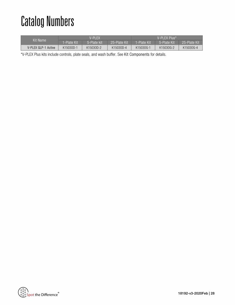
Catalog Numbers Kit Name
V-PLEX V-PLEX Plus* 1-Plate Kit 5-Plate kit 25-Plate Kit 1-Plate Kit 5-Plate Kit 25-Plate Kit
V-PLEX GLP-1 Active K1503OD-1 K1503OD-2 K1503OD-4 K1503OG-1 K1503OG-2 K1503OG-4
*V-PLEX Plus kits include controls, plate seals, and wash buffer. See Kit Components for details.
18192-v3-2020Feb | 29
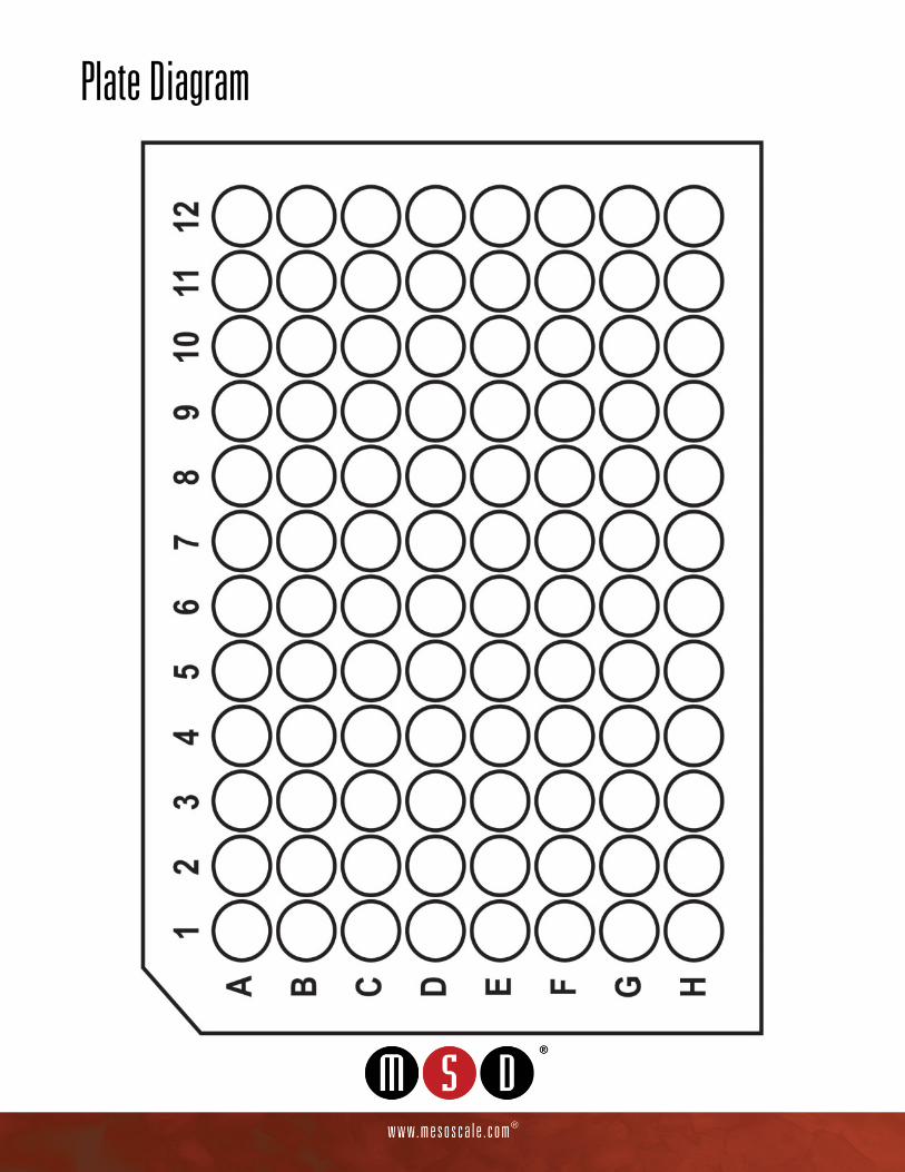
Plate Diagram


![Topical Administration of GLP-1 Receptor Agonists Prevents ...(a GLP-1 receptor [GLP-1R] agonist) prevent electroretino-graphy(ERG) abnormalities and morphological featuresrelated](https://static.fdocuments.in/doc/165x107/5f4839b6212d137c1c54d696/topical-administration-of-glp-1-receptor-agonists-prevents-a-glp-1-receptor.jpg)

![Original Article Dosimetry of [ Lu]-DO3A-VS-Cys -Exendin-4 ... · Original Article Dosimetry of [177Lu]-DO3A-VS-Cys40-Exendin-4 ... gon like peptide-1 receptor (GLP-1R). In the light](https://static.fdocuments.in/doc/165x107/5f077d867e708231d41d3dd0/original-article-dosimetry-of-lu-do3a-vs-cys-exendin-4-original-article.jpg)