GLOBAL ENDOMETRIAL ABLATION...
70
GLOBAL ENDOMETRIAL ABLATION TECHNOLOGY
Transcript of GLOBAL ENDOMETRIAL ABLATION...

GLOBAL ENDOMETRIAL ABLATION
TECHNOLOGY

Training: Part 1
Anatomy and Physiology

Female Anatomy – Normal Uterus
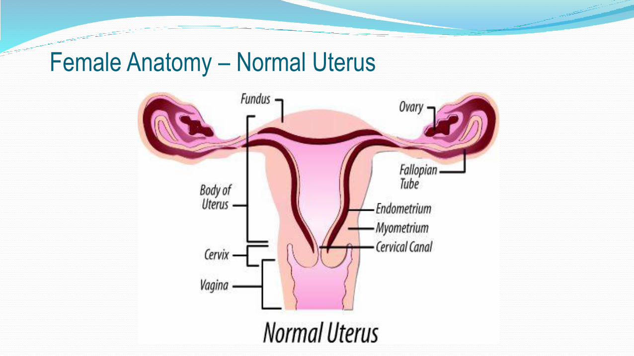
Female Anatomy – Normal Uterus
5
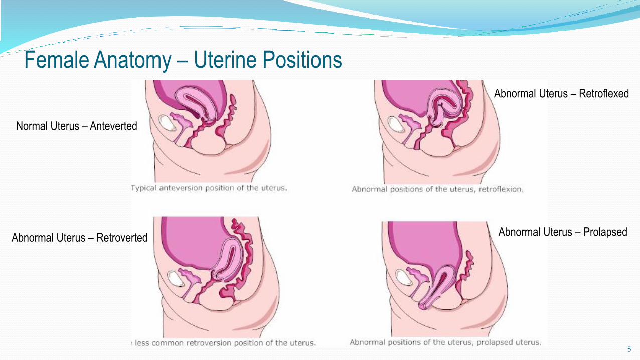
Female Anatomy – Uterine Positions
Normal Uterus – Anteverted
Abnormal Uterus – Retroverted
Abnormal Uterus – Retroflexed
Abnormal Uterus – Prolapsed
29/10/2014
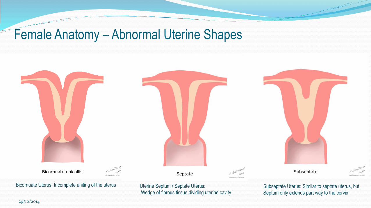
Female Anatomy – Abnormal Uterine Shapes
Bicornuate Uterus: Incomplete uniting of the uterus Uterine Septum / Septate Uterus:
Wedge of fibrous tissue dividing uterine cavitySubseptate Uterus: Similar to septate uterus, but
Septum only extends part way to the cervix
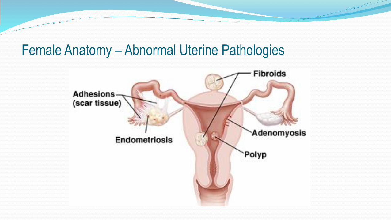
Female Anatomy – Abnormal Uterine Pathologies
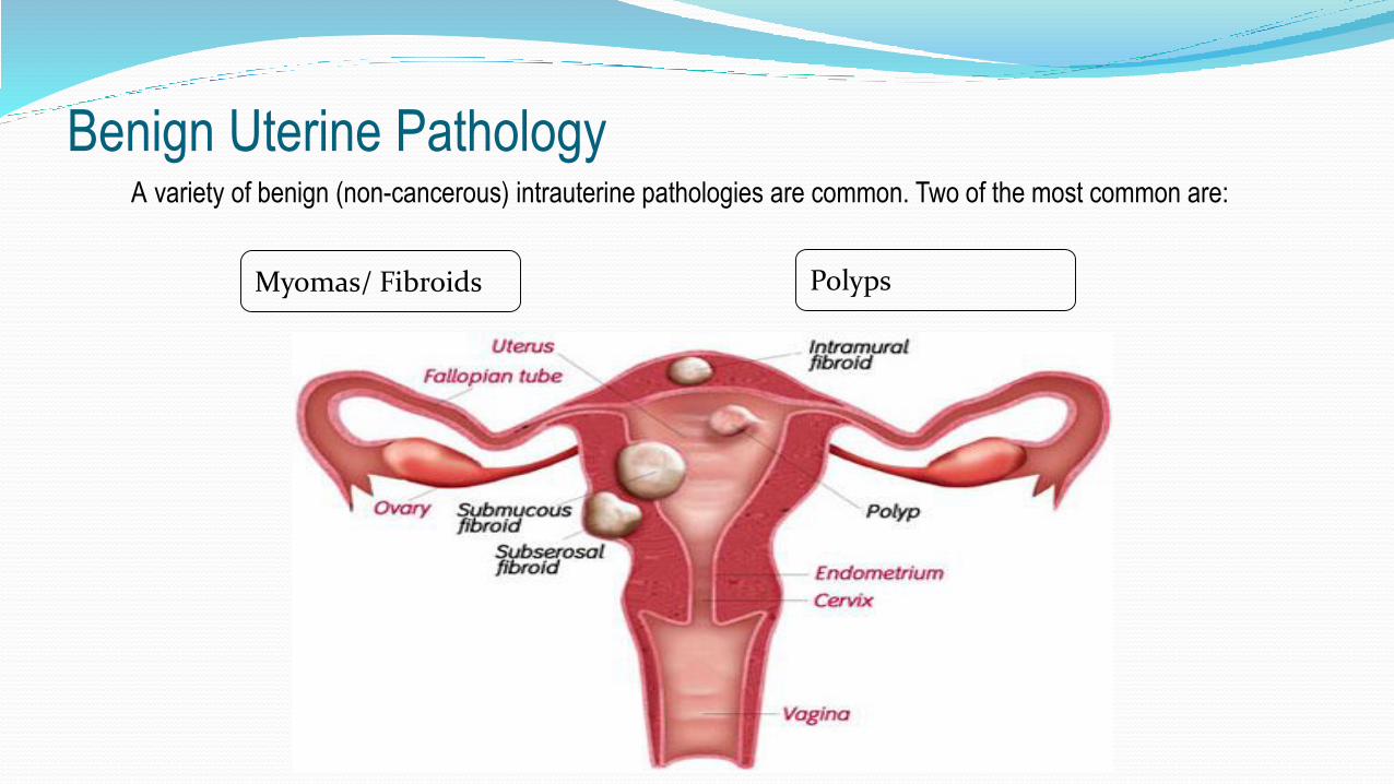
Benign Uterine PathologyA variety of benign (non-cancerous) intrauterine pathologies are common. Two of the most common are:
Myomas/ Fibroids Polyps

The most common and severe symptom is abnormal uterine bleeding
The cause is anovulation – due to a hormonal imbalance of
progesterone formation
Estrogen continues to stimulate the endometrium, causing overgrowth
and endometrial hyperplasia
Polyps
Benign Uterine Pathology
Endometrial or Uterine Polyps
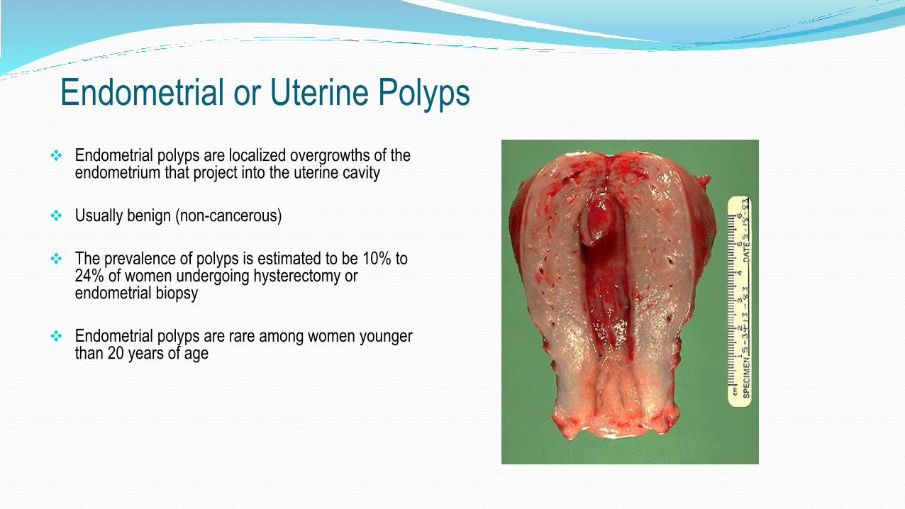
Endometrial polyps are localized overgrowths of the endometrium that project into the uterine cavity
Usually benign (non-cancerous)
The prevalence of polyps is estimated to be 10% to 24% of women undergoing hysterectomy or endometrial biopsy
Endometrial polyps are rare among women younger than 20 years of age
Endometrial or Uterine Polyps
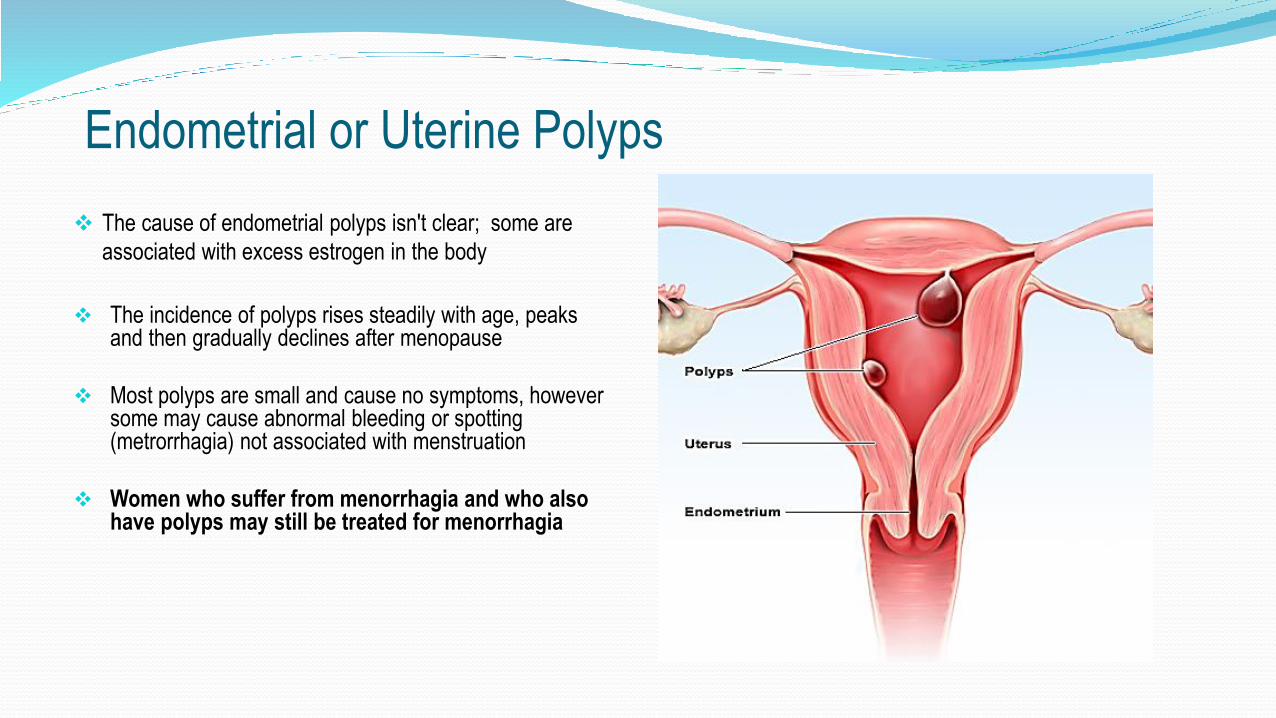
The cause of endometrial polyps isn't clear; some are
associated with excess estrogen in the body
The incidence of polyps rises steadily with age, peaks and then gradually declines after menopause
Most polyps are small and cause no symptoms, however some may cause abnormal bleeding or spotting (metrorrhagia) not associated with menstruation
Women who suffer from menorrhagia and who also have polyps may still be treated for menorrhagia
Endometrial or Uterine Polyps
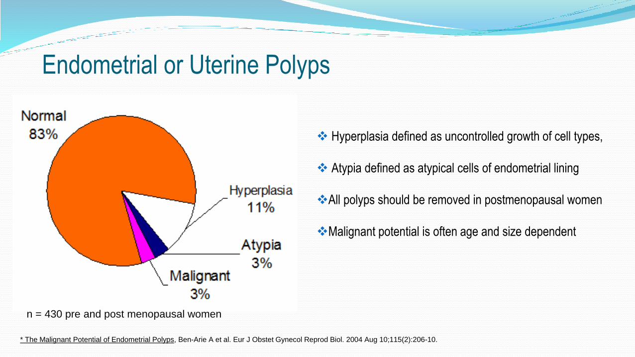
Hyperplasia defined as uncontrolled growth of cell types,
Atypia defined as atypical cells of endometrial lining
All polyps should be removed in postmenopausal women
Malignant potential is often age and size dependent
•* The Malignant Potential of Endometrial Polyps, Ben-Arie A et al. Eur J Obstet Gynecol Reprod Biol. 2004 Aug 10;115(2):206-10.
n = 430 pre and post menopausal women

Affects 20-50% of reproductive age women
Most common benign tumor in this population
Prevalence increases with age
Present in at least 5-10% of infertile patients
Sole factor identified in 1-2.4% of infertile patients
Fibroids are commonly called myomas or leiomyomas
Uterine Fibroids

Myomas/ Fibroids
Facts About Fibroids:
Most common cause of benign uterine enlargement
Occur in 70% of women by age 50
More common in African-American women
Arise from smooth muscle cells in the myometrium
Benign Uterine Pathology

Uterine
Fibroids

Submucosal Fibroids can be further classified based
on their location within the uterus:
Type 0 - Fibroids totally in cavity
Type 1 - More than 50% in cavity
Type 2 - Less than 50% in cavity
Uterine Fibroids European Society of Hysteroscopy Classification of
Submucous Myomas
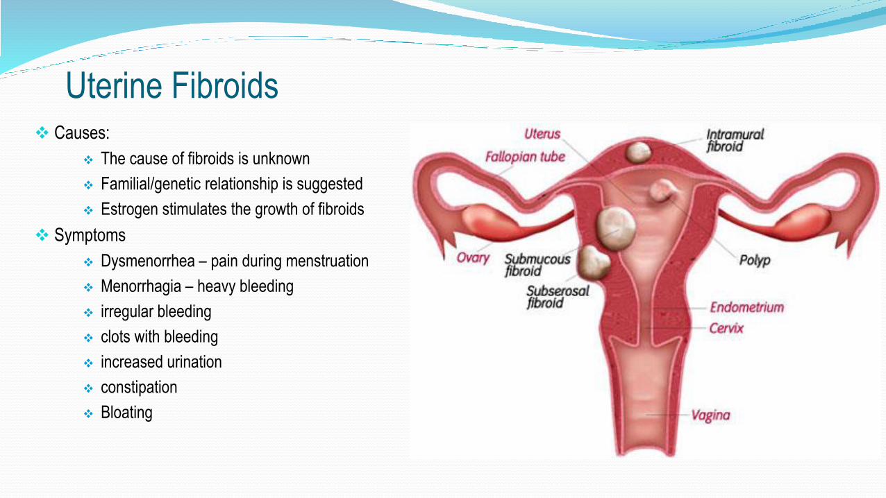
Causes:
The cause of fibroids is unknown
Familial/genetic relationship is suggested
Estrogen stimulates the growth of fibroids
Symptoms
Dysmenorrhea – pain during menstruation
Menorrhagia – heavy bleeding
irregular bleeding
clots with bleeding
increased urination
constipation
Bloating
Uterine Fibroids
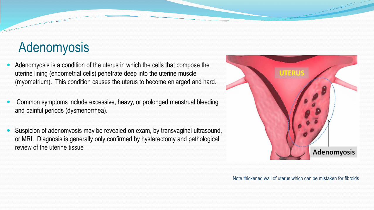
Adenomyosis Adenomyosis is a condition of the uterus in which the cells that compose the
uterine lining (endometrial cells) penetrate deep into the uterine muscle
(myometrium). This condition causes the uterus to become enlarged and hard.
Common symptoms include excessive, heavy, or prolonged menstrual bleeding
and painful periods (dysmenorrhea).
Suspicion of adenomyosis may be revealed on exam, by transvaginal ultrasound,
or MRI. Diagnosis is generally only confirmed by hysterectomy and pathological
review of the uterine tissue
Note thickened wall of uterus which can be mistaken for fibroids
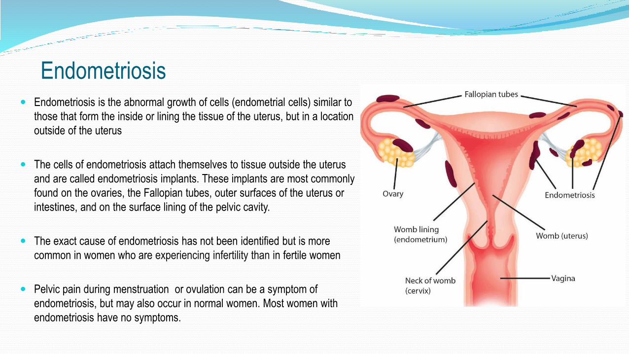
Endometriosis is the abnormal growth of cells (endometrial cells) similar to
those that form the inside or lining the tissue of the uterus, but in a location
outside of the uterus
The cells of endometriosis attach themselves to tissue outside the uterus
and are called endometriosis implants. These implants are most commonly
found on the ovaries, the Fallopian tubes, outer surfaces of the uterus or
intestines, and on the surface lining of the pelvic cavity.
The exact cause of endometriosis has not been identified but is more
common in women who are experiencing infertility than in fertile women
Pelvic pain during menstruation or ovulation can be a symptom of
endometriosis, but may also occur in normal women. Most women with
endometriosis have no symptoms.
Endometriosis

Training: Part 2
Menorrhagia

Excessively heavy or prolonged menstrual bleeding at normal intervals with total blood
loss exceeding 80 mL per cycle
OR
menses lasting longer than 7 days
Affects approx. 20% of all women
between the ages of 30 to 50
What is Menorrhagia? or Abnormal Uterine Bleeding
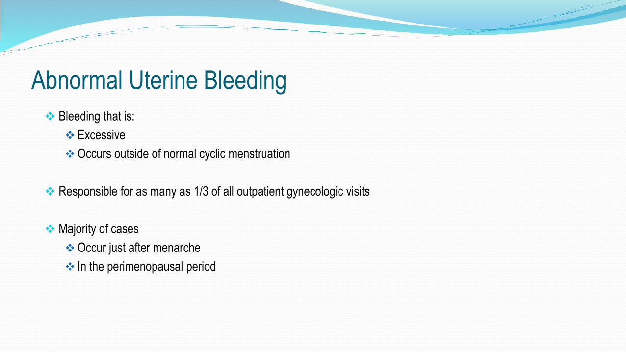
Abnormal Uterine Bleeding
Bleeding that is:
Excessive
Occurs outside of normal cyclic menstruation
Responsible for as many as 1/3 of all outpatient gynecologic visits
Majority of cases
Occur just after menarche
In the perimenopausal period
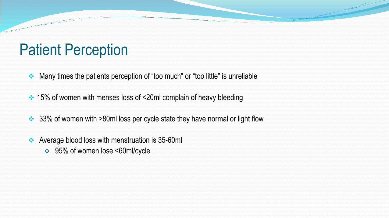
Patient Perception
Many times the patients perception of “too much” or “too little” is unreliable
15% of women with menses loss of <20ml complain of heavy bleeding
33% of women with >80ml loss per cycle state they have normal or light flow
Average blood loss with menstruation is 35-60ml
95% of women lose <60ml/cycle
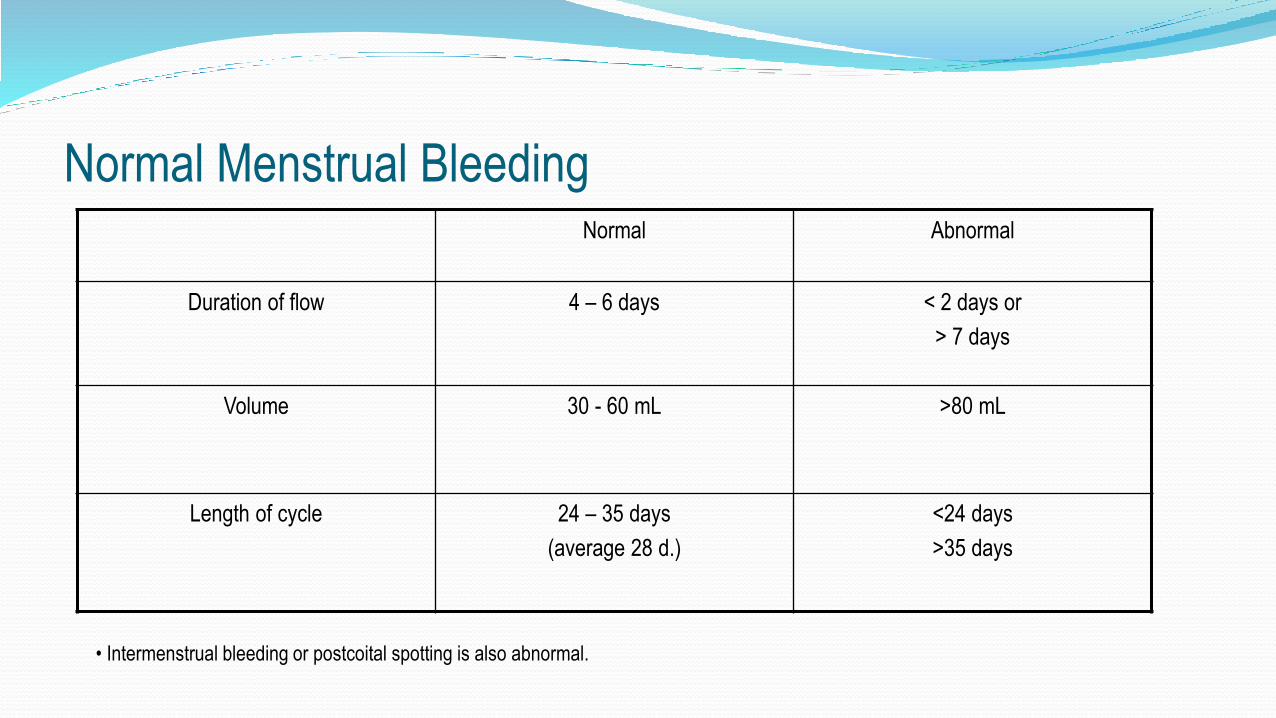
Normal Menstrual BleedingNormal Abnormal
Duration of flow 4 – 6 days < 2 days or
> 7 days
Volume 30 - 60 mL >80 mL
Length of cycle 24 – 35 days
(average 28 d.)
<24 days
>35 days
• Intermenstrual bleeding or postcoital spotting is also abnormal.
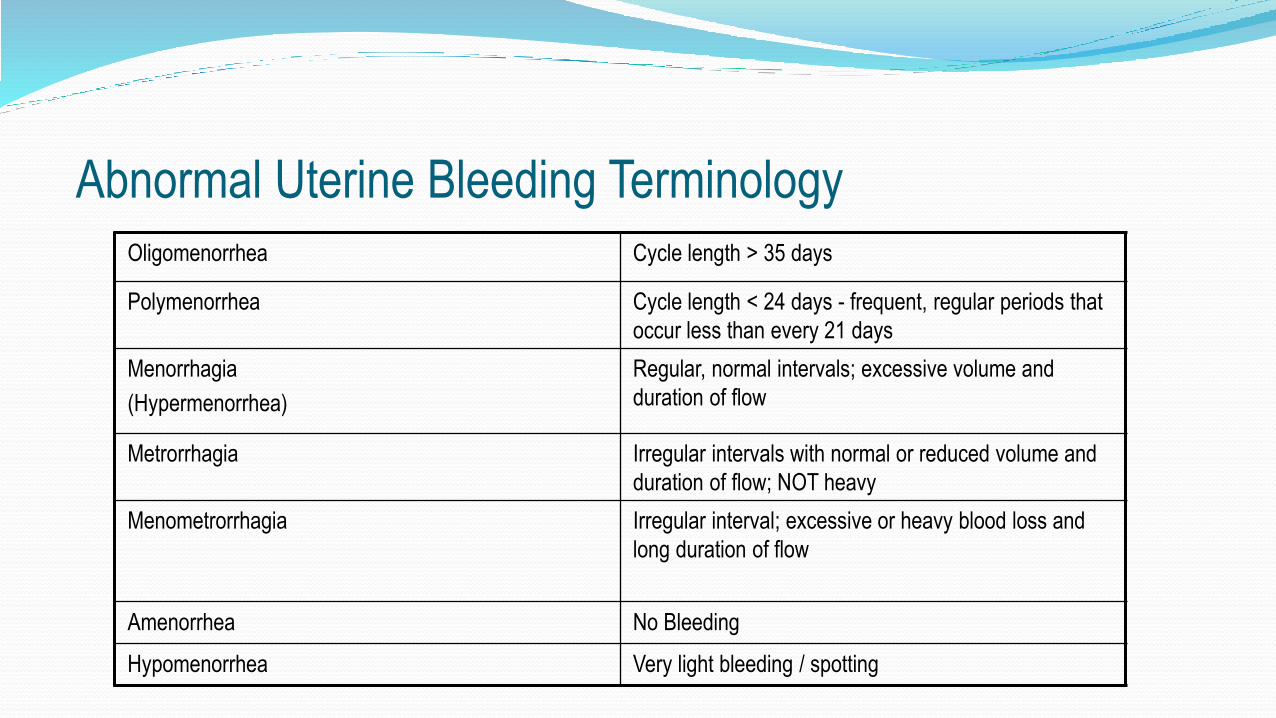
Oligomenorrhea Cycle length > 35 days
Polymenorrhea Cycle length < 24 days - frequent, regular periods that
occur less than every 21 days
Menorrhagia
(Hypermenorrhea)
Regular, normal intervals; excessive volume and
duration of flow
Metrorrhagia Irregular intervals with normal or reduced volume and
duration of flow; NOT heavy
Menometrorrhagia Irregular interval; excessive or heavy blood loss and
long duration of flow
Amenorrhea No Bleeding
Hypomenorrhea Very light bleeding / spotting
Abnormal Uterine Bleeding Terminology
Summary of AUB
Thorough history and evaluation is required to rule out abnormal conditions or pathology
Age and ovulatory status must be considered
Rule-out pregnancy!
Treatment failures require further evaluation
AUB: diagnosis of exclusion
Dysfunctional uterine bleeding (DUB) is heavy or irregular menstrual bleeding that is not
caused by an underlying anatomical abnormality, such as a fibroid, lesion, or tumor
DUB is the most common type of AUB
DUB – Dysfunctional Uterine Bleeding
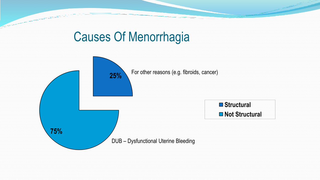
Causes Of Menorrhagia
25%
75%
Structural
Not Structural
DUB – Dysfunctional Uterine Bleeding
For other reasons (e.g. fibroids, cancer)

Step 1: Evaluate Anatomy
Transvaginal or Transabdominal ultrasound
Saline infusion Sonohysterogram
Diagnostic Hysteroscopy
Endometrial Biopsy / Dilation and Curretage
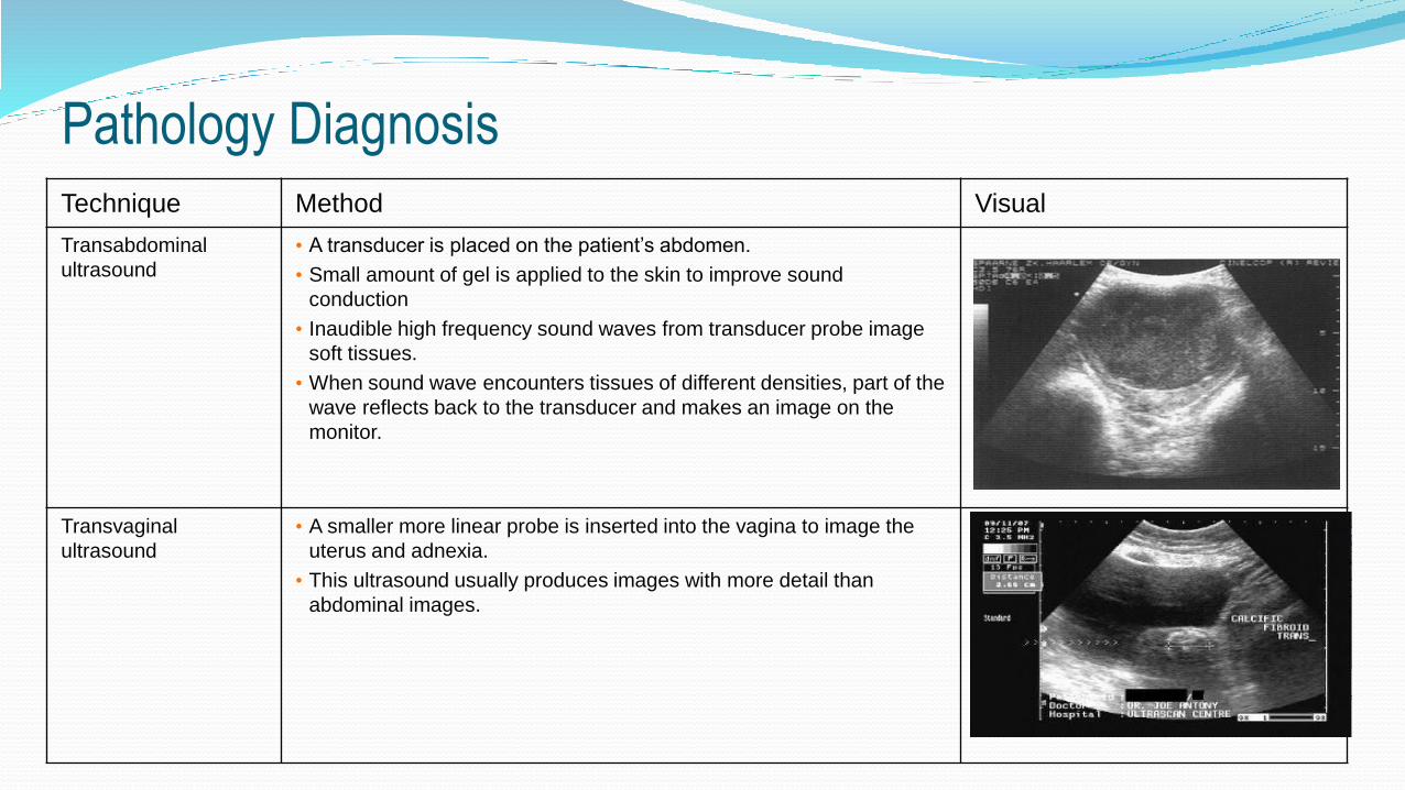
Technique Method Visual
Transabdominal
ultrasound
• A transducer is placed on the patient’s abdomen.
• Small amount of gel is applied to the skin to improve sound
conduction
• Inaudible high frequency sound waves from transducer probe image
soft tissues.
• When sound wave encounters tissues of different densities, part of the
wave reflects back to the transducer and makes an image on the
monitor.
Transvaginal
ultrasound
• A smaller more linear probe is inserted into the vagina to image the
uterus and adnexia.
• This ultrasound usually produces images with more detail than
abdominal images.
Pathology Diagnosis

Saline Infused Sonohysterography (SIS)
Transvaginal ultrasound following installation of saline into the uterus
Most useful for differentiating focal from diffuse endometrial abnormalities
Can help guide the decision of doing a hysteroscopy to evaluate a focal
Abnormality versus performing an endometrial biopsy or dilatation and curettage

Saline Infused Sonohysterography (SIS)
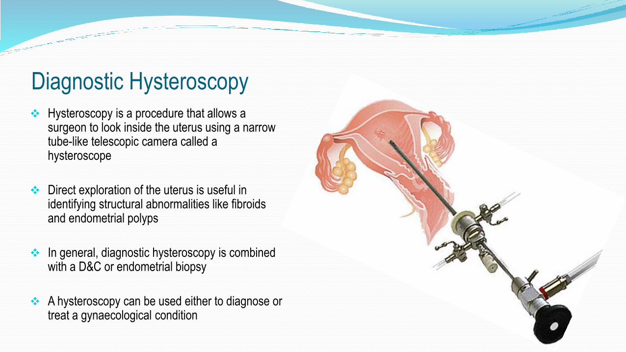
Diagnostic Hysteroscopy Hysteroscopy is a procedure that allows a
surgeon to look inside the uterus using a narrow tube-like telescopic camera called a hysteroscope
Direct exploration of the uterus is useful in identifying structural abnormalities like fibroids and endometrial polyps
In general, diagnostic hysteroscopy is combined with a D&C or endometrial biopsy
A hysteroscopy can be used either to diagnose or treat a gynaecological condition

HysteroscopyHysteroscopy is used to:
help find out what is causing symptoms, for example heavy
bleeding or pain
check for abnormal pathology (polyps or fibroids)
treat scar tissue (adhesions)
Insert or remove an intra-uterine system (IUS)
carry out a permanent form of contraception (Essure
Sterilisation)
Hysteroscopic morcellation of polyps, fibroids, septum, etc.
(TruClear, MyoSure)

Endometrial Biopsy In the office use a clear, flexible endometrial curette with
an inner plunger or piston that generates suction during
the procedure
Rates of obtaining an adequate endometrial sample
depends on the age of the patient
If inadequate sample is obtained, must use additional
diagnostic studies to fully evaluate the cause of the
vaginal bleeding
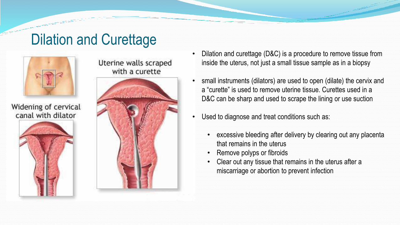
Dilation and Curettage• Dilation and curettage (D&C) is a procedure to remove tissue from
inside the uterus, not just a small tissue sample as in a biopsy
• small instruments (dilators) are used to open (dilate) the cervix and
a “curette” is used to remove uterine tissue. Curettes used in a
D&C can be sharp and used to scrape the lining or use suction
• Used to diagnose and treat conditions such as:
• excessive bleeding after delivery by clearing out any placenta
that remains in the uterus
• Remove polyps or fibroids
• Clear out any tissue that remains in the uterus after a
miscarriage or abortion to prevent infection

Treatment Goals for AUB
Alleviation of any acute bleeding
Prevention of future noncyclic bleeding
Decrease in the patient’s future risk of long-term health problems secondary to anovulation
Improvement in the patient’s quality of life

Training: Part 3
Treatment Options

Diagnosis of HMB based on more subjective factors:
- Negative impact on patient’s daily life
- Interferes with physical, emotional and/or social well being
Treatment Goal: Improved Quality of Life
Patient Satisfaction!
Patient Centered Definition of MenorrhagiaHeavy Menstrual Bleeding
A recent study published in April 2013 on “Women’s Attitudes towards HMB and their impact on
QOL”² researched 6179 women from 15 countries, aged 18-55 years old, and found that 39% of
those diagnosed with HMB believed there was no treatment available.
This Study also confirms that HMB has a profound negative impact on women’s lives and outlines
the need for increased education and information about available treatment options.
Patient Centered DefinitionHeavy Menstrual Bleeding

0%
10%
20%
30%
40%
50%
60%
Sex Work Party/Fun Event
Athletic Event Time with Friends/Family
Sometimes
Many times
National Women's Health Resource Center. National survey of 653 women, 35-49 with reported heavy periods and intact uterus; conducted Sept 29-Oct 12, 2005.
Impact of Heavy Menstrual Bleeding on Daily Activities
Treatment Options
Medical therapies
Hormone therapy – Mirena, Analogues, oral contraceptives
Surgical therapies
Hysterectomy
1st Generation Ablation (Hysteroscopic)
2nd Generation Ablation (Global)
• Medical therapy is often offered as a first line treatment ie. oral tablets to reduce blood loss by improving clotting,
hormone tablets or a levonorgestrel-releasing intrauterine system (LNG-IUS).
• Women who suffer with HMB who no longer want any more children and desire a more permanent, but less-invasive
solution, should consider endometrial ablation. Hormones are not designed for the treatment of menorrhagia.
• The Thermablate Endometrial Ablation System is an advanced, next-generation technology for the treatment of heavy
menstrual bleeding, and should be made available for women who have completed their families
Treatment Options

Levonorgestrel-Releasing Intrauterine System MIRENA IUD

Levonorgestrel-Releasing Intrauterine System MIRENA –
why it fails…
A recent study by Gupta (UK) compared the effectiveness of an LNG-IUS (MIRENA) to medical treatment in women
with menorrhagia. The primary finding of this study is:
• At two years, 36% of patients discontinued the use of the LNG-IUS
Consistent with the findings of Ewies (2009) showing:
• a 41% discontinuation rate of the Mirena Intrauterine System at 2 years2.
• Discontinuation rate increased as time went by, with 50% of patients discontinuing the use of an LNG-IUS by year
52.

Common reasons for discontinuation of the LNG-IUS:
• lack of effectiveness and irregular or prolonged bleeding
• progestin-related adverse events such as headaches, depression, acne
• sexual dissatisfaction due to lower abdominal pain• unscheduled bleeding, weight gain and decreased sex drive in women due to hormones
Levonorgestrel-Releasing Intrauterine System MIRENA –
why it fails…
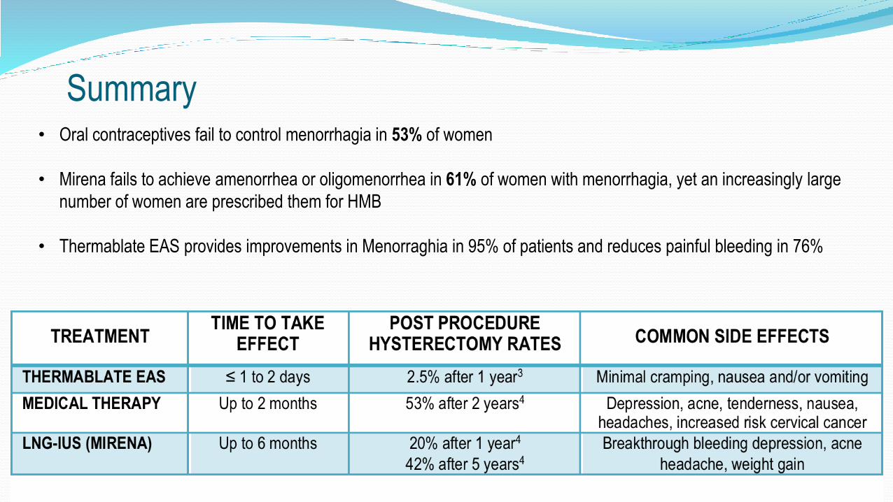
• Oral contraceptives fail to control menorrhagia in 53% of women
• Mirena fails to achieve amenorrhea or oligomenorrhea in 61% of women with menorrhagia, yet an increasingly large
number of women are prescribed them for HMB
• Thermablate EAS provides improvements in Menorraghia in 95% of patients and reduces painful bleeding in 76%
Summary

48
• Failure of medical management
• Intolerance of medical management
• Patient preference
• Physician preference
Indications for Ablation or Surgery
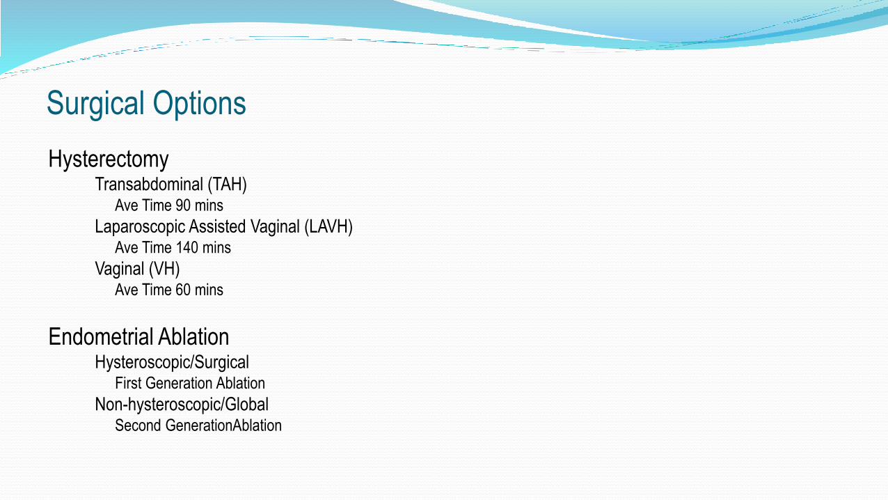
HysterectomyTransabdominal (TAH)
Ave Time 90 mins
Laparoscopic Assisted Vaginal (LAVH) Ave Time 140 mins
Vaginal (VH)Ave Time 60 mins
Endometrial AblationHysteroscopic/Surgical
First Generation Ablation
Non-hysteroscopic/Global Second GenerationAblation
Surgical Options

50
Major invasive operation!
• Second most frequently performed major surgical procedure among reproductive-aged women
• 30-45% of all hysterectomies are performed on women who suffer from menorrhagia
• By age 43, 10% chance of having a hysterectomy
• Average 6 weeks recovery time
• Average 10% complication rate
Hysterectomy
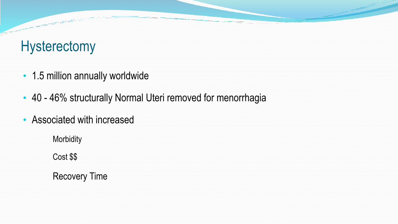
• 1.5 million annually worldwide
• 40 - 46% structurally Normal Uteri removed for menorrhagia
• Associated with increased
Morbidity
Cost $$
Recovery Time
Hysterectomy
52
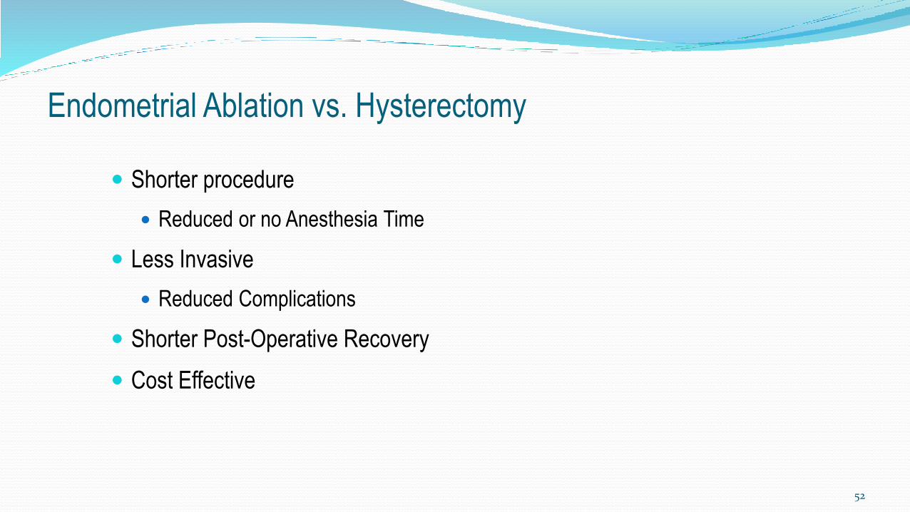
Shorter procedure
Reduced or no Anesthesia Time
Less Invasive
Reduced Complications
Shorter Post-Operative Recovery
Cost Effective
Endometrial Ablation vs. Hysterectomy
Endometrial Ablation
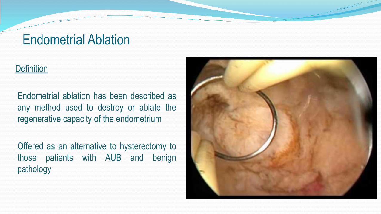
Definition
Endometrial ablation has been described as
any method used to destroy or ablate the
regenerative capacity of the endometrium
Offered as an alternative to hysterectomy to
those patients with AUB and benign
pathology
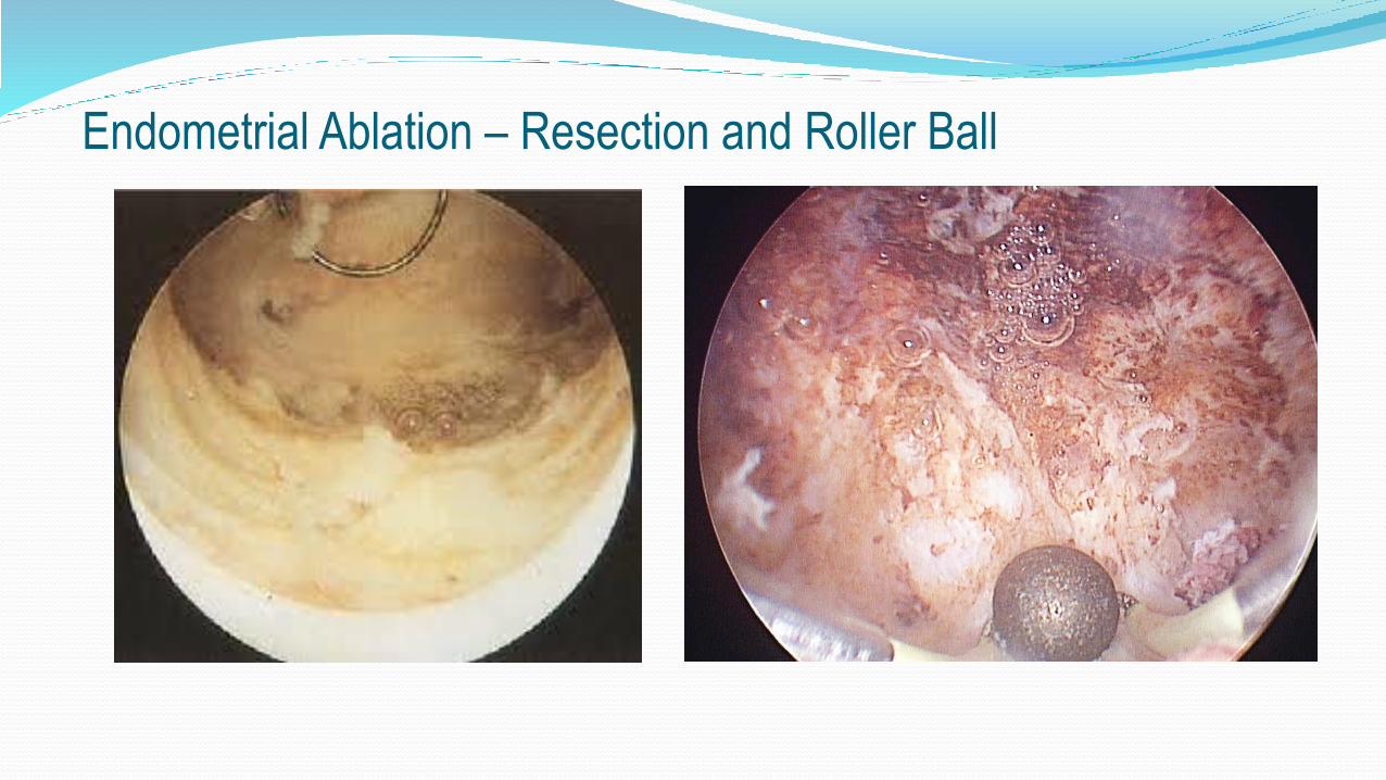
Endometrial Ablation – Resection and Roller Ball
55
• Performed under direct visualization
• Allows treatment of benign pathology such as fibroid removal
• Allows treatment of abnormal cavities
Hysteroscopic Ablation - TCRE, ROLLERBALL
ADVANTAGES
• Steep learning curve and highly skill dependent – need to do them frequently in order to become and stay proficient
• Higher failure rate in earlier cases
• Limited access to cornual areas
• Requires general anesthesia / O.R. setting
• Risk of fluid overload
• Risk of electrosurgical trauma and mechanical injury
• Visceral injuries or vulvar-vaginal burns
• Cervical laceration or uterine perforation
• Risk of hemorrhage
• Risk of gas or air embolism
Hysteroscopic Ablation - TCRE, ROLLERBALL
DISADVANTAGES

1st Generation Endometrial Ablation requires a skilled surgeon to manually ablate or
destroy the endometrial lining of the uterus
Complications common to 1st Generation
Ablation
1. Access (Mechanical Trauma)
1. Distend (Excessive Fluid - Drowning)
1. Energy (Thermal Burns)
Second generation Endometrial Ablation Technologies (SEATs)
Global Endometrial Ablation (GEA)
Non-Hysteroscopic Endometrial Ablation
Automated destruction of the endometrium without the use of operative hysteroscopy
1st Generation Endometrial Ablation Technologies were introduced in 1980’s as alternative to
hysterectomy
Global Ablation Technologies were introduced in 1990’s as alternative to 1st generation
SEATs: RATIONALE
Simpler
Safer
Faster
Consistent
Cost-effective
Minimal Analgesia Requirements (Office Procedure)

MENU FOR ENDOMETRIAL ABLATION
Laser Nd:YAG
Electrocute Rollerball
Slice Loop
Dice Loop with spurs
Plow Grooved rollerball
Poach Hot water (Balloons)
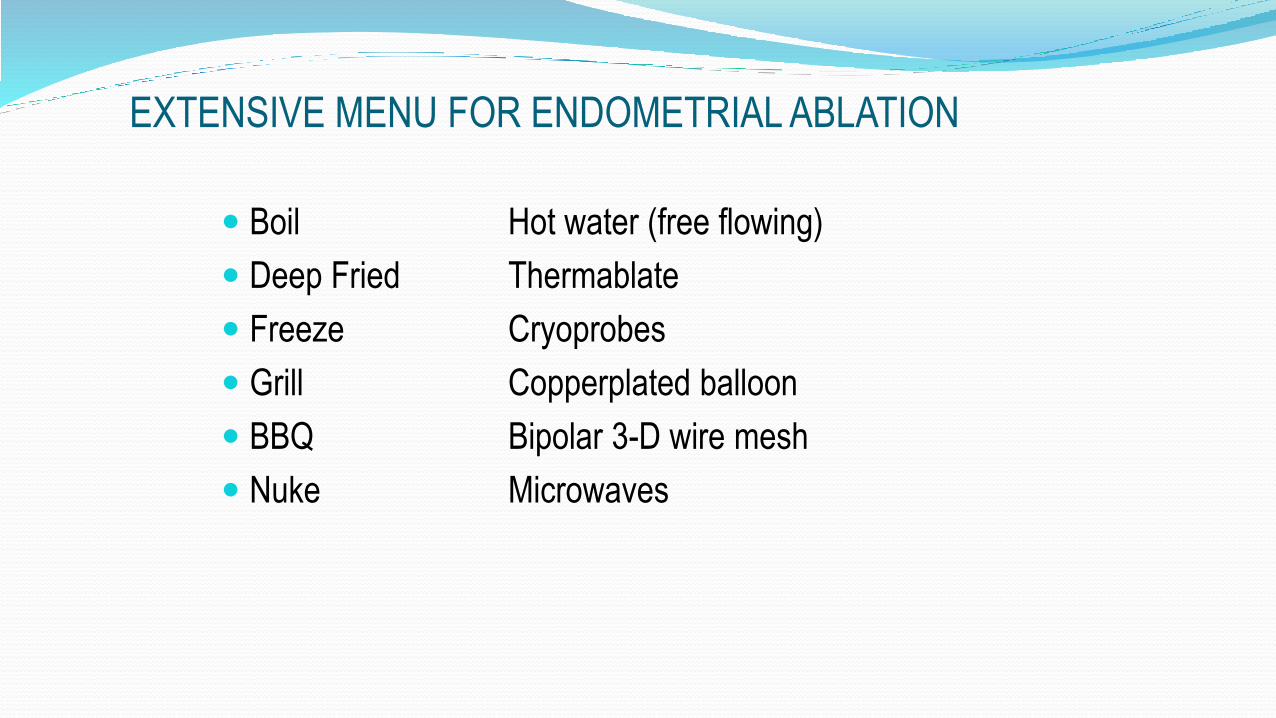
Boil Hot water (free flowing)
Deep Fried Thermablate
Freeze Cryoprobes
Grill Copperplated balloon
BBQ Bipolar 3-D wire mesh
Nuke Microwaves
EXTENSIVE MENU FOR ENDOMETRIAL ABLATION

Training: Part 4
Global Endometrial Ablation
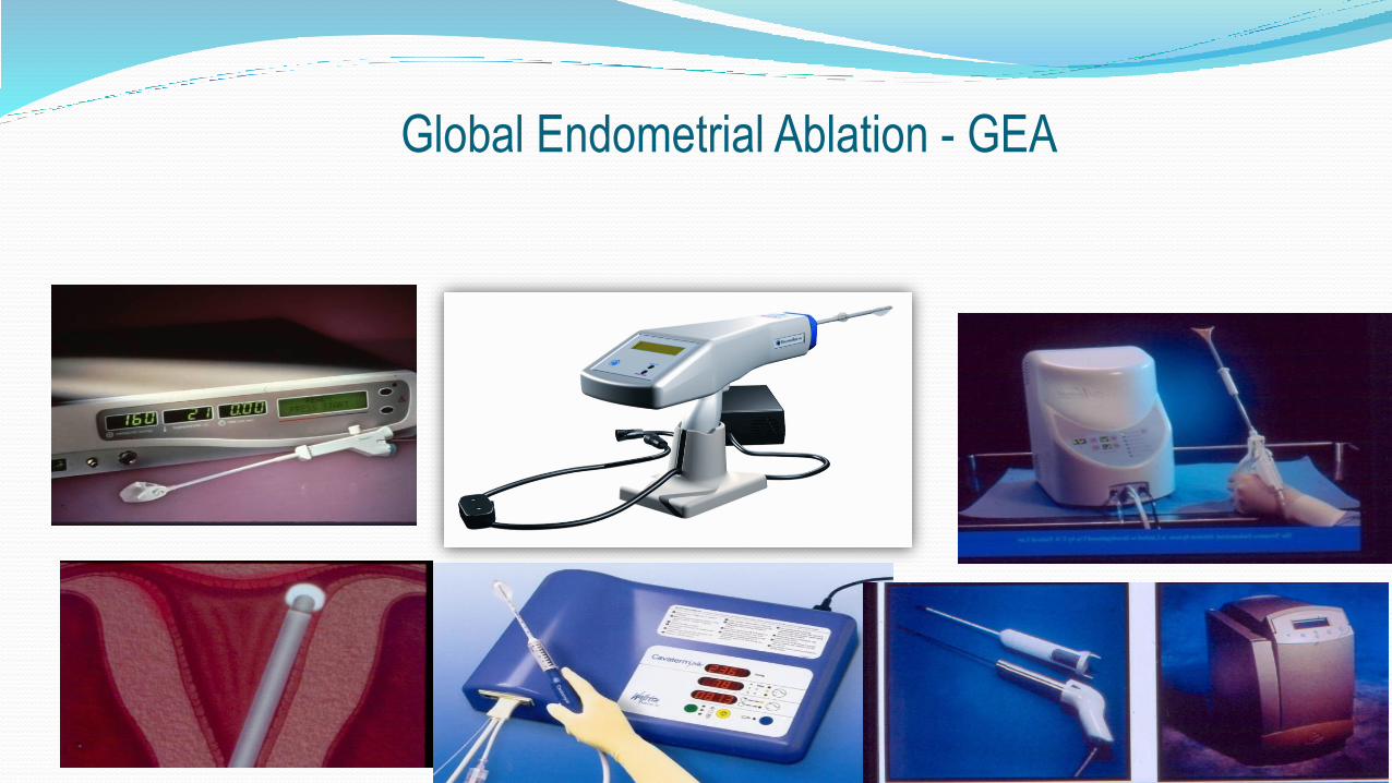
Global Endometrial Ablation - GEA

Current literature shows all 2nd generation techniques give similar results:
- 30-50% of patients achieve amenorrhea
- 80-90% report patient satisfaction after the treatment
Most Important Considerations:
- Patients safety
- Ability to use in outpatient setting!!!
- Versatility to use in an a variety of shape and size uterus
- low cost for treatment
Various Automated Ablation Technologies Now in the Market…
Studies show that 5-25% of women who undergo endometrial ablation will eventually receive
further surgery!
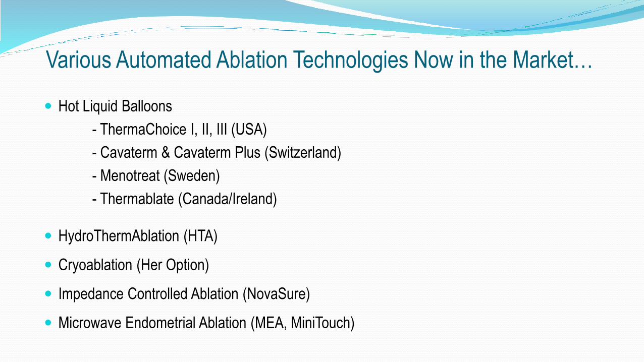
Hot Liquid Balloons
- ThermaChoice I, II, III (USA)
- Cavaterm & Cavaterm Plus (Switzerland)
- Menotreat (Sweden)
- Thermablate (Canada/Ireland)
HydroThermAblation (HTA)
Cryoablation (Her Option)
Impedance Controlled Ablation (NovaSure)
Microwave Endometrial Ablation (MEA, MiniTouch)
Various Automated Ablation Technologies Now in the Market…

The Ideal Endometrial Ablation System: Operator Skills:Short Learning Curve
Not skill dependent
Efficacy and Versatility:Highly effective, with satisfactory immediate and long-term results
Suitable for a variety of most women and uterine cavities
Anesthetic Parameters:Suitable for office procedures
Minimal cervical dilatation
Minimum post post-operative need for analgesia
Safety:Safety independent of operative skills
Device Instructions comply with recommendations of
international healthcare regulatory bodies
Economic parameters:Low capital cost
Low cost for treatment
Durable and easily maintainable equipment

Factors associated with Reduced
Success Rates
• Start of learning curve (more for TC)
• Patients age
• Uterine retroversion (↑chance of
subsequent hysterectomy)
• Preoperative endometrial thickness
of ≥4mm (recommend pre-treatment)
• Duration of menstruation Duration
≥9days (mean)
Factors not influencing success
Rates
• Presence of dysmenorrhea
• Longer duration of Treatment
More studies exist on Balloon Endometrial Ablation vs. any other 2nd generation technology:
- NO EVIDENCE documenting the superiority of one Balloon Endometrial Ablation Technique over another
- Clinical success rates, patients satisfaction and quality of life comparable to Hysteroscopic Ablation Techniques
Thermal Balloon Ablation Techniques


o First Global Ablation System on the market
o Approved by FDA in 1997
o Disposable silicone balloon catheter – circulating fluid
o Fluid (saline) heated to 87°C
o Manual adjustment of pressure and temperature
o 8 -10 minute treatment time
ThermachoiceTM Uterine Balloon Therapy System Johnson & Johnson / Gynecare

• A Thermachoice treatment requires much more Physician involvement
and has a much longer treatment time than one with Thermablate –
Less Suitable for Treatment Under Local Anesthesia
• Physician must manually monitor and control intra-uterine
pressure settings throughout the treatment by infusing the balloon
with cold saline
• Fluid is then heated within the uterine cavity; metallic heating
element inside balloon poses risk of electrical burn and/or
perforation
• Total treatment time 8 – 10 minutes because of the lower
treatment temperature of the fluid
ThermachoiceTM Uterine Balloon Therapy System Johnson & Johnson / Gynecare