Gliadin-Specific T-Cells Mobilized in the Peripheral Blood ... · Blood of Coeliac Patients by...
12
Review Gliadin-Specific T-Cells Mobilized in the Peripheral Blood of Coeliac Patients by Short Oral Gluten Challenge: Clinical Applications Stefania Picascia 1 , Roberta Mandile 2 , Renata Auricchio 2,3 , Riccardo Troncone 2,3 and Carmen Gianfrani 1,3, * Received: 14 September 2015; Accepted: 26 November 2015; Published: 2 December 2015 1 Institute of Protein Biochemistry-CNR, Via Pietro Castellino 111, Naples 80131, Italy; [email protected] 2 Department of Translational Medical Science (DISMET), Section of Pediatrics, University of Naples Federico II, Via S Pansini 5, Naples 80131, Italy; [email protected] (R.M.); [email protected] (R.A.); [email protected] (R.T.) 3 European Laboratory for the Investigation of Food-Induced Diseases (ELFID), University of Naples Federico II, Via S Pansini 5, Naples 80131, Italy * Correspondence: [email protected]; Tel.: +39-081-613-2265; Fax: +39-081-613-2277 Abstract: Celiac disease (CD) is a common lifelong food intolerance triggered by dietary gluten affecting 1% of the general population. Gliadin-specific T-cell lines and T-cell clones obtained from intestinal biopsies have provided great support in the investigation of immuno-pathogenesis of CD. In the early 2000 a new in vivo, less invasive, approach was established aimed to evaluate the adaptive gliadin-specific T-cell response in peripheral blood of celiac patients on a gluten free diet. In fact, it has been demonstrated that three days of ingestion of wheat-containing food induces the mobilization of memory T lymphocytes reactive against gliadin from gut-associated lymphoid tissue into peripheral blood of CD patients. Such antigen-specific T-cells releasing interferon-γ can be transiently detected by using the enzyme-linked immunospot (ELISPOT) assays or by flow cytometry tetramer technology. This paper discusses the suitability of this in vivo tool to investigate the repertoire of gluten pathogenic peptides, to support CD diagnosis, and to assess the efficacy of novel therapeutic strategies. A systematic review of all potential applications of short oral gluten challenge is provided. Keywords: celiac disease; gluten challenge; interferon-γ; ELISPOT 1. Introduction Celiac Disease (CD) is one of the most common food intolerances affecting almost 1% of worldwide population [1]. The disease develops in genetically predisposed subjects as a consequence of an abnormal immune response to wheat gluten and related prolamines of rye and barley. A decisive role in the pathogenesis is played by intestinal gliadin-specific T-cells whose presence seems to be specific of CD patients. Though the causative factor is a dietary protein, CD is considered a chronic inflammatory disorder characterized by autoimmune features. In fact, virtually all subjects with CD produce antibodies against the tissue transglutaminase (tTG) of IgA type which are the disease hallmark with diagnostic relevance [2]. For decades, CD has been considered prevalently an intestinal disease, and the enteropathy the main clinical and histological outcome. Accordingly, the evaluation of small intestinal histology has been for many years the only diagnostic tool in CD [3]. However, the high specificity and sensitivity of tTG IgA antibodies has recently led to a revision of the diagnostic criteria, especially for pediatric subjects. Based on these new guidelines from the ESPGHAN (The European Society for Paediatric Nutrients 2015, 7, 10020–10031; doi:10.3390/nu7125515 www.mdpi.com/journal/nutrients
Transcript of Gliadin-Specific T-Cells Mobilized in the Peripheral Blood ... · Blood of Coeliac Patients by...

Review
Gliadin-Specific T-Cells Mobilized in the PeripheralBlood of Coeliac Patients by Short Oral GlutenChallenge: Clinical Applications
Stefania Picascia 1, Roberta Mandile 2, Renata Auricchio 2,3, Riccardo Troncone 2,3 andCarmen Gianfrani 1,3,*
Received: 14 September 2015; Accepted: 26 November 2015; Published: 2 December 2015
1 Institute of Protein Biochemistry-CNR, Via Pietro Castellino 111, Naples 80131, Italy; [email protected] Department of Translational Medical Science (DISMET), Section of Pediatrics,
University of Naples Federico II, Via S Pansini 5, Naples 80131, Italy;[email protected] (R.M.); [email protected] (R.A.); [email protected] (R.T.)
3 European Laboratory for the Investigation of Food-Induced Diseases (ELFID),University of Naples Federico II, Via S Pansini 5, Naples 80131, Italy
* Correspondence: [email protected]; Tel.: +39-081-613-2265; Fax: +39-081-613-2277
Abstract: Celiac disease (CD) is a common lifelong food intolerance triggered by dietary glutenaffecting 1% of the general population. Gliadin-specific T-cell lines and T-cell clones obtained fromintestinal biopsies have provided great support in the investigation of immuno-pathogenesis ofCD. In the early 2000 a new in vivo, less invasive, approach was established aimed to evaluate theadaptive gliadin-specific T-cell response in peripheral blood of celiac patients on a gluten free diet.In fact, it has been demonstrated that three days of ingestion of wheat-containing food inducesthe mobilization of memory T lymphocytes reactive against gliadin from gut-associated lymphoidtissue into peripheral blood of CD patients. Such antigen-specific T-cells releasing interferon-γcan be transiently detected by using the enzyme-linked immunospot (ELISPOT) assays or by flowcytometry tetramer technology. This paper discusses the suitability of this in vivo tool to investigatethe repertoire of gluten pathogenic peptides, to support CD diagnosis, and to assess the efficacy ofnovel therapeutic strategies. A systematic review of all potential applications of short oral glutenchallenge is provided.
Keywords: celiac disease; gluten challenge; interferon-γ; ELISPOT
1. Introduction
Celiac Disease (CD) is one of the most common food intolerances affecting almost 1% ofworldwide population [1]. The disease develops in genetically predisposed subjects as a consequenceof an abnormal immune response to wheat gluten and related prolamines of rye and barley.A decisive role in the pathogenesis is played by intestinal gliadin-specific T-cells whose presenceseems to be specific of CD patients. Though the causative factor is a dietary protein, CD is considereda chronic inflammatory disorder characterized by autoimmune features. In fact, virtually all subjectswith CD produce antibodies against the tissue transglutaminase (tTG) of IgA type which are thedisease hallmark with diagnostic relevance [2].
For decades, CD has been considered prevalently an intestinal disease, and the enteropathy themain clinical and histological outcome. Accordingly, the evaluation of small intestinal histology hasbeen for many years the only diagnostic tool in CD [3]. However, the high specificity and sensitivityof tTG IgA antibodies has recently led to a revision of the diagnostic criteria, especially for pediatricsubjects. Based on these new guidelines from the ESPGHAN (The European Society for Paediatric
Nutrients 2015, 7, 10020–10031; doi:10.3390/nu7125515 www.mdpi.com/journal/nutrients

Nutrients 2015, 7, 10020–10031
Gastroenterology Hepatology and Nutrition), the evaluation of intestinal mucosa should be no morenecessary to make a diagnosis of CD in the presence of clear symptoms, genetics, and high anti-tTGtiters [4]. Although subjects with overt CD also have a high level of antibodies against gliadin,either for native (AGA) and deamidated (DGP) gliadin peptides, the AGA are not recommendedin the diagnosis of CD due to their low sensitivity and specificity. By contrast, the DGP-IgG have ahigher specificity and are recommended for the CD diagnosis in case of IgA deficiency, or in patientswith both anti-tTG and anti-endomysium (EMA) negative serology. Furthermore, the use of DGP issuggested especially for patients younger than two years. Notwithstanding, a diagnostic challenge isstill posed for those patients deliberately on gluten-free diets to which the intestinal histology andserum antibodies are not helpful. For these specific cases, and for other situations of diagnosticuncertainty, there is still a demand of novel approaches to make a clear and undoubted diagnosisof CD.
For both diagnostic purposes, and to study the mechanisms leading to CD, the demonstrationand characterization of gliadin-specific, pathogenic T-cell response is mandatory. In the early2000s, Anderson and co-workers established an in vivo approach to detect in peripheral bloodthe gluten-specific T-cells of intestinal origin by using the sensitive enzyme-linked immunospot(ELISPOT) assay, widely and successfully used to study antigen-specific T cells secreting cytokines, aswell as antibody-producing B cells, particularly in infectious diseases [5]. The procedure developedby Anderson and co-workers requires the oral administration of wheat bread for three days to celiacpatients on strict gluten free diet (GFD) and the collection of blood samples soon before and six daysafter the challenge started [6]. Since its first application, several studies have shown that the shortgluten challenge (SGC) quickly mobilizes T-cells in the blood of celiac patients on GFD that can berevealed by interferon-γ ELISPOT assays, or flow cytometry tetramers technology, thus suggestingits great clinical potentiality. In general, the clinical symptoms are not severe, and the serum levels ofCD-associated antibodies are unchanged, though in some patients morphological changes can occurafter three days of the gluten challenge [7–10].
2. Genetic Susceptibility, Clinical Spectrum, and Pathogenesis
The susceptibility to develop celiac disease is strongly influenced by inherited factors.The Human Leukocyte Antigen (HLA) class II genes encoding for DQ2.5 (DQA1*05 and DQB1*02alleles) and for DQ8 heterodimers (DQA1*03 and DQB1*0301 alleles) are the main risk factors [11].Although more than 90% of patients with celiac disease have the DQ2.5 genotype, and the remainingones carry either the DQ2.2 or the DQ8 genes, HLA class II account for about 40% of the genetic riskin CD [12–14]. Genome wide association studies (GWAS) have recently identified two genes, the B08and B39 of the HLA class I locus, and a large number of non-HLA genes associated to CD, almost allof them involved in the inflammatory pathways [15].
In CD patients the dietary ingestion of wheat gluten activates a strong immune responsecharacterized by the lymphocytic infiltration in the proximal part of the small bowel [11].Gluten-activated T lymphocytes populate both the epithelium and lamina propria, and play akey role in damaging the intestinal mucosa [11,16]. The consequence is the villous atrophy andcrypt hyperplasia that occurs within a variable window of time after the first gluten consumption.The intestinal damage can range from very mild, showing little or absent histological intestinallesions, to a complete villous flattening, according to Marsh-Oberhuber classification [17]. From theclinical point of view, CD can present in different forms [18]. In the “classical” form, the ingestion ofgluten induces an enteropathy mainly characterized by signs of malabsorption with different degreesof villous atrophy. Most common in adult age, CD may have a “non-classical” form, with no weightloss, nor classical symptoms. The disease may even be “subclinical”, with no symptoms albeit in thepresence of a villous atrophy. In addition, there are genetically predisposed individuals who havehigh anti-tTG titers, but normal small bowel mucosa. It has been reported that almost one third ofthese individuals with “potential celiac disease” will develop the overt disease within nine years [19].
10021

Nutrients 2015, 7, 10020–10031
Recent evidence has highlighted that the number of gluten-reactive T cells both in peripheralblood and in the small intestinal biopsy of CD patients positively correlated with the degree ofhistological intestinal damage. Similarly, the serum anti-TG IgA antibody levels have been foundto significantly correlate to the Marsh grade of mucosal damage [16,20].
Furthermore, it is well known that T lymphocytes reacting to specific gluten peptides andreleasing inflammatory cytokines, such as IFN-γ and IL-21, reside in the intestinal mucosa ofsubjects with CD but not in healthy controls [21]. These cells, mainly CD4+T lymphocytes, react tolong fragments (up to 30–40 amino acid residues) of gluten resistant to gastrointestinal enzymaticdegradation. These gluten peptides pass through the epithelial barrier via transcellular [22] orparacellular transport [23,24], this latter favored by an increased epithelial permeability mediatedby the release of zonulin, an intestinal peptide that is involved in the tight junction regulation [25].When in the lamina propria compartment, the gluten peptides become substrate for the enzyme tissuetransglutaminase type 2 (tTG2) [26]. In particular stress conditions, the tTG2 is released in theextracellular matrix, and acquires an open active form [27]. After the activation, tTG2 specificallyconverts glutamine residues (neutrally charged) in glutamic acid (negatively charged) residues.The deamidated peptides fit the binding pockets of both DQ2 and DQ8 molecules, having a strongaffinity for negative charged peptides [28]. As a consequence, the complex gluten peptide-HLADQ2/DQ8 is specifically recognized by CD4+ T lymphocytes bearing the α/β T-cell receptor (TCR)and activating the inflammatory cascade.
The great heterogeneity of gluten proteins accounts for the large diversity of T-cell epitopesfound to be active in celiac patients [29,30]. The identification of a complete repertoire ofgluten immunogenic sequences is mandatory to better understand either CD pathogenesis, and toprovide the bases for specific disease-targeted immuno-modulatory treatments. Among the severalimmunogenic sequences, three peptides were found the most active: the 33-mer from the α-gliadin(containing the DQ2.5-glia-α1a, DQ2.5-glia-α2 epitopes); the 17-mer from ω-gliadin (containing theDQ2.5-glia-ω-1, DQ2.5-glia-ω-2 epitopes); and the γ-gliadin DQ2.5-glia-γ-1 epitope [31–34]. Of note,many of the gluten T-cell stimulatory sequences have been identified thanks to the availability ofstable T-cell lines and T-cell clones raised from intestinal mucosa tissues. However, the intestinalT-cell cultures have several technical restrictions mainly due to: (i) the limited numbers of cells thatcan be obtained from intestinal biopsies, (ii) long time necessary to establish growing T-cell cultures.Because of that, there is the need to find new tools that allow to investigate the gluten-specific CD4+T-cell response in CD.
3. Current and Emerging Therapies
To date, the only valid treatment for celiac patients is the GFD [35], based on the strict avoidanceof wheat, rye, barley, and all related cereals, including spelt (a wheat variant). After a strict GFD,the intestine recovers a normal morphology and function, and concomitantly, all symptoms andserological disease markers disappear. If from one side the GFD allows the restoration of the intestinalphysiological function, from the other side it is expensive and provides several social restrictions, andcompliance to GFD is not optimal, particularly in adolescence [36,37]. Nutritional properties of glutenfree foods, as for example the high glycemic index and caloric power, increase the risk of treatedceliacs to develop nutritional alterations, obesity, or metabolic syndromes [38,39]. In addition, thereis a minority of patients that suffers from a refractory condition, in which the diet is not efficacious,and requires a pharmacological, anti-inflammatory treatment [40,41]. A deeper knowledge of CDpathophysiology has opened to the investigation of several therapeutic drug-based approachesin the last decade, some of them currently on clinical trial phase II to assess their efficacy [42].This promising scenario strongly demands the availability of a rapid, safe, and reproducible in vivoassay to assess the efficacy of emerging novel therapies to treat CD [43].
10022

Nutrients 2015, 7, 10020–10031
4. Gluten Oral Challenge as Tool to Monitor Intestinal Gluten-Reactive T-Cells
The gluten challenge is a clinical approach widely used in the last decades to have a diagnosisof celiac disease. It consists in the introduction of gluten containing foods in subjects previously ona gluten free diet, for a time frame necessary to provoke a clinical response (from two weeks up tofour months). About 75% of adults received a clear diagnosis in at least two weeks [44], howeverthe response rates and the onset of symptoms were highly variable among different patients [45,46].Either histological, serological, and symptomatic changes are evaluated, to monitor the efficacy ofgluten challenge. However, the extensive gluten challenge has some limitations, such as the risk ofthe overt disease induction (especially in younger patients), and the invasive endoscopy as final exam.Altogether, these findings have raised the need of alternative, less invasive, procedures to investigatethe role of gluten-specific T-cells in the pathogenesis of CD.
4.1. Interferon-γ ELISPOT Assay on Peripheral Blood Cells after a Three Day Gluten Challenge
For long time, all the efforts to isolate gliadin-specific memory T-cells from peripheral bloodsamples of CD patients gave poor results, due to the low frequency in the blood of gluten-primedintestinal CD4+ T-cells, and to a substantial functional differences (in particular a diverse HLArestriction) that has been reported between gliadin-specific T-cell clones raised from the gut orblood [26,47,48]. As consequence, peripheral blood samples have, for a long time, been considerednot optimal tissue material to study the anti-gluten T-cell immunity. At the beginning of this century,Anderson and co-workers published a study describing a new in vivo approach to analyze T-cellresponse to gluten in peripheral blood, overcoming in this way all the technical problems relatedto the use of intestinal T-cells [6]. This procedure requires the oral administration of bread slices(approximately 200 g/day) for three days to CD patients on a strict GFD, and blood samples obtainedat different time points during the gluten challenge. The gluten-reactive T-cells are monitored inperipheral blood mononuclear cells (PBMCs) by detecting those releasing IFN-γ, the prominentmediator of the inflammatory cascade in celiac mucosa, by ELISPOT assay. The ELISPOT is asensitive technique able to catch single cell secreting cytokine, or other immune mediators, uponspecific stimuli. In this pilot study, Anderson and co-workers found that the SGC rarely causesproblems, as only few volunteers, out of 16 adult CD patients enrolled, showed clinical symptomsof disease (usually mild), or had histological signs of intestinal mucosa inflammation. The classicalserological markers of CD, as the anti-endomysium and anti-tTG2 antibodies, remained negative afterthe SGC. The gluten challenge induced in celiac patients a transient IFN-γ response to tTG-treatedchymotrypsin-digested gliadin that was maximum six days after the volunteers began eating breadslices. A 660% increment of IFN-γ spot-forming cells (IFN-γ-SFC) was reported at day six comparedto responses before the challenge, whilst only a 37% of IFN-γ-SFC increment was detected in DQ2+control group. In addition, the gut origin of these circulating T lymphocytes, mobilized in responseto the gluten challenge, was supported by the expression of the α4β7 integrin [7,8], a classical markerof gut homing [49,50]. Similarly, the restriction of gluten response by HLA class II DQ2 molecules,associated to CD risk, was also demonstrated.
A subsequent study from our group performed in adolescent CD patients has reported that theSGC is a reproducible assay. To further demonstrate that the SGC is a valid instrument to investigatethe gluten induced immune response, 14 young celiac patients on GFD underwent two separategluten consumptions, with the same procedure described by Anderson et al. After three to fivemonths of gluten wash-out, the celiac cohort underwent a second cycle of wheat-containing foodchallenge. We found that the IFN-γ responses significantly increased in peripheral blood sampled sixdays after the second challenge, and interestingly, gliadin reactive cells were more frequent comparedto the first challenge, most likely due to the increased frequency of memory T-cells activated upon thefirst gluten exposure [8].
10023

Nutrients 2015, 7, 10020–10031
4.2. Interferon-γ ELISA Assay and Multiparametric Masscytometry to Monitor the Anti-Gluten T-CellsResponse after a Three Day Gluten Challenge
Other studies have reported in vitro read-outs different from ELISPOT assay to assess the specificimmune response elicited by gluten challenge. Ontiveros et al. have developed a whole blood assay todetect gluten-specific T-cells by dosing IFN-γ in the serum by ELISA after stimulation of blood withgluten/peptides [51]. The same research group has also analyzed the peripheral blood cell responseto gluten upon the three days of wheat consumption by measuring the cell proliferation and foundresults consistent with the IFN-γ ELISPOT findings [6].
Despite the central role given by HLA class II in CD, being the main genetic risk factor andthe key restriction molecules of pathogenic CD4+ T-cells, studies from our group have demonstratedthat gliadin contain peptides able to stimulate cytotoxic CD8+ T-cells in an antigen restricted mannerwhen presented on surface of antigen presenting cells (APC), such as B- or enterocytes by HLA classI molecules [52]. Of note, a more recent study from Mark Davis and co-workers using the potentmultiparametric CyTOF technology approach, that allows to monitor simultaneously more than 50different T-cell markers, showed that the three days gluten challenge induced in peripheral bloodof CD patients a remarkable increased of either TCRαβ- and TCRγδ- bearing CD8+ T lymphocytes,other than the CD4+ T-cells [53]. These lymphocytes, expressed the gut homing markers, such asCD103 (intestinal epithelial-homing markers αE) and β7-integrins, thus demonstrating their originfrom intestinal mucosa. The percentage of each cell subset mobilized by gluten intake varies amongsingle patients, but ranged from 1% up to 10% of total peripheral CD8+ cells. This keynote study hasdemonstrated that memory CD8+ T-cells are activated by the oral gluten challenge and circulate fromthe target intestinal tissue to peripheral blood. However, further studies are necessary to assess thegluten specificity of these CD8+ T cells mobilized by the SGC.
4.3. HLA-DQ2-Tetramers as Probe to Detect Gliadin-Specific Cells in Peripheral Blood
In the recent years, much attention has been paid to the use of tetramers technology to dissectspecific T-cell responses to a variety of antigenic sources [20]. Tetramers are composed by four majorhistocompatibility complex (MHC) molecules each of them loaded with a single antigenic peptide,labelled with fluoresceinated biotin-streptavidin complex. The MHC-peptide construct binds to asingle T-cell receptor on the surface membrane of cognate T-cells. When the tetramer is bound, thecells can be visualized by flow cytometry analysis [54]. This sensitive assay allows to quantify the cellfrequency, to assess their phenotype, or to separate the cell subset that specifically reacts to a singleantigen. Tetramer complexes have been widely and successfully used to study MHC class I-restrictedCD8+ T lymphocytes specific for infectious diseases or tumor antigens [55].
DQ2-gliadin-tetramer tests were first used by Raki and co-workers to monitor CD4+ Tlymphocyte specific for two immunodominant gluten epitopes, DQ2.5-glia-α1a and DQ2.5-glia-α2,in PBMCs of celiac patients underwent the SGC [10]. The response rate of such test (approximately85% sensitive and 100% specific evaluated in HLA-DQ2.5+ celiac patients vs. HLA-matched controls)is comparable to that found in IFN-γ ELISPOT assay [10]. Frequencies of positive cells identifiedafter gluten challenge is similar between the two approaches (number of IFN-γ secreting cells foundby ELISPOT ranging from 1 to 5000 in comparison to DQ2.5-glia-α1a tetramer positive cells ranging1:1000 and DQ2.5-glia-α2 tetramer positive cells 1:5000). Similarly to the IFN-γ ELISPOT findings,no tetramer positive cells were detected in DQ2+ healthy controls, either before or after the briefgluten exposure. Interestingly, in subjects with a diagnosis of CD, 5%–8% of total CD4+ cells werestained with tetramer specific for both DQ2.5 α epitopes [10]. More recently, other studies from thesame group have monitored gluten-specific T-cells in peripheral blood of celiac patients by tetramertechnology without the gluten oral challenge [20]. More specifically, gliadin-tetramer positive cellshave been detected in peripheral blood of both treated and untreated DQ2-positive subjects with CD.
In addition, a single cell-TCR sequence analysis performed on DQ2-gliadin-tetramer specificT-cells, mobilized upon the gluten challenge, has demonstrated how highly focused the TCR
10024

Nutrients 2015, 7, 10020–10031
repertoire is of CD4+ T-cells specific for the immunodominant gluten epitopes [53,56–58].Collectively, all these studies demonstrated the great potentiality of the tetramer technology as atool to investigate the anti-gluten T-cell responses. However, tetramer assay has both pros andcons. The main advantage is that it allows to quantify the antigen-specific cells independently bytheir immune function or activation state. More specifically, this technology can also monitor cellsnot releasing a specific cytokine [9]. However, despite the high sensitivity, tetramers allow theidentification of only cells specific for a single peptide, whereas ELISPOT assay allows simultaneousmonitoring for T cells reacting to a wider repertoire of gluten epitopes. Tetramer production,furthermore, is challenging, being laborious, expensive, and time consuming all factors that renderthis technology difficult in application, especially in a clinical practice context. Notwithstanding theabove advantages or disadvantages, it is evident that larger cohort of patients and healthy controlsare needed to validate the sensitivity of tetramer technology to diagnose CD, independently of thegluten challenge.
5. Translational Applications of the Short Gluten Oral Challenge
5.1. Identification of Gluten Immunogenic Peptides
Since the first description, the short oral gluten challenge has become an attractive tool forall researchers interested in the identification of the complete repertoire of gluten (and of otherprolamin) toxic sequences [32,59] (Table 1). Tye-Din and co-workers found a high degree ofT-cell peptide cross reactivity in adult celiacs underwent the SGC by screening a large library(almost 3000) of 20-mer peptides derived from gluten, hordein, and secalin [32]. Interestingly, thoughmany peptides were immunogenic, only the T-cell clones specific for three peptides containingfive epitopes (DQ2.5-glia-α1a/DQ2.5-glia-α2; DQ2.5-glia-ω-1/DQ2.5-glia-ω-2; DQ2.5-Hor-1) werefound responsible for the great majority of responses in adult CD, thus demonstrating a high T-cellstimulatory peptide redundancy.
A recent study from Hardy and co-workers [60] has expanded such peptide repertoire analysisto a pediatric cohort of CD patients. A comparable pattern of peptide recognition was found betweenchildren and adult with CD. These similarities in the nature of the T-cells induced by the in vivoSGC between pediatric and adult CD can have a great potentiality for the applications also in celiacchildren of the peptide-based therapy designed for adults.
5.2. Validation of Therapeutic Drugs
Many studies aimed to identify new strategies to detoxify wheat gluten, and several of theseare based on enzymatic technologies that degrade fragments or mask gluten immune-stimulatorysequences [34] (Table 1). The high content in proline and glutamine-rich peptides make glutenresistant to proteolysis by gastric, pancreatic, and intestinal brush border membrane enzymes.Partially digested gluten fragments stimulate the immune system and became toxic for celiacdisease patients [11]. The identification of a combination of enzymes that can break prolineand glutamine bounds is a fascinating goal for celiac researchers, and it represents an interestingfuture perspective for pharmaceutical sector that aims to produce oral drugs. To this specificpurpose, several gluten-specific proteases, called glutenases, have been isolated from bacteria,fungi, and cereals and are currently under clinical trial investigation. ALV003 is a promisingmixture of two glutenases which cleaves gluten fragments at site enriched in proline and glutamine:a cysteine-endoprotease derived from germinated barley seeds (EP-B2), able to breaks glutenprotein, and a prolyl endopeptidase (PEP) from S. capsulate (SC-PEP) that cleaves proline residues.When combined in 1:1 ratio these two glutenases maximized the enzymatic activity [61]. In a clinicaltrial, 20 patients with celiac disease on GFD were randomized to eat either gluten (16 g/day forthree days) pre-treated with ALV003, or gluten pre-treated with placebo. Patients who receivedALV003 gluten had significantly lower peripheral T-cell IFN-γ response to the immunodominant
10025
Nutrients 2015, 7, 10020–10031
α-gliadin 33-mer multi-epitope peptide, or whole gliadin, compared to the group that received theplacebo [62]. The relevance of the SGC to monitor drug efficacy has been demonstrated in a follow-upstudy, where a double blind, placebo-controlled trial was performed on 41 adults CD patientsrandomized to assume ALV003, or the placebo, along with a gluten daily intake (2 g/day) for sixweeks [63]. In this second study, the main clinical read-out was the evaluation of the small intestinalmucosa damage that appeared. Signs of lymphocytes activation, and intraepithelial infiltration ofCD3+ lymphocytes, both TCRα/β and TCRγ/δ, were found significantly increased only in theplacebo-treated patients, while these markers remained almost unchanged in ALV003 treated group.Though very promising, this drug shows an interesting expectative for its future application, furtherinvestigations are necessary to monitor the long term effects.
Other strategies have been developed to directly detoxify wheat flour, as the extensivehydrolysis during the sourdough fermentation with a mixture of acid bacteria proteases [64], or thetransamidation with methyl-lysine of specific glutamines that are target of the tTG [65] (Table 1).The demonstration of the immuno-stimulatory properties of fermented or transamidated wheat aftera short challenge may provide rapid and preliminary information about the safety and efficacy ofthese novel and promising strategies to produce wheat-based gluten free food for celiac disease.
5.3. Diagnostic Relevance
In recent times, a great attention has been paid to develop clinical practices less invasive thanendoscopy to diagnose celiac disease (Table 1). Moreover, the increased attention paid by the generalpublic to food related problems, as well as the improved distribution of gluten free foods, has spreadthe belief that gluten free diet coincides with a healthy life style; as a consequence more and morepeople voluntary exclude gluten from their diet without a clear diagnosis of celiac disease [66].This makes difficult for clinicians to formulate a definitive diagnosis of celiac disease in unclear cases,as both serological and histological tests revert to the normal value on GFD. Even the HLA genotypingof such individuals complaining gluten related disorders, and on arbitrary GFD, does not help tomake a definitive diagnosis of CD in DQ2 or DQ8 positive subjects, having this test only a negativeprediction value. To date, the only instrument to practice a correct diagnosis in such doubtful casesconsists in the evaluation of histological lesions after a long-term gluten challenge. A recent studyconducted in adults has shown that at least two weeks of gluten consumption allow a clear diagnosisof celiac disease in over 75% of CD population [44]. However, the time frame of the gluten challengeis variable, as indicated by several studies, and may be longer than two weeks. In fact it is clearthat sensitivity to gluten exposure varies greatly between coeliac patients and some may take muchlonger before showing signs of relapse [4,46]. This long-term gluten challenge makes this diagnosticprocedure difficult to practice. Moreover a long-term challenge is not suggested in children youngerthan five years old and during a pubertal growth spurt. The short-time (only three days) of thegluten challenge and the sensitivity of immunological tests offer an interesting perspective for usingthe SGC as a diagnostic tool. Pilot studies of gluten challenge as diagnostic tool for subjects coming tothe observation when they are already on gluten free diet came from Brottveit et al. [10]. The authorsenrolled 35 subjects with uncertain diagnosis and on GFD for at least four weeks and 13 patients withtreated CD. All the enrolled subjects underwent the SGC and endoscopy for biopsy sampling at day0 and day 4 of the gluten challenge. By the positive detection of tetramers the authors have observedgliadin-specific T-cells in 11/13 CD patients, and in only 2/35 with uncertain CD, thus identifying agroup of subjects clinically gluten-sensitive and yet negative to this test.
In order to validate the SGC as a procedure to support diagnosis of celiac disease in uncertaincases, it is mandatory the assessment of its sensitivity and specificity. Regarding the sensitivity,there is a large variability in the cut-off applied to define subject responders to the gluten challengeamong the different studies. In our recent experience of brief gluten challenge performed on acohort of 36 DQ2+ CD adolescent celiacs [8,60], each single patient was considered responsive tooral challenge when showed levels of INF-γ secreting cells in response to whole gliadin, and/or
10026

Nutrients 2015, 7, 10020–10031
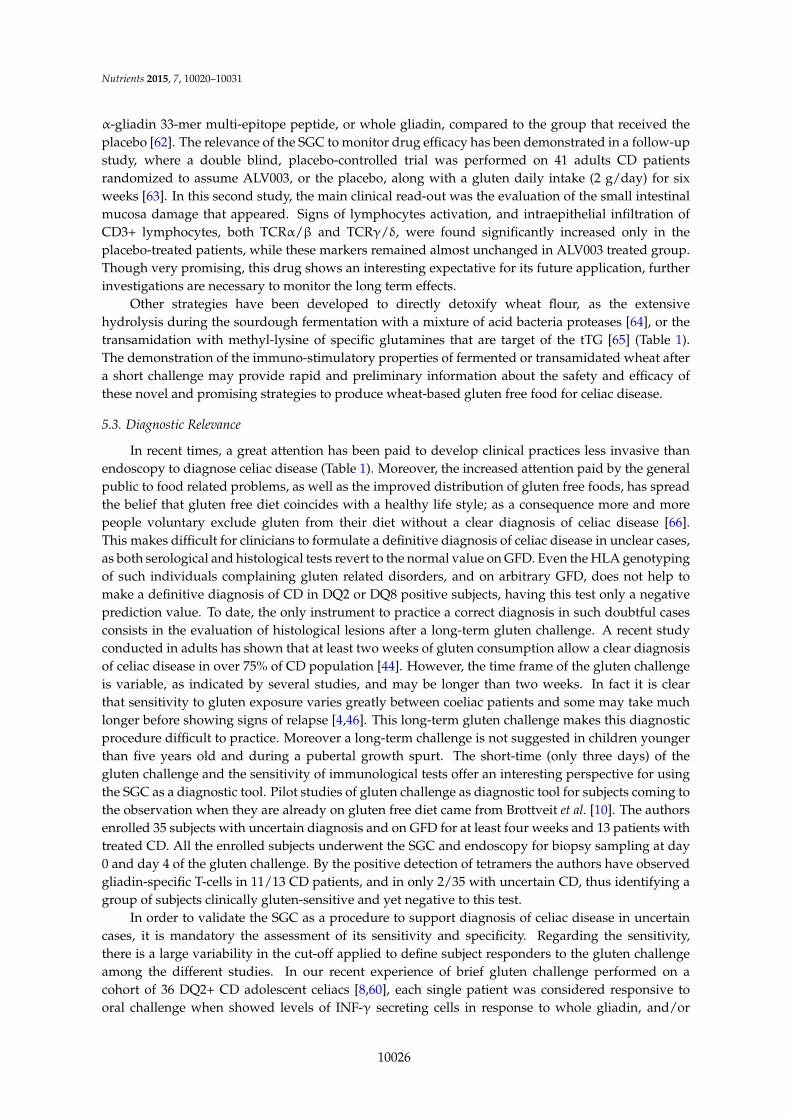
dominant gliadin peptides, that exceeded two-fold the INF-γ responses at day 0 (Fold Increase-FI ě 2)and a difference of SFC/well (∆SFC) of at least 10 between day 6 and day 0. Based on these criteria,we found that almost 73% of our 36 patients are responsive (Figure 1). Other studies reporting ahigher frequency of DQ2+ CD responders (by ELISPOT) ranging from 85% to 92% of respondercases [6–8,51]. These un-matched results, obtained in different celiac cohorts, highlighted how itis important to identify common and unique criteria to validate unequivocally the subjects thatpositively respond to gluten challenge (responders), and to distinguish from the non-responder ones.It is evident that further work is necessary to validate the SGC as diagnostic tool for celiac disease;it is particularly fundamental to expand such an analysis to HLA DQ2+ non celiac healthy control toassess its specificity, which is to date poorly investigated. Indeed the classical diagnostic approachcharacterized by the long gluten challenge, and monitored by serology and histology, is more stableand reliable than the brief challenge, despite that there is the need to have a shorter and less invasivetest to apply to the clinical practice. As stated above, the tetramers are promising diagnostic tools,however several technological limits still remain to be solved for their use large-scale.Nutrients 2015, 7, page–page
8
Figure 1. Percentage of subject responders to the short gluten challenge. A cohort of 36 Italian DQ2‐
positive young celiac patients [8,60] consumed 3–4 wheat bread slices (corresponding to 9–12 g of
gluten/die) for three days. Immunoreactivity was evaluated in peripheral blood at day 0 and day 6 by
INF‐γ‐ELISPOT assay, in response to either whole gliadin or immunodominant α‐gliadin peptides.
Table 1. Possible translational applications of brief gluten challenge.
Application Clinical/Research Purpose References
Diagnosis Confirmation of diagnosis in uncertain celiac disease cases [10,67]
Therapies Validation of new therapeutic drugs [62]
Therapies Validation of biochemical or enzymatic strategies to detoxify gluten. [64,65]
Therapies Searching of wheat cultivars with reduced immunotoxic gluten sequences [68]
Pathogenesis Identification of immunogenic gluten epitopes [32,60]
Pathogenesis Phenotypic analysis of cell population involved in celiac disease [53]
6. Conclusions
The short (three days) gluten challenge is a validated tool for the evaluation and monitoring in
peripheral blood of gluten‐specific T‐cell response that are elicited in the gut after gluten exposure.
Since its first demonstration in early 2000 [6], this in vivo procedure, less invasive than the endoscopy,
has allowed the screening of large peptide library and provided a great help for the characterization
of a repertoire of immunostimulatory gluten sequences, a fundamental step to constructing a peptide‐
base immunotherapy for the treatment of CD [29,32]. Furthermore, the short oral challenge has been
a powered instrument to demonstrate that gluten mobilizes intestinal CD8+ T‐cells, corroborating
their role in CD pathogenesis. The SGC is a promising tool to assess the efficacy of novel treatments
aimed to reduce the load of toxic gluten or of immunomodulatory drugs.
Finally, thanks to several sensitive assays to measure anti‐gluten T response in blood cells, as
IFN‐γ ELISPOT/ELISA or tetramers‐flow cytometry technologies, it is thinkable to apply this
innovative approach to clinical practice, in order to help specialists in making a correct and definitive
diagnosis of celiac disease in those cases in which subjects are arbitrarily on gluten free diet, or can
help the diagnosis in case of potential celiac disease. To make this technique adapt to clinical practice,
several parameters still have to be addressed. Further work is necessary to reach a high sensitivity,
to keep lowest number of false negative subjects. So far, the main limitations playing against the wide
use of oral challenge for clinical practice are less sensitivity and specificity compared to available
serology tests, and the high cost of ELISPOT and tetramers immune assays. Notwithstanding, these
limitations do not lessen the value of the oral challenge, as a rapid tool to assess the efficacy of several
alternative therapies currently under investigation.
Acknowledgments: This work has been supported by funding from CNR‐DSB Progetto Bandiera “InterOmics”
(MIUR PNR 2011‐2013).
Conflicts of Interest: The authors declare no conflict of interest.
Figure 1. Percentage of subject responders to the short gluten challenge. A cohort of 36 ItalianDQ2-positive young celiac patients [8,60] consumed 3–4 wheat bread slices (corresponding to 9–12 gof gluten/die) for three days. Immunoreactivity was evaluated in peripheral blood at day 0 and day 6by INF-γ-ELISPOT assay, in response to either whole gliadin or immunodominant α-gliadin peptides.
Table 1. Possible translational applications of brief gluten challenge.
Application Clinical/Research Purpose References
Diagnosis Confirmation of diagnosis in uncertain celiac disease cases [10,67]Therapies Validation of new therapeutic drugs [62]Therapies Validation of biochemical or enzymatic strategies to detoxify gluten. [64,65]Therapies Searching of wheat cultivars with reduced immunotoxic gluten sequences [68]
Pathogenesis Identification of immunogenic gluten epitopes [32,60]Pathogenesis Phenotypic analysis of cell population involved in celiac disease [53]
6. Conclusions
The short (three days) gluten challenge is a validated tool for the evaluation and monitoringin peripheral blood of gluten-specific T-cell response that are elicited in the gut after glutenexposure. Since its first demonstration in early 2000 [6], this in vivo procedure, less invasive thanthe endoscopy, has allowed the screening of large peptide library and provided a great help forthe characterization of a repertoire of immunostimulatory gluten sequences, a fundamental step toconstructing a peptide-base immunotherapy for the treatment of CD [29,32]. Furthermore, the shortoral challenge has been a powered instrument to demonstrate that gluten mobilizes intestinal CD8+
10027

Nutrients 2015, 7, 10020–10031
T-cells, corroborating their role in CD pathogenesis. The SGC is a promising tool to assess the efficacyof novel treatments aimed to reduce the load of toxic gluten or of immunomodulatory drugs.
Finally, thanks to several sensitive assays to measure anti-gluten T response in blood cells,as IFN-γ ELISPOT/ELISA or tetramers-flow cytometry technologies, it is thinkable to apply thisinnovative approach to clinical practice, in order to help specialists in making a correct and definitivediagnosis of celiac disease in those cases in which subjects are arbitrarily on gluten free diet, or canhelp the diagnosis in case of potential celiac disease. To make this technique adapt to clinical practice,several parameters still have to be addressed. Further work is necessary to reach a high sensitivity, tokeep lowest number of false negative subjects. So far, the main limitations playing against the wideuse of oral challenge for clinical practice are less sensitivity and specificity compared to availableserology tests, and the high cost of ELISPOT and tetramers immune assays. Notwithstanding, theselimitations do not lessen the value of the oral challenge, as a rapid tool to assess the efficacy of severalalternative therapies currently under investigation.
Acknowledgments: This work has been supported by funding from CNR-DSB Progetto Bandiera “InterOmics”(MIUR PNR 2011-2013).
Conflicts of Interest: The authors declare no conflict of interest.
References
1. Lundin, K.E.A.; Qiao, S.-W.; Snir, O.; Sollid, L.M. Coeliac disease—From genetic and immunological studiesto clinical applications. Scand. J. Gastroenterol. 2015, 50, 708–717. [CrossRef] [PubMed]
2. Korponay-Szabó, I.R.; Troncone, R.; Discepolo, V. Adaptive diagnosis of coeliac disease. Best Pract. Res.Clin. Gastroenterol. 2015, 29, 381–398. [CrossRef] [PubMed]
3. Walker-Smith, J.A.; Guandalini, S.; Schmitz, J.; Shmerling, D.H.; Visakorpi, J.K. Revised criteria fordiagnosis of coeliac disease. Arch. Dis. Child. 1990, 65, 909–911.
4. Husby, S.; Koletzko, S.; Korponay-Szabó, I.R.; Mearin, M.L.; Phillips, A.; Shamir, R.; Troncone, R.;Giersiepen, K.; Branski, D.; Catassi, C.; et al. European Society for Pediatric Gastroenterology, Hepatology,and Nutrition Guidelines for the Diagnosis of Coeliac Disease. J. Pediatr. Gastroenterol. Nutr. 2012, 54,136–160. [CrossRef] [PubMed]
5. Currier, J.R.; Kuta, E.G.; Turk, E.; Earhart, L.B.; Loomis-Price, L.; Janetzki, S.; Ferrari, G.; Birx, D.L.; Cox, J.H.A panel of MHC class I restricted viral peptides for use as a quality control for vaccine trial ELISPOT assays.J. Immunol. Methods 2002, 260, 157–172. [CrossRef]
6. Anderson, R.P.; Degano, P.; Godkin, A.J.; Jewell, D.P.; Hill, A.V. In vivo antigen challenge in celiac diseaseidentifies a single transglutaminase-modified peptide as the dominant A-gliadin T-cell epitope. Nat. Med.2000, 6, 337–342. [CrossRef] [PubMed]
7. Anderson, R.P.; van Heel, D.A.; Tye-Din, J.A.; Barnardo, M.; Salio, M.; Jewell, D.P.; Hill, A.V.S. T cells inperipheral blood after gluten challenge in coeliac disease. Gut 2005, 54, 1217–1223. [CrossRef] [PubMed]
8. Camarca, A.; Radano, G.; di Mase, R.; Terrone, G.; Maurano, F.; Auricchio, S.; Troncone, R.; Greco, L.;Gianfrani, C. Short wheat challenge is a reproducible in vivo assay to detect immune response to gluten.Clin. Exp. Immunol. 2012, 169, 129–136. [CrossRef] [PubMed]
9. Ráki, M.; Fallang, L.-E.; Brottveit, M.; Bergseng, E.; Quarsten, H.; Lundin, K.E.A.; Sollid, L.M. Tetramervisualization of gut-homing gluten-specific T cells in the peripheral blood of celiac disease patients.Proc. Natl. Acad. Sci. USA 2007, 104, 2831–2836. [CrossRef] [PubMed]
10. Brottveit, M.; Ráki, M.; Bergseng, E.; Fallang, L.-E.; Simonsen, B.; Løvik, A.; Larsen, S.; Løberg, E.M.;Jahnsen, F.L.; Sollid, L.M.; Lundin, K.E.A. Assessing possible celiac disease by an HLA-DQ2-gliadinTetramer Test. Am. J. Gastroenterol. 2011, 106, 1318–1324. [CrossRef] [PubMed]
11. Abadie, V.; Sollid, L.M.; Barreiro, L.B.; Jabri, B. Integration of genetic and immunological insights into amodel of celiac disease pathogenesis. Annu. Rev. Immunol. 2011, 29, 493–525. [CrossRef] [PubMed]
12. Van Heel, D.A.; Hunt, K.; Greco, L.; Wijmenga, C. Genetics in coeliac disease. Best Pract. Res.Clin. Gastroenterol. 2005, 19, 323–339. [CrossRef] [PubMed]
13. Sollid, L.M.; Thorsby, E. HLA susceptibility genes in celiac disease: Genetic mapping and role inpathogenesis. Gastroenterology 1993, 105, 910–922. [PubMed]
10028

Nutrients 2015, 7, 10020–10031
14. Karell, K.; Louka, A.S.; Moodie, S.J.; Ascher, H.; Clot, F.; Greco, L.; Ciclitira, P.J.; Sollid, L.M.; Partanen, J.HLA types in celiac disease patients not carrying the DQA1*05-DQB1*02 (DQ2) heterodimer: Results fromthe European Genetics Cluster on Celiac Disease. Hum. Immunol. 2003, 64, 469–477. [CrossRef]
15. Kumar, V.; Gutierrez-Achury, J.; Kanduri, K.; Almeida, R.; Hrdlickova, B.; Zhernakova, D.V.; Westra, H.J.;Karjalainen, J.; Ricaño-Ponce, I.; Li, Y.; et al. Systematic annotation of celiac disease loci refines pathologicalpathways and suggests a genetic explanation for increased interferon-gamma levels. Hum. Mol. Genet.2015, 24, 397–409. [CrossRef] [PubMed]
16. Bodd, M.; Ráki, M.; Bergseng, E.; Jahnsen, J.; Lundin, K.E.A.; Sollid, L.M. Direct cloning and tetramerstaining to measure the frequency of intestinal gluten-reactive T cells in celiac disease. Eur. J. Immunol.2013, 43, 2605–2612. [CrossRef] [PubMed]
17. Oberhuber, G.; Granditsch, G.; Vogelsang, H. The histopathology of coeliac disease: Time for a standardizedreport scheme for pathologists. Eur. J. Gastroenterol. Hepatol. 1999, 11, 1185–1194. [CrossRef] [PubMed]
18. Ludvigsson, J.F.; Leffler, D.A.; Bai, J.C.; Biagi, F.; Fasano, A.; Green, P.H.R.; Hadjivassiliou, M.; Kaukinen, K.;Kelly, C.P.; Leonard, J.N.; et al. The Oslo definitions for coeliac disease and related terms. Gut 2013, 62,43–52. [CrossRef] [PubMed]
19. Auricchio, R.; Tosco, A.; Piccolo, E.; Galatola, M.; Izzo, V.; Maglio, M.; Paparo, F.; Troncone, R.; Greco, L.Potential celiac children: 9-year follow-up on a gluten-containing diet. Am. J. Gastroenterol. 2014, 109,913–921. [CrossRef] [PubMed]
20. Christophersen, A.; Ráki, M.; Bergseng, E.; Lundin, K.E.; Jahnsen, J.; Sollid, L.M.; Qiao, S.-W.Tetramer-visualized gluten-specific CD4+ T cells in blood as a potential diagnostic marker for coeliacdisease without oral gluten challenge. United Eur. Gastroenterol. J. 2014, 2, 268–278. [CrossRef] [PubMed]
21. Gianfrani, C.; Auricchio, S.; Troncone, R. Adaptive and innate immune responses in celiac disease.Immunol. Lett. 2005, 99, 141–145. [CrossRef] [PubMed]
22. Matysiak-Budnik, T.; Moura, I.C.; Arcos-Fajardo, M.; Lebreton, C.; Ménard, S.; Candalh, C.; ben-Khalifa, K.;Dugave, C.; Tamouza, H.; van Niel, G.; et al. Secretory IgA mediates retrotranscytosis of intact gliadinpeptides via the transferrin receptor in celiac disease. J. Exp. Med. 2008, 205, 143–154. [CrossRef] [PubMed]
23. Tripathi, A.; Lammers, K.M.; Goldblum, S.; Shea-Donohue, T.; Netzel-Arnett, S.; Buzza, M.S.; Antalis, T.M.;Vogel, S.N.; Zhao, A.; Yang, S.; et al. Identification of human zonulin, a physiological modulator of tightjunctions, as prehaptoglobin-2. Proc. Natl. Acad. Sci. USA 2009, 106, 16799–16804. [CrossRef] [PubMed]
24. Lammers, K.M.; Lu, R.; Brownley, J.; Lu, B.; Gerard, C.; Thomas, K.; Rallabhandi, P.; Shea-Donohue, T.;Tamiz, A.; Alkan, S.; et al. Gliadin induces an increase in intestinal permeability and zonulin release bybinding to the chemokine receptor CXCR3. Gastroenterology 2008, 135, 194–204. [CrossRef] [PubMed]
25. Fasano, A.; Not, T.; Wang, W.; Uzzau, S.; Berti, I.; Tommasini, A.; Goldblum, S.E. Zonulin, a newlydiscovered modulator of intestinal permeability, and its expression in coeliac disease. Lancet 2000, 355,1518–1519. [CrossRef]
26. Molberg, O.; Mcadam, S.N.; Körner, R.; Quarsten, H.; Kristiansen, C.; Madsen, L.; Fugger, L.; Scott, H.;Norén, O.; Roepstorff, P.; et al. Tissue transglutaminase selectively modifies gliadin peptides that arerecognized by gut-derived T cells in celiac disease. Nat. Med. 1998, 4, 713–717. [CrossRef] [PubMed]
27. Pinkas, D.M.; Strop, P.; Brunger, A.T.; Khosla, C. Transglutaminase 2 undergoes a large conformationalchange upon activation. PLoS Biol. 2007. [CrossRef] [PubMed]
28. Sollid, L.M.; Jabri, B. Celiac disease and transglutaminase 2: A model for posttranslational modification ofantigens and HLA association in the pathogenesis of autoimmune disorders. Curr. Opin. Immunol. 2011,23, 732–873. [CrossRef] [PubMed]
29. Camarca, A.; del Mastro, A.; Gianfrani, C. Repertoire of gluten peptides active in celiac disease patients:Perspectives for translational therapeutic applications. Endocr. Metab. Immune Disord. Drug Targets 2012,12, 207–219. [CrossRef] [PubMed]
30. Wieser, H. Chemistry of gluten proteins. Food Microbiol. 2007, 24, 115–119. [CrossRef] [PubMed]31. Shan, L.; Molberg, Ø.; Parrot, I.; Hausch, F.; Filiz, F.; Gray, G.M.; Sollid, L.M.; Khosla, C. Structural basis for
gluten intolerance in celiac sprue. Science 2002, 297, 2275–2279. [CrossRef] [PubMed]32. Tye-Din, J.A.; Stewart, J.A.; Dromey, J.A.; Beissbarth, T.; van Heel, D.A.; Tatham, A.; Henderson, K.;
Mannering, S.I.; Gianfrani, C.; Jewell, D.P.; et al. Comprehensive, quantitative mapping of T cell epitopes ingluten in celiac disease. Sci. Transl. Med. 2010. [CrossRef] [PubMed]
10029

Nutrients 2015, 7, 10020–10031
33. Camarca, A.; Anderson, R.P.; Mamone, G.; Fierro, O.; Facchiano, A.; Costantini, S.; Zanzi, D.; Sidney, J.;Auricchio, S.; Sette, A.; et al. Intestinal T cell responses to gluten peptides are largely heterogeneous:Implications for a peptide-based therapy in celiac disease. J. Immunol. 2009, 182, 4158–4166. [CrossRef][PubMed]
34. Sollid, L.M.; Qiao, S.-W.; Anderson, R.P.; Gianfrani, C.; Koning, F. Nomenclature and listing of celiacdisease relevant gluten T-cell epitopes restricted by HLA-DQ molecules. Immunogenetics 2012, 64, 455–460.[CrossRef] [PubMed]
35. Sollid, L.M.; Lundin, K.E.A. Diagnosis and treatment of celiac disease. Mucosal Immunol. 2009, 2, 3–7.[CrossRef] [PubMed]
36. Greco, L.; Mayer, M.; Ciccarelli, G.; Troncone, R.; Auricchio, S. Compliance to a gluten-free diet inadolescents, or “what do 300 coeliac adolescents eat every day?”. Ital. J. Gastroenterol. Hepatol. 1997,29, 305–310. [PubMed]
37. Errichiello, S.; Esposito, O.; di Mase, R.; Camarca, M.E.; Natale, C.; Limongelli, M.G.; Marano, C.;Coruzzo, A.; Lombardo, M.; Strisciuglio, P.; et al. Celiac disease: Predictors of compliance with a gluten-freediet in adolescents and young adults. J. Pediatr. Gastroenterol. Nutr. 2010, 50, 54–60. [CrossRef] [PubMed]
38. Lamacchia, C.; Camarca, A.; Picascia, S.; Di Luccia, A.; Gianfrani, C. Cereal-based gluten-free food: How toreconcile nutritional and technological properties of wheat proteins with safety for celiac disease patients.Nutrients 2014, 6, 575–590. [CrossRef] [PubMed]
39. Diamanti, A.; Capriati, T.; Basso, M.S.; Panetta, F.; di Ciommo Laurora, V.M.; Bellucci, F.; Cristofori, F.;Francavilla, R. Celiac disease and overweight in children: An update. Nutrients 2014, 6, 207–220. [CrossRef][PubMed]
40. Ryan, B.M.; Kelleher, D. Refractory celiac disease. Gastroenterology 2000, 119, 243–251. [CrossRef] [PubMed]41. Malamut, G.; Cellier, C. Complications of coeliac disease. Best Pract. Res. Clin. Gastroenterol. 2015, 29,
451–458. [CrossRef] [PubMed]42. Kaukinen, K.; Lindfors, K.; Mäki, M. Advances in the treatment of coeliac disease: An immunopathogenic
perspective. Nat. Rev. Gastroenterol. Hepatol. 2014, 11, 36–44. [CrossRef] [PubMed]43. Ludvigsson, J.F.; Card, T.; Ciclitira, P.J.; Swift, G.L.; Nasr, I.; Sanders, D.S.; Ciacci, C. Support for patients
with celiac disease: A literature review. United Eur. Gastroenterol. J. 2015, 3, 146–159. [CrossRef] [PubMed]44. Leffler, D.; Schuppan, D.; Pallav, K.; Najarian, R.; Goldsmith, J.D.; Hansen, J.; Kabbani, T.; Dennis, M.;
Kelly, C.P. Kinetics of the histological, serological and symptomatic responses to gluten challenge in adultswith coeliac disease. Gut 2013, 62, 996–1004. [CrossRef] [PubMed]
45. Tack, G.J.; van de Water, J.M.W.; Bruins, M.J.; Kooy-Winkelaar, E.M.C.; van Bergen, J.; Bonnet, P.;Vreugdenhil, A.C.E.; Korponay-Szabo, I.; Edens, L.; von Blomberg, B.M.E.; et al. Consumption of glutenwith gluten-degrading enzyme by celiac patients: A pilot-study. World J. Gastroenterol. 2013, 19, 5837–5847.[CrossRef] [PubMed]
46. Mayer, M.; Greco, L.; Troncone, R.; Grimaldi, M.; Pansa, G. Early prediction of relapse during glutenchallenge in childhood celiac disease. J. Pediatr. Gastroenterol. Nutr. 1989, 8, 474–479. [CrossRef] [PubMed]
47. Gjertsen, H.A.; Sollid, L.M.; Ek, J.; Thorsby, E.; Lundin, K.E. T cells from the peripheral blood ofcoeliac disease patients recognize gluten antigens when presented by HLA-DR, -DQ, or -DP molecules.Scand. J. Immunol. 1994, 39, 567–574. [CrossRef] [PubMed]
48. Jensen, K.; Sollid, L.M.; Scott, H.; Paulsen, G.; Kett, K.; Thorsby, E.; Lundin, K.E. Gliadin-specific Tcell responses in peripheral blood of healthy individuals involve T cells restricted by the coeliac diseaseassociated DQ2 heterodimer. Scand. J. Immunol. 1995, 42, 166–170. [CrossRef] [PubMed]
49. Quiding-Järbrink, M.; Ahlstedt, I.; Lindholm, C.; Johansson, E.L.; Lönroth, H. Homing commitment oflymphocytes activated in the human gastric and intestinal mucosa. Gut 2001, 49, 519–525. [CrossRef][PubMed]
50. Berlin, C.; Berg, E.L.; Briskin, M.J.; Andrew, D.P.; Kilshaw, P.J.; Holzmann, B.; Weissman, I.L.; Hamann, A.;Butcher, E.C. Alpha 4 beta 7 integrin mediates lymphocyte binding to the mucosal vascular addressinMAdCAM-1. Cell 1993, 74, 185–195. [CrossRef]
51. Ontiveros, N.; Tye-Din, J.A.; Hardy, M.Y.; Anderson, R.P. Ex-vivo whole blood secretion of interferon (IFN)-γand IFN-γ-inducible protein-10 measured by enzyme-linked immunosorbent assay are as sensitive asIFN-γ enzyme-linked immunospot for the detection of gluten-reactive T cells in human leucocyte antigen(HLA)-DQ2¨ 5(+) -associated coeliac disease. Clin. Exp. Immunol. 2014, 175, 305–315. [PubMed]
10030

Nutrients 2015, 7, 10020–10031
52. Mazzarella, G.; Stefanile, R.; Camarca, A.; Giliberti, P.; Cosentini, E.; Marano, C.; Iaquinto, G.; Giardullo, N.;Auricchio, S.; Sette, A.; et al. Gliadin activates HLA class I-restricted CD8+ T cells in celiac disease intestinalmucosa and induces the enterocyte apoptosis. Gastroenterology 2008, 134, 1017–1027. [CrossRef] [PubMed]
53. Han, A.; Newell, E.W.; Glanville, J.; Fernandez-Becker, N.; Khosla, C.; Chien, Y.-H.; Davis, M.M. Dietarygluten triggers concomitant activation of CD4+ and CD8+ αβ T cells and γδ T cells in celiac disease.Proc. Natl. Acad. Sci. USA 2013, 110, 13073–13078. [CrossRef] [PubMed]
54. Altman, J.D.; Moss, P.A.; Goulder, P.J.; Barouch, D.H.; McHeyzer-Williams, M.G.; Bell, J.I.; McMichael, A.J.;Davis, M.M. Phenotypic analysis of antigen-specific T lymphocytes. Science 1996, 274, 94–96. [CrossRef][PubMed]
55. Klenerman, P.; Cerundolo, V.; Dunbar, P.R. Tracking T cells with tetramers: New tales from new tools.Nat. Rev. Immunol. 2002, 2, 263–272. [CrossRef] [PubMed]
56. Qiao, S.-W.; Ráki, M.; Gunnarsen, K.S.; Løset, G.-Å.; Lundin, K.E.A.; Sandlie, I.; Sollid, L.M.Posttranslational modification of gluten shapes TCR usage in celiac disease. J. Immunol. 2011, 187,3064–3071. [CrossRef] [PubMed]
57. Qiao, S.-W.; Christophersen, A.; Lundin, K.E.A.; Sollid, L.M. Biased usage and preferred pairing of α- andβ-chains of TCRs specific for an immunodominant gluten epitope in coeliac disease. Int. Immunol. 2014, 26,13–19. [CrossRef] [PubMed]
58. Broughton, S.E.; Petersen, J.; Theodossis, A.; Scally, S.W.; Loh, K.L.; Thompson, A.; van Bergen, J.;Kooy-Winkelaar, Y.; Henderson, K.N.; Beddoe, T.; et al. Biased T-cell receptor usage directed against humanleukocyte antigen DQ8-restricted gliadin peptides is associated with celiac disease. Immunity 2012, 37,611–621. [CrossRef] [PubMed]
59. Tanner, G.J.; Howitt, C.A.; Forrester, R.I.; Campbell, P.M.; Tye-Din, J.A.; Anderson, R.P. Dissectingthe T-cell response to hordeins in coeliac disease can develop barley with reduced immunotoxicity.Aliment. Pharmacol. Ther. 2010, 32, 1184–1191. [CrossRef] [PubMed]
60. Hardy, M.Y.; Girardin, A.; Pizzey, C.; Cameron, D.J.; Watson, K.A.; Picascia, S.; Auricchio, R.; Greco, L.;Gianfrani, C.; La Gruta, N.L.; et al. Consistency in Polyclonal T-cell Responses to Gluten Between Childrenand Adults with Celiac Disease. Gastroenterology 2015, 6, 1541–1552. [CrossRef] [PubMed]
61. Freeman, H.J. Emerging drugs for celiac disease. Expert Opin. Emerg. Drugs 2015, 20, 129–135. [CrossRef][PubMed]
62. Tye-Din, J.A.; Anderson, R.P.; Ffrench, R.A.; Brown, G.J.; Hodsman, P.; Siegel, M.; Botwick, W.;Shreeniwas, R. The effects of ALV003 pre-digestion of gluten on immune response and symptoms in celiacdisease in vivo. Clin. Immunol. 2010, 134, 289–295. [CrossRef] [PubMed]
63. Lähdeaho, M.-L.; Kaukinen, K.; Laurila, K.; Vuotikka, P.; Koivurova, O.-P.; Kärjä-Lahdensuu, T.;Marcantonio, A.; Adelman, D.C.; Mäki, M. Glutenase ALV003 attenuates gluten-induced mucosal injuryin patients with celiac disease. Gastroenterology 2014, 146, 1649–1658. [CrossRef] [PubMed]
64. Greco, L.; Gobbetti, M.; Auricchio, R.; di Mase, R.; Landolfo, F.; Paparo, F.; di Cagno, R.; de Angelis, M.;Rizzello, C.G.; Cassone, A.; et al. Safety for patients with celiac disease of baked goods made of wheat flourhydrolyzed during food processing. Clin. Gastroenterol. Hepatol. 2011, 9, 24–29. [CrossRef] [PubMed]
65. Gianfrani, C.; Siciliano, R.A.; Facchiano, A.M.; Camarca, A.; Mazzeo, M.F.; Costantini, S.; Salvati, V.M.;Maurano, F.; Mazzarella, G.; Iaquinto, G.; et al. Transamidation of wheat flour inhibits the response togliadin of intestinal T cells in celiac disease. Gastroenterology 2007, 133, 780–789. [CrossRef] [PubMed]
66. NPD Group. Dieting Monitor: Eating Patterns in America; NPD Group: Port Washington, NY, USA, 2013.67. Brottveit, M.; Beitnes, A.C.; Tollefsen, S.; Bratlie, J.E.; Jahnsen, F.L.; Johansen, F.E.; Sollid, L.M.;
Lundin, K.E.A. Mucosal Cytokine Response After Short-Term Gluten Challenge in Celiac Disease andNon-Celiac Gluten Sensitivity. Am. J. Gastroenterol. 2013, 108, 842–850. [CrossRef] [PubMed]
68. Gianfrani, C.; Camarca, A.; Mazzarella, G.; di Stasio, L.; Giardullo, N.; Ferranti, P.; Picariello, G.; RotondiAufiero, V.; Picascia, S.; Troncone, R.; et al. Extensive in vitro gastrointestinal digestion markedly reducesthe immune-toxicity of Triticum monococcum wheat: Implication for celiac disease. Mol. Nutr. Food Res.2015. [CrossRef] [PubMed]
© 2015 by the authors; licensee MDPI, Basel, Switzerland. This article is an openaccess article distributed under the terms and conditions of the Creative Commons byAttribution (CC-BY) license (http://creativecommons.org/licenses/by/4.0/).
10031