Git5 malabsorption syndrome
28
DR S C GAN FMHS/UTAR 10102012 1 GIT5: MALABSORPTION SYNDROME DR GAN SENG CHIEW Associate Professor FACULTY OF MEDICINE & HEALTH SCIENCES UNIVERSITY TUNKU ABDUL RAHMAN
-
Upload
gansc1 -
Category
Health & Medicine
-
view
332 -
download
3
Transcript of Git5 malabsorption syndrome

DR S C GAN FMHS/UTAR 10102012 1
GIT5: MALABSORPTION SYNDROME
DR GAN SENG CHIEWAssociate Professor
FACULTY OF MEDICINE & HEALTH SCIENCESUNIVERSITY TUNKU ABDUL RAHMAN

DR S C GAN FMHS/UTAR 10102012 2
Malabsorption
• The failure to absorb or digest normally one or more dietary constituents.
• Patients normally complain of diarrhea.• Most patients with malabsorption present
with a syndrome characterized by large, loose, foul-smelling stools and loss of weight.
• A wide variety of dosorders of the organs of digestion can cause malabsorption or maldigestion.

DR S C GAN FMHS/UTAR 10102012 3
Malabsorption syndromes encompass numerous clinical entities that result in chronic diarrhea, abdominal distention, and failure tothrive. Clinical malabsorption can be broken down into several distinct conditions, both congenital and acquired, that affect one or more of the different steps in the intestinal hydrolysis and subsequent transport of nutrients.The major site of absorption is the small intestine,.

DR S C GAN FMHS/UTAR 10102012 4
Digestive Disorder Examples
Pancreatic exocrine insufficiency
Chronic pancreatitis
Pancreatic carcinoma,
Bile acid insufficiency Small-bowel bacterial overgrowth
Crohn’s disease of the terminal ileum
Small-bowel disease:
a. Mucosal Disorder
b. Specific absorptive defects
Celic spruce; Collagenous sprue;
Tropical Sprue; Whipple’s disease;
Radiation enteritis; Ischemic disease; Intestinal lymphoma; Regional enteritis
(Crohn’s diisease); Amyloidosis.
Primary lactase deficiency
Abetalipoproteinemia
Lymphatic disorders Intestinal lymphangiectasia
Mixed defects in absorption
Zollinger-Ellison syndrome
Postgastrectomy disorders

DR S C GAN FMHS/UTAR 10102012 5
Pathophysiology • In general, the digestion and absorption of food
materials can be divided into 3 major phases: luminal, mucosal, and postabsorptive.
• The luminal phase is the phase in which dietary fats, proteins, and carbohydrates are hydrolyzed and solubilized by secreted digestive enzymes and bile. The mucosal phase relies on the integrity of the brush-border membrane of intestinal epithelial cells to transport digested products from the lumen into the cells. In the postabsorptive phase, reassembled lipids and other key nutrients are transported via lymphatics and portal circulation from epithelial cells to other parts of the body.
• Perturbation by disease processes in any of these phases frequently results in malabsorption.

DR S C GAN FMHS/UTAR 10102012 6
Luminal phase: Impaired nutrient hydrolysis
• The most common cause for impaired nutrient hydrolysis is pancreatic insufficiency due to chronic pancreatitis, pancreatic resection, pancreatic cancer, or cystic fibrosis. The resultant deficiencies in lipase and proteases lead to lipid and protein malabsorption, respectively.
• Inactivation of pancreatic enzymes by gastric hypersecretion, as seen in Zollinger-Ellison syndrome, is another cause.
• Inadequate mixing of nutrients, bile, and pancreatic enzymes, as seen in rapid intestinal transit, gastrojejunostomy, total and partial gastrectomy, or intestinal resection after mesenteric emboli or thrombosis, also causes impaired hydrolysis.
• Rarely, a failure to convert a proenzyme to active form, such as enterokinase and trypsinogen deficiencies, also can cause protein maldigestion and malabsorption.

DR S C GAN FMHS/UTAR 10102012 7
Luminal phase: Impaired micelle formation
• Impaired micelle formation causes a problem in fat solubilization and subsequent fat malabsorption. This impairment is due to different reasons, including (1) decreased bile salt synthesis from severe parenchymal liver disease (eg, cirrhosis); (2) impaired bile secretion from biliary obstruction or cholestatic jaundice (eg, primary biliary cirrhosis, primary sclerosing cholangitis); (3) impaired enterohepatic bile circulation, as seen in small bowel resection or regional enteritis; or (4) bile salt deconjugation due to small bowel bacterial overgrowth.
• Stasis of intestinal content caused by a motor abnormality (eg, scleroderma, diabetic neuropathy, intestinal obstruction), an anatomic abnormality (eg, small bowel diverticula, stricture, ischemia, blind loops), or small bowel contamination from enterocolonic fistulas can cause bacterial overgrowth.

DR S C GAN FMHS/UTAR 10102012 8
Luminal phase : Luminal availabil i ty and processing
• Luminal bacterial overgrowth can cause a decrease in the availability of substrates, including carbohydrates, proteins, and vitamins (eg, vitamin B-12, folate).
• Vitamin B-12 deficiency due to pernicious anemia is caused by a lack of intrinsic factor and by pancreatic enzyme deficiency.

DR S C GAN FMHS/UTAR 10102012 9
Mucosal phase : Impaired brush-border hydrolase activity
• Disaccharidase deficiency can lead to disaccharide malabsorption.
• Lactase deficiency, either primary or secondary, is the most common form of disaccharidase deficiency. Genetic factors determine primary lactase deficiency. Secondary lactase deficiency can be due to acute gastroenteritis (rotavirus and giardia infection), chronic alcoholism, celiac sprue, radiation enteritis, regional enteritis, or AIDS enteropathy.
• IgA deficiency (most common immunodeficiency) is due to decreased or absent serum and intestinal IgA, which clinically appears similar to celiac disease and is unresponsive to a gluten-free diet.

DR S C GAN FMHS/UTAR 10102012 10
• Acrodermatitis enteropathica is an autosomal recessive disease with selective inability to absorb zinc, leading to villous atrophy and acral dermatitis.
• Autoimmune enteropathy primarily diagnosed in children presenting with intractable secretory diarrhea and villous atrophy. Autoimmune enteropathy is due to antibodies directed against intestinal epithelial and goblet cells. Additional cell types affected by autoantibodies include islet and parietal cells.
• Other carbohydrase deficiencies, such as sucrase-isomaltase deficiency, may be the cause.

DR S C GAN FMHS/UTAR 10102012 11
Mucosal phase : Impaired nutrient absorption
• Inherited defects include glucose-galactose malabsorption, abetalipoproteinemia, cystinuria, and Hartnup disease.
• Acquired disorders are far more common and are caused by the following: (1) decreased absorptive surface area, as seen in intestinal resection of intestinal bypass; (2) damaged absorbing surface, as seen in celiac sprue, tropical sprue, Crohn's disease, AIDS enteropathy, chemotherapy, or radiation therapy; (3) infiltrating disease of the intestinal wall, such as lymphoma and amyloidosis; and (4) infections, including bacterial overgrowth, giardiasis, Whipple's disease, cryptosporidiosis, and microsporidiosis.

DR S C GAN FMHS/UTAR 10102012 12
Postabsorptive phase
Obstruction of the lymphatic system, both congenital (eg, intestinal lymphangiectasia, Milroy disease) and acquired (eg, Whipple disease, neoplasm [including lymphoma], tuberculosis), impairs the absorption of chylomicrons and lipoproteins and may cause fat malabsorption or a protein-losing enteropathy.

DR S C GAN FMHS/UTAR 10102012 13
Laboratory Studies Hematologic tests • A CBC count may reveal microcytic anemia due to
iron deficiency or macrocytic anemia due to vitamin B-12 or folate malabsorption.
• Serum iron, vitamin B-12, and folate concentrations may help establish a diagnosis.
• Prothrombin time may be prolonged because of malabsorption of vitamin K, a fat-soluble vitamin.
Electrolytes and chemistries • Malabsorption can involve electrolyte imbalances,
such as hypokalemia, hypocalcemia, hypomagnesemia, and metabolic acidosis.

DR S C GAN FMHS/UTAR 10102012 14
• Protein malabsorption may cause hypoproteinemia and hypoalbuminemia.
• Fat malabsorption can lead to low serum levels of triglycerides, cholesterol, and alpha- and beta-carotene.
• Westergren sedimentation rate is elevated in Crohn disease and Whipple disease.
Serology • No serologic tests are specific for malabsorption. • Serum antigliadin and antiendomysial antibodies
can be used to help diagnose celiac sprue. • Serum IgA can be used to rule out IgA deficiency. • Determination of fecal elastase and chymotrypsin
(2 proteases produced by the pancreas) can be used to try to distinguish between pancreatic causes and intestinal causes of malabsorption.

DR S C GAN FMHS/UTAR 10102012 15
Imaging Studies Small bowel barium studies • An abnormal small bowel pattern obtained from
barium studies of the upper gastrointestinal tract may reveal the nature of malabsorption.
• The mucosa pattern associated with celiac disease often becomes obliterated or coarsened.
• Flocculation of the barium occurs in the gut lumen.
• Small bowel dilatation and diverticulosis are frequently identified in scleroderma.
• Regional enteritis of the small intestine can lead to stricture, ulceration, and fistula formation.
• Other anatomic abnormalities, such as surgical changes or enterocolonic fistula, also can be detected on x-ray films.

DR S C GAN FMHS/UTAR 10102012 16
• CT scan of the abdomen: Performing this study may help detect evidence of chronic pancreatitis, such as pancreatic calcification or atrophy. Enlarged lymph nodes are seen in Whipple disease and lymphoma.
• Endoscopic retrograde cholangiopancreatogram (ERCP): This study helps document malabsorption due to pancreatic or biliary-related disorders.
• Plain abdominal x-ray film: Pancreatic calcifications are indicative of chronic pancreatitis.

DR S C GAN FMHS/UTAR 10102012 17
Upper endoscopy with small bowel mucosal biopsy
• Establishing a definitive diagnosis of malabsorption of the mucosal phase often can be achieved by histologic examination of biopsied mucosal specimens obtained during routine upper endoscopy.
• Villous atrophy is seen in celiac sprue, giardiasis, Crohn disease, Whipple disease, amyloidosis, abetalipoproteinemia, and lymphoma.

DR S C GAN FMHS/UTAR 10102012 18
Treatment Correction of nutritional deficiencies:
i. Supplementing various minerals, such as calcium, magnesium, iron, and vitamins, which may be deficient in malabsorption.
ii. Caloric and protein replacement.
iii. Medium-chain triglycerides can be used as fat substitutes because they do not require micelle formation for absorption and their route of transport is portal rather than lymphatic.
iv. In cases of massive resection and extensive regional enteritis, parenteral nutrition may become necessary.

DR S C GAN FMHS/UTAR 10102012 19
Treatment of causative diseases:
i. A gluten-free diet helps treat celiac disease.
ii. Similarly, a lactose-free diet helps correct lactose intolerance; supplementing the first bite of milk-containing food products with Lactaid also helps.
iii. Protease and lipase supplements are the therapy for pancreatic insufficiency.
iv. Antibiotics are the therapy for bacterial overgrowth.
v. Corticosteroids, anti-inflammatory agents, such as mesalamine, and other therapies are used to treat regional enteritis.

DR S C GAN FMHS/UTAR 10102012 20
Diarrhea
Diarrhea (from the Greek, δι ρροια meaning "flowing ὰthrough"), also spelled diarrhoea, is the condition of having three or more loose or liquid bowel movements per day. It is a common cause of death in developing countries and the second most common cause of infant deaths worldwide. The loss of fluids through diarrhea can cause dehydration and electrolyte imbalances. In 2009 diarrhea was estimated to have caused 1.1 million deaths in people aged 5 and over and 1.5 million deaths in children under the age of 5. Oral rehydration salts and zinc tablets are the treatment of choice and have been estimated to have saved 50 million children in the past 25 years.
DR S C GAN FMHS/UTAR 10102012 22
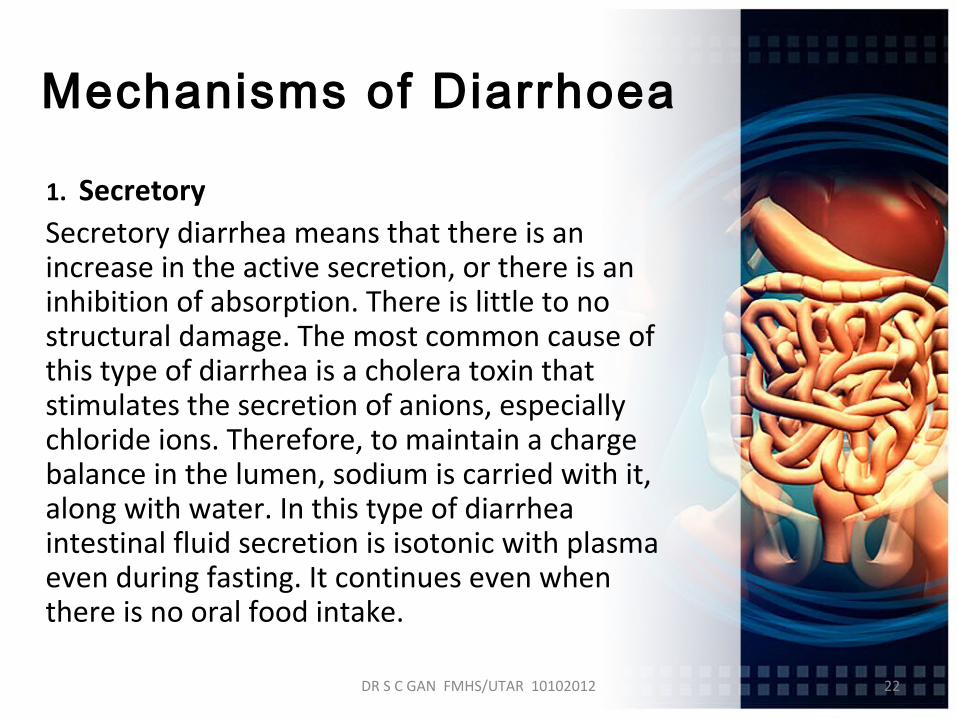
Mechanisms of Diarrhoea
1. Secretory Secretory diarrhea means that there is an
increase in the active secretion, or there is an inhibition of absorption. There is little to no structural damage. The most common cause of this type of diarrhea is a cholera toxin that stimulates the secretion of anions, especially chloride ions. Therefore, to maintain a charge balance in the lumen, sodium is carried with it, along with water. In this type of diarrhea intestinal fluid secretion is isotonic with plasma even during fasting. It continues even when there is no oral food intake.

DR S C GAN FMHS/UTAR 10102012 23
2. Osmotic Osmotic diarrhea occurs when too much water is drawn into
the bowels. This can be the result of maldigestion (e.g., pancreatic disease or celiac disease), in which the nutrients are left in the lumen to pull in water. Osmotic diarrhea can also be caused by osmotic laxatives (which work to alleviate constipation by drawing water into the bowels). In healthy individuals, too much magnesium or vitamin C or undigested lactose can produce osmotic diarrhea and distention of the bowel. A person who has lactose intolerance can have difficulty absorbing lactose after an extraordinarily high intake of dairy products. In persons who have fructose malabsorption, excess fructose intake can also cause diarrhea. High-fructose foods that also have a high glucose content are more absorbable and less likely to cause diarrhea. Sugar alcohols such as sorbitol (often found in sugar-free foods) are difficult for the body to absorb and, in large amounts, may lead to osmotic diarrhea. Diarrhea stops when offending agent (e.g. milk, sorbitol) is stopped.

DR S C GAN FMHS/UTAR 10102012 24
3. Exudative Exudative diarrhea occurs with the presence of blood and
pus in the stool. This occurs with inflammatory bowel diseases, such as Crohn's disease or ulcerative colitis, and other severe infections such as E. coli or other forms of food poisoning.
4. Motility-related Motility-related diarrhea is caused by the rapid movement
of food through the intestines (hypermotility). If the food moves too quickly through the gastrointestinal tract, there is not enough time for sufficient nutrients and water to be absorbed. This can be due to a vagotomy or diabetic neuropathy, or a complication of menstruation. Hyperthyroidism can produce hypermotility and lead to pseudodiarrhea and occasionally real diarrhea. Diarrhea can be treated with antimotility agents (such as loperamide). Hypermotility can be observed in people who have had portions of their bowel removed, allowing less total time for absorption of nutrients.

DR S C GAN FMHS/UTAR 10102012 25
5. Inflammatory Inflammatory diarrhea occurs when there is damage to the
mucosal lining or brush border, which leads to a passive loss of protein-rich fluids, and a decreased ability to absorb these lost fluids. Features of all three of the other types of diarrhea can be found in this type of diarrhea. It can be caused by bacterial infections, viral infections, parasitic infections, or autoimmune problems such as inflammatory bowel diseases. It can also be caused by tuberculosis, colon cancer, and enteritis.
6. Dysentery Generally, if there is blood visible in the stools, it is not
diarrhea, but dysentery. The blood is trace of an invasion of bowel tissue. Dysentery is a symptom of, among others, Shigella, Entamoeba histolytica, and Salmonella.

DR S C GAN FMHS/UTAR 10102012 26
Differential diagnosis
Diarrhea is most commonly due to viral gastroenteritis with rotavirus accounting for 40% of cases in children under five. In travelers however bacterial infections predominate.
It can also be the part of the presentations of a number of medical conditions such as: Crohn's disease or mushroom poisoning.
1. Infection
2. Malabsorption
3. Inflammatory Bowel Disease
4. Irritable Bowel Disease

DR S C GAN FMHS/UTAR 10102012 27
Other causes
• Diarrhea can be caused by chronic ethanol ingestion.
• Ischemic bowel disease. This usually affects older people and can be due to blocked arteries.
• Hormone-secreting tumors: some hormones (e.g., serotonin) can cause diarrhea if excreted in excess (usually from a tumor).
• Chronic mild diarrhea in infants and toddlers may occur with no obvious cause and with no other ill effects; this condition is called toddler's diarrhea.

DR S C GAN FMHS/UTAR 10102012 28
Evolution
According to two researchers, Nesse and Williams, diarrhea may function as an evolved expulsion defense mechanism. As a result, if it is stopped, there might be a delay in recovery. They cite in support of this argument research published in 1973 which found that treating Shigella with the anti-diarrhea drug (Co-phenotrope, Lomotil) caused people to stay feverish twice as long as those not so treated. The researchers indeed themselves observed that: "Lomotil may be contraindicated in shigellosis. Diarrhea may represent a defense mechanism".


![Clinical and Radiologic Considerations for Idiopathic ...€¦ · malignancy, malabsorption, and eating disorders) to SMA syndrome [4]. Additional common causes include aortic aneurysm](https://static.fdocuments.in/doc/165x107/605c4ff72c218f56110451ef/clinical-and-radiologic-considerations-for-idiopathic-malignancy-malabsorption.jpg)

![[Product Monograph Template - Standard]€¦ · Page 4 of 46 CONTRAINDICATIONS XENICAL (orlistat) is contraindicated in patients with chronic malabsorption syndrome, cholestasis and](https://static.fdocuments.in/doc/165x107/5b1544d87f8b9a1a398b84bc/product-monograph-template-standard-page-4-of-46-contraindications-xenical.jpg)