Giant cell tumour of bone: new treatments in development · EDUCATIONAL SERIES – RED SERIES NEW...
12
EDUCATIONAL SERIES – RED SERIES NEW TRENDS IN CLINICAL ONCOLOGY Giant cell tumour of bone: new treatments in development A. Lo ´pez-Pousa • J. Martı ´n Broto • T. Garrido • J. Va ´zquez Received: 5 December 2014 / Accepted: 18 December 2014 / Published online: 24 January 2015 Ó The Author(s) 2015. This article is published with open access at Springerlink.com Abstract Giant cell tumour of bone (GCTB) is a benign osteolytic tumour with three main cellular components: multinucleated osteoclast-like giant cells, mononuclear spindle-like stromal cells (the main neoplastic components) and mononuclear cells of the monocyte/macrophage line- age. The giant cells overexpress a key mediator in osteo- clastogenesis: the RANK receptor, which is stimulated in turn by the cytokine RANKL, which is secreted by the stromal cells. The RANK/RANKL interaction is predomi- nantly responsible for the extensive bone resorption by the tumour. Historically, standard treatment was substantial surgical resection, with or without adjuvant therapy, with recurrence rates of 20–56 %. Studies with denosumab, a monoclonal antibody that specifically binds to RANKL, resulted in dramatic treatment responses, which led to its approval by the United States Food and Drugs Adminis- tration (US FDA). Recent advances in the understanding of GCTB pathogenesis are essential to develop new treat- ments for this locally destructive primary bone tumour. Keywords Giant cell tumour of bone Á Pathophysiology Á Surgery Á Denosumab Introduction: new insights into pathophysiology Definition, epidemiology and natural course Giant cell tumour of bone (GCTB) is a rare osteolytic tumour that is responsible for approximately 6 % of all primary bone tumours. Reported annual incidence ranges between 1 and 6 per 10 million persons [1, 2] to approxi- mately 1 per million in the US, Western Australia, Japan and Sweden [3]. It typically affects adults aged between 20 and 50 years [4–6], with a slightly higher incidence among females (1.7 per 10 million in females versus 1.5 per 10 million in males) [1, 4–6]. GCTB is typically located in the epiphysis of bones, causing localised tenderness and swelling, reduced joint mobility, and pain that is often severe and intractable [6]. It usually develops in long bones but can also occur in unusual locations. The Enneking staging classification, based on radiological, histological and clinical features, is the most commonly used (Table 1) [7]. There is also a radiological grading system established by Campanacci et al. [8] that classifies GCTB into three radiographic types (I, intramedullary lesion confined to bone; II, thinned, expanded cortex, III, cortical breakout), and is roughly comparable with the staging system of Enneking et al. [7] Symptoms are variable; some patients may be asymp- tomatic until they develop a pathologic fracture while others complain of pain at the adjacent joint and limited range of motion. There may also be swelling and even a visible mass, if the tumour has grown for a long time. Other commonly reported symptoms include muscular or nerve pain [6]. The tumour is locally aggressive and destructive, and it grows rapidly, destroying bone and spreading into sur- rounding soft tissues [9]. If it is surgically resected, there is A. Lo ´pez-Pousa Hospital de la Santa Creu i Sant Pau, Barcelona, Spain A. Lo ´pez-Pousa (&) Medical Oncology Department, Hospital de la Santa Creu i Sant Pau, Autonomous University of Barcelona, Mas Casanovas 90, ES-08041 Barcelona, Spain e-mail: [email protected] J. M. Broto Hospital Universitari Son Espases, Palma de Mallorca, Spain T. Garrido Á J. Va ´zquez Amgen S.A., Barcelona, Spain 123 Clin Transl Oncol (2015) 17:419–430 DOI 10.1007/s12094-014-1268-5
Transcript of Giant cell tumour of bone: new treatments in development · EDUCATIONAL SERIES – RED SERIES NEW...
EDUCATIONAL SERIES – RED SERIES
NEW TRENDS IN CLINICAL ONCOLOGY
Giant cell tumour of bone: new treatments in development
A. Lopez-Pousa • J. Martın Broto •
T. Garrido • J. Vazquez
Received: 5 December 2014 / Accepted: 18 December 2014 / Published online: 24 January 2015
� The Author(s) 2015. This article is published with open access at Springerlink.com
Abstract Giant cell tumour of bone (GCTB) is a benign
osteolytic tumour with three main cellular components:
multinucleated osteoclast-like giant cells, mononuclear
spindle-like stromal cells (the main neoplastic components)
and mononuclear cells of the monocyte/macrophage line-
age. The giant cells overexpress a key mediator in osteo-
clastogenesis: the RANK receptor, which is stimulated in
turn by the cytokine RANKL, which is secreted by the
stromal cells. The RANK/RANKL interaction is predomi-
nantly responsible for the extensive bone resorption by the
tumour. Historically, standard treatment was substantial
surgical resection, with or without adjuvant therapy, with
recurrence rates of 20–56 %. Studies with denosumab, a
monoclonal antibody that specifically binds to RANKL,
resulted in dramatic treatment responses, which led to its
approval by the United States Food and Drugs Adminis-
tration (US FDA). Recent advances in the understanding of
GCTB pathogenesis are essential to develop new treat-
ments for this locally destructive primary bone tumour.
Keywords Giant cell tumour of bone � Pathophysiology �Surgery � Denosumab
Introduction: new insights into pathophysiology
Definition, epidemiology and natural course
Giant cell tumour of bone (GCTB) is a rare osteolytic
tumour that is responsible for approximately 6 % of all
primary bone tumours. Reported annual incidence ranges
between 1 and 6 per 10 million persons [1, 2] to approxi-
mately 1 per million in the US, Western Australia, Japan
and Sweden [3]. It typically affects adults aged between 20
and 50 years [4–6], with a slightly higher incidence among
females (1.7 per 10 million in females versus 1.5 per 10
million in males) [1, 4–6]. GCTB is typically located in the
epiphysis of bones, causing localised tenderness and
swelling, reduced joint mobility, and pain that is often
severe and intractable [6]. It usually develops in long bones
but can also occur in unusual locations. The Enneking
staging classification, based on radiological, histological
and clinical features, is the most commonly used (Table 1)
[7]. There is also a radiological grading system established
by Campanacci et al. [8] that classifies GCTB into three
radiographic types (I, intramedullary lesion confined to
bone; II, thinned, expanded cortex, III, cortical breakout),
and is roughly comparable with the staging system of
Enneking et al. [7]
Symptoms are variable; some patients may be asymp-
tomatic until they develop a pathologic fracture while
others complain of pain at the adjacent joint and limited
range of motion. There may also be swelling and even a
visible mass, if the tumour has grown for a long time. Other
commonly reported symptoms include muscular or nerve
pain [6].
The tumour is locally aggressive and destructive, and it
grows rapidly, destroying bone and spreading into sur-
rounding soft tissues [9]. If it is surgically resected, there is
A. Lopez-Pousa
Hospital de la Santa Creu i Sant Pau, Barcelona, Spain
A. Lopez-Pousa (&)
Medical Oncology Department, Hospital de la Santa Creu i Sant
Pau, Autonomous University of Barcelona, Mas Casanovas 90,
ES-08041 Barcelona, Spain
e-mail: [email protected]
J. M. Broto
Hospital Universitari Son Espases, Palma de Mallorca, Spain
T. Garrido � J. Vazquez
Amgen S.A., Barcelona, Spain
123
Clin Transl Oncol (2015) 17:419–430
DOI 10.1007/s12094-014-1268-5

a substantial probability of recurrence, which seems to be
greater in some locations associated with more difficult
treatment, such as the distal radius and the proximal femur
[10]. In the absence of treatment, the continued and
unchecked tumour growth leads to complete destruction of
the bone, physical deformity and the possibility of loss of
limb.
The most common site of metastasis is the lung,
occurring at a frequency of 1–6 % [11, 12]. Pulmonary
metastases are usually histologically benign and their
course is indolent. The standard of care is surgical resec-
tion, and prognosis is generally good. If resection is not
possible, they can be left untreated [6, 11, 13].
Rarely, in less than 1 % of cases, GCTB may undergo
malignant transformation that is known to result in a poor
prognosis for the patient [6, 14]. The malignancy may arise
as a result of dedifferentiation of the primary tumour or
secondary to radiation therapy (approximately 50 % of
cases) [11]. The most commonly observed transformation
is to a high-grade sarcoma, usually an osteosarcoma,
however, in rare cases this transformation may result in the
formation of a fibrosarcoma or a classically denominated
malignant fibrous histiocytoma. The mean time after initial
GCTB diagnosis to malignant transformation is around
19 years in patients with spontaneous transformation and
around 9 years in post-radiation cases [11].
Pathophysiology
The histopathology of GCTB reveals the presence of
marked haemorrhage and three major cell types: multinu-
cleated giant cells, stromal cells and mononuclear cells of
the monocyte/macrophage lineage [15, 16].
The spindle-like stromal cells are the main neoplastic
components, and appear to be activated by fibroblasts that
secrete type I and III collagen and possess parathormone
receptors [17, 18]. They promote giant cell formation by
expressing and secreting a variety of chemotactic factors
(cytokines such as interleukin [IL]-6, IL-8, IL-11, IL-17, IL-
34, basic fibroblast growth factor [b-FGF], tumour necrosis
factor [TNF]-a, vascular endothelial growth factor [VEGF],
macrophage colony-stimulating factor [M-CSF], RANKL,
cathepsin K; chemokines such as IL-8, TGF-b1 and stromal
derived factor-1 [SDF-1]; and enzymes such as matrix
metalloproteinase [MMP]-9 and MMP-13) [19–22]. All
these factors serve to engage and differentiate circulating
monocytes into macrophages [23]. Of these factors, SDF-1
appears to act as a chemoattractant involved in the recruit-
ment of monocytes [23]. Furthermore, some studies have
correlated the expression of VEGF and MMP-9 with the
extent of bone destruction and probability of recurrence [24].
Giant cells are directly responsible for the increased
bone resorption observed within the lesion [25, 26]. They
are considered to be reactive macrophages that have
acquired osteoclastic activity as a result of their stimulation
by stromal cells, which modifies their gene expression
pattern within the osseous environment [27]. Giant cells
also drive increased expression of a key mediator in
osteoclastogenesis: the RANK receptor [28]. Activation of
this receptor by RANKL, which is secreted by stromal
cells, promotes osteoclast formation, activation, function,
and survival [29–32]. Thus, leading to the increased level
of bone resorption observed within the GCTB lesion. In
addition, the activated osteoclasts, in turn, release tumour
growth factors into the bone microenvironment, initiating a
tumour/bone vicious cycle (Fig. 1a) [33–35].
Table 1 Enneking
classification of GCTB [7]Stage % Description
Stage I (latent) 15 Confined totally by bone
Asymptomatic
Inactive on bone scan
Histologically benign
Stage II (active) 70 Expanded cortex with no breakthrough
Symptomatic
Often have pathological fracture
Active on bone scan
Histologically benign
Stage III (aggressive) 15 Rapidly growing mass
Cortical perforation with soft tissue mass
May metastasize
Symptomatic
Extensive activity on bone scan
Histologically benign
Malignant Very rare Sarcomatous lesion contiguous with benign GCT
420 Clin Transl Oncol (2015) 17:419–430
123

The underlying cause of the increased RANKL expres-
sion by stromal cells is unknown, however, this phenom-
enon is reduced after elimination of the giant cells [32].
Conversely, giant cells are clearly dependent on RANKL
signalling by stromal cells [32, 36]. Thus, it is possible that
GCTB promotes a pathological variation of the normal
physiological interdependence of osteoblast and osteoclast
populations in bone [37, 38].
Cytogenetic abnormalities have been observed in up to
72 % of patients with GCTB, yet to date, no uniform
aberrations have been identified [15, 39, 40]. Telomeric
associations (reductions in telomere length with an average
loss of 500 base pairs) are the most frequent chromosomal
aberrations and the telomeres most commonly affected are
11p, 13p, 14p, 15p, 19q, 20q and 21p [39, 40].
There is also a hypothesis that the origin of GCTB could
be linked to a form of bone injury, as in some cases GCTB
appears in locations associated with prior trauma [41, 42].
In this scenario, GCTB could be considered a local reactive
condition secondary to a haemorrhage due to bone injury
and/or defective collagen in the matrix or in the vessel
wall. It is possible that the haemorrhage serves to provide
fresh monocytes and plasma proteins that initiate activation
of stromal cells, which in turn stimulate conversion of giant
cells into active osteoclasts [41]. Once the primary lesion
occurs, the stromal cells would be capable of re-forming
the tumour in secondary tumour sites or after surgical
removal, thanks to their proliferative and tumour-initiating
properties [17, 43, 44]. However, it seems that other
transformational factors are required since injection of
isolated stromal cells into immunocompromised mice does
not produce giant cells [45, 46]. Some studies suggest that
metastases could result from tumour emboli travelling to
distant sites [47, 48].
Treatment
Surgical treatment
Surgical removal of the tumour with wide excision or int-
ralesional curettage and placement of cement (polymethyl
methacrylate) has been historically the preferred treatment
for GCTB [10, 49–52]. The challenge with surgery is to
remove as much of the tumour as possible while leaving the
joint intact. Wide excision is associated with poor func-
tional outcome and greater surgical complications [53–55].
Therefore, intralesional curettage has been the mainstay of
treatment for the majority of patients with stage I or II
tumours. Wide excision is usually reserved for more
aggressive stage III tumours with extraosseous extension or
otherwise unresectable tumours [6, 56, 57]. However,
sometimes the tumour is unresectable or surgery is not
recommended due to age, patient comorbidities or risk of
severe morbidity, such as joint removal or loss of limbs.
Aggressive GCTBs may require wide excision and
reconstruction with a modular endoprosthesis; the most
commonly used synthetic grafts are made from polymethyl
methacrylate (PMMA). These grafts are known to generate
an exothermic reaction that increases thermal necrosis of
tumour cells and an inflammatory reaction, consequently
resulting in an improved patient recovery and tumour
removal [58, 59].
The main complications associated with surgery include
pathologic fracture and postoperative infection. Postoper-
ative infection occurs in 2–25 % of patients, and its inci-
dence is probably greater with more extensive surgery
involving en bloc resection and placement of an endo-
prosthesis [60–63]; whereas pathologic fracture is associ-
ated with an increased rate of recurrence and a poorer
functional outcome [64].
There is a recognised tendency for GCTBs to recur
locally in many cases following surgery, even in the soft
tissues adjacent to the primary bone location [5, 10, 65,
66]. In one of the largest published cohorts, a multicentre
retrospective study in 294 Scandinavian patients, Kivioja
et al. [52] reported recurrence rates ranging between 20 %
for patients with PMMA cementation following intrale-
sional curettage, and 56 % for patients without cementa-
tion. In contrast, wide excision is reported to be associated
with a lower risk of local recurrence (0–12 %) than int-
ralesional curettage (12–65 %) [5, 67–69]. The use of
improved surgical techniques, such as extensive mechani-
cal burr drilling of the tumour wall after curettage or
adjuvant cryoablation with liquid nitrogen, has further
decreased recurrence rates in some centres, but these
techniques have not yet been widely adopted. In the study
by Malawer et al. [70] only 2.3 % of patients recurred after
primary treatment with cryosurgery, although this per-
centage increased to 7.9 % when second-line treatments
were also considered.
Chemical adjuvant therapy
Currently, there is no standard or approved first-line med-
ical treatment for GCTB. Surgical treatment may be
combined with chemical adjuvant therapies. Some of the
treatments commonly applied to the affected area are:
alcohols [59, 71, 72], phenol [71, 73], hydrogen peroxide
[71, 74, 75], and zinc chloride [76]. Hydrogen peroxide has
been found to increase the penetration of phenol into the
surrounding tissues [75]. Use of chemical adjuvants has
been shown to reduce the percentage of recurrences in
some studies [77], although others failed to demonstrate
any impact [78]. Furthermore, these adjuvants must be
used with caution, to avoid chemical burns.
Clin Transl Oncol (2015) 17:419–430 421
123

Radiation therapy
Radiation therapy has been used to treat GCTB since 1932
[79] and its efficacy has been demonstrated by several
studies in patients for whom surgery was not feasible [80,
81]. Specialised techniques such as 3-D conformal radio-
therapy (RT) and intensity-modulated radiotherapy (IMRT)
have been associated with good local control rates in
Fig. 1 a Mechanism of
increased bone resorption in
GCTB: central role of the
RANK/RANKL interaction
[33–35]. b Proposed mechanism
of action of denosumab in
GCTB [96]
422 Clin Transl Oncol (2015) 17:419–430
123

patients with GCTB in locations that are not accessible by
surgical resection [82, 83]. However, some reports have
suggested an increased risk of malignant transformation
into post-radial sarcoma [84].
The better safety profile of the new drugs available to
inhibit osteoclastogenesis has decreased the use of RT in
GCTB [85].
Embolisation and laser photoablation
Embolisation is made by hyperselective catheterisation and
embolisation of the arteries that feed the pathological
lesion with the most appropriate embolic agent. Typically,
Gelfoam, polyvinyl alcohol (PVA) particles, and coils are
used for embolisation; other agents include tissue adhesive,
ethanol, and microfibrillar collagen. Occlusion of the ves-
sels decreases the volume of the tumour, but multiple
procedures are frequently necessary [86]. Photoablation
with an argon laser is another therapy that can lead to
successful tumour necrosis [87].
Given the high vascularity and morbidity associated with
surgical resection and/or radiation therapy, embolisation
has been reported to be useful within 24–48 h prior to these
therapies [88, 89], to prevent recanalisation. The combined
use of preoperative embolisation and adjuvants, including
radiation therapy and intraoperative phenol and nitrogen,
can decrease local recurrence to less than 10 % [90].
Serial embolisation is also used as primary treatment in
some patients with GCTB of the extremities, especially for
tumours with large cortical defects or joint involvement
and for those with large GCTBs of the sacrum. This pro-
cedure has a low morbidity rate and has been shown to be
effective in preserving function and relieving pain in
selected patients [91–93].
Drug therapy
Denosumab
In parallel with an improved understanding of the patho-
genesis of the tumour, other treatment options for GCTB
are continuously being explored. The discovery of the
involvement of the RANK/RANKL pathway has recently
led to the use of the monoclonal antibody denosumab [94].
To date, denosumab is the first and only drug approved by
the United States (US) Food and Drug Administration
(FDA) and, in Europe, by the European Medicine Agency
(EMA) for GCTB [95].
Indication Since June 2013, denosumab is indicated in
the US for treatment of adults and skeletally mature ado-
lescents with GCTB that is unresectable or where surgical
resection is likely to result in severe morbidity [95]. In
Europe, the European Medicines Agency also approved it
for GCTB in September 2014. In addition, denosumab is
indicated for the prevention of skeletal-related events in
patients with bone metastases from solid tumours in the US
and in the European Union [94, 95].
The recommended dose of denosumab in the GCTB
indication is 120 mg administered once every 4 weeks with
additional 120 mg doses on days 8 and 15 of the first
month of therapy. Denosumab is administered as a single
subcutaneous injection in the upper arm, upper thigh, or
abdomen [94, 95].
Mechanism of action Denosumab is a human monoclonal
antibody [immunoglobulin G2 (IgG2)] that targets and
binds RANKL with high affinity and specificity, preventing
activation of its receptor, RANK, on the surface of giant
cells, osteoclast precursors and osteoclasts. Prevention of
the RANK/RANKL interaction inhibits osteoclast forma-
tion, function, and survival, thereby decreasing bone
resorption in GCTB (Fig. 1b) [96].
Pharmacokinetic and pharmacodynamic properties Fol-
lowing subcutaneous administration, rapid and prolonged
absorption of denosumab has been shown [96, 97]. It has
been detected in the serum within 1 h of dosing and for up
to 9 months following a single dose (maximal serum
concentrations achieved between 5 and 21 days) [96, 97].
With multiple dosing (120 mg subcutaneously, every
4 weeks), there was an approximately twofold increase in
serum concentrations in treated patients with bone metas-
tases secondary to solid tumours. Steady state is attained by
6 months and, at steady state, the mean serum trough
concentration is 20.5 lg/mL (standard deviation 13.5 lg/
mL) and mean elimination half-life is 28 days [94].
Clinical development in GCT The safety and efficacy of
denosumab for the treatment of GCTB in adults or skele-
tally mature adolescents were demonstrated in two phase 2,
open-label studies. All patients received 120 mg of deno-
sumab subcutaneously every 4 weeks with additional doses
on days 8 and 15 of the first cycle of therapy [98, 99].
Efficacy A single-arm, open-label, pharmacodynamic
and proof of concept study evaluated the safety and effi-
cacy of denosumab in 37 patients C18 years with recurrent
or unresectable GCTB [99].
Eighty-six percent (95 % CI 70–95) of patients (n = 30)
met the criteria for tumour response (elimination of C90 %
of giant cells or no radiological progression of the target
lesion): 20 based on histology and 10 based on radiology.
Histological results showed near-complete or complete
elimination of giant cells in all patients for whom histology
was available. Improvement in functional status or reduced
pain were reported in 84 % of patients (95 % CI 66–95;
Clin Transl Oncol (2015) 17:419–430 423
123

n = 26), and 29 % of patients (95 % CI 14–48; n = 9) had
evidence of bone repair [99].
The second study was an open-label, single-arm, parallel-
cohort, proof of concept, and safety trial conducted in 282
adult patients with primary or recurrent GCTB distributed in
3 cohorts [98]: cohort 1, 170 patients who had surgically
unsalvageable disease as determined by the treating surgeon
(e.g. sacral, spinal, or multiple GCTB lesions including
pulmonary metastases); cohort 2, 101 patients with a plan-
ned surgery that was associated with severe morbidity (e.g.
joint resection, limb amputation, hemipelvectomy); and
cohort 3, 11 patients who transitioned from the previous
denosumab GCTB study [99] and continued denosumab
treatment on this study. The primary efficacy outcome
measures were time to disease progression in cohort 1 and
the proportion of patients without any surgery at 6 months in
cohort 2 [98]. An interim analysis was published when more
than 200 patients had had an opportunity to complete
6 months of treatment after enrolment [98].
At the time of the interim analysis, median time to dis-
ease progression in cohort 1 was not reached, and the best
response rate (complete or partial) determined by investi-
gator was 41 % in cohort 1 and 58 % in cohort 2 (Table 2).
In a retrospective, independent imaging analysis that
evaluated tumour response in patients from all three cohorts
who received imaging as part of their standard of care
(N = 190), the overall objective response rate (RECIST
1.1) was 25 % (95 % CI 19, 32), with all responses docu-
mented as partial responses. The estimated median time to
response was 3 months. In the 47 patients with an objective
response, median duration of follow-up was 20 months
(range 2–44 months), and 51 % (24/47) had a duration of
response lasting at least 8 months. Three patients experi-
enced disease progression following response. Combining
three different response criteria (RECIST, European Orga-
nization for Research and Treatment of Cancer (EORTC)
and Modified Choi criteria), the best objective response rate
was 72 % (Table 2) [98].
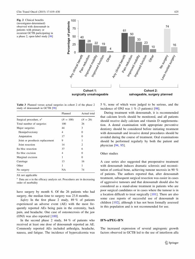
Clinical benefit was observed in 40 and 61 % of patients
in cohorts 1 and 2, respectively, with pain reduction the
most commonly observed benefit (Table 2; Fig. 2). Of the
100 patients in cohort 2 for whom surgery was planned at
baseline, 90 (90 %) patients had either no surgery (n = 74;
74 %) or underwent a less morbid procedure (n = 16;
16 %) compared with the surgical procedure planned at
baseline [98] (Table 3; Fig. 2). Median follow-up for
cohort 2 was 9.2 months (IQR 4.2–12.9). Of the 71 patients
who were on study for at least 6 months, 64 (90 %) did not
Table 2 Main results of the phase 2 study of denosumab in GCTB [98]
Best response (investigator-determined)
Cohort 1: surgically unsalvageable Cohort 2: salvageable, surgery planned
Complete response, % (n/N1) 5 (8/159) 18 (17/93)
Partial response, % (n/N1) 36 (57/159) 40 (37/93)
Stable disease, % (n/N1) 58 (93/159) 41 (38/93)
Disease progression, % (n/N1) 1 (1/159) 1 (1/93)
Best clinical benefit (investigator-determined)
Pain reduction, % (n/N) 28 (48/169) 50 (50/100)
Improved mobility, % (n/N) 22 (38/169) 33 (33/100)
Improved function, % (n/N) 19 (32/169) 23 (23/100)
Other, % (n/N) 4 (6/169) 10 (10/100)
Best response (independent imaging assessment)
Overall RECIST 1.1 EORTC Inverse Choi
Objective response (OR)a, % (n/N2) 72 (136/190) 25 (47/187) 96 (25/26) 76 (134/176)
Median time to OR, months 3.1 not reached 2.7 3
OR sustained C24 weeks, % (n/N2*) 68 (76/111) 24 (26/109) 92 (11/12) 75 (76/102)
Tumour controlb sustained C24 weeks, % (n/N2*) 98 (109/111) 99.1 (108/109) 100 (12/12) 99 (101/102)
N1 number of enrolled patients who received C1 dose of denosumab and had a disease status evaluation
N number of enrolled subjects who were eligible for the study and received C1 dose of denosumab
N2 Patients with C1 evaluable timepoint assessment
RECIST response evaluation criteria in solid tumours, EORTC European organization for research and treatment of cancer
*Patients with timepoint assessments C24 weeks aparta Objective response = complete ? partial responseb Tumour control = complete ? partial response ? stable disease
424 Clin Transl Oncol (2015) 17:419–430
123
have surgery by month 6. Of the 26 patients who had
surgery, the median time to surgery was 23.8 months.
Safety In the first phase 2 study, 89 % of patients
experienced an adverse event (AE) with the most fre-
quently reported AEs being pain in the extremity, back
pain, and headache. One case of osteonecrosis of the jaw
(ONJ) was also reported [100].
In the second phase 2 study, 84 % of patients who
received at least one dose of denosumab reported an AE.
Commonly reported AEs included arthralgia, headache,
nausea, and fatigue. The incidence of hypercalcemia was
5 %, none of which were judged to be serious, and the
incidence of ONJ was 1 % (3 patients) [98].
During treatment with denosumab, it is recommended
that calcium levels should be monitored, and all patients
should receive daily calcium and vitamin D supplementa-
tion. A dental examination with appropriate preventive
dentistry should be considered before initiating treatment
with denosumab and invasive dental procedures should be
avoided during the course of treatment. Oral examinations
should be performed regularly by both the patient and
physician [94, 95].
Other studies
A case series also suggested that preoperative treatment
with denosumab induces dramatic sclerosis and reconsti-
tution of cortical bone, achieving tumour necrosis in 90 %
of patients. The authors reported that, after denosumab
treatment, subsequent surgical resection was easier in cases
of aggressive tumours and that denosumab should also be
considered as a stand-alone treatment in patients who are
poor surgical candidates or in cases where the tumour is in
a location difficult to treat surgically [101]. There are also
some case reports of successful use of denosumab in
children [102], although it has not been formally assessed
in this population and is not recommended for use.
IFN-a/PEG-IFN
The increased expression of several angiogenic growth
factors observed in GCTB led to the use of interferon alfa
Table 3 Planned versus actual surgeries in cohort 2 of the phase 2
study of denosumab in GCTB [98]
Planned Actual total
Surgical procedure, na (N = 100) (N = 26)
Total number of surgeries 100 26
Major surgeries 44 3
Hemipelvectomy 4 0
Amputation 17 0
Joint or prosthesis replacement 9 1
Joint resection 14 2
En bloc resection 37 6
En bloc excision 4 0
Marginal excision 1 0
Curettage 13 16
Other 1 1
No surgery NA 74
NA not applicablea Data are n in the efficacy analysis set. Procedures are in decreasing
order of morbidity
4028 22 19
4
6150
3323
10
74
16
0
25
50
75
100
Perc
enta
ge o
f pat
ient
s
Cohort 1:surgically unsalvageable
Cohort 2:salvageable, surgery planned
Fig. 2 Clinical benefits
(investigator-determined)
observed with denosumab in
patients with primary or
recurrent GCTB participating in
a phase 2, open-label study [98]
Clin Transl Oncol (2015) 17:419–430 425
123

(IFN-a) as an anti-angiogenic agent. The first use was in
1995 [103], and since then several studies have reported
successful treatment of GCTB with this agent [104].
Pegylated (PEG)-IFN has also been shown to have anti-
GCTB activity. A few case reports have reported the effi-
cacy of interferon and pegylated interferon in the man-
agement of GCTB [105].
Bisphosphonates
Due to their anti-resorptive properties, some exploratory
studies tested the efficacy of bisphosphonates in GCTB. It
was shown that nitrogen-containing bisphosphonates
induce apoptosis in both giant cells and stromal cells
in vitro [106]. In a case–control study, pamidronate and
zoledronate reduced local tumour recurrence (4.2 vs 30 %
in the control group, p = 0.056) and controlled disease
progression when used orally or intravenously as adjuvant
therapy to intralesional curettage [107]. In 25 patients with
recurrent and metastatic GCTB treated with bisphospho-
nates, stabilisation of disease was achieved in most cases
refractory to conventional treatment [108]. In addition,
there are case reports of successful local administration of
zoledronic acid as adjuvant therapy during surgery [109].
However, they are not approved for use in this indication
and more evidence is needed.
Current guideline recommendations
NCCN
In 2013, the National Comprehensive Cancer Network
(NCCN) Clinical Practice Guidelines in Oncology for bone
cancer added a new section on GCTB.
According to the version 1.2015 of these guidelines,
workup begins with a history, physical examination, cross-
sectional imaging of the primary site, chest imaging, and
biopsy to confirm the diagnosis. Bone scan is considered
optional [110].
Regarding treatment (Table 4), the decision tree
depends on whether the disease is localised or metastatic.
For localised disease, the choice of surgery is next. If the
tumour is resectable, excision is the primary option. If the
tumour is resectable with unacceptable morbidity or unre-
sectable, the options include serial embolization (primarily
for tumours of the pelvis), denosumab, interferon, pegy-
lated interferon, and/or radiotherapy [110].
For metastatic disease, the feasibility of surgery deter-
mines the treatment options. If the tumour is resectable,
again the primary treatment pathway for localised disease
should be followed and excision of metastatic sites con-
sidered. If the tumour is unresectable, treatment options
include denosumab, interferon, pegylated interferon,
radiotherapy, or observation [110].
NCCN Guidelines also contain recommendations for
surveillance, which include physical examination, imaging
of the surgical site as clinically indicated, and chest
imaging every 6 months for 2 years and annually thereaf-
ter. For a resectable local tumour recurrence, chest imaging
and denosumab may be considered before surgery [110].
ESMO
The 2014 ESMO guidelines for bone sarcomas [111]
specify that treatment options for GCTB include intrale-
sional curettage with or without adjuvant or en bloc exci-
sion. They also mention that recent work has suggested that
denosumab obtains substantial tumour responses in large or
unresectable or metastatic GCTB. For this reason, deno-
sumab may be used to achieve cytoreduction allowing
potentially curative surgery, or also in unresectable and
rare metastatic disease, where treatment needs to be
maintained to avoid progression [111].
Regarding surveillance, the recommendation for low-
grade bone sarcomas such as GCTB, include follow-up
visits every 6 months for 2 years and then annually.
However, they comment that late metastases as well as
local recurrences and functional deficits may occur[10
years after diagnosis and that there is no universally
accepted stopping point for tumour surveillance [111].
Future expectations
The knowledge of GCTB pathophysiology is rapidly
evolving. The identification of the chemotactic factors
secreted by stromal cells and involved in monocyte trans-
formation into giant cells provides an opportunity to dis-
cover innovative treatments. The monoclonal antibody
denosumab is the first drug agent with proven efficacy in
GCTB by targeting one of these factors (RANKL). The
main pending questions with denosumab include the
evaluation of its possible benefits as neoadjuvant therapy
[112], the optimal duration and schedule of treatment at
long term to avoid recurrences, and its long-term safety.
Some angiogenesis inhibitors have also been tested, such as
calcitonin and interferon. IFN-a inhibits the expression of
b-FGF and IL-8, two angiogenic factors. Other candidate
therapies could be monoclonal antibodies directed against
the involved cytokines or enzymes, such as anti-IL6,
cathepsin inhibitors, anti-M-CSF or MMP-specific inhibi-
tors [113]. The newer antibody–drug conjugates (ADCs), a
novel class of highly potent drugs composed of an antibody
(a whole antibody or an antibody fragment) linked to a
cytotoxic drug could revolutionise treatment of GCTB
[114]. Although few ADCs are currently available [115],
426 Clin Transl Oncol (2015) 17:419–430
123

there are more than 20 compounds currently in clinical
development, specific for a wide range of biological targets
expressed by tumour cells [116]. It is hoped that, in the
near future, some of them could be suitable for GCTB, in
view of promising results in other cancers.
It also seems that targeting the neoplastic stromal cells
could fight directly against the origin of tumour. Therapies
blocking proliferation of stromal cells, such as drugs
inhibiting cell cycle progression or telomerase activity
could be effective. First, it would be necessary to identify
specific markers for the stromal cells.
Recent findings suggest that the haemorrhagic compo-
nent plays a fundamental role in the development of giant
cells. In some instances, GCTB could be a reactive con-
dition secondary to massive intraosseous haemorrhage,
which attracts monocytes and forces their quick prolifera-
tion and conversion into multinucleated cells. There is also
the hypothesis that poor matrix support to the vessels may
underlie the haemorrhage that precedes tumour formation.
Currently, the use of embolisation techniques and occlu-
sion of the vessels helps reduce recurrence. Other treat-
ments aimed to occlude the vessels and reinforce local
osseous matrix support, such as laser and hormone thera-
pies, could be also effective.
A more deep investigation on genetic predisposition
may help to identify individuals at higher recurrence risk,
in whom more aggressive therapies should be undertaken.
For example, amplification of 20q11.1 seems to be a
prognostic marker for adverse outcome [117] and warrants
further investigation.
Conclusions
GCTB is an aggressive primary osteolytic bone tumour that
causes substantial morbidity. GCTB tumours contain
osteoclast-like giant cells that express RANK and stromal
cells that express RANKL, a key mediator of osteoclast
formation, activation, function, and survival. Excessive
secretion of RANKL causes an imbalance in bone remod-
elling in favour of bone breakdown. Before the discovery of
denosumab, surgical intervention was the only definitive
therapy for patients with resectable tumours; however, it is
associated with significant morbidity. Currently, denosumab
Table 4 2015 NCCN recommendations for GCTB [110]
Giant cell tumour of the bone—NCCN guidelines (Version 1.2015)
Treatment Follow-up
Localised disease (primary or recurrent)
Resectable Excision (in recurrence: consider chest imaging
and/or denosumab prior to surgery)
Physical exam
Imaging of surgical site as clinically indicated
Chest imaging every 6 m for 2 years then annually
Resectable with unacceptable
morbidity
and/or
Unresectable
Serial embolization
and/or
Denosumab
and/or
IFN or PEG-IFN
and/or
RT
If stable/improved disease
Same follow-up as after excision
If stable/improved disease with incomplete healing
Excision (if resectable)
Continue on-treatment (if unresectable)
If progressive disease
Continue on-treatment
Metastatic disease (at presentation or recurrence)
Resectable Treat primary tumour
Consider excision of metastasis
Physical exam
Imaging of surgical site as clinically indicated
Chest imaging every 6 m for 2 years then annually
Unresectable Denosumab
and/or
IFN or PEG-IFN
and/or
RT
and/or
Observation
If stable/improved disease
Same follow-up as after excision
If stable/improved disease with incomplete healing
Excision (if resectable)
Continue on-treatment (if unresectable)
If progressive disease
Continue on-treatment
IFN interferon, NCCN national comprehensive cancer network, PEG pegylated, RT radiotherapy
Clin Transl Oncol (2015) 17:419–430 427
123

constitutes an effective therapeutic option for treatment of
adult patients with unresectable GCTB or in whom surgical
resection is likely to result in severe morbidity. Denosumab
provides objective tumour responses in 72 % of patients,
prolonging the time to surgery and reducing its morbidity in
those patients with planned interventions. Denosumab is
well tolerated, with ONJ and hypocalcemia; known risks are
observed at low rates. The increasing knowledge of the
molecular mechanisms involved in GCTB pathophysiology
provides an opportunity for using new targeted therapies that
may dramatically change the outcomes of GCTB in the next
years.
Acknowledgments Writing assistance was funded by Amgen S.A.
and provided by Dr. Neus Valveny from TFS Develop.
Conflict of interest TG and JV report being employees of Amgen
and receiving stocks and stock options from Amgen. The other
authors declare no conflict of interest.
Open Access This article is distributed under the terms of the
Creative Commons Attribution License which permits any use, dis-
tribution, and reproduction in any medium, provided the original
author(s) and the source are credited.
References
1. Beebe-Dimmer JL, Cetin K, Fryzek JP, Schuetze SM, Schwartz K. The epi-demiology of malignant giant cell tumors of bone: an analysis of data from theSurveillance, Epidemiology and End Results Program (1975–2004). RareTumors. 2009;1:e52. doi:10.4081/rt.2009.e52.
2. Larsson SE, Lorentzon R, Boquist L. Giant-cell tumor of bone. A demo-graphic, clinical, and histopathological study of all cases recorded in theSwedish Cancer Registry for the years 1958 through 1968. J Bone Joint SurgAm. 1975;57:167–73.
3. Liede A, Bach BA, Stryker S, Hernandez RK, Sobocki P, Bennett B, et al.Regional variation and challenges in estimating the incidence of giant celltumor of bone. J Bone Joint Surg Am. 2014;96(23):1999–2007. doi:10.2106/JBJS.N.00367.
4. Turcotte RE. Giant cell tumor of bone. Orthop Clin North Am.2006;37:35–51. doi:10.1016/j.ocl.2005.08.005.
5. Turcotte RE, Wunder JS, Isler MH, Bell RS, Schachar N, Masri BA, et al.Giant cell tumor of long bone: a Canadian Sarcoma Group study. Clin Orthop.2002;(397):248–58.
6. Mendenhall WM, Zlotecki RA, Scarborough MT, Gibbs CP, Mendenhall NP.Giant cell tumor of bone. Am J Clin Oncol. 2006;29:96–9. doi:10.1097/01.coc.0000195089.11620.b7.
7. Enneking WF, Spanier SS, Goodman MA. A system for the surgical staging ofmusculoskeletal sarcoma 1980. Clin Orthop. 2003;(415):4–18. doi:10.1097/01.blo.0000093891.12372.0f.
8. Campanacci M. Giant-cell tumor and chondrosarcomas: grading, treatmentand results (studies of 209 and 131 cases). Recent Results Cancer ResFortschritte Krebsforsch Prog Dans Rech Sur Cancer. 1976;54:257–61.
9. Novais EN, Shin AY, Bishop AT, Shives TC. Multicentric giant cell tumor ofthe upper extremities: 16 years of ongoing disease. J Hand Surg.2011;36:1610–3. doi:10.1016/j.jhsa.2011.06.032.
10. Errani C, Ruggieri P, Asenzio MAN, Toscano A, Colangeli S, Rimondi E,et al. Giant cell tumor of the extremity: a review of 349 cases from a singleinstitution. Cancer Treat Rev. 2010;36:1–7. doi:10.1016/j.ctrv.2009.09.002.
11. Bertoni F, Bacchini P, Staals EL. Malignancy in giant cell tumor of bone.Cancer. 2003;97:2520–9. doi:10.1002/cncr.11359.
12. Dominkus M, Ruggieri P, Bertoni F, Briccoli A, Picci P, Rocca M, et al. Histo-logically verified lung metastases in benign giant cell tumours—14 cases from asingle institution. Int Orthop. 2006;30:499–504. doi:10.1007/s00264-006-0204-x.
13. Okamoto Y, Mathew S, Daw NC, Neel MD, McCarville MB, Dome JS, et al.Giant cell tumor of bone with pulmonary metastases. Med Pediatr Oncol.2003;41:454–9. doi:10.1002/mpo.10258.
14. Nahal A, Ajlan A, Alcindor T, Turcotte R. Dedifferentiated giant cell tumourof bone in the form of low-grade fibroblastic osteogenic sarcoma: case reportof a unique presentation with follow-up. Curr Oncol. 2010;17:71–6.
15. Wulling M, Engels C, Jesse N, Werner M, Delling G, Kaiser E. The nature ofgiant cell tumor of bone. J Cancer Res Clin Oncol. 2001;127:467–74.
16. Werner M. Giant cell tumour of bone: morphological, biological and histoge-netical aspects. Int Orthop. 2006;30:484–9. doi:10.1007/s00264-006-0215-7.
17. Wulling M, Delling G, Kaiser E. The origin of the neoplastic stromal cell ingiant cell tumor of bone. Hum Pathol. 2003;34:983–93.
18. Roessner A, von Bassewitz DB, Schlake W, Thorwesten G, Grundmann E.Biologic characterization of human bone tumors III. Giant cell tumor of bone.A combined electron microscopical, histochemical, and autoradiographicalstudy. Pathol Res Pract. 1984;178:431–40.
19. Kumta SM, Huang L. Expression of VEGF and MMP-9 in giant cell tumor ofbone and other osteolytic lesions. Life Sci. 2003;73:1427–36. doi:10.1016/S0024-3205(03)00434-X.
20. Itonaga I, Schulze E, Burge PD, Gibbons CLMH, Ferguson D, Athanasou NA.Phenotypic characterization of mononuclear and multinucleated cells of giantcell reparative granuloma of small bones. J Pathol. 2002;198:30–6. doi:10.1002/path.1184.
21. Miyamoto N, Higuchi Y, Tajima M, Ito M, Tsurudome M, Nishio M, et al.Spindle-shaped cells derived from giant-cell tumor of bone support differ-entiation of blood monocytes to osteoclast-like cells. J Orthop Res Off PublOrthop Res Soc. 2000;18:647–54. doi:10.1002/jor.1100180418.
22. Morgan T, Atkins GJ, Trivett MK, Johnson SA, Kansara M, Schlicht SL, et al.Molecular profiling of giant cell tumor of bone and the osteoclastic locali-zation of ligand for receptor activator of nuclear factor kappaB. Am J Pathol.2005;167:117–28.
23. Liao TS, Yurgelun MB, Chang S-S, Zhang H-Z, Murakami K, Blaine TA,et al. Recruitment of osteoclast precursors by stromal cell derived factor-1(SDF-1) in giant cell tumor of bone. J Orthop Res Off Publ Orthop Res Soc.2005;23:203–9. doi:10.1016/j.orthres.2004.06.018.
24. Bridge JA, Neff JR, Bhatia PS, Sanger WG, Murphey MD. Cytogeneticfindings and biologic behavior of giant cell tumors of bone. Cancer.1990;65:2697–703.
25. Horton MA, Rimmer EF, Lewis D, Pringle JA, Fuller K, Chambers TJ. Cellsurface characterization of the human osteoclast: phenotypic relationship toother bone marrow-derived cell types. J Pathol. 1984;144:281–94. doi:10.1002/path.1711440410.
26. Flanagan AM, Nui B, Tinkler SM, Horton MA, Williams DM, Chambers TJ.The multinucleate cells in giant cell granulomas of the jaw are osteoclasts.Cancer. 1988;62:1139–45.
27. Udagawa N, Takahashi N, Akatsu T, Tanaka H, Sasaki T, Nishihara T, et al.Origin of osteoclasts: mature monocytes and macrophages are capable of dif-ferentiating into osteoclasts under a suitable microenvironment prepared bybone marrow-derived stromal cells. Proc Natl Acad Sci USA. 1990;87:7260–4.
28. Branstetter DG, Nelson SD, Manivel JC, Blay J-Y, Chawla S, Thomas DM,et al. Denosumab induces tumor reduction and bone formation in patients withgiant-cell tumor of bone. Clin Cancer Res Off J Am Assoc Cancer Res.2012;18:4415–24. doi:10.1158/1078-0432.CCR-12-0578.
29. Atkins GJ, Kostakis P, Vincent C, Farrugia AN, Houchins JP, Findlay DM,et al. RANK Expression as a cell surface marker of human osteoclast pre-cursors in peripheral blood, bone marrow, and giant cell tumors of bone.J Bone Miner Res Off J Am Soc Bone Miner Res. 2006;21:1339–49. doi:10.1359/jbmr.060604.
30. Huang L, Xu J, Wood DJ, Zheng MH. Gene expression of osteoprotegerinligand, osteoprotegerin, and receptor activator of NF-kappaB in giant celltumor of bone: possible involvement in tumor cell-induced osteoclast-like cellformation. Am J Pathol. 2000;156:761–7.
31. Kartsogiannis V, Zhou H, Horwood NJ, Thomas RJ, Hards DK, Quinn JM,et al. Localization of RANKL (receptor activator of NF kappa B ligand) mRNAand protein in skeletal and extraskeletal tissues. Bone. 1999;25:525–34.
32. Roux S, Amazit L, Meduri G, Guiochon-Mantel A, Milgrom E, Mariette X.RANK (receptor activator of nuclear factor kappa B) and RANK ligand areexpressed in giant cell tumors of bone. Am J Clin Pathol. 2002;117:210–6.doi:10.1309/BPET-F2PE-P2BD-J3P3.
33. Burgess TL, Qian Y, Kaufman S, Ring BD, Van G, Capparelli C, et al. Theligand for osteoprotegerin (OPGL) directly activates mature osteoclasts. J CellBiol. 1999;145:527–38.
34. Lacey DL, Timms E, Tan HL, Kelley MJ, Dunstan CR, Burgess T, et al.Osteoprotegerin ligand is a cytokine that regulates osteoclast differentiationand activation. Cell. 1998;93:165–76.
35. Yasuda H, Shima N, Nakagawa N, Yamaguchi K, Kinosaki M, Mochizuki S,et al. Osteoclast differentiation factor is a ligand for osteoprotegerin/osteo-clastogenesis-inhibitory factor and is identical to TRANCE/RANKL. ProcNatl Acad Sci USA. 1998;95:3597–602.
36. Atkins GJ, Haynes DR, Graves SE, Evdokiou A, Hay S, Bouralexis S, et al.Expression of osteoclast differentiation signals by stromal elements of giantcell tumors. J Bone Miner Res Off J Am Soc Bone Miner Res. 2000;15:640–9.doi:10.1359/jbmr.2000.15.4.640.
37. Cowan RW, Singh G. Giant cell tumor of bone: a basic science perspective.Bone. 2013;52:238–46. doi:10.1016/j.bone.2012.10.002.
38. Kim Y, Nizami S, Goto H, Lee FY. Modern interpretation of giant cell tumorof bone: predominantly osteoclastogenic stromal tumor. Clin Orthop Surg.2012;4:107–16. doi:10.4055/cios.2012.4.2.107.
428 Clin Transl Oncol (2015) 17:419–430
123

39. Gorunova L, Vult von Steyern F, Storlazzi CT, Bjerkehagen B, Folleras G,Heim S, et al. Cytogenetic analysis of 101 giant cell tumors of bone: non-random patterns of telomeric associations and other structural aberrations.Genes Chromosomes Cancer. 2009;48:583–602. doi:10.1002/gcc.20667.
40. Bardi G, Pandis N, Mandahl N, Heim S, Sfikas K, Willen H, et al. Chro-mosomal abnormalities in giant cell tumors of bone. Cancer Genet Cytogenet.1991;57:161–7.
41. Haque AU, Moatasim A. Giant cell tumor of bone: a neoplasm or a reactivecondition? Int J Clin Exp Pathol. 2008;1:489–501.
42. Langer F, Pritzker KP, Gross AE, Shapiro II. Giant cell tumor associated withtrauma. Clin Orthop. 1982;(164):245–8.
43. Goldring SR, Roelke MS, Petrison KK, Bhan AK. Human giant cell tumors ofbone identification and characterization of cell types. J Clin Invest.1987;79:483–91. doi:10.1172/JCI112838.
44. Byers VS, Levin AS, Johnston JO, Hackett AJ. Quantitative immunofluo-rescence studies of the tumor antigen-bearing cell in giant cell tumor of boneand osteogenic sarcoma. Cancer Res. 1975;35:2520–31.
45. James IE, Dodds RA, Olivera DL, Nuttall ME, Gowen M. Human osteo-clastoma-derived stromal cells: correlation of the ability to form mineralizednodules in vitro with formation of bone in vivo. J Bone Miner Res Off J AmSoc Bone Miner Res. 1996;11:1453–60. doi:10.1002/jbmr.5650111012.
46. Balke M, Neumann A, Szuhai K, Agelopoulos K, August C, Gosheger G,et al. A short-term in vivo model for giant cell tumor of bone. BMC Cancer.2011;11:241. doi:10.1186/1471-2407-11-241.
47. Tubbs WS, Brown LR, Beabout JW, Rock MG, Unni KK. Benign giant-celltumor of bone with pulmonary metastases: clinical findings and radiologicappearance of metastases in 13 cases. AJR Am J Roentgenol.1992;158:331–4. doi:10.2214/ajr.158.2.1729794.
48. Alberghini M, Kliskey K, Krenacs T, Picci P, Kindblom L, Forsyth R, et al.Morphological and immunophenotypic features of primary and metastaticgiant cell tumour of bone. Virchows Arch Int J Pathol. 2010;456:97–103.doi:10.1007/s00428-009-0863-2.
49. McDonald DJ, Sim FH, McLeod RA, Dahlin DC. Giant-cell tumor of bone.J Bone Joint Surg Am. 1986;68:235–42.
50. Saiz P, Virkus W, Piasecki P, Templeton A, Shott S, Gitelis S. Results of giantcell tumor of bone treated with intralesional excision. Clin Orthop.2004;424:221–6.
51. Malek F, Krueger P, Hatmi ZN, Malayeri AA, Faezipour H, O’Donnell RJ.Local control of long bone giant cell tumour using curettage, burring and bonegrafting without adjuvant therapy. Int Orthop. 2006;30:495–8. doi:10.1007/s00264-006-0146-3.
52. Kivioja AH, Blomqvist C, Hietaniemi K, Trovik C, Walloe A, Bauer HCF,et al. Cement is recommended in intralesional surgery of giant cell tumors: aScandinavian Sarcoma Group study of 294 patients followed for a mediantime of 5 years. Acta Orthop. 2008;79:86–93. doi:10.1080/17453670710014815.
53. Boons HW, Keijser LCM, Schreuder HWB, Pruszczynski M, Lemmens JAM,Veth RPH. Oncologic and functional results after treatment of giant celltumors of bone. Arch Orthop Trauma Surg. 2002;122:17–23.
54. Su Y-P, Chen W-M, Chen T-H. Giant-cell tumors of bone: an analysis of 87cases. Int Orthop. 2004;28:239–43. doi:10.1007/s00264-004-0564-z.
55. Guo W, Sun X, Zang J, Qu H. Intralesional excision versus wide resection forgiant cell tumor involving the acetabulum: which is better? Clin Orthop.2012;470:1213–20. doi:10.1007/s11999-011-2190-6.
56. Labs K, Perka C, Schmidt RG. Treatment of stages 2 and 3 giant-cell tumor.Arch Orthop Trauma Surg. 2001;121:83–6.
57. Saini R, Bali K, Bachhal V, Mootha AK, Dhillon MS, Gill SS. En blocexcision and autogenous fibular reconstruction for aggressive giant cell tumorof distal radius: a report of 12 cases and review of literature. J Orthop Surg.2011;6:14. doi:10.1186/1749-799X-6-14.
58. Futani H, Okumura Y, Fukuda Y, Fukunaga S, Hasegawa S, Yoshiya S. Giantcell tumor of the sternum: a case report and review of the literature. Anti-cancer Res. 2008;28:4117–20.
59. Oh JH, Yoon PW, Lee SH, Cho HS, Kim WS, Kim H-S. Surgical treatment ofgiant cell tumour of long bone with anhydrous alcohol adjuvant. Int Orthop.2006;30:490–4. doi:10.1007/s00264-006-0154-3.
60. Blackley HR, Wunder JS, Davis AM, White LM, Kandel R, Bell RS. Treat-ment of giant-cell tumors of long bones with curettage and bone-grafting.J Bone Joint Surg Am. 1999;81:811–20.
61. Liu HS, Wang JW. Treatment of giant cell tumor of bone: a comparison oflocal curettage and wide resection. Chang Yi Xue Za Zhi Chang Ji Nian YiYuan Chang Gung Med J Chang Gung Meml Hosp. 1998;21:37–43.
62. Rastogi S, Prashanth I, Khan SA, Trikha V, Mittal R. Giant cell tumor ofbone: is curettage the answer? Indian J Orthop. 2007;41:109–14. doi:10.4103/0019-5413.32040.
63. Yu X, Xu M, Song R, Fu Z, Liu X. Long-term outcome of giant cell tumors ofbone around the knee treated by en bloc resection of tumor and reconstructionwith prosthesis. Orthop Surg. 2010;2:211–7. doi:10.1111/j.1757-7861.2010.00089.x.
64. Deheshi BM, Jaffer SN, Griffin AM, Ferguson PC, Bell RS, Wunder JS. Jointsalvage for pathologic fracture of giant cell tumor of the lower extremity. ClinOrthop. 2007;459:96–104. doi:10.1097/BLO.0b013e31805d85e4.
65. Lee FY, Montgomery M, Hazan EJ, Keel SB, Mankin HJ, Kattapuram S.Recurrent giant-cell tumor presenting as a soft-tissue mass. A report of fourcases. J Bone Joint Surg Am. 1999;81:703–7.
66. Prosser GH, Baloch KG, Tillman RM, Carter SR, Grimer RJ. Does curettagewithout adjuvant therapy provide low recurrence rates in giant-cell tumors ofbone? Clin Orthop. 2005;(435):211–8.
67. Balke M, Schremper L, Gebert C, Ahrens H, Streitbuerger A, Koehler G, et al.Giant cell tumor of bone: treatment and outcome of 214 cases. J Cancer ResClin Oncol. 2008;134:969–78. doi:10.1007/s00432-008-0370-x.
68. Kafchitsas K, Habermann B, Proschek D, Kurth A, Eberhardt C. Functionalresults after giant cell tumor operation near knee joint and the cementradiolucent zone as indicator of recurrence. Anticancer Res.2010;30:3795–9.
69. Zhen W, Yaotian H, Songjian L, Ge L, Qingliang W. Giant-cell tumour ofbone. The long-term results of treatment by curettage and bone graft. J BoneJoint Surg Br. 2004;86:212–6.
70. Malawer MM, Bickels J, Meller I, Buch RG, Henshaw RM, Kollender Y.Cryosurgery in the treatment of giant cell tumor. A long-term followup study.Clin Orthop. 1999;(359):176–88.
71. Gortzak Y, Kandel R, Deheshi B, Werier J, Turcotte RE, Ferguson PC, et al.The efficacy of chemical adjuvants on giant-cell tumour of bone. An in vitrostudy. J Bone Joint Surg Br. 2010;92:1475–9.
72. Jones KB, DeYoung BR, Morcuende JA, Buckwalter JA. Ethanol as a localadjuvant for giant cell tumor of bone. Iowa Orthop J. 2006;26:69–76.
73. Eckardt JJ, Grogan TJ. Giant cell tumor of bone. Clin Orthop.1986;(204):45–58.
74. Nicholson NC, Ramp WK, Kneisl JS, Kaysinger KK. Hydrogen peroxideinhibits giant cell tumor and osteoblast metabolism in vitro. Clin Orthop.1998;347:250–60.
75. Ward WG Sr, Li G 3rd. Customized treatment algorithm for giant cell tumorof bone: report of a series. Clin Orthop. 2002;(397):259–70.
76. Lu Y, Fan Q, Wang Q. Treatment of giant cell tumor of bone. Iowa Orthop J.1988;8:39–42.
77. Durr HR, Maier M, Jansson V, Baur A, Refior HJ. Phenol as an adjuvant forlocal control in the treatment of giant cell tumour of the bone. Eur J SurgOncol J Eur Soc Surg Oncol Br Assoc Surg Oncol. 1999;25:610–8. doi:10.1053/ejso.1999.0716.
78. Trieb K, Bitzan P, Lang S, Dominkus M, Kotz R. Recurrence of curetted andbone-grafted giant-cell tumours with and without adjuvant phenol therapy.Eur J Surg Oncol J Eur Soc Surg Oncol Br Assoc Surg Oncol. 2001;27:200–2.doi:10.1053/ejso.2000.1086.
79. McCarthy EF. Giant-cell tumor of bone: an historical perspective. Clin Ort-hop. 1980;(153):14–25.
80. Malone S, O’Sullivan B, Catton C, Bell R, Fornasier V, Davis A. Long-termfollow-up of efficacy and safety of megavoltage radiotherapy in high-riskgiant cell tumors of bone. Int J Radiat Oncol Biol Phys. 1995;33:689–94.doi:10.1016/0360-3016(95)00159-V.
81. Micke O, Bruns F, Eich HT, Muecke R, Buentzel J, Willich N, et al. Radiationtherapy for giant cell tumors of bone: Long-term results of a multicenter studyin Germany. Int J Radiat Oncol Biol Phys. 2005;63:S108. doi:10.1016/j.ijrobp.2005.07.183.
82. Hug EB, Muenter MW, Adams JA, de Vries A, Rosenberg AE, Munzen-rider JE. 3-D-conformal radiation therapy for pediatric giant cell tumors ofthe skull base. Strahlenther Onkol Organ Dtsch Rontgenges Al.2002;178:239–44.
83. Roeder F, Timke C, Zwicker F, Thieke C, Bischof M, Debus J, et al. Intensitymodulated radiotherapy (IMRT) in benign giant cell tumors–a single institu-tion case series and a short review of the literature. Radiat Oncol Lond Engl.2010;5:18. doi:10.1186/1748-717X-5-18.
84. Brien EW, Mirra JM, Kessler S, Suen M, Ho JK, Yang WT. Benign giant celltumor of bone with osteosarcomatous transformation (‘‘dedifferentiated’’primary malignant GCT): report of two cases. Skeletal Radiol.1997;26:246–55.
85. Van der Heijden L, Dijkstra PDS, van de Sande MAJ, Kroep JR, Nout RA,van Rijswijk CSP, et al. The clinical approach toward giant cell tumor ofbone. Oncologist. 2014;19:550–61. doi:10.1634/theoncologist.2013-0432.
86. Layalle I, Flandroy P, Trotteur G, Dondelinger RF. Arterial embolization ofbone metastases: is it worthwhile? J Belge Radiol. 1998;81:223–5.
87. Lewis VO, Wei A, Mendoza T, Primus F, Peabody T, Simon MA. Argonbeam coagulation as an adjuvant for local control of giant cell tumor. ClinOrthop. 2007;454:192–7. doi:10.1097/01.blo.0000238784.98606.d4.
88. Onishi H, Kaya M, Wada T, Nagoya S, Sasaki M, Yamashita T. Giant celltumor of the sacrum treated with selective arterial embolization. Int J ClinOncol. 2010;15:416–9. doi:10.1007/s10147-010-0048-7.
89. Owen RJT. Embolization of musculoskeletal bone tumors. Semin IntervRadiol. 2010;27:111–23. doi:10.1055/s-0030-1253510.
90. Ruggieri P, Mavrogenis AF, Ussia G, Angelini A, Papagelopoulos PJ, MercuriM. Recurrence after and complications associated with adjuvant treatments forsacral giant cell tumor. Clin Orthop. 2010;468:2954–61. doi:10.1007/s11999-010-1448-8.
91. Emori M, Kaya M, Sasaki M, Wada T, Yamaguchi T, Yamashita T. Pre-operative selective arterial embolization as a neoadjuvant therapy for proximal
Clin Transl Oncol (2015) 17:419–430 429
123

humerus giant cell tumor of bone: radiological and histological evaluation.Jpn J Clin Oncol. 2012;42:851–5. doi:10.1093/jjco/hys090.
92. Lin PP, Guzel VB, Moura MF, Wallace S, Benjamin RS, Weber KL, et al.Long-term follow-up of patients with giant cell tumor of the sacrum treatedwith selective arterial embolization. Cancer. 2002;95:1317–25. doi:10.1002/cncr.10803.
93. Hosalkar HS, Jones KJ, King JJ, Lackman RD. Serial arterial embolization forlarge sacral giant-cell tumors: mid- to long-term results. Spine.2007;32:1107–15. doi:10.1097/01.brs.0000261558.94247.8d.
94. Amgen SA (2014) XGEVA� (denosumab) Summary of Product Character-istics (SmPC). http://www.ema.europa.eu/docs/en_GB/document_library/EPAR_-_Product_Information/human/002173/WC500110381.pdf. Accessed2 Oct 2014.
95. XGEVA� (denosumab) Prescribing information. http://www.accessdata.fda.gov/drugsatfda_docs/label/2013/125320s094lbl.pdf. Accessed 2 Oct 2014.
96. Bekker PJ, Holloway DL, Rasmussen AS, Murphy R, Martin SW, Leese PT,et al. A single-dose placebo-controlled study of AMG 162, a fully humanmonoclonal antibody to RANKL, in postmenopausal women. J Bone MinerRes Off J Am Soc Bone Miner Res. 2004;19:1059–66. doi:10.1359/JBMR.040305.
97. Body J-J, Facon T, Coleman RE, Lipton A, Geurs F, Fan M, et al. A study ofthe biological receptor activator of nuclear factor-kappaB ligand inhibitor,denosumab, in patients with multiple myeloma or bone metastases from breastcancer. Clin Cancer Res Off J Am Assoc Cancer Res. 2006;12:1221–8. doi:10.1158/1078-0432.CCR-05-1933.
98. Chawla S, Henshaw R, Seeger L, Choy E, Blay J-Y, Ferrari S, et al. Safety andefficacy of denosumab for adults and skeletally mature adolescents with giantcell tumour of bone: interim analysis of an open-label, parallel-group, phase 2study. Lancet Oncol. 2013;14:901–8. doi:10.1016/S1470-2045(13)70277-8.
99. Thomas D, Henshaw R, Skubitz K, Chawla S, Staddon A, Blay J-Y, et al.Denosumab in patients with giant-cell tumour of bone: an open-label, phase 2study. Lancet Oncol. 2010;11:275–80. doi:10.1016/S1470-2045(10)70010-3.
100. Aghaloo TL, Felsenfeld AL, Tetradis S. Osteonecrosis of the jaw in a patienton Denosumab. J Oral Maxillofac Surg Off J Am Assoc Oral Maxillofac Surg.2010;68:959–63. doi:10.1016/j.joms.2009.10.010.
101. Chakarun CJ, Forrester DM, Gottsegen CJ, Patel DB, White EA, Matcuk GRJr. Giant cell tumor of bone: review, mimics, and new developments intreatment. Radiogr Rev Publ Radiol Soc N Am Inc. 2013;33:197–211. doi:10.1148/rg.331125089.
102. Karras NA, Polgreen LE, Ogilvie C, Manivel JC, Skubitz KM, Lipsitz E.Denosumab treatment of metastatic giant-cell tumor of bone in a 10-year-oldgirl. J Clin Oncol Off J Am Soc Clin Oncol. 2013;31:e200–2. doi:10.1200/JCO.2012.46.4255.
103. Kaban LB, Mulliken JB, Ezekowitz RA, Ebb D, Smith PS, Folkman J.Antiangiogenic therapy of a recurrent giant cell tumor of the mandible withinterferon alfa-2a. Pediatrics. 1999;103:1145–9.
104. Kaban LB, Troulis MJ, Ebb D, August M, Hornicek FJ, Dodson TB. Anti-angiogenic therapy with interferon alpha for giant cell lesions of the jaws.J Oral Maxillofac Surg Off J Am Assoc Oral Maxillofac Surg.2002;60:1103–11 discussion 1111–1113.
105. Yasko AW. Interferon therapy for giant cell tumor of bone. Curr Opin Orthop.2006;17:568–72. doi:10.1097/BCO.0b013e328010913b.
106. Cheng YY, Huang L, Lee KM, Xu JK, Zheng MH, Kumta SM. Bisphos-phonates induce apoptosis of stromal tumor cells in giant cell tumor of bone.Calcif Tissue Int. 2004;75:71–7. doi:10.1007/s00223-004-0120-2.
107. Tse LF, Wong KC, Kumta SM, Huang L, Chow TC, Griffith JF. Bisphos-phonates reduce local recurrence in extremity giant cell tumor of bone: acase–control study. Bone. 2008;42:68–73. doi:10.1016/j.bone.2007.08.038.
108. Balke M, Campanacci L, Gebert C, Picci P, Gibbons M, Taylor R, et al.Bisphosphonate treatment of aggressive primary, recurrent and metastaticgiant cell tumour of bone. BMC Cancer. 2010;10:462. doi:10.1186/1471-2407-10-462.
109. Nishisho T, Hanaoka N, Endo K, Takahashi M, Yasui N. Locally administeredzoledronic acid therapy for giant cell tumor of bone. Orthopedics.2011;34:e312–5. doi:10.3928/01477447-20110526-22.
110. NCCN. Bone cancer—NCCN guidelines (Version 1.2015). http://www.nccn.org. Accessed 2 Oct 2014.
111. Group TESNW. Bone sarcomas: ESMO clinical practice guidelines fordiagnosis, treatment and follow-up. Ann Oncol. 2014;25:iii113–23. doi:10.1093/annonc/mdu256.
112. Agarwal A, Larsen BT, Buadu LD, Dunn J, Crawford R, Daniel J, et al.Denosumab chemotherapy for recurrent giant-cell tumor of bone: a case reportof neoadjuvant use enabling complete surgical resection. Case Rep OncolMed. 2013;2013:496351. doi:10.1155/2013/496351.
113. Cheng H, Clarkson PW, Gao D, Pacheco M, Wang Y, Nielsen TO. Thera-peutic antibodies targeting CSF1 impede macrophage recruitment in a xeno-graft model of tenosynovial giant cell tumor. Sarcoma. 2010;. doi:10.1155/2010/174528.
114. Li GN, Wang SP, Xue X, Qu XJ, Liu HP. Monoclonal antibody-related drugsfor cancer therapy. Drug Discov Ther. 2013;7:178–84.
115. Sievers EL, Senter PD. Antibody-drug conjugates in cancer therapy. AnnuRev Med. 2013;64:15–29. doi:10.1146/annurev-med-050311-201823.
116. Perez HL, Cardarelli PM, Deshpande S, Gangwar S, Schroeder GM, Vite GD,et al. Antibody-drug conjugates: current status and future directions. DrugDiscov Today. 2014;19:869–81. doi:10.1016/j.drudis.2013.11.004.
117. Smith LT, Mayerson J, Nowak NJ, Suster D, Mohammed N, Long S, et al.20q11.1 amplification in giant-cell tumor of bone: array CGH, FISH, andassociation with outcome. Genes Chromosomes Cancer. 2006;45:957–66.doi:10.1002/gcc.20354.
430 Clin Transl Oncol (2015) 17:419–430
123


![Recent developments in prostate cancer biomarker Austin ... · ness of prostate cancer and potential new treatments [15]. Molecular genetic approaches examining tumour gene expression](https://static.fdocuments.in/doc/165x107/604fc3385c266f307b2b650a/recent-developments-in-prostate-cancer-biomarker-austin-ness-of-prostate-cancer.jpg)