GI Physiology Part 1: Metabolic Pathways Part 2: GI Physiology Part 3: GI Disorders 1.
97
GI Physiology Part 1: Metabolic Pathways Part 2: GI Physiology Part 3: GI Disorders 1
-
Upload
cornelius-atkins -
Category
Documents
-
view
222 -
download
1
Transcript of GI Physiology Part 1: Metabolic Pathways Part 2: GI Physiology Part 3: GI Disorders 1.

GI Physiology
Part 1: Metabolic PathwaysPart 2: GI PhysiologyPart 3: GI Disorders
1

Simple and Complex Carbohydrates There are three main simple sugars (AKA
monosaccharides or simple carbohydrates) Glucose Fructose Galactose
If you join a glucose to any of these, you get a disaccharide Glucose + Glucose = Maltose Glucose + Galactose = Lactose Glucose + Fructose = Sucrose
2

Simple and Complex Carbohydrates If you join many monosaccharides and/or
disaccharides together, it is called a polysaccharide (AKA complex carbohydrate).
These are stored in the liver as glycogen. They can be broken down later into glucose as needed.
The storage form of glucose in plants is called starch.
When we eat starch, we convert it to glycogen and store it, or break it down to glucose to use it.
3

Glucagon and Insulin Glucagon, a hormone secreted by the pancreas, raises blood
glucose levels. Its effect is opposite that of insulin, which lowers blood
glucose levels. The pancreas releases glucagon when blood sugar (glucose)
levels fall too low. Glucagon causes the liver to beak down the stored glycogen
into glucose, which is released into the bloodstream. Since glycogen is being broken down, this process is called glycogenolysis. Don’t confuse this with glycolysis (break down of glucose to ATP)!
High blood glucose levels stimulate the release of insulin. Insulin allows glucose to be taken up and used by insulin-
dependent tissues. Thus, glucagon and insulin are part of a feedback system that
keeps blood glucose levels at a stable level. 4

GI Kit
Line up the white label organsPlace each colored substance under the proper organ

Stomach, Parietal Cells
Duodenum, K-Cells
Pancreas, acinar cells
Pancreas, Islets of LangerhansAlpha cells
Pancreas, Islets of LangerhansBeta cells
Pancreas, Islets of LangerhansDelta cells
Stomach, Chief Cells
Stomach, G-Cells
Salivary GlandsLiver
Stomach
Duodenum
KEYYellow = fat enzymeGreen = protein enzymeRed = sugar enzymeBlue = hormonesOrange = substances

LipaseLipase Lipase Lipase
Amylase AmylaseSucrase Maltase Lactase
PepsinTrypsin
Chymotrypsin
Carboxypeptidase
Somatostatin
Insulin Glucagon
Motilin GIP
CCK Secretin
Gastrin
Bicarbonate Mucus Prostaglandins
Intrinsic factor Bile HCl
Pancreas, acinar cells
Amylase
Trypsin
Chymotrypsin
CarboxypeptidaseLipase
Bicarbonate
Pancreas, Islets of LangerhansAlpha cells
Glucagon
Pancreas, Islets of LangerhansBeta cells
Insulin
Pancreas, Islets of LangerhansDelta cells
Somatostatin
KEY
Fat enzyme
Protein enzyme
Sugar enzyme
Hormone
Substances
Stomach
Stomach, Parietal Cells
Stomach, Chief Cells
Stomach, G-Cells
Lipase
Prostaglandins
Mucus
Pepsin
Gastrin
Intrinsic factor
HCl
Liver Salivary Glands
Amylase
Lipase
Lipase
Bile
KEY
Fat enzyme
Protein enzyme
Sugar enzyme
Hormone
Substances

Duodenum Duodenum, K-Cells
Sucrase
Maltase
Lactase
Secretin
CCK
Motilin
GIP
KEY
Fat enzyme
Protein enzyme
Sugar enzyme
Hormone
Substances

11
Organ Region of the Organ Substances FunctionPancreas Acinar cells Amylase (enzyme) Breaks down starch and carbohydrates into glucose
Acinar cells Lipase (enzyme) Breaks down fat into fatty acids
Acinar cellsProtease enzymes (trypsin,
chymotrypsin, carboxypeptidase)Breaks down proteins into amino acids and also kills intestinal parasites and bacteria
Acinar cells Bicarbonate (not an enzyme) Raises pH in duodenum
Islet of Langerhans;
Alpha cells glucagon (hormone)
Causes glycogenolysis, the process which breaks down glycogen into glucose to raise blood glucose. Also causes gluconeogenesis to make new glucose molecules
Islet of Langerhans;
Beta cells insulin (hormone)Removes glucose in bloodstream and brings it into cells. Lowers blood glucose levels.
Islet of Langerhans;
Delta cells Somatostatin (hormone) Inhibits gastrin, insulin, and glucagon (inhibits digestive system)
Liver Bile (a detergent) Emulsifies fat Lipase Breaks down fat
Salivary glands Amylase (enzyme) Breaks down starch and carbohydrates into glucose Lipase Breaks down fat
Stomach Mucous (not an enzyme) Protect the stomach liningProstaglandins (not an enzyme) Protect the stomach lining
Lipase Breaks down fat
Parietal cells HCl (not an enzyme) Allows Pepsinogen to be converted to pepsin, and it also kills bacteria
Parietal cells Intrinsic factor (not an enzyme)Allows Vit B12 to be absorbed, which is needed to make RBCs. Without it, you get megaloblastic (pernicous) anemia.
Chief cells Pepsinogen --> pepsin (enzyme) Breaks proteins into amino acids G cells Gastrin (hormone) Tells parietal cells to secrete HCl
Duodenum Secretin (hormone) Tells pancreas to secrete bicarbonate
CCK (hormone)Tells pancreas to secrete proteases and lipase, and tells gallbladder to release stored bile (stimulates fat and protein digestion)
Motilin (hormone) Initiates peristalsis and tells Chief cells to secrete pepsinogen
Maltase, Lactase, Sucrase (enzymes) Break down complex carbohydrates into glucose
K cells GIP (hormone)Tells pancreas to release insulin and also causes fat to be broken down into fatty acids

Glycolysis Glycolysis is the process where cells take in
glucose and break it down into pyruvate, and ATP is released.
This is how we get ATP from glucose. Fructose and galactose can also be broken down
into pyruvate and ATP. During glycolysis, NAD (an energy molecule) is
reduced to NADH. If you run out of NAD, glycolysis will stop. Therefore, we need to oxidize NADH to convert it back into NAD.
This can be done by aerobic or anaerobic respiration, or fermentation.
12
Glycolysis
Notice that 2 ATP molecules are used during glycolysis, but 4 are made (2 pyruvate molecules are made, each of which generates 2 ATP).
There is a net gain of 2 ATP molecules.
13

After Glycolysis Immediately upon finishing glycolysis, the cell must
continue respiration in either an aerobic or anaerobic direction; this choice is made based on the circumstances of the particular cell.
A cell that can perform aerobic respiration and which finds itself in the presence of oxygen will continue on to the aerobic citric acid cycle in the mitochondria.
If a cell able to perform aerobic respiration is in a situation where there is no oxygen (such as muscles under extreme exertion), it will move into anaerobic respiration.
Some cells such as yeast are unable to carry out aerobic respiration and will automatically move into a type of anaerobic respiration called alcoholic fermentation.
14

15
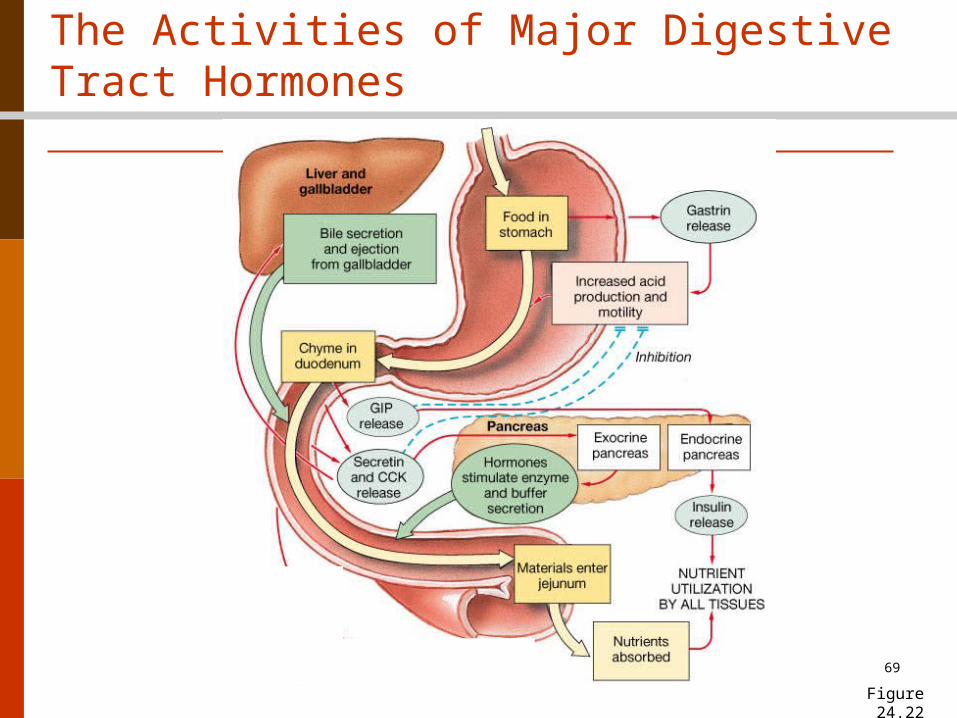
The Activities of Major Digestive Tract Hormones
Figure 24.22

Aerobic vs. Anaerobic Respiration
Aerobic respiration
(in the mitochondria)will result in 6 ATP’s.
Anaerobic respiration (in our cytoplasm) will result in only 2 ATP’s.
16More importantly, we get our NAD back, so glycolysis can continue.

Making ATP by Aerobic Respiration Takes place in the mitochondria Requires oxygen Breaks down glucose to pyruvate and produces ATP Waste products are CO2 and H2O (we exhale
them) The good thing about making ATP from our
mitochondria is that we can make a LOT of it. The bad things are that it takes longer to make it,
and it requires oxygen, and a muscle cell may have used up all the oxygen during a sprinting run.

Making ATP by Anaerobic Respiration Takes place in the cytoplasm Does not require oxygen Breaks down glucose to pyruvate and
produces ATP Waste product is lactic acid The good thing about making ATP this way
is that we can make it FAST. The bad thing is that it does not make
much ATP, and we deplete the reserves quickly.

Lactic Acid Build-up During strenuous workouts where oxygen becomes deficient, the
pyruvate product of glycolysis does not have enough oxygen to use for aerobic respiration, so it has to undergo anaerobic respiration.
The enzyme lactate dehydrogenase (LDH) is used to transfer hydrogen from the NADH molecule to the pyruvate molecule.
Pyruvate with the extra hydrogen is called lactate. Lactic acid is formed from lactate. This causes muscle aches and
fatigue. Lactic acid is deactivated by the addition of oxygen to it.
Therefore, breathing heavily adds the oxygen to our system to deactivate lactic acid, and the muscle pains go away. Warm water or ultrasound will also increase oxygenated blood to the muscles, easing muscle cramps from lactic acid.
When you add oxygen to lactic acid, it either goes back to being pyruvate, which is used to fuel the Krebs cycle (aerobic respiration), or it is converted to glucose in the liver. 19

ATP and Creatine Phosphate What do we do when we run out of ATP? Muscle fibers cannot stockpile ATP in preparation
for future periods of activity. However, they can store another high energy
molecule called creatine phosphate. Creatine phosphate is made from the excess ATP
that we accumulate when we are resting. During short periods of intense exercise, the
small reserves of ATP existing in a cell are used first.
Then creatine phosphate is broken down to produce ATP.
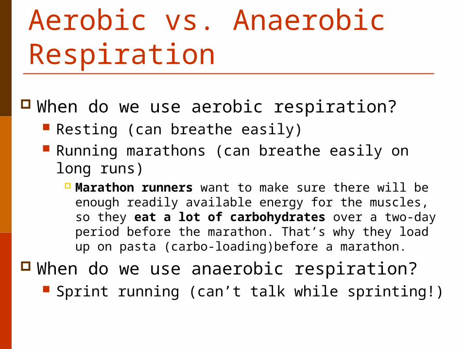
Aerobic vs. Anaerobic Respiration
When do we use aerobic respiration? Resting (can breathe easily) Running marathons (can breathe easily on long
runs) Marathon runners want to make sure there will be
enough readily available energy for the muscles, so they eat a lot of carbohydrates over a two-day period before the marathon. That’s why they load up on pasta (carbo-loading)before a marathon.
When do we use anaerobic respiration? Sprint running (can’t talk while sprinting!)
Gluconeogenesis Gluconeogenesis is a metabolic pathway that
results in the generation of new glucose from non-carbohydrate carbon substrates such as lactate, glycerol, and amino acids. Therefore, if we do not have enough glucose in our body, we will break down proteins (muscles) to make glucose.
It is one of the two main mechanisms to keep blood glucose levels from dropping too low (hypoglycemia).
The other means of maintaining blood glucose levels is through the degradation of glycogen (glycogenolysis).
22
23
24
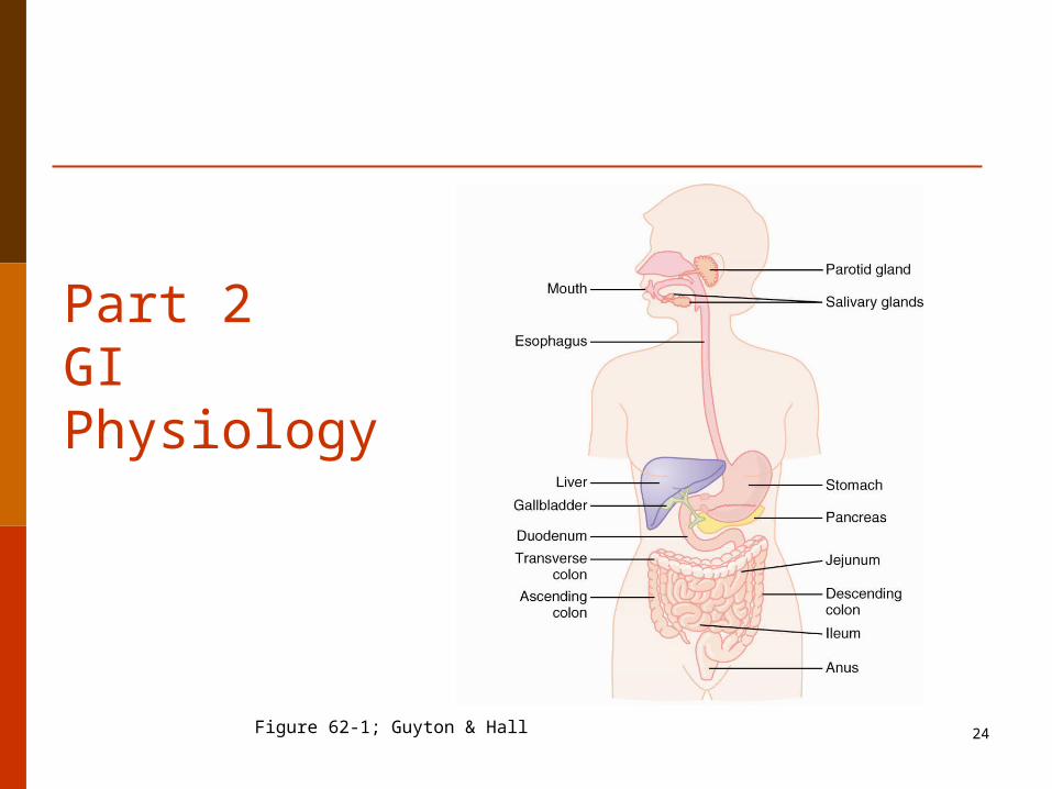
Part 2GI Physiology
Figure 62-1; Guyton & Hall

Digestion Problems Incomplete digestion may be a contributing factor
in the development of many ailments including flatulence, bloating, belching, food allergies, nausea, bad breath, bowel problems and stomach disorders.
Digestive enzymes are primarily responsible for the chemical breakdown of food and constitute a large portion of digestive secretions.
The human body makes approximately 22 different enzymes that are involved in digestion.
25

Digestive Enzymes Saliva is secreted in large amounts (1-1.5
liters/day) Salivary glands contain the enzyme
salivary amylase. This enzymes breaks starch into smaller
sugars and is stimulated by chewing. It is important to chew food thoroughly as
this is the first stage of the digestive process.
26

Saliva The saliva serves to clean the oral cavity
and moisten the food. It also contains digestive enzymes such as
salivary amylase, which aids in the chemical breakdown of polysaccharides such as starch into disaccharides such as maltose.
It also contains mucus, a glycoprotein which helps soften the food and form it into a bolus.
27

Swallowing The mechanism for swallowing is
coordinated by the swallowing center in the medulla oblongata and pons (in the brain stem).
The reflex is initiated by touch receptors in the pharynx (back of the throat)as the bolus of food is pushed to the back of the mouth.
28
Stomach The stomach is responsible for the digestion of
protein. Mucous cells (in the stomach) secrete mucous.
The pancreas secretes bicarbonate. Mucous, bicarbonate, and prostaglandins protect the stomach lining from being digested.
The parietal cells of the stomach secrete hydrochloric acid (gastric acid) and intrinsic factor.
Hydrochloric acid (HCl), along with pepsin (from the chief cells), breaks down proteins to their individual amino acids.
29
Downloaded from: StudentConsult (on 23 April 2010 06:51 PM)© 2005 Elsevier
Stomach Protection and Damage

Downloaded from: StudentConsult (on 23 April 2010 06:51 PM)© 2005 Elsevier

© 2005 Elsevier
Downloaded from: StudentConsult (on 23 April 2010 06:51 PM)© 2005 Elsevier
Stimuli for Stomach Secretions

Stomach Acid The acid itself does not break down food
molecules. It provides an optimum pH for the
activation of pepsin, and kills many microorganisms that are ingested with the food.
It can also denature proteins. The parietal cells of the stomach also
secrete a glycoprotein called intrinsic factor, which enables the absorption of vitamin B-12. 34

Stomach Acid Diseases Hypochlorhydria
Diseases associated with low gastric acidity: Asthma, coeliac disease, eczema, osteoporosis and
pernicious anemia. Hyperchlorhydria
Diseases associated with high gastric acidity: Heartburn, gas and ulcers
35
Hypochlorhydria Deficient hydrochloric acid secretion Causes malabsorption and may result in a
number of signs and symptoms. These include bloating, belching, flatulence,
nausea, a sense of fullness immediately after meals, indigestion, diarrhea, constipation, food allergies, anemia (Folic acid, vitamin B12 and iron will not be absorbed if there is too little acid), undigested food in stool, chronic intestinal parasites, abnormal flora and weak, peeling and cracked fingernails.
36

Small Intestine• Duodenum
– Absorption of minerals– Receives pancreatic digestive enzymes– Secretes hormones when acidic chyme enters duodenum
• Secretin– Tells pancreas to secrete bicarbonate– Tells liver to make bile
• Cholecystokinin (CCK)– Tells pancreas to release protein-digesting enzymes – Tells the gallbladder to release stored bile.– Therefore, it stimulates digestion of fat and protein.
• GIP– stimulates insulin secretion
• Motilin– Initiates peristalsis (increases GI motility)– Tells the Chief cells to secrete pepsinogen
– Secretes enzymes to break down polysaccharides• Maltase: breaks maltose down into glucose• Lactase: breaks lactose down to galactose plus glucose• Sucrase: breaks sucrose down into fructose plus glucose
37

Maltose Maltose (malt sugar) is made of two glucose molecules joined
together. Maltose is the disaccharide produced when amylase breaks
down starch.
Starch Maltose Glucose
Maltose is found in germinating seeds such as barley as they break down their starch stores to use for food.
It is also produced when glucose is caramelized (browning of sugar during cooking).
Foods containing maltose include malted milk shakes, malt liquor and beer. People who lack the maltase enzyme get diarrhea and gas if they ingest malt sugars.
38
Amylase Maltase

Lactose Lactose is needed for milk production. It is made in the body by combining glucose with
galactose. When milk products are consumed, lactose is
broken down by the enzyme lactase. Many Asian and Hispanic people lack the enzyme
lactase, so they are called lactose intolerant. If they consume milk products, they cannot break
down lactose, so the E. coli in the colon get the sugar. E. coli metabolism then causes gas. The person may have diarrhea as well.
39

Sucrose and Fructose Sucrose is table sugar Fructose is fruit sugar
All polysaccharide sugars and starches are broken down into glucose, which is needed by the body for metabolism.
40

Small Intestine Duodenum
When there is no more chyme entering the duodenum, it secretes glucose-dependent insulinotropic peptide (GIP).
GIP is synthesized by K cells, which are found in the duodenum and jejunum.
GIP stimulates insulin secretion. Insulin is in the blood stream. It takes the
absorbed sugars and pulls them into cells that need it.
GIP also causes fat to be broken down into fatty acids.
41

42
Lipid digestion and absorption Lipid digestion utilizes
lingual and pancreatic lipases, to release fatty acids and monoglycerides. Bile salts improve chemical
digestion by emulsifying lipid drops
Lipid-bile salt complexes called micelles are formed
43
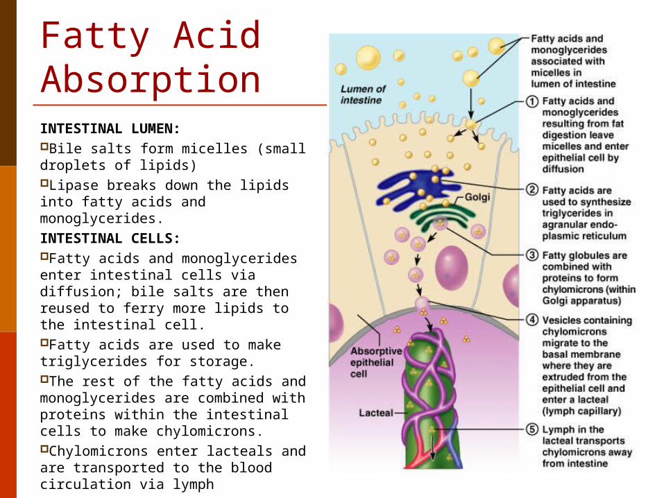
Fatty Acid AbsorptionINTESTINAL LUMEN:Bile salts form micelles (small droplets of lipids) Lipase breaks down the lipids into fatty acids and monoglycerides. INTESTINAL CELLS:Fatty acids and monoglycerides enter intestinal cells via diffusion; bile salts are then reused to ferry more lipids to the intestinal cell.Fatty acids are used to make triglycerides for storage.The rest of the fatty acids and monoglycerides are combined with proteins within the intestinal cells to make chylomicrons.Chylomicrons enter lacteals and are transported to the blood circulation via lymph

Protein and Fat Digestion During digestion, lipids are broken down into fatty
acids plus glycerol by the enzyme lipase. Proteins are degraded by various proteolytic
enzymes, and they break down into amino acids. Hydrolysis (water is added to break a bond) of
proteins occurs in the stomach by pepsin, and this process requires the presence of hydrochloric acid in the stomach, but the rest of the proteins are broken down in the intestine.
Both fat and protein digestion requires ATP.
44
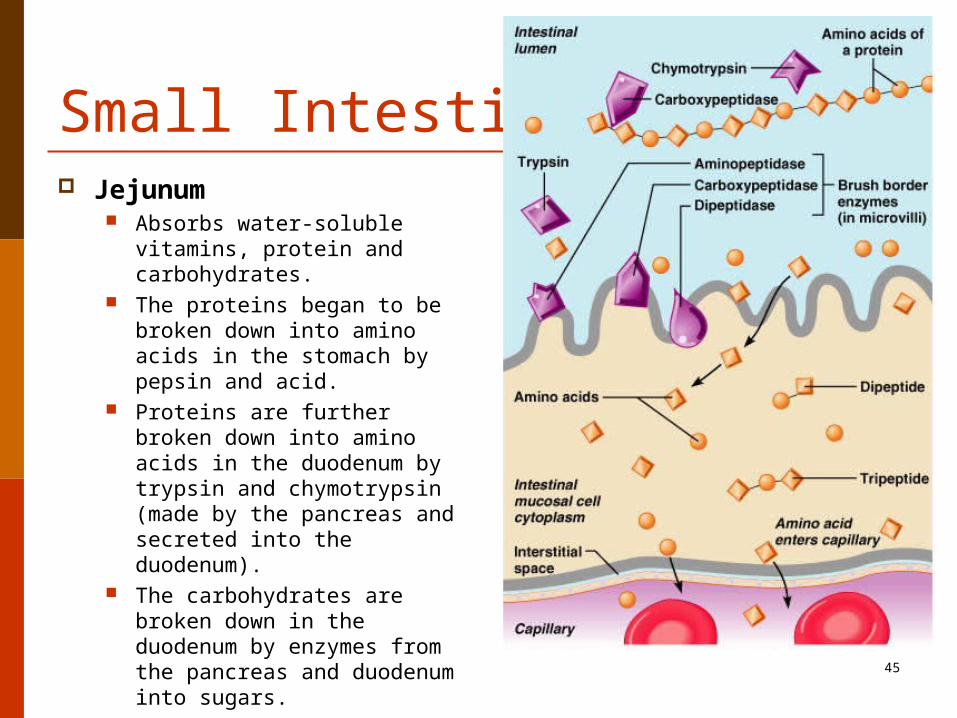
Small Intestine Jejunum
Absorbs water-soluble vitamins, protein and carbohydrates.
The proteins began to be broken down into amino acids in the stomach by pepsin and acid.
Proteins are further broken down into amino acids in the duodenum by trypsin and chymotrypsin (made by the pancreas and secreted into the duodenum).
The carbohydrates are broken down in the duodenum by enzymes from the pancreas and duodenum into sugars.
45

Small Intestine Ileum
Absorbs fat-soluble vitamins, fat, cholesterol, and bile salts.
Fats are broken down into fatty acids in the duodenum.
First, bile emulsifies the fat (breaks it down into droplets).
Then, lipase (made in the pancreas) breaks the fat into fatty acids, which are small enough to be absorbed.
46

Pancreas Enzymes The pancreas secretes about one and a half liters
of pancreatic juice a day! Pancreatic juice secretion is regulated by the
hormones secretin and cholecystokinin (CCK), which is produced by the walls of the duodenum upon detection of acid food, proteins and fats.
The enzymes produced by the pancreas include Lipases Amylases Proteases
47

Pancreas Enzymes Lipases
Digestion of fats, oils, and fat-soluble vitamins Amylases
Break down starch molecules into smaller sugars. Break down carbohydrates into maltose
Proteases Break down protein into smaller amino acids Proteases include trypsin, chymotrypsin and
carboxypeptidase. Proteases are also responsible for keeping the small
intestine free from parasites (intestinal worms, yeast overgrowth and bacteria).
A lack of proteases can cause incomplete digestion that can lead to allergies and the formation of toxins.
48

49
Regulation of Pancreatic Secretion Secretin and CCK are
released when fatty or acidic chyme enters the duodenum
CCK and secretin enter the bloodstream
Upon reaching the pancreas: CCK induces the secretion of
enzyme-rich pancreatic juice Secretin causes secretion of
bicarbonate-rich pancreatic juice
Vagal stimulation also causes release of pancreatic juice

50
The Pancreas
Exocrine function (98%) Acinar cells make,
store, and secrete pancreatic enzymes
Endocrine function – ( cells) release
somatostatin (inhibitory to gastrin, insulin, and glucagon)
β-cells –release insulin α-cells-Release glucagon

51
The Pancreas as an Endocrine Gland Insulin
Beta cells Skeletal muscle and
adipose tissue need functional glucose receptors
Promotes glucose uptake Prevents fat and glycogen
breakdown and inhibits gluconeogenesis
Increases protein synthesis
Promotes fat storage
Picture from:http://www.dkimages.com/discover/Home/Health-and-Beauty/Human-Body/Endocrine-System/Pancreas/Pancreas-1.html
Epi/Norepi inhibit insulin!Help maintain glucose levels during times of stress and increase lipase activity in order to conserve glucose levels

52
The Pancreas as an Endocrine Gland Glucagon
Increases blood glucose levels Maintains blood glucose
between meals and during periods of fasting by breaking down glycogen (stored in liver) into glucose.
Initiates glycogenolysis in liver (within minutes).
Stimulates gluconeogenesis. This process involves breaking down amino acids (proteins) into glucose.
Stimulates amino acid transport to liver to stimulate gluconeogenesis
Nervous tissue (brain) is heavily dependent on glucose levels!
Image from: http://www.dkimages.com/discover/previews/768/74261.JPG

Liver and Gallbladder The liver produces bile that is either stored by the
gallbladder or secreted into the small intestine. Bile emulsifies fats and fat-soluble vitamins. It also helps keep the small intestine free from
parasites. The liver does not make the digestive enzymes for
carbohydrates, amino acids and proteins (the pancreas and small intestine do that), but the liver does metabolize proteins, carbohydrates and cholesterol.
It also is responsible for the detoxification of toxins, drugs and hormones.
53

Large Intestine The large intestine absorbs water, electrolytes and some of
the final products of digestion. It allows fermentation due to the action of gut bacteria,
which break down the substances which remain after processing in the small intestine; some of the breakdown products are absorbed. In humans, these include most complex saccharides (at most three disaccharides are digestible in humans)
Food products that cannot go through the villi, such as cellulose (dietary fiber), are mixed with other waste products from the body and become hard and concentrated feces.
54

55
Physiology of the large intestine
Reabsorption of water and electrolytes
Coliform bacteria make: Vitamins – K, biotin, and B5
Organic wastes are left in the lumen – urobilinogens and sterobilinogens
Bile salts Toxins
Mass movements of material through colon and rectum Defecation reflex
triggered by distention of rectal walls

Coliforms Coliforms is the term used for the bacteria
that normally inhabit our colon (large intestine). E. coli is just one species of coliform.
A ratio of 80-85% beneficial to 15-20% potentially harmful bacteria generally is considered normal within the intestines.
Harmful microorganisms also are kept at a minimum by an extensive immune system comprising the gut-associated lymphoid tissue (GALT).
56
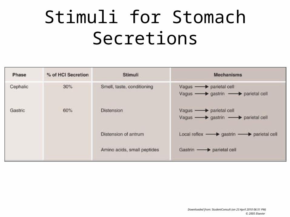
Phases of gastric secretion Cephalic phase Gastric phase Intestinal phase
57

Cephalic phase This phase occurs before food enters the stomach and
involves preparation of the body for eating and digestion. Sight and thought stimulate the cerebral cortex. Taste and
smell stimulus is sent to the hypothalamus and medulla oblongata.
After this it is routed through the vagus nerve and release of acetylcholine.
58
Gastric secretion at this phase rises to 40% of maximum rate. Acidity in the stomach is not buffered by food at this point and thus acts to stimulate Delta cells to secrete somatostatin. That causes the G cells to stop secreting gastrin. That caused the parietal cells to stop secreting HCl.

G cell secretion of gastrinD cell secretion of somatostatin
59

G cells and Gastrin G cells are found deep within the gastric glands of the
stomach. When food arrives in the stomach, the parasympathetic
nervous system is activated. This causes the vagus nerve to release a neurotransmitter called Gastrin-releasing peptide onto the G cells in the stomach.
Gastrin-releasing peptide, as well as the presence of proteins in the stomach, stimulates the release of gastrin from the G cells.
Gastrin tells parietal cells to increase HCl secretion, and it also stimulates other special cells to release histamine.
Gastrin also tells the chief cells to produce pepsinogen. Gastrin is inhibited by low pH (acid) in the stomach. When
enough acid is present, it turns off.60

Gastrin Gastrin is released in response to
Stomach distension Vagus nerve stimulation The presence of proteins or amino acids
Gastrin release is inhibited by The presence of enough HCl in the stomach
(negative feedback) Somatostatin also inhibits the release of
gastrin
61

D cells D cells can be found in the stomach,
intestine and the Islets of Langerhans in the pancreas.
When gastrin is present, D cells increase somatostatin output.
When D cells are stimulated by Ach, they decrease somatostatin output.
62

D cells Ach that comes from the Vegas nerve
branch that lands on D cells will decrease somatostatin output so that digestion can occur.
Ach that comes from the Vegas nerve branch that lands on G cells will cause gastrin to be released, and when gastrin is present but there is no food left, that excess gastrin will stimulate the D cells to increase somatostatin to try to turn off the system. 63

Somatostatin Somatostatin is also known as growth hormone-inhibiting
hormone. It suppresses the release of gastrointestinal hormones
Gastrin Cholecystokinin (CCK) Secretin GIP
It suppresses the release of pancreatic hormones. It slows down the digestive process. It inhibits insulin release. It inhibits the release of glucagon.
64

Gastric phase This phase takes 3 to 4 hours. It is stimulated by distension of the
stomach, presence of food in stomach and decrease in pH. Distention activates the vagus nerve. This activates the release of acetylcholine which stimulates the
release of more gastric juices. As protein enters the stomach, it binds to hydrogen ions, which
raises the pH of the stomach. Inhibition of gastrin and gastric acid secretion is lifted. This triggers G cells to release gastrin, which in turn stimulates
parietal cells to secrete gastric acid. Gastric acid is about 0.5% hydrochloric acid (HCl), which lowers
the pH to the desired pH of 1-3. Acid release is also triggered by acetylcholine and histamine.
65

Intestinal phase This phase has 2 opposing actions: the
excitatory and the inhibitory. Partially digested food fills the duodenum. This triggers gastrin to be released. It also triggers the enterogastric reflex,
which inhibits the Vagus nerve. This activates the sympathetic nervouse
system, which causes the pyloric sphincter to tighten to prevent more food from entering the duodenum.
66

67
Digestive Enzymes (fats in yellow, proteins in green, and sugars in red)
Salivary glands-amylase
Lipase
Stomachpepsin
Lipase
Liver Lipase
Duodenum sucrase maltase lactase
Pancreas amylase trypsin chymotrypsincarboxypeptidaseLipase
68
Digestive Hormones and Substances
Stomachgastrin
Intrinsic factor HCl Prostaglandins Mucous
Duodenum Secretin CCK GIP Motilin
Pancreas Glucagon Insulin Somatostatin Bicarbonate
LiverBile
69
The Activities of Major Digestive Tract Hormones
Figure 24.22

70
Organ Region of the Organ Substances FunctionPancreas Acinar cells Amylase (enzyme) Breaks down starch and carbohydrates into glucose
Acinar cells Lipase (enzyme) Breaks down fat into fatty acids
Acinar cellsProtease enzymes (trypsin,
chymotrypsin, carboxypeptidase)Breaks down proteins into amino acids and also kills intestinal parasites and bacteria
Acinar cells Bicarbonate (not an enzyme) Raises pH in duodenum
Islet of Langerhans;
Alpha cells glucagon (hormone)
Causes glycogenolysis, the process which breaks down glycogen into glucose to raise blood glucose. Also causes gluconeogenesis to make new glucose molecules
Islet of Langerhans;
Beta cells insulin (hormone)Removes glucose in bloodstream and brings it into cells. Lowers blood glucose levels.
Islet of Langerhans;
Delta cells Somatostatin (hormone) Inhibits gastrin, insulin, and glucagon (inhibits digestive system)
Liver Bile (a detergent) Emulsifies fat Lipase Breaks down fat
Salivary glands Amylase (enzyme) Breaks down starch and carbohydrates into glucose Lipase Breaks down fat
Stomach Mucous (not an enzyme) Protect the stomach liningProstaglandins (not an enzyme) Protect the stomach lining
Lipase Breaks down fat
Parietal cells HCl (not an enzyme) Allows Pepsinogen to be converted to pepsin, and it also kills bacteria
Parietal cells Intrinsic factor (not an enzyme)Allows Vit B12 to be absorbed, which is needed to make RBCs. Without it, you get megaloblastic (pernicous) anemia.
Chief cells Pepsinogen --> pepsin (enzyme) Breaks proteins into amino acids G cells Gastrin (hormone) Tells parietal cells to secrete HCl
Duodenum Secretin (hormone) Tells pancreas to secrete bicarbonate
CCK (hormone)Tells pancreas to secrete proteases and lipase, and tells gallbladder to release stored bile (stimulates fat and protein digestion)
Motilin (hormone) Initiates peristalsis and tells Chief cells to secrete pepsinogen
Maltase, Lactase, Sucrase (enzymes) Break down complex carbohydrates into glucose
K cells GIP (hormone)Tells pancreas to release insulin and also causes fat to be broken down into fatty acids

Where do the molecules go when you lose weight?
• Think of fat as essentially a long-chain hydrocarbon CH3-(CH2)n-CH3. When your body uses that fat as fuel (either because you need fuel to exercise, or because you're not eating enough new fuel to support what you're doing), it burns that fat to extract the energy from it. That "burn" isn't a metaphor. The chemistry that your body does is exactly equivalent to literally burning it, just under more controlled conditions.
71

Where do the molecules go when you lose weight?
• So, that hydrocarbon undergoes a controlled combustion with oxygen (O2) to produce a lot of energy, water (H2O), and carbon dioxide (CO2).
Or, in chemical form:CH3-(CH2)n-CH3 + (3/2n+7/2)O2 ----> (n+2) CO2 + (n+3) H2O + Energy
72

Where do the molecules go when you lose weight?
• So the carbon in the hydrocarbon goes to carbon dioxide and the hydrogen goes to water. But most of the mass of the hydrocarbon is carbon, so most of the mass gets converted to carbon dioxide, which is a gas and gets breathed out.
• Now this is incomplete, because lipids and fat really aren't just hydrocarbons. They have phosphates and nitrogen and other things too, and those parts don't get converted to gases for excretion. Excess nitrogen gets converted to urea, for example, which gets excreted in the urine. And protein produces a lot more impurities when it gets broken down (though generally the body prefers to recycle proteins rather than burn them for energy).
• But really, the way you lose most of your weight is just by breathing it off.
73

Good Website
• http://uh.edu/sibs/tutorial/ap2.htm#digestive

75
Part 3GI Disorders
Figure 62-1; Guyton & Hall

GI Disorders Peptic ulcers Pancreatitis Celiac Disease Inflammatory bowel disease (Crohn's disease and ulcerative colitis) Irritable bowel syndrome Appendicitis Diverticulitis Cancer Gastroenteritis ("stomach flu“); an inflammation of the stomach and
intestines Cholera (bacteria in sewage-contaminated food or water) Giardiasis (protozoa in contaminated drinking water) Yellow Fever (virus transmitted by tropical mosquito)
76

Peptic Ulcers Classification By Region/Location
Duodenum (called duodenal ulcer) Esophagus (called esophageal ulcer) Stomach (called gastric ulcer)
Classification by Type Type I: Ulcer along the body of the stomach, most often
along the lesser curve. Type II: Ulcer in the body in combination with duodenal
ulcers. Associated with acid oversecretion. Type III: In the pyloric region. Associated with acid
oversecretion. Type IV: Proximal gastroesophageal ulcer Type V: Can occur throughout the stomach. Associated with
chronic NSAID use (such as aspirin).77

78
Gastric and Duodenal ulcers
Peptic ulcers occur when damaging effects of acid and pepsin overcome ability of mucosa to protect itself Gastric ulcers - main problem is decreased ability of
mucosa to protect itself
Duodenal ulcers - main problem is exposure to increased amounts of acid and pepsin
Strengthensmucus, HCO3
- secretion, gastrin, PGs, epidermal growth
factor
WeakensH. pylori, aspirin, ethanol,
NSAIDs, bile salts

Two major causes of Peptic Ulcers:1) 60% of gastric and up to 90% of duodenal
ulcers are due to a bacterium called Helicobacter pylori. The body responds by increasing gastrin
secretion, which erodes the stomach lining.
2) NSAIDs (non-steroidal anti-inflammatory drugs, such as aspirin) block prostaglandin synthesis. Prostaglandins promote the inflammatory
reaction. They also are found in the stomach, protecting it from erosion.
79

Does stress cause ulcers? There is debate as to whether
psychological stress can influence the development of peptic ulcers.
Helicobacter pylori thrives in an acidic environment, and stress has been demonstrated to cause the production of excess stomach acid.
80

Diagnosis of Helicobacter pylori Urea breath test (noninvasive)
Patient drinks a tasteless liquid which contains a radioactive carbon atom as part of the substance that the bacteria breaks down. After an hour, the patient will be asked to blow into a bag that is sealed. If the patient is infected with H. pylori, the breath sample will contain radioactive carbon dioxide.
Biopsy Direct culture from a biopsy Histological examination and staining Direct detection of urease activity in a biopsy specimen by
rapid urease test Measurement of antibody levels in blood Stool antigen test
81

Differential Diagnosis (DDx) A differential diagnosis is a list of possible things that
may be causing a patient’s symptoms. DDx for H. pylori infection
Peptic ulcer Gastritis Stomach cancer Gastroesophageal reflux disease Pancreatitis Hepatic congestion Cholecystitis Biliary colic Inferior myocardial infarction Referred pain (pleurisy, pericarditis) Superior mesenteric artery syndrome
82

Risk and Transmission The lifetime risk for developing a peptic ulcer is
approximately 10%. In Western countries the prevalence of
Helicobacter pylori infections roughly matches age (i.e., 20% at age 20, 30% at age 30, 80% at age 80 etc.).
Prevalence is higher in third world countries. Transmission is by food, contaminated
groundwater, and through human saliva (such as from kissing or sharing toothbrushes or food utensils)
83

Treatment Younger patients with ulcer-like symptoms are often
treated with antacids or H2 antagonists (blocks the acid secretion of parietal cells).
Patients who are taking NSAIDs may also be prescribed a prostaglandin analogue (Misoprostol) to help prevent peptic ulcers.
When H. pylori infection is present, the most effective treatments are combinations of 2 antibiotics (e.g. Clarithromycin, Amoxicillin, Tetracycline, Metronidazole) and 1 proton pump inhibitor (PPI), sometimes together with a bismuth compound. An example of a PPI is Omeparazole (Prilosec).
84

Treatment Ranitidine (Zantac) and Cimetidine (Tagamet)
provide relief of peptic ulcers, heartburn, indigestion and excess stomach acid and prevention of these symptoms associated with excessive consumption of food and drink.
They decrease the amount of acid the stomach produces allowing healing of ulcers.
Sucralfate, (Carafate) and strawberries have also been used in successful treatment of peptic ulcers.
85

Pancreas Disorders Gestational Diabetes Type I diabetes Type II diabetes Pancreatitis Cancer Chronic pancreatitis
alcohol cystic fibrosis
Acute pancreatitis Gallstones
86

87
Disorders of the Pancreas: Diabetes Mellitus Gestational Diabetes Type I diabetes – develops
suddenly, usually before age 15 Destruction of the beta cells Skeletal tissue and adipose
cells must use alternative fuel and this leads to ketoacidosis
Hyperglycemia results in diabetic coma

88
Disorders of the Pancreas: Diabetes Mellitus Type II diabetes– adult
onset Usually occurs after age
40 Cells have lowered
sensitivity to insulin Controlled by dietary
changes and regular exercise

89
Pancreatic Failure
Digestion is abnormal when pancreas fails to secrete normal amounts of enzymes. Pancreatitis Removal of pancreatic head - malignancy
Without pancreatic enzymes - 60% fat not absorbed (steatorrhea) 30-40% protein and carbohydrates not absorbed

90
Pancreatitis Pancreatitis means inflammation of pancreas.
Autodigestion theory can explain condition.
Chronic pancreatitis - alcohol - most common cause in adults cystic fibrosis - most common cause in children
CF patients lack chloride transporter at apical membrane. Watery ductal secretion decreases which concentrates acinar secretions in ducts. Destroys pancreas gland by autodigestion.
Acute pancreatitis - Gallstones - most common cause

Celiac disease (Sprue; gluten intolerance) Genetic autoimmune disorder of the small intestine,
causing chronic diarrhea. The person is allergic to gluten. Causes destruction of microvilli and villi.
It is characterized by having pale, loose and greasy stools (steatorrhea) which are voluminous and malodorous.
It often presents with abdominal pain and cramping, abdominal distension, and sometimes mouth ulcers.
Without adjusting the diet, coeliac disease leads to an increased risk of adenocarcinoma (small intestine cancer).
91
Celiac disease (Sprue; gluten intolerance) They may develop ulcerative jejunitis and stricturing (narrowing as a
result of scarring with obstruction of the bowel). The changes in the bowel make it less able to absorb carbohydrates,
fats, minerals (calcium and iron), and the fat-soluble vitamins A, D, E, and K.
Anemia may develop in several ways: iron malabsorption may cause iron deficiency anemia, and folic acid and vitamin B12 malabsorption may give rise to megaloblastic anemia.
Calcium and vitamin D malabsorption may cause osteopenia (decreased mineral content of the bone) or osteoporosis (bone weakening and risk of fragility fractures).
A small proportion have abnormal coagulation due to vitamin K deficiency and are slightly at risk for abnormal bleeding.
Coeliac disease is also associated with bacterial overgrowth of the small intestine, which can worsen malabsorption or cause malabsorption despite adherence to treatment.
92

Celiac disease (Sprue; gluten intolerance) Celiac disease is caused by an allergy to gluten. Gluten is present in Wheat subspecies (such as spelt,
semolina and durum) and related species such as barley, rye, triticale and Kamut. A small minority of coeliac patients also react to oats. It is most probable that oats produce symptoms due to cross contamination with other grains in the fields or in the distribution channels. Generally, oats are therefore not recommended.
Other cereals such as maize (corn), millet, rice, and wild rice are safe for patients to consume, as well as non cereals such as amaranth, quinoa or buckwheat.
Non-cereal carbohydrate-rich foods such as potatoes and bananas do not contain gluten and do not trigger symptoms.
93

Gluten-free diet Several grains and starch sources are considered acceptable
for a gluten-free diet. The most frequently used are corn, potatoes, rice, and tapioca.
Various types of bean, soybean, and nut flours are sometimes used in gluten-free products to add protein and dietary fiber.
Almond flour is a low-carbohydrate alternative to flour, with a low glycemic index.
In spite of its name, buckwheat is not related to wheat; pure buckwheat is considered acceptable for a gluten-free diet, although many commercial buckwheat products are actually mixtures of wheat and buckwheat flours, and thus not acceptable.
Gram flour, derived from chickpeas, is also gluten-free (this is not the same as Graham flour made from wheat).
94

Gluten-free diet Gluten is used in foods in some unexpected ways, for
example as a stabilizing agent or thickener in products like ice-cream and ketchup.
People wishing to follow a completely gluten free diet must also take into consideration the ingredients of any over-the-counter or prescription medications and vitamins. Also, cosmetics such as lipstick, lip balms, and lip gloss may contain gluten and need to be investigated before use. Glues used on envelopes may also contain gluten.
Most products manufactured for Passover are gluten free. Exceptions are foods that list matzah as an ingredient, usually in the form of cake meal.
A blood test for IgA antiendomysial antibodies can detect celiac disease.
95

Uses of animal gut by humans The stomachs of calves have commonly been used as a source of rennet for
making cheese. The use of animal gut strings by musicians can be traced back to the third
dynasty of Egypt. In the recent past, strings were made out of lamb gut. With the advent of the modern era, musicians have tended to use strings made of silk, or synthetic materials such as nylon or steel. Some instrumentalists, however, still use gut strings in order to evoke the older tone quality. Although such strings were commonly referred to as "catgut" strings, cats were never used as a source for gut strings.
Sheep gut was the original source for natural gut string used in racquets, such as for tennis. Today, synthetic strings are much more common, but the best gut strings are now made out of cow gut.
Gut cord has also been used to produce strings for the snares which provide the snare drum's characteristic buzzing timbre.
"Natural" sausage hulls (or casings) are made of animal gut, especially hog, beef, and lamb. Similarly, Haggis is traditionally boiled in, and served in, a sheep stomach.
Chitterlings, a kind of food, consist of thoroughly washed pig's gut. The oldest known condoms, from 1640 AD, were made from animal intestine.
96

97