Getting back into exercise: the neck and shoulders
5
29 www.recreationcentral.eu 2017. tavasz rekreáció ÉLETMÓD / SZELLEMI REKREÁCIÓ Getting back into exercise: the neck and shoulders Figure 1. Dreamsme photo Abstract The physical inacvity combined with the body being subject to repeve work tasks and non-ergonomic seang postures results in today’s human being becoming less condioned and equipped to take part in sports. This then presents challenges to personal trainers and coaches – when the body is expected to perform pushes, pulls and overhead movements a musculoskel- etally imbalanced shoulder girdle, tho- racic and cervical spine, also presented in the Upper Crossed Syndrome, can lead to muscle, tendon, bursa and joint irritaons (Griegel-Morris et al. 1992; Grimsby – Gray, 1997; Jull et al, 2002; Ketola et al. 2002; Marcus et al. 2002; Rempel et al, 2006; Ber- naards et al, 2011) that could be aggravated by exercise. One approach to counter such ailments is the applicaon of the NASM stac and movement tests in conjuncon with the Correcve Exercise Connuum (CEx), which laer can be incorporated into a warm-up. Comprised of four stages – Self-Myofascial Release (SMR), stretches, isolated acva- on and muscle integraon – it promotes the acvaon and deacvaon of muscles, fascia and the nervous system. My aim is to discuss the importance of maintaining a good posture and therefore a healthy musculoskeletal system, with parcular reference to the neck and shoulders. Spe- cifically, improved muscle strength and flexibility around the shoulder blades and neck with NASM CEx can lead to a reduc- on of impingement processes, irritaons, tendonis and lessen the pain symptoms (Griegel-Morris et al, 1992; Grimsby and Gray, 1997; Jull et al, 2002; Lewis and Val- enne, 2007). Keywords: upper crossed syndrome, the NASM Correcve Exercise Connuum, physical inacvity, workplace ergonomics, forward head posture Questions How do work-induced musculoskeletal changes (such as an increased forward head posture and excessive kyphoc curve) affect the recreaonal and professional athlete? Would implemenng the NASM move- ment screens be enough to idenfy major musculoskeletal problems? If not, what other assessments could be undertaken? DOI: 10.21486/recreation.2017.7.1.3
Transcript of Getting back into exercise: the neck and shoulders
29
www.recreationcentral.eu 2017. tavaszrekreáció
ÉLETMÓD / szELLEMi rEkrEáciÓ
Getting back into exercise: the neck and shoulders
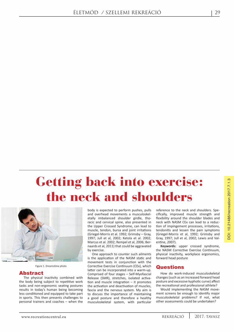
Figure 1. Dreamstime photo
Abstract The physical inactivity combined with
the body being subject to repetitive work tasks and non-ergonomic seating postures results in today’s human being becoming less conditioned and equipped to take part in sports. This then presents challenges to personal trainers and coaches – when the
body is expected to perform pushes, pulls and overhead movements a musculoskel-etally imbalanced shoulder girdle, tho-racic and cervical spine, also presented in the Upper Crossed Syndrome, can lead to muscle, tendon, bursa and joint irritations (Griegel-Morris et al. 1992; Grimsby – Gray, 1997; Jull et al, 2002; Ketola et al. 2002; Marcus et al. 2002; Rempel et al, 2006; Ber-naards et al, 2011) that could be aggravated by exercise.
One approach to counter such ailments is the application of the NASM static and movement tests in conjunction with the Corrective Exercise Continuum (CEx), which latter can be incorporated into a warm-up. Comprised of four stages – Self-Myofascial Release (SMR), stretches, isolated activa-tion and muscle integration – it promotes the activation and deactivation of muscles, fascia and the nervous system. My aim is to discuss the importance of maintaining a good posture and therefore a healthy musculoskeletal system, with particular
reference to the neck and shoulders. Spe-cifically, improved muscle strength and flexibility around the shoulder blades and neck with NASM CEx can lead to a reduc-tion of impingement processes, irritations, tendonitis and lessen the pain symptoms (Griegel-Morris et al, 1992; Grimsby and Gray, 1997; Jull et al, 2002; Lewis and Val-entine, 2007).
Keywords: upper crossed syndrome, the NASM Corrective Exercise Continuum, physical inactivity, workplace ergonomics, forward head posture
QuestionsHow do work-induced musculoskeletal
changes (such as an increased forward head posture and excessive kyphotic curve) affect the recreational and professional athlete?
Would implementing the NASM move-ment screens be enough to identify major musculoskeletal problems? If not, what other assessments could be undertaken?
DO
I: 10
.214
86/re
crea
tion.
2017
.7.1
.3

30
www.recreationcentral.eu2017. tavasz rekreáció
IntroductionA typical example of a detrimental pos-
ture for the body is familiar to many of us who have experienced a desk-based, office job. As the day progresses and the body tires, the torso begins to slouch, the up-per back hunches over the desk, the neck cranes out, the chest caves inwards and the shoulders fall forward. Chronic shoul-der and neck pain often stems from these long-standing bad posture and can often be tracked back to forward head posture and overly destabilized shoulder blades on the thoracic cavity (Gray – Grimsby, 2004) (Fig-ure 2). As a result, an important first step of treatment is considering workplace ergo-nomics. This is the science of designing the workplace, based on the impetus that poor workplace design leads to fatigue, a series of musculoskeletal disorders (MSDs) and pain syndromes (Marcus et al. 2002; Keto-la et al. 2002; Rempel et al. 2006; Morken et al. 2007; Holth et al. 2008). A systematic approach would be to remove the risk fac-tors that lead to musculoskeletal injuries at the workplace.
Figure 2. Wrong sitting posture. (Dreamstime photo)
The neck and shoulder problems has been placed in one group as several mus-cles act on both the shoulder girdle/blades and the cervical spine simultaneously and the musculoskeletal change of one will in-evitably influence the other (Griegel-Morris et al, 1992). For example, a forward head posture with a chronic excessive cervical curve of the neck forces the thoracic spine to develop a larger curve – that causes a lengthening of the back muscles, thereby putting them and the attaching shoulder blades into a biomechanically disadvan-taged position so that they cannot perform their physiological functions adequately and with the required strength (Gray and Grimsby, 2004, Bullock, Foster and Wright, 2005; Page et al, 2010). As a consequence, other muscles are forced to ‚take over’
which then eventually tighten up, develop trigger points and sometimes even atrophy due to impaired blood circulation (Page et al, 2010).
The increased weight of the head is borne by the neck extensors at the back of the neck and they subsequently contract more forcefully to prevent the head from falling any further forward – which, in so doing, causes pain (Kapandji, 1974) (Figure 3). At the same time, the front neck flexors, (with particular note to the deep neck flex-ors), which are supposed to help balance the head by pulling it forward, are made redundant and so weaken (disuse atrophy).
Figure 3. Forward head posture adapted from Kapandji, 1974. With each inch, the head is moved forward with the force needed to support its weight increases by ten lbs (4.5kg).
Upper Crossed Syndrome
Janda’s so-called upper crossed syn-drome (UCS) is also referred to as the shoulder girdle crossed syndrome. The term ‚crossed’ refers to the crossing pattern of the overactive muscles with the counter crossing of the underactive muscles. When viewed from the side, an X-pattern can be seen between these two sets of muscles. In UCS, the tightness of the upper trape-zius and levator scapula (neck extensors) at the back crosses with tightness of the sternocleidomastoid (SCM), pectoralis ma-jor and minor at the front. Weakness of the deep cervical flexors anteriorly crosses with weakness of the middle and lower tra-pezius, rhomboids and lower fibres of the serratus anterior at the back (Figure 4 a,b).
Figure 4: a) Janda’s Upper Crossed Syndrome b) typical posture in UCS c) muscles’ lengths in slouched posture
adapted from (www.fixtheneck.com), d) normal shoulder blade position, neither depressed nor elevated, neither protracted nor retracted, normal distance 10-12 cm apart, glenohumeral joint (GH) in neutral, pointing away horizontally, adapted from (www.fixtheneck.com)
These imbalances will manifest in for-ward head posture, increased cervical lordo-sis and thoracic kyphosis also coupled with winging of the scapulae, where the shoulder blades migrate to the side of the thoracic cavity and flare away from the ribcage. If a slouched posture is habitual, the rhomboids as well as the middle and lower trapezius muscles, (which depress and anchor the shoulder blades to the thoracic spinal col-umn of the back), stay elongated and weak-en (stretch weakness and atrophy) while the upper trapezius shortens, and the levator scapulae pulls the blades upwards towards the cervical spine (Figure 4c,d). These mus-cles will shorten and develop chronic trigger points and spasms to somewhat maintain glenohumeral centration (Page at al, 2010).
At the same time the chest caves inwards as the pectorals contract and shorten to take up the slack, and so in turn, rounding the shoulders and arms forward and inwards to the chest, pulling the shoulder blades to the sides and over into a protracted, forwardly rotated and anteriorly tilted position.
The outcome of these biomechanical changes is a shoulder blade(s) that is no longer able to serve as a stable platform for arm use, now unable to rotate freely under the arm(s), as it would do normally and the individual will be unable to raise their arm(s) fully overhead without compensations (Fin-ley and Lee, 2003; Gray and Grimsby, 2004) (Figure 5).
NASM Static and Dynamic Assessments
Some basic assessments can be imple-mented to identify faulty movement pat-terns of the upper body. These include the static posture analysis, overhead squat, then for further investigations the pec-toralis minor length test and pushing and pulling motions, in which we observe the head’s position relative to the shoulders and shoulder blades to the ribs. By using the ears, shoulders and the glenohumeral joint as markers (joint between the shoul-der blade and upper arm bone) the client with UCS will exhibit the following charac-teristics in a static assessment:
In the anterior view, the collarbone will drop down towards the arms or just about
Figure 5: overactive upper trapezius and levator scapulae (www.fixtheneck.com)
TANULMáNY
31
www.recreationcentral.eu 2017. tavaszrekreáció
TANULMáNY
stay horizontal. From the side view, the arm appears internally rotated and the head protrudes forward from the shoulders (Fig-ure 6), while from the posterior view a lean individual’s shoulder blade(s) will appear far apart (over 10-12cm is excessive) (Figure 4d). The medial boarders (towards spine) of the shoulder blades will be far from parallel and flare. The shoulder(s) will also seem to be raised.
Figure 6: NASM Static assessment lateral view and muscle imbalances adapted from NASM.
When assessing clients with UCS the NASM overhead squat is not a comprehen-sive test but can give us clues: note that the overhead arm raise can appear to be nor-mal, but upon closer look compromises of the thoracic cavity and the lower back can be observed from a side view – they are characterised by the flaring out of the lower ribs, a forward head posture and an exces-sive curve of the lower back (Figure 7a,b).
Figure 7: a) normal and b) impaired overhead arm raise (http://www.teamchiroames.com/blog/category/shoulder)
From the posterior view, the client el-evates one or both shoulders abnormally high as the upper trapezius overcompen-sates and the lower trapezius and serratus anterior becomes unable to pull the shoul-der blades sufficiently down enough – due to their lengthened state. Also note how, when instructing the client to engage the core (pull on their belly button) and flatten the ribcage, their arm(s) will fall forward.
Further AssessmentsA shortened pectoralis minor muscle
is common in the UCS imbalance and/or shoulder pain (Sahrmann, 2002). Assess its tightness as the client lies on his or her back on a firm surface with their arms at sides (Yesilyaprak et al, 2014). In this position, the
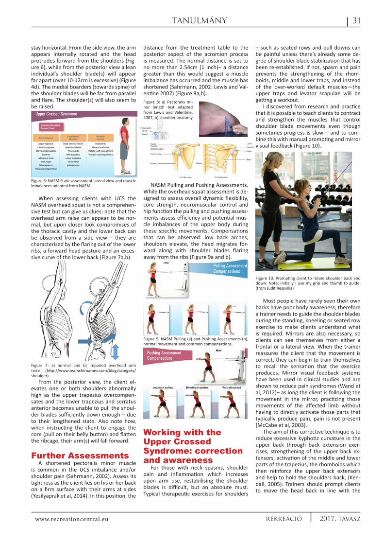
distance from the treatment table to the posterior aspect of the acromion process is measured. The normal distance is set to no more than 2.54cm (1 inch)– a distance greater than this would suggest a muscle imbalance has occurred and the muscle has shortened (Sahrmann, 2002: Lewis and Val-entine 2007) (Figure 8a,b).
NASM Pulling and Pushing Assessments. While the overhead squat assessment is de-signed to assess overall dynamic flexibility, core strength, neuromuscular control and hip function the pulling and pushing assess-ments assess efficiency and potential mus-cle imbalances of the upper body during these specific movements. Compensations that can be observed: low back arches, shoulders elevate, the head migrates for-ward along with shoulder blades flaring away from the ribs (Figure 9a and b).
Figure 9: NASM Pulling (a) and Pushing Assessments (b), normal movement and common compensations.
Working with the Upper Crossed Syndrome: correction and awareness
For those with neck spasms, shoulder pain and inflammation which increases upon arm use, restabilising the shoulder blades is difficult, but an absolute must. Typical therapeutic exercises for shoulders
– such as seated rows and pull downs can be painful unless there’s already some de-gree of shoulder blade stabilization that has been re-established. If not, spasm and pain prevents the strengthening of the rhom-boids, middle and lower traps, and instead of the over-worked default muscles—the upper traps and levator scapulae will be getting a workout.
I discovered from research and practice that it is possible to teach clients to contract and strengthen the muscles that control shoulder blade movements even though sometimes progress is slow – and to com-bine this with manual prompting and mirror visual feedback (Figure 10).
Figure 10. Prompting client to rotate shoulder back and down. Note: initially I use my grip and thumb to guide. (from Judit Ressinka)
Most people have rarely seen their own backs have poor body awareness; therefore a trainer needs to guide the shoulder blades during the standing, kneeling or seated row exercise to make clients understand what is required. Mirrors are also necessary, so clients can see themselves from either a frontal or a lateral view. When the trainer reassures the client that the movement is correct, they can begin to train themselves to recall the sensation that the exercise produces. Mirror visual feedback systems have been used in clinical studies and are shown to reduce pain syndromes (Wand et al, 2012)– as long the client is following the movement in the mirror, practicing those movements of the affected limb without having to directly activate those parts that typically produce pain, pain is not present (McCabe et al, 2003).
The aim of this corrective technique is to reduce excessive kyphotic curvature in the upper back through back extension exer-cises, strengthening of the upper back ex-tensors, activation of the middle and lower parts of the trapezius, the rhomboids which then reinforce the upper back extensors and help to hold the shoulders back, (Ken-dall, 2005). Trainers should prompt clients to move the head back in line with the
Figure 8: a) Pectoralis mi-nor length test adapted from Lewis and Valentine, 2007, b) shoulder anatomy
32
www.recreationcentral.eu2017. tavasz rekreáció
TANULMáNY
spine by tucking the chin in during exercise and roll the shoulders back with the chest lifted. However, it seems that for effective-ness what the trainer and clients achieves within the exercises has to be reinforced regularly and several times a day – while walking, sitting and driving. Ergonomical-ly-friendly work environments and habits are key to reducing the risk of workplace injuries and pain. Steps should be taken to orientate workstations with the principles from University of California in mind (UCLA Environment Health & Safety). It is advis-able to take regular breaks and get up, to move and stretch from time to time (Mar-tins de Oliveira et al, 2015). Once posture is improved and the head is held more in line with its base of support, the back and front neck muscles will be able to work together to keep the head steady and balanced with the least amount of effort.
NASM CEx ContinuumThis corrective program can be used as
a preparation for one’s main workout. In preparing muscles for this, show the client how to move better to improve coordina-tion – before applying speed and increased force during their core workout session. Assessment results can now be consid-ered when designing a program. The four-step corrective exercise process for upper crossed syndrome starts with increasing range of motion by inhibiting and relaxing the possible overactive muscles, improving local strength in underactive muscles, and assisting the client in learning to better con-trol the newly found range of motion, in integrated functional movement patterns. To improve shoulder blade stabilization to the thoracic spine in the CEx warm up – the rhomboids, serratus anterior, deep neck flexors and lower and middle trapezius need to be strengthened. This takes stress off the overactive upper trapezius and levator scapulae muscles, hence relaxes the neck (Clark and Lucett, 2011). Added benefits also include a possible decrease in pain and discomfort, stability of the upper torso, and improved physical performance in training and in play.
Step 1. SMR. Inhibit overactive muscles with Self-Myofascial Release
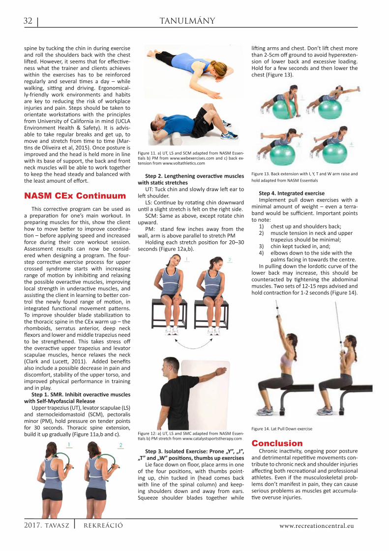
Upper trapezius (UT), levator scapulae (LS) and sternocleidomastoid (SCM), pectoralis minor (PM), hold pressure on tender points for 30 seconds. Thoracic spine extension, build it up gradually (Figure 11a,b and c).
Figure 11. a) UT, LS and SCM adapted from NASM Essen-tials b) PM from www.webexercises.com and c) back ex-tension from www.voltathletics.com
Step 2. Lengthening overactive muscles with static stretches
UT: Tuck chin and slowly draw left ear to left shoulder.
LS: Continue by rotating chin downward until a slight stretch is felt on the right side.
SCM: Same as above, except rotate chin upward.
PM: stand few inches away from the wall, arm is above parallel to stretch PM
Holding each stretch position for 20–30 seconds (Figure 12a,b).
Figure 12: a) UT, LS and SMC adapted from NASM Essen-tials b) PM stretch from www.catalystsportstherapy.com
Step 3. Isolated Exercise: Prone „Y”, „I”, „T” and „W” positions, thumbs up exercises
Lie face down on floor, place arms in one of the four positions, with thumbs point-ing up, chin tucked in (head comes back with line of the spinal column) and keep-ing shoulders down and away from ears. Squeeze shoulder blades together while
lifting arms and chest. Don’t lift chest more than 2-5cm off ground to avoid hyperexten-sion of lower back and excessive loading. Hold for a few seconds and then lower the chest (Figure 13).
Figure 13. Back extension with I, Y, T and W arm raise and
hold adapted from NASM Essentials
Step 4. Integrated exerciseImplement pull down exercises with a
minimal amount of weight – even a terra-band would be sufficient. Important points to note:
1) chest up and shoulders back; 2) muscle tension in neck and upper
trapezius should be minimal;3) chin kept tucked in, and;4) elbows down to the side with the
palms facing in towards the centre. In pulling down the lordotic curve of the
lower back may increase, this should be counteracted by tightening the abdominal muscles. Two sets of 12-15 reps advised and hold contraction for 1-2 seconds (Figure 14).
Figure 14. Lat Pull Down exercise
Conclusion Chronic inactivity, ongoing poor posture
and detrimental repetitive movements con-tribute to chronic neck and shoulder injuries affecting both recreational and professional athletes. Even if the musculoskeletal prob-lems don’t manifest in pain, they can cause serious problems as muscles get accumula-tive overuse injuries.

33
www.recreationcentral.eu 2017. tavaszrekreáció
TANULMáNY
Non-ergonomic work environments can lead to the development of the upper crossed syndrome. The corrective exer-cise plan, corrective exercise continuum, should be based on careful assessment and screening of the client’s posture with static and dynamic assessments. Key postures to recognise include the forward head pos-ture, excessive kyphotic curve and incorrect shoulder blade positioning.
Maintaining good posture can be diffi-cult at first – guidance and prompts from the trainer as well as mirrors will be needed. Clients should be encouraged to develop the habit of self-correcting these postures
during the process of strengthening pos-tural muscles, and continue that habit after good postural muscle strength has been achieved and improved posture becomes easier to maintain.
Specifically, improved muscle strength and flexibility around the shoulder blades and neck can help completely eliminate short –term pain, prevent and reduce chronic pain while improving musculoskele-tal balance evident in my own practice with long term clients. However it is noted by the author that long term intervention studies to date are few, inconclusive and tended to null findings in relation to exercise as a
workplace intervention to control neck/shoulder pain (Lowe and Dick, 2015) there-fore scientific evidence for long term posi-tive effects are still needed.
FootnotePlease consult your doctor if you notice
any unusual symptoms when doing any of these exercises. While performing the ex-ercises, please be aware of good body po-sitioning and control of movement. (Please refer back to my previous articles published here on description of MSDs and use of the movement assessments and the CEx.)
ReferencesBernaards, C.M. – Bosmans, J.E. – Hilde-
brandt, V.H. – Van Tulder, M.W. – Heymans, M.W. (2011): The cost-effectiveness of a lifestyle physical activity intervention in addition to a work style intervention on recovery from neck and upper limb symptoms and pain reduction in computer workers. Occupational and Envi-ronmental Medicine. 68. 265-272. doi: 10.1136/oem.2008.045450.
Bullock, M.P. – Foster, N.E. – Wright, C.C. (2005): Shoulder impingement: the effect of sitting posture on shoulder pain and range of motion. Manual Therapy. 10. 1. 28-37. doi: 10.1016/j.math.2004.07.002
Clark, M.A. – Lucett, S.C. (2011): NASM Essen-tials of Corrective Exercise Training. Lippincott Williams & Wilkins. 324-333.
Finley, M.A. – Lee, R.Y. (2003): Effect of sitting posture on 3-dimensional scapular kinematics measured by skin-mounted electromagnetic tracking sensors. Archives of Physical Medicine and Rehabilitation. 84. 4. 563-568. doi:10.1053/apmr.2003.50087.
Gray, J. – Grimsby, O. (2004): Interrelationship of the spine, rib cage, and shoulder. In Physical Therapy of the Shoulder. 4th edition. Edinburgh, Churchill Livingston, 138
Griegel-Morris, P. – Larson, K. – Mueller-Klaus, K. – Oatis, C.A. (1992): Incidence of common postural abnormalities in the cervical, shoulder, and thoracic regions and their association with pain in two age groups of healthy subjects. Phys-ical Therapy. 72. 6. 425-431.
Grimsby, O.– Gray, J. (1997): Interrelation of the spine to the shoulder girdle. In Physical Therapy of the Shoulder. 3rd edition. New York, Churchill Livingstone, 95-129.
Holth, H.S. – Werpen, H.K.B. – Zwart, J.A. – Hagen, K. (2008): Physical inactivity is associated with chronic musculoskeletal complaints 11 years later: Results from the Nord-Trondelag Health Study. BMC Musculoskeletal Disorders. 9. 159. doi: 10.1186/1471-2474-9-159
Jull, G. – Trott, P. – Potter, H. – Zito, G. – Niere, K. – Shirley, D. – Emberson, J. – Marschner, I. – Richardson, C. (2002): A randomized controlled trial of exercise and manipulative therapy for cervicogenic headache. Spine. 27.17. 1835–43.
Kapandji, I.A. (1974): The Physiology of the
Joints: The Trunk and the Vertebral Column. Vol-ume 3., Second Edition, Paris, Churchill Living-stone. doi: 10.1002/bjs.1800620542
Kendall, F.P. – McCreary Kendall, E. – Provance, P.G. – Rodgers McIntyre, M. – Romani, W.A. (2005): Muscle testing and function with pos-ture and pain. Fifth Edition. Lippincott Williams & Wilkins. 92.
Ketola, R. – Toivonen, R. – Häkkänen, M. – Luukkonen, R. – Takala, E.P. – Viikari-Juntura, E. (2002): Effects of ergonomic intervention in work with video display units. Scandinavian Journal of Work, Environment & Health. 28. 1. 18-24. doi:10.5271/sjweh.642
Lewis, J.S. – Valentine, R.E. (2007): The pec-toralis minor length test: a study of the in-tra-rater reliability and diagnostic accuracy in subjects with and without shoulder symp-toms. BMC Musculoskeletal Disorders. 8. 64. doi: 10.1186/1471-2474-8-64.
Lowe, B.D. – Dick, R.B. (2015): Workplace ex-ercise for control of occupational neck/shoul-der disorders: a review of prospective studies. Environmental Health Insights. 26. 75-95. doi: 10.4137/EHI.S15256.
Marcus, M. – Gerr, F. – Monteilh, C. – Ortiz, D.J. – Gentry, E. – Cohen, S. – Edwards, A. – Ensor, C. – Keinbaum, D. (2002): A prospective study of computer users: II. Postural risk factors for mus-culoskeletal symptoms and disorders. American Journal of Industrial Medicine. 41. 4. 236-49. doi: 10.1002/ajim.10067.
Martins de Oliveira, P.F. – Zicolau, E.A.A. – Cury-Boaventura, M.F. (2015): Stretch breaks in the work setting improve flexibility and grip strength and reduce musculoskeletal complaints. Motriz, Rio Claro. 21. 3. 263-273. doi: http://dx.doi.org/10.1590/S1980-65742015000300007
McCabe C.S. – Haigh R.C. – Ring E.F.J. – Halli-gan P.W. – Wall P.D. – Blake D.R. (2003): A con-trolled pilot study of the utility of mirror visual
feedback in the treatment of complex regional pain syndrome (type 1). Rheumatology. 42. 1. 97-101. doi: 10.1093/rheumatology/keg041.
Morken, T. – Magerøy, N. – Moen, B.E. (2007): Physical activity is associated with a low prevalence of musculoskeletal disorders in the Royal Norwegian Navy: a cross sectional study. BMC Musculoskeletal Disorders. 8. 56. doi:10.1186/1471-2474-8-56.
Page, P. – Frank C.C. – Lardner, R. (2010): As-sessment and treatment of muscle imbalances: The Janda Approach. Human Kinetics. 51, 180-181 and 195.
Rempel, D.M. – Krause, N. – Goldberg, R. – Benner, D. – Goldner, G.U. (2006): A randomised controlled trial evaluating the effects of two workstation interventions on upper body pain and incident musculoskeletal disorders among computer operators. Occupational and Environ-mental Medicine. 63. 5. 300–306. doi: 10.1136/oem.2005.022285.
Sahrmann, S. (2002): Diagnosis and treatment of movement impairment syndromes. London, Mosby. 211 and 338-340.
UCLA Environment Health & Safety – The Regents of the University of California (2012): http://ergonomics.ucla.edu/office-ergonom-ics/4-steps.html
Wand, B.M. – Tulloch, V.M. – George, P.J. – Smith, A.J. – Goucke, R. – O’Connell, N.E. – Moseley, G.L. (2012): Seeing it helps: movement-related back pain is reduced by visualization of the back during movement. Clin-ical Journal of Pain. 28. 7. 602-8. doi: 10.1097/AJP.0b013e31823d480c.
Yeşilyaprak, S.S. – Yüksel, E. – Kalpan, S. (2014): Influence of pectoralis minor muscle and upper trapez muscle tightness in scapular dyskinesis. Orthopaedic Journal of Sports Med-icine. 2. (3 Suppl). 2325967114S00149. doi: 10.1177/2325967114S00149.
Szerző: Ressinka Judit Title: Personal Trainer, Strength & Conditioning Coach
Munkahelye: Myhealthcare Clinic, Wandsworth Webpage: www.movepainfree.co.uk
E-mail cime: [email protected] Főbb kutatási területei: fájdalommentes mozgás
![Animalsekladata.com/_jLM27dZk4eKuErpqWbtwqmQr1M/anglais_lecons... · 2016-12-20 · Body parts HEAD NECK SHOULDERS ARM [hed] [nek] [ˈʃəʊldəɼz] [ɑːm] HAND CHEST BACK HIP ...](https://static.fdocuments.in/doc/165x107/5f8da54e81650a52fd3e31c9/2016-12-20-body-parts-head-neck-shoulders-arm-hed-nek-fldz-m.jpg)