Gestational Trophoblastic Disease (GTD) MAJOR NABILA AMIN ASSISTANT PROFESSOR CONSULTANT...
28
Gestational Trophoblastic Disease (GTD) MAJOR NABILA AMIN ASSISTANT PROFESSOR CONSULTANT GYNAECOLOGIST CMH RAWALPINDI
-
Upload
candace-gray -
Category
Documents
-
view
228 -
download
3
Transcript of Gestational Trophoblastic Disease (GTD) MAJOR NABILA AMIN ASSISTANT PROFESSOR CONSULTANT...

Gestational Trophoblastic Disease (GTD)
MAJOR NABILA AMINASSISTANT PROFESSOR
CONSULTANT GYNAECOLOGISTCMH RAWALPINDI
GTD Overview
• Heterogeneous group of related lesions• Arise from abnormal proliferation of
trophoblast of the placenta• Unique because the maternal lesions arise
from fetal (not maternal) tissue• Most GTD lesions produce the beta subunit
of human chorionic gonadotropin (B-hCG)
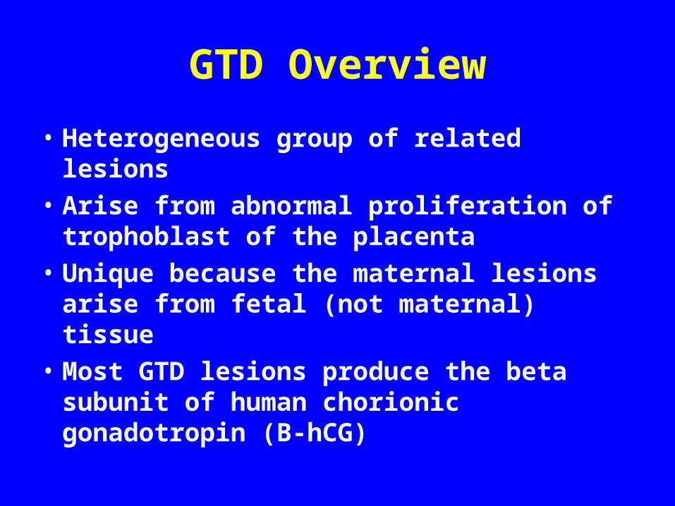
OverviewHydatidiform Mole:• Complete • Partial** BenignGestational Trophoblastic Neoplasia (GTN):• Persistent/Invasive Mole• Choriocarcinoma• Placental-Site Trophoblastic Tumor (PSTT)** Malignant

Hydatidiform Mole
• North America: 0.6-1.1 per 1000 pregnancies• Asia: 2-10 per 1000 (3x Western countries)• Difference possibly related low dietary intake
of carotene (vitamin A deficiency) and animal fat
• More common at reproductive extremes in age (>35y or <20y)
Hydatidiform Mole
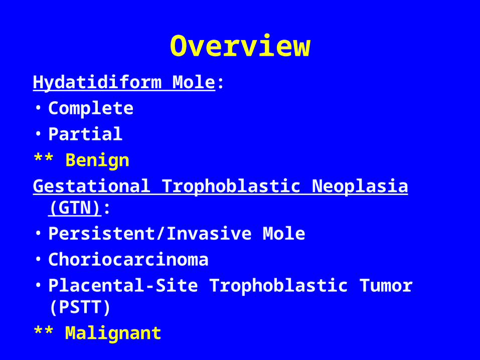
Risk Factors:• History of previous GTD
– If one previous mole, 1% chance of recurrence (vs. 0.1% in general population)
– If 2 previous moles, risk of recurrence increases to 16-28%
• Smoking• Vitamin A deficiency

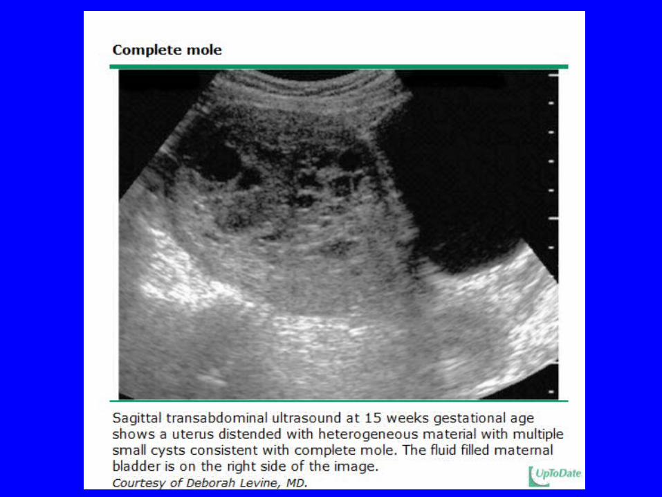
Hydatidiform MoleClinical Manifestations:• Vaginal bleeding/anemia• Enlarged uterus (size > dates)• Pelvic pain• Theca lutein cysts• Hyperemesis gravidarum• Hyperthyroidism• Preeclampsia <20 weeks gestation• Vaginal passage of hydropic vesicles


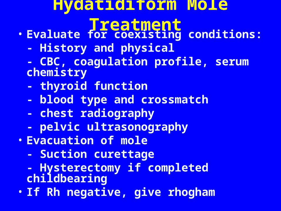
Hydatidiform Mole Treatment • Evaluate for coexisting conditions:
- History and physical- CBC, coagulation profile, serum chemistry- thyroid function- blood type and crossmatch- chest radiography- pelvic ultrasonography
• Evacuation of mole- Suction curettage- Hysterectomy if completed childbearing
• If Rh negative, give rhogham


Follow-Up Care – Molar Pregnancy• 80% of patients cured by evacuation• Follow B-hCG levels every two weeks until 3
consecutive tests negative• Then monthly B-hCG every month for 6-12 months• Avoid pregnancy for at least 6 months after first
normal B-hCG• Birth control during follow-up period• Subsequent Pregnancies:
– Send placenta for pathology– Check B- hCG 6 weeks postpartum

Gestational Trophoblastic Neoplasia (GTN)
• Persistent/Invasive Mole• Choriocarcinoma• Placental-Site Trophoblastic Tumor (PSTT)** Malignant

Risk Factors for GTN After Mole
• Preevacuation uterine size greater than gestationl age or larger than 20 weeks gestation
• Theca-lutein cysts larger than 6 cm• Age > 40 years• Serum hCG levels > 100,000 mIU/mL• Medical complications of molar pregnancy• Previous hydatidiform mole

Invasive Mole
• Myometrial invasion by hydatidiform mole• 1 in 15,000 pregnancies• 10-17% of hydatidiform moles will progress
to invasive moles
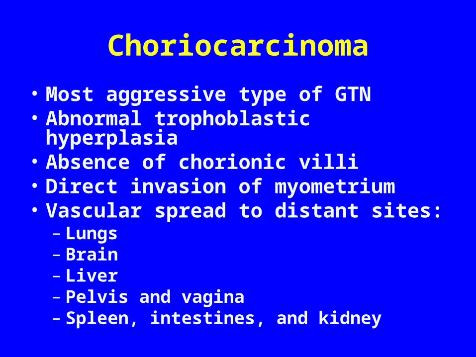
Choriocarcinoma• Most aggressive type of GTN• Abnormal trophoblastic hyperplasia• Absence of chorionic villi• Direct invasion of myometrium• Vascular spread to distant sites:
– Lungs – Brain – Liver– Pelvis and vagina– Spleen, intestines, and kidney

Choriocarcinoma• May come from any type of pregnancy
- 25% follow abortion or tubal pregnancy- 25% with term gestation- 50% from hydatidiform moles
• 2-3% of moles progress to choriocarcinoma• Incidence 1 in 40,000 pregnancies
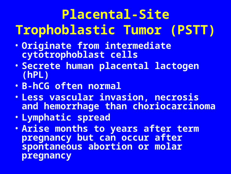
Placental-Site Trophoblastic Tumor (PSTT)
• Originate from intermediate cytotrophoblast cells
• Secrete human placental lactogen (hPL)• B-hCG often normal• Less vascular invasion, necrosis and
hemorrhage than choriocarcinoma• Lymphatic spread• Arise months to years after term pregnancy
but can occur after spontaneous abortion or molar pregnancy

Placental-Site Trophoblastic Tumor (PSTT)
• Most common symptom is vaginal bleeding• Tend to:
- Remain in uterus- Disseminate late- Produce low levels of B-hCG compared to other GTN- Be resistant to chemotherapy (treat with surgery)

Signs & Symptoms GTN
• Continued uterine bleeding, uterine perforation, enlarged irregular uterus, persistent bilateral ovarian enlargement
• From metastatic lesions: abdominal pain, hemoptysis, melena, increased intracranial pressure (headaches, seizures, hemiplegia), dyspnea, cough, chest pain

Diagnosis of GTN
• Increase or plateau in B-hCG after molar pregnancy
• Pathologic diagnosis by D&C or biopsy of metastatic lesions
• WARNING: biopsy of metastatic lesions can result in massive hemorrhage
• Metastatic workup: CXR (or CT chest), CT abdomen/pelvis +/- CT/MR of brain

Classification & Staging of GTD
• FIGO Staging– Describes anatomic distribution of disease
• World Health Organization (WHO) Scoring Index– Describes prognosis

FIGO Staging
Stage Description
I Disease confined to the uterus
II Disease extends outside the uterus but limited to genital structures (adnexa, vagina, and broad ligament)
III Disease extends to the lungs with or without genital tract involvement
IV Disease involves any other metastatic sites
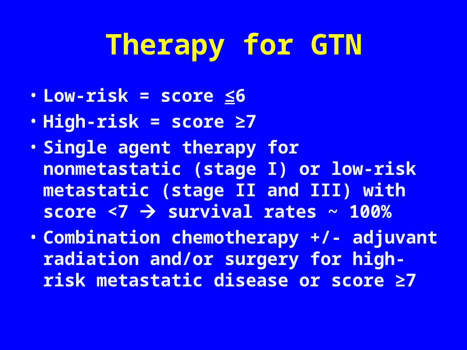
Therapy for GTN
• Low-risk = score ≤6• High-risk = score ≥7• Single agent therapy for nonmetastatic
(stage I) or low-risk metastatic (stage II and III) with score <7 survival rates ~ 100%
• Combination chemotherapy +/- adjuvant radiation and/or surgery for high-risk metastatic disease or score ≥7

Therapy: Nonmetastatic GTN• Single-agent with either methotrexate or
dactinomycin• Chemotherapy continued until hCG values normal
and then 2-3 cycles beyond• Change to alternative single-agent for hCG plateaus
above normal or toxicities• If significant elevation of hCG or new metastases,
switch to multiagent• 85-90% cured with initial regimen, <5% will require
hysterectomy for cure

Follow-up Care
• After completion of chemotherapy, follow serial hCG every 2 weeks for three months, then monthly for one year
• Physical examinations every 6-12 months and imaging as indicated

Reproductive Performance
• Most women resume normal ovarian function
• No increase risk of stillbirths, abortions, congenital anomalies, prematurity, or major obstetric complications
• No evidence of reactivation• At increased risk for development of second
episode

Summary
• Hydatidiform mole is a benign condition, 80% cured with suction D&C
• Malignant GTN:– Persistent or invasive mole– Choriocarcinoma– PSTT
• WHO score > 7 represents high-risk disease• GTN very sensitive to chemotherapy