Genotypic identification of cychloheximide resistance ...501-510)V9N12PT.… · Genotypic...
10
Genotypic identification of cychloheximide resistance yeasts isolated from clinical cases with superficial mycosis Kawther Mohammed Ali Hasan Science College for Women/ Babylon University/Hilla/ Iraq. Abstract : Most of Candida species cause many opportunistic infections, so that identification of Candida spp is indispensable to know the type of pathogen and determine the appropriate drug especially those with wide range antifungal resistance. Three species of Candida were isolated from clinical specimens on SDA with chloramphenicol and Cycloheximide C. albicans (60%), C. tropicalis (30%), and C. dubliniensis (10%). The study antifungal susceptibility of tow antifungal caspofungin (CAS) and fluconazole (FLU) for candidal isolates were shown the antifungal agent caspofungin good activity against Candida species. Primerpairs forwared CA- INT-L and reverse CA-INT-R was used to amplify a transposable intron region of the 25S DNA gene by using conventional PCR and used phylogenetic tree and sequencing analysis to study the genotypic relation among these species. In all isolates of Candida species appeared a sequence likeness between>99.4%.The results showed a close relationship by similarity between sequences of nucleotide of all isolates which belong to the same of species or different species. Keywords: Antifungal agent, Candida spp,(PCR), Transposable intron region, Sequencing. Introduction Yeasts such as Candida spp. are a normal flora on and within human body and it unsatisfactory inimmunocompetent hosts, but in moist conditions and immunocompromised patients, Candida spp. are converted to opportunistic fungi cause different diseases such as superficial infections. There are about more than one hundred species of the genus Candida, but just a few species cause human and animalinfections 1 .Nearly 90- 95% of infections caused bythese five species are C. albicans,C. glabrata,C. tropicalis, C.krusei and C. parapsilosis 2 . Recently there have been important changes due to dynamic triangle: the infecting fungi, the host factors, and the antifungal agents. Some of these changes include the increase of the immunocompromised patients number, so the increase in invasive fungal infections of these patients, and the emergence and development of new antifungal drugs and antifungal resistance. Therefor we need emergence of a standard antifungal susceptibility testfor best results of antifungal treatment 3,4 . Moreover, Tortorano 5 mentioned that infections with some Candida spp such as C. glabrata are associated with larger death rate than those associated with other pathogenic species of Candida. But maybe that correlated with types of infected patients more than reduced antifungal susceptibility 6 . Cycloheximide (actidione) isan antibiotic used in a large number of culture media for the isolation of fungi from medical samples, especially in the cases of dermatophytoses and in same the time, it inhibits non- pathogenic fungi such as saprophytic fungi (moulds and yeasts) 7 . Often yeasts such as Candida, especially C. albicans and C. tropicalis can grow in culture media with Cycloheximide and the mechanism of cycloheximide International Journal of PharmTech Research CODEN (USA): IJPRIF, ISSN: 0974-4304, ISSN(Online): 2455-9563 Vol.9, No.12, pp 501-510, 2016
Transcript of Genotypic identification of cychloheximide resistance ...501-510)V9N12PT.… · Genotypic...
Genotypic identification of cychloheximide resistance yeasts isolated from clinical cases with superficial mycosis
Kawther Mohammed Ali Hasan
Science College for Women/ Babylon University/Hilla/ Iraq.
Abstract : Most of Candida species cause many opportunistic infections, so that identification
of Candida spp is indispensable to know the type of pathogen and determine the appropriate
drug especially those with wide range antifungal resistance. Three species of Candida were
isolated from clinical specimens on SDA with chloramphenicol and Cycloheximide C. albicans (60%), C. tropicalis (30%), and C. dubliniensis (10%). The study antifungal susceptibility of
tow antifungal caspofungin (CAS) and fluconazole (FLU) for candidal isolates were shown the
antifungal agent caspofungin good activity against Candida species. Primerpairs forwared CA-INT-L and reverse CA-INT-R was used to amplify a transposable intron region of the 25S
DNA gene by using conventional PCR and used phylogenetic tree and sequencing analysis to
study the genotypic relation among these species. In all isolates of Candida species appeared a sequence likeness between>99.4%.The results showed a close relationship by similarity
between sequences of nucleotide of all isolates which belong to the same of species or different
species.
Keywords: Antifungal agent, Candida spp,(PCR), Transposable intron region, Sequencing.
Introduction
Yeasts such as Candida spp. are a normal flora on and within human body and it unsatisfactory inimmunocompetent hosts, but in moist conditions and immunocompromised patients, Candida spp. are
converted to opportunistic fungi cause different diseases such as superficial infections. There are about more
than one hundred species of the genus Candida, but just a few species cause human and
animalinfections1.Nearly 90- 95% of infections caused bythese five species are C. albicans,C. glabrata,C.
tropicalis, C.krusei and C. parapsilosis2.
Recently there have been important changes due to dynamic triangle: the infecting fungi, the host factors, and the antifungal agents. Some of these changes include the increase of the immunocompromised
patients number, so the increase in invasive fungal infections of these patients, and the emergence and
development of new antifungal drugs and antifungal resistance. Therefor we need emergence of a standard antifungal susceptibility testfor best results of antifungal treatment
3,4. Moreover, Tortorano
5 mentioned that
infections with some Candida spp such as C. glabrata are associated with larger death rate than those
associated with other pathogenic species of Candida. But maybe that correlated with types of infected patients
more than reduced antifungal susceptibility6.
Cycloheximide (actidione) isan antibiotic used in a large number of culture media for the isolation of
fungi from medical samples, especially in the cases of dermatophytoses and in same the time, it inhibits non-pathogenic fungi such as saprophytic fungi (moulds and yeasts)
7. Often yeasts such as Candida, especially C.
albicans and C. tropicalis can grow in culture media with Cycloheximide and the mechanism of cycloheximide
International Journal of PharmTech Research
CODEN (USA): IJPRIF, ISSN: 0974-4304, ISSN(Online): 2455-9563 Vol.9, No.12, pp 501-510, 2016
Kawther Mohammed Ali Hasan et al /International Journal of PharmTech Research, 2016,9(12):501-510; 502
and other antifungal resistance in these yeasts happens by either change of ribosomal proteins (rps) or contain a
drug efflux pump coding with Candida drug resistance genes (CDR), as well as multidrug resistance genes
(MDR) which responsible for resistance of yeast to a number of antifungal agents8,9
.
The diagnoses of Candida spp. by using traditional methods is sometimes problematic because the
taxonomy of the most Candidaspp have undergone changes that due to confuse in some taxonomic characters in
CHROMagar medium or biochemical reactions. So far, most research using molecular techniques as well as traditional diagnosis. Recently, several studies have demonstrated that sequence analysis of different regions of
rDNA is the golden method for Candida spp. identification. Imran and Al-Asadi10
were compared among
different species of Candida (35 isolates) from patients with conjunctivitis and diagnosed according to the CHROMagar medium in eight isolates were C. albicans with green color and 27 isolates were non-albicans
with white to the pink color colony as well as a molecular method with pair primer CAI microsatellite, which
that amplified all C.albicans isolates without others While Imran and Al-Shukry11
were diagnosed Candida spp
from patients with vaginal candidiasis by using RAPD-PCR techniques. Moreover, the sequencing of the ITS region of the Candidar DNA gene has appeared as a tool was more discriminatory for species identification,
addition to other molecular identification techniques12-15
The aim of this study is selective Candida species has been cyclohximide resistant and identifying by
using molecular methods as conventional PCR, phylogenetic tree and sequencing analysis.
2- Materials and methods
Sampling and fungal culture:-
A overall of 40 candidal isolates from clinical specimens were collected from patients with
dermatomycoses that clinically diagnosed by the dermatologist from the Unit of Mycology in Al- Marghan
Hospital of Hilla (Iraq). Samples were25 (62.5%) from female and 15 (37.5%) from male, which including 8 hair fragments,15 skin scrapings and 17 nail clippings, all samples were identified depending on culture with
Sabouraude,s Dextrose Agar (SDA) with tow antibiotic of Cycloheximide and chloramphenicol. The
identification of Candida species was on CHROMagar Candida medium (Liofilchem Lab, Italy),in
addition,other tests such as germ tube production, micromorphology on corn meal agar and grow in 45˚C and identification according to the Ellis et al.
16.
Antifungal susceptibility Epsilometer test (E-test):-
Antifungal susceptibility by E-test method was done on Muller- Hinton agar medium (MHA) of tow
antifungal caspofungin (CAS) and fluconazole (FLU) (Liofilchem Lab, Italy) for candidal isolates was determined according to the Clinical and Laboratory Standards Institute (CLSI, 2002)
17. The minimum
inhibitory concentrations (MICs) were determined following incubation at 35˚Cafter 24 h. and results were
confirmed by second reading after 84 h., breakpoint values have been read at the point of intersection between the edge of the inhibition zoon and the degree of concentration on MIC strip. The results recorded according to
the sensitivity and resistance standards, isolates with MIC ≤ 8 µg/ml for fluconazole and ≤ 1 µg/ml for
caspofungin were considered susceptible(S), isolates with MIC between 16 to 32 µg/ml for fluconazole only
were considered susceptible dose- dependent (SDD) and isolates with MIC ≥ 64 µg/ml for fluconazole and ≥ 2 µg/ml for caspofungin were considered resistant (R)
17.
Yeasts genomic DNA extraction:
The DNA of each isolate was extracted according to Imran (18), and fungal isolates DNA is stored at -
20 ˚C until use .
PCR assay:
The morphological results we're confident by using conventional PCR with primer pairs forward CA-
INT-Land reverse CA-INT-R for the region of transposable intron of 25S rDNA19
. DNA (1 μL) from Candida
isolates were mixed with PCR mixture, with a volume of the final reaction (25 μL) consisted of 2x Master Mix
(Promega) (12 μL) and primers (10 pmole) (2 μL) with free molecular- water. PCR condition were7 min of
Kawther Mohammed Ali Hasan et al /International Journal of PharmTech Research, 2016,9(12):501-510; 503
initial denaturation at 94˚C followed by 32 cycles with 40 sec of denaturation at 94˚C,45 sec of annealing at
55˚C and1 min of extension at 72˚C followed by7 min of the final extension at 72˚C. Mixture of PCR product
was amplified by using the thermal cycle tool for PCR(Labnet, origin of USA).
The products for PCRof the previous step were run on 1.5% agarose gel (Bio Basic, origin of Canada
Inc.). Electrophoresis were completed in TBE bufferat 100 V.,the gel was stained with 0.05% of ethidium
bromide. The DNA bands were exposed by using Desktop Gel documentation ultraviolet (Korea Com.).
Sequencing assay:
To study the relationship and similarity at the molecular level, the PCR products for isolates of Candida
were sequenced by sending to the Microgene Company, USA. The sequence alignment was compared with
sequences from NCBI Blast database to get the highest percentage of the match in the genus and species name of asexual phase of each isolate.
Sequencing analysis:
To reach the degree of genetic convergence among these isolates, multiple transposable intron region
of 25S rDNA, nitrogen base sequences were performed by using the Mega 6 software program. Phylogenetic
tree analysis applied by using an unweighted pair group method with arithmetic mean (UPGMA) software. Alignment of Candida spp. sequences was conducted by using BioEdit software.
3- Results
Fungal isolates:
Three species of Candida were isolated from clinical specimens on SDA with chloramphenicol and Cycloheximide (Table 1). Twenty four isolates ofC.albicans,12 isolates of C.tropicalis and4 isolates of
C.dubliniensis were identified by CHROMagar.
Table 1: Shows No. of Candida spp isolated from clinical specimens.
Candida spp Colony color on
CHROMagar
hair
fragment
skin
scrapings
nails
clipping
Total
No.(%)
C.albicans Light green 6 8 10 24 (60)
C. tropicalis Blue- pink 2 5 5 12 (30)
C. dubliniensis Dark green - 1 3 4 (10)
Antifungal susceptibility (E-test):
Table 2 showed the percentage of antifungal resistance and sensitive of Candida species for tow
antifungal agents caspofungin and fluconazole. While table 3 showed antifungal susceptibility MIC mg/L
values of caspofungin and fluconazole tested against candidal isolates.
Table 2:Percentage of antifungal resistance and sensitivity of Candida species.
Candida spp No. of isolates Caspofungin Fluconazole
C.albicans 24 24 (100%) S
0 (%) R
18 (75%) S
2 (8.3%) SDD
4(16.6%) R
C. tropicalis 12 9 (75%) S 3 (25%) R
8 (66.6%) S 2 (16.6%) SDD
2 (16.6%) R
C. dubliniensis 4 4 (100%) S
0 (%) R
0 (%) S
0 (%) SDD 4 (100%) R
Kawther Mohammed Ali Hasan et al /International Journal of PharmTech Research, 2016,9(12):501-510; 504
Table 3: Shows antifungal susceptibility MIC mg/L values of caspofungin and fluconazole tested against
candidal isolates.
Candida spp (No. of
isolates)
Caspofungin Fluconazole
Range MIC Range MIC
C.albicans (24) 0.125-1 0.5 0.5-32 8
C. tropicalis (12) 0.25-0.5 0.38 32-64 64
C. dubliniensis (4) 0.125-0.5 0.5 0.5-12 12
Molecular identification:
All isolates of Candida species were tested at a molecular level to confirm the identification of
cychloheximide resistance Candida. By using conventional PCR technique with primer pairs forward CA-INT-
L and reverse CA-INT-R for transposable intron region of 25S rDNA. In gel electrophoresis, the amplicons of transposable intron region for C. albicans,C. tropicalis and C. dubliniensis were around 480, 520-600 and1080
bp respectively. Satisfactory sequencing results were obtained, the isolates had been identified by comparison
those isolates sequencing with sequences from the NCBI Blast databases, the identification for all isolates in the
intron region was 99-100%. Our results of the sequencing analysis for candidal isolates was obtained depending on the basis of their transposable intron region sequences by using the MEGA6 software program. The results
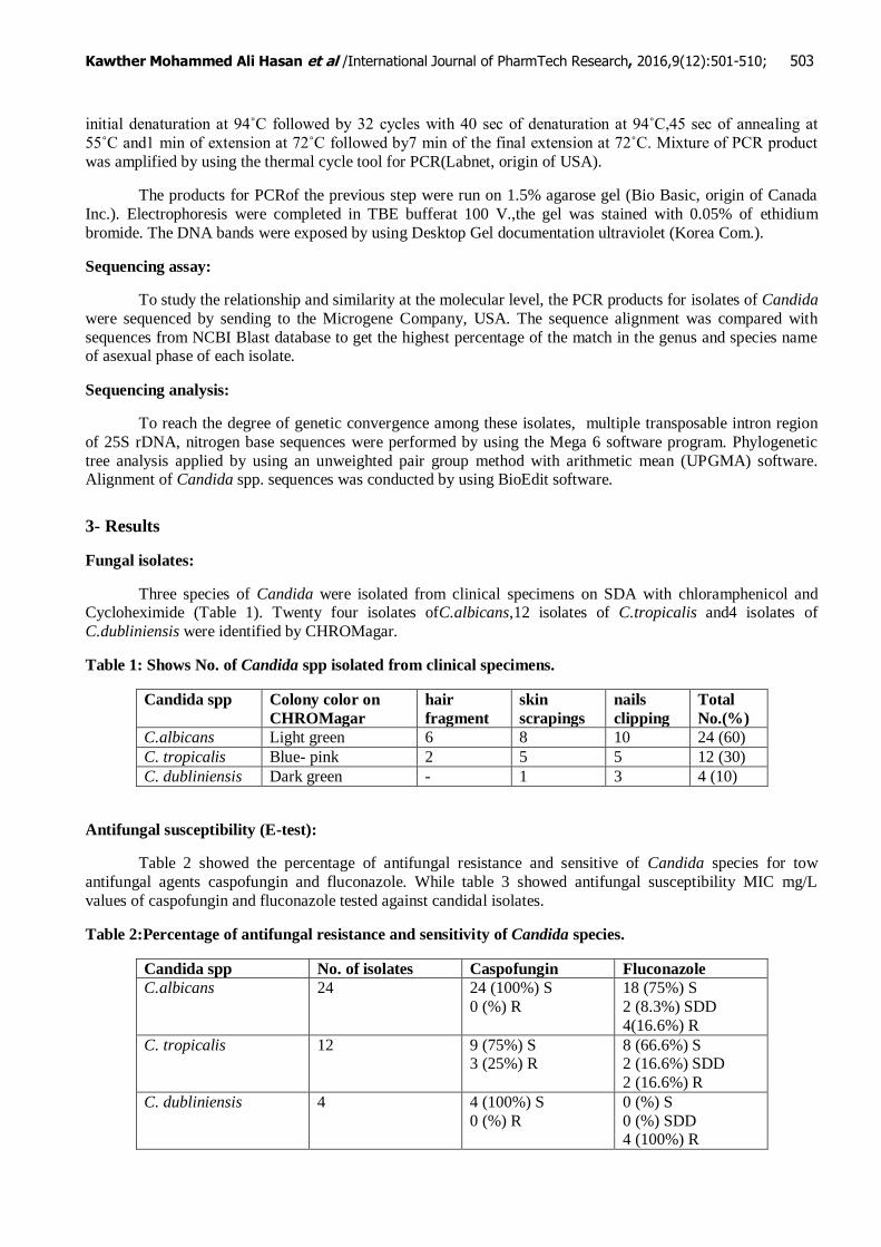
of the phylogenetic tree analysisfor twelve isolates of Candida spp. were observed three clusters of Candida
spp. C. albicans in clusters 1, C. dubliniensis in cluster 2 and C. tropicalis in cluster 3. Phylogenetic tree was showed high sequence similarity in the transposable intron region of rDNA between 99.9-100% similarities for
the isolates belong to one species (Fig. 1). Phylogenetic tree of C. albicans cluster 1 and C. dubliniensis cluster
2 showed high similarity of about 99.9%, but this group with C. tropicalis cluster 3 showed the similar of about 99.4%.
Fig. 1: Phylogenetic tree based on transposable intron region sequences of 25S rDNA for Candida spp.
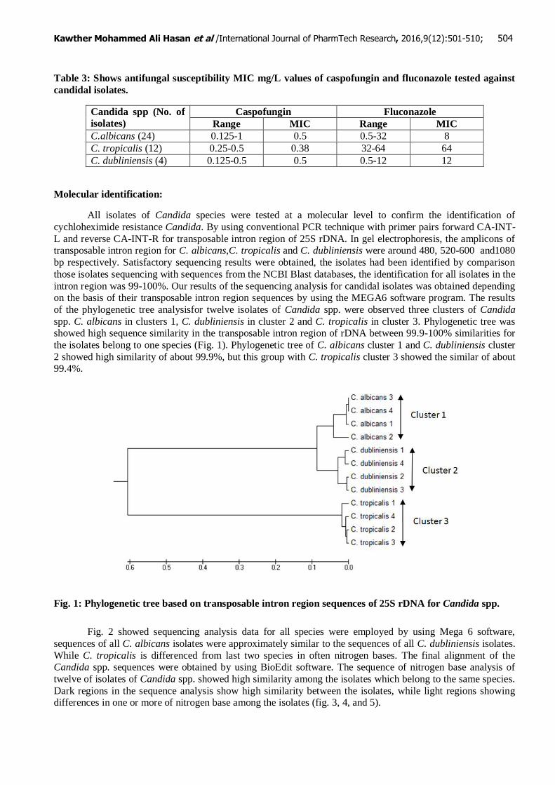
Fig. 2 showed sequencing analysis data for all species were employed by using Mega 6 software,
sequences of all C. albicans isolates were approximately similar to the sequences of all C. dubliniensis isolates.
While C. tropicalis is differenced from last two species in often nitrogen bases. The final alignment of the Candida spp. sequences were obtained by using BioEdit software. The sequence of nitrogen base analysis of
twelve of isolates of Candida spp. showed high similarity among the isolates which belong to the same species.
Dark regions in the sequence analysis show high similarity between the isolates, while light regions showing differences in one or more of nitrogen base among the isolates (fig. 3, 4, and 5).

Kawther Mohammed Ali Hasan et al /International Journal of PharmTech Research, 2016,9(12):501-510; 505
Fig. 2: sequence data for Candida species by using Mega 6 software, the star is showed similarity of
nitrogen base of all Candida species isolates. Number of nucleotide sequence is shown on the left and
right.
Kawther Mohammed Ali Hasan et al /International Journal of PharmTech Research, 2016,9(12):501-510; 506
Fig. 3: Alignment of transposable intron region sequences for C. albicans by using BioEdit software.
Nucleotide sequences are present in all intron regions are shaded in dark color.
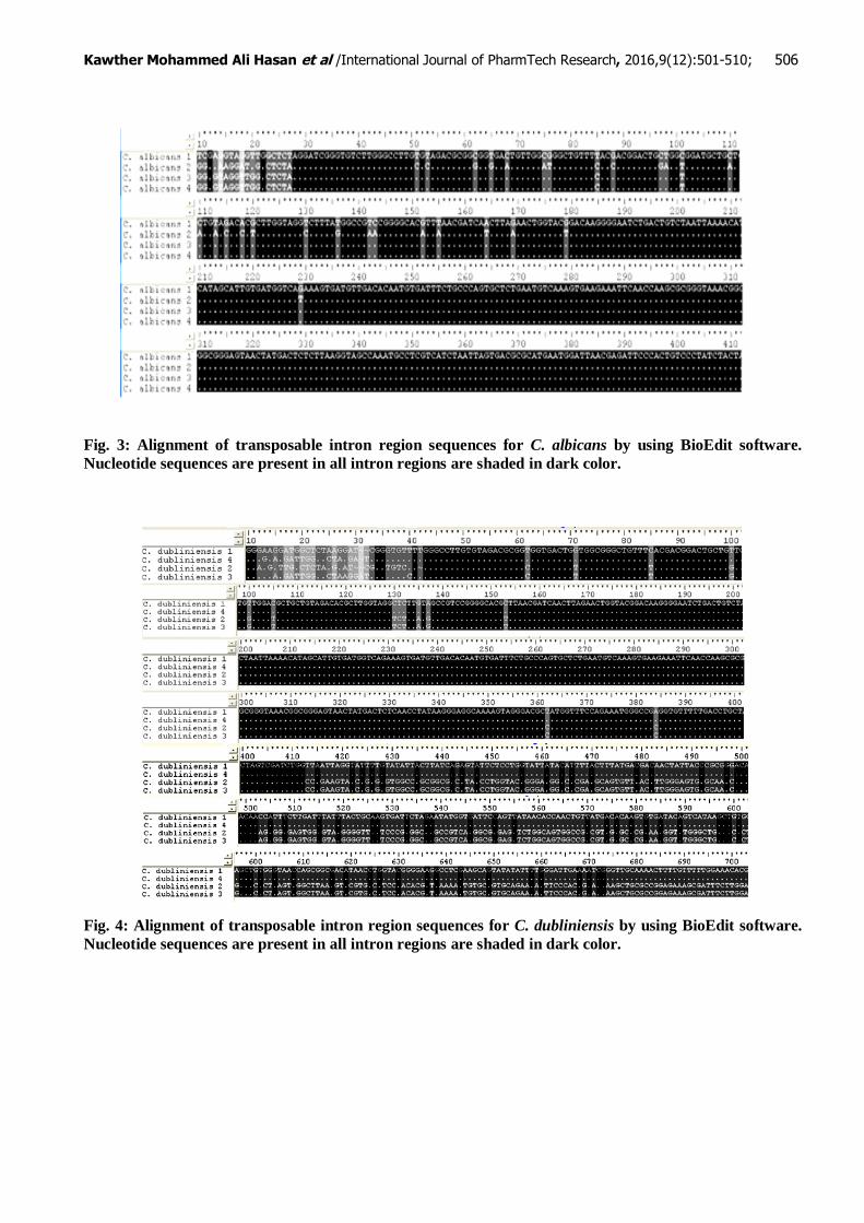
Fig. 4: Alignment of transposable intron region sequences for C. dubliniensis by using BioEdit software.
Nucleotide sequences are present in all intron regions are shaded in dark color.
Kawther Mohammed Ali Hasan et al /International Journal of PharmTech Research, 2016,9(12):501-510; 507
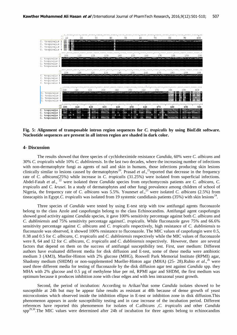
Fig. 5: Alignment of transposable intron region sequences for C. tropicalis by using BioEdit software.
Nucleotide sequences are present in all intron region are shaded in dark color.
4- Discussion
The results showed that three species of cychloheximide resistance Candida, 60% were C. albicans and
30% C. tropicalis while 10% C. dubliniensis. In the last two decades, where the increasing number of infections
with non-dermatophyte fungi as agents of nail and skin in humans, those infections producing skin lesions
clinically similar to lesions caused by dermatophytes20
. Prasad et al.,21
reported that decrease in the frequency rate of C. albicans(25%) while increase in C. tropicalis (31.25%) were isolated from superficial infections.
Abdel-Fatah et al., 22
were isolated three Candida species from onychomycosis patients are C. albicans, C.
tropicalis and C. krusei. In a study of dermatophytes and other fungi prevalence among children of school of Nigeria, the frequency rate of C. albicans was 5.5%. Youneset al.,
23 were isolated C. albicans (2.5%) from
tineacapitis in Egypt.C. tropicalis was isolated from 19 systemic candidiasis patients (35%) with skin lesions24
.
Three species of Candida were tested by using E-test strip with tow antifungal agents fluconazole
belong to the class Azole and caspofungin belong to the class Echinocandins. Antifungal agent caspofungin
showed good activity against Candida species, it gave 100% sensitivity percentage against both C. albicans and
C. dubliniensis and 75% sensitivity percentage againstC. tropicalis. While fluconazole gave 75% and 66.6% sensitivity percentage against C. albicans and C. tropicalis respectively, high resistance of C. dubliniensis to
fluconazole was observed, it showed 100% resistance to fluconazole. The MIC values of caspofungin were 0.5,
0.38 and 0.5 for C. albicans, C. tropicalis and C. dubliniensis respectively while the MIC values of fluconazole were 8, 64 and 12 for C. albicans, C. tropicalis and C. dubliniensis respectively. However, there are several
factors that depend on them on the success of antifungal susceptibility test. First, user medium: Different
authors have evaluated different media for disk diffusion and E-test, some of these media were antibiotic medium 3 (AM3), Mueller-Hinton with 2% glucose (MHG), Roswell Park Memorial Institute (RPMI) agar,
Shadomy medium (SHDM) or non-supplemented Mueller-Hinton agar (MHA) (25- 28).Rubio et al.,26
were
used three different media for testing of fluconazole by the disk diffusion agar test against Candida spp. they
MHA with 2% glucose and 0.5 µg of methylene blue per ml, RPMI agar and SHDM, the first medium was optimum because it produces inhibition zone with clear edges and with less intrazonal yeast growth.
Second, the period of incubation: According to Arikan3that some Candida isolates showed to be
susceptible at 24h but may be appear false results as resistant at 48h because of dense growth of yeast
microcolonies which observed inside the inhibition ellipse in E-test or inhibition zone in disk diffusion.This
phenomenon appears in azole susceptibility testing and in case increase of the incubation period. Different references have reported this phenomenon for isolates of C.albicans ,C. tropicalis and other Candida
spp29,30
.The MIC values were determined after 24h of incubation for three agents belong to echinocandins
Kawther Mohammed Ali Hasan et al /International Journal of PharmTech Research, 2016,9(12):501-510; 508
groups (anidulafungin, caspofungin and micafungin)against 404 isolates due to six species of Candida
31.And
three agents belong to azole groups (fluconazole, posaconazole and voriconazole) against 1056 isolates due to
five species of Candida32
.
Third, type of antifungal agentand fungal species: The correlation between antifungal agent and fungal
species is often variable. Several references reported that large number of antifungal agents against Candida
spp. was had been used successfully such as amphotericin B, azoles especially fluconazole and echinocandins especially caspofungin
33-36. Rodriguez-Tudela et al.,
37 reported that the correlation between two groups of
patients (candidemia 126 cases and oropharyngeal candidiasis 110 cases), who had been treated with different
doses of fluconazole, they were concluded that an exposure increase above the dose/MIC of 35.5 has increased the cure rate in patients, and cure rate was reached 100% when the dose/MIC was higher than 100.
Sometime, we noted that one species of Candida having variable resistance tothe multiple antifungal
agent and over different periods of time, such as C. glabrata was acquired resistance to all amphotericin B, flucytosine, voriconazole, fluconazole and caspofungin. This resistance was readily mutated in vivo in a single
patient38
. Pfaller and Diekema39
mentioned that C. glabrata and C. krusei demonstrates less susceptibility to
amphotericin B than other Candida species, especially when used E-test.
In our study, analysis of phylogenetic treeo f twelve isolates of Candida spp. displayed high sequence
similarity in the transposable intron region of 25S rDNA between 99.9-100% similarities for the isolates belong to one species (Fig. 2). While the difference in sequence between C. albicans and C. dubliniensis were shown
approximately 0.1%.Our results agreed with number of studies were demonstrated a high degree of genotypic
homogeneity between bothC.dubliniensis and C. albicans and these studies referred to the similarity of several
regions of rDNA for both C. albicans and C. dubliniensis as well as the similarity between the two species in some of the loci sequencing
18, 40-42. There are several studies were identified byCandida species by one or more
of molecular methods, Cirak et al.,43
were identified forty four isolates of Candida spp. due to five species by
using RFLP-PCR with three restriction enzymes: BfaI, DdeI and HaeIII. The last enzyme can be differentiated between C. albecans and non- C. albecans isolates, while BfaI can be differentiated among non- C. albecans
species. Xuet al.,13
were compared the API20C technique for identification of C. albicans and sequencing of the
ITS2 region for for identification of C. dubliniensis. Because of differences of amplicon length and base sequence among different species, Turenne et al.
44 found that differences in three nucleotide band in size of
ITS2 region of C. Krusei and C. albicans. Fujita et al.45
were determined the PCR fragment lengths of ITS
regions for six of Candida species by using both agarose gel electrophoresis and microchip electrophoresis.
While Abdel-Fatah et al.22
were used three molecular methods RAPD, ISSR and RFLP for identification three species for Candida and six species of dermatophytes isolated from patients with skin mycosis from Egypt and
Libya and for determination the genetic relationship among these species were analyzed phylogenetic tree for
RAPD and ISSR products, while in RFLP were used ITS1 and ITS4 primers and digested the PCR product with Hinf I and HaeIII enzymes.
References
1. Ho, K. and Cheng, T. (2010). Common superficial fungal infections a short review. Medical bulletin,
15(11): 23-27.
2. Pfaller, M.A. and Diekema, D.J. (2007). Epidemiology of invasive candidiasis: a persistent public health problem. ClinMicrobiol Rev, 20(1): 133-163.
3. Bille, J.; Marchetti, O. and Calandra, T. (2005). Changing face of health-care associated fungal
infections. CurrOpin Infect Dis 18: 314-319. 4. Arikan, S. (2007). Current status of antifungal susceptibility testing methods. Med Mycol, 45: 569-587.
5. Torttorano, A.; Penman, J.; Bemhardt, H.; Klingspor, L.; Kibbler, C.C. and et al. (2004). Epidemiology
of candidaemia in Europe: results of 28-month European Confederation of Medical Mycology
(ECMM) hospital based surveillance study. Eur J ClinMicrobiol Infect Dis, 23(4): 317-322. 6. Johnson, E.M. (2008). Issues in antifungal susceptibility testing. J antimicrobchemother, 61(1): 13-18.
7. Sutton, D.A. . (2003). Specimen collection, transport, and processing: mycology. In: Murray, P.R.;
Baron, E.J.; Jorgensen, J.H.; Pfaller, M.A. and Yolken, R.H. Manual of clinical microbiology. 8th ed.
Washington DC: American Society for Microbiology.
Kawther Mohammed Ali Hasan et al /International Journal of PharmTech Research, 2016,9(12):501-510; 509
8. Vandeputte, P.; Ferrari, S. and Coste, A. (2012). Antifungal resistance and new strategies to control
fungal infections.Internat J Microbiol, 26pp.
9. Ali, G.F.; Al-Hamadani, A.H. and Mohammad, A.W. (2015). Detection of cyclohximide resistant gene
in selected pathogenic fungi. J Contemp Med Sci, 1(4): 23-26. 10. Imran, Z.K. and Al-Asadi, Y.F. (2014). Multiple molecular markers for diagnosis of conjunctivitis
caused by Candida spp. in Iraq.Afr J Microbiol Res, 8(38):3482-3488.
11. Imran, Z.K. and Al-Shukry, H.N. (2014). Molecular diagnosis of vaginal candidiasis by polymerase chain reaction (PCR) and random amplification polymorphism DNA (RAPD-PCR) in Babylon
Province, Iraq.Afr J Microbiol Res, 8(6):496-502.
12. Elie, C.M.; Lott, T.J.; Reiss, E. and Morrison, C.J. (1998). Rapid identification of Candida species with species- specific DNA probes. J ClinMicrobiol 36(11): 3260-3265.
13. Xu, J.; Millar, B.C.; Moore, J.E.; McClurg, R.; Walker, M.J.; Evans, J.; Hedderwick, S. and McMullan,
R. (2002). Comparison of API20C with molecular identification of Candida spp isolated from
bloodstream infections. J ClinPathol, 55: 774-777. 14. Cornet, M.; Sendid, B.; Fradin, C.; Gaillardin, C.; Poulain, D. and Nguyen, H. (2011). Molecular
identification of closely related Candida species using two ribosomal intergenic spacer fingerprinting
methods. J Molecular Diagnostics, 13(1): 12-22. 15. Mohammadi, R. and Abdi, S. (2015). Molecular identification of Candida species isolated from gastro-
oesophageal candidiasis in tehran, Iran. GastroenterolHepatol bed Bench, 8(4): 288-293.
16. Ellis, D.; Davis, S.; Alexiou, H.; Handke, R. and Bartley, R. (2007). Description of medical fungi.2nd
ed. Mycology unit.Women´s and Children´s Hospital. North Adelaide, Australia. 17. Clinical and Laboratory Standard Institute CLSI (2002). Reference method for broth dilution antifungal
susceptibility testing of yeasts, Approved standard- second edition. CLSI document M27-A2.
Pennsylvania, USA. 18. Imran, Z.K. (2015). Candida albicans ssp. dubliniensis stat.et comb.Nov., a new combination for
Candida dubliniensis based on genetic criteria.Afr J Microbiol Res, 9(17): 1205-1214.
19. McCullough, M.J.; Clemons, K.V. and Stevens, D.A. (1999). Molecular and phenotypic characterization of genotype C. albicans subgroups and comparsion with Candida dubliniensis and
Candida stellatoidea. J ClinMicrobiol, 37: 417-421.
20. Aggarwal, A.; Arora, U. and Khanna, S. (2002). Clinical and mycological study of superficial mycoses
in Amritsar. Indian J Dermatol, 47(4): 218-220. 21. Prasad, N.; Mahapatra, A. and Chayani, N. (2013). Changing trends in the fungal isolates from clinical
specimens of suspected superficial mycosis.Inaian Medical Gazette, pp: 60-62.
22. Abdel-Fatah, B.E.; Moharram, A.M.; Moubasher, A.A.H. and Al-Ryani, M.A. (2013). Genetic relationships and isozyme profile of dermatophytes and Candida strains from Egypt and Libya. Afr. J.
Biotechnol., 12(29): 4554-4568.
23. Younes, A.E.H.; Mohamed, E.E.M.; Tawfik, K. M. and Ezzat, A.A. (2012). Tineacapitis in Assiut (Egypt).AA M J, 10(1): 45-54.
24. Bae, G.Y.; Chang, S.E.; Moon, K.C.; Lee, M.W.; Choi, J.H. and Koh, J.K. (2005). Clinicopathologic
review of 19 patients with systemic candidiasis with skin lesions.Int J Dermatol, 44(7): 550-555.
25. Lozano-Chiu, M.; Nelson, P. W.; Lancaster, M.; Pfaller, M.A. and Rex, J.H. (1997). Lot-to- lot variability of antibiotic medium 3 when used for susceptibility testing of Candida isolates to
amphotericin B. J ClinMicrobiol, 35:270-272.
26. Rubio, M.C.; Gil, J.; de Ocáriz, I.R.; Benito, R. and Rezusta, A. (2003). Comparison of results obtained by testing with three different agar media and by the NCCLS M27-A method for in vitro testing of
fluconazole against Candida spp. J ClinMicrobiol, 41(6): 2665-2668.
27. Capoor, M.R.; Rawat, D.; Nair, D. Deb, D. and Aggarwal, P. (2007). Evaluation of glucose-methylene-
blue-Mueller-Hinton agar for E-test minimum inhibitory concentration determination in Candida spp. Indian J Med Microbiol, 25(4): 432-433.
28. Espinel-Ingroff, A.; Canton, E.; Gibbs, D. and Wang, A. (2007). Correlation of neo-sensitable tablet
diffusion assay results on three different agar media with CLSI broth microdilution M27-A2 and disk diffusion M44-A results for testing susceptibilities of Candida spp. and Cryptococcus neoformans to
amphotericin B, caspofungin, fluconazole, Itraconazole, and voriconazole. J ClinMicrobiol, 45(3): 858-
864. 29. Liao, R.S.; Rennie, R.P. and Talbot, J.A. (2001). Novel fluorescent broth microdilution method for
fluconazole susceptibility testing of Candida albicans. J ClinMicrobiol, 39:2708-2712.
Kawther Mohammed Ali Hasan et al /International Journal of PharmTech Research, 2016,9(12):501-510; 510
30. Ostrosky-Zeichner, L.; Rex, J.H.; Pappas. P.G.; Hamill, R.J.; Larsen, R.A. (2003). Antifungal
susceptibility survey of 2,000 bloodstream Candida isolates in the United States. Antimicrob Agents
Chemother 47:3149-3154.
31. Pfaller, M.A.; Chaturvedi, V.; Diekema, D.J.; Ghannoum, M.A.; Holliday, N.M.; Killian, S.B.; Knapp, C.C.; Messer, S.A.; Miskov, A. and Ramani, R. (2008). Clinical evaluation of the sensititreYeastOne
colorimetric antifungal panel for antifungal susceptibility testing of the echinocandinsanidulafungin,
caspofungin, and micafungin. J ClinMicrobiol, 46(7): 2155-2159. 32. Pfaller, M.A.; Espinel-Ingroff, A.; Boyken, L.; Hollis, R.J.; Kroeger, J.; Messer, S.A.; Tendolkar, S.
and Diekema, D.J. (2011). Comparsion of the broth microdilution (BMD) method of the European
committee on antimicrobial susceptibility testing with the 24-hour CLSI BMD method for testing susceptibility of Candida species to fluconazole, posaconazole, and voriconazole by use of
epidemiological cutoff values. J ClinMicrobiol, 49(3): 845-850.
33. Peyron, F.; Favel, A.; Michel-Nguyen, A.; Gilly, M.; Regli, P. and Bolmstrom, A. (2001). Improved
detection of amphotericin B-resistant isolates of Candida lusitaniae by Etest. J ClinMicrobiol, 39(1): 339-342.
34. Betts, R.; Glasmacher, A.; Maertens, J.; Maschmeyer, G.; Vazquez, J.A. and et al. (2006). Efficacy of
caspofungin against invasive Candida or invasive Aspergillus infections in neutropenic patients. Cancer, 106(2): 466-473.
35. Park, B.J.; Arthington-skaggs, B.A.; Hajjeh, R.A.; Iqbal, N.; Ciblak, M.A. and et al. (2006). Evaluation
of amphotericin B interpretive breakpoints for Candida bloodstream isolates by correlation with
therapeutic outcome. Antimicrob Agents Chemother 50(4): 1287-1292. 36. Pfaller, M.A.; Diekema, D.J. and Sheehan, D.J. (2006). Interpretive breakpoints for fluconazole and
Candida revisited: ablueprint for the future of antifungal susceptibility testing. ClinMicrobiol Rev,
19(2): 435-447. 37. Rodriguez-Tudela, J.L.; Almirante, B.; Rodriguez-Pardo, D.; Laguna, F.; Donnelly, J.P.; Mouton, J.W.;
Pahissa, A. and Cuenca-Estrella, M. (2007). Correlation of the MIC and Dose/MIC ratio of fluconazole
to the therapeutic response of patients with mucosal candidiasis and candidemia.Antimicrob Agents Chemother, 51(10): 3599-3604.
38. Chapeland-Leclerc, F.; Hennequin, C.; Papon, N.; Noël, T.; Girard, A.; Socié, G.; Ribaud, P. and
Lacroix, C. (2010). Acquisition of fluctosine, azole, and caspofungin resistance in Candida glabrata
bloodstream isolates serially obtained from a hematopoietic stem cell transplant recipient. Antimicrob Agents Chemother, 54(3): 1360-1362.
39. Pfaller, M.A. and Diekema, D.J. (2004). Rare and emerging opportunistic fungal pathogens: concern
for resistance beyond Candida albicans and Aspergillus fumigates. J ClinMicrobiol, 42(10): 4419-4431.
40. Pujol, C.; Daniels, K.L.; Shawn, R.L.; Srikantha, T.; Joshua, B.R.; Geiger, J. and Soll, D.R. (2004). The
closely related species Candida albicans and Candida dubiniensis can mate. Eukaryot Cell, 3(4): 1015- 1027.
41. Jackson, A.P.; Gamble, J.A.; Yeomans, T.; Moran, G.P.; Saunders, D.; Harris, D.; Aslett, M.;
BarrellJ.F.; Butler, G.; Citiulo, F. Coleman, D.C. and et al. (2009). Comparative genomic of the fungal
pathogens Candida dubliniensis and C. albicans.Genome Res 19: 2231- 2244. 42. Costa, J.M.; Garcia-Hermoso, D.; Olivi, M.; Cabaret, O.; Farrugia, C.; Lecellier, G.; Dromer, F. and
Bretagne, S. (2010). Genotyping of Candida albicans using length fragment and high-resolution
melting analyses together with minisequencing of a polymorphic microsatellite locus. J Microbiol Methods, 80(3): 306-309.
43. Cirak, M.Y.; Kalkanci, A. and Kustimur, S. (2003). Use molecular methods in identification of Candida
species and evaluation of fluconazole resistance. MemInstOswaldo Cruz, 98(8): 1-11.
44. Turenne, C.Y.; Sanche, S.E.; Hoban, D.J.; Karlowsky, J.A. and Kabani, A.M. (1999). Rapididentification of fungi by using the internal transcribed spacer 2 genetic region and an automated
fluorescent capillary electrophoresis system. J ClinMicrobiol, 37: 1846-1851.
45. Fujita, S.; Senda, Y.; Nakaguspecieschi, S. and hashimoto, T. (2001). Multiplex PCR using internal transcribed spacer 1 and 2 regions for rapid detection and identification of yeast strains. J
ClinMicrobiol, 39(10): 3617-3622.
*****