Genomic and Immunogenic Protein Diversity of ...
17
Scotland's Rural College Genomic and Immunogenic Protein Diversity of Erysipelothrix rhusiopathiae Isolated From Pigs in Great Britain Forde, Taya L.; Kollanandi Ratheesh, Nichith; Harvey, William T.; Thomson, Jill R.; Williamson, Susanna; Biek, Roman; Opriessnig, Tanja Published in: Frontiers in Microbiology DOI: 10.3389/fmicb.2020.00418 First published: 13/03/2020 Document Version Publisher's PDF, also known as Version of record Link to publication Citation for pulished version (APA): Forde, T. L., Kollanandi Ratheesh, N., Harvey, W. T., Thomson, J. R., Williamson, S., Biek, R., & Opriessnig, T. (2020). Genomic and Immunogenic Protein Diversity of Erysipelothrix rhusiopathiae Isolated From Pigs in Great Britain: Implications for Vaccine Protection. Frontiers in Microbiology, 11, [418]. https://doi.org/10.3389/fmicb.2020.00418 General rights Copyright and moral rights for the publications made accessible in the public portal are retained by the authors and/or other copyright owners and it is a condition of accessing publications that users recognise and abide by the legal requirements associated with these rights. • Users may download and print one copy of any publication from the public portal for the purpose of private study or research. • You may not further distribute the material or use it for any profit-making activity or commercial gain • You may freely distribute the URL identifying the publication in the public portal ? Take down policy If you believe that this document breaches copyright please contact us providing details, and we will remove access to the work immediately and investigate your claim. Download date: 23. Mar. 2022
Transcript of Genomic and Immunogenic Protein Diversity of ...

Scotland's Rural College
Genomic and Immunogenic Protein Diversity of Erysipelothrix rhusiopathiae IsolatedFrom Pigs in Great BritainForde, Taya L.; Kollanandi Ratheesh, Nichith; Harvey, William T.; Thomson, Jill R.;Williamson, Susanna; Biek, Roman; Opriessnig, TanjaPublished in:Frontiers in Microbiology
DOI:10.3389/fmicb.2020.00418
First published: 13/03/2020
Document VersionPublisher's PDF, also known as Version of record
Link to publication
Citation for pulished version (APA):Forde, T. L., Kollanandi Ratheesh, N., Harvey, W. T., Thomson, J. R., Williamson, S., Biek, R., & Opriessnig, T.(2020). Genomic and Immunogenic Protein Diversity of Erysipelothrix rhusiopathiae Isolated From Pigs in GreatBritain: Implications for Vaccine Protection. Frontiers in Microbiology, 11, [418].https://doi.org/10.3389/fmicb.2020.00418
General rightsCopyright and moral rights for the publications made accessible in the public portal are retained by the authors and/or other copyright ownersand it is a condition of accessing publications that users recognise and abide by the legal requirements associated with these rights.
• Users may download and print one copy of any publication from the public portal for the purpose of private study or research. • You may not further distribute the material or use it for any profit-making activity or commercial gain • You may freely distribute the URL identifying the publication in the public portal ?
Take down policyIf you believe that this document breaches copyright please contact us providing details, and we will remove access to the work immediatelyand investigate your claim.
Download date: 23. Mar. 2022

fmicb-11-00418 March 12, 2020 Time: 18:55 # 1
ORIGINAL RESEARCHpublished: 13 March 2020
doi: 10.3389/fmicb.2020.00418
Edited by:John R. Battista,
Louisiana State University,United States
Reviewed by:Yoshihiro Shimoji,
National Institute of Animal Health,JapanHo To,
Nippon Institute for BiologicalScience, Japan
*Correspondence:Taya L. Forde
Specialty section:This article was submitted to
Evolutionary and GenomicMicrobiology,
a section of the journalFrontiers in Microbiology
Received: 25 November 2019Accepted: 27 February 2020
Published: 13 March 2020
Citation:Forde TL, Kollanandi Ratheesh N,
Harvey WT, Thomson JR,Williamson S, Biek R and Opriessnig T
(2020) Genomic and ImmunogenicProtein Diversity of Erysipelothrixrhusiopathiae Isolated From Pigs
in Great Britain: Implicationsfor Vaccine Protection.
Front. Microbiol. 11:418.doi: 10.3389/fmicb.2020.00418
Genomic and Immunogenic ProteinDiversity of Erysipelothrixrhusiopathiae Isolated From Pigs inGreat Britain: Implications forVaccine ProtectionTaya L. Forde1* , Nichith Kollanandi Ratheesh1, William T. Harvey1, Jill R. Thomson2,Susanna Williamson3, Roman Biek1 and Tanja Opriessnig4
1 Institute of Biodiversity, Animal Health & Comparative Medicine, University of Glasgow, Glasgow, United Kingdom,2 Disease Surveillance Centre, SAC Veterinary Services, Scotland’s Rural College, Edinburgh, United Kingdom, 3 SurveillanceIntelligence Unit, Animal and Plant Health Agency, Bury St Edmunds, United Kingdom, 4 The Roslin Institute, The Universityof Edinburgh, Midlothian, United Kingdom
Erysipelas, caused by the bacterium Erysipelothrix rhusiopathiae, is re-emerging inswine and poultry production systems worldwide. While the global genomic diversity ofthis species has been characterized, how much of this genomic and functional diversityis maintained at smaller scales is unclear. Specifically, while several key immunogenicsurface proteins have been identified for E. rhusiopathiae, little is known about theirpresence among field strains and their divergence from vaccines, which could result invaccine failure. Here, a comparative genomics approach was taken to determine thediversity of E. rhusiopathiae strains in pigs in Great Britain over nearly three decades, aswell as to assess the field strains’ divergence from the vaccine strain most commonlyused in British pigs. In addition, the presence/absence and variability of 13 previouslydescribed immunogenic surface proteins was determined, including SpaA which isconsidered a key immunogen. We found a high diversity of E. rhusiopathiae strains inBritish pigs, similar to the situation described in European poultry but in contrast to swineproduction systems in Asia. Of the four clades of E. rhusiopathiae found globally, threewere represented among British pig isolates, with Clade 2 being the most common. AllBritish pig isolates had one amino acid difference in the immunoprotective domain ofthe SpaA protein compared to the vaccine strain. However, we were able to confirmusing in silico structural protein analyses that this difference is unlikely to compromisevaccine protection. Of 12 other known immunogenic surface proteins of E. rhusiopathiaeexamined, 11 were found to be present in all British pig isolates and the vaccine strain,but with highly variable degrees of conservation at the amino acid sequence level,ranging from 0.3 to 27% variant positions. Moreover, the phylogenetic incongruenceof these proteins suggests that horizontal transfer of genes encoding for antigens is
Frontiers in Microbiology | www.frontiersin.org 1 March 2020 | Volume 11 | Article 418

fmicb-11-00418 March 12, 2020 Time: 18:55 # 2
Forde et al. Genomic Diversity of Erysipelothrix rhusiopathiae
commonplace for this bacterium. We hypothesize that the sequence variants in theseproteins could be responsible for differences in the efficacy of the immune response.Our results provide the necessary basis for testing this hypothesis through in vitro andin vivo studies.
Keywords: antigen, Erysipelothrix rhusiopathiae, genomics, Great Britain, pigs (Sus domesticus), vaccine, wholegenome sequencing
INTRODUCTION
Erysipelothrix rhusiopathiae, the causative agent of erysipelas,remains a persistent challenge for swine and poultry productionsystems worldwide. Although well-controlled for decadesthrough vaccination, erysipelas is re-emerging in severalEuropean and Asian countries (Eriksson et al., 2013; Kwok et al.,2014; Janßen et al., 2015; Ogawa et al., 2017). E. rhusiopathiae wasalso recently implicated in large-scale mortalities and populationdeclines in muskoxen and other wild ungulates in North America(Kutz et al., 2015; Forde T.L. et al., 2016). While Erysipelothrixspp. infection can impact all pig production stages and is acommon and significant cause of carcass condemnation, itstrue economic burden is largely unknown. The E. rhusiopathiaegenome is comprised of a single chromosome of about 1.8 millionbase pairs (MB), with an average GC content of 36.5% (Ogawaet al., 2011; Kwok et al., 2014; Tang et al., 2016). Comparativegenomic analysis of a diverse collection of E. rhusiopathiaeisolates from a wide range of host species recently led to a betterunderstanding of the global diversity and population structureof this species (Forde T. et al., 2016), including its propensityfor homologous recombination. Strains can be broadly dividedinto Clades 1, 2, and 3, as well as a clade phylogenetically“intermediate” to Clades 2 and 3. The genomic diversity ofE. rhusiopathiae at smaller (e.g. national) scales has only begunto be explored. However, studies conducted to date illustrate thatthis may range from highly related clonal strains (e.g. as seen inpigs in Japan; Ogawa et al., 2017), to phylogenetically diversestrains, as observed in poultry in Germany (Janßen et al., 2015).The degree of genomic diversity present could affect the abilityof vaccines to provide protection against the full spectrum ofcirculating field strains.
An important component of E. rhusiopathiae diversity thathas remained relatively unstudied is that of its surface proteins.Because of their high propensity for host-pathogen interactions,surface proteins often play an important role in virulencemechanisms, as well as in eliciting a host immune response,thereby representing potential vaccine candidates (Gamberiniet al., 2005). One of the most critical immunogens forE. rhusiopathiae identified to date is the surface protective antigen(Spa)A protein (Makino et al., 1998; Imada et al., 1999; Shimojiet al., 1999). Of the three different Spa types (A, B, and C)that have been found in Erysipelothrix spp. (To and Nagai,2007), SpaA is by far the most widely prevalent Spa type inE. rhusiopathiae. In a global collection of E. rhusiopathiae isolatesexamined from various host species, the spaA gene was foundin more than 90% of isolates (79/86), including all those frompigs and poultry (Forde T. et al., 2016). This corroborates several
international studies where the spaA gene was the exclusive Spatype found, including from pig outbreaks in Japan (n = 83) (Toet al., 2012), isolates from pigs in Australia (n = 44) (Eamens et al.,2006), and a large study of 165 predominantly poultry isolatesfrom Germany (Janßen et al., 2015). It has been shown that theN-terminal immunoprotective domain of this surface protein –also referred to as the hypervariable domain – is the componentresponsible for eliciting protective immunity (Figure 1; Imadaet al., 1999; Shimoji et al., 1999). SpaA also plays an importantrole in pathogenesis by increasing resistance to phagocytosisand promoting endothelial adherence (Harada et al., 2014;Borrathybay et al., 2015; Zhu et al., 2017a,b). Several variants(groups) of the SpaA protein have already been described, basedon amino acid differences within the immunoprotective domain(Uchiyama et al., 2014; Janßen et al., 2015; Ogawa et al., 2017).While tests have been conducted on both mice and pigs toassess cross-protection among different Spa types using strainsrepresenting multiple serotypes (To et al., 2010), it remainsunclear whether amino acid variants within the SpaA protein canresult in differences in protection. Moreover, the genetic sequenceof spaA of E. rhusiopathiae strains used in vaccines has not yetbeen characterized.
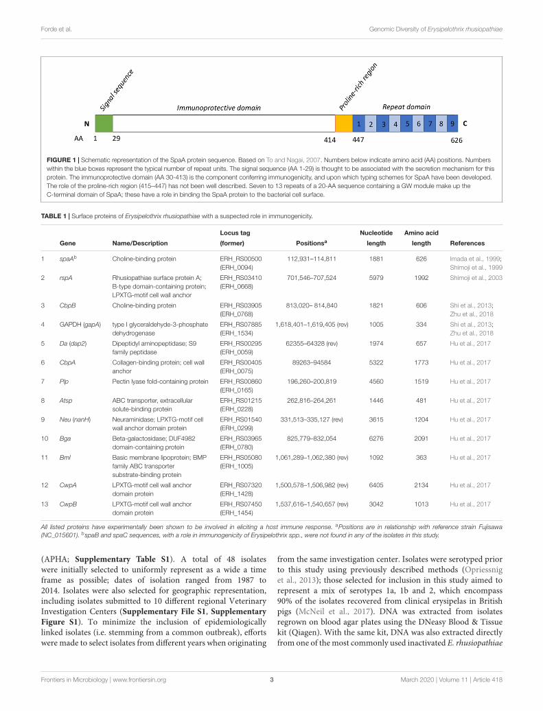
Additional surface proteins that have a demonstrated rolein immunogenicity of E. rhusiopathiae include rhusiopathiaesurface protein A (RspA) (Shimoji et al., 2003), choline bindingprotein B (cbpB) (Shi et al., 2013; Zhu et al., 2018) andGlyceraldehyde 3-phosphate dehydrogenase (GAPDH) (Zhuet al., 2018); these proteins have all elicited protection duringchallenge studies in mice, and the latter two also in pigs. Anadditional eight putative surface proteins were recently foundto give rise to varying degrees of protective immunity in mice(Hu et al., 2017; Table 1). As with SpaA variants, the exact roleand the extant diversity of these different surface proteins amongE. rhusiopathiae strains have yet to be determined.
The objectives of this study were to (1) characterize the overallgenomic diversity of E. rhusiopathiae in British pigs and assessthe amount of genetic divergence between field strains and acommonly used vaccine strain in the United Kingdom (UK), and(2) to quantify the amount of genetic diversity in Spa and otherimmunogenic surface proteins.
MATERIALS AND METHODS
Bacterial Isolates and Vaccine StrainArchived E. rhusiopathiae isolates from clinical pigerysipelas cases from across England and Wales wereprovided by the UK Animal and Plant Health Agency
Frontiers in Microbiology | www.frontiersin.org 2 March 2020 | Volume 11 | Article 418
fmicb-11-00418 March 12, 2020 Time: 18:55 # 3
Forde et al. Genomic Diversity of Erysipelothrix rhusiopathiae
FIGURE 1 | Schematic representation of the SpaA protein sequence. Based on To and Nagai, 2007. Numbers below indicate amino acid (AA) positions. Numberswithin the blue boxes represent the typical number of repeat units. The signal sequence (AA 1-29) is thought to be associated with the secretion mechanism for thisprotein. The immunoprotective domain (AA 30-413) is the component conferring immunogenicity, and upon which typing schemes for SpaA have been developed.The role of the proline-rich region (415–447) has not been well described. Seven to 13 repeats of a 20-AA sequence containing a GW module make up theC-terminal domain of SpaA; these have a role in binding the SpaA protein to the bacterial cell surface.
TABLE 1 | Surface proteins of Erysipelothrix rhusiopathiae with a suspected role in immunogenicity.
Locus tag Nucleotide Amino acid
Gene Name/Description (former) Positionsa length length References
1 spaAb Choline-binding protein ERH_RS00500(ERH_0094)
112,931–114,811 1881 626 Imada et al., 1999;Shimoji et al., 1999
2 rspA Rhusiopathiae surface protein A;B-type domain-containing protein;LPXTG-motif cell wall anchor
ERH_RS03410(ERH_0668)
701,546–707,524 5979 1992 Shimoji et al., 2003
3 CbpB Choline-binding protein ERH_RS03905(ERH_0768)
813,020– 814,840 1821 606 Shi et al., 2013;Zhu et al., 2018
4 GAPDH (gapA) type I glyceraldehyde-3-phosphatedehydrogenase
ERH_RS07885(ERH_1534)
1,618,401–1,619,405 (rev) 1005 334 Shi et al., 2013;Zhu et al., 2018
5 Da (dap2) Dipeptidyl aminopeptidase; S9family peptidase
ERH_RS00295(ERH_0059)
62355–64328 (rev) 1974 657 Hu et al., 2017
6 CbpA Collagen-binding protein; cell wallanchor
ERH_RS00405(ERH_0075)
89263–94584 5322 1773 Hu et al., 2017
7 Plp Pectin lyase fold-containing protein ERH_RS00860(ERH_0165)
196,260–200,819 4560 1519 Hu et al., 2017
8 Atsp ABC transporter, extracellularsolute-binding protein
ERH_RS01215(ERH_0228)
262,816–264,261 1446 481 Hu et al., 2017
9 Neu (nanH) Neuraminidase; LPXTG-motif cellwall anchor domain protein
ERH_RS01540(ERH_0299)
331,513–335,127 (rev) 3615 1204 Hu et al., 2017
10 Bga Beta-galactosidase; DUF4982domain-containing protein
ERH_RS03965(ERH_0780)
825,779–832,054 6276 2091 Hu et al., 2017
11 Bml Basic membrane lipoprotein; BMPfamily ABC transportersubstrate-binding protein
ERH_RS05080(ERH_1005)
1,061,289–1,062,380 (rev) 1092 363 Hu et al., 2017
12 CwpA LPXTG-motif cell wall anchordomain protein
ERH_RS07320(ERH_1428)
1,500,578–1,506,982 (rev) 6405 2134 Hu et al., 2017
13 CwpB LPXTG-motif cell wall anchordomain protein
ERH_RS07450(ERH_1454)
1,537,616–1,540,657 (rev) 3042 1013 Hu et al., 2017
All listed proteins have experimentally been shown to be involved in eliciting a host immune response. aPositions are in relationship with reference strain Fujisawa(NC_015601). bspaB and spaC sequences, with a role in immunogenicity of Erysipelothrix spp., were not found in any of the isolates in this study.
(APHA; Supplementary Table S1). A total of 48 isolateswere initially selected to uniformly represent as a wide a timeframe as possible; dates of isolation ranged from 1987 to2014. Isolates were also selected for geographic representation,including isolates submitted to 10 different regional VeterinaryInvestigation Centers (Supplementary File S1, SupplementaryFigure S1). To minimize the inclusion of epidemiologicallylinked isolates (i.e. stemming from a common outbreak), effortswere made to select isolates from different years when originating
from the same investigation center. Isolates were serotyped priorto this study using previously described methods (Opriessniget al., 2013); those selected for inclusion in this study aimed torepresent a mix of serotypes 1a, 1b and 2, which encompass90% of the isolates recovered from clinical erysipelas in Britishpigs (McNeil et al., 2017). DNA was extracted from isolatesregrown on blood agar plates using the DNeasy Blood & Tissuekit (Qiagen). With the same kit, DNA was also extracted directlyfrom one of the most commonly used inactivated E. rhusiopathiae
Frontiers in Microbiology | www.frontiersin.org 3 March 2020 | Volume 11 | Article 418

fmicb-11-00418 March 12, 2020 Time: 18:55 # 4
Forde et al. Genomic Diversity of Erysipelothrix rhusiopathiae
vaccines in the UK (referred to hereafter as the vaccine strain;see Commercial Products Used). We also endeavored to performDNA extraction from a second commercial vaccine, however,this vaccine did not test positive by E. rhusiopathiae probe-basedqPCR, and DNA of sufficient quantity (as measured by Qubit)could not be extracted despite multiple attempts.
Library Preparation, Sequencing andAssemblyLibrary preparation, sequencing and de novo assembly wereperformed by MicrobesNG (Birmingham, UK). Libraries wereprepared using the Nextera XT v2 kit and sequenced on theIllumina HiSeq2500 platform, generating 250 base pair paired-end reads. Reads were trimmed using Trimmomatic (Bolger et al.,2014), and assembled de novo using SPAdes v. 3.7.0 (Bankevichet al., 2012) with default settings. Assembly quality metrics (i.e.number of contigs, N50) were obtained using QUAST (Gurevichet al., 2013). Three of the isolates submitted had poor sequencingquality indicative of contamination (i.e. unexpected total lengthand GC content) and were excluded from further analyses,resulting in 45 whole genome E. rhusiopathiae sequences (WGS)from British pigs. Since key questions in this study were relatedto surface antigens and their relationship with a current vaccinestrain, the vaccine extract was submitted for sequencing twice ontwo independent runs for variant confirmation.
Population StructureTo place the newly sequenced isolates (i.e. the vaccine strain andthe 45 British pig isolates from which high quality sequence datawere obtained) within the broader global population structure ofE. rhusiopathiae, a phylogenetic tree was estimated that includedan additional 75 isolates from Clades 2, 3 and the intermediateclade (Forde T. et al., 2016) (Supplementary Table S1). TheFujisawa reference genome (NC_015601.1), the first completehigh-quality genome for E. rhusiopathiae (Ogawa et al., 2011),was also included; this is a virulent serotype 1a strain isolatedfrom a pig in Japan prior to 1985. Serotype was already knownfor 10/75 of the global isolates. In silico serotype testing wasalso done to detect isolates of serotypes 1a, 1b, 2, and 5. Inbrief, BLAST searches of all de novo assemblies were doneusing primer pairs described for each serotype (Shiraiwa et al.,2018). A BLAST search was considered positive for a givenserotype if there were matches for both forward and reverseprimers across their full length with a maximum of one SNPdifference, and that yielded the expected amplicon length (i.e.distance between primers). The phylogeny – based on core singlenucleotide polymorphisms (SNPs) – was built using Nullarbor1
(Seemann et al.) implemented through the CLIMB computingplatform for microbial genomics (Connor et al., 2016). TheNullarbor pipeline uses the program Freebayes v1.1.0 for variantcalling across all isolates with respect to a reference genome. Itsubsequently builds a core SNP alignment using Snippy v3.2,where “core sites” are genomic positions that are present in allthe included isolates. This core alignment comprised 3490 SNPs.FastTree v2.1.10 was then called within Nullarbor to infer the
1https://github.com/tseemann/nullarbor
phylogeny using maximum-likelihood based on the GTR + G4model of substitution.
Variability of Spa and Other SurfaceProtein SequencesSpa genes were searched for in all newly sequenced isolatesby performing BLASTn searches using a custom database ofspaA, spaB, and spaC nucleotide sequences from differentserotypes available on GenBank (Supplementary Tables S1, S2),implemented within Geneious v. 11.0.5 (Kearse et al., 2012).A BLAST hit was considered positive for a particular Spatype if it had greater than 95% pairwise identity; in practice,homologous Spa types had ∼98% pairwise identity, whileheterologous types generally had ∼90% identity or lower.Nucleotide sequences of the spaA gene based on the BLASThits were extracted from all de novo assemblies (n = 121). spaAsequences were similarly extracted from seven whole genomesequences of E. rhusiopathiae available on GenBank: Fujisawa,SY1027 (NC_021354.1), GXBY-1 (NZ_CP014861.1), WH13013(NZ_CP017116.1), NCTC7999 (NZ_UFYF01000001.1),NCTC8163 (NZ_LR134439.1), and ML101 (NZ_CP029804.1).Finally, to further explore the variability of this surface protein,an additional 215 publicly available spaA nucleotide sequenceswere downloaded (Supplementary Table S2), resulting in atotal of 343 spaA sequences. Translations were performed eitherusing the transeq program from EMBOSS (Rice et al., 2000), orwithin Geneious, using translation Table 11 for bacteria. Aminoacid sequences were aligned using MUSCLE (Edgar, 2004),implemented within Geneious, and any amino acid variants wererecorded in comparison with the Fujisawa reference sequence.A phylogenetic tree was estimated from the protein alignmentusing a Neighbor-Joining method implemented in GeneiousTree Builder, using the Jukes-Cantor model.
The amino acid variability within 12 additional surfaceproteins that have been shown to play a role in E. rhusiopathiaeimmunogenicity was also examined (Table 1). A custom BLASTdatabase of the nucleotide sequences of these genes was createdbased on the sequences from the Fujisawa reference genomewithin the program Geneious. Each de novo assembly fromBritish pig isolates and the vaccine strain was queried toidentify and extract the homologous gene sequences, whichwere then translated to amino acid sequences and alignedwithin Geneious along with the Fujisawa reference sequence.Phylogenies for each protein were estimated as described forSpaA. Variants of these different surface proteins were identifiedbased on clustering observed in mid-point rooted phylogenies(Supplementary File S2).
Conformation of SpaA Protein VariantsTo assess the potential structural impact of SpaA protein variantsidentified, the 3-D protein structure of a typical group 1 SpaA(Figure 4) was initially modeled using the I-TASSER platform(Roy et al., 2010). I-TASSER uses LOMETS, a meta-threadingserver that combines several threading programs to detectstructural templates from the Protein Data Bank (PDB) usingthreading or fold recognition. The threading-aligned regions
Frontiers in Microbiology | www.frontiersin.org 4 March 2020 | Volume 11 | Article 418

fmicb-11-00418 March 12, 2020 Time: 18:55 # 5
Forde et al. Genomic Diversity of Erysipelothrix rhusiopathiae
of these templates provide the building blocks and spatialrestraints for the prediction of the target protein, with unalignedareas predicted ab initio. Full-length structural models werepredicted by I-TASSER, and the best structural model – identifiedthrough comparison of C-scores – was used for further analysis.C-scores are typically bounded between −5 and 2 with highervalues indicating greater confidence in the predicted modeland −1.5 acting as a useful threshold above which more than90% of predictions are correct (Roy et al., 2010). To assess thestructural similarity of the SpaA structural model to existing PDBstructures, template modeling score (TM-score) and root-mean-square deviation (RMSD) were calculated across aligned Cα
atoms using TM-align v20170708 (Zhang and Skolnick, 2005).TM-scores are bounded between 0 and 1 where 1 indicatesperfect structural alignment, scores above 0.5 assume roughlythe same fold while scores below 0.2 correspond to randomlychosen unrelated proteins (Zhang and Skolnick, 2005). Mutationswere introduced using the PyMOL molecular graphics system(Schrödinger2), where the most common rotamer was selectedand energy minimization was performed in the locality ofthe mutated site.
Epitope scores were predicted per-residue based on aminoacid identity, surface exposure and side chain orientation usingBEpro (Sweredoski and Baldi, 2008). Amino acid diversity wascalculated using an alignment of all available E. rhusiopathiaeSpaA protein sequences (n = 343) (including residues 1–447 ofthe N-terminal region). Diversity at each amino acid position wascalculated as the true diversity at q = 2 (Inverse Simpson index),whereby 1 indicates a fully conserved position and numbersgreater than 1 indicate increasing diversity. Amino acid diversitywas visualized on the structural model using PyMOL.
RESULTS
Sequencing, Population Structure andSerotypeAmong the 45 newly sequenced E. rhusiopathiae isolates fromBritish pigs and the vaccine strain, a minimum mean sequencingdepth of 26X was achieved, with a mean of 82X. The majorityof the newly sequenced strains from British pigs fell withinClade 2 (32/45 = 71.1%), as did the vaccine strain (Figure 2).Five isolates (11.1%) were in Clade 3, while the remaining17.8% (8/45) were within the intermediate clade. None of theisolates belonged to Clade 1. The average number of core SNPdifferences separating the pig isolates from one-another was435 (median = 377, range 0–767). This was comparable to thenumber of core SNPs separating the vaccine strain from thefield strains (median = 413, range 328–655). The core SNPprofile of the vaccine strain was 100% identical across the twosequencing rounds. No clear geographic clustering was evidentbased on submitting veterinary center. Similarly, no obvioustemporal clustering was apparent, indicating that rather thanstrain turnover, multiple divergent strains have remained incirculation throughout the study period (Figure 2). The genetic
2https://pymol.org
distance (based on core SNPs) of the British pig isolates fromthe vaccine strain did not change appreciably over time, thusproviding no evidence for potential vaccine-induced selection(Supplementary File S1, Supplementary Figure S2). Thephenotypically determined serotypes for the British pig isolatesand vaccine were confirmed in silico in 40 of 46 isolates, withsix discrepancies (Supplementary Table S1). However, upon re-testing, the phenotypic serotype of these isolates matched thatpredicted by in silico testing. Among the global collection, theFujisawa reference strain was confirmed as serotype 1a in silico,while 19 isolates were designated as serotype 1b, 27 as serotype 2,17 as serotype 5, and 12 had no BLAST hits for any of the primerpairs, suggestive of belonging to a serotype other than 1a, 1b, 2 or5. This corroborated with the previously determined serotype foreight isolates, including one serotype 9 for which none of theseprimer pairs matched. Two mismatches between phenotypicand in silico serotype were found: 1. Isolate HC-585, previouslyclassified as serotype 1a, did not have any primer pair matches;2. Isolate Mew22, a serotype “N” (i.e. does not react with any ofthe panel of antisera), was classified as a serotype 1b. The reverseprimer sequence for serotype 5 was found to consistently haveone SNP difference with all serotype 5 isolates. The distributionof different serotypes within the phylogeny is shown in Figure 3,where there was limited correlation with Clade.
Occurrence and Variability of SpaProteinsThe spaA gene was present in all the newly sequenced isolates,while spaB and spaC genes were not detected. In the translatedSpaA protein sequences, substantial diversity was found withinthe immunoprotective or “hypervariable” domain, which spansamino acids 30–413 (Figure 1). All SpaA amino acid sequences(n = 343) were initially classified into the five SpaA groupspreviously described (Janßen et al., 2015) (SupplementaryTable S3), which are based upon amino acid variants inthis domain. Beyond the previously reported positions, threeadditional variable amino acid sites were present in at least fiveisolates, and thus considered discriminatory. These were variantsat position 97 (N/I), position 109 (N/H), and position 139 (Q/K).There were also several amino acid variants (n = 53) present infewer than five isolates (Supplementary Table S4). One isolate(20767, from a wild bird) had a highly divergent SpaA protein,with 19 unique amino acids. If these sites are not considered,there were 34 variable sites, four of which had two different aminoacid variants (3 alleles).
The phylogenetic tree of the different SpaA protein variantssuggests that there are two main groups (Figure 4), and wefound that these groups are correlated with the overall populationstructure of E. rhusiopathiae (Figures 3, 5). We therefore proposea simplified nomenclature for SpaA groups, where Group 1has amino acid variants at positions 55 (V/I), 70 (K/N), 178(G/D), 195 (D/N) and 303 (G/E) in relation with the Fujisawareference, consistent with the previous scheme proposed byJanßen et al. (2015). All other previously described groupsare minor variants of what we now refer to as Group 2.These variants appear to arise multiple times throughout the
Frontiers in Microbiology | www.frontiersin.org 5 March 2020 | Volume 11 | Article 418

fmicb-11-00418 March 12, 2020 Time: 18:55 # 6
Forde et al. Genomic Diversity of Erysipelothrix rhusiopathiae
FIGURE 2 | Diversity of Erysipelothrix rhusiopathiae in British pigs as shown within a global phylogenetic context. This maximum likelihood phylogeny is based oncore single nucleotide polymorphisms (SNPs) identified through the Nullarbor pipeline. British pig isolates (n = 45) are represented by blue circles at branch tips, whilethe vaccine strain is represented by a red star. Branches with no symbol are isolates from a broad global collection (n = 75) from various host species andgeographic locations representing Clades 2, 3 and the intermediate clade. An isolate from Clade 1 was used to determine the appropriate rooting position (notshown). The Fujisawa strain was used as the reference genome. Spatio-temporal diversity of the British pig isolates is illustrated by color bars to the outside of thephylogeny, with the inner color representing the year range of isolation, and the outer color bar representing the veterinary center to which the isolate was submitted.
phylogeny (Supplementary File S1, Supplementary Figure S3);this is suggestive of either parallel evolution (homoplasy)or recombination. A detailed description of how this SpaAclassification relates to previously described groups is found inSupplementary Table S3 and Supplementary File S3.
All isolates within the SpaA group 1 belonged to Clade 2 orthe intermediate clade. All Clade 2 isolates possessed SpaA group1 except for a single British pig isolate (swine100, Figure 3).Since the majority of the British pig isolates belonged to Clade2, SpaA group 1 was the variant found in the majority of thesestrains (82%; Supplementary Table S1). The vaccine strain wasa variant of Group 1, with an additional amino acid differenceat position 109 (N/H). This variant was also found in sevenother sequences from GenBank; however, none of the British pigisolates investigated in this study shared this difference.
Predicted Structure of SpaAWe aimed to assess the potential functional implications ofvariant residues detected within SpaA. However, given thatthe structure of SpaA is unknown and its function poorlyunderstood, its structure was predicted as an exploratory exercise.A full-length structural model was produced using I-TASSER,a program that predicts structure using a combination ofthreading to existing protein structures and ab initio modeling.The majority of the highest ranked structural templates forthe SpaA protein detected by I-TASSER were surface-locatedcholine-binding proteins of Streptococcus pneumoniae, eitherphosphorylcholine esterase (Pce) also known as choline-bindingprotein E (CbpE) (PDB ID: 2BIB; Hermoso et al., 2005) orcholine-binding protein F (CbpF) (PDB IDs: 2V04, 2V05; Molinaet al., 2009). Other Firmicutes templates from Clostridium difficile
Frontiers in Microbiology | www.frontiersin.org 6 March 2020 | Volume 11 | Article 418

fmicb-11-00418 March 12, 2020 Time: 18:55 # 7
Forde et al. Genomic Diversity of Erysipelothrix rhusiopathiae
FIGURE 3 | Relationship of SpaA group, serotype, and immunogenic surface proteins to phylogenetic population structure. This phylogenetic tree is based on coresingle nucleotide polymorphisms (SNPs), and was estimated using the Nullarbor pipeline (same sequence data as in Figure 2). British pig isolates are represented byblue circles at branch tips, while the vaccine strain is represented by a red star. SpaA group (1 or 2) is shown by the yellow/orange color strip. Serotype (1a, 1b, 2, 5)as determined in silico is shown by the purple/green color strip. Those where phenotypic testing was not performed or where in silico test results differed are notshown. Variants of 8 different immunogenic surface proteins are shown by the black/gray color strips for all British pig isolates. Those shown in black are those whichcluster most closely with the variant found in the vaccine strain; isolates in dark gray represent isolates with surface protein variants in outlier groups(Supplementary Table S1, Supplementary File S1).
Frontiers in Microbiology | www.frontiersin.org 7 March 2020 | Volume 11 | Article 418

fmicb-11-00418 March 12, 2020 Time: 18:55 # 8
Forde et al. Genomic Diversity of Erysipelothrix rhusiopathiae
FIGURE 4 | Discriminatory amino acid positions within the immunoprotective domain of the SpaA protein of Erysipelothrix rhusiopathiae. Amino acid (AA) variantpositions with respect to the Fujisawa reference sequence are listed in the top row, with the amino acid found in the reference sequence shown in the second row.The phylogenetic tree shown to the left shows the relatedness among the SpaA sequence variants. Variants in each group are shown in blue boxes, with the AAchange indicated.
FIGURE 5 | Relationship of Spa type and SpaA group to the population structure of Erysipelothrix spp. This figure is adapted from Forde T. et al., 2016. The tree isrooted to other genera of the family Erysipelotrichaceae. Spa type and SpaA group are indicated in red. SpaA group is based on discriminatory amino acid variants(Figure 4). ∗spaA and spaB have been reported only once each in Erysipelothrix tonsillarum (Pomaranski et al., 2017).
and Leuconostoc mesenteroides were also identified. The highest-ranked structural model predicted using I-TASSER had a C-scoreof −1.30 indicating a good degree of confidence in the predictedstructure (Roy et al., 2010).
Comparison with solved PDB structures showed that thestructural model of E. rhusiopathiae SpaA most closely resembledthe Pce protein of S. pneumoniae (Hermoso et al., 2005) (TM-score = 0.82, RMSD = 1.50 Å for 524 aligned Cα atoms, 17.6%amino acid sequence identity in structurally aligned region). TheS. pneumoniae Pce comprises an N-terminal catalytic module(300 residues) and a C-terminal tail (228 residues) responsiblefor choline-binding (Hermoso et al., 2005). The hypervariabledomain of the SpaA structural model (residues 30–413) alignedto the Pce catalytic module (TM-score = 0.75, RMSD = 1.88 Å for303 aligned Cα atoms), while the C-terminal domain (residues448–626) aligned to the Pce choline-binding domain (TM-score = 0.98, RMSD = 0.37 Å for 177 aligned Cα atoms).
While the function of the N-terminal domain is unknown,we hypothesized that it might be similar to that of its closesthomolog, which we found to be the Pce protein of S. pneumoniae.As such, we examined the proximity of variable SpaA positions
to the residues of the structural model homologous to thosecomprising the Pce catalytic site. Residues of the S. pneumoniaePce protein catalytic domain active site were mapped to the SpaAstructural model using TM-align so that the proximity of variableSpaA sites to the potential active site could be calculated. Thestructural model of the E. rhusiopathiae SpaA protein is shownin Figure 6, colored according to amino acid diversity calculatedacross the alignment of available sequences. Of the residues withthe highest amino acid diversity (55, 70, 101, 178, 195, 203, 303,426, and 435, Inverse Simpson index > 1.3), positions 70, 178and 303 were closest to residues of the potential active site withdistances of under 10 Å (for comparison, the distance betweencovalently bound residues was 3.5–4 Å). For position 109, theposition at which most E. rhusiopathiae sequences from Britishpigs differ relative to the vaccine (Asn – His), the lowest distanceto a residue aligned to the Pce protein catalytic domain active sitewas 13.3 Å which is beyond the range expected to be structurallyimpacted by a mutation between His and Asn at that position.
The extent to which SpaA is recognized by antibodies ofthe humoral immune system is not known. To investigate thepotential for variant residues to influence antibody recognition,
Frontiers in Microbiology | www.frontiersin.org 8 March 2020 | Volume 11 | Article 418

fmicb-11-00418 March 12, 2020 Time: 18:55 # 9
Forde et al. Genomic Diversity of Erysipelothrix rhusiopathiae
FIGURE 6 | Structural model of Erysipelothrix rhusiopathiae SpaA proteinpredicted using I-TASSER. Residues structurally aligned to the active site ofthe S. pneumoniae Pce protein are colored magenta while the remainingresidues of the N-terminal domain (residues 1–447) are colored following thelegend according to amino acid diversity (Inverse Simpson index) calculatedfrom 343 SpaA sequences sourced from newly sequenced isolates and publicsequence databases. Residues of the C-terminal domain (residues 448–626)are colored gray. (A) Cartoon representation of whole structural modelshowing secondary structure. (B) Close up ribbon representation ofN-terminal domain with spheres highlighting the nine residues with highestamino acid diversity (Inverse Simpson index > 1.3) and with residuesstructurally aligned to the active site of the S. pneumoniae Pce protein(magenta) shown in stick format. Figure created using Pymol (Schrödinger).
the degree to which their location within the structural modelshowed epitope characteristics was assessed. Thus, to evaluatethe potential of the variant residues influencing recognition bythe humoral immune system, the structural model was used togenerate epitope scores that reflect the likelihood of each residuebelonging to a B-cell epitope. Epitope scores, which reflect thelikelihood of each residue belonging to a B-cell epitope, werecalculated for each residue within the SpaA structural modelbased on surface exposure, amino acid identity and side chainorientation (Sweredoski and Baldi, 2008). Of the nine high-diversity positions, 55 and 435 were both placed among thehighest predicted epitope scores, at the 94th and 93rd percentile,respectively, but there was no consistent relationship betweendiversity and epitope scores. The epitope score associated withposition 109 on the SpaA was at only the 8th percentile calculatedacross all positions, suggesting the position is unlikely to beimportant for antibody recognition. For all positions, aminoacid diversity, predicted secondary structure, epitope score, andaligned S. pneumoniae Pce protein active site residues are detailedin Supplementary Table S6.
Occurrence and Variability of OtherSurface ProteinsOf the 12 other E. rhusiopathiae surface proteins examined, 11were present in all isolates from British pigs and the vaccine(i.e. were core genes). However, the degree to which aminoacid sequences were conserved varied greatly among proteins
(Table 2; Supplementary Table S5; Supplementary File S2). Ofthe surface proteins examined, GAPDH was the most conserved,with only a single variant across the 334 amino acid protein,found in a single isolate (swine74). Atsp and Da were also highlyconserved, with 6 and 13 variant positions, respectively. It isnoteworthy that three of the six variant positions in Atsp wereunique to the vaccine strain. Of the 52 variant positions identifiedin Bga, three of these were unique to the vaccine strain. Oneisolate (swine98) also had a mutation conferring a premature stopcodon for this protein. A slightly higher proportion of sites variedin Neu (3.4%), Bml (3.8%) and rspA (4.8%); the latter had foursites unique to the vaccine. CbpA showed much higher diversity,with variants at 162 of 1773 amino acid positions (9.1%), but noneof which were unique to the vaccine.
The only proteins with any insertions or deletions identifiedamong the British pig isolates were CbpB, Plp and CwpB. InCbpB, one isolate (swine78) had a deletion of 20 amino acids,corresponding to the loss of a repeat unit. In Plp, an insertionof either 4 amino acids (16 strains) or 12 amino acids (twostrains) was present. In CwpB, a single isolate (swine103) hada deletion of 4 amino acids (positions 971–974), while anotherisolate (swine18) had an insertion of 7. This protein also hadthe highest proportion of variant positions (27%), although themajority of the variant sites (83%) were due to two isolateswhose sequences differed greatly from the others (swine18and swine101; Supplementary Table S5 and SupplementaryFile S2). Finally, one surface protein examined was variablypresent among the isolates. Complete nucleotide sequences forCwpA (5322 bp) were present in only 13 isolates, as well as inan additional 6 isolates that were missing a stop codon. Theremaining 27 isolates had BLAST hits for this sequence rangingfrom 758 – 3116 bp (median = 2610); the vaccine strain had a hitlength of 3116 bp. Some of the BLAST hits for the surface proteinsinvestigated were only partial sequences spread across multiplecontigs (i.e. had not been completely assembled) (Table 2). Theaffected isolates had lower quality assemblies overall (swine94,50, and 40), as shown by the lowest N50 values among the newlysequenced isolates (Supplementary Table S1).
The phylogenetic trees generated for the different surfaceproteins (Supplementary File S2) did not show concordancewith the core SNP phylogeny. This is illustrated in Figure 3,where for each surface protein, the isolates that cluster mostclosely with the protein sequence of the vaccine strain are shown.There were no major changes to the distribution of the differentsurface protein variants over the timeframe of the study period(Supplementary File S1, Supplementary Figure S4).
DISCUSSION
Diversity of Circulating E. rhusiopathiaeStrains in Pigs in Great Britain and TheirRelation to a Commonly Used VaccineStrainWhile the global genomic diversity of E. rhusiopathiae has beencharacterized, how much of this genomic and functional diversity
Frontiers in Microbiology | www.frontiersin.org 9 March 2020 | Volume 11 | Article 418

fmicb-11-00418 March 12, 2020 Time: 18:55 # 10
Forde et al. Genomic Diversity of Erysipelothrix rhusiopathiae
TABLE 2 | Amino acid (AA) sequence diversity in surface proteins related to immunogenicity of Erysipelothrix rhusiopathiae.
Protein BLAST hit results where codinglength # Variant AAs* # variant AAs Insertions/deletions and sequence was spread across
Surface protein (AAs) (% of total AAs) unique to vaccine premature stop codons multiple contigs+
rspA 1992 96 (4.8%) 4 swine40 (3 contigs) swine94 (3 contigs)
cbpB 606 27 (4.5%) 1 Deletion 20AAs swine78 (1 repeatunit)
GAPDH (gapA) 334 1 (0.3%) 0
Dipeptidyl aminopeptidase(Da, dap2)
657 13 (2%) 0
CbpA 1773 162 (9.1%) 0
Plp 1519–1531 91 (6%) 0 Insertion of either 4 AAs (16 strains)or 12 AAs (2 strains)
swine40 (2 contigs)
Atsp 481 6 (1.2%) 3 swine50 (2 contigs)
Neuraminidase (Neu, nanH) 1204 41 (3.4%) 1
Beta-galactosidase (Bga) 2091 52 (2.6%) 3 swine98 stop codon at AA Pos 658 swine40 (3 contigs) swine50 (2 contigs)swine94 (3 contigs)
Bml 364 14 (3.8%) 0 swine40 (2 contigs)
CwpA 2134 N/A N/A Full length BLAST hits in 13 isolates(5322 bp) Missing stop codon in 6isolates Remaining 27 isolates withBLAST hits ranging from 758 – 3116 bp(median = 2610)
CwpB 1009–1020 276 (27%) 1 Insertion 7AAs swine18 Deletion4AAs swine103
swine40 (2 contigs)
Isolates were examined from British pigs (n = 45) and a commercially available vaccine strain in comparison with the Fujisawa reference strain. *Comparisons are made inrelationship with reference strain Fujisawa (NC_015601). +Complete coding sequences were found in all isolates (based on BLASTn searches of the de novo assemblednucleotide sequences) unless otherwise stated. bp = base pairs.
is maintained at smaller scales was unclear. This study providesa valuable contribution to our understanding of E. rhusiopathiaegenomics, and in particular provides novel data on the diversityof surface antigens. Moreover, fully sequencing a commercialvaccine strain of E. rhusiopathiae has provided valuable insightsinto its relationship to circulating field strains.
Throughout the 27-year time period from which sampleswere collected for this study (1987 – 2014), phylogeneticallydiverse strains of E. rhusiopathiae were isolated from British pigs(Figure 2). Similarly high diversity was previously observed inpoultry isolates from Germany based on multi-locus sequencetyping (Janßen et al., 2015), while the diversity of E. rhusiopathiaecirculating in pigs in Asia has been shown to be restricted tostrains within the intermediate clade (Ogawa et al., 2017). Thelower diversity of E. rhusiopathiae in Asian countries couldbe due a more recent introduction of the pathogen, and/ordifferences in animal trade, pig breeding, herd managementor biosecurity. The majority of the British pig isolates belongto Clade 2, which differs from what was previously found inNorth America, where all pig and poultry isolates investigated(n = 14) belonged to Clade 3 (Forde T. et al., 2016), suggestingdifferent E. rhusiopathiae clades have become dominant on thesecontinents. The vaccine strain also belonged to Clade 2. We foundno evidence that E. rhusiopathiae strains in circulation in GreatBritain have diverged from the vaccine strain over the studyperiod. It would have been interesting to investigate the strainsassociated with other commercially available vaccines, however,we were unable to detect and extract E. rhusiopathiae DNA fromthe second vaccine tested. Whether there is a component of
the vaccine adjuvant (e.g. aluminum hydroxide) that inhibiteddetection of DNA by Qubit and PCR is unknown. None of theisolates we sequenced from British pigs in this study belongedto E. rhusiopathiae Clade 1, emphasizing the rarity of strainsfrom this clade causing disease in the major production speciesworldwide; in a global collection of isolates previously sequenced(Forde T. et al., 2016), only 8% (7/86) belonged to Clade 1, andnone of these were from pigs or poultry.
Spa Type and Correlation WithPopulation Structure and ClinicalDiseaseNo spaB or spaC genes were found among the E. rhusiopathiaeisolates from British pigs. Given the lack of Clade 1 isolates, thisfinding was expected and is consistent with previous observationsthat Clade 1 isolates carry the spaB gene, whereas Clade 2,3 and intermediate isolates carry the spaA gene (Figure 5;Forde T. et al., 2016). To date, spaC has only been associatedwith Erysipelothrix sp. strain 2, and not E. rhusiopathiae. Thepredominance of the spaA gene in isolates associated with clinicalerysipelas has been supported in several studies (Eamens et al.,2006; To et al., 2012; Janßen et al., 2015). It therefore seemsthat spaB and spaC (i.e. isolates belonging to E. rhusiopathiaeClade 1 and E. sp. Strain 2) are less relevant when consideringvaccine design for production species. However, given thatE. rhusiopathiae Clade 1 is more commonly isolated from marinemammals in captivity (Opriessnig et al., 2013; Forde T. et al.,2016), and Erysipelothrix spp. carrying the spaC gene has recently
Frontiers in Microbiology | www.frontiersin.org 10 March 2020 | Volume 11 | Article 418

fmicb-11-00418 March 12, 2020 Time: 18:55 # 11
Forde et al. Genomic Diversity of Erysipelothrix rhusiopathiae
been associated with disease in fish (Pomaranski et al., 2017),protecting against Erysipelothrix spp. carrying these Spa typescould be of greater relevance for aquatic species. It was previouslysuggested that Spa proteins are likely important virulence factorsassociated with the pathogenic potential of E. rhusiopathiae incomparison to the less pathogenic E. tonsillarum (To and Nagai,2007), and until recently, no Spa-related genes or proteins hadbeen found in E. tonsillarum (To and Nagai, 2007; Shen et al.,2010). To our knowledge, the occurrence of spaA and spaB genesin E. tonsillarum has only been documented in one paper, amongisolates from ornamental fish (Pomaranski et al., 2017). However,given that the species was defined based only on sequencingof the gyrB gene, it is conceivable that this isolate may havebeen misclassified.
SpaA Diversity, Correlation WithPopulation Structure, and Relevance forVaccination and PathogenicityThis study represents the largest examination of the diversityof SpaA protein sequences in E. rhusiopathiae conducted todate, based on a collection of publicly available sequences andde novo whole genome sequence assemblies (n = 343). Withthis extensive collection of sequences, we were able to identifyadditional amino acid variants that could further discriminateSpaA protein sequences in comparison to previous typingschemes (Uchiyama et al., 2014; Janßen et al., 2015). We werealso able to assess for correlation of these groups with populationstructure (Figure 5). SpaA group 1 was only present in Clade2 and the intermediate clade, but was absent from the Clade3 isolates examined (Figure 3). Janßen et al. (2015) found astrong correlation between SpaA group 1 and their ST complex9. It is very likely that ST-9 corresponds with Clade 2, sincewe found that all Clade 2 isolates had this SpaA variant withone exception. We propose amalgamating all other previouslydescribed SpaA groups into a single group, forthwith referredto as SpaA group 2, since variants within this group are limitedto only one or two amino acid differences. Moreover, thesevariants appear to be unconstrained by the core phylogeny(Supplementary File S1). Such incongruent patterns could ariseeither through parallel evolution (homoplasy), or, more likely,through horizontal gene transfer, given the apparent propensityfor recombination observed in this species.
Whether the observed differences within the SpaA proteincould be responsible for differential immune protection – orfor differences in virulence – remains an important area forfuture study. For instance, the vaccine strain tested in this studybelonged to Group 1 SpaA along with the majority of British pigisolates, but had an amino acid change from N→H at position109. Whether this could result in differential immune response isunknown; however, based on the predicted structure of SpaA, wefound that this position is unlikely to represent an epitope.
Isolates with methionine at position 203 (i.e. Janßen Group5, Uchiyama Group 1) were rare among the isolates examinedin this study. This variant was present in one British pig isolate(Supplementary Tables S1, S3) and in three WGS available onGenBank (GXBY-1, WH13013 and ML101), but in none of the
global collection of isolates (n = 75). It was previously foundin a handful of pig isolates from Germany (n = 4) (Janßenet al., 2015). Otherwise, this variant has been mostly limitedto strains from China (Supplementary Table S2) and Japan,where it is widespread and has been isolated from pigs withacute, subacute and chronic infections (To et al., 2012; Uchiyamaet al., 2017) and where it appears to be increasing in prevalence(Ogawa et al., 2017). There is, however, not yet any evidence ofthis variant being associated with an increase in virulence (Zouet al., 2015). One particular variant that would be worthy offurther investigation is that of alanine at position 195 (previouslyclassified as Group 3 by Uchiyama et al., 2014). This is thevariant carried by the pathogenic reference strain SY1027 fromChina (Kwok et al., 2014), and was the variant carried by theE. rhusiopathiae strain associated with large-scale muskox die-offs in northern Canada (Kutz et al., 2015; Forde T.L. et al.,2016). This variant has also been reported among pigs in Japan,although is apparently being replaced by newer lineages (Ogawaet al., 2017). Whether this amino acid change confers increasedvirulence would be valuable to explore.
Predicted SpaA Protein StructureWe found that the amino acid sequence of the E. rhusiopathiaeSpaA protein predicts a protein structure most closely resemblingthat of choline-binding proteins of Streptococcus pneumoniae.Indeed, the homology in the C-terminal region of theseproteins had previously been described (Borrathybay et al.,2015). This region – containing tandem repeats includingthe GW motif – is a motif conserved across Gram-positivebacteria that allows proteins to bind to choline residuesof techoic acid on the cell surface (Jedrzejas, 2001). It isbelieved that this motif is what allows SpaA to bind tophosphorylcholine of the of E. rhusiopathiae capsule (Haradaet al., 2014), facilitating binding to host endothelial cells.To our knowledge, no studies have previously examined thestructure of the N-terminal region of SpaA, which is theportion shown to be involved in its protective immunogenicity.Among proteins for which the structure has been determined,we found the N-terminal portion of the S. pneumoniae Pceprotein was the closest match, although with lower homologyin comparison with the C-terminal portion. S. pneumoniaePce modifies the distribution of phosphoryl choline on thebacterial surface, impairing the ability of the host immunesystem to efficiently bind, providing a mechanism for immuneescape (Hermoso et al., 2005). Given the predicted structuralsimilarity, it is possible that SpaA has a similar function, thoughthis hypothesis would need to be further explored throughfunctional assays.
Working with the hypothesis that SpaA may have a similarfunction to S. pneumoniae Pce, we explored whether SpaApositions with high amino diversity, including those differingbetween groups 1 and 2, were located near to a possible activesite. The N-terminal module of the Pce protein contains abinuclear Zn2 + catalytic center (Hermoso et al., 2005). Withinthis active center, particular residues have been shown to beinvolved in substrate binding and catalysis, facilitating teichoicacid hydrolysis which releases phosphoryl choline moieties
Frontiers in Microbiology | www.frontiersin.org 11 March 2020 | Volume 11 | Article 418

fmicb-11-00418 March 12, 2020 Time: 18:55 # 12
Forde et al. Genomic Diversity of Erysipelothrix rhusiopathiae
reducing recognition by the immune system. In SpaA, the variantamino acid residues at positions 70, 178 and 303 were thoseclosest to potential active site, as based on homology with theS. pneumoniae Pce protein. Since these are all variants thatdistinguish between SpaA groups 1 and 2, it is possible that thesevariants confer functional differences between these groups.
Alternatively, we considered that variable positions may haveplayed a role in recognition by the humoral immune system.To do so, we assessed whether diverse amino acid sites tendedto occupy locations within the structural model that exhibitedsignatures of epitope regions. With the notable exception ofposition 55, a variant that differs between groups 1 and 2, andposition 435 that possessed very high predicted epitope scores,the diverse positions did not show a general tendency toward highepitope scores, suggesting pressure from the humoral immunitymay not be a major driver of the observed genetic diversity. Therole of SpaA in immune recognition has not been characterized ingreat detail. In vitro experiments and challenge studies to assesscross-protection, wherein isolates from different SpaA groups areincluded, would be valuable.
Variability in Immunogenic SurfaceProteinsOur study provides novel information about the sequencediversity of different immunogenic surface proteins ofE. rhusiopathiae. The degree of variability differed substantiallyamong the 12 proteins we investigated, ranging from proteinsthat were nearly completely conserved across all 45 isolatesand the vaccine strain (GAPDH) to 27% of the amino acidsshowing variation (CwpB), as well as some proteins with smallinsertions and deletions (Plp and CwpB). Coding sequences forthese proteins were found to be core genes, with the exceptionof CwpA which was variably present. Based on the phylogenetictrees estimated for each protein (Supplementary File S1),variants of the different proteins did not correlate with thephylogenetic relationship among isolates (Figure 3), suggestingthat horizontal transfer of genes encoding for antigens iscommonplace for E. rhusiopathiae.
We investigated whether there was any evidence ofantigenic divergence (Bart et al., 2014) between field strainsof E. rhusiopathiae and a common vaccine strain, as this couldpotentially be of concern for vaccine efficacy. However, we didnot observe any such temporal shift in surface protein variantsfor any of the antigens we examined (Supplementary File S1).The vaccine strain had unique amino acids in 5 of the 12 surfaceproteins examined – rspA, Bga, Atsp, Neu and CwpB (i.e. thatwere not present among the 45 pig isolates). Of particularnote is in Atsp, where this included three of only six variantsacross the protein and among all isolates. The impact of thesedifferences – if any – would be valuable to explore. The mostconserved surface protein – GAPDH – has been confirmed tobe expressed at the cell surface (Shi et al., 2013), and like SpaA,RspA and RspB, has a role in endothelial adhesion (Zhu et al.,2017c). It was recently found to provide good protection inboth murine and porcine challenge models (Zhu et al., 2018).These authors suggested that given its proven role as a protective
antigen, in addition to being a housekeeping gene and thushighly conserved – as confirmed by this study – GAPDH couldmake a good candidate for a subunit vaccine. Indeed, subunitvaccines based on surface-exposed proteins have been proposedas a potential alternative to traditional live or bacterin-basedvaccines (Hu et al., 2017).
Serotype as an Epidemiological andImmunological MarkerE. rhusiopathiae isolates have historically been described by theirserotype, which is based on testing for agglutination of an isolateagainst a panel of serotype-specific antisera using a double agar-gel diffusion method (Kucsera, 1973). It was recently determinedthat a chromosomal region encoding a putative pathway forpolysaccharide biosynthesis is responsible for defining theantigenicity of the major serotypes 1a, 1b, 2 and 5, and thatthese can be distinguished based on their genetic composition atthis locus (Ogawa et al., 2018; Shiraiwa et al., 2018). However,this present study lends further support to the fact that serotypeis inappropriate for assessing the genetic relatedness amongisolates at larger scales (e.g. at a national level). It has even beenreported that the same serotypes can be found across multipleErysipelothrix species (Takahashi et al., 2008; McNeil et al., 2017).Given the lack of phylogenetic informativeness of serotype, itmay not be the most relevant feature by which to primarilyclassify isolates. Rather, a hierarchical approach should be taken(i.e. Figure 5), wherein Erysipelothrix spp. isolates are describedby species, clade and/or Spa type, followed by SpaA group andserotype. The value of serotype as an epidemiological markerat smaller scales (e.g. within or between farm spread) would bevaluable to assess.
Whether serotype is a relevant trait for predicting cross-protection among strains remains unknown and warrants furtherstudy. It has been suggested that vaccines based on serotype2 strains confer protection against serotype 1 and 2 strains inpigs, as well as other serotypes to a variable degree (Woodet al., 1981; Takahashi et al., 1984; Sawada and Takahashi, 1987;Kitajima et al., 1998). Since serotypes 1 and 2 are those mostcommonly associated with clinical disease globally (Wood andHarrington, 1978; Cross and Claxton, 1979; Takahashi et al.,1996; Opriessnig et al., 2004; Coutinho et al., 2011; McNeil et al.,2017), these challenge studies suggest that currently availablevaccines should confer protective immunity against a variety ofstrains. However, since these pig challenge studies have onlyincluded one representative strain per serotype, the potential forconfounding is very high (i.e. the possibility that these findingsare based on immunogenic features other than serotype), makingit difficult to assess the likelihood or relevance of serotype-associated cross protection.
The newly reported PCR scheme (Shiraiwa et al., 2018)for distinguishing among serotypes 1a, 1b, 2 and 5 wassuccessfully applied in silico to determine the serotype ofsequenced isolates for which serotype was not previously known.Initial discrepancies between phenotypic and in silico resultsfor some British pig isolates were resolved upon re-runningthe agglutination tests on fresh subcultures, suggesting that the
Frontiers in Microbiology | www.frontiersin.org 12 March 2020 | Volume 11 | Article 418

fmicb-11-00418 March 12, 2020 Time: 18:55 # 13
Forde et al. Genomic Diversity of Erysipelothrix rhusiopathiae
in silico method may produce more accurate and reproducibleresults than standard methods. However, as the authors ofthis PCR scheme previously noted and as we found for globalisolate MEW22, one limitation is that certain serovars thatwould be phenotypically untypeable (“N”) using the doubleagar-gel diffusion method may be assigned to a serotype. Thislikely represents the original serotype that no longer yieldsserotype-specific antigen due to only minor genetic differencesthat result in changes in antigen-antibody reactions. Furtherstudies will be valuable to expand our understanding of thegenetic basis for other serotypes and the molecular toolsavailable for distinguishing them. We found that the reverseprimer sequence for serotype 5 had one nucleotide discrepancywith all isolates of this serotype in the global collection(n = 17); this sequence should therefore be updated to 5′-GAAATAATGCCAATAGATGGAGCACC-3′.
Vaccine Cross ProtectionVaccination success is multifactorial, and includes factors relatedto delivery (e.g. maintenance of cold chain), host-related factors(e.g. age, health status, genetic factors), and factors related tothe pathogen. This study highlights important knowledge gapsassociated with pathogen-related factors for E. rhusiopathiaevaccination, namely what are the determinants of protectiveimmunity, and what features confer cross-protection. Protectioninduced by E. rhusiopathiae vaccination is via both cell-mediated and humoral immunity (Shimoji, 2000; Pomorska-Mól et al., 2012). Protective immunity is more difficult toelucidate, as it can only be assessed by challenge studies. Itis likely stimulated via a complex array of antigens, includingproteins and polysaccharides. Ultimately, it would be valuablefor producers and clinicians to know what vaccine(s) wouldbe suitable to provide protection against the specific strainsfound on a farm, but information to make such a judgmentis currently lacking. The surface proteins explored in thisstudy have all been shown to be immunogenic, but whetherdifferences in their structure or their presence or absencecan impact a strain’s ability to confer a protective immuneresponse against different strains remains unknown. In mostchallenge studies that have been conducted in both mice andpigs, the majority of E. rhusiopathiae vaccines have provided aprotective immune response against a range of different strains.The key question for the field may therefore be ‘What geneticand/or antigenic differences between strains would result in afailure to provide cross protection?’. There is evidence that themost common vaccine strains are not consistently protectiveagainst strains assigned to serotypes 9 or 10 (Wood, 1979;Wood et al., 1981; Takahashi et al., 1984), however, thesestrains are likely sufficiently rare in field conditions to makethis a minor concern (Haesebrouck et al., 2004). It has beenwell recognized that gram-positive bacterial cell wall antigens,including lipoteichoic acid, have immunomodulating properties(Shimoji et al., 2019). It is possible that serotype differences inE. rhusiopathiae result in different immunostimulatory potencies(i.e. have a role as adjuvants), but this requires further studiesfor confirmation. Whether phylogenetic distance (e.g. differencesin clade) impacts the ability of a vaccine to confer protective
immunity is an important area of study that has yet to beexplored. For instance, it is not known whether the vaccinestrain (Clade 2) would be protective against more divergentisolates from Clade 3. Similarly, the relevance of surface proteinvariants – including SpaA groups – for immune cross-protectionis unknown. This could initially be explored using serum fromimmunized pigs, and testing for antibody responses againstdifferent strains through ELISA. A further complication is thepossibility of mixed infection, which we previously showed tobe quite common for E. rhusiopathiae infection, at least in wildungulates (Forde T.L. et al., 2016). If a host were infected withmultiple strains, particular variants might gain hold if they areundetected by the immune system. The inclusion of multiplestrains in a single vaccine could potentially overcome someof these issues.
CONCLUSION
In this study, we show that diverse E. rhusiopathiae strains havebeen circulating in pigs in Great Britain over the past few decades,and that the average genetic distance of these field strains fromthe vaccine strain has remained relatively stable over time. Thisstudy provides further support that SpaA is the most relevant Spatype associated with clinical erysipelas in production species (i.e.pigs and poultry); suggestions to update the nomenclature relatedto SpaA groups based on amino acid variants are provided. Weprovide novel data on the degree of conservation of variousimmunogenic surface proteins, and demonstrate that horizontalgene transfer has likely contributed to the observed diversity.Research into which variables are relevant for conferring cross-protection will be critical for understanding the relevance of thisdiversity for vaccine development. As the agricultural industryis working to reduce the use of antibiotics due to increasingconcerns related to antimicrobial resistance, the availability andselection of effective vaccines is increasingly important. Variantsidentified in this study could serve as a basis for guiding theselection of strains to be included in future in vitro and in vivochallenge studies.
Commercial Products UsedThe sequenced vaccine was Porcilis Ery R©, MSD Animal Health,which contains serotype 2 strain M2. This strain, either as amonovalent vaccine or in combination with vaccines againstparvovirus ± Leptospira spp., reportedly accounts for more thanhalf of the total market in United Kingdom vaccines againstE. rhusiopathiae (R. Warin, Hipra, personal communication).The second vaccine from which DNA was not successfullyextracted was Eryseng R©, Hipra.
DATA AVAILABILITY STATEMENT
The sequence data generated for this study can be foundin the European Nucleotide Archive under accessionnumber PRJEB34953.
Frontiers in Microbiology | www.frontiersin.org 13 March 2020 | Volume 11 | Article 418

fmicb-11-00418 March 12, 2020 Time: 18:55 # 14
Forde et al. Genomic Diversity of Erysipelothrix rhusiopathiae
AUTHOR CONTRIBUTIONS
TF and TO conceived, designed, and coordinated the study. JTand SW provided sample collections and associated metadata.TF performed laboratory work and drafted the manuscript. TF,NK, WH, and RB performed and/or advised on data analysis.All authors contributed to editing the manuscript and approvedthe final version.
FUNDING
This study was supported by the John Robertson BequestFund. During the time of this study, TLF was supportedby a Marie Skłodowska-Curie fellowship (659223 – MEBA)from the European Union’s Horizon 2020 Research andInnovation Program and by a Biotechnology and BiologicalSciences Research Council (BBSRC) Future Leader Fellowship
(FORDE/BB/R012075/1). TO was supported by BBSRC RoslinInstitute Strategic Program Control of Infectious Diseasesfunding (BBS/E/D/20002173 and BBS/E/D/20002174). WH wassupported by the Medical Research Council and BBSRC undergrant MR/R024758/1.
ACKNOWLEDGMENTS
Thanks to Seymour Lopez for assistance with some of thepreliminary data analyses.
SUPPLEMENTARY MATERIAL
The Supplementary Material for this article can be foundonline at: https://www.frontiersin.org/articles/10.3389/fmicb.2020.00418/full#supplementary-material
REFERENCESBankevich, A., Nurk, S., Antipov, D., Gurevich, A. A., Dvorkin, M., Kulikov, A. S.,
et al. (2012). SPAdes: a new genome assembly algorithm and its applicationsto single-cell sequencing. J. Comput. Biol. 19, 455–477. doi: 10.1089/cmb.2012.0021
Bart, M. J., Harris, S. R., Advani, A., Arakawa, Y., Bottero, D., Bouchez, V.,et al. (2014). Global population structure and evolution of Bordetella pertussisand their relationship with vaccination. mBio 5:e01074. doi: 10.1128/mBio.01074-14
Bolger, A. M., Lohse, M., and Usadel, B. (2014). Trimmomatic: a flexibletrimmer for Illumina sequence data. Bioinformatics 30, 2114–2120. doi: 10.1093/bioinformatics/btu170
Borrathybay, E., Gong, F.-J., Zhang, L., and Nazierbieke, W. (2015). Role of surfaceprotective antigen A in the pathogenesis of Erysipelothrix rhusiopathiae strainC43065. J. Microbiol. Biotechnol. 25, 206–216. doi: 10.4014/jmb.1407.07058
Connor, T. R., Loman, N. J., Thompson, S., Smith, A., Southgate, J., Poplawski, R.,et al. (2016). CLIMB (the Cloud Infrastructure for Microbial Bioinformatics):an online resource for the medical microbiology community. BioRxiv[preprint]. doi: 10.1101/064451
Coutinho, T. A., Imada, Y., Barcellos, D. E. S. N., Oliveira, S. J., and Moreno,A. M. (2011). Phenotypic and molecular characterization of recent and archivedErysipelothrix spp. isolated from Brazilian swine. Diagn. Microbiol. Infect. Dis.69, 123–129. doi: 10.1016/j.diagmicrobio.2010.09.012
Cross, G. M., and Claxton, P. D. (1979). Serological classification of Australianstrains of Erysipelothrix rhusiopathiae isolated from pigs, sheep, turkeys andman. Aust. Vet. J. 55, 77–81. doi: 10.1111/j.1751-0813.1979.tb15170.x
Eamens, G. J., Forbes, W. A., and Djordjevic, S. P. (2006). Characterisationof Erysipelothrix rhusiopathiae isolates from pigs associated with vaccinebreakdowns. Vet. Microbiol. 115, 329–338. doi: 10.1016/j.vetmic.2006.02.015
Edgar, R. C. (2004). MUSCLE: multiple sequence alignment with high accuracy andhigh throughput. Nucleic Acids Res. 32, 1792–1797. doi: 10.1093/nar/gkh340
Eriksson, H., Nyman, A.-K., Fellström, C., and Wallgren, P. (2013). Erysipelas inlaying hens is associated with housing system. Vet. Rec. 173:18. doi: 10.1136/vr.101388
Forde, T., Biek, R., Zadoks, R., Workentine, M. L., De Buck, J., Kutz, S., et al. (2016).Genomic analysis of the multi-host pathogen Erysipelothrix rhusiopathiaereveals extensive recombination as well as the existence of three generalistclades with wide geographic distribution. BMC Genomics 17:461. doi: 10.1186/s12864-016-2643-0
Forde, T. L., Orsel, K., Zadoks, R. N., Biek, R., Adams, L. G., Checkley, S. L., et al.(2016). Bacterial genomics reveal the complex epidemiology of an emergingpathogen in arctic and boreal ungulates. Front. Microbiol. 7:1759. doi: 10.3389/fmicb.2016.01759
Gamberini, M., Gómez, R. M., Atzingen, M. V., Martins, E. A. L., Vasconcellos,S. A., Romero, E. C., et al. (2005). Whole-genome analysis of Leptospirainterrogans to identify potential vaccine candidates against leptospirosis. FEMSMicrobiol. Lett. 244, 305–313. doi: 10.1016/j.femsle.2005.02.004
Gurevich, A., Saveliev, V., Vyahhi, N., and Tesler, G. (2013). QUAST: qualityassessment tool for genome assemblies. Bioinformatics 29, 1072–1075. doi: 10.1093/bioinformatics/btt086
Haesebrouck, F., Pasmans, F., Chiers, K., Maes, D., Ducatelle, R., and Decostere,A. (2004). Efficacy of vaccines against bacterial diseases in swine: what can weexpect? Vet. Microbiol. 100, 255–268. doi: 10.1016/j.vetmic.2004.03.002
Harada, T., Ogawa, Y., Eguchi, M., Shi, F., Sato, M., Uchida, K., et al. (2014).Phosphorylcholine and SpaA, a choline-binding protein, are involved in theadherence of Erysipelothrix rhusiopathiae to porcine endothelial cells, but thisadherence is not mediated by the PAF receptor. Vet. Microbiol. 172, 216–222.doi: 10.1016/j.vetmic.2014.04.012
Hermoso, J. A., Lagartera, L., González, A., Stelter, M., García, P., Martínez-ripoll,M., et al. (2005). Insights into pneumococcal pathogenesis from the crystalstructure of the modular teichoic acid phosphorylcholine esterase Pce. 12,533–538. doi: 10.1038/nsmb940
Hu, Y.-F., Zhao, D., Yu, X.-L., Hu, Y.-L., Li, R.-C., Ge, M., et al. (2017).Identification of bacterial surface antigens by screening peptide phage librariesusing whole bacteria cell-purified antisera. Front Microbiol 8:82. doi: 10.3389/fmicb.2017.00082
Imada, Y., Goji, N., Ishikawa, H., Kishima, M., and Sekizaki, T. (1999). Truncatedsurface protective antigen (SpaA) of Erysipelothrix rhusiopathiae serotype 1aelicits protection against challenge with serotypes 1a and 2b in pigs. Infect.Immun. 67, 4376–4382. doi: 10.1128/iai.67.9.4376-4382.1999
Janßen, T., Voss, M., Kühl, M., Semmler, T., Philipp, H.-C., and Ewers, C. (2015).A combinational approach of multilocus sequence typing and other moleculartyping methods in unravelling the epidemiology of Erysipelothrix rhusiopathiaestrains from poultry and mammals. Vet. Res. 46:84. doi: 10.1186/s13567-015-0216-x
Jedrzejas, M. J. (2001). Pneumococcal virulence factors: structure and function.Microbiol. Mol. Biol. Rev. 65, 187–207. doi: 10.1128/MMBR.65.2.187-207.2001
Kearse, M., Moir, R., Wilson, A., Stones-Havas, S., Cheung, M., Sturrock, S.,et al. (2012). Geneious Basic: an integrated and extendable desktop softwareplatform for the organization and analysis of sequence data. Bioinformatics 28,1647–1649. doi: 10.1093/bioinformatics/bts199
Kitajima, T., Oishi, E., Amimoto, K., Ui, S., Nakamura, H., Okada, N., et al. (1998).Protective effect of NaOH-extracted Erysipelothrix rhusiopathiae vaccine inpigs. J. Vet. Med. Sci. 60, 9–14. doi: 10.1292/jvms.60.9
Kucsera, G. (1973). Proposal for standardization of the designations used forserotypes of Erysipelothrix rhusiopathiae (Migula) Buchanan. Int. J. Syst.Bacteriol. 23, 184–188. doi: 10.1099/00207713-23-2-184
Frontiers in Microbiology | www.frontiersin.org 14 March 2020 | Volume 11 | Article 418

fmicb-11-00418 March 12, 2020 Time: 18:55 # 15
Forde et al. Genomic Diversity of Erysipelothrix rhusiopathiae
Kutz, S., Bollinger, T., Branigan, M., Checkley, S., Davison, T., Dumond, M., et al.(2015). Erysipelothrix rhusiopathiae associated with recent widespread muskoxmortalities in the Canadian arctic. Can. Vet. J. 56, 560–563.
Kwok, A. H., Li, Y., Jiang, J., Jiang, P., and Leung, F. C. (2014). Complete genomeassembly and characterization of an outbreak strain of the causative agent ofswine erysipelas – Erysipelothrix rhusiopathiae SY1027. BMC Microbiol. 14:176.doi: 10.1186/1471-2180-14-176
Makino, S., Yamamoto, K., Murakami, S., Shirahata, T., Uremura, K., Sawada,T., et al. (1998). Properties of repeat domain found in a novel protectiveantigen. SpaA, of Erysipelothrix rhusiopathiae. Microb. Pathog. 25, 101–109.doi: 10.1006/mpat.1998.0216
McNeil, M., Gerber, P. F., Thomson, J., Williamson, S., and Opriessnig, T. (2017).Serotypes and Spa types of Erysipelothrix rhusiopathiae isolates from British pigs(1987 to 2015). Vet. J. 225, 13–15. doi: 10.1016/j.tvjl.2017.04.012
Molina, R., González, A., Stelter, M., Inmaculada, P., Kahn, R., Morales, M., et al.(2009). Crystal structure of CbpF, a bifunctional choline-binding protein andautolysis regulator from Streptococcus pneumoniae. EMBO Rep. 10, 246–251.doi: 10.1038/embor.2008.245
Ogawa, Y., Ooka, T., Shi, F., Ogura, Y., Nakayama, K., Hayashi, T., et al. (2011).The genome of Erysipelothrix rhusiopathiae, the causative agent of swineerysipelas, reveals new insights into the evolution of firmicutes and the orga-nism’s intracellular adaptations. J. Bacteriol. 193, 2959–2971. doi: 10.1128/JB.01500-10
Ogawa, Y., Shiraiwa, K., Nishikawa, S., Eguchi, M., and Shimoji, Y. (2018).Identification of the chromosomal region essential for serovar-specific antigenand virulence of serovar 1 and 2 strains of Erysipelothrix rhusiopathiae. Infect.Immun. 86:e00324-18. doi: 10.1128/IAI.00324-18
Ogawa, Y., Shiraiwa, K., Ogura, Y., Ooka, T., Nishikawa, S., Eguchi, M., et al.(2017). Clonal lineages of Erysipelothrix rhusiopathiae responsible for acuteswine erysipelas in japan identified by using genome-wide single-nucleotidepolymorphism analysis. Appl. Environ. Microbiol. 83:e00130-17. doi: 10.1128/AEM.00130-17
Opriessnig, T., Hoffman, L. J., Harris, D. L., Gaul, S. B., and Halbur, P. G. (2004).Erysipelothrix rhusiopathiae: genetic characterization of midwest US isolatesand live commercial vaccines using pulsed-field gel electrophoresis. J. Vet.Diagn. Invest. 16, 101–107. doi: 10.1177/104063870401600202
Opriessnig, T., Shen, H. G., Bender, J. S., Boehm, J. R., and Halbur, P. G. (2013).Erysipelothrix rhusiopathiae isolates recovered from fish, a harbour seal (Phocavitulina) and the marine environment are capable of inducing characteristiccutaneous lesions in pigs. J. Comp. Pathol. 148, 365–372. doi: 10.1016/j.jcpa.2012.08.004
Pomaranski, E. K., Reichley, S. R., Yanong, R., Shelley, J., Pouder, D. B., Wolf, J. C.,et al. (2017). Characterization of spaC-type Erysipelothrix sp. isolates causingsystemic disease in ornamental fish. J. Fish Dis. 41, 49–60. doi: 10.1111/jfd.12673
Pomorska-Mól, M., Markowska-Daniel, I., and Pejsak, Z. (2012).Effect of age and maternally-derived antibody status on humoraland cellular immune responses to vaccination of pigs againstErysipelothrix rhusiopathiae. Vet. J. 194, 128–130. doi: 10.1016/j.tvjl.2012.03.009
Rice, P., Longden, I., and Bleasby, A. (2000). EMBOSS: the european molecularbiology open software suite. Trends Genet. 16, 276–277. doi: 10.1016/s0168-9525(00)02024-2
Roy, A., Kucukural, A., and Zhang, Y. (2010). I-TASSER: a unified platform forautomated protein structure and function prediction. Nat. Protoc. 5, 725–738.doi: 10.1038/nprot.2010.5
Sawada, T., and Takahashi, T. (1987). Cross protection of mice and swineinoculated with culture filtrate of attenuated Erysipelothrix rhusiopathiaeand challenge exposed to strains of various serovars. Am. J. Vet. Res. 48,239–242.
Shen, H. G., Bender, J. S., and Opriessnig, T. (2010). Identification of surfaceprotective antigen (spa) types in Erysipelothrix reference strains and diagnosticsamples by spa multiplex real-time and conventional PCR assays. J. Appl.Microbiol. 109, 1227–1233. doi: 10.1111/j.1365-2672.2010.04746.x
Shi, F., Ogawa, Y., Sano, A., Harada, T., Hirota, J., Eguchi, M., et al. (2013).Characterization and identification of a novel candidate vaccine proteinthrough systematic analysis of extracellular proteins of Erysipelothrixrhusiopathiae. Infect. Immun. 81, 4333–4340. doi: 10.1128/IAI.00549-13
Shimoji, Y. (2000). Pathogenicity of Erysipelothrix rhusiopathiae: virulence factorsand protective immunity. Microbes Infect. 2, 965–972. doi: 10.1016/s1286-4579(00)00397-x
Shimoji, Y., Mori, Y., and Fischetti, V. A. (1999). Immunological characterizationof a protective antigen of Erysipelothrix rhusiopathiae: identification of theregion responsible for protective immunity. Infect. Immun. 67, 1646–1651.
Shimoji, Y., Ogawa, Y., Osaki, M., Kabeya, H., Maruyama, S., Mikami, T.,et al. (2003). Adhesive surface proteins of Erysipelothrix rhusiopathiae bindto polystyrene, fibronectin, and type I and IV collagens. J. Bacteriol. 185,2739–2748. doi: 10.1128/jb.185.9.2739-2748.2003
Shimoji, Y., Ogawa, Y., Tsukio, M., Shiraiwa, K., Nishikawa, S., and Eguchi,M. (2019). Genome-wide identification of virulence genes in Erysipelothrixrhusiopathiae: use of a mutant deficient in a tagF homolog as a safe oralvaccine against swine erysipelas. Infect. Immun. 18:e673-19. doi: 10.1128/IAI.00673-19
Shiraiwa, K., Ogawa, Y., Nishikawa, S., Eguchi, M., and Shimoji, Y. (2018).Identification of serovar 1a, 1b, 2, and 5 strains of Erysipelothrix rhusiopathiaeby a conventional gel-based PCR. Vet. Microbiol. 225, 101–104. doi: 10.1016/j.vetmic.2018.09.014
Sweredoski, M. J., and Baldi, P. (2008). PEPITO: improved discontinuous B-cellepitope prediction using multiple distance thresholds and half sphere exposure.Bioinformatics 24, 1459–1460. doi: 10.1093/bioinformatics/btn199
Takahashi, T., Fujisawa, T., Umeno, A., Kozasa, T., Yamamoto, K., and Sawada,T. (2008). A taxonomic study on Erysipelothrix by DNA-DNA hybridizationexperiments with numerous strains isolated from extensive origins. Microbiol.Immunol. 52, 469–478. doi: 10.1111/j.1348-0421.2008.00061.x
Takahashi, T., Nagamine, N., Kijima, M., Suzuki, S., Takagi, M., Tamura, Y.,et al. (1996). Serovars of Erysipelothrix strains isolated from pigs affected witherysipelas in Japan. J. Vet. Med. Sci. 58, 587–589. doi: 10.1292/jvms.58.587
Takahashi, T., Takagi, M., Sawada, T., and Seto, K. (1984). Cross protection in miceand swine immunized with live erysipelas vaccine to challenge exposure withstrains of Erysipelothrix rhusiopathiae of various serotypes. Am. J. Vet. Res. 45,2115–2118.
Tang, H.-B., Xie, J., Wang, L., Liu, F., and Wu, J. (2016). Complete genomesequence of Erysipelothrix rhusiopathiae strain GXBY-1 isolated from acuteswine erysipelas outbreaks in south China. Genom Data 8, 70–71. doi: 10.1016/j.gdata.2016.04.006
To, H., and Nagai, S. (2007). Genetic and antigenic diversity of the surfaceprotective antigen proteins of Erysipelothrix rhusiopathiae. Clin. VaccineImmunol. 14, 813–820. doi: 10.1128/CVI.00099-07
To, H., Sato, H., Tazumi, A., Tsutsumi, N., Nagai, S., Iwata, A., et al. (2012).Characterization of Erysipelothrix rhusiopathiae strains isolated from recentswine erysipelas outbreaks in Japan. J. Vet. Med. Sci. 74, 949–953. doi: 10.1292/jvms.11-0533
To, H., Someno, S., Nagai, S., Koyama, T., and Nagano, T. (2010). Immunizationwith truncated recombinant protein SpaC of Erysipelothrix rhusiopathiae strain715 serovar 18 confers protective immunity against challenge with variousserovars. Clin. Vaccine Immunol. 17, 1991–1997. doi: 10.1128/CVI.00213-10
Uchiyama, M., Shimazaki, Y., Isshiki, Y., Kojima, A., Hirano, F., Yamamoto, K.,et al. (2017). Pathogenic characterization of Erysipelothrix rhusiopathiae Met-203 type SpaA strains from chronic and subacute swine erysipelas in Japan.J. Vet. Med. Sci. 79, 18–21. doi: 10.1292/jvms.16-0164
Uchiyama, M., Yamamoto, K., Ochiai, M., Yamamoto, T., Hirano, F., Imamura,S., et al. (2014). Prevalence of Met-203 type spaA variant in Erysipelothrixrhusiopathiae isolates and the efficacy of swine erysipelas vaccines in Japan.Biologicals 42, 109–113. doi: 10.1016/j.biologicals.2013.12.002
Wood, R. L. (1979). Specificity in response of vaccinated swine and mice tochallenge exposure with strains of Erysipelothrix rhusiopathiae of variousserotypes. Am. J. Vet. Res. 40, 795–801.
Wood, R. L., Booth, G. D., and Cutlip, R. C. (1981). Susceptibility of vaccinatedswine and mice to generalized infection with specific serotypes of Erysipelothrixrhusiopathiae. Am. J. Vet. Res. 42, 608–614.
Wood, R. L., and Harrington, R. Jr. (1978). Serotypes of Erysipelothrixrhusiopathiae isolated from swine and from soil and manure of swine pens inthe United States. Am. J. Vet. Res. 39, 1833–1840.
Zhang, Y., and Skolnick, J. (2005). TM-align: a protein structure alignmentalgorithm based on the TM-score. Nucleic Acids Res. 33, 2302–2309. doi: 10.1093/nar/gki524
Frontiers in Microbiology | www.frontiersin.org 15 March 2020 | Volume 11 | Article 418

fmicb-11-00418 March 12, 2020 Time: 18:55 # 16
Forde et al. Genomic Diversity of Erysipelothrix rhusiopathiae
Zhu, W., Cai, C., Wang, Y., Li, J., Wu, C., Kang, C., et al. (2017a). Characterizationof roles of SpaA in Erysipelothrix rhusiopathiae adhesion to porcineendothelial cells. Microb. Pathog. 113, 176–180. doi: 10.1016/j.micpath.2017.10.020
Zhu, W., Wang, Y., Cai, C., Li, J., Chao, W., and Jin, M. (2017b). Erysipelothrixrhusiopathiae recruit host plasminogen via the major protective antigen SpaA.FEMS Microbiol. Lett. 364:fnx036. doi: 10.1093/femsle/fnx036
Zhu, W., Wu, C., Kang, C., Cai, C., Wang, Y., Li, J., et al. (2018). Evaluation of theprotective efficacy of four newly identified surface proteins of Erysipelothrixrhusiopathiae. Vaccine 36, 8079–8083. doi: 10.1016/j.vaccine.2018.10.071
Zhu, W., Zhang, Q., Li, J., Wei, Y., Cai, C., Liu, L., et al. (2017c). Glyceraldehyde-3-phosphate dehydrogenase acts as an adhesin in Erysipelothrix rhusiopathiaeadhesion to porcine endothelial cells and as a receptor in recruitment ofhost fibronectin and plasminogen. Vet. Res. 48:16. doi: 10.1186/s13567-017-0421-x
Zou, Y., Zhu, X., Muhammad, H. M., Jiang, P., and Li, Y. (2015). Characterizationof Erysipelothrix rhusiopathiae strains isolated from acute swine erysipelasoutbreaks in Eastern China. J. Vet. Med. Sci. 77, 653–660. doi: 10.1292/jvms.14-0589
Conflict of Interest: The authors declare that the research was conducted in theabsence of any commercial or financial relationships that could be construed as apotential conflict of interest.
Copyright © 2020 Forde, Kollanandi Ratheesh, Harvey, Thomson, Williamson,Biek and Opriessnig. This is an open-access article distributed under the termsof the Creative Commons Attribution License (CC BY). The use, distribution orreproduction in other forums is permitted, provided the original author(s) and thecopyright owner(s) are credited and that the original publication in this journalis cited, in accordance with accepted academic practice. No use, distribution orreproduction is permitted which does not comply with these terms.
Frontiers in Microbiology | www.frontiersin.org 16 March 2020 | Volume 11 | Article 418