Genome Organization of Covid-19 and Emerging Severe Acute ...
9
Genome Organization of Covid-19 and Emerging Severe Acute Respiratory Syndrome Covid-19 Outbreak: A Pandemic Address for correspondence: Hanuman Singh Dagur, Department of Biotechnology, Jaipur National University, Rajasthan, India Phone: +91 8349572145 E-mail: [email protected] Submitted Date: February 22, 2020 Accepted Date: April 14, 2020 Available Online Date: April 15, 2020 © Copyright 2020 by Eurasian Journal of Medicine and Oncology - Available online at www.ejmo.org OPEN ACCESS This work is licensed under a Creative Commons Attribution-NonCommercial 4.0 International License. T he World Health Organization (WHO) announced Co- vid-19 disease as pandemic. It is a novel infection with serious clinical manifestations, including death, and it has transmitted over 204 countries and territories (Fig. 1). About sixteen lakh people are infected (Fig. 2) and about one lakh people have died till April 08, 2020 (Table 1). [1] Covid-19 is contagious and highly progressive infectious which can af- fect healthy persons after trivial contacts. [2] It has been es- tablished that Covid-19 is caused by severe acute respirato- ry syndrome virus known as SARS-CoV-2. Coronaviruses are diverse with single-strand, positive-sense RNA. It has been shown that coronaviruses can be found around the world in bats, birds, cats, dogs, pigs, mice, horses, whales, and hu- mans. [3] SARS-CoV-2 is the third zoonotic coronavirus which has created a frightening crisis around the world on human health. [4,5] Initial cases of covid-19 occurred in Wuhan, Hu- bei Province, China, in December 2019 and mushroomed over the globe. Human-to-human transmission has been observed during covid-19 infection since the middle of December 2019. Mostly older people (mean age 45 year) The World Health Organization (WHO) announced Covid-19 disease as a pandemic caused by SARS-CoV-2 which has reached 204 countries and territories. Around 15 lakh people are infected and about 1 lakh have died till April 8, 2020. A global social and economic stress have been observed. SARS-CoV-2 is a rapidly evolved zoonotic single strand, posi- tive sense RNA virus belonging to the genus betacoronavirus which contains structural and nonstructural proteins. It transmits through airborne droplets and rapidly spreads through person-to-person contacts in the human population. It causes both upper and lower respiratory tract disease to be associated with more severe illnesses, such as bronchitis, bronchiolitis, pneumonia, organ failure and respiratory distress syndrome. The symptoms of Covid-19 are fever, tired- ness, dry cough, sore throat and difficulty in breathing. 5-10% people require critical care subjected to ventilation. Medicines are being used in combinations worldwide to overcome the disease. On the basis of research findings, we hypothesize that antimalarial drugs, regular BCG vaccination and higher temperature may reduce the infectivity of Covid-19. Physical distancing is a potential way to control community transmission of the pandemic. Vaccination is required to mitigate Covid-19 virus, while vaccine development is subject to further research. Keywords: COVID-19, physical distancing, transmission Hanuman Singh Dagur, 1 Saurabh Singh Dhakar 2 1 Department of Biotechnology, Jaipur National University, Rajasthan, India 2 Deparmant of Biosciences and Bioengineering, Indian Institute of Technology, Bombay, India Abstract DOI: 10.14744/ejmo.2020.96781 EJMO 2020;4(2):107–115 Review Cite This Article: Dagur HS, Dhakar SS. Genome Organization of Covid-19 and Emerging Severe Acute Respiratory Syn- drome Covid-19 Outbreak: A Pandemic. EJMO 2020;4(2):107–115.
Transcript of Genome Organization of Covid-19 and Emerging Severe Acute ...

Genome Organization of Covid-19 and Emerging Severe Acute Respiratory Syndrome Covid-19 Outbreak:A Pandemic
Address for correspondence: Hanuman Singh Dagur, Department of Biotechnology, Jaipur National University, Rajasthan, India Phone: +91 8349572145 E-mail: [email protected] Date: February 22, 2020 Accepted Date: April 14, 2020 Available Online Date: April 15, 2020©Copyright 2020 by Eurasian Journal of Medicine and Oncology - Available online at www.ejmo.orgOPEN ACCESS This work is licensed under a Creative Commons Attribution-NonCommercial 4.0 International License.
The World Health Organization (WHO) announced Co-vid-19 disease as pandemic. It is a novel infection with
serious clinical manifestations, including death, and it has transmitted over 204 countries and territories (Fig. 1). About sixteen lakh people are infected (Fig. 2) and about one lakh people have died till April 08, 2020 (Table 1).[1] Covid-19 is contagious and highly progressive infectious which can af-fect healthy persons after trivial contacts.[2] It has been es-tablished that Covid-19 is caused by severe acute respirato-ry syndrome virus known as SARS-CoV-2. Coronaviruses are
diverse with single-strand, positive-sense RNA. It has been shown that coronaviruses can be found around the world in bats, birds, cats, dogs, pigs, mice, horses, whales, and hu-mans.[3] SARS-CoV-2 is the third zoonotic coronavirus which has created a frightening crisis around the world on human health.[4,5] Initial cases of covid-19 occurred in Wuhan, Hu-bei Province, China, in December 2019 and mushroomed over the globe. Human-to-human transmission has been observed during covid-19 infection since the middle of December 2019. Mostly older people (mean age 45 year)
The World Health Organization (WHO) announced Covid-19 disease as a pandemic caused by SARS-CoV-2 which has reached 204 countries and territories. Around 15 lakh people are infected and about 1 lakh have died till April 8, 2020. A global social and economic stress have been observed. SARS-CoV-2 is a rapidly evolved zoonotic single strand, posi-tive sense RNA virus belonging to the genus betacoronavirus which contains structural and nonstructural proteins. It transmits through airborne droplets and rapidly spreads through person-to-person contacts in the human population. It causes both upper and lower respiratory tract disease to be associated with more severe illnesses, such as bronchitis, bronchiolitis, pneumonia, organ failure and respiratory distress syndrome. The symptoms of Covid-19 are fever, tired-ness, dry cough, sore throat and difficulty in breathing. 5-10% people require critical care subjected to ventilation. Medicines are being used in combinations worldwide to overcome the disease. On the basis of research findings, we hypothesize that antimalarial drugs, regular BCG vaccination and higher temperature may reduce the infectivity of Covid-19. Physical distancing is a potential way to control community transmission of the pandemic. Vaccination is required to mitigate Covid-19 virus, while vaccine development is subject to further research.Keywords: COVID-19, physical distancing, transmission
Hanuman Singh Dagur,1 Saurabh Singh Dhakar2
1Department of Biotechnology, Jaipur National University, Rajasthan, India2Deparmant of Biosciences and Bioengineering, Indian Institute of Technology, Bombay, India
Abstract
DOI: 10.14744/ejmo.2020.96781EJMO 2020;4(2):107–115
Review
Cite This Article: Dagur HS, Dhakar SS. Genome Organization of Covid-19 and Emerging Severe Acute Respiratory Syn-drome Covid-19 Outbreak: A Pandemic. EJMO 2020;4(2):107–115.

108 Dagur et al., SARS-CoV-2: A Pandemic / doi: 10.14744/ejmo.2020.96781
get more infected as compared to younger one. The basic reproductive number (Ro) is estimated to be 2.0 (95% CI, 1.4 to 3.9) and Covid-19 doubles in size approximately ev-ery 7.4 days. (2020_10). The WHO has estimated mortality rate to be ~3.48 for Covid-19. Evidences showed that SARS-CoV-2 can survive from 3 hours in aerosol up to 72 hours on plastic, stainless steel, copper and cardboard surfaces.[6] In March, 2003 an outbreak was observed in Hong Kong, causing a large amount of death due to respiratory disease which was named as SARS by the World Health Organiza-tion (WHO). In SARS, the neutrophil count and lactate dehy-drogenase enzyme level were used as disease identification markers.[7] A similar outbreak was caused by Middle East re-spiratory syndrome coronavirus (MERS-CoV) in September 2012[8] which was not transmissible among humans like the SARS-CoV epidemic in 2003.[9] Serologic and genomic data confirmed that camels and bats act as intermediate hosts for human MERS-CoV and SARS-CoV, respectively.[10, 11] Re-cently, various kinds of studies were conducted worldwide which indicate that Malayan pangolin is the closest inter-mediate host (Fig. 3) for SARS-CoV-2 after Bat.[12]
The most common symptoms of COVID-19 are fever, tired-ness, and dry cough. Body temperature more than 38 de-gree Celsius (high fever) has been observed in covid-19 infected patients. Different case studies have shown that 20-25% people don’t develop any symptoms and don’t show any symptoms while they are infected, consequently they promote community transmission. Clinical investiga-
Table 1. Worldwide Covid-19 infected cases in various stages
Attributes Cases (In Numbers) Percentage (%)
Currently Infected 1.137.187 71.74Case with Outcome 447.795 28.26Mild Condition 1.088.244 96Recovered/Discharge 353.075 79Serious/Critical 48.953 4Deaths 94.720 21Total = Infected + Case Outcome, Outcome = Death + Recovered, Mild condition % = Non Critical/Currently Infected X 100, Recovered Patients % = Recovered/Case with Outcome X 100, Death % = Patients Died/Case with Outcome X100. Serious = Critical Cases/Currently Infected X100.
Figure 1. Data shows Corona patient numbers in different countries till 08 April, 2020.
Infe
cted
per
son
Infected person number
0.0
Uni
ted
Stat
es
Uni
ted
King
dom
Turk
eyBe
lgiu
mSw
itzer
land
Net
heda
nds
Cana
daBr
azil
Port
ugal
Aust
riaSo
uth
Kore
aIs
rael
Russ
iaSw
eden
Nor
way
Irela
ndAu
stra
liaIn
dia
Chile
Den
mar
kCz
ech
Repu
blic
Pola
ndRo
man
iaEc
uado
rPe
ruPa
kist
anJa
pan
Mal
aysi
aTh
e Ph
ilipp
ines
Luxe
mbo
urg
Indo
nesi
aSa
udi A
rabi
aM
exic
oSe
rbia
Uni
ted
Ara
b Em
irate
sFi
nlan
dTh
aila
ndPa
nam
aQ
atar
Dom
inic
an R
epub
licCo
lom
bia
Ger
man
yFr
ance
Chin
aIra
n
Spai
nIta
ly
50.000
1.00.000
1.50.000
2.00.000
2.50.000
3.00.000
3.50.000
4.00.000
4.50.000
5.00.000
Figure 2. Date wise increasing numbers of active cases are depicted on the Y-axis till 08, April 2020.
Num
ber o
f Cas
es
Active Corona Virus Cases
0
200000
400000
600000
800000
1000000
1200000
22-J
an
29-J
an
5-Fe
b
12-F
eb
19-F
eb
26-F
eb
4-M
ar
Date/Day
11-M
ar
18-M
ar
25-M
ar
1-A
pr
8-A
pr

109EJMO
tions have revealed that most of the people recover from the disease.[13] Researchers showed interest in the presence of virus in feces but no evidence has been found yet.[14] Na-sal, throat swab and sputum samples are being used for diagnostic purposes by health specialists. The data so far suggest that the virus has a case fatality risk around 1%; this rate would make it many times more severe than typi-cal seasonal influenza, putting it somewhere between the 1957 influenza pandemic (0.6%) and the 1918 influenza pandemic (2%).[15] The Covid-19 pandemic caused world-wide shortage of hospital beds, ICU beds, and ventilators. The SARS-CoV-2 has a large economic and social impact on the human population. As still no potential therapy has been developed so lockdown has been implemented by different countries to weaken the viral spread.[16]
Classification
2019-nCoV named as SARS-CoV-2 by considering its taxo-nomic and genomic relationships with the species of se-vere acute respiratory syndrome-related coronaviruses.[17] Phylogenetic studies have established that SARS-CoV-2 was distinct from SARS-CoV in a phylogeny of the com-plete RNA-dependent RNA polymerase (RdRp) gene and virus belongs to the subgenus Sarbecovirus (Fig. 4).[18] The genome of the SARS-CoV-2 showed similarity with bat-derived coronavirus strains, bat-SL-CoVZC45 and bat-SL-CoVZXC21, including the virus that caused the SARS out-break of 2003.[19]
Structure and Genome Organization In the past two decades, covid-19 is the third rapidly evolved zoonotic RNA coronavirus, emerged in the human
population.[5, 20, 21] Zhang, Wu et al. conducted metagenom-ic study and established that Malayan Pangolin coronavi-rus (SARS-CoV2-like) show 91.02% and 90.55% similarity at genomic level to SARS-CoV-2 and BatCoV, respectively. This study has revealed the Pangolin-CoV as the most closely re-lated CoV to SARS-CoV-2 after BatCoV. In Pangolin-CoV and SARS-CoV-2 five conserved amino acid residues interact with human ACE2, but in Bat-CoV mutations are present in four amino acid. A putative furin recognition sequence mo-tif at the S1/S2 cleavage site was observed in the Covid-19, which was absent in both Pangolin-CoV and BatCoV.[12] The genome of Covid-19 has been sequenced and deposited to NCBI reported from different laboratories across the world. Updated sequence information has been provided in the Table 2. It has been found that covid-19 has a single, positive, sense stranded RNA. It contains 29.811 bp longs nucleotides (cDNA) broken down as follows: 8.903 (29.86%) adenosines, 5.482 (18.39%) cytosines, 5.852 (19.63%) gua-nines, and 9.574 (32.12%) thymines. Five mutations have been identified including T8782C (in ORF1a, codons AGT to AGC, silent mutation), T9561C (in ORF1a, codons TTA to
Table 2. List of genomes sequenced by different countries
Accession Number Strain/Origin
MN988668 2019-nCoV WHU01 NC_045512 Wuhan-Hu-1MN938384.1 2019-nCoV_HKU-SZ-002a_2020MN975262.1 2019-nCoV_HKU-SZ-005b_2020MN988713.1 2019-nCoV/USA-IL1/2020MN994467.1 2019-nCoV/USA-CA1/2020MN994468.1 2019-nCoV/USA-CA2/2020MN997409.1 2019-nCoV/USA-AZ1/2020MN985325.1 2019-nCoV/USA-WA1/2020MT072688 SARS0CoV-2/61-TW/human/2020/NPLMT106054 2019/nCoV/USA-TX1/2020MT012098.1 SARS-CoV-2/human/IND/29/2020MT050493.1 SARS-CoV-2/human/IND/166/2020
Figure 3. Potential intermediate host of novel coronavirus named as SARS-CoV-2.
a
b
Figure 4. Taxonomic classification of Covid-19 (SARS-CoV-2). It be-longs to the subgenus sarbecovirus of genus betacoronavirus.

110 Dagur et al., SARS-CoV-2: A Pandemic / doi: 10.14744/ejmo.2020.96781
TCA, non-silent mutation), C15607T (in ORF1b, codons CTA to TTA, silent mutation), C28144T (in ORF8b, codons TCA to TTA, non-silent mutation), and T29095C (in Nucleocap-sid, codons TTT to TTC, silent mutation).[22] Different types of protein sequences have been identified in the viral ge-nome which code for various structural and nonstructural proteins. Structural proteins include RNA dependent RNA polymerase (RdRp), Spike protein (S), Envelope protein (E), Membrane protein (M), Nucleocapsid protein, 5' UTR and 3' UTR. Nonstructural proteins such as ORF3a, ORF7a, and ORF8 function as accessory proteins playing a role in viral pathogenesis.[23] In a few studies, it was shown that muta-tion in SARS CoV-2 was present at nucleotide level in gene S, nsp1, nsp3 and nsp15 but not at amino acid level.[24, 25] In addition, the SARS-CoV-2 strain found in the US, the Nucleocapsid (N) protein gene has three mutations (28881G>A, 28882G>A, and 28883G>C). Studies have re-vealed that the N protein of SARS-CoV is responsible for the formation of the helical structure during virion assem-bly. The N protein has potential value in vaccine develop-ment because it may cause immune response. Also, muta-tions associated with spike glycoprotein have been found. The significant SNP mutation (23403A>G) occurred in the gene encoding spike glycoprotein (S protein: D614G). The mutation D614G is located in the putative S1–S2 junction region near the furin recognition site (R667) for the cleav-age of S protein when the virus enters or exits cells.[26] Spike glycoprotein structure was predicted in SARS-CoV-2 (Fig.
5) and two conformations, closed and open deduced via cryo-electron microscopy deposited to the protein data bank with pdb id 6VXX and 6VYB, respectively. It has been suggested that SARS-CoV polyclonal antibodies may in-hibit SARS-CoV-2 spike mediated entry into cells.[27] It has been found that the S1 domain of spike protein mediates an initial high-affinity association with their ACE2 receptor.[28, 29] Walls et al and Zhou et al. have demonstrated experi-mentally that SARS-CoV-2 uses ACE2 (human angiotensin-converting enzyme 2) to enter inside the target cell and shows a similar affinity towards ACE2 as SARS-CoV.[30] Con-sequently, the spike protein has been considered a viral tar-get. The Mechanisms of viral entry may include membrane fusion upon receptor binding and induced conformational changes in spike protein followed by cathepsin L (CTSL) proteolysis which leads to activation of membrane fusion within endosomes.[31, 32] Yadav et al. demonstrates that the SARS-CoV-2 shares approximate 96% whole genome iden-tity with a BatCoV (RaTG13), from Rhinolophus affinis from Yunnan Province.[33]
Transmission
The epidemic of 2019 novel coronavirus (now called SARS-CoV-2, causing the disease Covid-19) has expanded from Wuhan throughout China and is being exported to a growing number of countries (Fig. 6),[13] some of which have seen onward transmission.[34] It has been seen in the transmission of sars-cov-2 in Thailand, where taxi driver get infected from close contact of Chinese tourists who wore masks, which indicates asymptomatic transmission.[35] Lo-cal transmission has been identified as a case of locally transmitted infection in Taiwan from a wife (who returned from Wuhan, China) to her husband.[36] Research findings have been shown that the potential cause for spreading of covid-19 infection is human to human transmission by close contact via airborne droplets generated by cough-ing and sneezing (Fig. 7).[37] Tedros Ghebreyesus, director-general of the WHO, said that 41% of the Covid-19 cases in Wuhan resulted from hospital-related transmission.[38] Also, it has been observed that during this pandemic, the im-portant thing is to separate clinicians providing care from those making triage decisions.[39] Subclinical symptomat-ic[40] or asymptomatic persons are being considered poten-tial sources of Covid-19 infection may warrant a reassess-ment of transmission dynamics of the current outbreak.[41] Study conducted in various countries demonstrates that Covid-19 shows sustained transmission and supports the implementation of physical distancing measures to rapidly control the outbreak. Worldwide data of infected persons established that older people (mean age 45 year) get more infected as compared to younger one (Table 3).[42] Also, case studies have been confirmed that Covid-19 occurred in children, causing moderate-to-severe respiratory illness, in the early phase of the SARS-CoV-2.[43, 44] It has been found that transmission of SARS-CoV-2 is affected by high tem-Figure 5. Schematic structural representation of Covid-19 (SARS CoV 2).

111EJMO
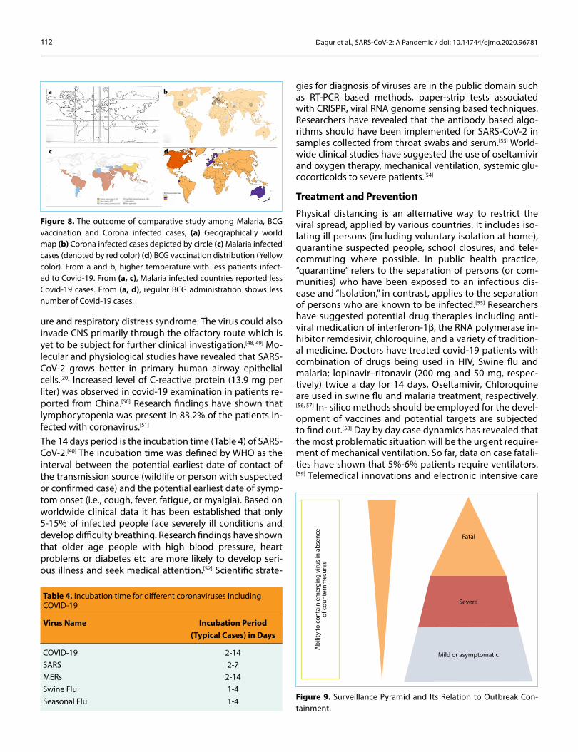
perature, antimalarial drugs and regular BCG vaccination (Fig. 8).[45–47]
Diagnosis
Covid-19 is pandemic in the human population and causes both upper and lower respiratory tract disease to be asso-ciated with more severe illnesses, such as bronchitis, bron-chiolitis, pneumonia, exacerbations of asthma, organ fail-
Figure 6. Transmission history of SARS-CoV-2 across the world.
Figure 7. Local transmission among the human population. (a) Droplets containing coronavirus, (b) Person in droplet contact, (c) Virus entry in person through mouth and nasal route, (d) Virus infected person, (e, g) Infected person spread the virus through sneezing or coughing, (h) Person to person transmission of virus through arial droplet, (f) Infected person transfers the viral load on the surface.
Table 3. Death rate among different age groups
Age (in years) Death Rate (%)
80+ 14.8070-79 860-69 3.6050-59 1.3040-49 0.4030-39 0.2020-29 0.2010-19 0.200-9 No fatalities
Death Rate = (number of deaths/number of cases) = probability of dying if infected by the virus (%). This probability differs depending on the age group. The percentages shown in the table do not have to add up to 100%, as they do NOT represent the share of deaths by age group. Rather, it represents, for a person in a given age group, the risk of dying if infected with COVID-19.
112 Dagur et al., SARS-CoV-2: A Pandemic / doi: 10.14744/ejmo.2020.96781
ure and respiratory distress syndrome. The virus could also invade CNS primarily through the olfactory route which is yet to be subject for further clinical investigation.[48, 49] Mo-lecular and physiological studies have revealed that SARS-CoV-2 grows better in primary human airway epithelial cells.[20] Increased level of C-reactive protein (13.9 mg per liter) was observed in covid-19 examination in patients re-ported from China.[50] Research findings have shown that lymphocytopenia was present in 83.2% of the patients in-fected with coronavirus.[51] The 14 days period is the incubation time (Table 4) of SARS-CoV-2.[40] The incubation time was defined by WHO as the interval between the potential earliest date of contact of the transmission source (wildlife or person with suspected or confirmed case) and the potential earliest date of symp-tom onset (i.e., cough, fever, fatigue, or myalgia). Based on worldwide clinical data it has been established that only 5-15% of infected people face severely ill conditions and develop difficulty breathing. Research findings have shown that older age people with high blood pressure, heart problems or diabetes etc are more likely to develop seri-ous illness and seek medical attention.[52] Scientific strate-
gies for diagnosis of viruses are in the public domain such as RT-PCR based methods, paper-strip tests associated with CRISPR, viral RNA genome sensing based techniques. Researchers have revealed that the antibody based algo-rithms should have been implemented for SARS-CoV-2 in samples collected from throat swabs and serum.[53] World-wide clinical studies have suggested the use of oseltamivir and oxygen therapy, mechanical ventilation, systemic glu-cocorticoids to severe patients.[54]
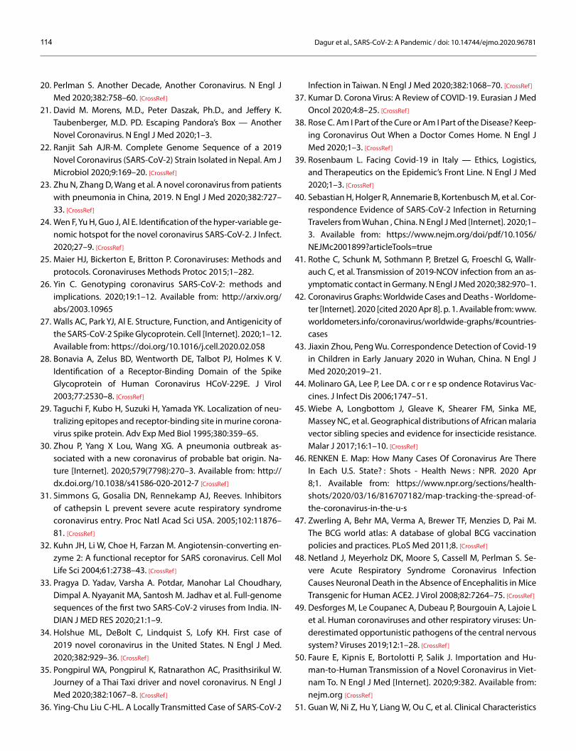
Treatment and Prevention Physical distancing is an alternative way to restrict the viral spread, applied by various countries. It includes iso-lating ill persons (including voluntary isolation at home), quarantine suspected people, school closures, and tele-commuting where possible. In public health practice, “quarantine” refers to the separation of persons (or com-munities) who have been exposed to an infectious dis-ease and “Isolation,” in contrast, applies to the separation of persons who are known to be infected.[55] Researchers have suggested potential drug therapies including anti-viral medication of interferon-1β, the RNA polymerase in-hibitor remdesivir, chloroquine, and a variety of tradition-al medicine. Doctors have treated covid-19 patients with combination of drugs being used in HIV, Swine flu and malaria; lopinavir–ritonavir (200 mg and 50 mg, respec-tively) twice a day for 14 days, Oseltamivir, Chloroquine are used in swine flu and malaria treatment, respectively.[56, 57] In- silico methods should be employed for the devel-opment of vaccines and potential targets are subjected to find out.[58] Day by day case dynamics has revealed that the most problematic situation will be the urgent require-ment of mechanical ventilation. So far, data on case fatali-ties have shown that 5%-6% patients require ventilators.[59] Telemedical innovations and electronic intensive care
Figure 9. Surveillance Pyramid and Its Relation to Outbreak Con-tainment.
Abi
lity
to c
onta
in e
mer
ging
viru
s in
abs
ence
of c
ount
ernm
esur
es
Mild or asymptomatic
Severe
Fatal
Figure 8. The outcome of comparative study among Malaria, BCG vaccination and Corona infected cases; (a) Geographically world map (b) Corona infected cases depicted by circle (c) Malaria infected cases (denoted by red color) (d) BCG vaccination distribution (Yellow color). From a and b, higher temperature with less patients infect-ed to Covid-19. From (a, c), Malaria infected countries reported less Covid-19 cases. From (a, d), regular BCG administration shows less number of Covid-19 cases.
a
c
b
d
Table 4. Incubation time for different coronaviruses including COVID-19
Virus Name Incubation Period (Typical Cases) in Days
COVID-19 2-14SARS 2-7MERs 2-14Swine Flu 1-4Seasonal Flu 1-4

113EJMO
unit (e-ICU) monitoring programs are ideal to fight Co-vid-19.[60] Epidemiologic investigations, risk assessment, surveillance, rapid diagnostic tests and vigilance (Fig. 9) are required by all countries to combat the potential outbreak.[61] In this pandemic, specific recommendations have been suggested for allocating medical resources such as maximize benefits, prioritize health workers, be responsive to evidence and recognize research participa-tion.[62]
ConclusionBoth N and S proteins are the most abundant proteins in SARS-CoV-2 and can be potential targets of covid-19. Com-bined studies on antimalarial drugs, regular administration of BCG vaccine, and geographical variation in temperature can help researchers to find potential solutions to combat Covid-19. The Covid-19 pandemic caused worldwide short-age of hospital beds, ICU beds, and ventilators. More and more epidemiologic studies are needed to address risk factors for infection, severity of disease and the efficacy of infection-control measures. These types of studies should help us refine and focus our efforts to control and treat Covid-19. The SARS-CoV-2 has a large economic and social impact on the human population. Several aspects of the covid-19 outbreak are not yet fully understood, including transmission dynamics and the full spectrum of clinical ill-ness. We need to save lives now while also improving the way we respond to outbreaks in general. Governments and industries will need to come to an agreement: during a pandemic, vaccines and antivirals can’t simply be sold to the highest bidder. Prevention is the ultimate solution for saving lives from such a pandemic.
Disclosures
Peer-review: Externally peer-reviewed.
Conflict of Interest: None declared.
Acknowledgement: We acknowledge Mr. Kaushal Singh Dhaked (Master of Fine Art) for designing figure of local trans-mission among the human population. We would like to thank Mr. Prem Pal Singh (PhD in Electronics at JNU, Jaipur), Mr. Chan-dra Pal Uikey and Ms. Pooja Dhaker for their support. We dedi-cate this review article to the health community for fighting against SARS-CoV-2.
References1. Coronavirus Update (Live): 1,621,771 Cases and 97,185 Deaths
from COVID-19 Virus Pandemic - Worldometer [Internet]. [cit-ed 2020 Apr 9]. p. 1. Available from: www.worldometers.info/coronavirus/
2. Antonio GE, Wong KT, Hui DSC, Lee N, Yuen EHY, Wu A, et al. Imaging of severe acute respiratory syndrome in Hong Kong. Am J Roentgenol 2003;181:11–7. [CrossRef ]
3. Lefkowitz EJ, Dempsey DM, Hendrickson RC, Orton RJ, Siddell
SG, Smith DB. Virus taxonomy: The database of the Interna-tional Committee on Taxonomy of Viruses (ICTV). Nucleic Ac-ids Res 2018;46(D1):D708–17. [CrossRef ]
4. Li Q, Guan X, Wu P, Wang X, Zhou L, Tong Y, et al. Early Trans-mission Dynamics in Wuhan, China, of Novel Coronavirus-In-fected Pneumonia. N Engl J Med 2020;382:1199–207. [CrossRef ]
5. Munster VJ, Koopmans M, van Doremalen N, van Riel D, de Wit E. A novel coronavirus emerging in China - Key questions for impact assessment. N Engl J Med 2020;382:692–4. [CrossRef ]
6. Patients L, Taylor D, Lindsay AC, Halcox JP. correspondence Niacin Compared with Ezetimibe. N Engl J Med 2020;0–3.
7. Nelson Lee, David Hui, Alan Wu, Paul Chan, Peter Cameron, et al. A Major Outbreak of Severe Acute Respiratory Syndrome in Hong Kong. N Engl J Med 2003;1986–94. [CrossRef ]
8. Assiri A, McGeer A, Perl TM, Price CS, Al Rabeeah AA, et al. Hos-pital outbreak of middle east respiratory syndrome coronavi-rus. N Engl J Med 2013;369:407–16. [CrossRef ]
9. Memish ZA, Zumla AI, Al-Hakeem RF, Al-Rabeeah AA, Stephens GM. Family cluster of middle east respiratory syndrome corona-virus infections. N Engl J Med 2013;368:2487–94. [CrossRef]
10. Azhar EI, El-Kafrawy SA, Farraj SA, Hassan AM, Al-Saeed MS, et al. Evidence for camel-to-human transmission of MERS coro-navirus. N Engl J Med 2014;370:2499–505. [CrossRef ]
11. Cui J, Li F, Shi ZL. Origin and evolution of pathogenic coronavi-ruses. Nat Rev Microbiol [Internet]. 2019;17:181–92. Available from: http://dx.doi.org/10.1038/s41579-018-0118-9 [CrossRef ]
12. Zhang T, Wu Q, Zhang Z. Probable Pangolin Origin of SARS-CoV-2 Associated with the COVID-19 Outbreak. Curr Biol [In-ternet]. 2020;1–6. Available from: https://doi.org/10.1016/j.cub.2020.03.022 [CrossRef ]
13. Coronavirus [Internet]. [cited 2020 Apr 10]. p. 1. Available from: www.who.int/health-topics/coronavirus#tab=tab_3
14. Drosten C, Günther S, Preiser W, Van der Werf S, Brodt HR, et al. Identification of a novel coronavirus in patients with severe acute respiratory syndrome. N Engl J Med 2003;348:1967–76.
15. Gates B. New engla nd journal. 2020;1–3. 16. Duan H WS& YC. Coronavirus: limit short-term economic dam-
age. Nature [Internet]. 2020;7796. Available from: https://www.nature.com/articles/d41586-020-00522-6 [CrossRef ]
17. Gorbalenya AE, Baker SC, Baric RS, de Groot RJ, Drosten C, et al. The species Severe acute respiratory syndrome-related coro-navirus: classifying 2019-nCoV and naming it SARS-CoV-2. Nat Microbiol 2020;5(March). [CrossRef ]
18. Ren L-L, Wang Y-M, Wu Z-Q, Xiang Z-C, Guo L, Xu T, et al. Iden-tification of a novel coronavirus causing severe pneumonia in human. Chin Med J (Engl). 2020;1.
19. Lu R, Zhao X, Li J, Niu P, Yang B, et al. Genomic characterisa-tion and epidemiology of 2019 novel coronavirus: implica-tions for virus origins and receptor binding. Lancet [Internet]. 2020;395:565–74. Available from: http://dx.doi.org/10.1016/S0140-6736(20)30251-8 [CrossRef ]
114 Dagur et al., SARS-CoV-2: A Pandemic / doi: 10.14744/ejmo.2020.96781
20. Perlman S. Another Decade, Another Coronavirus. N Engl J Med 2020;382:758–60. [CrossRef ]
21. David M. Morens, M.D., Peter Daszak, Ph.D., and Jeffery K. Taubenberger, M.D. PD. Escaping Pandora’s Box — Another Novel Coronavirus. N Engl J Med 2020;1–3.
22. Ranjit Sah AJR-M. Complete Genome Sequence of a 2019 Novel Coronavirus (SARS-CoV-2) Strain Isolated in Nepal. Am J Microbiol 2020;9:169–20. [CrossRef ]
23. Zhu N, Zhang D, Wang et al. A novel coronavirus from patients with pneumonia in China, 2019. N Engl J Med 2020;382:727–33. [CrossRef ]
24. Wen F, Yu H, Guo J, Al E. Identification of the hyper-variable ge-nomic hotspot for the novel coronavirus SARS-CoV-2. J Infect. 2020;27–9. [CrossRef ]
25. Maier HJ, Bickerton E, Britton P. Coronaviruses: Methods and protocols. Coronaviruses Methods Protoc 2015;1–282.
26. Yin C. Genotyping coronavirus SARS-CoV-2: methods and implications. 2020;19:1–12. Available from: http://arxiv.org/abs/2003.10965
27. Walls AC, Park YJ, Al E. Structure, Function, and Antigenicity of the SARS-CoV-2 Spike Glycoprotein. Cell [Internet]. 2020;1–12. Available from: https://doi.org/10.1016/j.cell.2020.02.058
28. Bonavia A, Zelus BD, Wentworth DE, Talbot PJ, Holmes K V. Identification of a Receptor-Binding Domain of the Spike Glycoprotein of Human Coronavirus HCoV-229E. J Virol 2003;77:2530–8. [CrossRef ]
29. Taguchi F, Kubo H, Suzuki H, Yamada YK. Localization of neu-tralizing epitopes and receptor-binding site in murine corona-virus spike protein. Adv Exp Med Biol 1995;380:359–65.
30. Zhou P, Yang X Lou, Wang XG. A pneumonia outbreak as-sociated with a new coronavirus of probable bat origin. Na-ture [Internet]. 2020;579(7798):270–3. Available from: http://dx.doi.org/10.1038/s41586-020-2012-7 [CrossRef ]
31. Simmons G, Gosalia DN, Rennekamp AJ, Reeves. Inhibitors of cathepsin L prevent severe acute respiratory syndrome coronavirus entry. Proc Natl Acad Sci USA. 2005;102:11876–81. [CrossRef ]
32. Kuhn JH, Li W, Choe H, Farzan M. Angiotensin-converting en-zyme 2: A functional receptor for SARS coronavirus. Cell Mol Life Sci 2004;61:2738–43. [CrossRef ]
33. Pragya D. Yadav, Varsha A. Potdar, Manohar Lal Choudhary, Dimpal A. Nyayanit MA, Santosh M. Jadhav et al. Full-genome sequences of the first two SARS-CoV-2 viruses from India. IN-DIAN J MED RES 2020;21:1–9.
34. Holshue ML, DeBolt C, Lindquist S, Lofy KH. First case of 2019 novel coronavirus in the United States. N Engl J Med. 2020;382:929–36. [CrossRef ]
35. Pongpirul WA, Pongpirul K, Ratnarathon AC, Prasithsirikul W. Journey of a Thai Taxi driver and novel coronavirus. N Engl J Med 2020;382:1067–8. [CrossRef ]
36. Ying-Chu Liu C-HL. A Locally Transmitted Case of SARS-CoV-2
Infection in Taiwan. N Engl J Med 2020;382:1068–70. [CrossRef ]
37. Kumar D. Corona Virus: A Review of COVID-19. Eurasian J Med Oncol 2020;4:8–25. [CrossRef ]
38. Rose C. Am I Part of the Cure or Am I Part of the Disease? Keep-ing Coronavirus Out When a Doctor Comes Home. N Engl J Med 2020;1–3. [CrossRef ]
39. Rosenbaum L. Facing Covid-19 in Italy — Ethics, Logistics, and Therapeutics on the Epidemic’s Front Line. N Engl J Med 2020;1–3. [CrossRef ]
40. Sebastian H, Holger R, Annemarie B, Kortenbusch M, et al. Cor-respondence Evidence of SARS-CoV-2 Infection in Returning Travelers from Wuhan , China. N Engl J Med [Internet]. 2020;1–3. Available from: https://www.nejm.org/doi/pdf/10.1056/NEJMc2001899?articleTools=true
41. Rothe C, Schunk M, Sothmann P, Bretzel G, Froeschl G, Wallr-auch C, et al. Transmission of 2019-NCOV infection from an as-ymptomatic contact in Germany. N Engl J Med 2020;382:970–1.
42. Coronavirus Graphs: Worldwide Cases and Deaths - Worldome-ter [Internet]. 2020 [cited 2020 Apr 8]. p. 1. Available from: www.worldometers.info/coronavirus/worldwide-graphs/#countries-cases
43. Jiaxin Zhou, Peng Wu. Correspondence Detection of Covid-19 in Children in Early January 2020 in Wuhan, China. N Engl J Med 2020;2019–21.
44. Molinaro GA, Lee P, Lee DA. c or r e sp ondence Rotavirus Vac-cines. J Infect Dis 2006;1747–51.
45. Wiebe A, Longbottom J, Gleave K, Shearer FM, Sinka ME, Massey NC, et al. Geographical distributions of African malaria vector sibling species and evidence for insecticide resistance. Malar J 2017;16:1–10. [CrossRef ]
46. RENKEN E. Map: How Many Cases Of Coronavirus Are There In Each U.S. State? : Shots - Health News : NPR. 2020 Apr 8;1. Available from: https://www.npr.org/sections/health-shots/2020/03/16/816707182/map-tracking-the-spread-of-the-coronavirus-in-the-u-s
47. Zwerling A, Behr MA, Verma A, Brewer TF, Menzies D, Pai M. The BCG world atlas: A database of global BCG vaccination policies and practices. PLoS Med 2011;8. [CrossRef ]
48. Netland J, Meyerholz DK, Moore S, Cassell M, Perlman S. Se-vere Acute Respiratory Syndrome Coronavirus Infection Causes Neuronal Death in the Absence of Encephalitis in Mice Transgenic for Human ACE2. J Virol 2008;82:7264–75. [CrossRef ]
49. Desforges M, Le Coupanec A, Dubeau P, Bourgouin A, Lajoie L et al. Human coronaviruses and other respiratory viruses: Un-derestimated opportunistic pathogens of the central nervous system? Viruses 2019;12:1–28. [CrossRef ]
50. Faure E, Kipnis E, Bortolotti P, Salik J. Importation and Hu-man-to-Human Transmission of a Novel Coronavirus in Viet-nam To. N Engl J Med [Internet]. 2020;9:382. Available from: nejm.org [CrossRef ]
51. Guan W, Ni Z, Hu Y, Liang W, Ou C, et al. Clinical Characteristics

115EJMO
of Coronavirus Disease 2019 in China. N Engl J Med 2020;1–13. 52. WHO | Novel Coronavirus [Internet]. 2020 [cited 2020 Apr 10].
Available from: www.who.int/csr/don/archive/disease/novel_coronavirus/en/
53. Drosten C, Meyer B, Müller MA, Corman VM, Al-Masri et al. Transmission of MERS-coronavirus in household contacts. N Engl J Med 2014;371:828–35. [CrossRef ]
54. Mitjà O, Clotet B. Use of antiviral drugs to reduce COVID-19 transmission. Lancet Glob Heal [Internet]. 2020;2019–20. Avail-able from: http://dx.doi.org/10.1016/S2214-109X(20)30114-5
55. Rothstein MA, Coughlin CN. Ensuring compliance with quar-antine by undocumented immigrants and other vulner-able groups: Public health versus politics. Am J Public Health. 2019;109:1179–83. [CrossRef ]
56. Cao B, Wang Y, Wen D, Liu W, Wang J, Fan G. A Trial of Lopina-vir–Ritonavir in Adults Hospitalized with Severe Covid-19. N Engl J Med 2020;1–13.
57. The Times of India. Rajasthan doctors cure coronavirus patient with HIV drugs. 2020;1–21. Available from: https://timesofind-
ia.indiatimes.com/city/jaipur/city-docs-cure-corona-patient-with-hiv-drugs/articleshow/74584859.cms
58. Seema M. T Cell Epitope-Based Vaccine Design for Pan-demic Novel Coronavirus 2019-nCoV. Chemrxiv [Internet]. 2020; Available from: https://chemrxiv.org/articles/T_Cell_Epitope-Based_Vaccine_Design_for_Pandemic_Novel_Coronavirus_2019-nCoV/12029523
59. Robert D. Truog, Christine Mitchell, R.N., and George Q. Daley MD. The Toughest Triage — Allocating Ventilators in a Pan-demic. N Engl J Med 2020;1–3.
60. Judd E. Hollander, M.D., and Brendan G. Carr MD. Perspective: Virtually Perfect? Telemedicine for Covid-19. 2020;1–3. Avail-able from: NEJM.org [CrossRef ]
61. Zaki AM, Van Boheemen S, Bestebroer TM, Osterhaus ADME, Fouchier RAM. Isolation of a novel coronavirus from a man with pneumonia in Saudi Arabia. N Engl J Med 2012;367:1814–20.
62. Emanuel EJ, Persad G, Upshur R, Thome B, Parker M et al. Fair Allocation of Scarce Medical Resources in the Time of Co-vid-19. N Engl J Med 2020;1–7. [CrossRef ]