GENITOURINARY SYSTEM Michelle Gardner NUR-224. URINARY SYSTEM.
99
GENITOURINARY SYSTEM Michelle Gardner NUR-224
-
Upload
joel-hoover -
Category
Documents
-
view
218 -
download
3
Transcript of GENITOURINARY SYSTEM Michelle Gardner NUR-224. URINARY SYSTEM.

GENITOURINARY SYSTEM
Michelle GardnerNUR-224

URINARY SYSTEM

ASSESSMENT OF THE URINARY SYSTEM
Subjective Dataa. Good communication skillsb. Avoid medical terminologyc. Anxiety/embarrassment –
“forget/deny”--

ASSESSMENT DATA
Past Health Historya. Presence/history of diseases r/t
urologic problems – DM, HTNb. Neurologic conditions – back injury,
stroke, traumac. Urinary problems – BPH, renal
calculi, cancer, infection
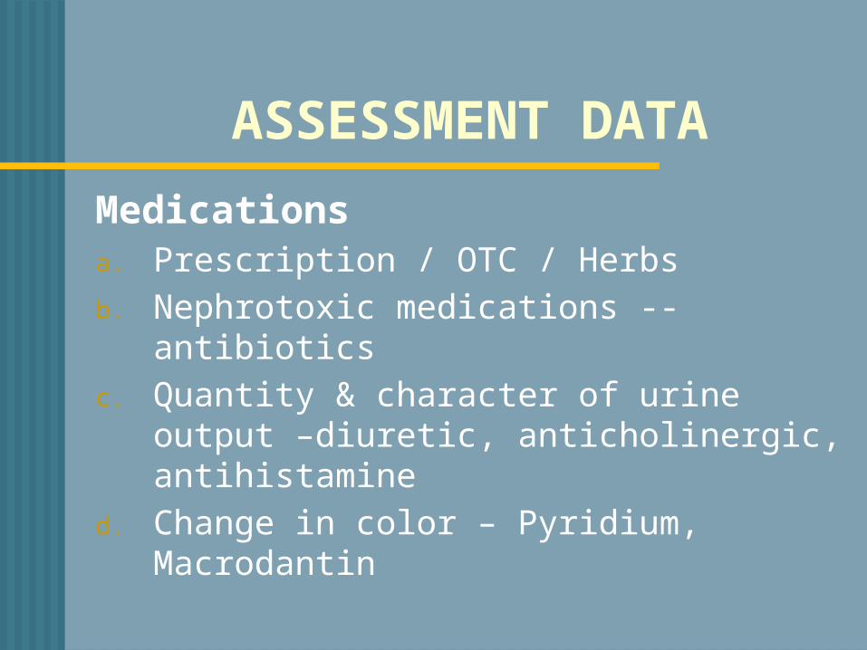
ASSESSMENT DATA
Medicationsa. Prescription / OTC / Herbsb. Nephrotoxic medications -- antibioticsc. Quantity & character of urine output –
diuretic, anticholinergic, antihistamined. Change in color – Pyridium, Macrodantin
ASSESSMENT DATA
Surgerya. Previous hospitalizations r/t
urologic diseaseb. Pelvic surgeriesc. Urinary instrumentationd. Urinary problems during past
pregnanciese. Radiation/chemotherapy

ASSESSMENT DATA Pain Changes in voiding Affects of aging on the urinary
system a. Decrease muscle tone b. Decrease bladder capacity c. Prostate enlargement
d. Changes in metabolism
BLOOD CHEMISTRIES
Blood Chemistries Serum Creatinine: 0.6 – 1.2mg/dlo End product of muscle & protein
metabolismo Excellent indicator of kidney functiono Renal disease results in increase
creatinine

BLOOD CHEMISTRIES
BUN/Blood Urea Nitrogen: 7-18mg/dlo Used to identify renal problemso Nonrenal factors may increase BUN a. Fever b. Dehydration c. High protein diet d. Athletic activity e. Drugs and vitamins (acetaminophen,
ibuprofen, vitamin D)
DIAGNOSTIC STUDIES KUB (kidneys, ureters, bladder)o X- ray exam of abdomen & pelviso Used to detect abnormalities
o Urinary calculi o Cystso Tumorso Hydronephrosis

DIAGNOSTIC STUDIES IVP (INTRAVENOUS PYLEOGRAM)
Urographyo Intravenous injection of radiopaque
imaging dyeo X-ray imaging of dye through upper
and lower urinary system

INTRAVENOUS UROGRAPHY

INTRAVENOUS UROGRAPHY
Patient preparation:o Consent form o Cathartic/enema the night beforeo Identify allergies – shellfish,
iodineo Pre-medicate–antihistamine
(Benadryl) o NPO 8 hr. before procedure o Transitory effects – contrast medium

INTRAVENOUS UROGRAPHY
Post-procedureo Monitor vital signso Assess for s/s anaphylactic reactionso Monitor urine outputo Force fluids

RENAL ANGIOGRAPHY RENAL ANGIOGRAM:o Catheter inserted into femoral arteryo Contrast material injected through the
cathetero Visualize renal blood vessels Findings :1. Renal artery stenosis2. Differentiate renal cysts from tumors3. Evaluate hypertension
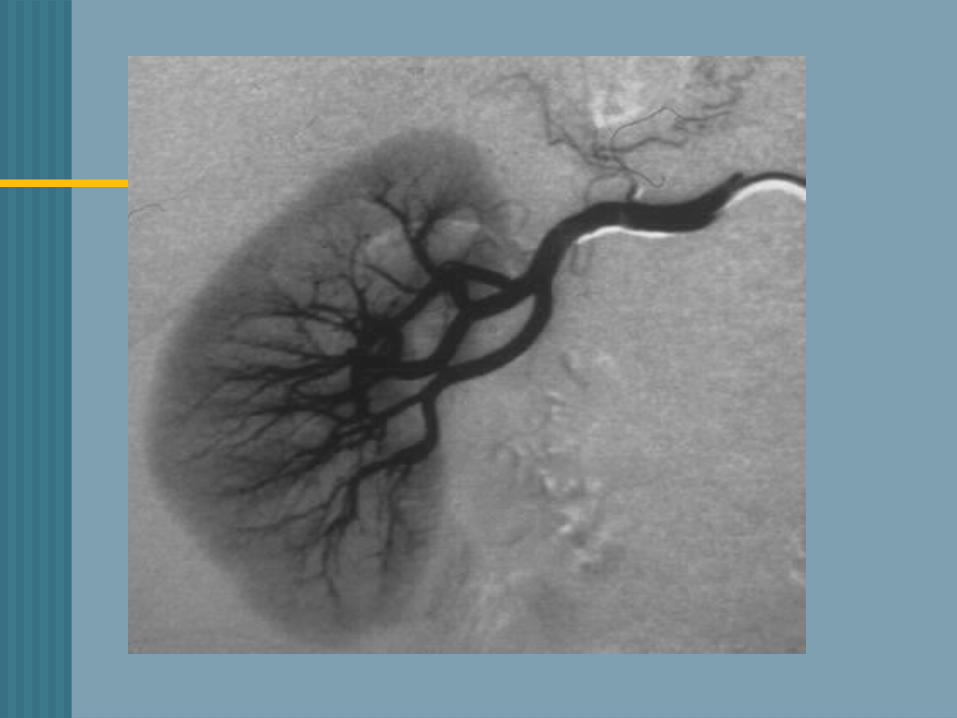

RENAL ANGIOGRAPHY Patient preparationo Consent formo Cathartic/enema the evening beforeo Assess allergic reactiono Mark peripheral pulses

RENAL ANGIOGRAPHY Post-Procedureo Monitor vital signso Pressure dressing over insertion siteo Assess insertion site - o Bedrest with affected leg straighto Palpate peripheral pulses

RENAL BIOPSY Done as a needle biopsy with needle
insertion into lower lobe of the kidney OR open biopsy via small flank incision
o Obtain renal tissue to determine type of renal disease
o Kidneys are vascular organs – hemorrhage/complication

RENAL BIOPSY
Patient preparationo Consent form signedo NPO status 8 hrs. prior to testo Assess baseline coagulation statuso Medications that may alter clotting
function

RENAL BIOPSY

RENAL BIOPSYPost-Procedureo Pressure dressing appliedo Check puncture site – swelling/tendernesso Prone position for 30-60 minuteso Monitor vital signso Observe for gross bleedingo Assess for flank pain, Hgb./Hct. levelso Avoid lifting heavy object/strenuous
activity – 7 days

UROLOGIC ENDOSCOPIC PROCEDURES
o Visualize/inspect the interior of the urethra and bladder with a tubular lighted scope (cystoscope)
o Used to:a. Treat bleeding lesionsb. Insert ureteral cathetersc. Remove calculi d. Obtain biopsy specimens


CYSTOSCOPY Patient preparationo Signed consent formo NPO prior to the procedureo Local topical anesthetic o Lithotomy position – leg cramps

CYSTOSCOPY Post-procedureo Expected side effects - burning on
urination, blood-tinged urine, urinary frequency
o Encourage increased fluids o Warm sitz bath o Mild analgesics


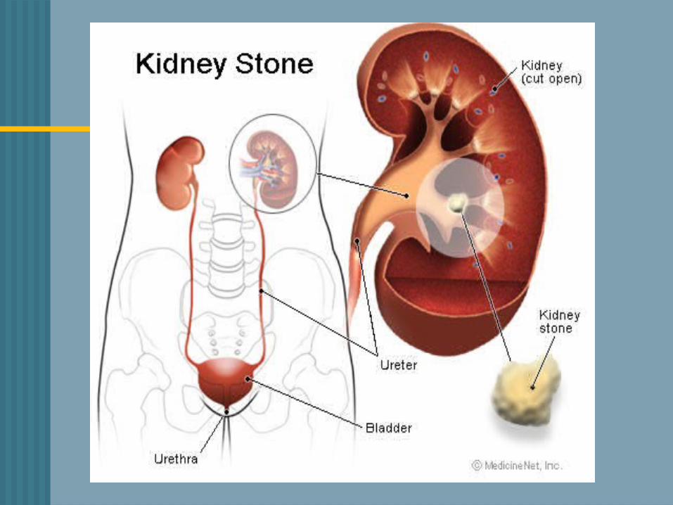
RENAL CALCULI

UROLITHIASIS/NEPHROLITHIASIS
500,000 people in the U.S. have kidney stone disease
Incidence is highest in Southern & Midwest states.
Occurs between the 3rd-5th decade of life. Recurrence of stones – 50% of pts. More common in men than in women
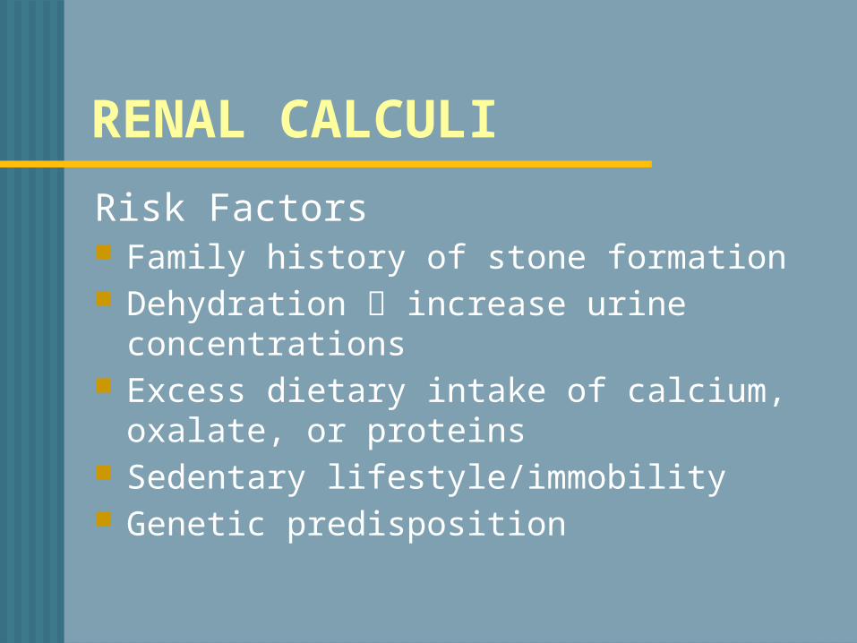
RENAL CALCULI
Risk Factors Family history of stone formation Dehydration increase urine
concentrations Excess dietary intake of calcium, oxalate,
or proteins Sedentary lifestyle/immobility Genetic predisposition

RENAL CALCULIo Stones can be found anywhere from
kidney to bladdero Vary in size o Factors that contribute to urolithiasis * supersaturation * nucleation

RENAL CALCULI
Pathophysiology Concentration of an insoluble salt is
high in the urine supersaturation Crystals form from supersaturated
urine Growth continues by aggregation to
form larger particles – stone formation

RENAL CALCULI

RENAL CALCULI 4 Major Categories of Stones1. Calcium 2. Oxalate3. Uric acid4. Cystine
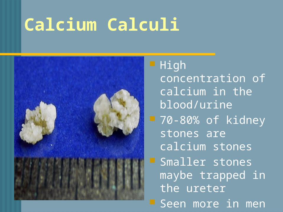
Calcium Calculi
High concentration of calcium in the blood/urine
70-80% of kidney stones are calcium stones
Smaller stones maybe trapped in the ureter
Seen more in men

Calcium Calculi (Oxalate)Risk factors Hypercalciuria/hypercalcemia,
immobility, vit.D, urine intoxication, dehydration
Management Thiazide diuretics Limit foods that acidify urine Hydration/exercise

Uric Acid Stones

Uric Acid Calculio Urine
concentration of uric acid is high
o Common in menCauses: 1.Gout2. Increased
dietary intake of purine
3.Acid urine
1. Reduce dietary purines– sardines, mussels, organ meats, aged cheese
2. Administer allopurinol (Zyloprim)
3. Reduce urinary concentration of uric acid

Struvite Calculi

Struvite Calculi (Staghorn) 15-20 % of stones -
magnesium/ammonium/phosphateRisk Factors UTIs, esp Proteus infections Stones are large fill renal pelvisManagement Antibiotics Surgical intervention/lithotripsy

Cystine Calculio Make up 1-2% of
all stoneso Caused by
genetic defecto Tend to form in
acid urineo Stones appear
during childhood / adolescence
o Rare in adults
o Increase hydrationo Low-protein diet
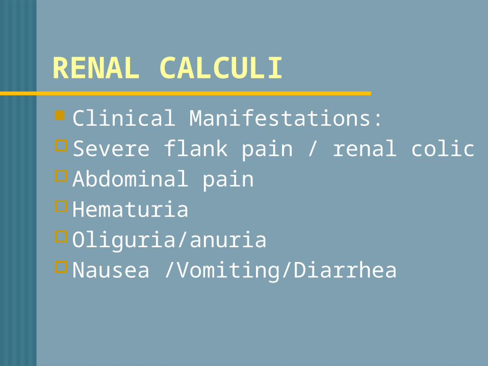
RENAL CALCULI Clinical Manifestations:o Severe flank pain / renal colic o Abdominal paino Hematuria o Oliguria/anuria o Nausea /Vomiting/Diarrhea
RENAL CALCULIDiagnostic Studies:o Urinalysiso 24 hr urinary measurement for calcium,
uric acido X-ray - KUB o Renal Ultrasonographyo CT Scan
RENAL CALCULIManagement
Pain management o Opiod analgesics – Morphineo NSAID Toradalo Comfort measureso Increase fluid intake (oral/intravenous)

RENAL CALCULI Stones may pass spontaneously Stones larger than 4mm are unlikely
to pass through the ureter Chemical analysis of the stone to
determine the composition of the stone
STRAIN ALL URINE
RENAL CALCULI THERAPUETIC INTERVENTIONS ESWL-Extracorporeal shock-wave lithotripsyo Non-invasive procedureo External shock-waves break up the stone o No damage to surrounding tissueo Stones are fragmented into fine sando Fragments are excreted in the urineo All urine is strained -- chemical analysiso Anesthesia is necessary


RENAL CALCULIo Cystoscopy passed – removes stones
located in the ureter close to the bladder o Stone removed -- grasping basket, forcepso Stent may be placedo Foley catheter -- facilitate passage stone
fragments o Minimal complications


RENAL CALCULI After episode of urolithiasis a. Increase fluid intake – 3000ml/day b. High urine output – 2L/day c. Water is the preferred fluid d. Avoid tea, coffee, colas e. Limit foods high in oxalate, calcium, & purinesSTRAIN ALL URINE -

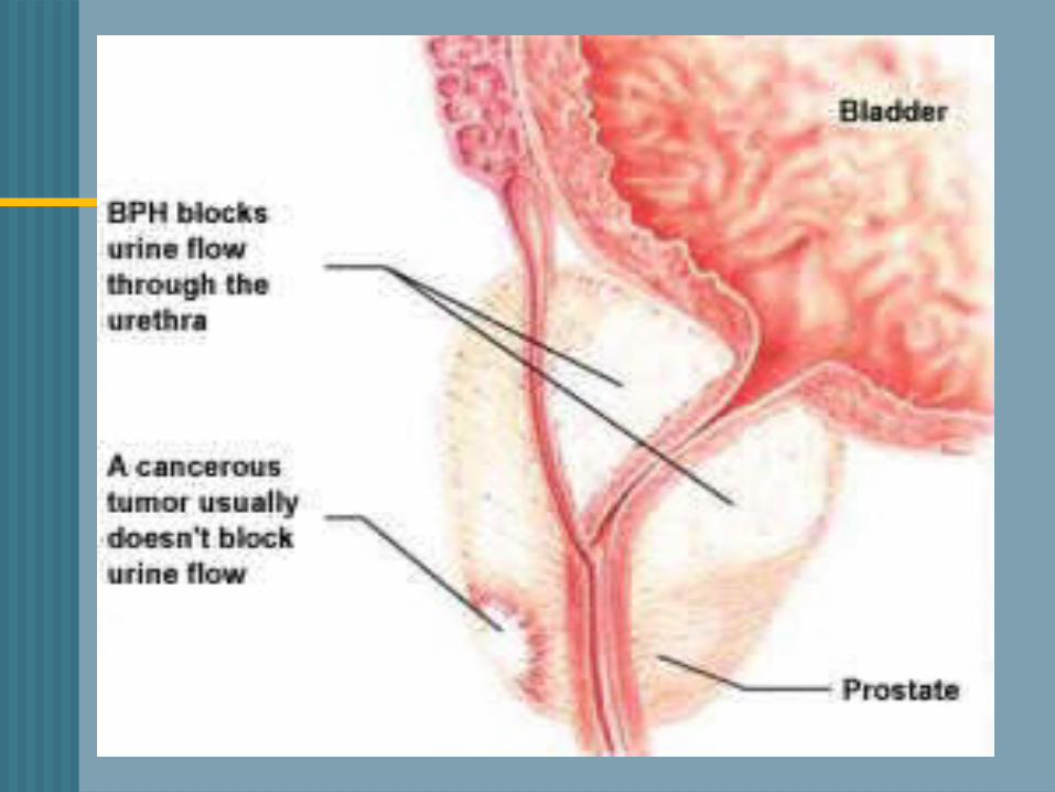
BENIGN PROSTATIC HYPERPLASIA (BPH) Age–related, nonmalignant enlargement
of the prostate gland• Enlargement of the prostate gland --
compress the urethra/bladder This impedes the normal flow of urine Begins at the age of 40 and continues
slowly throughout the rest of life Symptoms appear slightly earlier in Afro-
American men
BPH
Begins with small layers in the periuretheral gland
Prostate enlarges through formation /growth of nodules and enlargement of glandular cells
Enlargement compresses against the urethra urologic symptoms
Changes occur over a long period of time
BPH
Clinical Manifestations Difficulty starting urinary stream Urinary frequency Nocturia Leakage or dribbling of urine Urgency

BPH
Complications Urinary retention Urinary tract infections Bladder stones

BPHDiagnostic Studies History & physical exam Urinalysis/ C&S Digital rectal exam (DRE) Prostatic Specific Antigen(PSA) -- R/O Prostate Cancer Serum Creatinine
MEDICATION THERAPY
ALPHA-ADRENERGIC BLOCKERS
Relax the smooth muscle of the bladder neck and prostate
• Improves urine flow• Relax smooth muscle of the prostate

BPH
ALPHA-ADRENERGIC BLOCKERS Flomax- (tamsulosin) Cardura - (doxazosin) Hytrin – (terazosin) Uroxatral – (alfuzosin)Side effect
orthostatic hypotension dizziness
BPH
5 ALPHA-REDUCTASE INHIBITORS Decreases the size of the prostate
gland Proscar (finasteride) Avodart – (dutasteride) Side effect - *decreases libido, *erectile dysfunction
BPH
Minimally Invasive Therapy
used when medication not effective relieves the manifestations of BPH less invasive than traditional surgery

BPH Transuretheral Needle Ablation
(TUNA)
Low-wave radio frequency – to burn away a region of the enlarged prostate
Improves the flow of urine 70% of pt. show marked improvement Little pain Early return to regular activities

TUNA
BPHTransuretheral Resection of the
Prostate (TURP) Removal of inner prostate tissue Most common procedureAdvantages1. No external incision made2. Shorter hospitalization3. Complications – clot retention,
hemorrhage, infection, catheter obstruction

TURP

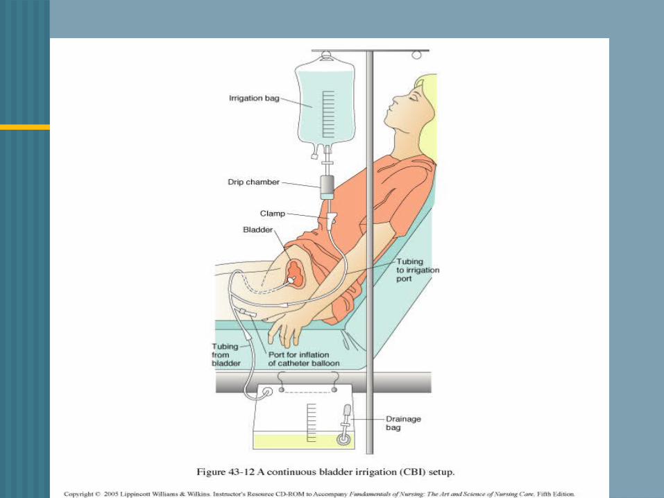
CONTINOUS BLADDER IRRIGATION
3- way drainage system- useful in irrigating the bladder & preventing clot formation
3000 ml sterile normal saline Irrigation -- consist of continuous inflow &
outflow of solution & drainage Maintain patency of catheter & tubing Urine drainage – light pink Blood clots are expected 1st 24-36hrs.
after surgery

CBI
Catheter removal – assess amount, color and
consistency of urine may experience burning on
urination, dribbling is common

BPH
Complications Hemorrhage
Obstructed catheter
Urinary incontinence

URINARY DIVERSIONS Procedure performed to divert urine
from the bladder to a new exit site – STOMA
Used to treat a. Cancer of the bladder b. Congenital anomalies c. Trauma to the bladder d. Neurogenic bladder

URINARY DIVERSIONS 2 CATERGORIES1. Incontinent urinary diversion2. Continent urinary diversion
URINARY DIVERSIONS INCONTINENT DIVERSION• Urine drains through an opening
created in the abdominal wall• An appliance is needed • Most common – Ileal Conduit

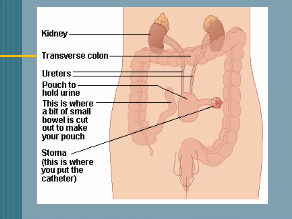
URINARY DIVERSIONS INCONTINENT DIVERSIONS – ileal
conduit• Ureters are excised from the bladder
& resected to a part of the ileum• Proximal end is sewn closed• Distal end created to form a stoma• Remaining intestinal segments –
anatomosed


URINARY DIVERSIONS INCONTINENT DIVERSIONS• Stents -- prevent occlusion from
post-surgical edemaDisadvantages:• Requires a external collection device • Visible stoma

URINARY DIVERSIONS INCONTINENT DIVERSIONSPre-op Management• Discuss social aspects of living with a
stoma 1. Clothing2. Changes in body image3. Odor4. Sexuality 5. Exercise
URINARY DIVERSIONS
INCONTINENT DIVERSIONSPost-op Management• Assess for complications a. Paralytic ileus/SBO• Make sure urinary stents are draining• U/O < 30cc/hr – dehydration/obstruction• Hematuria –1st 24-48 hours• Mucous threads in urine – normal
occurrence

URINARY DIVERSIONS
Post-op management (cont’d)• Check stoma color– beefy red• Increase fld. intake• Empty pouch when 1/3 full/q2-3 hr.• Meticulous skin care • Avoid foods that give strong odor–
cheese, eggs, asparagus
URINARY DIVERSIONS CONTINENT DIVERSIONS• Intra-abdominal urinary reservoir• Self catheterize every 4-6 hours• No need external attachments • Reservoirs constructed from
different parts of the ileum/colon• Kock, Indiana, Charleston pouch

URINARY DIVERSION CONTINENT DIVERSIONSPost-op Management• Teach patient to catheterize pouch• Irrigate pouch • Adhere to strict catheterization
schedule• Enterostomal therapy nurse


QUESTION
A patient returns to the unit following a TURP . His urinary drainage bag is filled with dark red fluid with obvious bloods clots. And he is having bladder spasms. What would you do first?a. Assess his intake/output since surgeryb. Administer pain medication as orderedc. Report your assessment to the urologistd. Nothing, these are manifestations that are
expected following a TURP

QUESTION The nurse evaluates her teaching as
effective when a patient with a newly continent ileal diversion is able to do which of the following?
a. Demonstrate care for the collection deviceb. State the importance of reporting cloudy
urine to the physicianc. Demonstrate self-catherization of the stomad. Identify factors that contribute to this
condition


Urinary Tract Cancers
Prostate CancerCancer of the Bladder

Cancer of the Prostate Most common cancer among men
after skin cancer Highest incidence in African-
American men Risk Factors
increases rapidly after age 50 Family history High intake of red meat and high fat
dairy products

Cancer of the ProstateSigns and Symptoms
Often asymptomatic As malignancy enlarges, may have
symptoms of urinary obstruction Blood in urine, semen and painful
ejaculation may occur C/O back and hip pain, weight loss,
anemia, oliguria may indicate metastases

Cancer of the ProstateAssessment and Diagnosis
Screening tools DRE PSA
Normal: 0-4 ng/mL Transrectal Ultrasound (TRUS) Biopsy

Cancer of the ProstateTreatment
Surgical removal of the prostate TURP Laproscopic radical prostatectomy
Radiation Teletherapy Brachytherapy
Hormone Therapy Casodex DES
Chemotherapy

Cancer of the Bladder Most commonly seen in ages 50-70
Transitional-cell carcinoma of the bladder
Papilillomatous growths in the bladder Risk factors
Cigarette smoking (twice as much) Environmental carcinogens Frequent/recurrent bacterial infections History of urogenital cancers


Cancer of the BladderAssessment and Diagnosis
Hematuria Bladder irritability Pelvic or back pain Diagnostic tests:
Cystoscopy Ultrasound/CT Biopsies
Cancer of the BladderTreatment
Transurethral Resection of Bladder Tumor (TURBT)
Chemotherapy/Radiation BCG Methotrexate/5-FU/
vinblastin/Adriamycin Cystectomy
Cancer of the BladderTreatment
Transurethral Resection of Bladder Tumor (TURBT)
Chemotherapy/Radiation BCG Methotrexate/5-FU/
vinblastin/Adriamycin/cisplatin Cystectomy
Partial Radical