Genital tuberculosis in a Woman Manifested as Secondary ...
4
Journal of Diagnostic Pathology 2018;13(2):10-14 10 Case Report Genital tuberculosis in a Woman Manifested as Secondary Subfertility Wickramaratne E.K.D.M. 1 , Idirisinghe K.A.P. 2 , Samaranayake K.U. 2 1 Department of Pathology, Faculty of Medicine, University of Peradeniya, Sri Lanka. 2 District General Hospital, Nawalapitiya, Sri Lanka. DOI: http://doi.org/10.4038/jdp.v13i2.7757 Submitted on 21.04.2018 Accepted for publication on 03.08.2018 Summary A 35-year-old mother of one child, presented with inability to conceive for two years and a chronic lower abdominal pain. Laparoscopy revealed extensive adhesions in the bilateral adnexal regions and adhesion of uterus to the bowel. Subsequently, she underwent a total abdominal hysterectomy and bilateral salpingo-oophorectomy. Histopathological examination of endometrium, myometrium, both ovaries and fallopian tubes revealed several granulomata with caseous necrosis. Special stain for acid fast bacilli was negative. However, based on the presence of characteristic caseating granulomata, the diagnosis is most likely tuberculosis. Keywords: Genital tuberculosis, subfertility Introduction Tuberculosis (TB) is an infectious disease caused by the bacterium Mycobacterium tuberculosis, which remains as a major public health problem despite the advances in antibiotic treatment and vaccination. Tuberculosis can be categorized into pulmonary and extrapulmonary forms based on the affected organ. Genital tuberculosis (GTB) represents 15- 20% of cases of extrapulmonary TB and affects 12.1% of patients with pulmonary TB (1). GTB mainly affects women in the reproductive age group (20-40 years). Usually, this is a silent disease and the most common presentation amongst these patients is subfertility. In addition to that, menstrual disturbances, dyspareunia and abdominal pain may occur. Approximately 5-10% subfertile women worldwide have GTB. The worldwide incidence varies from less than 1% in developed countries to more than 13% in developing countries (2,3). In our literature review, accurate statistics of GTB among subfertile women in Sri Lanka could not be found. We report a case of a middle-aged female who was investigated for secondary subfertility and found to have GTB. Author for correspondence: E.K.D.M. Wickramaratne. Department of Pathology, Faculty of Medicine, University of Peradeniya, Sri Lanka. E-mail: [email protected] This is an open access article licensed under a Creative Commons Attribution-ShareAlike 4.0 International License (CC BY-SA 4.0), which permits unrestricted use, distribution and reproduction in any medium, provided the original author and source are attributed and materials are shared under the same license.
Transcript of Genital tuberculosis in a Woman Manifested as Secondary ...

Journal of Diagnostic Pathology 2018;13(2):10-14
10
Case Report
Genital tuberculosis in a Woman Manifested as Secondary Subfertility
Wickramaratne E.K.D.M.1, Idirisinghe K.A.P.2, Samaranayake K.U.2
1Department of Pathology, Faculty of Medicine, University of Peradeniya, Sri Lanka. 2District General Hospital, Nawalapitiya, Sri Lanka.
DOI: http://doi.org/10.4038/jdp.v13i2.7757 Submitted on 21.04.2018 Accepted for publication on 03.08.2018
Summary
A 35-year-old mother of one child, presented with inability to conceive for two years and a chronic lower abdominal pain. Laparoscopy revealed extensive adhesions in the bilateral adnexal regions and adhesion of uterus to the bowel. Subsequently, she underwent a total abdominal hysterectomy and bilateral salpingo-oophorectomy.
Histopathological examination of endometrium, myometrium, both ovaries and fallopian tubes revealed several granulomata with caseous necrosis. Special stain for acid fast bacilli was negative. However, based on the presence of characteristic caseating granulomata, the diagnosis is most likely tuberculosis.
Keywords: Genital tuberculosis, subfertility
Introduction
Tuberculosis (TB) is an infectious disease caused by the bacterium Mycobacterium tuberculosis, which remains as a major public health problem despite the advances in antibiotic treatment and vaccination. Tuberculosis can be categorized into pulmonary and extrapulmonary forms based on the affected organ. Genital tuberculosis (GTB) represents 15-20% of cases of extrapulmonary TB and affects 12.1% of patients with pulmonary TB (1). GTB mainly affects women in the reproductive age group (20-40 years). Usually, this is a silent disease and the most common presentation
amongst these patients is subfertility. In addition to that, menstrual disturbances, dyspareunia and abdominal pain may occur. Approximately 5-10% subfertile women worldwide have GTB. The worldwide incidence varies from less than 1% in developed countries to more than 13% in developing countries (2,3). In our literature review, accurate statistics of GTB among subfertile women in Sri Lanka could not be found.
We report a case of a middle-aged female
who was investigated for secondary subfertility and found to have GTB.
Author for correspondence: E.K.D.M. Wickramaratne. Department of Pathology, Faculty of Medicine, University of Peradeniya, Sri Lanka. E-mail: [email protected]
This is an open access article licensed under a Creative Commons Attribution-ShareAlike 4.0 International License
(CC BY-SA 4.0), which permits unrestricted use, distribution and reproduction in any medium, provided the
original author and source are attributed and materials are shared under the same license.

Wickramaratne Journal of Diagnostic Pathology 2018;13(2):10-14
12
Case Report
Clinical History
A 35-year-old previously well housewife and a mother of one child, presented with inability to conceive for two years. She complained about a lower abdominal pain but did not have menstrual disturbances, chronic cough and loss of weight or fever. Her first pregnancy was eight years ago and was uneventful. She was investigated for secondary sub fertility. Initial investigations revealed a white cell count of 6x10⁹/l with 64% neutrophils and 26% lymphocytes. Haemoglobin was 9.8g/dl. Chest X-ray, ESR and Mantoux were not done. Liver and renal function tests were normal. She underwent a laparoscopy as part of the investigation for sub fertility in January 2019, which revealed extensive adhesions in the adnexal region involving both ovaries and adhesion of uterus to the bowel. Subsequently she underwent a total abdominal hysterectomy and bilateral salpingo-oophorectomy.
Pathological features
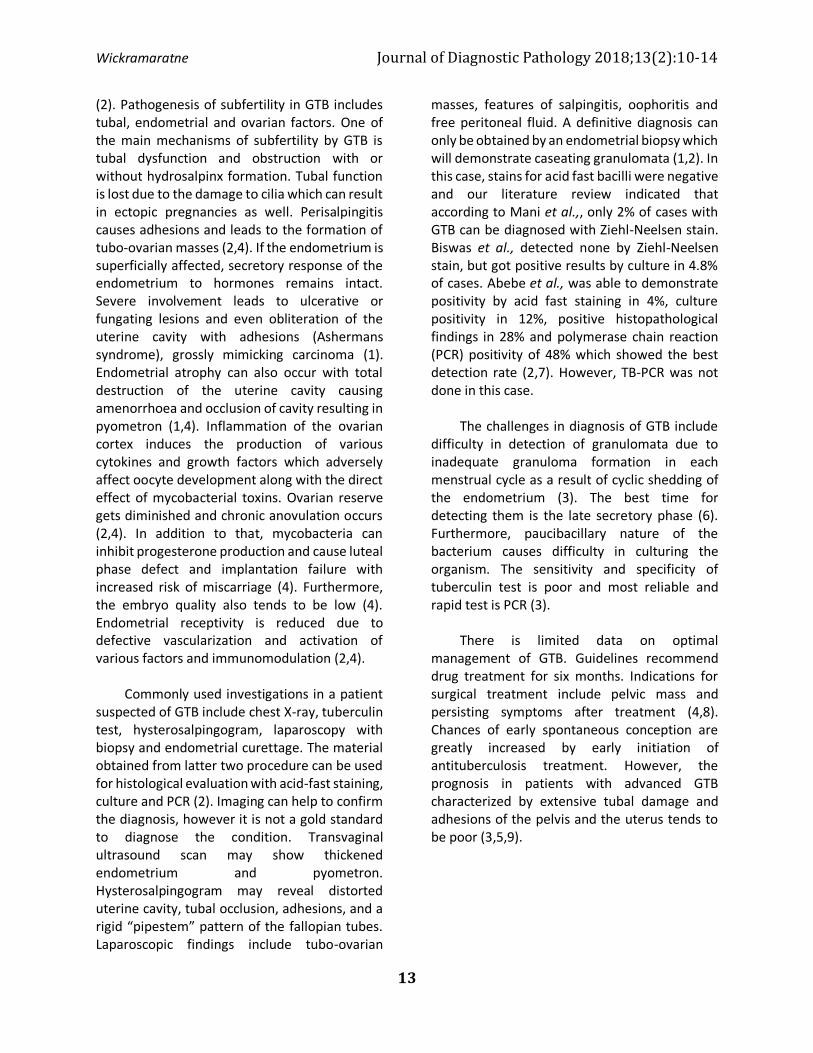
The uterus and cervix were grossly unremarkable. Fallopian tubes and ovaries were adhered together; however, the cut sections did not reveal significant abnormalities. Sections through the endometrium, myometrium, both ovaries and fallopian tubes revealed several granulomata composed of epithelioid histiocytes as well as Langhans type and foreign body type giant cells, surrounded by lymphocytes and some showing extensive caseous type central necrosis (Figures A to D). In addition to that there were features of bilateral chronic salpingitis. Special stains for acid fast bacilli (Ziehl-Neelsen stain) and fungi (Grocott and PAS) were negative.
Discussion
GTB is frequently acquired by
haematogenous spread or lymphatic spread from an extragenital site. Interestingly, genital tract is almost never a primary infection site (1). Most frequent sites of involvement are the fallopian tubes (90-100%), endometrium (50-60%) and ovaries (30%) similar to present case (3,4,5). Involvement of cervix, vulva and vagina is less common (6). In the early stages pelvic organs appear normal. Initially, the ampullary region of the fallopian tubes shows changes and later the fimbria get swollen. In the endometrium, the changes are usually focal and in advanced cases there is ulceration and intrauterine adhesions. Ovaries may adhere to adjacent pelvic organs giving rise to adnexal masses as in this case (6). Subfertility is defined as inability to conceive after one year of unprotected intercourse and in a female this can be due to various factors such as ovulation problems, tubal blockage or uterine problems due to endocrine disorders, endometriosis or various infections
A) Endometrium revealing coalescent caseating granulomas(x4); B) Myometrium showing a large granuloma with central caseation(x4); C) Left ovary showing a granuloma(x4); D) Several granulomas in right fallopian tube(x4)
Wickramaratne Journal of Diagnostic Pathology 2018;13(2):10-14
13
(2). Pathogenesis of subfertility in GTB includes tubal, endometrial and ovarian factors. One of the main mechanisms of subfertility by GTB is tubal dysfunction and obstruction with or without hydrosalpinx formation. Tubal function is lost due to the damage to cilia which can result in ectopic pregnancies as well. Perisalpingitis causes adhesions and leads to the formation of tubo-ovarian masses (2,4). If the endometrium is superficially affected, secretory response of the endometrium to hormones remains intact. Severe involvement leads to ulcerative or fungating lesions and even obliteration of the uterine cavity with adhesions (Ashermans syndrome), grossly mimicking carcinoma (1). Endometrial atrophy can also occur with total destruction of the uterine cavity causing amenorrhoea and occlusion of cavity resulting in pyometron (1,4). Inflammation of the ovarian cortex induces the production of various cytokines and growth factors which adversely affect oocyte development along with the direct effect of mycobacterial toxins. Ovarian reserve gets diminished and chronic anovulation occurs (2,4). In addition to that, mycobacteria can inhibit progesterone production and cause luteal phase defect and implantation failure with increased risk of miscarriage (4). Furthermore, the embryo quality also tends to be low (4). Endometrial receptivity is reduced due to defective vascularization and activation of various factors and immunomodulation (2,4). Commonly used investigations in a patient suspected of GTB include chest X-ray, tuberculin test, hysterosalpingogram, laparoscopy with biopsy and endometrial curettage. The material obtained from latter two procedure can be used for histological evaluation with acid-fast staining, culture and PCR (2). Imaging can help to confirm the diagnosis, however it is not a gold standard to diagnose the condition. Transvaginal ultrasound scan may show thickened endometrium and pyometron. Hysterosalpingogram may reveal distorted uterine cavity, tubal occlusion, adhesions, and a rigid “pipestem” pattern of the fallopian tubes. Laparoscopic findings include tubo-ovarian
masses, features of salpingitis, oophoritis and free peritoneal fluid. A definitive diagnosis can only be obtained by an endometrial biopsy which will demonstrate caseating granulomata (1,2). In this case, stains for acid fast bacilli were negative and our literature review indicated that according to Mani et al.,, only 2% of cases with GTB can be diagnosed with Ziehl-Neelsen stain. Biswas et al., detected none by Ziehl-Neelsen stain, but got positive results by culture in 4.8% of cases. Abebe et al., was able to demonstrate positivity by acid fast staining in 4%, culture positivity in 12%, positive histopathological findings in 28% and polymerase chain reaction (PCR) positivity of 48% which showed the best detection rate (2,7). However, TB-PCR was not done in this case. The challenges in diagnosis of GTB include difficulty in detection of granulomata due to inadequate granuloma formation in each menstrual cycle as a result of cyclic shedding of the endometrium (3). The best time for detecting them is the late secretory phase (6). Furthermore, paucibacillary nature of the bacterium causes difficulty in culturing the organism. The sensitivity and specificity of tuberculin test is poor and most reliable and rapid test is PCR (3). There is limited data on optimal management of GTB. Guidelines recommend drug treatment for six months. Indications for surgical treatment include pelvic mass and persisting symptoms after treatment (4,8). Chances of early spontaneous conception are greatly increased by early initiation of antituberculosis treatment. However, the prognosis in patients with advanced GTB characterized by extensive tubal damage and adhesions of the pelvis and the uterus tends to be poor (3,5,9).

Wickramaratne Journal of Diagnostic Pathology 2018;13(2):10-14
14
Conclusion Awareness about GTB is important in the workup of a subfertile patient in a tuberculosis endemic country like Sri Lanka as this is one of the treatable causes of subfertility. Moreover, failure to recognize this condition in early stages will cause irreversible damage to the reproductive organs resulting in permanent inability to conceive.
References 1. Djuwantono T. et al.Female genital
tuberculosisand infertility: serial cases report in Bandung,Indonesia and literature review.BMC Res Notes 2017; 10:683.
2. Eftekhar M. et al. Mycobacterium tuberculosis infection in women with unexplained infertility. Int J Reprod BioMed 2015; 13(12):749-754.
3. Shende P.et al.Genital tuberculosis and infertility. Int J Reprod Contracept Obstet Gynecol. 2017; 6(8):3514-3517.
4. Sharma JB, Dharmendra S, Agarwal S, Sharma E.Genital tuberculosis and infertility. Fertil Sci Res 2016; 3:6-18.
5. Al eryani AA,Abdelrub AS, Al Harazi AH. Genital tuberculosis is common among females with tubal factor infertility: Observational study. Alexandria Journal of Medicine2015; 51:321–324.
6. Grace GA, Devaleenal DB, Natrajan M. Genital tuberculosis in females.Indian J Med Res 2017; 145: 425-436.
7. Abebe M, Lakew M, Kidane D, Lakew Z, Kiros K, Harboe M. Female genital tuberculosis in Ethiopia. Int J Gynaecol Obstet 2004; 84: 241-246.
8. Hassoun A, Female genital tuberculosis: Uncommon presentationof tuberculosis in the United States. The American Journal of Medicine 2005; 118: 1295-1299.
9. Sharma JB. Current Diagnosis and Management of Female GenitalTuberculosis. The Journal of Obstetrics and Gynecology of India 2015; 65(6):362–371.