JB Accepts, published online ahead of print on 1 March 2013 J

1
Title: Carbonic anhydrase-related protein VIII deficiency is associated with a
distinctive lifelong gait disorder in waddles mice
Author affiliation: Yan Jiao*, Jian Yan†, Yu Zhao‡, Leah Rae Donahue§, Wesley G.
Beamer§, Xinmin Li¶, Bruce A. Roe , Mark S. LeDoux‡, Weikuan Gu*
*Departments of Orthopaedic Surgery- Campbell Clinic and Pathology, University of
Tennessee Health Science Center, Memphis, Tennessee; †Department of Biology, University of Memphis, Memphis, Tennessee; ‡Departments of
Neurology and Anatomy and Neurobiology, University of Tennessee Health Science
Center, Memphis, Tennessee; §The Jackson Laboratory, Bar Harbor, Maine; ¶Functional Genomics Facility, University of Chicago, Chicago, Illinois;
Department of Chemistry and Biochemistry, University of Oklahoma, Norman, Oklahoma
Running title: Gene deficiency in waddles mice
Key words: Carbonic anhydrase-related protein VIII gene; mutation; Waddles; Ataxia; High-
Throughput Screening.
Corresponding author: Weikuan Gu, PhD., University of Tennessee Health Science
Center, A331 Coleman Building, 956 Court Avenue, Memphis, TN 38163. Phone: 901-448-
2259; Fax: 448-3343; E-Mail: [email protected]
Genetics: Published Articles Ahead of Print, published on August 22, 2005 as 10.1534/genetics.105.044487

2
ABSTRACT
The waddles (wdl) mouse is a unique animal model that exhibits ataxia and appendicular
dystonia without pathological abnormalities of either the central or peripheral nervous
systems. A 19 bp deletion in exon 8 of the carbonic anhydrase-related protein VIII gene
(Car8) was detected by high-throughput temperature gradient capillary electrophoresis
heteroduplex analysis of PCR amplicons of genes and ESTs within the wdl locus on mouse
Chr 4. Although regarded as a member of the carbonic anhydrase gene family, the
encoded protein (CAR8) has no reported enzymatic activity. In normal mice, CAR8 is
abundantly expressed in cerebellar Purkinje cells as well as several other cell groups.
Compatible with nonsense-mediated decay of mutant transcripts, CAR8 is virtually absent
in mice homozygous for the wdl mutation. These data indicate that the wdl mouse is a
Car8 null mutant and that CAR8 plays a central role in motor control.

3
Introduction
Rodents, mainly spontaneously mutant mice with movement disorders, have
revealed molecules and cellular pathways involved in motor control. Examples of deficient
or defective proteins in mice with motor dysfunction include the Scn8a (type VIII alpha
subunit of voltage-gated sodium channel) in MedJ mice (Burgess et al, 1995), Grid2 (delta2
glutamate receptor) in lurcher mice (Zuo et al, 1997), Kcnj6 (potassium inwardly-rectifying
channel subfamily J member 6) in weaver mice (Patil et al, 1995), Agtpbp1 (ATP/GTP
binding protein 1) in Purkinje cell degeneration mice (Fernandez-Gonzalez et al, 2002), and
Cacna1a (P/Q type calcium channel α-1A subunit) in tottering mice (Fletcher et al, 1996).
Although these models have shed considerable light on signaling and second messenger
pathways, the relationship between cellular dysfunction due to these mutations and motor
control at the systems level remains unclear. This disconnect is largely due to the
ubiquitous expression of the encoded proteins within neural tissues. Furthermore, many
mutant rodents with movement disorders exhibit overt structural and histological
abnormalities of the central nervous system, extra-neural disease, or early death. Thus, in
many cases, the mutant gene may have deleterious effects on several local-area neural
networks in addition to systemic effects which severely compromise attempts to correlate
genotype with particular motor phenotypes (e.g., ataxia, Parkinsonism, dystonia, spasticity,
or myoclonus). Identification of additional mutant genes with restricted expression in neural
tissue and causally associated with distinct motor syndromes may help to bridge the gaps
between the molecular and systems neurobiology of movement disorders. As part of this
effort, we chose to study waddles (wdl) mice.
The wdl mutation was discovered at The Jackson Laboratory (TJL) in 1995 in
C57BL/KS mice (http://www.jax.org/mmr/waddler.html). The wdl phenotype is very similar
4
to that of another model, waddler (wd), which was discovered in 1959 and is now thought to
be extinct (Yoon 1959). The gait of wdl mice is characterized by wobbly side-to-side ataxic
movements that are readily visualized when mice reach two weeks of age. The gait
disorder of wdl mice persists throughout their lifespan.
In addition to ataxia, wdl mice exhibit frequent tail elevation and intermittent Straub tail.
During ambulation, the trunks of wdl mice are abnormally elevated, particularly their caudal
portions. Resting forelimb and hindlimb tone is normal. However, action dystonia with
apparent co-contraction of knee and elbow flexors and extensors is exacerbated by
ambulation (Jinnah and Hess 2004). This appendicular dystonia produces nearly straight
limbs with minimal flexion at the knee and elbow joints, elevation of the pelvis, and a
"bouncy" or "waddling" motion during ambulation, particularly at higher velocities.
Occasionally, wdl mice fall to their sides.
Pathological examination of wdl mice at TJL was unremarkable except for one isolated
case of hydrocephalus. In addition, vision and hearing were normal in the mutants. An
early genetic study indicated that wdl mice were autosomal recessive mutants. Linkage
mapping at TJL placed the wdl mutation in close approximation to the wd locus on mouse
Chromosome 4, although tests for allelism were not conducted since wd is extinct.
MATERIALS AND METHODS
Mice: One homozygous waddles (wdl/wdl) mouse and its heterozygous littermate (+/wdl)
from (LRD), Mouse Mutant Stock Resource colonies at TJL, were sent to (WG) University
of Tennessee Health Science Center (UTHSC). Then, a breeding colony was established
by mating them at UTHSC. Experimental animal procedures and mouse husbandry were
performed in accordance with the National Institute of Health’s Guide for the Care and Use
of Laboratory Animals and approved by the UTHSC Institutional Animal Care and Use
Committee.

5
Motor Function Examination: Adult (2-4 months) wdl/wdl mice and wild-type (+/+)
littermates were used for quantitative analyses of motor function. The open field behavior
of additional wdl mice along with heterozygote (+/wdl) and +/+ littermates was observed in
both home cages and bedless arenas. Digital videos were analyzed for the presence of
distinguishing behaviors. ANOVA (Analysis of Variance) was conducted for the significance
between the homozygous wdl (wdl/wdl) and their normal littermates (+/wdl or +/+)
whenever is necessary.
Rotarod. Mice were acclimated to a rotarod (San Diego Instruments) rotating at 5
revolutions per minute (rpm) for 5 min prior to data acquisition. Three 2-min trials were
performed at each target speed (5, 10, 20, 30, 40 and 50 rpm) with an inter-trial interval of
5 min. The rotarod was accelerated to target speeds over 1 min. Median values were
used for statistical comparisons.
Footprint analysis. Mouse forepaws and hindpaws were dipped in non-toxic water-
based paints. Mice were then allowed to run down a runway lined with white paper. Three
trials were performed with inter-trial intervals of at least 5 min. Two to four steps from the
middle portion of each run were measured for 1), stride length; 2), hind-base width (the
distance between the right and left hindlimb strides); 3), front-base width (the distance
between the right and left forelimb strides) and 4), overlap between forepaw and hindpaw
placement. At least 7 steps were measured for each mouse. Mean values were used for
statistical analyses.
Tail suspension. This test involved the response of each mouse to 1 min of suspension
from the tail. Some mice with neurological dysfunction will exhibit hindlimb and/or forelimb
clasping during this maneuver.

6
Righting reflex. To obtain righting reflex times, mice were placed in the supine position
and then released. The time required for all four limbs to contact the tabletop was
measured for three trials. Median values were used for statistical comparisons.
Vertical rope climbing. Mice were acclimated to a vertical 40-cm long, 10-mm thick
rope prior to testing. The bottom of the rope was suspended 15 cm above a padded base
and the top entered an escape box. Three trials with a 5-min inter-trial interval were
completed for each mouse and median times were used for statistical analyses.
Raised-beam task. Mice were acclimated to an 80-cm long, 26-mm wide beam
elevated 50 cm above a padded foundation. A 60W lamp at the start served as an
aversive stimulus whereas the opposite end of the beam entered an escape box. Slips
were counted as mice traversed the beam. Falls were counted as 5 slips.
Genome information:
Information on microsatellite markers and their locations were obtained from the MGD
search forms (http://www.informatics.jax.org/searches/markerform.shtml). The locations of
microsatellite markers on genome sequences and physical chromosomal distances were
obtained by searching the Ensembl mouse genome database
(http://www.ensembl.org/Mus_musculus). Data used in the paper is according to the
updated information as of March 20, 2005.
High-Throughput Screening of the wdl Locus: Liver genomic DNA (gDNA) from +/+,
+/wdl)), and homozygous (wdl/wdl) mice was extracted for temperature gradient capillary
electrophoresis (TGCE) and sequence analysis. Based on the Ensembl and National
Center for Biotechnology Information (NCBI) databases, primer pairs flanking the exons of
known and predicted genes (including ESTs) within the wdl locus were designed with

7
Primer3 software (http://www-genome.wi.mit.edu/cgi-bin/primer/primer3_www.cgi). Primers
were located approximately 100 bp 5’ or 3’ to each exon and, in general, produced 300-400
bp amplicons. Several primer pairs were required for complete coverage of some exons.
PCR amplification of gDNA was performed in a 96-well plate format and consisted of 30-35
cycles at three-temperatures: strand denaturation at 96°C for 30 sec, primer annealing at
54-60°C for 60 sec, and primer extension at 72°C for 120 sec. TGCE (SpectruMedix; State
College, PA) was used to analyze amplicons from +/+, +/wdl, and wdl/wdl mice mixtures.
The SpectruMedix system includes a high-throughput capillary electrophoresis instrument
capable of analyzing 96 samples every 140 min. Heteroduplex analysis was subsequently
performed offline using SpectruMedix software. Amplicons from wdl mice were sequenced
if they differed from normal.
RT-PCR: Total RNA was extracted from cerebellum and liver with Trizol reagent
(Invitrogen). Total RNA integrity was confirmed with the Agilent Bioanalyzer 2100.
Reverse transcription and PCR were conducted using a One-Step RT-PCR kit from
Invitrogen. Reactions were performed in a total volume of 50 µl with 8ng/µl of total RNA
and 0.2 µM forward (CCAAAACAATTCCATGCTTTAAT) and reverse
(GTATGAATTCCAGAAGCTGTGGT) primers used to amplify exons 6 to 9 of Car8. First,
cDNA synthesis and pre-denaturation were performed in single cycles at 50°C for 40 min
and 94°C for 2 min. Next, PCR amplification was performed for 35 cycles: 94°C for 30 sec,
54-58°C for 36 sec and 72°C for 2 min.
DNA sequencing: DNA sequencing was conducted to verify the deletion in the gDNA and
cDNA of Car8. PCR products from both genomic and cDNA were purified using an
AMPure PCR Purification Kit (Agencourt Beverly, MA) and the purified products were
sequenced using a BigDye® Terminator v3.1 Cycle Sequencing (Applied Biosystems Inc.,

8
Foster City, CA). A total of 5 ul sequencing reactions including 2µl Big Dye (plus Half-BD),
10 to 23 ng of purified DNA template and 1 to 3 pmoles of either forward or reverse
universal sequencing primers, were incubated for 37 cycles at 96°C for 180 sec, 50°C for
30 sec, and 60°C for 180 sec. Unreacted primers were removed by ethanol-acetate
precipitation (3.75% 3M NaOAc, 87.5% Non- denatured 100% EtOH, and 8.75% dH2O, pH
4.6,). The labeled products were dissolved in 0.02 mM EDTA in HiDi formamide prior to
electrophoretically loading onto the SpectruMedix 96 capillary sequencing system. The
same primers in the amplification of DNA fragments from either genomic DNA or mRNA
were also used in the sequencing. Sequencing was conducted two times to verify the result
for either gDNA or cDNA.
Expression of Recombinant CAR8: Normal (Accession number: NM_007592) and
mutant Car8 cDNA was amplified (forward primer:
CACCATGGCTGACCTGAGCTTCATTG, reverse primer:
CTGAAAGGCCGCTCGGATGACTCTAT) and cloned into Invitrogen's pET102/D-TOPO
vector in-frame for transcription. A single colony of E. coli BL21 (DE3) transformed with the
Car8 expression vector was inoculated into 10 ml of Luria–Bertani medium containing 100
µg/ml ampicillin; this was incubated at 37°C for 10 h. Then, 0.5 ml of the culture was
inoculated into 50 ml of Luria–Bertani medium containing 100µg/ml ampicillin. This culture
was incubated at 37°C until it reached an OD600 of 0.7. At that point, isopropyl -D-
thiogalactoside was added to a final concentration 0.5 mM and incubation was continued at
37°C for 3 h; cells were harvested by centrifugation. Protein products were analyzed on
8% SDS-PAGE gels. Calculated molecular weights for normal and mutant CAR8 fusion
proteins are 46.09 and 45.49 kD, respectively.

9
Northern-Blot Hybridization: After isolation of total RNA from mouse cerebella, mRNA
(from 20 mice: +/+: 7, wdl/wdl: 7, +/wdl: 6)was extracted and purified with the
MicroPoly(A)Purist Kit from Ambion (Austin, TX). The mRNA was electrophoretically
resolved on denaturing gels and transferred to positively charged nylon membranes.
Radiolabeled (32P-UTP) complementary RNA (cRNA) probes were generated by in vitro
transcription using T7 RNA polymerase. The location of Car8 probe in the cDNA of Car 8
gene is from 205bp to 650bp. After ultraviolet cross-linking, blots were pre-hybridized and
then hybridized overnight with both Car8 and β-actin cRNA probes. After washing, blots
were exposed to Kodak Biomax MR radiographic film prior to development.
Antibody Production: A rabbit polyclonal antibody to CAR8 was generated by
immunizing rabbits with a peptide sequence unique to CAR8 (DANGEYQSPINLNSREC)
and encoded by nucleotides 27-71 from the second exon of Car8 (AnaSpec; San Jose,
CA). This peptide shows no sequence similarity with other carbonic anhydrases. Serum
was immunoaffinity purified.
Western Blot Analysis: Cerebellar cortex was harvested from mice, rinsed in PBS, and
homogenized in chilled NP-40 lysis buffer containing a protease inhibitor cocktail (Sigma).
Lysates were clarified by centrifugation. Protein concentrations were determined with the
Bio-Rad DC Protein Assay kit using BSA standards. Equal amounts of proteins were
electrophoretically resolved on 4-20% Tris-HCl Criterion pre-cast gels (Bio-Rad), and then
transferred onto Immunoblot PVDF membranes (Bio-Rad). Membranes were washed in
TBST (Tris-buffered saline with 0.1% Tween), blocked in 5% nonfat dry milk and then
incubated in affinity-purified rabbit anti-CAR8 (1:2000) or rabbit anti-IP3R1 (1:2000, A.G.
Scientific; San Diego) antibodies. After washing, blots were incubated in horseradish
peroxidase (HRP) conjugated secondary antibodies (1:5000, Amersham). Targeted

10
proteins were visualized with the ECL Plus chemiluminescent kit from Amersham. For
loading controls, membranes were stripped and re-probed with a mouse anti-β-tubulin
antibody (1:5000, Chemicon)
Immunocytochemistry: Mice and rhesus monkeys were perfusion-fixed with saline/4%
paraformaldehyde. Brains were post-fixed and then cryoprotected in 30%
sucrose/phosphate buffer (PB). Mouse brains and monkey cerebella were sectioned
parasagitally on a cryostat and collected onto Superfrost®-Plus glass slides (Fisher).
Mouse spinal cords were sectioned horizontally. Brain sections were processed for
immunocytochemical detection of CAR8 (affinity-purified rabbit antibody, 1:20,000),
calbindin (rabbit polyclonal antibody, 1:5000, Chemicon) and IP3R1 (rabbit polyclonal
antibody, 1:2000). For light microscopic visualization, tissues were sequentially exposed to
a biotinylated goat anti-rabbit polyclonal antibody (Vector, 1:500), streptavidin and
diaminobenzidine. Cy2- and rhodamine red-X (RRX, Jackson ImmunoResearch)
conjugated secondary antibodies were applied for confocal microscopy.
RESULTS
Motor Dysfunction in wdl Mice: To characterize the motor function of wdl mice, several
experiments were performed to compare the mutants with +/+ littermates. There were no
differences between wdl and +/+ mice in their responses to tail suspension. Furthermore,
righting times and vertical rope climbing, respectively, did not differ between the mutants
and their normal littermates, suggesting that the wdl mice have normal axial and
appendicular motor power. In contrast, the results of footprint analysis (Fig. 1A and B),
rotarod (Fig. 1C) and the raised-beam task were compatible with significant motor disability
in wdl mice. Although traversal times on the raised-beam task did not differ significantly
between wdl mice and +/+ littermates, the mutants had many more slips (19.0 +/- 1.7) than
11
that of normal (0.8 +/- 0.3) littermates (p < 0.0001). In wdl mice, paws were abnormally
everted and hindpaw (red) placement was rostral to the location of forepaw (green) prints
(Fig. 1A). However, there were no differences in either stride length or the distance of paw
overlap between wdl and +/+ mice (Fig. 1B). At all speeds, wdl mice showed shorter
latencies to fall off the rotarod than +/+ littermates (p < 0.05, for all).
High-Throughput Screening of the wdl Locus on Chr 4: According to data from the
Jackson Laboratory web page (http://www.informatics.jax.org/menus/marker_menu.shtml),
the recombination estimates with standard errors and the best gene order were centromere
- D4Mit149 1.79 +/-1.02 cM - wdl - 0.81+/-0.80 cM [D4Mit181, D4Mit99] - 7.65+/-4.16 cM -
D4Mit97 - 2.52+/-2.47cM - D4Mit39 -1.62+/-1.60cM - D4Mit5. To begin with, we searched
the Ensembl database (http://www.ensembl.org/Mus_musculus) to locate genetic markers
flanking the wdl locus. Initially, D4mit149 (at 0 cM), the nearest marker on the centromeric
side of the wdl locus, was mapped between 3584271bp and 3584381 bp. D4mit181 (at 2.5
cM), the nearest marker on the telomeric side, was mapped between 9500798 and
9500930 bp (Fig. 2A). Another marker on the telomeric side, D4mit99, was mapped
between 12496147 bp and 12496289 bp. However, the initial mapping data from TJL was
based on 86 F2 mice. By examining the genomic information, we found that the region from
centromere to 12,500,000 bp is a gene poor region with 68 genes and 41 ESTs, mainly
from the region between 6,500,000 bp and 12,500,000 bp (Fig. 2A). Those genetic
elements were then analyzed with TGCE. Every exon and at least 50 bps of intron
sequences on both sides from wdl and normal mice were amplified and carefully compared
on SpectruMedix system. During this screening process, we found that only the Car8 exon
8 amplicon was smaller in wdl mice than in +/+ littermates (forward primer:
AATTGTCTCCCAAAATCCCATC; reverse primer: CAGCATGCTTTCTTAACCACTG) (Fig.
2B). Every DNA fragment from other genes was the same between wdl and its +/+

12
littermates. After verification of this difference by PCR amplification of Car8 exon 8 (Fig.
2C), we conducted a comparison of PCR products from eight mouse strains and wdl/wdl
mice (Fig. 2D), and the data indicated that the deletion found in wdl mice did not exist in
any of these normal mouse strains (Fig. 2E). At that point, we felt confident that there was a
deletion within the exon 8 of Car8 that caused the wdl disease. We then sequenced exon 8
and discovered that 19 nucleotides were deleted in the mutant mice (Fig. 2F).
Car8 Mutation: Car8 has 9 exons (ENSMUSG00000041261). Total RNA from normal and
wdl mice was extracted for RT-PCR amplification of 3’ Car8 transcript (exons 6-9) (Fig.3A).
Smaller amplicons were found in cerebella (Fig. 3A) and extra-neural tissue (liver, lung, and
kidney) from wdl mice (Data not shown). Direct sequencing of Car8 cDNA from normal and
wdl mice confirmed the 19-nucleotide deletion (Fig. 3B). In the region of deletion, 15
nucleotides are flanked by two AAGG motifs. The deleted 19 nucleotides include the 15
nucleotides and one AAGG motif, leaving the other AAGG motif in the genome. Thus, the
deletion either starts at first AAGG motif and stops at the second AAGG motif or starts
immediately after the first AAGG motif and stops after the second motif. Due to frame
shifting, wdl Car8 cDNA is predicted to encode a protein that is 29 amino acids shorter than
+/+ CAR8 secondary to elimination of 50 +/+ amino acids and addition of 21 novel residues
before terminating in a stop codon (Fig. 3B).
Car8 Transcript and Protein (CAR8): To determine the molecular impact of the Car8
deletion mutation, we compared mutant and +/+ Car8 at the transcriptional and
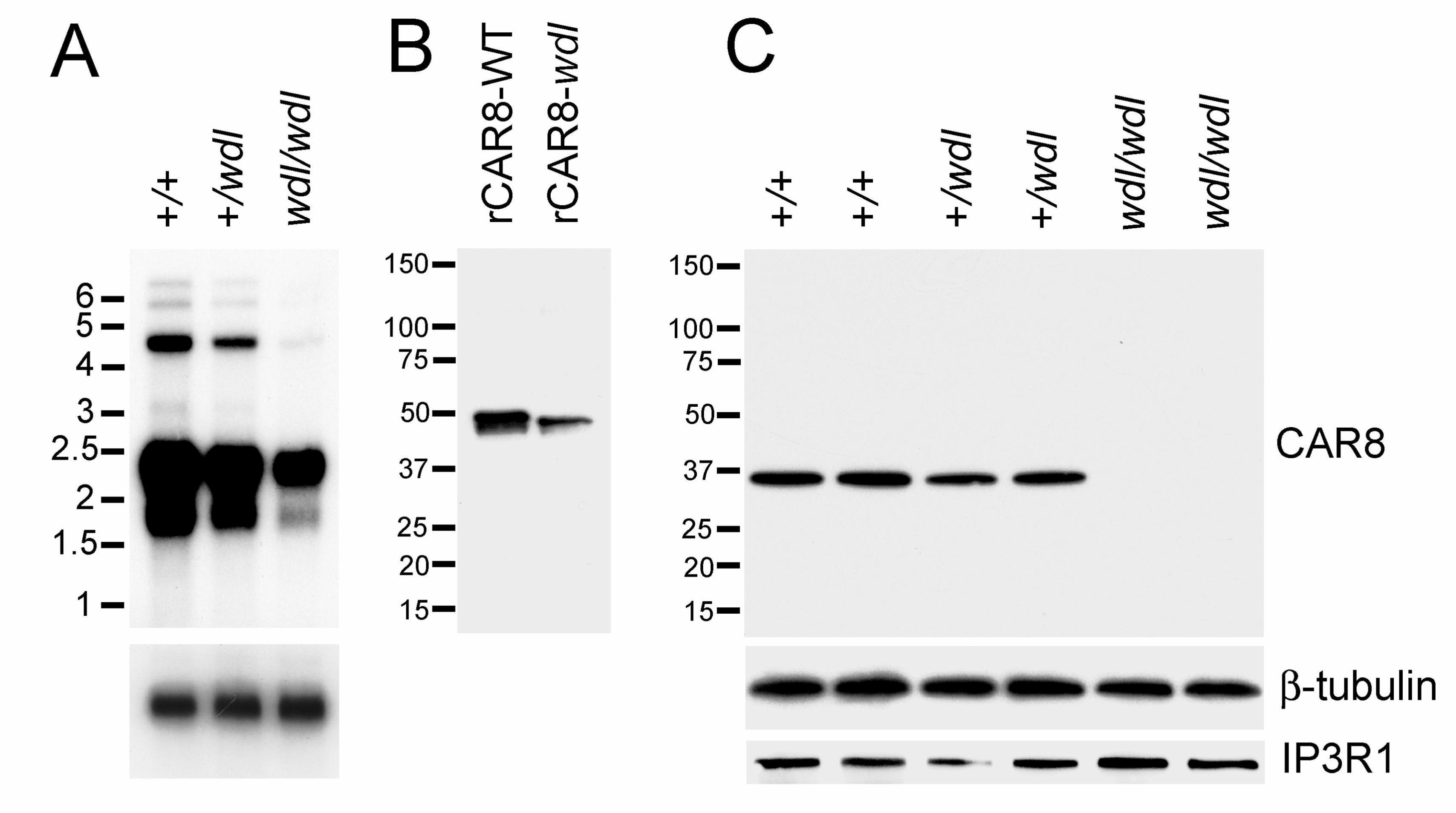
translational levels. Six bands were identified on Northern blotting of mRNA from both +/+
and wdl mice (Fig. 4A). However, a striking global reduction of Car8 transcript was detected
in wdl mice (Fig. 4A). The larger bands on Northern were barely detectable in the mutants.
Quantities of Car8 mRNA were intermediate in +/wdl animals. With Western blotting, our

13
affinity-purified rabbit polyclonal antibody to CAR8 identified both full-length recombinant
+/+ and wdl CAR8 in different sizes (Fig. 4B). In contrast, we were unable to detect CAR8
in cerebellar tissue lysates from wdl mice although a Purkinje cell marker, the type 1
inositol 1,4,5-triphosphate receptor (IP3R1), was expressed at normal levels in the mutants
(Fig. 4C).
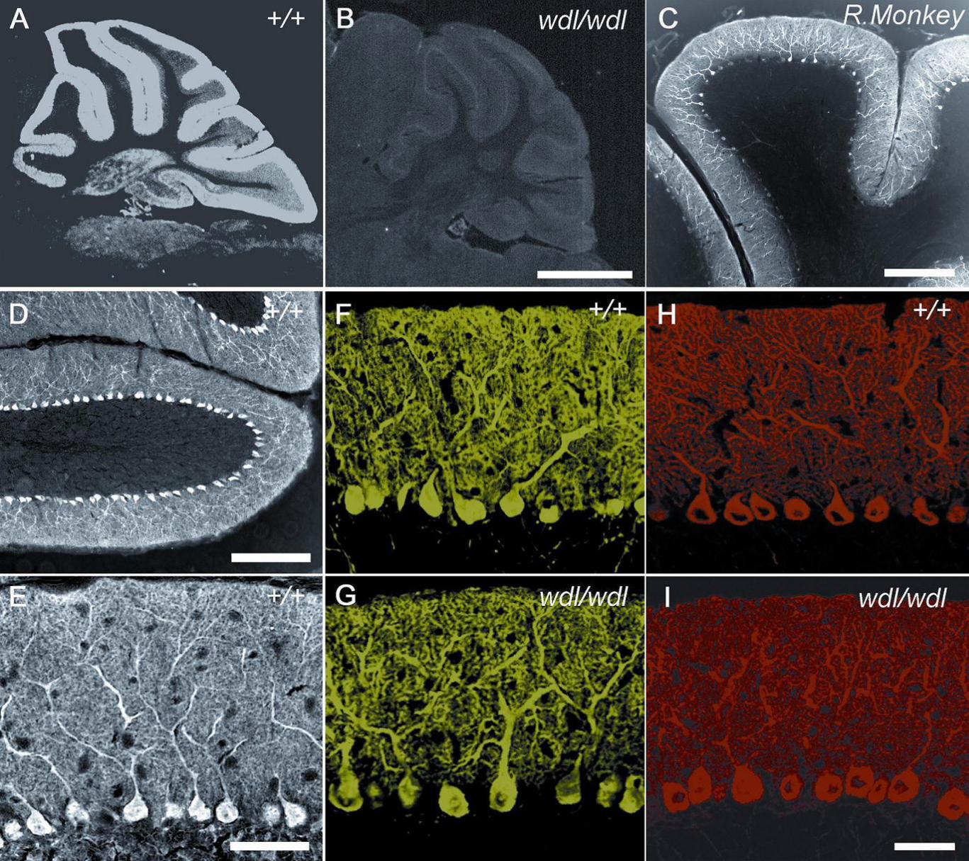
Immunolocalization of CAR8: Immunohistochemistry was then used to examine the
patterns of CAR8 expression in +/+ and wdl mice (Fig. 5). In +/+ mice, CAR8 was
expressed in cerebellar Purkinje cells as well as in cerebellar nuclei and brainstem (Fig.
5A). Immunohistochemically, CAR8 expression was greatest in cerebellar Purkinje cells
and minimal to absent in the spinal cord and cerebral cortex. In line with the Western
blotting results, CAR8 expression was virtually undetectable in wdl mice (Fig. 5B). The
potential importance of CAR8 to humans is suggested by its prominent expression in
Purkinje cells from a non-human primate (Fig. 5C). CAR8 is abundantly present in the
somas, axons, and entire dendritic arbors of cerebellar Purkinje cells (Fig. 5D and E). The
common Purkinje cell marker, calbindin, showed immunoreactivity in cerebellar cortex that
did not differ between normal and mutant mice (Fig. 5F and G). Furthermore, at the level of
confocal microscopy, the density and intracellular localization of IP3R1 was normal in wdl
mice (Fig. 5H and I).
DISCUSSION
Our data strongly suggests that the 19bp deletion in Car8 described here is the causally
associated with the wdl phenotype. We provide several lines of evidence to support this
claim. First, Car8 is located within the genetic region of wdl locus. Second, the Car8
deletion was the only defect detected among genes and ESTs within the wdl locus from wdl
mice. Because the wdl mutation was derived from the C57BLKS inbred strain and there are
14
no other difference between wdl mice and their littermates, it is unlikely that another
mutation causes the movement disorder exhibited by wdl mice. Third, work on the cDNA
sequence agreed with the genomic data. Lastly, we showed a virtual absence of CAR8
expression in wdl mice but not wild-type littermates.
Our molecular analysis of the wdl mouse model has shown that CAR8 is essential for
motor control. Moreover, the relative preponderance of CAR8 in cerebellar Purkinje cells
suggests that the wdl mouse can be used as a tool to precisely investigate the effects of
Purkinje cell dysfunction on local and wide-area motor networks. In contrast to several
other mouse mutants with ataxia, there is no loss or overt morphological abnormalities of
Purkinje cells in wdl mice. Although CAR8 is a member of the carbonic anhydrase family of
zinc metalloenzymes that catalyze the reversible hydration of CO2, CAR8 lacks catalytic
activity by virtue of missing critical amino acid residues required for zinc binding (Sjoblom et
al, 1996; Taniuchi et al, 2002). Recombinant CAR8 generated by introducing R117H and
E115Q mutations into the wild-type protein is able to bind zinc and catalyze the hydration of
CO2. CAR8 may contribute to the pathophysiology of ataxia in other mouse models. For
instance, Kelly and colleagues (Kelly et al, 1979) reported absence of Car8 transcript in the
cerebella of lurching mice years before the actual causal mutation was identified.
The reduced Car8 transcript and barely detectable CAR8 protein in wdl mice is
indicative of nonsense-mediated decay (NMD). NMD, first documented by Losson and
Lacroute over twenty years ago (Losson and Lacroute 1979), is a proofreading mechanism
that enables eukaryotic cells to detect and degrade mRNAs that contain premature
termination codons (PTCs). About one-third of inherited genetic disorders and many forms
of cancer are caused by frame-shift or nonsense mutations, which result in the generation
of PTCs (Frischmeyer and Dietz 1999; Holbrook et al, 2003). Although mRNA containing a
PTC may initially be translated into a truncated protein, cells can initiate the NMD

15
mechanism to recognize and degrade the mutant transcripts if the truncated protein is
deleterious (Wagner and Lykke-Andersen 2002).
With respect to the cellular effects of CAR8 deficiency, it has been shown that CAR8
binds to the modulatory domain of inositol 1,4,5-triphosphate receptor 1 (IP3R1), which is
an intracellular IP3-gated Ca2+ channel (Hirota et la, 2003). CAR8 inhibits IP3 binding to
IP3R1 by reducing the affinity of the receptor for IP3 (Hirota et la, 2003). IP3 is an
intracellular second messenger for calcium release. Increased cytosolic free calcium
concentration is a stimulatory signal for diverse calcium dependent mechanisms such as
secretion, contraction, or alterations in membrane excitability. Therefore, CAR8 deficiency
may cause ataxia by altering calcium homeostasis and disturbing the normal physiology of
cerebellar Purkinje cells.
Our study also demonstrates a useful strategy for simplifying positional cloning.
Most procedures employed in traditional positional cloning have been labor intensive and
time consuming. As described in the material and methods section, our strategy includes
identifying a target genomic region based on linkage mapping, identifying every gene and
biologically functional element within the candidate region, TGCE heteroduplex analysis
(Chou et al., 2005; Girald-Rosa et al; 2005), confirmation of suspected mutations by cDNA
sequencing and, finally, investigated of the encoded protein. Using a similar approach, we
recently identified the causal mutation in a mouse disease model of spontaneous fractures
(Jiao et al., 2005). In that study, mapping of the mutation locus was followed by positional
cloning. In this study, mapping information on the TJL webpage allowed us to define a
target genomic region. The mutant gene was discovered in less than half a year from the
start of the project, thereby offering the possibility of rapid identification of mutations with
only crude mapping data. . We believe that our strategy will be been particularly useful for
familial human diseases with small kindreds. Furthermore, in the setting of rodent models

16
with spontaneous mutations, mutations can be rapidly identified without the excessive cost
and time associated with the extensive breeding programs required for high-resolution
mapping. .
In summary, we have identified a19-bp deletion in exon 8 of Car8 in wdl mice using
a positional candidate cloning approach. This loss-of-function mutation of Car8 may
underlie the ataxic phenotype of wdl mice. Since were did not find an obvious change in the
morphology or Purkinje cells or the distribution of the CAR8 binding target, IP3R1, the
cellular pathways by which CAR8 deficiency causes ataxia and dystonia remain uncertain.
Thus, the role of CAR8 cerebellar neurophysiology warrants additional study.
ACKNOWLEDGEMENTS:
Major support for this work was from Center of Genomics and Bioinformatics (W.G.)
and Center in Connective Tissue Research (W.G.), at University of Tennessee Health
Science Center. Additional support was from Dystonia Medical Research Foundation
(M.S.L.); Veterans Administration at Memphis Medical Center (W.G); National Institute of
Arthritis and Musculoskeletal and Skin Diseases, National Institutes of Health (R01
AR51190 to W.G; RR01183 L.R.D.); National Eye Institute, National Institutes of Health
(R01 EY12232 to M.S.L.; R01 EYO15073 to L.R.D.); National Institute of Neurological
Diseases and Stroke, National Institutes of Health (R01 NS048458 to M.S.L.). Special
recognition and gratitude are offered to Belinda Harris of The Mouse Mutant Resource at
The Jackson Laboratory for providing miceand Dr. Feng Jiao of University of Tennessee
Health Science Center, for mouse breeding, tissue collection, and management for this
work.
17
References:
1. Burgess, D. L., Kohrman, D. C., Galt, J., Plummer, N. W., Jones, J. M., Spear, B. and Meisler, M.
H. (1995) Nat. Genet. 10: 461-465. 2. Zuo, J., De Jager, P. L., Takahashi, K. A., Jiang. W., Linden,
D. J. and Heintz, N. (1997) Nature 388: 769-773.
3. Patil, N., Cox, D. R., Bhat, D., Faham, M., Myers, R. M. and Peterson, A. S. (1995) Nat. Genet.
11: 126-129.
4. Fernandez-Gonzalez, A., La Spada, A. R., Treadaway, J., Higdon, J. C., Harris, B.S., Sidman,
R. L., Morgan, J. I. and Zuo, J. (2002) Science 295: 1904-1906.
5. Fletcher, C. F., Lutz, C. M., O'Sullivan, T. N., Shaughnessy, J. D., Jr., Hawkes, R., Frankel, W.
N., Copeland, N. G., and Jenkins, N. A. (1996) Cell 87: 607-617
6. Yoon, C. H. (1959) J. Hered. 50: 238-244.
7. Jinnah, H. A. and Hess, E. J. (2004) in Animal Models of Movement Disorders, ed. LeDoux, M.S.
(Elsevier, San Diego), pp. 55-72.
8. Sjoblom, B., Elleby, B., Wallgren, K., Jonsson, B. H. and Lindskog, S. (1996) FEBS Lett. 398:
322-325.
9. Taniuchi, K., Nishimori, I., Takeuchi, T., Ohtsuki, Y. and Onishi S. (2002) Brain Res. Mol. Brain
Res. 109: 207-215.
10. Kelly, C., Nogradi, A., Walker. R., Caddy, K., Peters, J. and Carter, N. (1994) Biochem. Soc.
Trans. 22: 359S. 11. Losson, R. and Lacroute, F. (1979) Proc Natl Acad Sci USA 76: 5134-5137
12. Frischmeyer, P. A. and Dietz, H. C. (1999) Hum. Mol. Genet. 8: 1893-1900
13. Holbrook, J. A., Neu-Yilik, G., Hentze, M.W. and Kulozik, A. E. (2004) Nat. Genet. 36: 801-808.
14. Wagner, E. and Lykke-Andersen, J. (2002) J. Cell Sci. 115: 3033-3038.
15. Hirota, J., Ando, H., Hamada, K. and Mikoshiba, K. (2003) Biochem J. 372: 435-441.
16. Hess, E. J. and Jinnah, H. A. (2004) in Animal Models of Movement Disorders, ed. LeDoux,
M.S. (Elsevier, San Diego), pp. 265-278.17. Sethi, K. D. and Jankovic, J. (2002) Mov Disord. 17:
150-153.
18. Kuoppamaki, M., Giunti, P., Quinn, N., Wood, N.W. and Bhatia, K. P. (2003) Mov Disord. 18:
200-206.

18
19. Desaiah, D., Vig, P. J., Subramony, S. H. and Currier, R. D. (1991) Brain Res. 552: 36-40.20.
Lin X., Antalffy B., Kang D., Orr H. T. and Zoghbi H. Y. (2000) Nat. Neurosci. 3: 157-163.
21. Fureman, B. E., Jinnah, H. A. and Hess, E. J. (2002) Pharmacol. Biochem. Behav. 73: 631-637.
22. Fletcher, C. F., Tottene, A., Lennon, V. A., Wilson, S. M., Dubel, S. J., Paylor, R., Hosford, D.
A., Tessarollo, L., McEnery, M. W., Pietrobon, D., et al. (2001) FASEB J. 15: 1288-1290.
23. Takahashi, K. and Kitamura, K. (1999) Biochem. Biophys. Res. Commun. 261: 773-778.
24. Matsumoto, M., Nakagawa, T., Inoue, T., Nagata, E., Tanaka, K., Takano, H., Minowa, O.,
Kuno, J., Sakakibara, S., Yamada, M., et al. (1996) Nature 379:168-171.25. Su, A. I., Wiltshire, T.,
Batalov, S., Lapp, H., Ching, K. A., Block, D., Zhang, J., Soden, R., Hayakawa, M., Kreiman, G., et
al. (2004) Proc. Natl. Acad. Sci. U. S. A. 101: 6062-6067.
26. Lakkis, M. M., O'Shea, K. S., Tashian, R. E. (1997) J. Histochem. Cytochem. 45: 657-662.
27. Bataller, L., Sabater, L., Saiz, A., Serra, C., Claramonte, B. and Graus, F. (2004) Ann. Neurol.
56: 575-579.28. Lu, S. H., Takeuchi, T., Fujita, J., Ishida, T., Akisawa, Y., Nishimori, I., Kohsaki, T.,
Onishi, S., Sonobe, H., Ohtsuki, Y. (2004) Lung Cancer 44: 273-280.29. Miyaji, E., Nishimori, I.,
Taniuchi, K., Takeuchi, T., Ohtsuki, Y., Onishi, S. (2003) J. Pathol. 201: 37-45.30. Boehning, D.,
Patterson, R. L., Sedaghat, L., Glebova, N. O., Kurosaki, T. and Snyder, S. H. (2003) Nature Cell
Biol. 5: 1051-1061.31. Jiao, Y., Li, X., Beamer, W. G., Yan, J., Tong, Y., GoldoWitz, D., Roe, B.
and Gu, W. (2005) Mammalian Genome. 16: 20-31.32. Chou, LS, Gedge F, Lyon E. Complete
gene scanning by temperature gradient capillary electrophoresis using the cystic fibrosis
transmembrane conductance regulator gene as a model. J Mol Diagn. 2005 Feb;7(1):111-20.
33. Girald-Rosa W, Vleugels RA, Musiek AC, Sligh JE. High-throughput mitochondrial genome
screening method for nonmelanoma skin cancer using multiplexed temperature gradient capillary
electrophoresis. Clin Chem. 2005 Feb;51(2):305-11. Epub 2004 Dec 8.

19
FIGURE LEGENDS
FIGURE 1. Behavioral analysis of wdl mice and +/+ littermates. (A) Representative
footprint patterns from +/+ and wdl/wdl mice. (B) Quantitative footprint analysis. (C)
Latency to fall off the rotarod plotted versus speed in revolutions per minute (RPM). (*, p <
0.05). The error bar denotes “standard deviation”.
FIGURE 2. A genomic map of the wdl locus (A) shows the relative locations of
microsatellite markers. TGCE detected an abnormality in exon 8 of the Car8 gene (B).
PCR amplification of Car8 exon 8 generated a smaller band in wdl/wdl mice, a larger band
in +/+ mice, and two bands in +/wdl mice (C). Comparison of PCR products from eight
mouse strains and wdl/wdl mice (D) revealed that the deletion does not exist in any normal
mouse strains (E). DNA sequence analysis revealed a 19 bp deletion in Car8 exon 8 from
wdl/wdl mice (F).
FIGURE 3. Analysis of Car8 transcript with RT-PCR. (A) Amplification of Car8 transcript from total
RNA extracted from cerebella of +/+ and wdl/wdl mice. (B) Derived amino acid sequences of
normal (black) and mutant CAR8 (brown).
FIGURE 4. The wdl Car8 mutation has dramatic effects at the transcriptional and
translational levels. (A) Northern blot analysis of cerebellar mRNA . (B) Western blot
analysis of recombinant wild-type (rCAR8-WT) and mutant (rCAR8-wdl) proteins. (C)
Western blot analysis of cerebellar lysates from +/+, +/wdl and wdl/wdl mice; β-tubulin was
the loading control.
FIGURE 5. Immunocytochemistry for CAR8 and Purkinje cell markers in +/+ and wdl/wdl
mice and a R.Monkey (rhesus monkey). CAR8 in the cerebellum from +/+ (A, D, E) and
wdl/wdl (B) mice and a rhesus monkey (C). Also shown is calbindin (F, G) and IP3R1 (H, I)
expression in +/+ (F, H) and wdl/wdl (G, I) mice. Scale bars: A-B, 1 mm; C, 500µm; D,
100µm; E-I, 50µm.