Genetics Mitosis. Cell Theory Principle “Where a cell exists, there must have been a preexisting...
31
Genetics Mitosis
-
Upload
beverly-mccormick -
Category
Documents
-
view
230 -
download
0
Transcript of Genetics Mitosis. Cell Theory Principle “Where a cell exists, there must have been a preexisting...

Genetics
Mitosis

Cell Theory Principle
“Where a cell exists, there must have been a preexisting cell”
“Omnis cellula e cellula” (all cells come from cells)

Necessary Characteristic of Life: Cell Reproduction unicellular organism
reproduces entire organism
multicellular growth embryonic development replacement of damaged or dead cells

Binary FissionReproduction in Bacteria genome (hereditary endowment)
single, circular dsDNA associated with proteins single chromosome
replication averages minutes-3 hours

Steps of Binary Fission chromosome replication begins, one
copy of the origin moves rapidly tothe other end
one copy of the origin is now at eachend of the cell; the cell elongates
replication finishes, the plasmamembrane grows inward and anew cell wall is formed
resulting in two daughter cells

Eukaryotes vs. Prokaryotes larger cell size smaller cell sizegreater quantity of DNA lesser
quantity of DNAHistones other proteinsseveral chromosomes one chromosome

DNA Quantity in Eukaryotes Amount of DNA has no direct relationship to
the complexity of that organism ex.
Alligators have more DNA than humans frogs have more DNA than humans

DNA of Eukaryotes
Chromosomes discrete entities condensed DNA + protein state found when DNA division occurs
Chromatin mass of DNA loose coils of DNA + protein state found when DNA replication occurs

Drawing of a Chromosome
short arms
long arms
centromere
sister chromatids
www.biologycorner.com
sister chromatids
kinetochore (protein)

Eukaryotic Chromosome
composition DNA = 40% protein = 60%
average human chromosome 5 cm (2.5cm = 1 in)
46 chromosomes in each cell 2 meters of DNA 6 Billion base pairs

Karyotype
particular array of chromosomes chromosomes of one cell are arranged
according to: size number type information
extra or missing DNA gender of individual
www.biotechnologyonline.gov.au/images/contentpages/karyotype.jpg

Human Cells
somatic cells = body cells 46 chromosomes diploid (2n)
gametes = sex cells (sperm, ova) 23 chromosomes haploid number (1n or n)

Homologous Chromosomes
pair of chromosomes one paternal, one maternal

Sister Chromatids
identical replicated forms of one chromosome held together by one centromere

Chromosomes
Homologous (2) maternal, paternal
Replicated (2) 4 sister chromatids
http://www.phschool.com/science/biology_place/labbench/lab3/images/homologs.gif

Autosome vs. Sex Chromosome autosomal chromosomes
22 pairs
sex chromosomes 1 pair, XX or XY

Cell Cycle
Interphase G1 phase S G2 phase
M (mitotic) phase mitosis (P, M, A, T) cytokinesis

Interphase
Preparing a cell for division
G1 phase (first gap) growth of cell, enough cytosol for 2
S (synthesis) DNA replication, DNA in chromatin
G2 phase (second gap) chromosome condensation microtubule synthesis

M Phase
Mitosis (karyokinesis) - divide DNA, precise division prophase (prometaphase) metaphase anaphase telophase
Cytokinesis - divide the cytoplasm, rough division

MitosisProphase-Prometaphase Prophase
DNA condenses nucleoli disappear sister chromatids
visible spindle is forming centrosomes migrate
Prometaphase nuclear envelope disappear centromeres are at poles condensation increases kinetochore present microtubules attach to kinetochore

Metaphase
centrosomes conveneat the equator (plate)
kinetochores have bothmicrotubules from the opposite poles attachedto each one

Anaphase
proteins maintaining thechromosomes attachedsplit
daughter chromosomes move to the poles
cell elongates

Telophase
2 daughter nuclei form nuclear envelopes form nucleoli reappear chromosomes become
less condense END of Mitosis

Animals Plants
Cytokinesis(division of cytoplasm)
cleavage furrow cell plate
microfilaments form contractile ring and cell pinches in two
vesicles form plasma membrane, cell wall material is deposited

Cell Cycle Duration
varies from 1-24 hours example human liver cell
Interphase: G1 phase…………......9 hrs. S…………………..…10 hrs. G2 phase……..………2 hrs
M phase: mitosis……50 min. Cytokinesis……….....10 min

Controls of Cell Division Cells in tissue culture do not divide if 1. essential nutrients are missing (growth factors)
2. poisons that inhibit protein (microtubules) synthesis are present
3. cells are crowded
4. they cannot progress beyond the restriction point in late G1
5. insufficient concentration of MPF (maturation promoting factor, a complex of proteins)
6. they have reached maximum division (20-50x for cells in culture)

Tumor
mass of transformed cells benign tumor
cells remain at original site lump can be completely removed
malignant tumor invasive cells can impair functions of other organs

Cancer Cells
express abnormal cell division 1. density-dependent inhibition is
absent 2. divide excessively, immortal
HeLa cells 1951 3. can invade other tissues
(metastasis) 4. genetically transformed 5. can form malignant tumors 6. can stop dividing at random
points in the cycle
cancer cells
normal cells
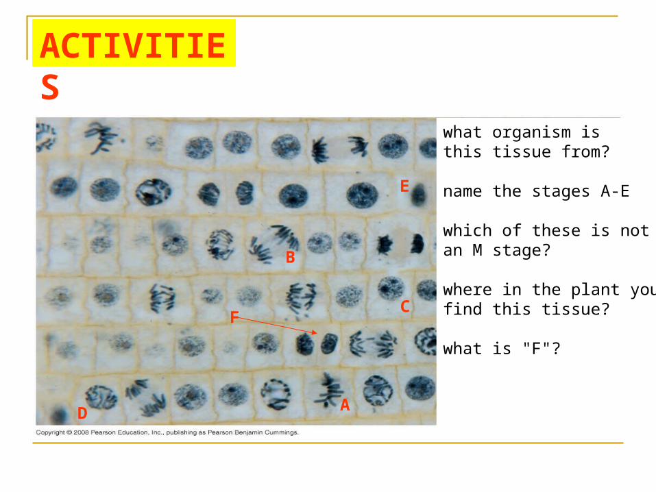
ACTIVITIES
A
B
C
D
E
what organism isthis tissue from?
name the stages A-E
which of these is notan M stage?
where in the plant youfind this tissue?
what is "F"?
F

name the stage
what stage comes before and after it?
if this is a somaticcell and n=10, how many chromosomeswill it have?

name the stage
is it part of mitosis?
give a visible characteristic.
describe what happens during
this stage
The End