Genetics Hearing Loss - zaums.ac.ir
148
Transcript of Genetics Hearing Loss - zaums.ac.ir

Geneticsand
Hearing Loss

A Singular Audiology TextbookJeffrey L. Danhauer, Ph.D.
Audiology Editor
Geneticsand
Hearing Loss
Edited by
Charles I. Berlin, Ph.D.
Kenneth and Frances Barnes Bullington Professor of Hearing ScienceDirector, Kresge Hearing Research Laboratory of the South
Louisiana State University Health Sciences CenterDepartment of Otolaryngology and Biocommunication
New Orleans, Louisiana
Bronya J. B. Keats, Ph.D.
Professor and Acting HeadDepartment of Biometry and Genetics
Molecular and Human Genetics Center of ExcellenceLouisiana State University Health Sciences Center
New Orleans, Louisiana

Singular Publishing GroupThomson Learning401 West A Street, Suite 325San Diego, California 92101-7904
Singular Publishing Group, Inc., publishes textbooks, clinical manuals, clinical refer-ence books, journals, videos, and multimedia materials on speech-language patholo-gy, audiology, otorhinolaryngology, special education, early childhood, aging, occupa-tional therapy, physical therapy, rehabilitation, counseling, mental health, and voice.For your convenience, our entire catalog can be accessed on our web-site athttp//www.singpub.com. Our mission to provide you with materials to meet the dailychallenges of the everchanging health care/educational environment will remain oncourse if we are in touch with you. In that spirit, we welcome your feedback on ourproducts. Please telephone (1-800-521-8545), fax (1-800-774-8398), or e-mail([email protected]) your comments and requests to us.
© 2000 by Singular Publishing Group
Typeset in 10/12 Palatino by So Cal GraphicsPrinted in the United States of America by RR Donnelly
All rights, including that of translation, reserved. No part of this publication maybe reproduced, stored in a retrieval system, or transmitted in any form or by anymeans, electronic, mechanical, recording, or otherwise, without the prior writtenpermission of the publisher.
Library of Congress Cataloging-in-Publication DataGenetics and hearing loss / edited by Charles I. Berlin and Bronya Keats.
p. ; cm. — (Singular audiology textbook)Includes bibliographical references and index.ISBN 0-7693-0103-7 (hardcover : alk. paper)
1. Deafness—Genetic aspects—Congresses. 2. Deafness—Etiology—Congresses. 3.Ear—Abnormalities—Congresses. I. Berlin, Charles I. II. Keats, Bronya J. B. III. Singularaudiology text
[DNLM: 1. Hearing Disorders—genetics—Congresses. WV 270 G32785 2000]RF292.5.G46 2000617.8'042—dc21
99-045186

Contents
Foreword by Charles I. Berlin and Bronya J. B. Keats vii
Contributors xi
Chapter 1 Of Mice and Men (and Myosins) 1
Karen P. Steel, Tim J. Self, Xue-Zhong Liu, Karen B. Avraham, and Steve D. M. Brown
Chapter 2 The Myosin-15 Molecular Motor is Necessary for 31Hearing in Humans and Mice: A Review of DFNB3 andShaker 2
Thomas B. Friedman, Frank J. Probst, Edward R. Wilcox,John T. Hinnant, Yong Liang, Aihui Wang, Thomas D. Barber, Anil K. Lalwani, David W. Anderson, I. Nyoman Arhya, Sunaryana Winata, Sukarti Moeljapowiro,James H. Asher, Jr., David Dolan, Yehoash Raphael, Robert A. Fridell, and Sally A. Camper
Chapter 3 Mouse Models and Progress in Human Deafness Research 47
Stacy Drury, Daryl Scott, Zhining Den, Margaret M.DeAngelis, Mark A. Batzer, Val C. Sheffield, Richard J. H.Smith, Prescott L. Deininger, and Bronya J. B. Keats
Chapter 4 Physical Maps as Molecular Tools to Identify Disease 61Genes
Margaret M. DeAngelis, Chadwick J. Donaldson, Gregory M. Ditta, Lauren M. Buckley, John P. Doucet,Zhining Den, Stacy Drury, Mary Z. Pelias, Prescott L. Deininger, Bronya J. B. Keats, and Mark A. Batzer
Chapter 5 Gene Therapy for the Treatment of Hearing Disorders 73
Julia L. Cook, Grace B. Athas, and Prescott L. Deininger
vv

Chapter 6 Transmitters in the Cochlea: ATP as a Neuromodulator 87in the Organ of Corti
Richard P. Bobbin, Anthony P. Barnes, Chu Chen,Prescott L. Deininger, Christopher S. LeBlanc, andMargaret S. Parker
Chapter 7 Adaptation to Deafness in a Balinese Community 111
John T. Hinnant
Appendix 125
Index 129

Foreword
This is the fifth book in the Kresge-Mirmelstein Award cycle publishedand supported by Singular Publishing Group. The series was conceived bythe late Rona Mirmelstein, who died in November of 1992, and Dr. CharlesBerlin. Each book presents the proceedings of an annual symposium heldto honor an outstanding auditory scientist, who is chosen by a consensusof peers. At the symposium, the honoree presents his or her research, andcompanion papers are prepared by other scientists with similar interests;all of the scientists contribute chapters to the commemorative volume, andthe proceeds go to support next year’s award.
This volume honors the genetic studies of Karen P. Steel, Ph.D., of theMedical Research Council’s Institute of Hearing Research. Dr. Steel was se-lected by the previous winners of this prestigious prize to join their augustranks. The previous winners were Dr. William Brownell for his discoveriesof outer hair cell motility, Dr. Robert Wenthold for his work in moleculartransmitters in sensory systems, Dr. David Kemp for his discovery of otoa-coustic emissions, and Dr. M. Charles Liberman for his studies of the audi-tory efferent system.
The symposium, entitled “Of Mice and Men: Genes, Deafness, andOtolaryngology” was held on September 27, 1998. It opened with greet-ings from the LSU Health Sciences Center Chancellor, Dr. Mervin L. Trail,the Dean of the Medical School, Dr. Robert L. Marier, and Otolaryngolo-gy—Head and Neck Surgery Department Head, Dr. Daniel W. Nuss, andwas attended by a group of approximately 60 people. The speakers pre-sented the results of their research in the genetics of hearing and also re-viewed studies ranging from the identification and characterization ofgenes involved in hearing impairment to the potential for effective thera-pies based on knowledge of gene structure and function. Because thechapters in this book were written after the symposium, the authors haveincluded additional information and thus the chapter titles may differfrom the titles of the symposium presentation.
The first speaker at the symposium was our award winner Dr. KarenP. Steel. Her title was “A Mousey Business” and in her talk she highlightedsome of the mouse studies that have advanced understanding of heredi-tary hearing impairment in humans. Dr. Steel’s chapter title, “Of Mice and
viivii

Men (and Myosins),” reflects the important homology between the mouseand the human genomes and the usefulness of mouse models for humandeafness. Her mentor, the legendary Malkeot Deol, described several ofthe hearing-impaired mouse strains that have facilitated research on hu-man hearing. Dr. Steel has studied many of these mouse mutants, includ-ing shaker-1, jerker, deafness, quivering, splotch, whirler, and Bronx waltzer.These studies have been instrumental in advancing our understanding ofthe biological basis of hearing impairment.
Among our other distinguished speakers was Dr. Thomas Friedman,Chief of NIDCD’s Laboratory of Molecular Genetics. His presentation, en-titled “Identifying DFNB3” described the search for the gene responsiblefor deafness in members of families in Bali; the B in this symbol indicatesthat the mode of inheritance of the deafness in these families is autosomalrecessive and DFNB3 was the third gene to be localized to a specific chro-mosomal region in families with this inheritance pattern. In his chapter Dr.Friedman clearly describes the relevance of parallel studies in the shaker-2mouse to the identification of DFNB3.
One of Dr. Friedman’s collaborators, Dr. John Hinnant, an anthropolo-gist at Michigan State University, studied the culture of the deaf inhabi-tants of Bali. They are born deaf and have adopted a sign language that isunique to their area. Dr. Hinnant contributed a chapter about these studiesand this unique communication method. It is one of hundreds of such signlanguages reported around the world, and a sample is presented in the ac-companying CD-ROM prepared from Dr. Hinnant’s tapes.
Since many readers of this book may not have seen either AmericanSign Language nor an equally valuable tool for teaching language to thedeaf called Cued Speech (or Cued Language, see Fleetwood and Met-zger’s book, Cued Language Structure, Calliope Press, Silver Spring, Mary-land, 1998), samples are also included on the CD-ROM. The ASL samplesare provided courtesy of Ilene Miner, M.S.W., a social worker fluent in ASLwho specializes in counseling with Deaf patients in general and with Ush-er syndrome patients in particular. She has been particularly instrumentalin sensitizing us to the importance of code-switching ability in well-inte-grated Deaf people. The Cued Speech samples were provided by Dr.Catherine Quenin of the National Cued Speech Association. Both of theseesteemed colleagues have taught us that we must never lose sight of theenormous variety of unique and successful adaptations that humans canmake to a loss of auditory input. These adaptations can serve every bit aswell as hearing and vocal output to convey language, literacy, and culture,and are viable strategies with their own intrinsic value.
Other speakers at the symposium were Drs. Bronya Keats, MarkBatzer, Prescott Deininger, and Richard Bobbin who each presented ongo-ing work in their laboratories and contributed a chapter to this book. Inaddition, the symposium featured posters on genetics and deafness and
viii GENETICS AND HEARING LOSS

otoacoustic emissions by Drs. Peter Rigby, Matthew Money, Jer-MinHuang, and their collaborators, as well as related informal talks on Headand Neck Cancer and genetics by Drs. Nuss, Kluka, and Friedlander.
This entire series was made possible through the foresight and gen-erosity of the Mirmelstein family and their friends. Sadly, Dr. HowardMirmelstein died on October 11, 1999. The date was especially poignant,coming soon after his 80th birthday (9/14/99) and on the morning of thefifth anniversary of his wedding to Betty Mirmelstein. This book is dedi-cated to him and to his family who have supported our work and our clin-ical, scientific, and fund-raising goals for many years.
Finally, no book after 1997 in this series is complete without acknowl-edging the far-sighted, thoughtful and committed munificence of FrancesBarnes Bullington. Mrs. Bullington, who is a retired speech pathologist,funded the Regents-supplemented Professorship which Dr. Berlin nowholds, and has made a bequest to fund a Chair as well. At this time ourBoard of Regents accepts petitions to supplement donated funds of$600,000 so that they reach the $1 million needed to support a Chair in per-petuity. It is this generosity of purse as well as spirit, and commitment toour work, that will ultimately leave our Otolaryngology Department withboth a Professorship and a Chair funded in the names of Kenneth andFrances Barnes Bullington. We will be forever indebted to her.
Charles I. Berlin, Ph.D.Kenneth and Frances Barnes Bullington Professor of Hearing Science
Professor of Otolaryngology—Head and Neck SurgeryDirector, Kresge Hearing Research Laboratory of the South
Bronya J. B. Keats, Ph.D.Professor of Biometry and Genetics
Director, LSUHSC Molecular and Human Genetics Center of ExcellenceActing Head, Department of Biometry and Genetics
FOREWORD ix

David W. AndersonLaboratory of Molecular GeneticsNational Institute on Deafness and
Other Communication DisordersNational Institutes of HealthRockville, Maryland
I. Nyoman ArhyaBiochemistry and Microscopic
AnatomyFaculty of MedicineUdayana UniversityBali, Indonesia
James H. Asher, Jr.Department of ZoologyMichigan State UniversityEast Lansing, Michigan
Grace B. AthasDepartment of OtoloaryngologyTulane University Medical CenterNew Orleans, Louisiana
Karen B. AvrahamDepartment of Human GeneticsSackler School of MedicineTel Aviv UniversityTel Aviv, Israel
Thomas D. BarberLaboratory of Molecular GeneticsNational Institute on Deafness and
Other Communication DisordersNational Institutes of HealthRockville, Maryland
Anthony P. BarnesNeuroscience Center of Excellence
Louisiana State University HealthSciences Center
New Orleans, Louisiana
Mark A. BatzerNeuroscience Center of ExcellenceDepartments of Pathology,
Biometry and Genetics, andBiochemistry and MolecularBiology
Stanley S. Scott Cancer CenterLouisiana State UniversityHealth Sciences Center
New Orleans, Louisiana
Richard P. BobbinDepartment of
Otorhinolaryngology andBiocommunication
Kresge Hearing ResearchLaboratory of the South
Louisiana State University HealthSciences Center
New Orleans, Louisiana
Steve D. M. BrownMRC Mammalian Genetics UnitHarwell, DidcotOxfordshireUnited Kingdom
Lauren M. BuckleyNeuroscience Center of ExcellenceDepartment of PathologyLouisiana State University Health
Sciences CenterNew Orleans, Louisiana
xi
Contributors

Sally A. CamperDepartment of Human GeneticsUniversity of Michigan Medical
SchoolAnn Arbor, Michigan
Chu ChenKresge Hearing Research
Laboratory of the SouthDepartment of
Otorhinolaryngology andBiocommunication
Neuroscience Center of Excellence
Louisiana State University HealthSciences Center
New Orleans, Louisiana
Julia L. CookLaboratory of Molecular
GeneticsAlton Ochsner Medical
FoundationNew Orleans, Louisiana
Margaret M. DeAngelisNeuroscience Center of ExcellenceLouisiana State University Health
Sciences CenterNew Orleans, Louisiana
Prescott L. DeiningerDepartment of Environmental
Health SciencesTulane Cancer CenterTulane Medical CenterNew Orleans, Louisiana
Zhining DenDepartment of Biometry and
GeneticsLouisiana State University Health
Sciences CenterNew Orleans, Louisiana
Gregory M. DittaDepartment of PathologyLouisiana State University Health
Sciences CenterNew Orleans, Louisiana
David DolanKresge Hearing Research InstituteUniversity of MichiganAnn Arbor, Michigan
Chadwick J. DonaldsonDepartments of Pathology,
Biometry and GeneticsLouisiana State University Health
Sciences CenterNew Orleans, Louisiana
John P. DoucetMolecular Genetics SectionDepartment of Biological SciencesNicholls State UniversityThibodeaux, Louisiana
Stacy DruryDepartment of Biometry and
GeneticsLouisiana State University Health
Sciences CenterNew Orleans, Louisiana
Robert A. FridellLaboratory of Molecular GeneticsNational Institute on Deafness
and Other CommunicationDisorders
National Institutes of HealthRockville, Maryland
Thomas B. FriedmanChiefLaboratory of Molecular GeneticsNational Institute on Deafness
and Other CommunicationDisorders
National Institutes of HealthRockville, Maryland
John T. HinnantDepartment of Religious StudiesMichigan State UniversityEast Lansing, Michigan
Bronya J. B. KeatsDepartment of Biometry and
GeneticsMolecular and Human Genetics
Center
xii GENETICS AND HEARING LOSS

Kresge Hearing ResearchLaboratory of the South
Louisiana State University HealthSciences Center
New Orleans, Louisiana
Anil K. LalwaniLaboratory of Molecular OtologyEpstein Laboratories Department of Otolaryngology—
Head and Neck SurgerySan Francisco, California
Christopher S. LeBlancNeuroscience Center of ExcellenceKresge Hearing Research
Laboratory of the SouthDepartment of
Otorhinolaryngology andBiocommunication
Louisiana State University HealthSciences Center
New Orleans, Louisiana
Yong LiangLaboratory of Molecular
GeneticsNational Institute on Deafness
and Other CommunicationDisorders
National Institutes of HealthRockville, Maryland
Xue-Zhong LiuMRC Mammalian Genetics UnitHarwell, DidcotOxfordshireUnited Kingdom
Sukarti MoeljapowiroLaboratory of BiochemistryFaculty of BiologyThe Inter University Center for
BiotechnologyGadjah Mada UniversityYogyakarta, Indonesia
Margaret S. ParkerKresge Hearing Research
Laboratory of the South
Department ofOtorhinolaryngology andBiocommunication
Louisiana State University HealthSciences Center
New Orleans, Louisiana
Mary Z. PeliasDepartment of Biometry and
GeneticsLouisiana State University Health
Sciences CenterNew Orleans, Louisiana
Frank J. ProbstDepartment of Human
GeneticsUniversity of Michigan Medical
SchoolAnn Arbor, Michigan
Yehoash RaphaelDepartment of Human GeneticsUniversity of Michigan Medical
SchoolAnn Arbor, Michigan
Tim J. SelfMRC Institute of Hearing
ResearchUniversity Park, NottinghamUnited Kingdom
Val C. SheffieldThe University of IowaDepartment of PediatricsIowa City, Iowa
Richard J. H. SmithThe University of IowaDepartment of OtolaryngologyIowa City, Iowa
Karen P. SteelMRC Institute of Hearing ResearchUniversity Park, NottinghamUnited Kingdom
Aihui WangLaboratory of Molecular GeneticsNational Institute on Deafness and
Other Communication Disorders
CONTRIBUTORS xiii

National Institutes of HealthRockville, Maryland
Edward R. WilcoxLaboratory of Molecular GeneticsNational Institute on Deafness
and Other CommunicationDisorders
National Institutes of HealthRockville, Maryland
Sunaryana WinataBiochemistry and Microscopic
AnatomyFaculty of MedicineUdayana UniversityBali, Indonesia
xiv GENETICS AND HEARING LOSS
DEAFNESS IN HUMANS1
Genetic deafness is a highly heterogeneous disease: Many different genesare involved. The auditory system is highly complex, so we might expectthat many different genes would be involved in its development and func-tion. However, deafness is not a lethal condition, so mutations in many ofthe genes specifically involved in hearing will result in a viable individualwith impaired hearing. The high degree of heterogeneity is therefore notsurprising. In fact, genetic deafness is a good example of Murphy’s Law:Whatever can go wrong will go wrong. The result is that deafness is themost common sensory impairment in the human population. Around 1 in750 children have a significant, early-onset (prelingual) hearing impair-ment (Fortnum & Davis, 1997), and it is estimated that at least half of theseimpairments have a simple genetic basis. Furthermore, single gene muta-tions have been demonstrated to cause hearing loss with onset in adult-hood in some families, and late-onset deafness with a simple genetic basismay well prove to be far more common than we presently imagine (Steel,1998b).
Some forms of genetic deafness occur in combination with other fea-tures, allowing the disease to be defined as a specific syndrome. Over
1
Of Mice and Men(and Myosins)
Karen P. Steel, Tim J. Self, Xue-Zhong Liu,Karen B. Avraham, and Steve D. M. Brown
1This chapter is based loosely on a talk presented in New Orleans in September 1998 at theKresge Hearing Research Institute. The aim is to give a broad impression of the usefulnessof the mouse as a model for studying human genetic deafness, using some examples drawnlargely from our own work, rather than to present a comprehensive review of the geneticsof deafness in the mouse. The chapter includes some more recently published data in addi-tion to work presented at the talk.
1

400 distinct clinically defined syndromes including hearing impairmentare listed in Online Mendelian Inheritance in Man (OMIM 1999; Keats &Berlin, 1999), and although hearing loss is a minor feature in some ofthese syndromes (e.g., Charcot-Marie-Tooth disease, osteogenesis imper-fecta, etc.), in others the deafness is one of the major features. Some of themost common syndromes with deafness include Waardenburg syn-drome (with pigmentary anomalies and widely spaced eyes), TreacherCollins syndrome (with craniofacial anomalies), Usher syndrome (withretinitis pigmentosa), Pendred syndrome (with goiter), Alport syndrome(with kidney defects), and branchio-oto-renal syndrome (with craniofa-cial and kidney defects). As these syndromes have been investigated indetail, their clinical features have sometimes led to the delineation ofseveral discrete subtypes. Studies mapping the mutations to particularchromosomal regions led to further subdivision as it was realized thatseveral different genes could underlie each clinical subtype. A good ex-ample of this is Usher syndrome, which was first split into type 1, withsevere or profound hearing impairment, preadolescent onset of retinitispigmentosa and balance dysfunction, and type II, which features moder-ate hearing impairment with later onset of retinitis pigmentosa, and noproblems with balance. It was later established that a third type existed,showing progressive hearing loss, variable progression of retinitis pig-mentosa and variable involvement of balance. As the responsible geneswere mapped, it emerged that at least six different genes could be in-volved in Usher type 1, three in Usher type II, and one in Usher type III.For some of these syndromes, one or more of the responsible genes havebeen identified (Van Camp & Smith, 1999).
However, the majority of cases of deafness are nonsyndromic, withno obvious associated features to aid diagnosis It has for a long timebeen realized that there are many genes underlying nonsyndromic deaf-ness (e.g., Morton, 1991), and early estimates of the numbers involvedbased on population studies suggested there were around 100. Only inthe past few years have some of these been localized to particular chro-mosomal regions, providing evidence that many different loci are indeedinvolved. Over 60 loci scattered around the genome have now been de-fined, including dominant, recessive, X-linked, and mitochondrialmodes of inheritance, and it seems likely that many further loci remainto be discovered (Van Camp & Smith, 1999). In the past 2 years there hasbeen remarkable progress in identifying some of the genes involved, sothat we now know the DNA sequence and mutations for around 15 ofthe genes. The types of molecules encoded by these deafness genes varyfrom transcription factors through channel components and motor mole-cules to extracellular matrix molecules (Tables 1–1 and 1–2; see Steel &Bussoli, 1999, for a review).
One surprising feature in human nonsyndromic deafness that hasemerged in the past couple of years is the preponderance of mutations inthe GJB2 gene, encoding connexin 26, in some populations. Western pop-
2 GENETICS AND HEARING LOSS

Table 1–1. Some molecules involved in nonsyndromic deafness.
Molecule Inheritance Type of Protein
Connexin 26 Dom+Rec Channel componentConnexin 31 Dom+Rec Channel componentConnexin 30 Dom Channel componentKCNQ4 Dom Channel componentPendrin Rec+Pendred Ion transporterMyosin 7A Dom+Rec+Usher Motor moleculeMyosin 15 Rec Motor moleculeDiaphanous Dom Cytoskeletal proteinPOU3F4 X-linked Rec Transcription factorPOU4F3 Dom Transcription factor�-tectorin Dom+Rec Extracellular matrixCoch Dom Extracellular matrixOtoferlin Rec Synapse componentDFNA5 Dom Novel
Note: See Steel and Bussoli (1999) for references and further details. Dom =dominant, Rec = recessive.
Table 1–2. Some molecules involved in syndromic deafness.
Molecule Other Affected Sites Syndrome Type of Protein
Connexin 32 Peripheral nerves CMT Channel componentATP6B1 Kidney RTA Ion pumpPendrin Thyroid Pendred Ion transporterKVLQT1 Heart JLS Channel componentKCNE1 Heart JLS Channel componentMyosin 7A Retina Usher 1B Motor moleculeEYA1 Kidney, jaw BOR Transcription factorPAX3 Pigmentation WS1 Transcription factorMITF Pigmentation WS2 Transcription factorSOX10 Pigmentation, gut WS4 Transcription factorEDNRB Pigmentation, gut WS4 ReceptorEDN3 Pigmentation, gut WS4 LigandFGFR3 Skull CSS ReceptorTreacle Skull and jaw TCS Trafficking proteinNorrin Eye, brain Norrie Extracellular matrixUSH2A Retina Usher 2A Extracellular matrixCollagens 4 Kidney Alport Extracellular matrixCollagen 2 Eye, joints, palate Stickler Extracellular matrixDDP Muscle DFN1 Mitochondrial protein
Note: See Steel and Bussoli (1999) for references and further details.
3
ulations seem to have relatively high frequencies of GJB2 mutations, es-pecially the 35delG mutation. In Spanish and Italian populations thisgene may account for up to 50% of cases of recessive nonsyndromicchildhood deafness (e.g., Denoyelle et al., 1997; Estivill et al., 1998; Kelleyet al., 1998; Morell et al., 1998; Zelante et al., 1997). In contrast, this geneis rarely involved in deafness in Asian populations. The small size of thegene, which makes screening for mutations relatively simple, togetherwith the relatively high frequency of mutations in some populationsmakes molecular testing a viable option to aid diagnosis and geneticcounselling when this is sought. However, there are a number of uncer-tainties in interpreting the results of mutation screening, making it neces-sary to act with considerable caution in predicting the hearing status ofnewborn or unborn children on the basis of molecular tests, at least untilwe understand more about the reasons for the variability (e.g., Steel1998a; Denoyelle et al., 1999).
WHY MICE?
Despite the progress in identifying deafness genes in humans outlinedabove, it is very difficult to establish the mechanisms directly involved inhearing impairment in humans. If we are ever to devise treatments forhearing impairment, it will be necessary to understand the biological ba-sis of the disorder, and mice are ideal models for moving toward thisgoal. Many approaches that are not feasible in humans can be used inmice, such as making a detailed study of the development of the defect toestablish the site of the earliest anomalies, performing invasive electro-physiological investigations like measuring endocochlear potentials orsingle hair cell studies, using a genetic approach to define a region con-taining a deafness gene by making a congenic region by targeted breed-ing, or carrying out experimental manipulations such as knocking out aparticular gene or introducing a specific mutation into the genome. Thesetechniques can be used to dissect the pathological process and are so use-ful that, whenever a deafness gene is identified in humans and there ap-pears to be no homologous mouse mutant, transgenic techniques areused to create a suitable model mouse. Furthermore, mice have provenvery helpful in providing candidate genes for involvement in humandeafness, and several human deafness genes have been identified only af-ter the gene was identified in a deaf mouse mutant (e.g., Waardenburgsyndrome types I and II, Usher syndrome 1B, DFNB3, DFNA15; see Prob-st & Camper, 1999, for references).
Mice are thus very useful for studying the biological basis of geneticdeafness, but are they good models for human deafness? The answer iswith very few exceptions yes. The mammalian inner ear is unlike that ofany other vertebrate in having a highly specialized cochlea with inner andouter hair cells, specialized supporting cells reinforced with microtubule
4 GENETICS AND HEARING LOSS

bundles, and a pigmented stria vascularis that generates a high resting po-tential, the endocochlear potential, in the fluid bathing the sensory haircells. Therefore, a model system should ideally be a mammal, and themouse is the obvious choice, having a cochlea that is virtually identical(except in size) to that of humans. The mouse and human genomes areboth well-characterized, and the similarities are extensive, suggesting thatgenes identified in the mouse will almost always have a human ortho-logue, and vice versa. Many mouse mutants with defects of the auditorysystem are already in existence, and many of these have associated fea-tures making them comparable to human deafness syndromes. Thus, thereare both humans and mice with deafness with pigmentary anomalies,with skeletal malformations like digit defects, with pinna malformations,or with other craniofacial anomalies. Deaf mouse mutants are also similarto humans in the broad range of types of auditory defect observed, insofaras this can be determined in humans. For example, both mice and humanscan have primary conductive defects due to middle ear malformations,gross malformations of the whole or part of the labyrinth, primary organof Corti defects, primary dysfunction of endolymph production, or hear-ing impairment due to a central auditory system abnormality. These obser-vations suggest that the same biological processes are highly likely to beinvolved in the two species, making the mouse a good model. In the fewcases where mice and humans with mutations in the equivalent deafnessgene differ in their phenotype, the reasons for these differences can be triv-ial and unrelated to hearing impairment (e.g., the homozygous Gjb2 mu-tant mouse dies early in development because of differences in placentalstructure compared with humans, while GJB2 mutations are a commoncause of recessive deafness in humans), or the differences can be potential-ly instructive in pointing to factors such as modifier genes that alter the ex-pression of a mutant gene but are present as different alleles in the twospecies (e.g., Steel & Smith, 1992).
Despite the value of deaf mouse mutants for understanding humanhereditary deafness, the full potential of using them as models has not yetbeen realized. Indeed, if we line up comparable stretches of mouse and hu-man chromosomes, there is often no deaf mouse mutant corresponding toa particular human deafness locus, suggesting that we have not yet foundthe mouse model for that form of human deafness. This is a problem thatwe will return to at the end of the chapter. Likewise, among the deafnessgenes identified in the mouse, not all have an obvious human homologue,which is perhaps not surprising because to date the mutations responsiblefor only a small proportion of the cases of human genetic deafness havebeen localized or identified.
In this chapter, we will take just a few selected examples of how deafmutant mice have been used to advance our understanding of the patho-logical processes in the ear in genetic deafness, as an illustration of thesorts of approaches that might be useful in our long-term goal of develop-ing treatment strategies for human hereditary deafness. Several recent re-
OF MICE AND MEN (AND MYOSINS) 5

views cover a broader range of mouse models for hereditary deafness(Bussoli & Steel, 1999; Fekete 1999; Holme & Steel, 1999; Kiernan & Steel,2000; Probst & Camper, 1999; Steel, 1995.)
MYOSINS AND DEAFNESS
Shaker1
Two of the earliest deafness genes to be identified as affecting sensory haircells turned out to encode unconventional myosin molecules, and bothwere found by positional cloning of mutations in deaf mouse mutants. Thefirst of these was the Myo7a gene, encoding myosin VIIA (Gibson et al.,1995). Myo7a was found to be mutated in several alleles of the shaker 1 mu-tant, a classic deaf mouse mutant first described in the 1920s by Lord andGates (1929). Shaker 1 mice show deafness and balance defects leading tohyperactivity, head-tossing, and circling behavior, and had previouslybeen reported to show progressive degeneration of both cochlear andvestibular hair cells (Deol, 1956). Several other mutations at the same locushave been discovered over the past few years, and although a number ofthese have been lost, at least nine are available in addition to the originalmutation. The overt balance defects make new mutations easy to detect,which along with the relatively large size of the gene probably accountsfor the large number of alleles.
Myosin VIIA is a very large molecule of 2,215 amino acids. Mutationshave been determined in seven sh1 alleles in total, including the originalspontaneous mutation, one further spontaneous mutation, and five ENU-induced mutations (Gibson et al., 1995; Mburu et al., 1997; see Table 1–3).ENU (N-ethyl-N-nitrosourea) is a powerful mutagen that leads to singlebase changes, and the two spontaneous mutations also are single basechanges. Five of the mutations occur in the 5’ part of the gene coding forthe motor domain of the molecule, a region that is conserved among allmyosins and contains the actin-binding and ATP-binding regions, and twomutations affect the tail domain, the most divergent part of the myosinmolecule, thought to determine the specificity of the myosin for interac-tions with other molecules (Figure 1–1). The amounts of myosin VIIA inkidney and testis, two organs that express the Myo7a gene, vary betweenalleles from near-normal levels in the original allele to effective nulls intwo of the mutations (Hasson, Walsh, et al., 1997; Liu, Udovichenko,Brown, Steel, & Williams, 1999; Table 1–3), and we assume that similar lev-els are present in the inner ears of these mutants. The reduced amounts ofmyosin VIIA produced from the mutant alleles may contribute to their ef-fects on phenotype, in addition to the effects of the mutation on anymyosin produced. However, heterozygotes for all seven alleles studiedshow normal hearing, suggesting that 50% of the normal amount ofmyosin VIIA is sufficient for hair cells to function.
6 GENETICS AND HEARING LOSS

Myosin VIIA is found in several tissues, including kidney, testis, lung,retina and hair cells of the inner ear (El-Amraoui et al., 1996; Gibson et al.,1995; Hasson, Heintzelman, Santos-Sacchi, Corey, & Mooseker, 1995; Has-son, Gillespie, et al., 1997; Hasson, Walsh, et al., 1997; Liu, Vansant, et al.,1997), but there are no obvious kidney, testis, or lung abnormalities in hu-mans or mice with Myo7a mutations. In the retina, myosin VIIA is locatedin the pigmented retinal epithelium and the connecting cilium of photore-ceptor cells (El-Amraoui et al., 1996; Liu, Ondek, & Williams, 1998; Liu,Vasant, et al., 1997). In hair cells of the inner ear, myosin VIIA is located inthe cytoplasm and cuticular plate, and was found to be particularly con-centrated within the vesicle-rich region around the cuticular plate (the per-icuticular necklace), and at the membranes of the stereocilia (Hasson,Gillespie, et al., 1997).
Myosin VIIA Mutations in Humans
The human version of the myosin VIIA gene, MYO7A, was found to bemutated in humans with Usher syndrome type 1B shortly after the identi-fication of the mouse gene (Weil et al., 1995). Usher syndrome 1B involvessevere or profound congenital hearing impairment, balance dysfunction,and progressive retinitis pigmentosa starting before adolescence. This phe-notype corresponds to the hearing and balance defects in shaker 1 mice, butthe mouse mutants do not show any obvious retinal degeneration during
OF MICE AND MEN (AND MYOSINS) 7
Table 1–3. Shaker 1 mutations and resulting protein levels.
Allele Mutation Protein Level
Myo7ash1 CGG�CCG 93%Arg502Pro
Myo7a6J CGT�CCT 21%Arg241Pro
Myo7a26SB TTT�ATT 18–46%Phe1800Ile
Myo7a3336SB TGT�TGA 13%Cys2182stop
Myo7a816SB IVS16nt-2a�g 6.3%del (646-655)
Myo7a4494SB IVS6nt+2t�a� <1%stop 5aa downstream
Myo7a4626SB CAG�TAG <1%Gln720stop
Note: Mutations are from Mburu et al. (1997) and protein levels are fromkidney and testis (Hasson, Walsh, et al., 1997b).

500
IQ coiledcoil
myo IV, myo XII
1,000 1,500 2,000
band 4.1
A. thaliana CBPS. tubersum, N. tabacum[kinesin tail homology]
head domain
myo X
Src homology 3 (SH3)
Gly25Arg
Thr165Met809delC
Ile668Stop
Arg756Tyr
IVS27nt-1g-c
IVS29nt-2t>a
Gly955Ser
Gly2137Glu
Cys 31Stop
75delG(4Fs)
Arg302His
Ala397Asp
6J Arg241Pro
4494SB IVS6nt-2t-a
sh1 Arg502Pro
816SB IVS16nt-2a-g
4626SB Gln720Stop
26SBPhe1800Ile
3336SB Cys2182Stop
shaker 1mutations
IVS5+1g>aGly214Arg
IVS18+1>aAla826Thr
Arg1861Stop
2065delC
2119-2215del2kb120del G(Fs)
Arg212 HisArg212 Cys
Glu314Stop
468+GlnPro503Leu532delA(14Fs)
Tyr333Stop
Glu450Gln
IVS13nt-8c>g
Arg634StopArg1240Gln
724delC Arg1602Gln
Arg1743Trp
Arg150Stop
652del-�6 Gln234Stop
ATP binding site Actin binding site
Leu651Pro Arg1602Gln
IVS3nt-2a-g Arg244Pro Val1199insT (28Fs)
2658del-�9 (�AKK)
DFNB2
Atypical USH
USH1B
Met599Ile
Arg669Stop
DFNA11
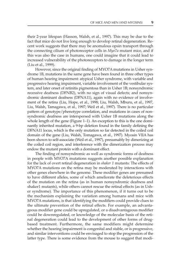
Figure 1–1. Spectrum of MYO7Amutations that lead to Usher syn-drome type 1B, atypical Usher syn-drome, nonsyndromic dominantand recessive deafness (see Liu,Hope, et al., 1998, for details ofsources). The horizontal bar in themiddle represents the myosin VIIAmolecule, with the motor head do-main on the left (dark spots indicat-ing the ATP binding site and theactin binding site), followed by theIQ domain and the coiled coil re-gion, and the tail domain on theright, with different shaded bars in-dicating regions of sequence simi-larity (key to shade coding is givenat the bottom). The position of theseven known shaker1 mouse muta-tions are shown at the bottom, andthe human mutations at the top.Scale bar represents numbers ofamino acids. (Reprinted with per-mission from “Mutations in theMyosin VIIA Gene Cause a WidePhenotypie Spectrum IncludingAtypical Usher Syndrome,” byLiu, Hope, et al., 1998, Fig. 2,American Journal of Human Ge-netics, 63, 909–912. Copyright1998 University of Chicago Press.)
8
A. thaliana CBPS. tubersum, N. tabacum[kinesin tail homology]
their 2-year lifespan (Hasson, Walsh, et al., 1997). This may be due to thefact that mice do not live long enough to develop retinal degeneration. Re-cent work suggests that there may be anomalous opsin transport throughthe connecting cilium of photoreceptor cells in Myo7a mutant mice, and ifthis was also the case in humans, one could imagine that it could lead toincreased vulnerability of the photoreceptors to damage in the longer term(Liu et al., 1999).
However, since the original finding of MYO7A mutations in Usher syn-drome 1B, mutations in the same gene have been found in three other typesof human hearing impairment: atypical Usher syndrome, with variable andprogressive hearing impairment, variable involvement of the vestibular sys-tem, and later onset of retinitis pigmentosa than in Usher 1B; nonsyndromicrecessive deafness (DFNB2), with no sign of visual defects; and nonsyn-dromic dominant deafness (DFNA11), again with no evidence of involve-ment of the retina (Liu, Hope, et al., 1998; Liu, Walsh, Mburu, et al., 1997,Liu, Walsh, Tamagawa, et al., 1997; Weil et al., 1997). There is no particularpattern of genotype/phenotype correlation, and mutations in cases of non-syndromic deafness are interspersed with Usher 1B mutations along thewhole length of the gene (Figure 1–1). An exception to this is the one domi-nantly inherited mutation, a 9-bp deletion found in the family defining theDFNA11 locus, which is the only mutation so far detected in the coiled coildomain of the gene (Liu, Walsh, Tomagawa, et al., 1997). Myosin VIIA hasbeen shown to self-associate (Weil et al., 1997), presumably by dimerizing atthe coiled coil region, and interference with the dimerization process mayendow the mutant protein with a dominant effect.
The finding of nonsyndromic as well as syndromic forms of deafnessin people with MYO7A mutations suggests another possible explanationfor the lack of overt retinal degeneration in shaker 1 mutants: The effects ofMYO7A mutations on the retina may be moderated by interactions withother genes elsewhere in the genome. These modifier genes are presumedto have different alleles, some of which ameliorate the deleterious effectsof the mutation on the retina (as in human nonsyndromic deafness andshaker1 mutants), while others cannot rescue the retinal effects (as in Ush-er syndrome). The importance of this phenomenon, if it turns out to bethe mechanism explaining the variation among humans and mice withMYO7A mutations, is that identifying the modifiers could provide clues tothe ultimate prevention of the retinal effects. For example, an advanta-geous modifier gene could be upregulated, or a disadvantageous modifiercould be downregulated, or knowledge of the molecular basis of the reti-nal degeneration could lead to the development of other forms of drug-based treatment. Furthermore, the same modifiers might determinewhether the hearing impairment is congenital and stable, or is progressive,and similar interventions could be envisaged to stop the progression of thelatter type. There is some evidence from the mouse to suggest that modi-
OF MICE AND MEN (AND MYOSINS) 9
fiers may influence the effects of the original, mildest Myo7a mutation sofar characterized (Myo7ash1), in that changing the genetic background ofthis mutation alters the progression of the hearing loss and leads to histo-logical differences in the organ of Corti (Deol, 1956; Emmerling & Sobkow-icz, 1990).
Role of Myosin VIIA in Hair Cell Developmentand Function
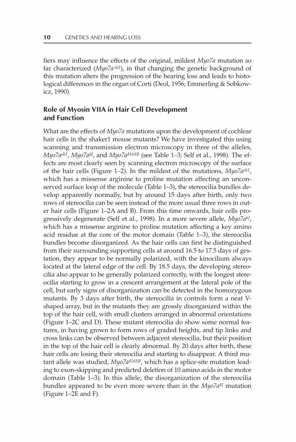
What are the effects of Myo7a mutations upon the development of cochlearhair cells in the shaker1 mouse mutants? We have investigated this usingscanning and transmission electron microscopy in three of the alleles,Myo7ash1, Myo7a6J, and Myo7a816SB (see Table 1–3; Self et al., 1998). The ef-fects are most clearly seen by scanning electron microscopy of the surfaceof the hair cells (Figure 1–2). In the mildest of the mutations, Myo7ash1,which has a missense arginine to proline mutation affecting an uncon-served surface loop of the molecule (Table 1–3), the stereocilia bundles de-velop apparently normally, but by around 15 days after birth, only tworows of stereocilia can be seen instead of the more usual three rows in out-er hair cells (Figure 1–2A and B). From this time onwards, hair cells pro-gressively degenerate (Self et al., 1998). In a more severe allele, Myo7a6J,which has a missense arginine to proline mutation affecting a key aminoacid residue at the core of the motor domain (Table 1–3), the stereociliabundles become disorganized. As the hair cells can first be distinguishedfrom their surrounding supporting cells at around 16.5 to 17.5 days of ges-tation, they appear to be normally polarized, with the kinocilium alwayslocated at the lateral edge of the cell. By 18.5 days, the developing stereo-cilia also appear to be generally polarized correctly, with the longest stere-ocilia starting to grow in a crescent arrangement at the lateral pole of thecell, but early signs of disorganization can be detected in the homozygousmutants. By 3 days after birth, the stereocilia in controls form a neat V-shaped array, but in the mutants they are grossly disorganized within thetop of the hair cell, with small clusters arranged in abnormal orientations(Figure 1–2C and D). These mutant stereocilia do show some normal fea-tures, in having grown to form rows of graded heights, and tip links andcross links can be observed between adjacent stereocilia, but their positionin the top of the hair cell is clearly abnormal. By 20 days after birth, thesehair cells are losing their stereocilia and starting to disappear. A third mu-tant allele was studied, Myo7a816SB, which has a splice-site mutation lead-ing to exon-skipping and predicted deletion of 10 amino acids in the motordomain (Table 1–3). In this allele, the disorganization of the stereociliabundles appeared to be even more severe than in the Myo7a6J mutation(Figure 1–2E and F).
10 GENETICS AND HEARING LOSS

Can the pathology of the shaker 1 mutants tell us anything about therole of myosin VIIA in hair cells? We have addressed several possible func-tions by examining the mutants. First, we wondered if myosin VIIA mightbe involved in the condensation of the cuticular plate, which appears
OF MICE AND MEN (AND MYOSINS) 11
Figure 1–2. Scanning electron micrographs from three different shaker 1 alleles. A.Control outer hair cell stereocilia bundle at 15 days after birth, showing three rowsof stereocilia. B. Myo7ash1/Myo7ash1 mutant outer hair cell bundle, showing onlytwo rows of stereocilia. C. Control organ of Corti at 3 days old. D. LittermateMyo7a6J/Myo7a6J mutant at 3 days old, showing disorganized hair bundles. E. Tworows of outer hair cells from a control mouse at 3 days after birth. F. Two rows ofouter hair cells from a mutant Myo7a816SB/Myo7a816SB cochlea, with extensive dis-organization of the stereocilia bundles.
Scale bars represent: A,B 1�m, C,D 10�m, E,F 1�m.

shortly before birth as a dense, organelle-free plate below the apical sur-face of the hair cells, anchoring the rootlets of the stereocilia. If there is adelay or abnormality in the condensation of this plate, then the stereociliamight be free to move around within the top of the hair cell rather than be-come anchored to form the V-shaped array. Transmission electron mi-croscopy of developing hair cells in the mutants showed that the cuticularplate did condense at the correct time, suggesting this hypothesis was notthe explanation for the disorganization of stereocilia bundles (Self et al.,1998). Furthermore, there was no obvious difference in the ultrastructuralrelationship between stereocilia rootlets and the dense material of the cu-ticular plate, so there was no indication from this approach that the stereo-cilia were more loosely anchored in the cuticular plate than normal.
Second, the apical surface of hair cells appears to be a region of muchendocytotic and exocytotic activity, and the resulting membrane turnovercould control the position of the stereocilia, so we investigated whetherthere was evidence for a difference in this activity between mutants andcontrols that might account for the disorganization. Transmission electronmicroscopy showed the presence of endocytotic pits at the apical surfacesof both mutant and control hair cells, and freeze-fracture of apical profilesalso showed ample pits in mutants (Richardson et al., 1997). When thesefreeze-fracture images were quantified, there was no difference in thenumbers of pits on each hair cell between mutants and controls, arguingagainst this hypothesis (Table 1–4). However, the total apical surface areaof mutant hair cells was significantly greater than in controls, which couldindicate that membrane turnover was not completely normal in mutants(Table 1–4; Richardson et al., 1997). A further indication that membraneprocesses were not quite normal in mutants came from the observationthat radiolabeled aminoglycoside antibiotics were not taken up in largequantities by mutant hair cells, whereas normal hair cells seem specificallyto take up these drugs (Richardson et al., 1997). The explanation for thelack of uptake of aminoglycosides by Myo7a6J mutant hair cells is not obvi-ous (see Richardson et al., 1997, for discussion), but the finding clearly im-plicates myosin VIIA in the process. Finally, ferritin uptake by Myo7a6J mu-tant hair cells in organ culture was normal, indicating that fluid-phase
12 GENETICS AND HEARING LOSS
Table 1–4. Endocytotic pits in apical profiles of basal OHCs in culture.
Pits per Apex Apex Area, �2 Pits per �2
+/Myo7a6J 102+42 38+10 2.73+0.84
Myo7a6J/Myo7a6J 97+25 46+9* 2.17+0.58*
*Significantly different from heterozygote (p<0.05); n = 29 and 25.
Source: Data from Richardson et al. (1997).

uptake was probably normal, in contrast to the aminoglycoside uptake,which was not normal (Richardson et al., 1997). This difference could re-sult from different mechanisms of uptake, with ferritin being taken up in anonspecific manner and aminoglycosides being taken up by specific re-ceptors which in turn depend on normal myosin VIIA function (Richard-son et al., 1997). In summary, there is little evidence for a general abnor-mality in membrane turnover at the apical surface of Myo7a6J hair cellsthat could account for the stereocilia disorganization.
Third, we examined carefully the relationship between the kinociliumand the arrangement of the stereocilia bundle, because the location of thekinocilium at the point of the V-shape led previous investigators to specu-late that the kinocilium might control bundle formation. As already men-tioned, the kinocilium appears to be correctly located at the lateral pole ofthe hair cells from the earliest stages studied. However, in the Myo7a6J andMyo7a816SB mutants at 3 days after birth, we saw no obvious relationshipbetween the position of the kinocilium and the clusters of stereocilia, indi-cating that either the kinocilium has no role in organizing the stereociliabundle or that it requires functional myosin VIIA molecules to carry outthis role.
Fourth, we investigated the suggestion that myosin VIIA might pro-vide the intracellular anchor of cross links between adjacent stereocilia(Hasson, Gillespie, et al., 1997). The reason for this hypothesis was thatmyosin VIIA was found to be enriched at the membranes of stereocilia,and in frog sacculus hair cells it was particularly concentrated in a bandjust above the basal tapers, corresponding to the location of a particulartype of interstereocilia link called the ankle link (Hasson, Gillespie, et al.,1997a). In mammals, these ankle links are distributed along the length ofthe stereocilium rather than being concentrated near the ankles, andmyosin VIIA is also evenly distributed along the stereocilium. If thesecrosslinks between adjacent stereocilia were abnormal in some way in theshaker1 mutants, this could possibly lead to drifting apart of clusters ofstereocilia as we observed in some of the shaker1 alleles. We looked atcrosslinks qualitatively by field emission electron microscopy, and foundno obvious differences between Myo7a6Jor Myo7ash1 mutants and controlsin the appearance of these links. We also examined ultrathin sectionsacross the stereocilia bundle and measured distances between stereociliawithin clusters in Myo7a6J mutants and compared these with controls, butthere was no significant difference in spacing between mutants and con-trols (Self & Steel, unpublished data). These observations did not supportthe suggestion that crosslinks might be the mediator of stereocilia bundledisorganization, at least in the Myo7a6J allele.
Thus, we have not yet been able to establish decisively the role ofmyosin VIIA in hair cell development, but we have succeeded in ruling outsome of the more obvious hypotheses to explain the disorganized hair bun-
OF MICE AND MEN (AND MYOSINS) 13
dle. What of hair cell function? Gross recordings of compound action po-tentials, cochlear microphonics, and summating potentials in the three al-leles described above correlated broadly with the ultrastructural observa-tions. The two most severely affected alleles, Myo7a6J and Myo7a816SB,showed very little or no response to tone-burst stimuli, while the originalsh1 allele, Myo7ash1, which has the mildest ultrastructural defects of the al-leles studied, showed some responses for a short period after the normaltime of onset around 12 to 14 days after birth, although these were never ofnormal threshold and quickly deteriorated as the mutants aged (Harvey1989; Self et al., 1998; Steel & Harvey, 1992). However, measurements oftransducer currents from single outer hair cells in organ culture at 1 to 3days after birth revealed that, at this stage, transducer currents could be ob-tained from Myo7a6J mutants and were of similar size to those obtained fromcontrols (Richardson et al., 1997). Single hair cell recordings from neonatalcultured specimens are likely to be very helpful in understanding the rolenot only of myosin VIIA but also of other molecules involved in geneticdeafness where hair cells seem to be the primary target of the mutations.
Snell’s Waltzer
The second gene to be identified as affecting hair cells directly surprisinglyalso turned out to encode an unconventional myosin molecule, myosin VI.The mutant is Snell’s waltzer, another of the classic deaf mouse mutants,first described by Deol and Green in 1966. It shows progressive hair cellloss and deafness, combined with hyperactivity, head-tossing, and circlingindicative of vestibular dysfunction. The myosin VI gene, Myo6, was iden-tified by positional cloning, and an intragenic deletion of 130 bp was dis-covered in the mutant allele, leading to a frameshift and predicted trunca-tion of the product (Avraham et al., 1995). No myosin VI protein could bedetected in any tissue, suggesting it is a null allele. A second allele in-volved an inversion associated with greatly reduced myosin VI proteinlevels, but this allele has not been so well characterized so we describe on-ly the original allele below. The human MYO6 gene has been isolated, butno mutations in families with hearing impairment have yet been discov-ered (Avraham et al., 1997).
Myosin VI is widely expressed in many tissues, but only inner ear de-fects have been noted in Snell’s waltzer mutants, suggesting that themyosin VI molecule is redundant in other locations (Avraham et al., 1995;Buss et al., 1998; Hasson, Gillespie et al., 1997). In the inner ear, myosin VIis found in hair cells and is particularly concentrated in the vesicle-richpericuticular necklace and in the cuticular plate (Hasson, Gillespie et al.,
14 GENETICS AND HEARING LOSS

1997). In mammals, it was not detected in stereocilia, and the recent reportthat myosin VI runs in the opposite direction along actin filaments (to-ward the minus end) compared with other myosin molecules might ex-plain why it does not enter stereocilia while other hair cell myosins do(Wells et al., 1999).
We have looked at the Snell’s waltzer cochlea to investigate the role ofmyosin VI in hair cell development (Self et al., 1999). Like myosin VIIA,myosin VI is involved in the development of stereocilia bundles, but themechanism seems to be different, with early stereocilia fusion being aprominent feature. In Snell’s waltzer hair cells at birth, stereocilia bundlesappear largely normal, with the tallest stereocilia arranged in a crescentshape at the lateral edge of the cell apex, but minor signs of disorganiza-tion (a “swirling” appearance) can be found in a few cells. Transmissionelectron microscopy at this stage shows a few places where apical hair cellmembranes are slightly raised between adjacent stereocilia (Self et al.,1999). One day later, more of the hair bundles are disorganized, and signsof fusion can be seen by scanning electron microscopy. By 3 days afterbirth, extensive fusion can be seen, associated with obvious disorganiza-tion of bundles (Figure 1–3A and B). The fusion appears to start at the baseand progress toward the tips of the stereocilia, as if the membranes are zip-ping up. By 7 days, most hair cells have only a few fused stereocilia (Fig-ure 1–3 C–F), and these continue to fuse and also grow in length, so that by20 days the hair cells sport giant fused stereocilia, sometimes withswellings at or near their tips (Figure 1–4) (Self et al., 1999).
The fusion of membranes between stereocilia seems to be the majorpathological feature in Snell’s waltzer, and could presumably account forthe other observed changes (disorganization and swirling of bundles,growth of giant stereocilia, ultimate degeneration of hair cells). We haveconsidered several possible explanations for the fusion. First, abnormalmembrane turnover might lead to a net loss of apical membrane, and con-sequent pulling up of membrane between stereocilia. This seems unlikely,because stereocilia continue to grow during the period studied, ratherthan being forced back into the cell to minimize apical membrane area.Furthermore, we see no evidence of abnormal membrane turnover in mu-tant hair cells: Endocytotic pits of normal ultrastructural appearance aredetected at the apical surface, and the membrane dye FM1-43 is taken upby mutant as well as by control hair cells, suggesting active endocytosisoccurs in mutants, although neither of these approaches was quantified(Self et al., 1999).
A second explanation might be that myosin VI could serve to anchorthe stereocilia rootlets into the cuticular plate, and in its absence the stereo-cilia could pull out of the plate, dragging membrane with them. This also
OF MICE AND MEN (AND MYOSINS) 15

seems an unlikely explanation, because we might expect the stereocilia toappear to extend (by scanning electron microscopy of the upper surface ofthe hair cell) before the fusion is observed, but instead we see fusion prior
16 GENETICS AND HEARING LOSS
Figure 1–3. Scanning electron micrographs of the Snell’s waltzer mouse mutant. A.Control outer hair cells at 3 days of age. B. Myo6sv/Myo6sv mutant outer hair cells,showing fusion of stereocilia starting from the base, and disorganization of the bun-dle. C. Control inner hair cells from a 7-day-old mouse. D. Myo6sv/Myo6sv mutantinner hair cells, showing further fusion of stereocilia and excess growth. E. Controlorgan of Corti at 7 days old. F. Myo6sv/Myo6sv organ of Corti at 7 days of age, show-ing extensive disorganization and fusion of stereocilia bundles in all rows of haircells.
Scale bars represent: A,B,C,D, 1�m, E,F, 10�m.

Figure 1–4. Scanning electron micrographs of further Snell’s waltzer hair cells. A. 12-day-old mutant inner hair cells. B. 20-day-old mu-tant inner hair cells, showing the growth of giant stereocilia.
Scale bar represents 1�m.
17
to extension. Furthermore, we saw no obvious detachment of stereociliarootlets by transmission electron microscopy in neonatal stages.
Third, myosin VI might be involved in the delivery of components tothe growing stereocilia, because unconventional myosins are often as-sumed to have a primary cargo-carrying role, and in the Drosophila em-bryo, the myosin VI homologue has been shown directly to mediate vesi-cle transport (Mermall, McNally, & Miller, 1994; Mermall, Post, &Mooseker, 1998). If the delivery of stereocilia components is not properlybalanced, the result might be the uncontrolled growth of stereocilia thatwe observe. However, it is difficult to imagine how this explanation couldaccount for the initial fusion of membranes.
Our fourth and favored hypothesis is that myosin VI might normallybe involved in anchoring the apical cell membrane to the actin in the cutic-ular plate (Self et al., 1999). In this model, the motor end of the myosin VImolecule would attach to the abundant actin in the cuticular plate, whilethe tail of the myosin would attach directly or indirectly to the apicalmembrane, serving to hold down the membrane between adjacent stereo-cilia. The natural tendency of a lipid-rich cell membrane in an aqueous en-vironment would be to form a sphere with a minimal surface area to mini-mize surface tension, and any other configuration must be activelymaintained. The arrangement of stereocilia, with closely apposed mem-branes, seems likely to require very active maintenance to avoid the ten-dency to fusion. In the absence of adequate anchoring, the membranes be-tween stereocilia may proceed to zip up, rising to the tops of the stereociliaand leading to fusion. This explanation does not account for the continuedgrowth of stereocilia to form giant protrusions on the apical surface, butonce fusion has started, the whole process of stereocilia growth and main-tenance will likely be disrupted, and the excess growth may be secondaryto the fusion.
In summary, the myosin VI molecule appears to have a role in haircell development and function that is distinct from the role of myosinVIIA, as might be expected from the fact that both are essential moleculesfor normal cochlear function. A further unconventional myosin, myosinXV (Myo15), has more recently also been discovered to underlie bothmouse and human deafness, in the shaker 2 mouse mutant and the reces-sive deafness DFNB3 (Probst et al., 1998; Wang et al., 1998). In the shaker 2mutants, a further distinct stereocilia defect is found: They are much short-er than normal. These three molecules are among the first that have beendemonstrated to have an essential role in hair cells and represent the startof the process of unraveling the molecular basis of auditory function. Incontrast to the visual or olfactory systems, we know very little about themolecular basis of hair cell function, and a genetic approach is proving tobe very helpful in identifying essential molecular components.
18 GENETICS AND HEARING LOSS

IONIC HOMEOSTASIS INTHE COCHLEAR DUCT
It is perhaps not surprising that there has been a great deal of focus on haircells, and in particular hair cell degeneration, in investigating the cellularbasis of hearing impairment. Much effort in the past has been expended incounting degenerating and missing hair cells and correlating these countswith auditory function. With the extended use of more detailed ways ofassessing the state of the cochlear duct, such as scanning and transmissionelectron microscopy, it has become clear that hair cell degeneration israrely, if ever, the cause of hearing impairment in hereditary deafness, butthat instead the hair cells may have distinct developmental or functionaldefects that cause the hearing impairment and lead to secondary hair celldegeneration.
However, one of the striking realizations from the research in both hu-man and mouse genetic deafness over the past few years has been the im-portance of the rest of the cochlear duct, and in particular the roles of dif-ferent cell types in maintaining cochlear homeostasis (Steel & Bussoli,1999). It is not a new observation that the stria vascularis is important forcochlear function (e.g., Schuknecht, 1974; Steel & Bock, 1983), but the num-ber of different deafness genes that appear to have a primary role inhomeostasis is surprisingly larger than the number so far identified thataffect hair cells directly (Steel & Bussoli, 1999). This is an area where workon animal models has been critical to understanding the processes in-volved, because, for example, measuring the resting potential (the endo-cochlear potential, normally around 80 to 100 mV) in the endolymphbathing the upper surfaces of the sensory hair cells is too invasive to be car-ried out in humans, yet it provides key information about the functionalbasis of the deafness.
Endocochlear potential is necessary for normal hair cell function inthe mammalian cochlea, as it provides a large potential difference acrossthe top of the hair cells, from the positive potential in scala media to thenegative interior of the hair cell, and animals with reduced or absent endo-cochlear potentials have raised thresholds for cochlear responses. The po-tential is generated by the stria vascularis on the lateral wall of thecochlear duct and depends also on adequate electrical resistance of theboundaries of scala media. It is an electrogenic potential generated by thepumping of potassium ions into scala media by the stria. Endolymph has ahigh potassium, low sodium content, and it is thought that, when thetransducer channels in the sensory hair cells are opened by sound stimula-tion, the current passing into the cell down the electrical gradient is mostlycarried by potassium, as it is the predominant cation available. It is be-lieved that the potassium flowing into the hair cells is recycled through the
OF MICE AND MEN (AND MYOSINS) 19

supporting cells and fibrocytes of the cochlear duct, back to the stria vas-cularis for pumping back into the endolymph (e.g., Kikuchi, Kimura, Paul,& Adams, 1995; Spicer & Schulte, 1996) (Figure 1–5). The ionic composi-tion of the endolymph and the endocochlear potential are to some extentseparate aspects of the system, as the high potassium content is estab-lished before the endocochlear potential during development, andvestibular endolymph has a similar high potassium, low sodium contentbut has no high resting potential like cochlear endolymph.
The first attempts to record endocochlear potentials in animal modelsof hereditary deafness with strial defects were reported by Suga and Hat-tler, who looked at two deaf white cats and three deaf dalmatian dogs, andfailed to record any endocochlear potential (Suga & Hattler 1970). Howev-er, the Reissner’s membrane had collapsed extensively in these animals,and there was no indication that the recording electrode had actuallypassed through the small remaining open area of scala media. Later meas-urements of a reduced or absent endocochlear potential in the viable dom-inant spotting mouse mutant (Wv/Wv, since identified as the Kit gene en-coding a growth factor) were corroborated by serial 1 micron-thicksections through the cochlea, to show that the hole in the lateral wall of thecochlear duct made by the recording electrode did open into an open scalamedia (Steel & Barkway, 1989; Steel, Barkway, & Bock, 1987). Since theseearly reports, several other mouse mutants have been shown to have anopen scala media but a reduced or absent endocochlear potential, such asfurther alleles of the Kit gene (Cable, Huszar, Jaenisch, & Steel, 1994), thesteel-dickie mutant (Sld, since identified as the ligand of the Kit growth fac-tor, Mgf; Steel, Davidson, & Jackson, 1992), the light allele of brown(Tyrp1lt; Cable, Jackson, & Steel, 1993), the varitint-waddler mutant (VaJ;Cable & Steel, 1998), and most recently the Pou3f4 mouse mutant (Minowaet al., 1999; Steel, 1999).
The Kit, Mgf, VaJ, and Tyrp1lt mouse mutants mentioned above havein common pigmentation anomalies, such that parts or all of their bodiesare devoid of the pigment cells, melanocytes, leading to white coats andalso to a lack of melanocytes in the stria vascularis in their cochleas. Thecells themselves are missing, rather than being present but unable to pro-duce pigment as in albinos, and we have demonstrated in the Kit and Mgfmutant alleles we studied that the precursors of these melanocytes migratefrom the neural crest as usual, but then frequently fail to survive when theyreach their target tissue, such as the inner ear (Cable, Jackson, & Steel, 1995;Steel et al., 1992). The absence of melanocytes is invariably associated withabsence of endocochlear potential, suggesting a causative relationship (Ca-ble, Huszar, Jaenisch, & Steel, 1994). Melanocyte-like cells form the interme-diate cells of the stria, scattered between the marginal cells on the lumenalside and the basal cells adjacent to the fibrocytes forming the outer wall ofthe cochlear duct. Their role in generating the endocochlear potential is not
20 GENETICS AND HEARING LOSS

21
Figure 1–5. Illustration of the cochlear duct, with the proposed routes for potassi-um recycling indicated with arrows. Dark shading represents the expression of KC-NQ4 in outer hair cells, which may allow potassium to be removed from basolater-al hair cell membranes. Pale grey regions represent the areas connected by gapjunctions, thought to permit the flow of potassium back to the stria vascularis, andthe gap junctional connexins encoded by GJB2, GJB3, and GJB6 are expressed inparts of this cell network. The transcription factor gene POU3F4 is also thought tobe involved in potassium recycling by allowing extensive intercellular contacts andis expressed in the spiral ligament, among other places. The marginal cells of thestria vascularis are shaded medium gray, and these express the SLC12A2 gene ontheir basolateral surfaces and the KCNQ1 and KCNE1 genes on their lumenal sur-faces; the cotransporter encoded by SLC12A2 participates in potassium pumpinginto the marginal cell, while the products of the KCNQ1 and KCNE1 genes togetherform a channel through which potassium can flow into the endolymph, restoringthe high potassium concentration there. (ihc, inner hair cells; ohc, outer hair cells;p, pillar cells; d, Deiter’s cells; h, Hensen’s cells; c, Claudius cells; os, outer spiralsulcus cells; sp, spiral prominance; sl, spiral limbus; b, basal cells of stria vascu-laris; i, intermediate [melanocyte-like] cells of the stria vascularis; m, marginal cellsof the stria vascularis; id, interdental cells; tm, tectorial membrane; slm, spiral lim-bus; is, inner sulcus cells.) (Adapted with permission from “Genes Involved in Deaf-ness,” by R. H. Holmes and K. P. Steel, 1999. Current Opinion in Genetics and De-velopment, 9, 304–314. Figure prepared by Sarah Holme and Ralph Holme.)

known, but whatever their function is, it does not appear necessary for thesecretion of some sort of endolymph, because there is an open scala mediaand no collapse of Reissner’s membrane in the mutants. Other mouse mu-tants (and other animals with deafness and white spotting of the coat) havea similar pigmentation defect, due to a lack of melanocytes, so may alsohave reduced or absent endocochlear potentials, but such measurementshave not yet been reported (see Steel, 1995, for a review).
As mentioned above, a number of deafness genes have recently beenidentified, in both human and mouse, that appear to function directly inpotassium recycling in the cochlear duct (for review, see Holt & Corey,1999; Steel, 1999; Steel & Bussoli, 1999) (see Figure 1–5). KCNQ4 encodes apotassium channel expressed in outer hair cells and is thought to be in-volved in removing potassium from the cells via their basolateral mem-branes (Kubisch et al., 1999); the gene is mutated in a form of autosomaldominant deafness in humans. From here the potassium is believed topass through an extensive network of gap junctions between the support-ing cells of the organ of Corti and the fibrocytes forming the spiral liga-ment on the lateral wall, back to the stria vascularis ( Figure 1–5). At leastthree connexin genes involved in forming these gap junctions have beenfound to be mutated in different types of human genetic deafness. Theseinclude the GJB2 gene, encoding connexin 26, mutated in both recessiveand dominant forms of nonsyndromic deafness; the GJB3 gene, encodingconnexin 31, mutated in dominant nonsyndromic deafness; and the GJB6gene, encoding the connexin 30 molecule, which has most recently beenreported to underlie another type of dominant nonsyndromic deafness(Denoyelle et al., 1998; Grifa et al., 1999; Kelsell et al., 1997; Xia et al., 1999).
The Pou3f4 mouse mutant, with a targeted deletion of the gene, pro-vides evidence that this gene is involved in some aspect of potassium recy-cling, although the mouse has a reduced endocochlear potential and nocollapse of scala media, suggesting that endolymph secretion can still bemaintained in the mutant (Minowa et al., 1999). The only ultrastructuraldefect reported was a reduction in the intercellular contacts between adja-cent fibrocytes in the spiral ligament on the lateral wall, corresponding tothe site of expression of this transcription factor, which is restricted to mes-enchymal cells around the inner ear, including the spiral ligament fibro-cytes. It is possible that these restricted contacts inhibit potassium recy-cling to some extent.
Having reached the stria, the potassium is pumped into the marginalcells across their basolateral membranes by a Na-K-ATPase acting in con-cert with a Na-K-Cl cotransporter (e.g., Wangemann, 1995). This cotrans-porter has recently been identified as the product of the Slc12a2 gene,because this gene was found to be mutated in two alleles at the shaker-with-syndactylism (sy) locus; in the original sy mutation, the gene is com-pletely deleted, and in the syns allele, there is an insertion of an additional
22 GENETICS AND HEARING LOSS

adenine in a string of six adenine bases, leading to a frameshift and prema-ture termination codon (Dixon et al., 1999). We found in the syns mutant, aspreviously reported for the sy mutant (Deol, 1963), that there is extensivecollapse of Reissner’s membrane and the walls of the vestibular compart-ments of the inner ear, suggesting that there is complete failure of en-dolymph secretion (Dixon et al., 1999). A knockout of the same gene hasalso been reported with the same collapse of endolymphatic compart-ments (Delpire, Lu, England, Dull, & Thorne, 1999).
Finally, the KCNQ1 (KvLQT1) and KCNE1 (ISK) genes are both in-volved in forming a potassium channel at the lumenal surface of the mar-ginal cells, which allows the potassium pumped into the cell to flow out,down its electrochemical gradient, into the endolymph, restoring thepotassium concentration there. Both genes have been found to be mutatedin different families with Jervell and Lange-Nielsen syndrome (Neyroud etal., 1997; Schultze-Bahr et al., 1997). In the mouse mutant with the Isk geneinactivated, there is a failure of endolymph secretion and collapse of Reiss-ner’s membrane (Vetter et al., 1996).
In addition to the genes involved in potassium recycling, two othergenes are likely to be involved in maintaining endolymphatic homeosta-sis. The pendrin gene (PDS) is mutated in Pendred syndrome and somecases of nonsyndromic deafness; it is expressed in a small group of epithe-lial cells just below the stria vascularis (spiral prominance and outer sulcusregion), and the gene is believed to be involved in iodide and chloridetransport (Everett, Morsli, Wu, & Green, 1999). Another gene, ATP6B1 en-coding the B1 subunit of the H+-ATPase proton pump, is probably in-volved in adjusting the pH of the endolymph by pumping hydrogen ionsinto scala media; this gene is expressed in the interdental cells, on the up-per surface of the spiral limbus just below the attachment of the tectorialmembrane, and mutations in humans cause renal tubular acidosis withdeafness (Karet et al., 1999).
The number of genes now known to be involved in controlling ionichomeostasis within the cochlear duct and known to be vital for normal au-ditory function does suggest that this is an aspect of cochlear function thatneeds careful study. In particular, the finding that one of the genes, theGJB2 gene encoding connexin 26, is quite frequently involved in nonsyn-dromic deafness in the human population suggests that this will become amuch more active area of research in the future.
MOVING FORWARD
All areas of genetic research are moving forward very rapidly, and the im-minent release of extensive sequence of the human genome will revolu-
OF MICE AND MEN (AND MYOSINS) 23

tionize progress. However, identifying the function of genes discovered bysequence analysis will still require much empirical laboratory work. Anumber of deafness genes have been found by targeted knockout bygroups interested in a particular gene family, but many others have beenidentified by starting with a mutant mouse (or human) and identifying thegene by positional cloning. The advantage of the latter approach is thatyou know before starting whether there is a phenotype (deafness) that isof interest to you, although the disadvantage is that positional cloning stillrepresents a considerable effort. However, positional cloning in the mouseis still somewhat easier than positional cloning of human deafness genes.Having identified a mouse deafness gene, it is a relatively straightforwardstep to clone the orthologous human gene and search for mutations in it indeaf humans.
A limiting factor in our progress in identifying deafness genes is thefact that there is a gap between the number of mouse mutants that wewould expect to find and the number available. This may be becausemouse mutants with deafness alone would not normally be detected byanimal handlers, so new deaf mutants are likely to be lost. We know thesemutants are missing because when we compare the locations of the knowndeafness genes in the human genome with the corresponding regions ofthe mouse genome, there are many cases where there is no potential deafmouse model mapping to the appropriate chromosomal region. Equally,there are many deaf mouse mutants for which no human equivalent hasyet been found, such as the Myo6 mutation described earlier.
One way forward that we have adopted is to generate many moredeaf mouse mutants by a programme of random mutagenesis. Male miceare injected with a powerful mutagen, N-ethyl-N-nitrosourea (ENU), mat-ed to normal females, and the offspring screened for new dominant muta-tions. The new deaf mutants are detected by their failure to respond withan ear flick (Preyer reflex) to a brief, high-frequency toneburst. New mu-tants with balance defects are picked up by their characteristic hyperac-tive, head-tossing, and/or circling behavior, and these often have hearingimpairment in addition to their vestibular dysfunction. This approach hasbeen successful in producing many new mutants, some with completelynew phenotypes, and we are localizing the mutations as well as character-izing the auditory defects in them all. We anticipate that some of these atleast should provide models for known human forms of genetic deafness,and facilitate the search for the responsible gene.
Having mouse models for each form of human deafness will allow usto make progress in understanding the underlying pathological processesat a molecular and cellular level, and it is only when we have this under-standing that we can devise ways of treating this most common form ofsensory impairment in our population.
24 GENETICS AND HEARING LOSS

Acknowledgments: KPS would like to thank the many collaborators, in Notting-ham and around the world, who have worked with her over the years, and helpedto develop the ideas and expertise we now have in our studies of deaf mouse mu-tants. However, she would also like to take this opportunity to thank several otherpeople who have made important contributions in shaping the direction of herwork: Mr Savory and Mr Rix, the teachers who first introduced her to biology atschool; Dorothy Hodgkin, whose lecture to sixth-form school students about solv-ing the insulin structure provided much early inspiration; Malkiat Deol, her Ph.D.advisor, who first introduced her to deaf mouse mutants; Bill Kimberling, whosesheer enthusiasm first persuaded her that identifying deafness genes was worth-while; and Steve Brown, who agreed to help. KPS is supported by the MRC, De-feating Deafness, and the European Commission (Contracts BMH4-CT96-1324 andCT97-2715).
This chapter is dedicated to the memory of Professor Malkiat S. Deol, whodied in June 1999.
REFERENCES
Avraham, K. B., Hasson, T., Sobe, T., Balsara, B., Testa, J. R., Skvorak, A. B., Morton,C. C., Copeland, N. G., & Jenkins, N. A. (1997). Characterisation of unconven-tional MYO6, the human homologue of the gene responsible for deafness inSnell’s waltzer mice. Human Molecular Genetics, 6, 1225–1231.
Avraham, K. B., Hasson, T., Steel, K. P., Kingsley, D. M., Russell, L. B., Mooseker, M.S., Copeland, N. G., & Jenkins, N. A. (1995). The mouse Snell’s waltzer deafnessgene encodes an unconventional myosin required for structural integrity of in-ner hair cells. Nature Genetics, 11, 369–375.
Buss, F., Kendrick-Jones, J., Lionne, C., Knoght, A. E., Côté, G. P., & Luzio, J. P.(1998). The localisation of myosin VII at the Golgi complex and leading edge offibroblasts and its phosphorylation and recruitment into membrane ruffles ofA431 cells after growth factor stimulation. Journal of Cell Biology, 143, 1535–1545.
Bussoli, T. J., & Steel, K. P. (1999). Gene expression in the developing ear.http://www.ihr.mrc.ac.uk/Hereditary/genetable/index.html.
Cable, J., Huszar, D., Jaenisch, R., & Steel, K. P. (1994). Effects of mutations at the Wlocus (c-kit) on inner ear pigmentation and function in the mouse. Pigment CellResearch, 7, 17–32.
Cable, J., Jackson, I. J., Steel, K. P. (1993). Light (Blt), a mutation causing melanocytedeath, leads to strial dysfunction in the inner ear. Pigment Cell Research, 6,215–225.
Cable, J., Jackson, I. J. & Steel, K. P. (1995). Mutations at the W locus affect survivalof neural crest-derived melanocytes in the mouse. Mechanisms of Development,50, 139–150.
Cable J., & Steel, K. P. (1998). Combined cochleo-saccular and neuroepithelial ab-normalities in the Varitint-waddler-J (VaJ) mouse. Hearing Research, 123, 125–136.
Delpire, E., Lu, J., England, R., Dull, C., & Thorne, T. (1999). Deafness and imbal-ance associated with inactivation of the secretory Na-K-2Cl cotransporter. Na-ture Genetics, 22, 192–195.
OF MICE AND MEN (AND MYOSINS) 25
Denoyelle, F., LinaGranade, G., Plauchu, H., Bruzzone, R., Chaib, H., LeviAcobas,F., Weil, D., & Petit, C. (1998). Connexin 26 gene linked to a dominant deafness.Nature, 393, 319–320.
Denoyelle F., Marlin, S., Weil, D., Moatti, L., Chauvin, P., Garabédian, E. N., & Petit,C. (1999). Clinical features of the prevalent form of childhood deafness, DFNB1,due to a connexin-26 gene defect: Implications for genetic counselling. Lancet,353, 1298–1303.
Denoyelle, F., Weil, D., Maw, M. A., Wilcox, S. A., Lench, N. J., Allen-Powell, D. R.,Osborn, A. H., Dahl, H. H. M., Middleton, A., Houseman, M. J., Dodé, C., Mar-lin, S., Boulila-ElGaïed, A., Grati, M., Ayadi, H., BenArab, S., Bitoun, P., Lina-Granade, G., Godet, J., Mustapha, M., Loiselet, J., El-Zir, E., Aubois, A., Joan-nard, A., Levilliers, J., Garabédian, E. N., Mueller, R. F., Gardner, R. J. M., &Petit, C. (1997). Prelingual deafness: High prevalence of a 30delG mutation inthe connexin 26 gene. Human Molecular Genetics, 6, 2173–2177.
Deol, M. S. (1956). The anatomy and development of the mutants pirouette, shak-er1 and waltzer in the mouse. Proceedings of the Royal Society London, Ser. B, 145,206–213.
Deol, M. S. (1963). The development of the inner ear in mice homozygous for shak-er-with-syndactylism. Journal of Embryology and Expimental Morphology, 11,493–512.
Deol, M. S., & Green, M. C. (1966). Snell’s waltzer, a new mutation affecting behav-ior and the inner ear in the mouse. Genetic Research Cambridge, 8, 339–345.
Dixon, M. J., Gazzard, J., Chaudhry, S. S., Sampson, N., Schulte, B. A., & Steel, K. P.(1999). Mutation of the basolateral Na-K-Cl cotransporter gene Slc12a2 results indeafness in mice. Human Molecular Genetics, 8, 1579–1584.
El-Amraoui, A., Sahly, I., Picaud, S., Sahel, J., Abitbol, M., & Petit, C. (1996). HumanUsher 1B/mouse shaker1: The retinal phenotype discrepancy explained bypresence/absence of myosin VIIA in the photoreceptor cells. Human MolecularGenetics, 5, 1171–1178.
Emmerling, M. R., & Sobkowicz, H. M. (1990). Acetylcholinesterase-positive inner-vation in cochleas from two strains of shaker1 mice. Hearing Research, 47, 25–38.
Estivill, X., Fortina, P., Surrey, S., Rabionet, R., Melchionda, S., D’Agruma, L.,Mansfield, E., Rappaport, E., Govea, N., Milà, M., Zelante, L., & Gasparini, P.(1998). Connexin-26 mutations in sporadic and inherited sensorineural deaf-ness. Lancet, 351, 394–398.
Everett, L. A., Morsli, H., Wu, D. K., & Green, E. D. (1999). Expression pattern of amouse ortholog of the Pendred’s syndrome gene (Pds) suggests a key role forPendrin in the inner ear. Procedings of the National Academy of Science, USA, 96,9727–9732.
Fekete, D. M. (1999). Development of the vertebrate ear: Insight from knockoutsand mutants. Trends in Neurosciences, 22, 263–269.
Fortnum, H., & Davis, A. (1997). Epidemiology of permanent childhood hearingimpairment in Trent region, 1985-1993. British Journal of Audiology, 31, 409–446.
Gibson, F., Walsh, J., Mburu, P., Varela, A., Brown, K. A., Antonio, M., Beisel, K. W.,Steel, K. P., & Brown, S. D. M. (1995). A type VII myosin encoded by the mousedeafness gene shaker-1. Nature, 374, 62–64.
Grifa, A., Wagner, C. A., D’Ambrosio, L., Melchionda, S., Bernardi, F., Lopez-Bigas,N., Rabionet, R., Arbones, M., Della Monica, M., Estivill, X., Zelante, L., Lang, F.,
26 GENETICS AND HEARING LOSS
& Gasparini, P. (1999). Mutations in GJB6 cause nonsyndromic autosomal domi-nant deafness at DFNA3 locus. Nature Genetics, 23, 16–18.
Harvey, D. (1989). Structural and functional development of the cochlea in normal(CBA/Ca) and hearing impaired shaker1 (sh1/sh1) mice [Ph.D. thesis]. University ofNottingham, UK.
Hasson, T., Gillespie, P. G., Garcia, J. A., MacDonald, R. B., Zhao, Y., Yee, A. G.,Mooseker, M. S., & Corey, D. P. (1997). Unconventional myosins in inner earsensory epithelia. Journal of Cell Biology, 137, 1287–1307.
Hasson, T., Heintzelman, J., Santos-Sacchi, J., Corey, D. P., & Mooseker, M. S. (1995).Expression in cochlea and retina of myosin VIIa, the gene product defective inUsher syndrome type 1B. Procedings of the National Academy of Science, USA, 92,9815–9819.
Hasson, T., Walsh, J., Cable, J., Mooseker, M. S., Brown, S. D. M., & Steel, K. P.(1997). Effects of shaker-1 mutations on myosin-VIIa protein and mRNA expres-sion. Cell Motility and the Cytoskeleton, 37, 127–138.
Holme, R. H., & Steel, K. P. (1999). Genes involved in deafness. Current Opinion inGenetics and Development, 9, 309–314.
Holt, J. R., & Corey, D. P. (1999). Ion channel defects in hereditary hearing loss.Neuron, 22, 217–219.
Karet, F. E., Finberg, K. E., Nelson, R. D., Nayir, A., Mocan, H., Sanjad, S. A., Rod-riguez-Soriano, J., Santos, F., Cremers, C. W. R. J., Di Pietro, A., Hoffbrand, B. I.,Winiarski, J., Bakkaloglu, A., Ozen, S., Dusunsel, R., Goodyer, P., Hulten, S. A.,Wu, D. K., Skvorak, A. B., Morton, C. C., Cunningham, M. J., Jha, V., & Lifton, R.P. (1999). Mutations in the gene encoding B1 subunit of H+-ATPase cause renaltubular acidosis with sensorineural deafness. Nature Genetics, 21, 84–90.
Keats, B. J. B., & Berlin, C. I. (1999). Genomics and hearing impairment. Genome Re-search, 9, 7–16.
Kelley, P. M., Harris, D. J., Comer, B. C., Askew, J. W., Fowler, T., Smith, S. D., &Kimberling, W. J. (1998). Novel mutations in the connexin 26 gene (GJB2) thatcause autosomal recessive (DFNB1) hearing loss. American Journal of Human Ge-netics, 62, 792–799.
Kelsell, D. P., Dunlop, J., Stevens, H. P., Lench, N. J., Liang, J. N., Parry, G., Mueller,R. F., & Leigh, I. M. (1997). Connexin26 mutations in hereditary nonsyndromicsensorineural deafness. Nature, 387, 80–83.
Kiernan, A., & Steel, K. P. (in press). Mouse homologues for human deafness. In K.Kitamura & K. P. Steel (Eds.), Genetics in otolaryngology. Basel, Switzerland:Karger.
Kikuchi, T., Kimura, R. S., Paul, D. L., & Adams, J. C. (1995). Gap junctions in therat cochlea: Immunohistochemical and ultrastructural analysis. Anatomy andEmbryology, 191, 101–118.
Kubisch, C., Schroeder, B. C., Friedrich, T., Lütjohann, B., El-Amraoui, A., Marlin,S., Petit, C., & Jentsch, T. J. (1999). KCNQ4, a novel potassium channel ex-pressed in sensory outer hair cells, is mutated in dominant deafness. Cell, 96,437–446.
Liu, X., Ondek, B., & Williams, D. S. (1998). Mutant myosin VIIa causes defectivemelanosome distribution in the RPE of shaker1 mice. Nature Genetics, 19,117–118.
OF MICE AND MEN (AND MYOSINS) 27

Liu, X., Udovichenko, I. P., Brown, S. D. M., Steel, K. P., & Williams, D. S. (1999).Myosin VIIa is required for normal opsin distribution in the photoreceptor cili-um: Implications for blindness in Usher syndrome. Journal of Neuroscience, 19,6267–6274.
Liu, X., Vansant, G., Udovichenko, I. P., Wolfrum, U., & Williams, D. S. (1997).Myosin VIIA, the product of the Usher 1B syndrome gene, is concentrated inthe connecting cilia of photoreceptor cells. Cell Motility and the Cytoskeleton, 37,240–252.
Liu, X.-Z., Hope, C., Walsh, J., Newton, V., Ke, X. M., Liang, C. Y., Xu, L. R., Zhou, J.M., Trump, D., Steel, K. P., Bundey, S., & Brown, S. D. M. (1998). Mutations inthe myosin VIIA gene cause a wide phenotypic spectrum including atypicalUsher syndrome. American Journal of Human Genetics, 63, 909–912.
Liu, X.-Z., Walsh, J., Mburu, P., Kendrick-Jones, J., Cope, J., Steel, K. P., & Brown, S.D. M. (1997). Mutations in the myosin VIIA gene causing nonsyndromic reces-sive deafness. Nature Genetics, 16, 188–190.
Liu, X.-Z., Walsh, J., Tamagawa, Y., Kitamura, K., Nishizawa, M., Steel, K. P., &Brown, S. D. M. (1997). Autosomal dominant nonsyndromic deafness caused bya mutation in the myosin VIIA gene. Nature Genetics, 17, 268–269.
Lord, E. M., & Gates, W. H. (1929). Shaker, a new mutation of the house mouse(Mus musculus). American Naturalist, 63, 435–442.
Mburu, P., Liu, X. Z., Walsh, J., Saw, D., Cope, J. T. V., Gibson, F., Kendrick-Jones, J.,Steel, K. P., & Brown, S. D. M. (1997). Mutation analysis of the mouse myosinVIIA deafness gene—A putative myosin motor-kinesin tail hybrid. Genes andFunction, 1, 191–203.
Mermall, V., McNally, J. G., & Miller, K. G. (1994). Transport of cytoplasmic vesiclescatalysed by an unconventional myosin in living Drosophila embryos. Nature,369, 560–562.
Mermall, V., Post, P. L., & Mooseker, M. S. (1998). Unconventional myosins in cellmovement, membrane traffic, and signal transduction. Science, 279, 527–533.
Minowa, O., Ikeda, K., Sugitani, Y., Oshima, T., Nakai, S., Katori, Y., Suzuki, M., Fu-rukawa, M., Kawase, T., Zheng, Y., Ogura, M., Asada, Y., Watanabe, K., Yamana-ka, H., Gotoh, S., Nishi-Takeshima, M., Sugimoto, T., Kikuchi, T., Takasaka, T., &Noda, T. (1999). Altered cochlear fibrocytes in a mouse model of DFN3 nonsyn-dromic deafness. Science, 285, 1408–1411.
Morell, R. J., Kim, H. J., Hood, L. J., Goforth, L., Friderici, K., Fisher, R., Van Camp,G., Berlin, C. I., Oddoux, C., Ostrer, H., Keats, B., & Friedman, T. B. (1998). Mu-tations in the connexin 26 gene (GJB2) among Ashkenazi Jews with nonsyn-dromic recessive deafness. New England Journal of Medicine, 339, 1500–1505.
Morton, N. E. (1991). Genetic epidemiology of hearing impairment. In R. J. Ruben,T. R. Van De Water, & K. P. Steel (Eds.), Genetics of hearing impairment. Annalsof the New York Academy of Science, 630, 16–31.
Neyroud, N., Tesson, F., Denjoy, I., Leibovici, M., Donger, C., Barhanin, J., Faure, S.,Gary, F., Coumel, P., Petit, C., Schwartz, K., & Guicheney, P. (1997). A novel mu-tation in the potassium channel gene KVLQT1 causes the Jervell and Lange-Nielsen cardioauditory syndrome. Nature Genetics, 15, 186–189.
Online Mendelian Inheritance in Man, OMIM™ Center for Medical Genetics,Johns Hopkins University (Baltimore, MD) and National Center for Biotechnol-
28 GENETICS AND HEARING LOSS

ogy Information, National Library of Medicine (Bethesda, MD), (1999). WorldWide Web URL: http://www.ncbi.nlm.nih.gov/Omim
Probst, F. J., & Camper, S. A. (1999). The role of mouse mutants in the identificationof human hereditary hearing loss genes. Hearing Research, 130, 1–6.
Probst, F. J., Fridell, R. A., Raphael, Y., Saunders, T. L., Wang, A., Liang, Y., Morell,R. J., Touchman, J. W., Lyons, R. H., Noben-Trauth, K., Friedman, T. B., &Camper, S. A. (1998). Correction of deafness in shaker2 mice by an unconven-tional myosin in a BAC transgene. Science, 280, 1444–1447.
Richardson, G. P., Forge, A., Kros, C. J., Fleming, J., Brown, S. D. M., & Steel, K. P.(1997). Myosin VIIA is required for aminoglycoside accumulation in cochlearhair cells. Journal of Neuroscience, 17, 9506–9519.
Schuknecht, H. F. (1974). Pathology of the ear. Cambridge, MA: Harvard UniversityPress.
Schulze-Bahr, E., Wang, Q., Wedekind, H., Haverkamp, W., Chen, Q. Y., Sun, Y. L.,Rubie, C., Hordt, M., Towbin, J. A., Borggrefe, M., Assmann, G., Qu, X. D.,Somberg, J. C., Breithardt, G., Oberti, C., & Funke, H. (1997). KCNE1 mutationscause Jervell and Lange-Nielsen syndrome. Nature Genetics, 17, 267–268.
Self, T., Mahony, M., Fleming, J., Walsh, J., Brown, S. D. M., & Steel, K. P. (1998).Shaker-1 mutations reveal roles for myosin VIIA in both development and func-tion of cochlear hair cells. Development, 125, 557–566.
Self, T., Sobe, T., Copeland, N. G., Jenkins, N. A., Avraham, K. B., & Steel, K. P.(1999). Role of myosin VI in the development of cochlear hair cells. Developmen-tal Biology, 214, 331–341.
Spicer, S. S., & Schulte, B. A. (1996). The fine structure of spiral ligament cells re-lates to ion return to the stria and varies with place-frequency. Hearing Research,100, 80–100.
Steel, K. P. (1995). Inherited hearing defects in mice. Annual Review of Genetics, 29,675–701.
Steel, K. P. (1998a). A new era in the genetics of deafness. New England Journal ofMedicine, 339, 1545–1547.
Steel, K. P. (1998b). Progress in progressive deafness. Science, 279, 1870–1871.Steel, K. P. (1999). The benefits of recycling. Science, 285, 1363–1364.Steel, K. P., & Barkway, C. (1989). Another role for melanocytes: Their importance
for normal stria vascularis development in the mammalian inner ear. Develop-ment, 107, 453–463.
Steel, K. P., Barkway, C., & Bock, G. R. (1987). Strial dysfunction in mice withcochleo-saccular abnormalities. Hearing Research, 27, 11–26.
Steel, K. P., & Bock, G. R. (1983). Hereditary inner ear abnormalities in animals. Re-lationships with human abnormalities. Archives of Otolaryngology, 109, 22–29.
Steel, K. P., & Bussoli, T. J. (1999). Deafness genes: Expressions of surprise. Trends inGenetics, 15, 207–211.
Steel, K. P., Davidson, D. R., & Jackson, I. J. (1992). TRP-2/DT, a new earlymelanoblast marker, shows that steel growth factor (c-kit ligand) is a survivalfactor. Development, 115, 1111–1119.
Steel, K. P., & Harvey, D. (1992). Development of auditory function in mutant mice.In R. Romand (Ed.), Development of audiology and vestibular systems (Vol. 2, pp.221–242). Amsterdam: Elsevier Press.
OF MICE AND MEN (AND MYOSINS) 29

Steel, K. P., & Smith, R. J. H. (1992). Normal hearing in splotch (Sp/+), the mousehomologue of Waardenburg syndrome type 1. Nature Genetics, 2, 75–79.
Suga, F., & Hattler, K. W. (1970). Physiological and histopathological correlates ofhereditary deafness in animals. Laryngoscope, 80, 81–104.
Van Camp, G., & Smith, R. J. H. (1999). Hereditary hearing loss homepage. Available:World Wide Web URL: http://dnalab-www.uia.ac.be/dnalab/hhh/
Vetter, D. E., Mann, J. R., Wangemann, P., Liu, J. Z., McLaughlin, K. J., Lesage, F.,Marcus, D. C., Lazdunski, M., Heinemann, S.F., & Barhanin, J. (1996). Inner eardefects induced by null mutation of the isk gene. Neuron, 17, 1251–1264.
Wang, A., Liang, Y., Fridell, R. A., Probst, F. J., Wilcox, E. R., Touchman, J. W., Mor-ton, C. C., Morell, R. J., Noben-Trauth, K., Camper, S. A., & Friedman, T. B.(1998). Association of unconventional myosin MYO15 mutations with humannonsyndromic deafness DFNB3. Science, 280, 1447–1451.
Wangemann, P. (1995). Comparison of ion transport mechanisms between vestibu-lar dark cells and strial marginal cells. Hearing Research, 90, 149–157.
Weil, D., Blanchard, S., Kaplan, J., Guilford, P., Gibson, F., Walsh, J., Mburu, P.,Varela, A., Levilliers, J., Weston, M. D., Kelley, P. M., Kimberling, W. J., Wage-naar, M., Levi-Acobas, F., Larget-Piet, D., Munnich, A., Steel, K. P., Brown, S. D.M., & Petit, C. (1995). Defective myosin VIIA gene responsible for Usher syn-drome type 1B. Nature, 374, 60–61.
Weil, D., Küssel, P., Blanchard, S., Lévy, G., Levi-Acobas, F., Drira, M., Ayadi, H., &Petit, C. (1997). The autosomal recessive isolated deafness, DFNB2, and theUsher 1B syndrome are allelic defects of the myosin VIIA gene. Nature Genetics,16, 191–193.
Wells, A. L., Lin, A., Chen, L. Q., Safer, D., Cain, S. M., Hasson, T., Carraghers, B. O.,Milligan, R. A., & Sweeney, H. L. (1999). Myosin VI is an actin-based motor thatmoves backwards. Nature, 401, 505–508.
Xia, J. H., Liu, C. Y., Tang, B. S., Pan, Q., Huang, L., Dai, H. P., Zhang, B. R., Xie, W.,Hu, D. X., Zheng, D., Shi, X. L., Wang, D. A., Xia, K., Yu, K. P., Liao, X. D., Feng,Y., Yang, Y. F., Xiao, J. Y., Xie, D. H., & Huang, J. Z. (1998). Mutations in the geneencoding gap junction protein beta-3 associated with autosomal dominant hear-ing impairment. Nature Genetics, 20, 370–373.
Zelante, L., Gasparini, P., Estivill, X., Melchionda, S., D’Agruma, L., Govea, N.,Milá, M., Della Monica, M., Lutfi, J., Shohat, M., Mansfield, E., Delgrosso, K.,Rappaport, E., Surrey, S., & Fortina, P. (1997). Connexin26 mutations associatedwith the most common form of nonsyndromic deafness (DFNB1) in Mediter-raneans. Human Molecular Genetics, 6, 1605–1609.
30 GENETICS AND HEARING LOSS
In the early 1990s, several research groups realized independently that thegenetic, molecular, and conceptual tools were finally available to effective-ly map and identify mouse and human genes for hereditary hearing im-pairment. In mice, many mutated genes causing hearing loss phenotypeswere available, and some were likely to be orthologues of defective genesthat cause deafness in humans (Steel, 1995). Identifying mouse genes fordeafness was bound to give insight into mammalian auditory function ingeneral, a long-held, prescient viewpoint of Dr. Karen Steel. A particularstrength of the mouse as a model system is that the number of parent tooffspring transmissions can be very large, permitting the responsiblegenes to be rapidly mapped to a small chromosomal interval. Moreover,the mouse and human auditory systems are very similar.
In contrast to mice, a genetic analysis of human hereditary hearingimpairment requires identification of multigenerational families or an iso-lated population containing several hearing-impaired individuals. Heredi-tary deafness in humans is one of the most common neurosensory deficits,so ascertaining small families is straightforward, but they often do nothelp in mapping genes. Despite this obstacle, over 60 genes for nonsyn-
2
The Myosin-15 MolecularMotor is Necessary for Hearingin Humans and Mice: A Review
of DFNB3 and shaker-2
Thomas B. Friedman, Frank J. Probst, Edward R. Wilcox,John T. Hinnant, Yong Liang, Aihui Wang, Thomas D. Barber,
Anil K. Lalwani, David W. Anderson, I. Nyoman Arhya,Sunaryana Winata, Sukarti Moeljapowiro, James H. Asher, Jr.,
David Dolan, Yehoash Raphael, Robert A. Fridell, Sally A. Camper
31

dromic deafness have now been mapped, and many more remain to bediscovered.
This chapter describes the novel experimental strategies used to identi-fy DFNB3, the third human gene for hereditary nonsyndromic recessivedeafness to be mapped. The story begins in Bengkala, Bali, Indonesia, a vil-lage in a rural farming area where there are 48 profoundly deaf individuals(Figure 2–1A). Bengkala is an old village, as evidenced by metallic plaques(prasasti) from 1178 AD commemorating its incorporation. In 1992, the largeproportion of deaf individuals (2.2%) living in Bengkala was brought to theattention of the late Dr. James H. Asher, Jr. by two Indonesian academicphysicians, I. Nyoman Arhya and Sunaryana Winata. Jim was teaching ahuman genetics course in Indonesia. Sunaryana was in attendance andmentioned Bengkala to Jim, prompting a visit by them to Bengkala thatyear. Thus began a 6-year project to identify the gene responsible for hered-itary deafness in this village, as well as anthropological and linguistic stud-ies of the people of Bengkala. The next year a few of us began an interdisci-plinary investigation of hereditary deafness in this village.
The deaf individuals of Bengkala use a unique sign language that isunderstood by most of the 2254 members of the hearing population of thevillage. In the next chapter, details of the social lives of the people ofBengkala and their sign language are described as well as being illustratedon the CD-ROM accompanying this book.
DEAFNESS IN BENGKALA ISCONGENITAL AND PROFOUND
The deaf population of Bengkala is one of a few examples of a relativelylarge group of deaf people who are well integrated into their local commu-nity. The hearing and deaf residents live in residential clusters based ondescent from common male ancestors. Babies born to deaf parents arechecked immediately and periodically thereafter for signs of deafness byclapping hands, beating on water buckets, or gamelan orchestra cymbals.A child is considered deaf if there is no startle response to the loud noise.Deaf and hearing children are socialized together, and learn the uniqueBengkala sign language from one another. Before hearing children are ableto speak fluently they have been seen signing with their deaf playmates.
There are currently 24 deaf males and 24 deaf females in Bengkala.Some hearing parents from Bengkala have both hearing and deaf children.Seventeen congenitally deaf individuals in Bengkala have two hearingparents. When both parents are deaf, with one exception, all of the chil-dren are born profoundly deaf (Figure 2–1A). The explanation is geneticcomplementation. When a deaf person from Bengkala marries a deaf per-
32 GENETICS AND HEARING LOSS

son from another village, they often have hearing children, suggesting thatthe deafness segregating in most of the other nearby Balinese villages isnot due to mutations at the DFNB3 locus.
Given what we know about the hereditary deafness in Bengkala, themost parsimonious explanation for this pattern of inheritance is an autoso-mal recessive mutation of a single gene (Winata et al., 1995). Fortuitously,as we began to gather clinical data on deafness in Bengkala and to initiatean effort to map the responsible gene, we discovered several consan-guineous families living in India with congenital deafness linked toDFNB3. We also found a genetic link to a deaf strain of mice known asshaker-2.
DFNB3 Maps to Chromosome 17 at p11.2
Over many generations the mutation causing deafness in Bengkala hasbeen disseminated among many of the villagers. Assortative mating, thepreference of deaf individuals for deaf spouses, has also increased thenumber of deaf individuals in Bengkala. Approximately 2.2% of the peo-ple of Bengkala are congenitally deaf. Other such communities with a highproportion of hereditary deafness have been described (Groce, 1985; Shu-man, 1980). The molecular basis of the hearing loss is now known for thevillage of Adamarobe, Ghana (a mutation of GJB2, connexin 26) (Brobby,Muller-Myhsok, & Horstmann, 1998) and for Bengkala, Bali (Wang et al.,1998).
In isolated populations and in consanguineous families, affected indi-viduals with recessively inherited diseases most likely carry two copies ofthe same mutation. That is, they are homozygous by descent for the samemutant allele of DFNB3. Based on this assumption, the strategy we used tomap DFNB3 was to identify, via a genome wide screen, a region of ho-mozygosity in all affected individuals from Bengkala. Homozygositymapping was proposed as a strategy decades ago but has only recentlybeen popularized (Kruglyak, Daly, & Lander, 1995; Lander & Botstein,1987; Smith, 1953) and is now frequently used to refine the map positionsof genes (Aksentijevich et al., 1993; Ben Hamida et al., 1993; Pollak et al.,1993).
DNA samples were obtained from deaf and hearing people ofBengkala in June 1993. That fall, in the laboratory of Dr. James Weber at theMarshfield Medical Research Foundation, two of us (TBF and YL) mappedDFNB3 to a 12 cM interval of the pericentromeric region of chromosome17 (Friedman et al., 1995). Further work refined DFNB3 to a 4-5 centiMor-gan (cM) region of the short arm (p) of chromosome 17 in band 11.2 (Lianget al., 1998). The 17p11.2 region is rich in genes, with a daunting number ofgood DFNB3 candidates (Liang et al., 1998). One gene adjacent to theDFNB3 critical region is PMP22. A duplication or point mutations of
A REVIEW OF DFNB3 AND shaker-2 33

34

35
Figure 2–1. A. Partial genealogy of families with deaf individuals from Bengkala, Bali (Liang et al., 1998). Blackened symbols represent con-genitally deaf individuals. The hatched symbol (individual 48C) denotes deafness that likely was acquired and not genetic since this individualis not homozygous for or even carrying a MYO15 mutation. According to her relatives, individual 48C lost her hearing in childhood. An aster-isk indicates an individual living in Bengkala but originating from another Balinese village. B. Two consanguineous Indian families, I-1924 andM-21, with profound congenital deafness linked to DFNB3, have mutations in MYO15 (Figure modified from Liang et al., 1998).

PMP22 cause Charcot-Marie Tooth disease. Pmp22, the mouse ortholog ofPMP22, is on mouse chromosome 11. Very near Pmp22 on chromosome 11is the shaker-2 locus. Like humans homozygous for DFNB3, shaker-2 miceare profoundly deaf due to a recessive mutation (Dobrovolskaia-Zavasd-kaia, 1928). We hypothesized that shaker-2 and DFNB3 are orthologousgenes. At the University of Michigan’s Department of Human Genetics,two of us (SC and FP) began an effort to rapidly clone the shaker-2 locus asthe remainder of the team worked to further refine the map position ofDFNB3.
DFNB3 Families in India
Genotyping additional DFNB3 families might reveal recombinant chromo-somes that would permit refinement of the DFNB3 region, thus reducingthe number of DFNB3 candidate genes that would have to be screened formutations. Additional DFNB3 families would also help demonstrate acausal connection between a putative DFNB3 mutation and deafness ineach DFNB3 family. As we were refining the map position of DFNB3, twoof us (EW and AL) and our research colleagues from the district of Kolha-pur, Maharashtra State, India were ascertaining consanguineous familiesfrom India with profound nonsyndromic recessive deafness. The deafnesssegregating two of these families (Figure 2–1B) proved to be linked to ge-netic markers in the 17p11.2 region. These families were crucial in estab-lishing a causal connection between mutations of a candidate DFNB3 geneand the deafness segregating in these families (Liang et al., 1998; Wang etal., 1998).
Functional Cloning of shaker-2, the Mouse Homologue of DFNB3
The phenotype of shaker-2 mice includes circling behavior and profounddeafness. The circling and head-tossing behavior of shaker-2 mice is indica-tive of a severe vestibular defect. The shaker-2 gene was mapped in a 500meiosis mouse cross, providing a resolution of 0.2 cM on chromosome 11(�400 to 800 kilobase pairs (kb) of DNA). Because of the conserved synte-ny between mouse chromosome 11 and human chromosome 17, we wereable to utilize markers for human genes from the corresponding humangene map (Liang et al., 1998; Wang et al., 1998) to refine the mouse geneticmap. Several mouse YACs (yeast artificial chromosomes) and BACs (bacte-rial artificial chromosomes) carrying the genomic DNA that span the shak-er-2 region were identified. The next step usually undertaken in this sort ofeffort is to screen all of the BACs for genes with a mutation responsible forthe shaker-2 phenotype. However, these methods are labor intensive, espe-cially when applied to large genomic regions. To clone shaker-2 expedi-tiously we employed a strategy involving “gene rescue” coupled with
36 GENETICS AND HEARING LOSS
large scale DNA sequencing (Probst et al., 1998). BACs from the shaker-2region were injected into the pronuclei of homozygous shaker-2 fertilizedeggs. These eggs were then transferred to wild type foster mothers. In do-ing this experiment, we hoped to recover a transgenic shaker-2 mouse withnormal hearing and no circling behavior. Would a BAC containing the en-tire shaker-2 gene correct the deafness and circling behavior of shaker-2even if the BAC was integrated into a chromosomal site other than thenormal location (ectopic site) of the shaker-2 gene on chromosome 11?
The first BAC from the shaker-2 critical region that we began injectinginto fertilized eggs was BAC425p24. Remarkably, one transgenic shaker-2homozygous mouse injected with this BAC was born that did not circleand exhibited a startle response to sound. This mouse, named Sebastian,was mated with several shaker-2 females. Approximately half of Sebast-ian’s offspring could hear. DNA analysis confirmed that all of the hearingoffspring inherited BAC425p24 while all of the deaf offspring did not (i.e.nontransgenic shaker-2 mice). The location of shaker-2 was now refinedfrom �400–800 kb on chromosome 11 to just the 140 kb of DNA containedin BAC425p24. The NIH Intramural Sequencing Center and SeqWright(Houston, TX) determined the DNA sequence of most of BAC425p24.Analysis of the sequence of this BAC predicted the presence of two genes.The first is the mouse ortholog of a previously described human GTP-binding protein gene, Drg2 (Schenker, Lach, Kessler, Calderara, & Trueb,1994). We sequenced the coding region of this gene in DFNB3 probandsand found no mutations in deaf individuals from Bengkala or from thetwo DFNB3 Indian families. The second gene predicted to reside onBAC425p24 is a novel unconventional myosin, which was named Myo15.The human orthologue was named MYO15.
Myosins are motor-proteins that bind cytoskeletal actin and hy-drolyze ATP to produce force and unidirectional movement on actin.There are many different types of myosin heavy chain genes in fungi,plants, and animal cells (Mermall, Post, & Mooseker, 1998). Myosin geneshave an evolutionarily conserved motor domain at the amino terminusand a C-terminal tail domain that is divergent between myosin classes(Figure 2–2). Myosin genes are subdivided into conventional myosins(myosin class II of muscle and nonmuscle cells) and unconventionalmyosins (classes I and III–XV) (Mermall et al., 1998; Wang et al., 1998).
There is precedent for mutations in unconventional myosin genescausing both dominant and recessive nonsyndromic deafness. Mutationsof Myo6 cause deafness in mice (Avraham et al., 1995) and mutations ofMYO7A cause dominant and recessive nonsyndromic deafness (DFNA11and DFNB2) as well as Usher syndrome type 1B in humans (Adato et al.,1997; Liu et al., 1997; Mburu et al., 1997; Weil et al., 1995). In general, un-conventional myosins have a role in intracellular transport of cargo suchas specific proteins, RNA, and organelles and are involved in the processes
A REVIEW OF DFNB3 AND shaker-2 37
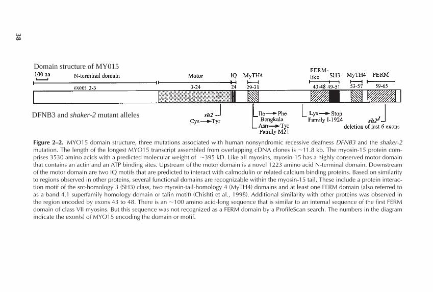
38
Figure 2–2. MYO15 domain structure, three mutations associated with human nonsyndromic recessive deafness DFNB3 and the shaker-2mutation. The length of the longest MYO15 transcript assembled from overlapping cDNA clones is �11.8 kb. The myosin-15 protein com-prises 3530 amino acids with a predicted molecular weight of �395 kD. Like all myosins, myosin-15 has a highly conserved motor domainthat contains an actin and an ATP binding sites. Upstream of the motor domain is a novel 1223 amino acid N-terminal domain. Downstreamof the motor domain are two IQ motifs that are predicted to interact with calmodulin or related calcium binding proteins. Based on similarityto regions observed in other proteins, several functional domains are recognizable within the myosin-15 tail. These include a protein interac-tion motif of the src-homology 3 (SH3) class, two myosin-tail-homology 4 (MyTH4) domains and at least one FERM domain (also referred toas a band 4.1 superfamily homology domain or talin motif) (Chishti et al., 1998). Additional similarity with other proteins was observed inthe region encoded by exons 43 to 48. There is an �100 amino acid-long sequence that is similar to an internal sequence of the first FERMdomain of class VII myosins. But this sequence was not recognized as a FERM domain by a ProfileScan search. The numbers in the diagramindicate the exon(s) of MYO15 encoding the domain or motif.
Domain structure of MY015
DFNB3 and shaker-2 mutant alleles

of endocytosis, regulating ion channels, localizing calmodulin, and cross-linking extensions of filopodia (Baker & Titus, 1998; Mermall et al., 1998;Titus, 1998). However, the precise roles of Myo6, MYO7A and MYO15 inthe hearing process are unknown.
The genomic structure of mouse Myo15 and human MYO15 is nowknown and consists of 66 exons (Liang et al., 1999), 16 more than wereidentified than when Wang and co-workers (1998) first reported the partialsequence of this gene (Figure 2–2). The sequence of mouse Myo15 allowedus to screen for the mutations responsible for the shaker-2 phenotype. Inshaker-2 mice a single nucleotide change was found in codon 1779 withinexon 20. This G to A transition mutation in the motor domain of myosin-15results in a cysteine to tyrosine substitution. The vast majority of all con-ventional and unconventional myosins have a cysteine at this position(http://www.mrc-lmb.cam.ac.uk/myosin/myosin.html) (Cope, Whis-stock, Rayment, & Kendrick-Jones, 1996). Additionally, a second allele ofshaker-2, shaker-2J, was discovered at the Jackson Laboratory in 1993 (Cook& Davisson, 1993). Like the original shaker-2 mice, shaker-2J mice are pro-foundly deaf and exhibit circling behavior. Molecular genetic analysis hasdemonstrated that shaker-2J mice have a deletion that removes the last sixexons and the polyadenylation signal of the Myo15 gene (Anderson et al.,submitted).
Myo15 is Expressed in the Sensory Organs of the Inner Ear
The location of Myo15 mRNA expression was determined by in-situ hy-bridization of paraffin sections of 17.5-day post coitum wild type(C57BL/6J) mouse embryos (Figure 2–3). The tissue was hybridized withan anti-sense radiolabeled riboprobe specific for exons 48 to 51 in the tailregion of Myo15. The results demonstrate that Myo15 has a limited distri-bution to discrete patches of the cochlea, the saccule, and the utricle consis-tent with the positions of the sensory epithelium. The localization ofMyo15 to these sensory organs provides further evidence that the muta-tions discovered in Myo15 are responsible for the phenotypes observed inshaker-2 and shaker-2J mice.
Mutations of Human MYO15 Cause Deafness
The sequence of the Myo15 gene was used to isolate the human MYO15gene (Wang et al., 1998). Three human genomic cosmid clones containingMYO15 were isolated and 90 kb of DNA was sequenced. Predicted humanMYO15 exons were identified and confirmed by sequencing humanMYO15 cDNA synthesized from human fetal inner ear mRNA (generouslyprovided by Dr. C. Morton) and from fetal brain and adult pituitary glandmRNA. MYO15 DNA was also sequenced from probands of the three
A REVIEW OF DFNB3 AND shaker-2 39

DFNB3 families to identify the human MYO15 mutations that cause deaf-ness. Mutations in MYO15 were found which co-segregated with deafnessin each of the three DFNB3 families (Wang et al., 1998) (Figure 2–2). Themutations of MYO15 in Bengkala villagers and in the M-21 Indian familyresult in single amino acid substitutions within the first of two myosin-tail-homology 4 domains (MyTH4). This domain is similar in amino acid se-quence to regions found in some other unconventional myosins includinghuman MYO7A, C. elegans Myo12, bovine myosin-X and AcanthamoebaHMW myosin-IV. The function of the MyTh4 domain is unknown (Mer-
40 GENETICS AND HEARING LOSS
Figure 2–3. In-situ hybridization of a Myo15 probe to 17.5 dayembryonic mouse inner ear. The sections were prepared by fixingthe heads in 4% paraformaldehyde and embedding them in paraf-fin, which was then cut at 10 micrometers in the sagittal planeand hybridized with a radiolabeled anti-sense riboprobe specificfor exons 48 to 51 of the Myo15 cDNA. The results were visual-ized by coating the tissue with a thin layer of photographic emul-sion. The emulsion was exposed for one week, then developedand fixed. The section on the left was photographed in dark field.The section is oriented with anterior (A) to the left and dorsal (D)towards the top. In the inner ear, Myo15 mRNA is localized in theutricle (U), saccule (S), and cochlea (C). The section was exam-ined under high magnification by bright field microscopy. The re-gions indicated by the arrows contain a high density of grains inthe photographic emulsion. The other bright areas lack any appre-ciable quantity of grains and are due instead to the presence ofpigments or the refractivity of the tissue. There was no signal ob-served when the corresponding sense riboprobe was used. Theimage to the right is a paint-filled 17-day embryonic mouse innerear (courtesy of Dr. Doris K. Wu) that is used to indicate the corre-sponding structures referred to in the tissue section.
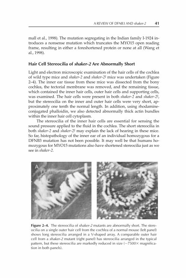
mall et al., 1998). The mutation segregating in the Indian family I-1924 in-troduces a nonsense mutation which truncates the MYO15 open readingframe, resulting in either a foreshortened protein or none at all (Wang etal., 1998).
Hair Cell Stereocilia of shaker-2 Are Abnormally Short
Light and electron microscopic examination of the hair cells of the cochleaof wild type mice and shaker-2 and shaker-2J mice was undertaken (Figure2–4). The inner ear tissue from these mice was dissected from the bonycochlea, the tectorial membrane was removed, and the remaining tissue,which contained the inner hair cells, outer hair cells and supporting cells,was examined. The hair cells were present in both shaker-2 and shaker-2J,but the stereocilia on the inner and outer hair cells were very short, ap-proximately one tenth the normal length. In addition, using rhodamine-conjugated phalloidin, we also detected abnormally thick actin bundleswithin the inner hair cell cytoplasm.
The stereocilia of the inner hair cells are essential for sensing thesound pressure applied to the fluid in the cochlea. The short stereocilia inboth shaker-2 and shaker-2J may explain the lack of hearing in these mice.So far, histopathology of the inner ear of an individual homozygous for aDFNB3 mutation has not been possible. It may well be that humans ho-mozygous for MYO15 mutations also have shortened stereocilia just as wesee in shaker-2.
A REVIEW OF DFNB3 AND shaker-2 41
Figure 2–4. The stereocilia of shaker-2 mutants are abnormally short. The stere-ocilia on a single outer hair cell from the cochlea of a normal mouse (left panel)shows long stereocilia arranged in a V-shaped array. A comparable outer haircell from a shaker-2 mutant (right panel) has stereocilia arranged in the typicalpattern, but these stereocilia are markedly reduced in size (�7500� magnifica-tion in both panels).

EPIDEMIOLOGY OF DFNB3
Once the mutation in MYO15 segregating in Bengkala was identified, itbecame possible to determine the carrier frequency of this mutationamong hearing individuals from Bengkala who have no known deaf rela-tives. In an earlier paper (Friedman et al., 1995) we referred to these indi-viduals as representative Bengkala individuals (RBIs). Of a sample of 48RBIs, we discovered that the DFNB3 mutation is carried by 25%. This ishigher than the predicted carrier rate for DFNB3 of 17.2% made by Winataand co-workers before DFNB3 was cloned and before we could directlydetermine the carrier rate (Winata et al., 1995). A 25% carrier rate for themissense mutation of MYO15 predicts that among random marriages be-tween two hearing people from Bengkala, 1.6% of the children will bedeaf. This was calculated by multiplying the probability of two hearingparents from Bengkala being carriers (0.25 � 0.25) by the probability oftheir child being homozygous for the DFNB3 mutation (0.25) or (0.25 �0.25 � 0.25 = 0.0156). This means we estimate that approximately 1.6% ofthe children born to hearing parents in Bengkala who have no deaf rela-tives will be profoundly deaf at birth. The higher than predicted incidenceof deafness in Bengkala (2.2%) is, in part, due to assortative marriagesamong the deaf villagers. As a point of comparison, in the United States,epidemiological studies indicate that about 1 in 1–2,000 children is bornwith a severe hearing impairment, and approximately 50% are estimatedto have a genetic cause for their hearing impairment (Marazita et al., 1993;Morton, 1991).
To what extent do mutant alleles of DFNB3 contribute to profounddeafness in various populations worldwide? The missense allele ofMYO15 causing congenital hearing loss in Bengkala, Bali is, so far, a pri-vate mutation, but clearly other mutations of MYO15 contribute to deaf-ness beyond Bali. Two unrelated families segregating congenital hearingloss from India have been found to be homozygous for two different mu-tations of MYO15 (Wang et al., 1998). Preliminary data suggest that muta-tions at the DFNB3 locus may account for about 5% of congenital pro-found deafness in a sample of 82 families from India and Pakistansegregating nonsyndromic recessive deafness. The contribution of MYO15mutations to profound hereditary deafness in North America is unknown.There is a bias in the types of families with hearing loss that have been as-certained, as the probands are typically profoundly deaf. It will be interest-ing, although difficult, to identify families with mild hereditary hearingloss. It is tempting to speculate that there are “mild” mutant alleles ofMYO15 that cause either mild congenital hearing loss or progressive hear-ing loss.
42 GENETICS AND HEARING LOSS

FUTURE STUDIES
The presence of inner and outer hair cells in the shaker-2 mouse, albeit withabnormally short stereocilia, offers the hope that the deafness phenotypemay be reversed by treating these cells directly as opposed to germ linetherapy. If a functional copy of Myo15 could be introduced into the haircells, would the stereocilia be restored to their normal lengths? If so,would the mouse then gain the ability to hear? These possibilities are nowunder investigation, and this work may eventually lead to treatments forpeople with DFNB3-related hearing loss due to mutations of MYO15.
Acknowledgments: The hospitality, consent and participation in this project of thefamilies from Bali and India are appreciated. This work was supported by the Na-tional Institute on Deafness and Other Communication Disorders (Z01 DC 00035-01, Z01 DC 00038-01, and R01 DC 01634)) to TBF, EW, and YR, respectively, and theNational Institute of Child Health and Human Development (R01 HD30428) toSAC. FJP is supported by a University of Michigan Rackham Fellowship. Jim Ash-er is included here as an author with permission of his wife, Doris. Jim died in May1996 just days before our second trip to Bengkala to collect more clinical data andblood samples. We dedicate this chapter to his memory.
REFERENCES
Adato, A., Weil, D., Kalinski, H., Pel-Or, Y., Ayadi, H., Petit, C., Korostishevsky, M.,& Bonne-Tamir, B. (1997). Mutation profile of all 49 exons of the humanmyosin VIIA gene, and haplotype analysis, in Usher 1B families from diverseorigins. American Journal of Human Genetics, 61(4), 813–821.
Aksentijevich, I., Pras, E., Gruberg, L., Shen, Y., Holman, K., Helling, S., Prosen,L., Sutherland, G. R., Richards, R. I., Ramsburg, M. et al. (1993). Refined map-ping of the gene causing familial Mediterranean fever, by linkage and ho-mozygosity studies. American Journal of Human Genetics, 53(2), 451–461.
Avraham, K. B., Hasson, T., Steel, K. P., Kingsley, D. M., Russell, L. B., Mooseker,M. S., Copeland, N. G., & Jenkins, N. A. (1995). The mouse Snell’s waltzerdeafness gene encodes an unconventional myosin required for structural in-tegrity of inner ear hair cells. Nature Genetics, 11(4), 369–375.
Baker, J. P., & Titus, M. A. (1998). Myosins: Matching functions with motors. Cur-rent Opinion in Cell Biology, 10(1), 80–86.
Ben Hamida, C., Doerflinger, N., Belal, S., Linder, C., Reutenauer, L., Dib, C.,Gyapay, G., Vignal, A., Le Paslier, D., Cohen, D. et al. (1993). Localization ofFriedreich ataxia phenotype with selective vitamin E deficiency to chromo-some 8q by homozygosity mapping. Nature Genetics, 5(2), 195–200.
Brobby, G. W., Muller-Myhsok, B., & Horstmann, R. D. (1998). Connexin 26R143W mutation associated with recessive nonsyndromic sensorineural deaf-ness in Africa. New England Journal of Medicine, 338(8), 548–550.
A REVIEW OF DFNB3 AND shaker-2 43

Chishti, A. H., Kim, A. C., Marfatia, S. M., Lutchman, M., Hanspal, M., Jindal, H.,Liu, S. C., Low, P. S., Rouleau, G. A., Mohandas, N., Chasis, J. A., Conboy, J. G.,Gascard, P., Takakuwa, Y., Huang, S. C., Benz, E. J., Jr., Bretscher, A., Fehon, R.G., Gusella, J. F., Ramesh, V., Solomon, F., Marchesi, V. T., Tsukita, S., Hoover,K. B. et al. (1998). The FERM domain: A unique module involved in the link-age of cytoplasmic proteins to the membrane. Trends in Biochemical Sciences,23(8), 281–282.
Cook, S. A., & Davisson, M. T. (1993). Re-mutation to shaker-2 (sh2). MouseGenome, 91, 312.
Cope, M. J. T., Whisstock, J., Rayment, I., & Kendrick-Jones, J. (1996). Conserva-tion within the myosin motor domain: Implications for structure and func-tion. Structure, 4(8), 969–987.
Dobrovolskaia-Zavasdkaia, N. (1928). L’irradiation des testicules et l’hereditechez la souris. Archives of Biology, 38, 457–501.
Friedman, T. B., Liang, Y., Weber, J. L., Hinnant, J. T., Barber, T. D., Winata, S.,Arhya, I. N., & Asher, J. H., Jr. (1995). A gene for congenital, recessive deafnessDFNB3 maps to the pericentromeric region of chromosome 17. Nature Genet-ics, 9(1), 86–91.
Groce, N. E. (1985). Everyone here spoke sign language: Hereditary deafness onMartha’s Vineyard. Cambridge, MA: Harvard University Press.
Kruglyak, L., Daly, M. J., & Lander, E. S. (1995). Rapid multipoint linkage analy-sis of recessive traits in nuclear families, including homozygosity mapping.American Journal of Human Genetics, 56(2), 519–527.
Lander, E. S., & Botstein, D. (1987). Homozygosity mapping: A way to map hu-man recessive traits with the DNA of inbred children. Science, 236(4808),1567–1570.
Liang, Y., Wang, A., Belyantseva I. A., Anderson, D. W. , Probst, F. J., Barber, T. D.,Miller, W., Touchman, J. W., Long, J., Sullivan, S. L., Sellers, J. R., Camper, S.A., Lloyd, R. V., Kachar, B., Friedman, T. B., & Fridell, R. A. (1999). Characteri-zation of the human and mouse unconventional myosin XV genes responsiblefor hereditary deafness DFNB3 and shaker 2. Genomics 61, 243–258.
Liang, Y., Wang, A., Probst, F. J., Arhya, I. N., Barber, T. D., Chen, K. S., Desh-mukh, D., Dolan, D. F., Hinnant, J. T., Carter, L. E., Jain, P. K., Lalwani, A. K.,Li, X. C., Lupski, J. R., Moeljopawiro, S., Morell, R., Negrini, C., Wilcox, E. R.,Winata, S., Camper, S. A., & Friedman, T. B. (1998). Genetic mapping refinesDFNB3 to 17p11.2, suggests multiple alleles of DFNB3, and supports homolo-gy to the mouse model shaker-2. American Journal of Human Genetics, 62(4),904–915.
Liu, X. Z., Walsh, J., Mburu, P., Kendrick-Jones, J., Cope, M. J., Steel, K. P., &Brown, S. D. (1997). Mutations in the myosin VIIA gene cause non-syndromicrecessive deafness. Nature Genetics, 16(2), 188–190.
Marazita, M. L., Ploughman, L. M., Rawlings, B., Remington, E., Arnos, K. S., &Nance, W. E. (1993). Genetic epidemiological studies of early-onset deafnessin the U.S. school-age population. American Journal of Medical Genetics, 46(5),486–491.
Mburu, P., Liu, X. Z., Walsh, J., Saw, D., Jr., Cope, M. J., Gibson, F., Kendrick-Jones, J., Steel, K. P., & Brown, S. D. (1997). Mutation analysis of the mousemyosin VIIA deafness gene. Genes and Function, 1(3), 191–203.
44 GENETICS AND HEARING LOSS

Mermall, V., Post, P. L., & Mooseker, M. S. (1998). Unconventional myosins in cellmovement, membrane traffic, and signal transduction. Science, 279(5350),527–533.
Morton, N. E. (1991). Genetic epidemiology of hearing impairment. Annals of theNew York Academy of Sciences, 630, 16–31.
Pollak, M. R., Chou, Y. H., Cerda, J. J., Steinmann, B., La Du, B. N., Seidman, J. G.,& Seidman, C. E. (1993). Homozygosity mapping of the gene for alkaptonuriato chromosome 3q2. Nature Genetics, 5(2), 201–204.
Probst, F. J., Fridell, R. A., Raphael, Y., Saunders, T. L., Wang, A., Liang, Y., Morell,R. J., Touchman, J. W., Lyons, R. H., Noben-Trauth, K., Friedman, T. B., &Camper, S. A. (1998). Correction of deafness in shaker-2 mice by an unconven-tional myosin in a BAC transgene. Science, 280(5368), 1444–1447.
Schenker, T., Lach, C., Kessler, B., Calderara, S., & Trueb, B. (1994). A novel GTP-binding protein which is selectively repressed in SV40 transformed fibro-blasts. Journal of Biological Chemistry, 269(41), 25447–25453.
Shuman, M. K. (1980). Culture and deafness in a Maya Indian village. Psychiatry,43(4), 359–370.
Smith, C. A. B. (1953). The detection of linkage in human genetics. Journal of theRoyal Statistical Society, 15B, 153–184.
Steel, K. P. (1995). Inherited hearing defects in mice. Annual Review of Genetics, 29,675–701.
Titus, M. A. (1998). Coming to grips with a multitude of myosins. Trends in CellBiology, 8(4), 171–172.
Wang, A., Liang, Y., Fridell, R. A., Probst, F. J., Wilcox, E. R., Touchman, J. W.,Morton, C. C., Morell, R. J., Noben-Trauth, K., Camper, S. A., & Friedman, T.B. (1998). Association of unconventional myosin MYO15 mutations with hu-man nonsyndromic deafness DFNB3. Science, 280(5368), 1447–1451.
Weil, D., Blanchard, S., Kaplan, J., Guilford, P., Gibson, F., Walsh, J., Mburu, P.,Varela, A., Levilliers, J., Weston, M. D. et al. (1995). Defective myosin VIIAgene responsible for Usher syndrome type 1B. Nature, 374(6517), 60–61.
Winata, S., Arhya, I. N., Moeljopawiro, S., Hinnant, J. T., Liang, Y., Friedman, T.B., & Asher, J. H., Jr. (1995). Congenital non-syndromal autosomal recessivedeafness in Bengkala, an isolated Balinese village. Journal of Medical Genetics,32(5), 336–343.
A REVIEW OF DFNB3 AND shaker-2 45


For most of us mice are pesky creatures that invade our homes and startleus in the middle of the night. However, for many scientists mice providepowerful model systems for the study of mammalian physiology and ge-netics, which often enhance insight into human biology (Meisler, 1996).The study of the genetic causes of deafness is one research area that hasbenefitted substantially from the use of mouse models. This chapter dis-cusses mouse models for human genetic deafness and focuses on our cur-rent studies of the dn, or deafness, mouse.
WHY THE MOUSE?
The similarities that make the mouse an excellent model system for hu-man genetic research are not evident at first glance. However, closer exam-ination reveals that the physiologic structures, including the cochlea andvestibular system, are remarkably similar between human and mouse,both in the developmental process and the functional adult structures. TheDNA sequence and the organization of the genome itself reveal that weare not nearly as different from the mouse, at the molecular level, as wemay think. Most of our genes are quite similar in DNA sequence, withsome sharing upwards of 99% identity to the mouse counterparts, or ortho-logues. Additionally, the order and relationship of individual genes toneighboring genes in the human and mouse genomes are conserved. If, forexample, genes A and B are located next to one another on a human chro-
47
3
Mouse Models and Progress inHuman Deafness Research
Stacy Drury, Daryl Scott, Zhining Den,Margaret M. DeAngelis, Mark A. Batzer,
Val Sheffield, Richard Smith,Prescott L. Deininger, and Bronya J. B. Keats

mosome, they are likely to be located next to each other on a mouse chro-mosome. Several computer databases (e.g., http://www3.ncbi.nlm.nih.gov)now provide lists of the locations of conserved regions and the genes theycontain, making the identification of these similar genes even simpler. Thesimultaneous search for new disease genes in the human and mousegenomes has dramatically increased the speed with which disease-causinggenes are mapped, identified, and characterized.
MOUSE MODELS AND DEAFNESS
More than 25 mouse models of deafness have been described and the loca-tions of the defective genes within the mouse genome have been deter-mined (Steel, 1995). Several of these mouse models map to the orthologousregions of human deafness loci and a database that lists these mouse mod-els and the relevant human deafness loci is available (Van Camp & Smith,1999; http://dnalab-www.uia.ac.be/dnalab/hhh). Mouse models haveproven indispensable in the identification of three nonsyndromic deafnessgenes, two of which have been discussed in the preceding chapters of thisbook, and mouse models promise to be equally useful for other deafnessgenes (Petit, 1996). They offer several advantages over attempts to identifygenes using only the human genome. First, the large number of controlledmatings that can be set up with mice allows the development of a detailedgenetic map. Second, samples for tissue-specific gene analysis can be ac-cessed much more easily in mice than humans. And finally the selectivecorrection or selective inactivation of specific genes using transgenic tech-nology permits an exploration of gene function previously unavailable.This powerful tool has elucidated genes involved in the development of theauditory system for which naturally occurring mutations do not yet exist.
The first human deafness gene identified with the aid of a mousemodel was the gene MYO7A that encodes myosin VIIa. Mutations in theorthologous mouse gene were found in the shaker-1 mouse, and subse-quently mutations in MYO7A were detected in patients with Usher syn-drome type 1B and with nonsyndromic forms of hearing impairment(DFNB2 and DFNA11). MYO7A was the first gene identified that is in-volved in auditory transduction (Gibson et al., 1995, Liu, Walsh, Mburu,1997; Liu, Walsh, & Tamagawa, 1997; Weil et al., 1995, 1997). Mappingstudies in the shaker-1 mouse and in Usher syndrome type 1B families pro-vided the locations of the defective genes and suggested they were proba-bly orthologues. The more definitive mapping information available forthe shaker-1 mutation localized the gene to a much smaller critical region inthe mouse genome than the human data allowed and thus led to the isola-tion of the myosin VIIa gene in the mouse first. The significant cytoskeletalabnormalities in the shaker-1 mouse made this gene a likely candidate, and
48 GENETICS AND HEARING LOSS

the identification of the mouse gene provided the critical candidate for thehuman gene.
Myosins are mechanoenzymes that bind to actin and hydrolyze ATPto produce force. They are involved in a wide range of functions includingmovement, cell process extension, cytoskeletal structure, phagocytosis,signal transduction, and muscle movement (Probst et al., 1998). The detec-tion of mutations in the gene encoding myosin VIIa, an unconventionalmyosin, in the shaker-1 mouse and patients with several types of hearingimpairment supports the importance of myosin in hearing. Since the iden-tification of myosin VIIa, two other unconventional myosins have beenimplicated in the development of the auditory system (Avraham et al.,1995; Probst et al., 1998).
The second human deafness gene identified with the aid of a mousemodel was DFNA15 (Vahava et al., 1998). DFNA15 is an autosomal domi-nant, adolescent onset, nonsyndromic hearing loss condition. The mousemodel, in this case, is the result of a targeted gene knockout of the transcrip-tion factor POU4F3. Homozygous knockouts are profoundly deaf (Erk-man et al., 1996; Xiang et al., 1997). Analysis of a family with hearing-im-paired members mapped DFNA15 to a large region on the long arm ofchromosome 5, but the presence of POU4F3 in the orthologous region onmouse chromosome 18 made it a strong candidate. Although no clearfunction of POU4F3 has been determined, it belongs to a family of tran-scription factors with a POU homeodomain and a POU specific domainthat are both involved in binding DNA (Steel, 1998). Analysis of thePOU4F3 sequence in DFNA15 family members revealed an eight base pairdeletion leading to a premature termination of the POU4F3 gene productand removing the homeodomain. This mutation probably alters the func-tion of the gene product by preventing DNA binding, but how this alter-ation leads to hearing impairment has yet to be elucidated.
Perhaps the most powerful feature of a mouse model is the ability tocorrect a phenotype in vivo using transgenic technology. This technologyled to the identification of the gene encoding myosin XV as the causativegene in both the shaker-2 mouse and DFNB3 families (Probst et al., 1998;Wang et al., 1998). The shaker-2 mouse is an x-ray induced mouse mutationon chromosome 11 associated with profound deafness and circling behav-ior indicative of vestibular dysfunction. It was likely that the two geneswere orthologues because four different genes in the shaker-2 critical re-gion mapped to the same human region located on the short arm of chro-mosome 17, which contained DFNB3. Transfer of the bacterial artificial chro-mosome (BAC) contig spanning the shaker-2 critical region revealed a single140-kilobase BAC that segregated with the rescue of both the deafness andthe vestibular phenotype. Sequence analysis of the entire BAC led to theconcurrent identification of myosin 15 as the shaker-2 and DFNB3 genes.As the shaker-2 mice exhibit dysmorphic stereocilia that are shortened but
MOUSE MODELS AND PROGRESS IN HUMAN DEAFNESS RESEARCH 49
normally arrayed and the presence of an abnormal actin bundle in the haircells, it is hypothesized that myosin XV is involved in the maintenanceand organization of the actin within the sensory hair cells (Probst et al.,1998). Taken together, these three examples highlight the effectiveness ofmouse models in the detection of human disease genes.
Many additional mouse deafness models exist that are not yet associ-ated with human deafness conditions, including some for which thecausative gene has been identified (Avraham et al., 1995; Street, McKee-Johnson, Fonseca, Tempel, & Noben-Trauth, 1998). Using transgenic tech-nology to “knockout” or inactivate specific genes, new mouse models ofdeafness have been created that identify yet more genes crucial for thefunction and/or the development of the auditory system, similar to themethod used in the pou4f3 model. Knockouts important for developmentinclude Math 1, a gene essential for the generation of the inner hair cells ofthe cochlea, and the winged helix transcription factor Fkh10, a transcrip-tional regulator necessary for the early development of both the cochleaand the vestibulum (Hulander, Wurst, Carlsson, & Enerback, 1998). Addi-tional knockouts of genes important for the correct functioning of the au-ditory system include Slc12a2, a secretory Na/K/2Cl co-transporter pro-tein involved in endolymph secretion, and ATP6B1, an H+ ATPaseinvolved in active proton secretion required to maintain endolymph pH,as well as distal nephron acid secretion in the kidney (Delpire, England,Dull, & Thorne, 1999; Karet et al., 1999). These knockout mouse modelsidentify genes involved in the auditory system and highlight the impor-tance of the maintenance of ion concentrations for correct auditory func-tion and the role of transcription factors in the development of the compo-nents of the auditory system. As the number of genes involved in hearingincreases we will be able to construct an interacting gene model for the de-velopment of the auditory and vestibular systems.
The Deafness, dn Mouse
The dn mouse is a model for human deafness with no other anomalies andno vestibular malformations. The dn mouse arose naturally in the curly-tail (ct) stock (Deol & Kocher, 1958) and has sensorineural deafness. Thedeafness phenotype in the dn mouse is inherited in an autosomal recessivemanner, and dn/dn homozygotes are never able to hear. These mice do notexhibit the Preyer startle reflex or an auditory brainstem response (ABR)and are profoundly deaf from birth (Keats et al., 1995). In these mice onecan see dramatic changes in the cochlear structure. There is marked degen-eration of the organ of Corti, stria vascularis, and occasionally the saccularmacula beginning at or before birth (Pujol, Shnerson, Lenoir, & Deol,1983). By day 45 after birth, there is complete degeneration of the organ ofCorti and no recognizable cell types remain. Interestingly, between day 45and day 90, there is marked regeneration of the nonsensory cells, but only
50 GENETICS AND HEARING LOSS

in the apical portion of the organ of Corti. One hypothesis for this local re-generation is the specific presence of GABA-nergic olivocochlear neuronsonly in the apical portion of the organ of Corti, which may promote regen-eration (Bock & Steel, 1983; Pujol et al., 1983; Webster, 1985, 1992).
Electron microscopy studies show additional changes of the cochleaand inner ear of dn/dn homozygous mice. Transmission electron micro-scopic studies show shrunken and distorted inner and outer hair cells withvacuoles present in the cytoplasm. Scanning electron microscopy also de-tects an irregular arrangement of stereocilia, loss of most stereocilia, andgiant cilia formation, similar to studies in another deaf mouse, the Snell’swaltzer. The gene that causes deafness in the Snell’s waltzer mouse has beenidentified as myosin VI, but a human deafness condition associated withmyosin VI has not yet been found (Avraham et al., 1995). Finally the num-ber of neurons in the spiral ganglion of dn mice is only 23% of that seen inheterozygous and normal hearing mice (Webster, 1985). The dramaticchanges in the hair cells and the presence of abnormal stereocilia providestructural explanations for the electrophysiologic findings in the dn/dn ho-mozygotes.
The endocochlear potential is normal in dn mice, demonstrating thatsound is able to travel from the outer ear to the tympanic membrane.However, they exhibit no cochlear microphonics, suggesting a primarycochlear defect. Additionally, dn mice have preserved central auditoryfunction on direct stimulation of the cranial nerve VIII, the vestibular-cochlear nerve, which is responsible for the transmission of sound to thebrain (Bock, Franks, & Steel, 1982). Together these findings point to a fun-damental defect in the functioning of the cochlear hair cells (Bock & Steel,1983).
Interestingly although an occasional dn/dn mouse has been foundwith mild vestibular abnormalities, none exhibit the circling behaviorcharacteristic of vestibular dysfunction seen in all other mouse deaf mu-tants (Bock & Steel, 1983; Brown & Steel, 1994). As the vestibular and thecochlear structures arise from the same embryological structures it is notsurprising that deafness is often associated with balance problems. Thelack of a vestibular problem in the dn mouse suggests that the gene caus-ing deafness is likely to be involved either in the differentiation of the haircells or the maintenance and functioning of the hair cells.
The Physical Mapping of the dn Region
Keats et al. (1995) localized the dn gene to the region containing markersD19Mit14, D19Mit96, D19Mit111, D19Mit60, and D19Mit14 on mousechromosome 19 and hypothesized the presence of a chromosomal inver-sion that disrupted the dn gene and led to the deafness phenotype. Withthis hypothesis in mind we began the construction of a physical map of
MOUSE MODELS AND PROGRESS IN HUMAN DEAFNESS RESEARCH 51

this region to help us identify the structural changes on the dn chromo-some as well as look for candidate genes that map to this region of thegenome. The recombination data indicated that one of the breakpoints ofthe inversion is located between the genetic markers D19Mit14 andD19Mit96 (Vinas et al., 1998), and we therefore constructed a physical con-tig of this region.
Screening of the Whitehead mouse yeast artificial chromosome (YAC) li-brary with markers from across the region identified a 460-kilobase YAC(91C10) that was positive for both D19Mit14 and D19Mit96. This resultdemonstrates that D19Mit14 and D19Mit96 are probably within an inter-val of less than 460 kilobases. To further define the interval we used thepolymerase chain reaction (PCR) to isolate clones from a total genomicmouse BAC library commercially available from Research Genetics, whichcontained the five markers D19Mit14, D19Mit96, D19Mit111, D19Mit60,and D19Mit41. Using PCR we determined which BACs overlapped witheach other and were able to complete the contig (Figure 3–1).
The construction of this contig gave us the tools to further narrow theregion of the breakpoint using fluorescent in situ hybridization (FISH). Thistechnique demonstrated that BAC 124J3 hybridized to two different loca-
52 GENETICS AND HEARING LOSS
Figure 3–1. Physical map of the mouse chromosome 19 region that is likely tocontain the dn gene. One YAC and 8 BACs that span this region are shown withtheir approximate locations below the entire chromosome. Specific genes that havebeen identified are in italics and are shown with line through the BACs on whichthey are known to be present.

tions on the dn chromosome 19, a result that is consistent with an inversionbreakpoint being present on this BAC (Figure 3–2). Pursuing our hypothe-sis that the inversion disrupted the dn gene we have now focused our genesearch on this BAC.
Candidate Genes in the dn Regionand the Homology to DFNB7/11
The region of mouse chromosome 19 containing the dn gene is ortholo-gous to a region of human chromosome 9 that contains the autosomal re-cessive nonsyndromic hearing impairment genes DFNB7 and DFNB11
MOUSE MODELS AND PROGRESS IN HUMAN DEAFNESS RESEARCH 53
Figure 3–2. Fluorescent in situ hybridization (FISH) experi-ment of a cell from a dn/dn homozygous mouse using BAC124J3 in blue and another BAC in a different chromosomalregion in red as probes. There are four distinct blue signalsbut only two red signals, indicating that BAC124J3 is hy-bridizing to two distinct locations on each of the homologouschromosomes, while the other BAC is only hybridizing to asingle location on both homologous chromosomes. This pat-tern of hybridization strongly supports the existence of an inversion.

(Jain et al., 1995; Scott et al., 1996). As it is likely that the mouse and humangenes are orthologues, we began analyzing genes that are expressed in thecochlea and are located in the dn and the DFNB7/11 critical regions.
As the human EST mapping project has progressed faster than themouse-mapping project and there exists a well-established human cochlearcDNA library, we were able to identify several human cDNAs that mappedto the DFNB7/11 region. Two of these cDNAs (ZNF216 and TMEM2) wereshown to be new cochlear-expressed genes (Scott et al., 1998, 1999). ZNF216shares homology with a gene that is involved in the development of thevessel endothelium from precursor cells and also with several genes thatcontain putative zinc finger domains. These domains are often found inDNA binding proteins. The function of TMEM2 is unknown but it containsat least one putative transmembrane domain. Sequence analysis using theNCBI Blast database (http://www.ncbi.nlm.nih.gov) identified the homol-ogous mouse ESTs. Using both the mouse and the human EST sequenceswe were able to confirm the map location of both genes within the respec-tive critical regions. Through direct sequence analysis of affectedDFNB7/11 individuals we were able to exclude both TMEM2 and ZNF216from causing deafness in these patients. In the mouse, we compared RNAexpression in the brain of the dn mouse with that in the wild type (ct)mouse. Through Northern and rtPCR analysis we determined that therewas no alteration in amount, size, or sequence of the RNA between thetwo mouse strains. Given these findings it is unlikely that either of thesetwo genes are responsible for the deafness phenotype. However as thesegenes are both expressed in the cochlea, as well as a variety of other tis-sues, they may still play a role in hearing.
The extensive amount of DNA sequence and gene information avail-able over the internet has dramatically enhanced the process of candidategene analysis and more ESTs and cDNAs are being discovered andmapped every day. As well as ZNF216 and TMEM2 we have also eliminat-ed arrestin as the defective gene. The protein encoded by this gene isthought to have an inhibitory role in the activated phototransduction cas-cade by interacting with either rhodopsin or a light-dependent cGMPphosphodiesterase. We are continuing to study other genes that map to thedn and the DFNB7/11 critical regions in order to identify the causativegene.
THE FUTURE AND TRANSGENIC MICE
The identification of genes that lead to diseases is an important area of re-search, but it is only the beginning of the process of understanding the de-fect and finding a therapy or cure for the disease. Once a gene has beenidentified at the sequence level the difficult process of dissecting the func-
54 GENETICS AND HEARING LOSS

tion of the protein encoded by this gene begins. One needs to examine notonly the level of RNA and protein expression in different organs, but alsothe time in development at which they are expressed and the precise local-ization of the gene product both within the tissue and within the cell itself.Where exactly and at what times in the development of the cochlea is thedn gene expressed? Is expression limited to hair cells and the supportingcells in the cochlea or is there a wider expression distribution? What othergenes interact with the dn gene and why is there degeneration of the cellsafter birth? These are just a few of the questions one can begin to answeronce the gene is identified.
Perhaps the greatest advantage of using mouse models for research isthe ability to put genes back into animals and see if one can correct thephenotype. Transgenic technology has rapidly advanced in the last 10 to15 years. We have the ability now to not only put genes in and turn themoff, but also to manipulate their expression at specific developmental timepoints and in specific tissues (Camper, 1987; Hogan, Beddington, Costanti-ni, & Lacy, 1994).
We recently created transgenic mice containing BAC 124J3. By breed-ing these mice to dn/dn homozygotes we hope to cross the transgene ontothe deaf mouse line and thereby correct the deafness phenotype. This tech-nique has already proven successful in the shaker-2 mouse and was instru-mental in the identification of that gene (Probst et al., 1998). The existenceof a transgenic model will greatly facilitate studies of the function of the dngene as well as contribute to our understanding of the auditory system.
The search for deafness genes is far from over and it seems that thereare more deafness loci than ever with new genes continually being identi-fied. With the identification of genes causing deafness, research to deter-mine their interactions with each other and the role that each plays in theauditory system is under way. Each gene is a small piece of the puzzle thatwill eventually give us a clear picture of how the intricate and delicate au-ditory and vestibular systems function and develop, and how a defect in aparticular gene can lead to malfunctions in that process. It is only througha clear understanding of the molecular basis of deafness that we can de-sign effective therapies and cures for this, the most common sensorydeficit.
GLOSSARY
Bacterial artificial chromosome: Similar to the yeast artificial chromo-some but containing a smaller amount of genomic DNA and replicatingin bacteria instead of yeast.
cDNA: RNA is reverse transcribed into a single strand of DNA using re-verse transcriptase and then made double stranded using the poly-
MOUSE MODELS AND PROGRESS IN HUMAN DEAFNESS RESEARCH 55

merase chain reaction. This double-stranded DNA represents the se-quence of the gene without the introns.
contig: Overlapping pieces of genomic DNA contained in smaller vectorsthat are easy to manipulate to produce large amounts of a specific frag-ment of DNA.
EST: Expressed sequence tag. This is a small piece of RNA that is alsomade into double-stranded DNA and represents part of a gene. SeveralESTs can be in a contig to give an entire cDNA sequence that representsthe full length of the original RNA derived from a particular gene.
FISH: Fluorescent in situ hybridization. Using a small piece of DNA, aprobe that is labeled with fluorescent antibodies, one hybridizes theprobe to whole cells and a bright signal fluorescent dot is seen whereverthe matching DNA sequence is found in the particular cell.
Inversion: A large fragment of a chromosome is inverted relative to therest of the chromosome. For example, if there was a gene order: A B CD, and an inversion occurred between A and D, the new gene orderwould be: A C B D.
Northern: A method to detect changes in RNA size or amounts from a par-ticular tissue or developmental time point of an animal. RNA is extract-ed from the tissue and electrophoresed in an agarose gel depending onthe size of the RNA. Next the RNA is transferred and immobilized ontoa nitrocellulose or nylon membrane. This membrane can then be hy-bridized with specific genes that are tagged with a radioactive markerso if they are present, a specific size and intensity of radioactive signalwill be observed.
Orthologue: One of a set of homologous genes in different species.rtPCR: reverse transcriptase PCR. A method used to amplify small quanti-
ties of RNA from a particular sample and convert the RNA to the morestable double-stranded DNA for further analysis. RNA is first extractedfrom a sample and then converted into single-stranded DNA using re-verse transcriptase. Next the single-stranded DNA is amplified andconverted to double-stranded DNA using the polymerase chain reac-tion, PCR.
Syntenic: The physical presence together on the same chromosome of twoor more gene loci, whether or not they are close enough together forlinkage to be determined.
Targeted gene knockout: The insertion of a gene or a piece of genomicDNA into a specific region of another animal’s genome where it inter-rupts the endogenous DNA and stops the correct function of that gene.
Transgenic: The addition of a gene or a piece of genomic DNA directly in-to a fertilized egg of another species, which is then incorporated into arandom place in that animal’s genome and passed on to all the off-spring of that animal.
56 GENETICS AND HEARING LOSS

Yeast artificial chromosome (YAC): A vector, similar to a normal yeastchromosome, that contains a large piece of genomic DNA from anotherspecies. The fragments range in size from 50 kilobase pairs to 1.5megabase pairs of DNA. This vector can be replicated to produce largeamounts of the specific fragment of DNA through the growing up oflarge amounts of yeast that contain the vector.
REFERENCES
Avraham, K. B., Hasson, T., Steel, K. P., Kingsley, D. M., Russell, L. B., Mooseker, M.S., Copeland, N. G., & Jenkins, N. A. (1995). The mouse Snell’s waltzer deafnessgene encodes an unconventional myosin required for structural integrity of in-ner ear hair cells. Nature Genetics, 11, 369–375.
Bock, G. R., Franks, M. P., & Steel, K. P. (1982). Presence of central auditory functionin the dn mouse. Brain Research, 23, 608–612.
Bock, G. R., & Steel, K. P. (1983). Inner ear pathology in the deafness mutant mouse.Acta Otolaryngologica, 96, 39–47.
Brown, S. D. M., & Steel, K. P. (1994). Genetic deafness—progress with mousemodels. Human Molecular Genetics, 3,1453–1456.
Camper, S. A. (1987). Research applications of transgenic mice. Biotechniques, 5,638–650.
Delpire, E., England, R., Dull, C., & Thorne, T. (1999). Deafness and imbalance as-sociated with the inactivation of the secretory Na-K-2Cl co-transporter. NatureGenetics, 22, 192–195.
Deol, M. S., & Kocher, W. (1958). A new gene for deafness in the mouse. Heredity,12, 463–466.
Erkman, L., McEvilly, R. J., Luo, L., Ryan, A. K., Hooshmand, F., O’Connell, S. M.,Keithley, E. M., Rapaport, D. H., Ryan, A. F., & Rosenfeld, M. G. (1996). Role oftranscription factors Brn-3.1 and Brn-3.2 in auditory and visual system develop-ment. Nature, 381, 603–606.
Gibson, F., Walsh, J., Mburu, P., Varela, A., Brown, K. A., Antonio, M., Beisel, K. W.,Steel, K. P., & Brown, S. D. M. (1995). A type VII myosin encoded by the mousedeafness gene Shaker-1. Nature, 374, 62–64.
Hogan, B., Beddington, R., Costantini, F., & Lacy, E. (1994). Manipulating the mouseembryo. New York: Cold Spring Harbor Laboratory Press.
Hulander, M., Wurst, W., Carlsson, P., & EnerBack, S. (1998). The winged helixtranscription factor Fkh10 is required for normal development of the inner ear.Nature Genetics, 20, 374–376.
Jain, P. K., Fukushima, K., Deshmukh, D., Arabandi, R., Thomas, E., Kumar, S., Lal-wani, A. K., Ploplis, B., Skarka, H., Srisailapathy, C. R. S., Wayne, S., Zbar, R. I.S., Verma, I. C., Smith, R. J. H., & Wilcox, E. R. (1995). A human recessive neu-rosensory nonsyndromic hearing impairment locus is a potential homologue ofthe murine deafness (dn) locus. Human Molecular Genetics, 4, 391–394.
Karet, F. E., Finberg, K. E., Nelson, R. D., Nayir, A., Mocan, H., Sanjad, S. A., Rod-riguez-Soriano, J., Santos, F., Cremers, C. W., DiPietro, A., Hoffbrand, B. I.,
MOUSE MODELS AND PROGRESS IN HUMAN DEAFNESS RESEARCH 57
Winiarski, J., Bakkaloglu, A., Ozen, S., Dusunsel, R., Goodyer, P., Hulton, S. A.,Wu, D. K., Skvorak, A. B., Morton, C. C., Cunningham, M. J., Jha, V., & Lifton, R.P. (1999). Mutations in the gene encoding B1 subunit of H+-ATPase cause renaltubular acidosis with sensorineural deafness. Nature Genetics, 21, 84–90.
Keats, B. J. B., Nouri, N., Huang, J. M., Money, M., Webster, D. B., & Berlin, C. I.(1995). The deafness locus (dn) maps to mouse chromosome 19. MammalianGenome, 6, 8–10.
Liu, X. Z., Walsh, J., Mburu, P., Kendrick-Jones, J., Cope, M. J. T. V., Steel, K. P., &Brown, S. D. M. (1997). Mutations in the myosin VIIA gene cause non-syn-dromic recessive deafness. Nature Genetics, 16, 188–190.
Liu, X. Z., Walsh, J., Tamagawa, Y., Kitamura, K., Nishizawa, M., Steel, K. P., &Brown, S. D. M. (1997). Autosomal dominant non-syndromic deafness (DFNA11)caused by a mutation in the myosin VIIA gene. Nature Genetics, 17, 268.
Meisler, M. H. (1996). The role of the laboratory mouse in the Human Genome Pro-ject. American Journal of Human Genetics, 59, 764–771.
Petit, C. (1996). Genes responsible for human hereditary deafness: Symphony of athousand. Nature Genetics, 14, 385–391.
Probst, F. J., Fridell, R. A., Raphael, Y., Saunders, T. L., Wang, A., Liang, Y., Morell,R. J., Touchman, J. W., Lyons, R. H., Noben-Trauth, K., Friedman, T. B., &Camper, S. A. (1998). Correction of deafness in shaker-2 mice by an unconven-tional myosin in a BAC transgene. Science, 280, 1444–1447.
Pujol, R., Shnerson, A., Lenoir, M., & Deol, M. S. (1983). Early degeneration of sen-sory and ganglion cells in the inner ear of mice with uncomplicated geneticdeafness (dn): Preliminary observations. Hearing Research, 12, 57–63.
Scott, D. A., Carmi, R., Elbedour, K., Yosefsberg, S., Stone, E. M., & Sheffield, V. C.(1996). An autosomal recessive nonsyndromic hearing loss locus identified byDNA pooling using two inbred Bedouin kindreds. American Journal of HumanGenetics, 59, 385–391.
Scott, D. A., Drury, S., Bishop, J., Swiderski, R. E., Carmi, R., Ramesh, A., Elbedour,K., Srisailapathy, C. R., Lovett, M., Keats, B. J., Smith, R. H., & Sheffiefld, V. C.(1999). Refining the DFNB7/11 deafness interval using intragenic polymor-phisms in a novel cochlear expressed gene, TMEM2. Submitted.
Scott, D. A., Greinwald, J. H., Marietta, J. R., Drury, S. S., Swiderski, R. E., Vinas, A.M., DeAngelis, M. M., Carmi, R., Ramesh, A., Kraft, M. L., Elbedour, K.,Skworak, A. B., Friedman, R. A., Srikumari Srisailapathy, C. R., Verhoeven, K.,VanCamp, G., Lovett, M., Deininger, P. L., Batzer, M. A., Morton, C. C., Keats, B.J. B., Smith, R. J. H., & Sheffield, V. (1998). Identification and mutation analysisof a cochlear-expressed, zinc finger protein gene at the DFNB7/11 and dn hear-ing-loss-loci on human chromosome 9q and mouse chromosome 19. Gene, 215,461–469.
Steel, K. (1995). Inherited hearing defects in mice. Annual Review of Genomics, 29,675–701.
Steel, K. P. (1998). Progress in progressive hearing loss. Science, 279, 1870–1871.Street, V. A., McKee-Johnson, J. W., Fonseca, R. C., Tempel, B. L., & Noben-Trauth,
K. (1998). Mutations in a plasma membrane Ca2+-ATPase gene cause deafnessin deafwaddler mice. Nature Genetics, 19, 390–394.
Vahava, O., Morell, R., Lynch, E. D., Weiss, S., Kagan, M. E., Ahituv, N., Morrow, J.E., Lee, M. K., Skvorak, A. B., Morton, C. C., Blumenfeld, A., Frydman, M.,
58 GENETICS AND HEARING LOSS

Friedman, T. B., King, M., & Avraham, K. B. (1998). Mutation in transcriptionfactor POU4F3 associated with inherited progressive hearing loss in humans.Science, 279, 1950–1954.
Van Camp, G., & Smith, R. J. H. (1999). Hereditary Hearing Loss Homepage. Avail-able: World Wide Web URL: http://dnalab-www.uia.ac.be/dnalab/hhh
Vinas, A. M., Drury, S. S., DeAngelis, M. M., Den, Z., Huang, J. M., Berlin, C. I.,Hunt, J. D., Batzer, M. A., Deininger, P. L., & Keats, B. J. B. (1998). The mousedeafness locus (dn) is associated with an inversion on chromosome 19. Biochimi-ca and Biophysica Acta, 1407, 257–262.
Wang, A., Liang, Y., Fridell, R. A., Probst, F. J., Wilcox, E. R., Touchman, J. W., Mor-ton, C. C., Morell, R. J., Noben-Trauth, K., Camper, S. A., & Friedman, T. B.(1998). Association of unconventional moysin MYO15 mutations with humannonsyndromic deafness DFNB3. Science, 280, 1447–1450.
Webster, D. B. (1985). The spiral ganglion and cochlear nuclei of deafness mice.Hearing Research, 18, 19–27.
Webster, D. B. (1992). Degeneration followed by partial regeneration of the organof Corti in deafness (dn/dn) mice. Experimental Neurology, 115, 27–31.
Weil, D., Blanchard, S., Kaplan, J., Guilford, P., Gibson, F., Walsh, J., Mburu, P.,Varela, A., Levilliers, J., Weston, M. D., Kelley, P. M., Kimberling, W. J., Wage-naar, M., Levi-Acobas, F., Larget-Piet, D., Munnich, A., Steel, K. P., Brown, S. D.M., & Petit. C. (1995). Defective myosin VIIA gene responsible for Usher syn-drome type 1B. Nature, 374, 60–61.
Weil, D., Kussel, P., Blanchard, S., Levy, G., Levi-Acobas, F., Drira, M., Ayadi, H., &Petit, C. (1997). The autosomal recessive isolated deafness, DFNB2, and theUsher 1B syndrome are allelic defects of the myosin-VIIA gene. Nature Genetics,16, 191–193.
Xiang, M., Gan, L., Li, D., Chen, Z. Y., Zhou, L., O’Malley, B. W., Klein, W., &Nathans, J. (1997). Essential role of POU-domain factor Brn-3c in auditory andvestibular hair cell development. Proceedings of the National Academy of Science,94, 9445–9450.
MOUSE MODELS AND PROGRESS IN HUMAN DEAFNESS RESEARCH 59


Physical and genetic maps of markers are essential tools for gene localiza-tion and identification using the strategy known as positional cloning(Collins, 1991). The goal of positional cloning is to find a disease gene by de-termining its physical location in the genome without any knowledge of thefunction of the gene. Initially, linkage analysis of family data localizes thedisease gene to a chromosomal region and determines the flanking geneticmarkers. From this information a physical map of genomic clones containingthe flanking markers and the putative disease gene is constructed. Candidategenes are identified from the overlapping genomic clones and then subjectedto mutational analysis in affected patients. This chapter reviews work fromour laboratories to identify one of the genes responsible for Usher syndrome.
USHER SYNDROME
The Usher syndromes are a group of autosomal recessive disorders char-acterized by congenital sensorineural hearing impairment and progressiveretinitis pigmentosa usually resulting in blindness. Although relativelyrare in the general population, estimated at 4.4 per 100,000 in the UnitedStates (Boughman, Vernon, & Shaver, 1983), Usher syndrome accounts for3–6% of children born with profound hearing impairment and accountsfor over 50% of the adult deaf-blind population (Smith et al., 1994).
61
4
Physical Maps as MolecularTools to Identify Disease Genes
Margaret M. DeAngelis, Chadwick J. Donaldson, Gregory M. Ditta, Lauren M. Buckley, John P. Doucet,
Zhining Den, Stacy Drury, Mary Z. Pelias, Prescott L. Deininger, Bronya J. B. Keats,
and Mark A. Batzer

Three types of Usher syndrome have been differentiated based on clini-cal variation with respect to severity of hearing loss and vestibular impair-ment (Smith et al., 1992). Usher syndrome type I, the most severe form, isdistinguishable from Usher syndrome types II and III by a severe-to-pro-found deafness and an absence of vestibular function (Smith et al., 1994).Usher syndrome type II patients present with moderate-to-severe congeni-tal hearing loss (Smith et al., 1994), whereas the hearing loss in Usher syn-drome type III is progressive (Davenport & Omen, 1977). Patients withUsher syndrome types II and III have normal vestibular function. All pa-tients with Usher syndrome develop retinitis pigmentosa but the rate ofprogression may be variable. Histopathological studies of temporal bonesof Usher patients show extensive degeneration of the hair cells of the or-gan of Corti and also the spiral ganglion cells (Cremers & Dellerman, 1988;Shinkawa & Nadol, 1986).
The chromosomal locations of nine of the genetic loci responsible forthe Usher syndromes have been previously reported; however, only twoof the genes have been identified to date (Van Camp & Smith, 1999). In1995, the gene responsible for Usher syndrome type IB on 11q was foundto encode an unconventional or nonmuscle myosin gene (Weil et al., 1995).Recently, the gene responsible for Type IIA on 1q was found to encode aprotein with extracellular matrix motifs (Eudy et al., 1998). Type I Ushersyndrome occurs in populations throughout the world, including a smallsouthwestern Louisiana population descended from French-speaking emi-grants exiled from Acadia. Our studies focus on the 11p locus for type IUsher syndrome in families of Acadian ancestry.
THE ACADIANS
In the early 1600s French fisherman from the northern coastal regions ofFrance (Brittany, Normandy) settled in the Canadian territory known asAcadia (now Nova Scotia and surrounding areas). At the time of their ex-pulsion (known as “Le Grand Dérangement”) in 1755 by the British, theAcadian population had grown from a few hundred to nearly 20,000.Over the 40 years following this expulsion, approximately 4000 Acadiansmade their way to Louisiana (Rushton, 1979). These displaced people set-tled along the banks of the Mississippi River between New Orleans andBaton Rouge and on the plains among the bayous of southwesternLouisiana, where they remained relatively isolated because of linguistic,religious, and cultural cohesiveness, as well as geographic isolation.These factors are believed to have contributed to the higher frequency ofUsher syndrome type I in the Acadian population than in the generalpopulation.
62 GENETICS AND HEARING LOSS

LINKAGE ANALYSIS
The gene for the Acadian form of Usher syndrome (USH1C) was local-ized to the p15.1-p14 region of chromosome 11 (Smith et al., 1992) andshown by linkage analysis to lie between the genetic markers D11S861and D11S899, a distance of approximately 2 million base pairs of DNA(Keats, Nouri, Pelias, Deininger, & Litt, 1994). Ayyagari et al. (1996) re-fined the USH1C region to D11S902 to D11S1888. All affected individualsof Acadian ancestry have shown linkage to this locus (USH1C), and theyare homozygous for the same haplotype at closely linked genetic markerssuggesting a single mutation in the Acadian population (Keats et al.,1992). Nouri, Risch, Pelias, Litt, and Keats (1994) estimated that theUSH1C mutation arose in the Acadians approximately 15 generationsago.
The family data analyzed in this study were described by Keats et al.(1994). The 46 affected individuals have a confirmed diagnosis of Ushersyndrome type I and are of Acadian ancestry. They belong to 27 nuclearfamilies from which a total of 54 disease and 50 normal chromosomeswere available for analysis. Ten new genetic markers (D11S1397, D11S902,D11S4096, D11S4160, D11S4099, D11S1890, D11S4138, D11S4130,D11S1888, D11S1310) from the Whitehead Institute/MIT Center forGenome Research integrated map (Dib et al., 1996; Hudson et al., 1995)that are in the vicinity of the USH1C locus were used to refine the criticalregion by haplotype analysis. The 54 disease chromosomes had identicalhaplotypes from D11S902 to D11S4099. However, two chromosomes had adifferent allele at D11S1397 and one chromosome had a different allele atD11S1890. These results are consistent with the USH1C locus lying be-tween D11S1397 and D11S1890, a physical distance of less than 400 kb onchromosome 11 (DeAngelis et al., 1998). More recently, DNA samples fromadditional Acadian Usher patients have enabled us to refine the critical re-gion to a 200 kb interval between D11S4160 and D11S1890.
LIBRARY SCREENING ANDCONTIG CONSTRUCTION
Bacterial artificial chromosomes (BACs) have a number of properties thatmake them highly desirable for physical mapping. BACs are capable offaithful propagation of DNA fragments >300 kb in size (Ashworth et al.,1995; Kim et al., 1996; Shizuya et al., 1992). The BAC libraries are well suit-ed for closing gaps in yeast artificial chromosome (YAC) maps using se-quence tagged sites (STSs) as anchors to provide high resolution templatesfor subsequent large scale DNA sequence analysis (Ashworth et al., 1995;Kim et al., 1996).
PHYSICAL MAPS AS MOLECULAR TOOLS TO IDENTIFY DISEASE GENES 63

Individual clones were isolated from total human genomic BAC li-braries commercially available from Research Genetics (Releases I, II, andIII). This library contains approximately 221,000 independent clones (aver-age human DNA insert size 130 kb) arranged in a microtiter format of 72superpools representing ninefold redundancy of the human genome. Thehuman BAC library was screened by polymerase chain reaction (PCR) am-plification of 21 genetic markers (D11S1397, D11S902, D11S4096, D11S4160,D11S1981, D11S1059, D11S921, D11S228, D11S4099, D11S1890, D11S4138,D11S4130, D11S1888, D11S1310, and D11S2450, 18b1(R), 966e8(L), KCNC1,239h11 (L), BIR, and MYOD1) using primer sets previously described(Ayyagari et al., 1996; Dib et al., 1996; Hudson et al., 1995; URL:http://gestec.swmed.edu/human.html) and commercially available orcustom made [Research Genetics]. All PCR amplifications were performedon BAC DNA and subjected to electrophoresis on an agarose gel to identi-fy clones that contained each genetic marker (DeAngelis et al., 1998).Primers for 17 microsatellite markers (D11S1397, D11S902, D11S4096, BIR,D11S4160, D11S1981, D11S1059, D11S921, D11S1228, D11S4099, D11S1890,18b1 (R), 966e8(L), D11S4138, D11S4130, KCN1, D11S1888) successfullyamplified PCR products. No clones were obtained using markersD11S1310, D11S2450, 239h11 (L), and MYOD1. A total of 76 BACs were iso-lated using the 17 STSs, and 60 of these BACs were used to construct thecontig across the region containing the USH1C candidate region (DeAnge-lis et al., 1998).
The region from D11S1397 to D11S1888 is covered contiguously witha minimum of seven BAC clones that span approximately 600 kb (Figure4–1). Our complete map (comprising 60 BACs) represents a tenfold aver-age coverage of the D11S1397-D11S1888 USH1C candidate region (DeAn-gelis et al., 1998) and is concordant with most of the other genetic andphysical maps of this area (Ayyagari et al., 1996; DeAngelis et al., 1998;URL: http://www-genome.wi.mit.edu). The physical map of the shortarm of chromosome 11 is our molecular tool to search for candidategenes.
CANDIDATE GENE ANALYSIS
There are several well-established methods that are complementary in na-ture for identifying coding sequence in a genomic region. Two of thesemethods are exon trapping and large-scale DNA sequencing of wholeclones. DNA sequences generated from either of these methods are sub-jected to computational analyses to either predict coding sequence orsearch for identity with previously reported coding sequences in publiclyavailable DNA sequence databases via the internet. Reverse-transcription-
64 GENETICS AND HEARING LOSS

65
Figure 4–1. A BAC minimal tiling contig encompassing the USH1C critical region. The region depicted here from D11S1397 toD11S1888 is covered contiguously and minimally by seven BACs that span an approximate distance of 600 kb. The USH1C bracketedregion indicates the area of the map that has been subjected to large-scale DNA sequencing. A scale of physical distance is denoted onthe top line of the figure. Genetic markers are spaced along the genetic map in the second line of the figure according to the approxi-mate physical distance between them. BAC clones are represented by horizontal lines beneath the genetic map of the region. Individ-ual clones are identified by plate and well addresses directly beneath and to the left of the line segment and are scaled according tosize. Open circles on a clone denote positive amplification for the genetic markers denoted by gray rectangles on the genetic map.
PCR (RT-PCR) is then used on human tissues and cell lines to demonstrateexperimentally that the putative exons are actually protein coding. Thesemethods are the first steps towards obtaining full-length cDNAs from can-didate genes.
The first method we chose was exon trapping across our physicalmap of the USH1C candidate region. Exon trapping was performed onnine BACs spanning the USH1C critical region (DeAngelis et al., 1998). Pu-tative exons were subjected to DNA sequence analysis (see Appendix). Abasic local alignment search tool (BLAST) sequence comparison with con-firmed exons revealed that only one shared a high degree of identity to aknown gene, the human nuclear EF-hand acidic (NEFA) gene (Barnikol-Watanabe, 1994). To assess the possibility that expression of NEFA was as-sociated with Acadian Usher syndrome, RT-PCR was performed on sever-al human cell lines, including two lymphoblastoid cell lines derived fromperipheral lymphocytes of patients with USH1C (lines GM10618 andGM10354 of the NIGMS Mutant Cell Repository), (DeAngelis et al., 1998).
We also performed comprehensive sequence analysis on cloned RT-PCR material to (1) confirm the identity of RT-PCR products as NEFA, (2)confirm the published sequence of NEFA cDNA, and (3) determine the nu-cleotide sequence of NEFA coding sequence from affected individuals. Incomparing the full-length cDNA sequences that we generated, with thepublished NEFA cDNA sequence, no evidence for mutations in NEFAcDNAs from either affected patient was observed (DeAngelis et al., 1998).Therefore, we concluded that the NEFA gene is not likely to be responsiblefor Acadian Usher syndrome.
In parallel with exon trapping, we performed large-scale DNA se-quencing on a BAC containing both flanking genetic markers D11S4160and D11S1890. Computational analysis via BLAST and GRAIL, revealed22 putative exons, 10 of which have statistically significant homology toknown Expressed Sequence Tag (EST) clones in the NCBI and TIGR data-bases (http://www.tigr.org/tdb/hgi/hgi.html). Furthermore, the putativecoding exons obtained by computational analysis have been analyzed ex-perimentally for coding potential utilizing RT-PCR on several cell lines aswell as human tissues from the brain and retina. This has not only enabledus to identify “true” coding exons but also to study the expression patternof the various exons in order to prioritize them for further study. Of the 22putative coding sequences predicted by computational analysis, RT-PCRanalysis showed that 12 were true coding exons (Table 4–1). Furthermore10 of the 12 exons have statistically significant matches to known ex-pressed sequence tags (ESTs) in the NCBI (GenBank) and TIGR databases(Table 4-1). Unlike the NEFA gene that we localized to the USH1C criticalregion, the potential exons that we obtained from large-scale DNA se-quencing did not share significant nucleotide identity with any previously
66 GENETICS AND HEARING LOSS

Table 4–1. Primer sequences (oligonucleotides) and NCBI/TIGR identification numbers for potential exons derived from singlepass sequence data of one BAC in the USH1C candidate gene region.
AnnealingExon GenBank I.D. TIGR I.D. Primer Sequence (5'–3') Temp. (°C) Size (bp)
Exon #1 N45082 THC150504 FOR: ATGAACAGCCAGTGGATTAGAA 60°C 376W00864 REV: TGATTGAGGTATAGGAAGGAAAAA
Exon #2 N49920 N49920 FOR: TACCACTCTACACAAAAGGCTACA 57°C 170REV: GGGTTTCTGCACTATATGATTGAG
Exon #3 AA340458 EST45944 FOR: ACTGTCCTCCCTGCGTGGTG 60°C 224REV: TGGGGAACTTGGACGACTTT
Exon #4 N30385 N30385 FOR: TGGGGGCTCAAGGCATAG 57°C 182REV: AAAAGAAAAGAAAGAGCGAATAG
Exon #5 AA708024 EST183098 FOR: ACCTGGCCTTATCCTTTCTTTTA 55°C 122REV: GTGGGATTATGGGATTTGTTTG
Exon #6 AA730310 FOR: GGAAACAGGTTAAGGTCACATAGT 57°C 188REV: AGAAGAGTAAACGGGACATCAAAT
Exon #7 AA680420 THC94253 FOR: GAGTTTAATGATTGCAGGGTGACA 60°C 239AA554066 AA554066 REV: AATAAAACTACTGACTCTACTGGTT90277 AA001305R94708
Exon #8 H78008 THC292724 FOR: AGTGTGGATTGCTCTGAA 52°C 737AA043051 REV: ATGATTATGTTATTGATGAAGG
Exon #9 T67639 T67639 FOR: TTGGGTGGAAAAAGGAAACTG 55°C 141REV: GAAGAATTACCTACAAGAAACTG
(continued)
67

Table 4–1. (continued)
AnnealingExon GenBank I.D. TIGR I.D. Primer Sequence (5'–3') Temp. (°C) Size (bp)
Exon #10 AA383629 EST97035 FOR: AAAAGCAGCAAGATCAAATA 53°C 154REV: TATTTTAGGGAGATATTGCTTC
Exon #11 GRAIL 2: 0.92 (Excellent) FOR: GTCTTCCCTTCCACCATACATAA 58°C 231REV: GCCACAGCACCTACACT
Exon #12 GRAIL 2: 0.90 (Excellent) FOR: TCGAAGCAAGACAGAAACCAAAGA 60°C 145REV: GGCAAAATATAAAAGAAGGGAAGG
Note: FOR denotes the forward or left primer and REV denotes the reverse or left primer. Annealing temperature is defined as the optimal temper-ature at which the primers hybridize to the target DNA sequence being amplified during a PCR reaction. Note that identification numbers are sub-ject to change as the sequence data bases are updated on a daily basis. The computer search tool GRAIL is also subject to change based on up-dates of the analysis program.
68

sequenced genes contained in the NCBI or TIGR databases. In addition,we detected no changes in exon-specific RT-PCR or genomic PCR prod-ucts from affected and unaffected individuals. Therefore we needed to ob-tain full-length cDNA clones in order to derive more sequence informationfor full-length mutation analysis in affected patients.
Presently, the exons we have identified are being used for (1) direct se-lection of a human fetal cochlear cDNA library and human adult retinalcDNA library, (2) Rapid amplification of cDNA ends (RACE) analysis of ahuman adult retinal cDNA library, and (3) PCR analysis of a human fetalbrain library for obtaining full-length cDNA clones. Full-length cDNAsobtained by any of these methods will subsequently be used for mutation-al analysis in patients to precisely identify the gene responsible forUSH1C. The identification of the USH1C gene will eventually provide im-portant information for use in genetic counseling, as well as major ad-vances in our understanding of hearing and vision.
Acknowledgments: This research was supported by a grant from the Foundationfor Fighting Blindness to BJBK, MZP and MAB.
APPENDIX
There are two fundamental ways of finding genes in genomic sequence:(1) Homology-based methods search for similar sequences in currentdatabases using the basic local alignment search tool (BLAST; URL:http://www.ncbi.nlm.nih.gov/; Altschul et al., 1990). (2) Feature recog-nition methods find potential protein coding regions using tools such asthe Gene Recognition and Analysis Internet Link (GRAIL; URL:http://avalon.epm.ornl.gov/Grail-1.3/; Uberbacher & Mural, 1991).BLAST and GRAIL should be used together to effectively analyze sequencedata. The BLAST programs used by the National Center for BiotechnologyInformation (NCBI) and The Institute for Genome Research (TIGR) work byfirst looking for similar segments between a query sequence and a databasesequence, and then evaluating the significance of these matches.
GLOSSARY
Allele. One of the different forms of a gene at a given locus.Bacterial artificial chromosome (BAC). A low copy number cloning vec-
tor propagated in bacteria that can carry DNA inserts 300 kilobase pairsor greater in length.
PHYSICAL MAPS AS MOLECULAR TOOLS TO IDENTIFY DISEASE GENES 69
Complementary DNA (cDNA). DNA synthesized from an mRNA tem-plate, using reverse transcriptase.
DNA Clone. A segment of exogenous DNA inserted into a vector mole-cule, such as a phage or plasmid, and replicated to form many copies.
ESTs. “Expressed sequence tags” are partial sequences that are derivedfrom cDNA libraries, with the objective of examining all expressedgenes.
Exon. The transcribed regions of the gene that are present in mature mRNA and usually contain coding information.
Exon trapping. A method by which the protein coding portions of genom-ic DNA are expressed and cloned in vitro (Buckler et al., 1991).
GenBank. A comprehensive repository of sequence data and associatedannotation built and distributed by the National Center for Biotechnol-ogy Information (NCBI) at the National Library of Medicine in Bethes-da, Maryland.
Genotype. The alleles at specific genetic loci.Haplotype. The combination of a group of alleles from two or more loci on
the same chromosome, usually inherited as an unit. Linkage. The association of alleles from closely spaced genes on the same
chromosome from parent to offspring.Polymerase chain reaction (PCR). A technique for amplifying segments of
DNA. The method depends on the use of two oligonucleotide DNAprimers and repeated cycles (30–40) of DNA replication using heat sta-ble DNA polymerase.
RACE. “Rapid amplification of cDNA ends,” a method by which full-length cDNA clones can be generated from knowledge of only a smallportion of coding sequence.
Reverse transcription polymerase chain reaction (RT-PCR). A techniquefor amplifying mRNA for the purpose of obtaining cDNA. The methodgenerally depends on the use of an oligo(dT) primer. This primer pro-vides a 3’ end that is used for extension by the enzyme reversetranscriptase.
STS. “Sequence-tagged site,” a distinct location in the human genome, de-fined by two oligonucleotide primers that can be used in a PCR reactionto amplify this unique location in the genome. The sequence of theprimers defines a marker at a particular site.
Yeast artificial chromosome (YAC). A cloning vector propagated in yeast,that can carry large DNA inserts up to one megabase in length.
REFERENCES
Altschul, S. F., Gish, W., Miller, W., Myers, E. W., & Lipman, D. (1990). Basic localalignment search tool. Journal of Molecular Biology, 215, 403–410.
70 GENETICS AND HEARING LOSS

Ashworth, L. K., Alegria-Hartman, M., Burgin, M., Devlin, L., Carrano A. V., &Batzer, M. A. (1995). Assembly of high-resolution bacterial artificial chromo-some, P1-derived artificial chromosome and cosmid contigs. Analytical Biochem-istry, 224, 564–571.
Ayyagari, R., Nestorowicz, A., Li, Y., Chandrasekharappa, S., Chinault, C., van Tu-inen, P., Smith, R. J. H., Hejtmancik, J. F., & Permutt, M. A. (1996). Constructionof a YAC contig encompassing the Usher syndrome type 1C and familial hyper-insulinism loci on chromosome 11p14–15.1. Genome Research, 6, 504–514.
Barnikol-Watanabe, S., Grob, N., Gotz, H., Henkel, T., Karabinos, A., Kratzin, H.,Barnikol, H., & Hilschmann, N. (1994). Human protein NEFA, a novel DNAbinding/EF-hand/leucine zipper protein: Molecular cloning and sequenceanalysis of the cDNA, isolation and characterization of the protein. BiologicalChemistry Hoppe-Seyler, 375, 497–512.
Boughman, J. A., Vernon, M., & Shaver, K. A. (1983). Usher syndrome: Definitionand estimate of prevalence from two high-risk populations. Journal of ChronicDiseases, 36, 595–603.
Buckler, A., Chang, D., Graw, S., Brook, J., Haber, D., Sharp, P., & Houseman, D.(1991). A strategy to isolate mammalian genes based on RNA splicing. Proceed-ings of the National Academy of Sciences, USA, 88, 4005–4009.
Collins, F. S. (1991). Of needles and haystacks: Finding human disease genes by po-sitional cloning. Clinical Genetics, 39, 615–623.
Cremers, C. W. R. J., & Dellerman, W. J. W. (1988). Usher’s syndrome temporalbone pathology. International Journal of Pediatric Otorhinolaryngology, 16, 23–30.
Davenport, S. L. H., & Omen, G. S. (1977). The heterogeneity of Usher syndrome [Pub-lication 426, abstract 215, pp. 87–88]. Amsterdam: Excerpta Medica Foundation,International Congress Series.
DeAngelis, M. M., Doucet, J. P., Drury, S., Sherry, S. T., Robichaux, M. B., Den, Z.,Pelias, M. Z., Ditta, G. M., Keats, B. J. B., Deininger, P. L. & Batzer, M. A. (1998).Assembly of a high-resolution map of the Acadian Usher syndrome region andlocalization of the nuclear EF-hand acidic gene. Biochimica et Biophysica Acta,1407, 84–91.
Dib, C., Faur, S., Fizames, C., Samson, D., Drouot, N., Vignal, A., Millasseau, P.,Marc, S., Hazan, J., Seboun, E., Lathrop, M., Gyapay, G., Morissette, J., & Weis-senbach, J. (1996). A comprehensive genetic map of the human genome basedon 5,264 microsatellites. Nature, 380, 2–154.
Eudy, J. D., Weston, M. D., Yao, S., Hoover, D. M., Rehm, H. L., Ma-Edmonds, M.,Yan, D., Ahmad, I., Cheng, J. J., Ayuso, C., Cremers, C., Davenport, S., Moller,C., Talmadge, C. B., Beisel, K. W., Tamayo, M., Morton, C. C., Swaroop, A., Kim-berling, W. J., & Sumegi, J. (1998). Mutation of a gene encoding a protein withextracellular matrix motifs in Usher syndrome type IIa. Science, 280, 1753–1757.
Hudson T. J., Stein, L. D., Gerety, S. S., Ma, J., Castle, A. B., Silva, J., Slonim, D. K.,Baptista, R., Kruglyak, L., Xu, S.-H., Hu, X., Colbert, A. M. E., Rosenberg, C,Reeve-Daly, M. P., Rozen, S., Hui, L., Wu, X., Vestergard, C., Wilson, K. M., Bae,J. S., Maitra, S., Ganiatsas, S., Evans, C. A., DeAngelis, M. M., Ingalls, K. A.,Nahf, R. W., Horton, L. T., Anderson, M. O., Collymore, A. J., Ye, W., Kouy-oumjian, V., Zemsteva, I. S., Tam, J., Devine, R., Courtney, D. F., Renaud, M. T.,Nguyen, M., O’Connor, T. J., Fizames, C., Faure, S., Gyapay, G., Dib, C., Moris-sette, J., Orlin, J. B., Birren, B. W., Goodman, N., Weissenbach, J., Hawkins, T. L.,
PHYSICAL MAPS AS MOLECULAR TOOLS TO IDENTIFY DISEASE GENES 71
Foote, S., Page, D. C., & Lander, E. S. (1995). An STS-based map of the humangenome. Science, 270, 1945–1954.
Keats, B. J. B., Nouri, N., Pelias, M. Z., Deininger, P. L., & Litt, M. (1994). Tightlylinked flanking microsatellite markers for the Usher syndrome type 1 locus onthe short arm of chromosome 11. American Journal of Human Genetics, 54,681–686.
Kim, U.-J., Birren, B. W., Slepak, T., Mancino, V., Boysen, C., Kang, H.-L., Simon, M.I., & Shizuya, H. (1996). Construction and characterization of a human bacterialartificial chromosome library. Genomics, 34, 213–218.
Nouri, N., Risch, J., Pelias, M. Z., Litt, M., & Keats, B. J. B. (1994). Predicting the ageof mutation for Usher syndrome type 1 in the Acadian population. AmericanJournal of Human Genetics, 55(Suppl.), A160.
Rushton, W. F. (1979). The Cajuns: From Acadia to Louisiana. New York: Farrar StrausGiroux.
Shinkawa, H., & Nadol, J. B. (1986). Histopathology of the inner ear in Usher’s syn-drome as observed by light and electron microscopy. Annals of Otology, Rhinolo-gy, and Laryngology, 95, 313–318.
Shizuya, H., Birren B., Kim, U.-J., Mancino, V., Slepak, T., Tachiiri, Y., & Simon, M.(1992). Cloning and stable maintenance of 300-kilobase fragments of humanDNA in Escherichia coli using an F-factor-based vector. Proceedings of the NationalAcademy of Sciences USA, 89, 8794–8797.
Smith, R. J. H., Berlin, C. I., Hejtmancik, J. F., Keats, B. J. B., Kimberling, W. J.,Lewis, R. A., Möller, C. G., Pelias, M. Z., & Tranebjaerg, L. (1994). Clinical diag-nosis of the Usher syndromes. American Journal of Medical Genetics, 50, 32–38.
Smith, R. J. H., Lee, E. C., Kimberling, W. P., Daiger, S. P., Pelias, M. Z., Keats, B. J.B., Jay, M., Bird, A., Reardon, W., & Guest, M. (1992). Localization of two genesfor Usher syndrome type 1 to chromosome 11. Genomics, 14, 995–1002.
Uberbacher, E. C., & Mural, R. J. (1991). Locating protein-coding regions in humanDNA by multiple neural sensorineural network approach. Proceedings of the Na-tional Academy of Sciences, USA, 88, 11261–11265.
Van Camp, G., & Smith, R. J. H. (1999). Hereditary hearing loss homepage. http://dnalab-www.uia.ac.be/dnalab/hhh
Weil, D., Blanchard, S., Kaplan, J., Guilford, P., Gibson, F., Walsh, J., Mburu, P.,Varela, A., Levilliers, J., Weston, M. D., Kelley, P. M., Kimberling, W. J., Wage-naar, M., Levi-Acobas, F., Larget-Piet, D., Munnich, A., Steel, K. P., Brown, S. D.M., & Petit, C. (1995). Defective myosin VIIA gene responsible for Usher syn-drome type 1B. Nature, 374, 60–61.
72 GENETICS AND HEARING LOSS

Gene therapy, or in vivo gene transfer, has tremendous potential to treat orcure human disease. The term gene therapy refers to the therapeutic deliv-ery, not only of genes, but also of other nucleic acids (RNAs, derivatizedDNAs and RNAs, oligonucleotides, etc.) that might modify gene expres-sion. Gene therapy involves the delivery of nucleic acids into cells viatransduction or transfection (viral vector- or nonviral vector-mediated nu-cleic acid delivery, respectively) to permit the production or repression ofRNAs and/or proteins in either a continuous or regulated manner. Pro-teins generated from introduction of exogenous genes may function tocompensate for abnormal or missing gene products characteristic of genet-ic diseases or to alleviate symptoms of nongenetic diseases through sever-al different avenues. For example, overexpression of a gene product en-dogenous to a cell may augment the growth or tumor suppressor capacityof the cell. In addition, expression of foreign gene products can function toinhibit or augment endogenous gene expression, slow the rate of cellgrowth, inhibit viral replication, or induce cell death. Alternatively, it ispossible to treat cells or tissues with “antisense” oligonucleotides or to in-troduce DNAs that express antisense RNAs, either class of which can an-neal with specific complementary RNAs and decrease expression of thecorresponding gene. These act either by inducing specific degradation orblocking translation of mRNAs (Gewirtz, Sokol, & Ratajczak, 1998; Kil-patrick & Phylactou, 1998; Laitinen & Yla-Herttuala, 1998; Morishita et al.,1998). Such treatments can be used to block aberrantly expressed genes, asmight occur during viral infections or other disease states.
At the close of 1998, there existed 261 FDA/NIH submitted and/orapproved human gene therapy protocols in the USA and 34 international
73
5
Gene Therapy for theTreatment of Hearing Disorders
Julia L. Cook, Grace B. Athas, Prescott L. Deininger

protocols (compiled in Human Gene Therapy, 1998). An examination ofthese studies shows that ~60% are for treatment of cancers, ~12% are formonogenic genetic diseases, and ~10% for treatment of infectious diseases.None of these protocols directly addresses hearing disorders. Some of thereasons for the current lack of clinical trials in this field are discussed at theend of the chapter.
Viral vectors are the most commonly used vehicles for in vivo genedelivery (see Friedmann, 1997; Karavanas, Marin, Salmons, Gunzberg, &Piechaczak, 1998; Robbins & Ghivizzani, 1998, for general reviews); theprototypes are the Moloney murine leukemia virus-based retroviral vec-tors (Gunzberg et al., 1998; Robbins & Ghivizzani, 1998). In NIH/FDA-ap-proved protocols to date, retroviral vectors or retroviral vector producercells have been most often proposed (>45% of protocols) as the mode oftransduction. These vectors, however, are not readily taken up and ex-pressed by the postmitotic sensory neuroepithelium of the inner ear and,therefore, will not be discussed further. We will instead focus on the viralvector systems that have shown the most promise in introducing genes tothe inner ear. Adenoviral vectors (Ad vectors) are proposed in ~20% andpox viral vectors in 7% of the approved human protocols, to date the sec-ond and third most prevalent transduction mechanisms, respectively. Ap-plications of naked DNAs and lipid-delivered DNAs account for ~20% ofapproved protocols (see Felgner, 1997; Gewirtz et al., 1998; Treco & Selden,1995, for a general review of these methodologies). Most lipids designedfor delivery of DNA are cationic. Lipids such as DOTMA (N-[1-(2,3-dioley-loxy)propyl]N,N,N-trimethylammonium choride) coat DNA to neutralizethe negatively charged DNA backbone so that it is more readily internal-ized by cells (Treco & Selden, 1995).
In this chapter, we focus on systems that have already shown somepromise for treatment of the inner ear.
ADENOVIRAL (AD) VECTORS
Adenoviruses, the etiologic agents of upper respiratory infections, possessa double-strand DNA genome of ~36 kb that encodes immediate-earlyproteins, (E1a and E1b), early proteins (E2–E4), and late proteins (Greber,Willetts, Webster, & Hellenius, 1993). These viruses attach to cells by theirfiber coat protein through two identified and widely-distributed receptors(Bergelson et al., 1997; Hong, Karayan, Tournier, Curiel, & Boulanger, 1997;Tomko, Xu, & Philipson, 1997) and are internalized. Because the viralgenome does not integrate into the cellular genome, cells can be “cured” ofthe viral infection, resulting in eventual loss of the transgene. Ad vectorshave been derived from adenovirus by removal of genes whose packagingfunctions can be complemented in trans by recombinant cell lines express-
74 GENETICS AND HEARING LOSS
ing those genes. Removal of early genes, E1a and E1b (and sometimes E3),the products of which are required for replication, permits insertion of 7–8kb of DNA into these vectors. Such vectors are referred to as first genera-tion Ad vectors. In theory, they should be replication incompetent outsidethe recombinant cell lines that provide complementary functions. In prac-tice, at high multiplicity of infection, cellular transactivators with compen-satory E1-like activity may permit Ad vector replication (Imperiale, Kao,Feldman, Nevins, & Strickland, 1984). Accumulation of viral gene prod-ucts may then initiate cytopathic effects and immune responses that maylimit the duration of gene expression. These include activation of bothCD8+ cytotoxic T lymphocytes and CD4+ T cells, the latter of which are re-quired for development of a humoral immune response (Yang, Haecker,Su, & Wilson 1996). Thus, Ad gene therapy vectors permit only (predomi-nantly) transient expression of the transgene, and treated individuals maydevelop an immune response which limits the effectiveness of successivetreatments with adenoviral-based vectors.
Limitations of the first generation Ad vectors inspired the design ofsecond (Engelhardt, Ye, Doranz, & Wilson, 1994; Zhou, O’Neal, Morral, &Beaudet, 1996), third (Brough, Lizonova, Hsu, Kulesa, & Kovesdi, 1996),and fourth (Ayres, Thomson, Merino, Balderes, & Figurski, 1993; Gu, Zou,& Rajewsky, 1993; Holt & May, 1993; Lieber, He, Kirillova, & Kay, 1996)generation, and gutless (Kochanek et al., 1996) Ad vectors in an effort toreduce cytotoxic effects and premature loss of transgene expression. Thesevectors were generated by deletion of progressively more viral genes,which were replaced in trans, in specific cell lines, to allow vector propaga-tion. These vector systems accommodate recombinant inserts of anywherefrom 10 kb to 28 kb, allowing expression of more, or larger, transgenes in asingle vector. Some of these vectors have also permitted improved dura-tion of gene expression and have been less immunogenic. Expression hasbeen maintained in excess of 70 days with some of these vectors (Engel-hardt et al., 1994).
Adenoviral vectors are being used with increasing frequency for sev-eral reasons. The first is the ubiquitous nature of the receptors; most mam-malian cells, including those that are quiescent, can be targeted. This canbe very important because, at any given time, only a small proportion ofcells in the adult mammalian body are actively growing. Furthermore,some cell types, such as neurons, are terminally differentiated and nolonger capable of proliferation. A second advantage is that Ad vectors canbe purified to high titers (1011 to 1012 viable particles/ml for many deriva-tives), allowing infection of the majority of cells in a tissue. Potential disad-vantages are the limited duration of transgene expression due to (a) cyto-pathic immune responses and (b) the fact that the vectors do not integrateinto chromatin and may be lost during proliferation and cell turnover. Thedisadvantages may be minimized by the use of vectors deleted with re-
GENE THERAPY FOR THE TREATMENT OF HEARING DISORDERS 75

spect to all viral genes; development and optimization of new vectors areongoing.
Several studies have successfully utilized adenoviral vectors to trans-duce genes into the inner ear (Mondain et al., 1998; Raphael, Frisancho, &Roessler, 1996). These studies were all designed merely to demonstrate thedegree to which various tissues in the inner ear can be infected with theseviruses. The investigators employed a second generation Ad vector,Ad.RSVntlacZ, which carries �-galactosidase as the transgene and express-es it using the Rous Sarcoma Virus (RSV) promoter. Because the promoterallows expression in a broad range of tissues, expression of the �-galactosi-dase gene acts as an indicator or reporter for cells that have been infectedby the virus. Cells expressing �-galactosidase in their nuclei can be readilystained, and the number and types of cells expressing in a tissue can bevisualized.
Raphael et al. (1996) injected the Ad vector into the cochlea of guinea pigs through the round window membrane (see Figure 5–1 fordiagram). Seven days following the injection of approximately 108 aden-oviral particles, the cochlea was harvested and stained with X-gal for �-galactosidase activity. Figure 5–2 shows the wide extent of staining ob-tained and demonstrates that a high percentage of cells throughout thecochlear spiral can be infected with this vector system.
ADENO-ASSOCIATED VIRUS
Adeno-associated virus (AAV) is a defective parvovirus that replicates inthe presence of adenovirus or herpesvirus helpers. The host-range and tis-sue-specificity appear to be largely determined by the helper virus proper-ties (Hermonat, Labow, Wright, Berns, & Muzyczka, 1984). The adeno-as-sociated virus has a linear single-strand DNA genome of 4.7 kb. The DNAis flanked by 145 bp ITR (inverted terminal repeats), which are requiredfor encapsidation of the virus. The wild-type genome preferentially inte-grates into human chromosome 19q13.3. The genome consists of rep(DNA replication), and lip and cap genes (encode proteins involved incapsid formation). Hermonat and Muzyczka (1984) first demonstratedthat AAV could be used as a transducing vehicle. They ligated theneomycin phosphotransferase selectable marker under control of the SV40promoter into an AAV plasmid genome deleted with respect to lip and capgenes. This plasmid replicated efficiently when transfected into ade-novirus-infected cells. Packaging was accomplished by cotransfection of aplasmid (constructed to be too large to package efficiently) encoding re-quired functions, although recombination between complementary re-gions in the two plasmids did result in limited wild-type genome genera-tion. Significantly, despite the fact that wild-type AAV is silent (latent)
76 GENETICS AND HEARING LOSS

Figure 5–1. Introduction of vectors to the cochlea. This diagram shows the placement of an osmotic pump for intro-duction of reagents through a hole that can be drilled through the bony bulla structure for access to the cochlea. Theosmotic pump allows injection of reagents over a prolonged period of time. Alternatively, vectors may be directly in-jected. (From “Osmotic Pump Implant for Chronic Infusion of Drugs into the Inner Ear,” by J. N. Brown, J. M. Miller,R. A. Altschuler, and A. L. Nuttall, 1993. Hearing Research, 70, 167–172. Copyright 1993 Elsevier Science. Reprint-ed with permission.)
77

when integrated, transgenes are efficiently expressed from the recombi-nant adeno-associated viral vector genomes. Other important features arethat (a) titers of 1011 to 1012 infectious particles/ml can be obtained, (b) vir-tually any mammalian cell can be transfected, and (c) AAV are nonpatho-genic and illicit a minimal immune response. The major limitation is thatthe packaging limit is ~5 kb.
78 GENETICS AND HEARING LOSS
Figure 5–2. Transgene expression in the cochlea. This figureshows B-galactosidase expression in a guinea pig cochlea 7days after injection of an Ad vector through the round window.These tissues were fixed and reacted with X-gal to display thedistribution of B-galactosidase expression. Expression extendsfrom the base to the tip of the cochlear spiral. (From “Adenovi-ral-Mediated Gene Transfer into Guinea Pig Cochlear Cells inVivo,” by Y. Raphael, J. C. Frisancho, and B. J. Roessler, 1996,Neuroscience Letters, 207, 137–141. Copyright 1996 by Neu-roscience Letters. Reprinted with permission.)

Lalwani and colleagues have used recombinant AAV vectors for genetransfer into the guinea pig cochlea (Lalwani, Walsh, Reilly, Muzyczka, &Mhatre, 1996; Lalwani et al., 1997; Lalwani, Walsh, Carvalho, Muzyczka, &Mhatre, 1998; Lalwani, Walsh, Reilly, et al., 1998). Their first vector, pTR-MLP�,encodes the reporter �-galactosidase and expresses the transgene underthe control of the adenovirus 2 major late protein. This AAV vector was in-fused into the perilymphatic space of Hartley guinea pigs through acatheter placed in the basal turn of the cochlea (i.e., via a cochleostomy).An osmotic pump (Figure 5–1) was used to deliver 105 AAV particles in avolume of 100 µl over 8.3 days. �-galactosidase expression was detectedby immunohistochemistry in the spiral limbus, spiral ligament, spiral gan-glion cells, and the organ of Corti in the perfused cochlea, with a weakerand similar pattern of staining in the uninjected contralateral cochlea. Theauthors hypothesized that the appearance of staining on the contralateralside may be due to either a hematogenous spread, a spread through thebone marrow space of the temporal bone, or through cerebrospinal fluidthat is in contact with the perilymphatic space through the cochlear aque-duct. The size of AAV may allow its spread in these compartments morereadily than the larger Ad and HSV vectors. The viral spread of AAV toother tissues raises safety issues that must be addressed when consideringthis virus as a vehicle for gene transfer in the cochlea.
The same investigators have also used the green fluorescent protein(GFP) gene from aequioria vitoria as an alternative reporter gene in similarstudies (Lalwani et al., 1997). The advantage of the GFP as a reporter isthat it can be studied with fluorescent microscopy on living tissues anddoes not require any detection system that utilizes enzymatic assays or an-tibodies. In this study, utilizing the vector AAV-hrGFP(UF2), expression ofthe GFP was driven by the cytomegalovirus (CMV) immediate/early genepromoter and enhancer. Infusion of this vector system via cochleostomyresulted in intense fluorescence in the spiral ganglia, spiral ligament andlimbus, Reissner’s membrane, and organ of Corti.
HERPES SIMPLEX VIRUS (HSV) VECTORS
The HSV genome is a linear double-strand DNA of 152 kb encoding atleast 75 gene products. HSV vectors have been used for gene transfer intoexplanted spiral ganglia and spiral ganglia in vivo (Geschwind et al., 1996;Staecker, Kopke, Malgrange, Lefebvre, & Van De Water, 1996). For theseapplications, defective HSV plasmids possessing an HSV origin of replica-tion and a packaging signal (the plasmid is referred to as an amplicon)have been used (Geller & Breakefield, 1988; Geller & Freese, 1990). In the“defective HSV vector” system, the plasmid is packaged in a helper cellline, which typically supplies most of the required functions for replication(one gene, the IE3 gene, however, is supplied by a helper virus). The re-combinant cells are transfected with plasmid and superinfected with a
GENE THERAPY FOR THE TREATMENT OF HEARING DISORDERS 79

helper virus encoding IE3. IE3 is typically mutated (point mutation ordeletion) such that its function is temperature sensitive. Growth occurs atthe permissive temperature (typically 31° C), while no growth occurs atthe nonpermissive temperature of 37° C (mammalian body temperature).In practice, some helper virus is generated during propagation of the HSVvector ex vivo, and during and after introduction in vivo. The ratio ofhelper virus to replication-incompetent HSV vector determines the cyto-toxic effects and the experimental outcome. Major advantages of this sys-tem include (a) the ability of these vectors to readily infect postmitoticcells, (b) high levels of gene expression from multiple tandem copies of thetransgene (typically 10–40, depending upon insert size) within the ampli-con, and (c) an insert capacity of 15 kb or more.
Two studies have taken advantage of the neurotropism of the HSVvectors to infect cells in the spiral ganglion, both in vitro and in vivo(Geschwind et al., 1996; Staecker, Gabaizadeh, Federoff, & Van De Water,1998). These laboratories created defective HSV vectors that possess thegene for brain-derived neurotrophic factor (BDNF). BDNF is important tothe maturation and function of mammalian auditory neurons and plays animportant role in innervation of the developing cochlea (Ernfors, Duan,ElShamy, & Canlon, 1996). BDNF has previously been shown to protectagainst the loss of auditory neurons when the inner ear is damaged(Staecker et al., 1996). One of these vectors also possessed the gene for �-galactosidase, so that the vector could express both BDNF for neural pro-tection and a reporter gene for determining the type and proportion of in-fected cells. To test for biological activity, the constructs were incubatedwith growth factor-dependent cell lines, neuronal cells, and spiral gan-glion explant cultures. Both vector constructs were capable of supportingcell survival and growth, and neurite extension. They were shown to infectboth neuronal and nonneuronal cells, resulting in production of functionalBDNF.
The BDNF-producing herpes vectors were then used in a mousemodel in which the inner ear was subjected to an ototoxic neomycin treat-ment through a cochleostomy. The ototoxic treatment destroys the haircells that normally produce neuroprotective factors and results in neuronaldegeneration. Subsequently, the ear in these animals was infused with theherpes vector or saline. Four weeks after injection of these agents, strongexpression of �-galactosidase was detected by immunohistochemistry,particularly in the spiral ganglion. This included about 50% of the neuralcells, as well as non-neural cells in the spiral ganglion, endothelial cells,and the stria vascularis. Most importantly, numbers of auditory neuronswere greatly enhanced in organs that received the BDNF-producing her-pes vector. This study provides an exciting example of how gene therapymay successfully be used in a clinical situation.
80 GENETICS AND HEARING LOSS

NON-VIRAL GENE THERAPY
In a recent study, the cationic liposome, dimethyldioctadecylammoniumbromide (DDAB), was employed to introduce the reporter gene, �-galac-tosidase, into the guinea pig cochlea (Wareing et al., 1999). Liposomes coatDNA molecules and assist their uptake into cells. Complexes were intro-duced to the cochea either by direct injection or infusion through acochleostomy, and transgene expression was examined at 1, 3, 7, and 14days. Immunoreactivity to polyclonal �-galactosidase antibodies was de-tected at 3, 7, and 14 days. Nearly all tissue types were positive for �-galac-tosidase expression, including the tissue surrounding the cochlear duct;the bony modiolus from base to apex; and spiral ligament, spiral limbus,and organ of Corti (including outer hair cells as well as supporting cells,Reissner’s membrane, and auditory neurons within the spiral ganglion).The stria vascularis did not display �-galactosidase immunoreactivity. Theintensity of �-galactosidase expression was greater at 7 days versus 14days. Animals infused with liposomes devoid of the �-galactosidase plas-mid, as well as saline-injected animals, displayed no immunoreactivity. Inaddition, animals that were given a direct slow injection (10 µl over 20min) exhibited no inflammatory reaction as detected by T-cell immunore-activity. Animals infused with �-galactosidase liposomes using thecochleostomy and mini-osmotic pump (100 µl over 8.3 days) showed fi-brosis and an acute immune response localized at the site of thecochleostomy.
The presence of reporter gene in the contralateral ear was also as-sessed by PCR on DNA extracted from paraffin-embedded cochlear sec-tions. The 450-bp �-galactosidase amplification product was detected inthe ipsilateral (injected) cochlea at 1, 3, 7, and 14 days, but not in the con-tralateral cochlea. The absence of reporter gene in the contralateral cochleamay be due to the large size (260–560 nm in diameter) of liposome com-plexes which may prevent dissemination of the nature observed in studies(previously cited) with AAV (15–20 nm diameter).
Another nonviral approach that has been used to modify gene expres-sion in the inner ear involves antisense oligonucleotides. These moleculesare designed to pair with and degrade or inhibit translation of specific mRNA molecules. Thus, in theory, it is possible to down-regulate the ex-pression of any specific gene with this approach. In practice, a number ofnonspecific effects have been associated with many of these oligonu-cleotides (Flanagan, 1998), and developments to improve both the efficacyand specificity of this approach are ongoing. Unlike viral delivery systems,which can express genes for long periods of time, antisense oligonu-cleotides are generally fairly unstable and are only effective if continuous-ly delivered. Thus, most studies have used an osmotic minipump deliverysystem. Antisense oligonucleotides have been effectively delivered to the
GENE THERAPY FOR THE TREATMENT OF HEARING DISORDERS 81

inner ear to decrease expression of the GluR2 AMPA receptor (d’Aldin etal., 1998), FGF3 (Frenz & Liu, 1998), and NGF, BDNF and NT-3 (Frenz &Liu, 1998; Staecker et al., 1996). Although this approach has been used pre-dominantly as a research tool to test the influence of these genes on the de-velopment and function of the inner ear, these examples demonstrate thepotential to use such a delivery system in a therapeutic manner as well.
THE FUTURE OF GENE THERAPYFOR HEARING DISORDERS
A number of factors make gene therapy for hearing disorders very attrac-tive. Several issues are also likely to limit the utility of gene therapy forhearing disorders. The biggest attraction is that the inner ear contains afluid-filled space that accesses all of the relevant cell types for treatment.This allows efficient delivery of gene therapy vectors to a very high pro-portion of the cells in the inner ear, as demonstrated by all of the studiesdiscussed above (see Figure 5–2). In addition, gene therapy vectors can bereadily contained within this structure, minimizing the potential dangersof altering gene expression in other parts of the body with potentially ad-verse effects. Vector and delivery systems are available that will allow theefficient delivery of genes to a broad range of cell types in the inner ear. Al-though future improvements are likely, vector design does not seem to bea limiting factor at this point. Ultimately, vector refinements that lessen cy-totoxic effects, or allow for increased length of expression or more highlyregulated expression of the transgenes, will allow therapies for a broaderrange of disorders. However, the studies in which BDNF expression wasfound to protect against nerve damage in response to antibiotic treatments(Geschwind et al., 1996; Staecker et al., 1998) demonstrate that practical ef-fects can be obtained with gene therapy in the inner ear.
One factor that will limit human gene therapy trials for inner ear de-fects for some time is that hearing disorders tend not to be life threatening.Most of the early gene therapy trials have been performed for life-threat-ening disorders because of the many uncertainties of applying novel tech-nologies. It is critical that no side effect of the treatment be worse than thedisease being treated. As investigators gain confidence in the safety ofgene therapy, protocols will begin to appear for less serious disorders.
One of the biggest hurdles to overcome in gene therapy for hearingdisorders is the complex and delicate nature of the hearing apparatus it-self. This structure develops in the embryo through a series of very com-plex cellular interactions. Many hearing disorders involve genetic defectsor traumas that alter the development of the inner ear. It is likely to be verydifficult to find gene therapy approaches that can appropriately direct thecomplex development of these structures. It would be a challenge to direct
82 GENETICS AND HEARING LOSS

gene therapy in a controlled enough manner to repair developmental de-fects in the embryo, even were there not tremendous difficulties and po-tential risks associated with gene delivery at early developmental stages.Thus, gene therapies to repair developmental difficulties or to regeneratecomplex hair cell structures are likely to be difficult or impossible in thenear future.
Currently the types of gene therapies that seem promising are thosethat involve either genetic defects or traumas that result in degeneration ofcochlear structures after they have developed. Introducing genes that pro-tect the cochlea from degeneration is already possible for some forms ofnerve degeneration, and it seems quite possible to effectively replace de-fective genes that can contribute to these later onset forms of deafness.
The beauty of gene delivery as a therapeutic option is that, with anappropriate understanding of the biology of the inner ear, we should havean incredible wealth of options to use in new therapies. The limitations arethat we still know too little about the interacting factors that might proveuseful when encoded by therapeutic transgenes, as well as the fine pointsof delivery and transgene expression optimization.
REFERENCES
Anonymous. (1998). Human gene marker/therapy clinical protocols. Human GeneTherapy, 9, 2805–2852.
Ayres, E. K., Thomson, V. J., Merino, G., Balderes, D., & Figurski, D. H. (1993). Pre-cise deletions in large bacterial genomes by vector-mediated excision (VEX).The trfA gene of promiscuous plasmid RK2 is essential for replication in severalgram-negative hosts. Journal of Molecular Biology, 230, 174–185.
Bergelson, J. M., Cunningham, J. A., Droguett, G., Kurt-Jones, E. A., Krithivas, A.,Hong, J. S., Horwitz, M. S., Crowell, R. L., & Finberg, R. W. (1997). Isolation of acommon receptor for Coxsackie B viruses and adenoviruses 2 and 5. Science,275, 1320–1323.
Brough, D. E., Lizonova, A., Hsu, C., Kulesa, V. A., & Kovesdi, I. (1996). A genetransfer vector-cell line system for complete functional complementation ofadenovirus early regions E1 and E4. Journal of Virology, 70, 6497–6501.
d’Aldin, C., Caicedo, A., Ruel, J., Renard, N., Pujol, R., & Puel, J. L. (1998). Anti-sense oligonucleotides to the GluR2 AMPA receptor subunit modify excitatorysynaptic transmission in vivo. Brain Research and Molecular Brain Research, 55,151–164.
Engelhardt, J. F., Ye, X., Doranz, B., & Wilson, J. M. (1994). Ablation of E2A in re-combinant adenoviruses improves transgene persistence and decreases inflam-matory response in mouse liver. Proceedings of the National Academy of Science,USA, 91, 6196–6200.
Ernfors, P., Duan, M. L., ElShamy, W. M., & Canlon, B. (1996). Protection of audito-ry neurons from aminoglycoside toxicity by neurotrophin-3. Nature Medicine, 2,463–467.
GENE THERAPY FOR THE TREATMENT OF HEARING DISORDERS 83

Felgner, P. L. (1997). Nonviral strategies for gene therapy. Scientific American, 276,102–106.
Flanagan, W. M. (1998). Antisense comes of age. Cancer Metastasis Review, 17(2),169–176.
Frenz, D. A., & Liu, W. (1998). Role of FGF3 in otic capsule chondrogenesis in vitro:An antisense oligonucleotide approach. Growth Factors, 15, 173–182.
Friedmann, T. (1997). Overcoming the obstacles to gene therapy. Scientific American,276, 96–101.
Geller, A. I., & Breakefield, X. O. (1988). A defective HSV-1 vector expresses Esch-erichia coli beta-galactosidase in cultured peripheral neurons. Science, 241,1667–1669.
Geller, A. I., & Freese, A. (1990). Infection of cultured central nervous system neu-rons with a defective herpes simplex virus 1 vector results in stable expressionof Escherichia coli betagalactosidase. Proceedings of the National Academy of ScienceUSA, 87, 1149–1153.
Geschwind, M. D., Hartnick, C. J., Liu, W., Amat, J., Van De Water, T. R., & Federoff,H. J. (1996). Defective HSV-1 vector expressing BDNF in auditory ganglia elicitsneurite outgrowth: Model for treatment of neuron loss following cochlear de-generation. Human Gene Therapy, 7, 173–182.
Gewirtz, A. M., Sokol, D. L., & Ratajczak, M. Z. (1998). Nucleic acid therapeutics:State of the art and future prospects. Blood, 92, 712–736.
Greber, U. F., Willetts, M., Webster, P., & Helenius, A. (1993). Stepwise dismantlingof adenovirus 2 during entry into cells. Cell, 75, 477–486.
Gu, H., Zou, Y. R., & Rajewsky, K. (1993). Independent control of immunoglobulinswitch recombination at individual switch regions evidenced through Cre-loxP-mediated gene targeting. Cell, 73, 1155–1164.
Gunzburg, W. H., Karle, P., Mrochen, S., Sparmann, G., Saller, R., Klein, D., Uckert,W., & Salmons, B. (1998). Regulated gene expression after retroviral vector-me-diated delivery of cancer-relevant therapeutic genes. Recent Results in Cancer Re-search, 144, 116–126.
Hermonat, P. L., Labow, M. A., Wright, R., Berns, K. I., & Muzyczka, N. (1984). Ge-netics of adeno-associated virus: Isolation and preliminary characterization ofadeno-associated virus type 2 mutants. Journal of Virology, 51, 329–339.
Hermonat, P. L., & Muzyczka, N. (1984). Use of adeno-associated virus as a mam-malian DNA cloning vector: Transduction of neomycin resistance into mam-malian tissue culture cells. Proceedings of the National Academy of Science, USA,81, 6466–6470.
Holt, C. L., & May, G. S. (1993). A novel phage lambda replacement Cre-lox vectorthat has automatic subcloning capabilities. Gene, 133, 95–97.
Hong, S. S., Karayan, L., Tournier, J., Curiel, D. T., & Boulanger, P. A. (1997). Aden-ovirus type 5 fiber knob binds to MHC class I alpha2 domain at the surface ofhuman epithelial and B lymphoblastoid cells. EMBO Journal, 16, 2294–2306.
Imperiale, M. J., Kao, H. T., Feldman, L. T., Nevins, J. R., & Strickland, S. (1984).Common control of the heat shock gene and early adenovirus genes: Evidencefor a cellular E1A-like activity. Molecular and Cell Biology, 4, 867–874.
Karavanas, G., Marin, M., Salmons, B., Gunzburg, W. H., & Piechaczyk, M. (1998).Cell targeting by murine retroviral vectors. Critical Reviews in Oncology andHematology, 28, 7–30.
84 GENETICS AND HEARING LOSS

Kilpatrick, M. W., & Phylactou, L. A. (1998). Towards an RNA-based therapy forMarfan syndrome. Molecular Medicine Today, 4, 376–381.
Kochanek, S., Clemens, P. R., Mitani, K., Chen, H. H., Chan, S., & Caskey, C. T.(1996). A new adenoviral vector: Replacement of all viral coding sequences with28 kb of DNA independently expressing both full-length dystrophin and beta-galactosidase. Proceedings of the National Academy of Science USA, 93, 5731-5736.
Laitinen, M., & Yla-Herttuala, S. (1998). Vascular gene transfer for the treatment ofrestenosis and atherosclerosis. Current Opinions in Lipidology, 9, 465–469.
Lalwani, A. K., Han, J. J., Walsh, B. J., Zolotukhin, S., Muzyczka, N., & Mhatre, A.N. (1997). Green fluorescent protein as a reporter for gene transfer studies in thecochlea. Hearing Research, 114, 139–147.
Lalwani, A. K., Walsh, B. J., Carvalho, G. J., Muzyczka, N., & Mhatre, A. N. (1998).Expression of adeno-associated virus integrated transgene within the mam-malian vestibular organs. American Journal of Otology, 19, 390-395.
Lalwani, A., Walsh, B., Reilly, P., Carvalho, G., Zolotukhin, S., Muzyczka, N., &Mhatre, A. (1998). Long-term in vivo cochlear transgene expression mediatedby recombinant adeno-associated virus. Gene Therapy, 5, 277–281.
Lalwani, A. K., Walsh, B. J., Reilly, P. G., Muzyczka, N., & Mhatre, A. N. (1996). De-velopment of in vivo gene therapy for hearing disorders: Introduction of adeno-associated virus into the cochlea of the guinea pig. Gene Therapy, 3, 588–592.
Lieber, A., He, C. Y., Kirillova, I., & Kay, M. A. (1996). Recombinant adenoviruseswith large deletions generated by Cre-mediated excision exhibit different bio-logical properties compared with first-generation vectors in vitro and in vivo.Journal of Virology, 70, 8944–8960.
Mondain, M., Restituito, S., Vincenti, V., Gardiner, Q., Uziel, A., Delabre, A., Math-ieu, M., Bousquet, J., & Demoly, P. (1998). Adenovirus-mediated in vivo genetransfer in guinea pig middle ear mucosa. Human Gene Therapy, 9, 1217–1221.
Morishita, R., Nakagami, H., Taniyama, Y., Matsushita, H., Yamamoto, K., Tomita,N., Moriguchi, A., Matsumoto, K., Higaki, J., & Ogihara, T. (1998). Oligonu-cleotide-based gene therapy for cardiovascular disease. Clinical Chemistry andLaboratory Medicine, 36, 529–534.
Raphael, Y., Frisancho, J. C., & Roessler, B. J. (1996). Adenoviral-mediated genetransfer into guinea pig cochlear cells in vivo. Neuroscience Letters, 207, 137–141.
Robbins, P. D., & Ghivizzani, S. C. (1998). Viral vectors for gene therapy. Pharmacol-ogy Therapy, 80, 35–47.
Staecker, H., Gabaizadeh, R., Federoff, H., & Van De Water, T. R. (1998). Brain-de-rived neurotrophic factor gene therapy prevents spiral ganglion degenerationafter hair cell loss. Otolaryngology—Head and Neck Surgery, 119, 7–13.
Staecker, H., Kopke, R., Malgrange, B., Lefebvre, P., & Van De Water, T. R. (1996).NT-3 and/or BDNF therapy prevents loss of auditory neurons following loss ofhair cells. Neuroreport, 7, 889–894.
Staecker, H., Van De Water, T. R., Lefebvre, P. P., Liu, W., Moghadassi, M., Gali-novic-Schwartz, V., Malgrange, B., & Moonen, G. (1996). NGF, BDNF and NT-3play unique roles in the in vitro development and patterning of innervation ofthe mammalian inner ear. Brain Research, Developmental Brain Research, 92, 49–60.
Tomko, R. P., Xu, R., & Philipson, L. (1997). HCAR and MCAR: The human andmouse cellular receptors for subgroup C adenoviruses and group B coxsack-ieviruses. Proceedings of the National Academy of Science, USA, 94, 3352–3356.
GENE THERAPY FOR THE TREATMENT OF HEARING DISORDERS 85
Treco, D. A., & Selden, R. F. (1995). Non-viral gene therapy. Molecular Medicine To-day, 1, 314–321.
Wareing, M., Mhatre, A. N., Pettis, R., Han, J. J., Haut, T., Pfister, M. H., Hong, K.,Zheng, W. W., & Lalwani, A. K. (1999). Cationic liposome mediated transgeneexpression in the guinea pig cochlea. Hearing Research, 128, 61–69.
Yang, Y., Haecker, S. E., Su, Q., & Wilson, J. M. (1996). Immunology of gene therapywith adenoviral vectors in mouse skeletal muscle. Human Molecular Genetics, 5,1703–1712.
Zhou, H., O’Neal, W., Morral, N., & Beaudet, A. L. (1996). Development of a com-plementing cell line and a system for construction of adenovirus vectors withE1 and E2a deleted. Journal of Virology, 70, 7030–7038.
86 GENETICS AND HEARING LOSS

The functions of various chemicals in the cochlea are slowly being defined.For example, there is no doubt that acetylcholine (ACh) functions as themajor neurotransmitter that the medial olivocochlear neurons release ontothe outer hair cells (OHCs) where it interacts with nicotinic receptors con-taining the �9 subunit to reduce the afferent output of the cochlea (see re-views: Bobbin, 1996, 1997; Bobbin & LeBlanc, 1999). Likewise there is nodoubt that glutamate (Glu) functions as the primary neurotransmitter re-leased by the inner hair cells to induce action potentials in the primary au-ditory nerve (Bledsoe, Bobbin, & Puel, 1988; Bobbin, Bledsoe, Winbery, &Jenison, 1985; Eybalin, 1993; Puel, 1995). In contrast, the neurotransmitteror neuromodulatory role of adenosine triphosphate (ATP) in the cochlea isyet to be completely defined (see reviews: Bobbin, 1996, 1997; Bobbin,Chen, Nenov, & Skellett, 1998; Housley, 1997). The role of ATP in thecochlea may be extensive because receptors for ATP appear to be on al-most every cell in the cochlea. In various organ systems extracellular ATPhas been suggested to serve as a neurotransmitter, neuromodulator, cyto-toxin and mitogen (Burnstock, 1990; Dubyak & El-Moatassium, 1993;Fredholm, 1995; Wang, Huang, Heller, & Heppel, 1994; Zoeteweij, Van deWater, De Bont, & Nagelkerke, 1996). In the cochlea, ATP may act as: (1) aneuromodulator of cochlear mechanics (Bobbin et al., 1998; Chen, Skellett,Fallon, & Bobbin, 1998; Skellett, Chen, Fallon, Nenov, & Bobbin, 1997); (2)a regulator of potassium levels in the endolymph (Housley, 1997); (3) a mi-togen, stimulating the proliferation of fibrocytes existing in the cochlea(Bobbin, Chu, Skellett, Campbell, & Fallon, 1997); and (4) a cytotoxin
87
6
Transmitters in the Cochlea:ATP as a Neuromodulator
in the Organ of Corti
Richard P. Bobbin, Anthony P. Barnes, Chu Chen, Prescott L. Deininger, Christopher S. LeBlanc,
and Margaret S. Parker

killing cells exposed to large amounts of ATP (Bobbin, et al., 1997). Thischapter focuses on ATP and its role in the organ of Corti.
THE LOCATION OF ATP RECEPTORS IN THE COCHLEA
Both metabotropic (G protein mediated; P2Y) and ionotropic (P2X) ATP re-ceptors may be present in the cochlea (Housley et al., 1999; Mockett, Bo,Housley, Thorne, & Burnstock, 1995; Xiang, Bo, & Burnstock, 1999). Defini-tive molecular biology evidence (mRNA levels) has been presented to showthat the machinery for making P2X receptor protein is present in cells in theorgan of Corti (Housley, Luo, & Ryan, 1998; Parker, Larroque, Campbell,Bobbin, & Deininger, 1998), spiral limbus, inner sulcus, spiral prominenceand Reissner’s membrane with low to undetectable levels in the stria vascu-laris (Housley et al., 1998). To date, a similar study has not been undertakento describe the distribution of mRNA for P2YRs in the cochlea. However,there is physiological evidence for P2YRs in cells in the lateral wall of thecochlea (Ogawa & Schacht, 1995), marginal cells of the stria vascularis (Liu,Kozakura, & Marcus, 1995; Suzuki et al., 1995), in the organ of Corti(Niedzielski & Schacht, 1992) and in individual cells in the organ of Corti(e.g., Deiters’ cells: Ashmore & Ohmori, 1990; Dulon, 1995; Dulon, Moataz,& Mollard, 1993; Moataz, Saito, & Dulon, 1992; OHCs: Ashmore & Ohmori,1990; Ikeda, Saito, Nishiyama, Takasaka, 1991; Mammano et al., 1999; Nilles,Jarlebark, Zenner, & Heilbronn, 1994; Hensen’s cells: Ashmore & Ohmori,1990; Dulon et al., 1993; Sugasawa, Erostegui, Blanchet, & Dulon, 1996b;IHCs: Dulon, Mollard, & Aran, 1991; Sugasawa, Erostegui, Blanchet, & Du-lon, 1996a; Yamashita, Amano, & Kumaawa, 1993; and pillar cells: Ashmore& Ohmori, 1990). In many cases, the physiological evidence was that appli-cation of ATP induced an increase in intracellular free calcium levels, whichstill occurred when extracellular calcium levels were reduced. From thesestudies the investigators concluded that the ATP receptors accounting forthe increase in free calcium in the absence of extracellular calcium was P2Yreceptors, which can release calcium from intracellular stores via a G proteinmechanism (Ashmore & Ohmori, 1990; Dulon et al., 1993; Nilles et al., 1994Sugasawa et al., 1996a, 1996b). There are other mechanisms that will explainthe results. For instance, lowering extracellular calcium will not prevent ATPapplication from inducing an entrance of sodium into the cell via P2X recep-tors, which will result in a cellular depolarization. In skeletal muscle, for ex-ample, a depolarization can release calcium from intracellular stores withouta G protein mechanism (Henzi & MacDermott, 1992; Rios & Brum, 1987).Thus the increase in intracellular calcium induced by ATP may not requirethe presence of P2Y receptors on the cells. Definitive evidence will be ob-tained with the demonstration in the cells of either the synthesis machinery(e.g., mRNA) or the protein for P2Y receptors.
Ionotropic responses to extracellular application of ATP presumably viaP2X2 ATP receptors have been recorded from several different cells isolated
88 GENETICS AND HEARING LOSS
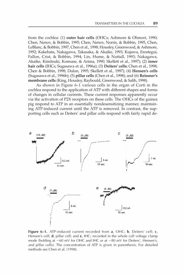
from the cochlea: (1) outer hair cells (OHCs; Ashmore & Ohmori, 1990;Chen, Nenov, & Bobbin, 1995; Chen, Nenov, Norris, & Bobbin, 1995, Chen,LeBlanc, & Bobbin, 1997, Chen et al., 1998; Housley, Greenwood, & Ashmore,1992; Kakehata, Nakagawa, Takasaka, & Akaike, 1993; Kujawa, Erostegui,Fallon, Crist, & Bobbin, 1994; Lin, Hume, & Nuttall, 1993; Nakagawa,Akaike, Kimitsuki, Komune, & Arima, 1990; Skellett et al., 1997); (2) innerhair cells (IHCs; Sugasawa et al., 1996a); (3) Deiters’ cells: Chen et al., 1998;Chen & Bobbin, 1998; Dulon, 1995; Skellett et al., 1997); (4) Hensen’s cells(Sugasawa et al., 1996b); (5) pillar cells (Chen et al., 1998); and (6) Reissner’smembrane cells (King, Housley, Raybould, Greenwood, & Salih, 1998).
As shown in Figure 6–1 various cells in the organ of Corti in thecochlea respond to the application of ATP with different shapes and formsof changes in cellular currents. These current responses apparently occurvia the activation of P2X receptors on these cells. The OHCs of the guineapig respond to ATP in an essentially nondesensitizing manner, maintain-ing ATP-induced current until the ATP is removed. In contrast, the sup-porting cells such as Deiters’ and pillar cells respond with fairly rapid de-
TRANSMITTERS IN THE COCHLEA 89
Figure 6–1. ATP-induced current recorded from a, OHC; b, Deiters’ cell; c,Hensen’s cell; d, pillar cell; and e, IHC; recorded in the whole cell voltage clampmode (holding at �60 mV for OHC and IHC or at �80 mV for Deiters’, Hensen’s,and pillar cells). The concentration of ATP is given in parenthesis. For detailedmethods see Chen et al. (1998).

sensitization where the current is greatly reduced almost immediately af-ter the maximum current is produced. Hensen’s cells and IHCs responddifferently from OHCs and supporting cells. Such variations in phenotypebetween cells are thought to be due to differences in the subunit composi-tion of the P2X receptors. Splicing of individual subunit mRNA and theexpression of these different splice variants assembled into the receptorscontributes to this variance in subunit composition of the receptors andtheir response to stimuli (e.g., Partin, Fleck, & Mayer, 1996).
TYPES OF P2X RECEPTORS IN THECOCHLEA IN THE ORGAN OF CORTI
Seven subunits of the P2X receptors (i.e., P2X1 – P2X7) and thirteen P2Y-likereceptor protein sequences (i.e., P2Y1-11, tp2y and fb1) have been isolated(Barnard, Simon, & Webb, 1997; King, Townsend-Nicholson, & Burnstock,1998; North & Barnard, 1997). To date the P2X2 subunit is the only receptortype mRNA that has been identified in the cochlea (Brandle et al., 1997;Chen & Bobbin, 1998; Housley, Greenwood, Bennett, & Ryan, 1995; Parker,Bobbin, & Deininger, 1997; Parker et al., 1998). In addition, several splicevariants of ionotropic ATP receptors have been described (Brandle, et al.,1997; Housley et al., 1995; Koshimizu, Tomic, Van Goor, & Stojilkovic, 1998;Parker et al., 1997, 1998; Simon et al., 1997; Troyanovskaya & Wackym, 1998).
Since we and others have used the guinea pig as a test subject, it wasimportant to discover the type of ATP receptors present in the guinea pigcochlea. Towards this end, we screened a guinea pig library kindly sup-plied to us by Wilcox and Fex (1992) for ATP receptors utilizing probes ofrat sequences kindly supplied to us (P2X2 by Brake, Wagenbach, & Julius,1994). Utilizing this procedure we isolated and sequenced three clones.Two of the clones proved to be sequences that are very similar in sequenceto two variants, P2X2-1 and P2X2-2 , that have been isolated from rat tissueand so we have called them guinea pig P2X2-1 and P2X2-2 (Figure 6–2). Thethird is unusual in that it retained a “rat intron” that was spliced out of therat sequence and so we have called it P2X2-3 (Figure 6–2). To date such asplice variant has not been described elsewhere in the literature.
To determine the distribution of the three P2X2 variants, and to see ifthe splice variant, P2X2-3, was expressed in tissue other than in the cochlea,total RNA was obtained from guinea pig cerebellum, brain (cerebrum),kidney, liver, and testes as well as cochlea. Briefly, 50 mg of tissue (or twocochlea) were homogenized with TRIzol reagent (Life Technologies) andthe RNA was extracted. RNA was separated by chloroform extraction, pre-cipitated with isopropanol, washed in ethanol, and dissolved in DEPC-treated water. The RNA was treated with Dnase I (Life Technologies) to re-move any contaminating chromosomal DNA. The cDNA was thensynthesized from mRNA with oligo(dT)12-18 primers using reverse tran-
90 GENETICS AND HEARING LOSS

scriptase (Life Technologies). As a negative control, the reverse transcrip-tase enzyme was omitted from matching RNA samples (“no RT” control).
Nested PCR was carried out on first strand cDNA by first utilizing aset of primers that amplified DNA contained in both regions of interest,i.e., intron VIII and/or spliced putative exon 11. The outer primer set con-tained forward primer ttcacagagctggcacacag at nucleotide 785 and reverseprimer aggaccagaagttcagagct at nucleotide 1541. PCR products from thisreaction were subjected to a second round of PCR by utilizing an inner setof primers, which also amplified the regions of interest (nested PCR). Theinner primer set contained forward primer tttggggtctgtgggtgtgg at nu-cleotide 856 and reverse primer tcatcccactgcaaccctaa nucleotide 1511.
PCR products were analyzed by 2% agarose gel electrophoresis. Inaddition to the “no RT” controls, negative controls included water insteadof cDNA. Positive controls included PCR performed on plasmid DNA ofeach of the three guinea pig P2X variants (data not shown).
As seen in Figure 6–3A and 6–3B, the PCR amplification of cDNAfrom brain and cerebellum yielded strong P2X2-2 (405 bp) and P2X2-3 (675
TRANSMITTERS IN THE COCHLEA 91
Figure 6–2. Organization of the guinea pig and rat P2X2 cDNA from exon 8 to the 3'end (poly A tail). The nucleotide sequence of rat and guinea pig variant P2X2-1 cDNAis generated from exon 8 and 9 (intron VIII spliced out) and all of exon 11 (11a, 11b,and 11c). The nucleotide sequence of rat and guinea pig variant P2X2-2 cDNA isgenerated from exon 8 and 9 (intron VIII spliced out) and a portion of exon 11 (11aand 11c; 11b spliced out). The nucleotide sequence of the guinea pig P2X2-3 variantcDNA is generated from exon 8, intron VIII, exon 9, exon 10, and all of exon 11(11a, 11b, and 11c). GenBank data base accession numbers are: AF053327 forP2X2-1, AF053328 for P2X2-2 and AF053329 for P2X2-3. (From “Novel Variant of theP2X2 ATP Receptor From the Guinea Pig Organ of Corti,” by M. S. Parker, M. L. Lar-roque, J. M. Campbell, R. P. Bobbin, & P. L. Deininger, 1998, p. 67. Hearing Research,121, 62–70. Copyright 1998 by Hearing Research. Reprinted with permission.)
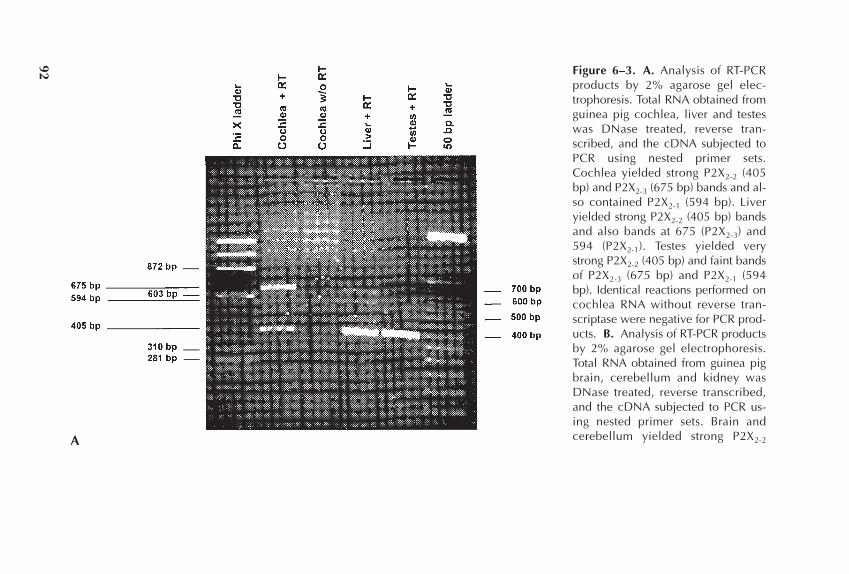
Figure 6–3. A. Analysis of RT-PCRproducts by 2% agarose gel elec-trophoresis. Total RNA obtained fromguinea pig cochlea, liver and testeswas DNase treated, reverse tran-scribed, and the cDNA subjected toPCR using nested primer sets.Cochlea yielded strong P2X2-2 (405bp) and P2X2-3 (675 bp) bands and al-so contained P2X2-1 (594 bp). Liveryielded strong P2X2-2 (405 bp) bandsand also bands at 675 (P2X2-3) and594 (P2X2-1). Testes yielded verystrong P2X2-2 (405 bp) and faint bandsof P2X2-3 (675 bp) and P2X2-1 (594bp). Identical reactions performed oncochlea RNA without reverse tran-scriptase were negative for PCR prod-ucts. B. Analysis of RT-PCR productsby 2% agarose gel electrophoresis.Total RNA obtained from guinea pigbrain, cerebellum and kidney wasDNase treated, reverse transcribed,and the cDNA subjected to PCR us-ing nested primer sets. Brain andcerebellum yielded strong P2X2-2A
92
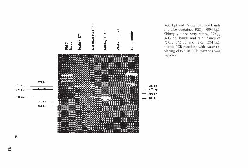
(405 bp) and P2X2-3 (675 bp) bandsand also contained P2X2-1 (594 bp).Kidney yielded very strong P2X2-2
(405 bp) bands and faint bands ofP2X2-3 (675 bp) and P2X2-1 (594 bp).Nested PCR reactions with water re-placing cDNA in PCR reactions wasnegative.
B
93

bp) bands and contained a moderate P2X2-1 (594 bp) band. Kidney yieldeda strong P2X2-2 (405 bp) band and faint P2X2-3 (675 bp) and P2X2-1 (594 bp)bands . Liver yielded strong P2X2-2 (405 bp) bands and faint P2X2-3 (675bp) and P2X2-1 (594 bp) bands. Testes yielded strong P2X2-2 (405 bp) andfaint P2X2-3 (675 bp) and P2X2-1 (594 bp) bands. Cochlea yielded strongP2X2-2 (405 bp) and P2X2-3 (675 bp) bands and a faint P2X2-1 (594 bp) band.Identical reactions performed on RNA from all the tissues without reversetranscriptase or with water replacing cDNA were negative for PCR prod-ucts. In summary, the splice variant P2X2-3 is strongly expressed in cochlea,brain, and cerebellum, and is weakly expressed in kidney, liver, and testes.
Presumably, the 3P2X2-3 splice variant found in guinea pig cannot befound in the rat. The reason is that the molecular mechanism that pro-duces this variant in the guinea pig is the inclusion in guinea pig of a re-gion analogous to rat intron VIII (Parker et al., 1998). The rat intron con-tains a stop codon. Therefore, even if the rat intron was expressed,presumably it would result in a truncated protein lacking the secondtransmembrane spanning region of the receptor.
This raises the question of why the guinea pig makes this splice vari-ant and the rat does not and what effect this variant has on the function ofthe cochlea. To date there are only preliminary data on this question. Chenet al. (1997) failed to obtain responses to application of ATP in most ratouter hair cells. The exceptions were very small cells that were difficult toidentify as OHCs and may not have been OHCs. In contrast these authorsobtained large responses in guinea pig outer hair cells and in Deiters’ cellsfrom both rat and guinea pig. This suggests that the reason for the reducedincidence or absence of a response to ATP in rat OHCs may have been dueto the absence of the P2X2-3 splice variant in rat. It does appear that P2X2mRNA is present in rat outer hair cells (Housley et al., 1998) but the pro-tein may not be expressed. Thus it will be interesting to look at the distrib-ution of the P2X2-3 splice variant and see if this subunit is present in guineapig OHCs.
As shown in Figure 6–1, the OHC of guinea pig responds to the appli-cation of ATP with an inward current that is nondesensitizing. Thus thequestion arises as to whether the P2X2-3 splice variant is nondesensitizingor imparts nondesensitizing characteristic to the expressed receptor. To ex-amine this hypothesis, the three splice variants were individually andtransiently expressed in human embryonic kidney cells (HEK293). Theproperties of the three P2X2 receptor variants were characterized using thewhole-cell voltage-clamp technique. The results indicate that the applica-tion of ATP induced inward currents in HEK293 cells expressing homo-meric receptors containing each of the three splice variants in a dose-de-pendent manner (Figure 6–4). The ATP induced currents in HEK293 cellsexpressing P2X2-1 and P2X2-2 variants were large, about the same ampli-tude and desensitized rapidly. The responses had characteristics similar to
94 GENETICS AND HEARING LOSS

those of the receptors on Deiters’ cells in both rat and guinea pig (Chen etal., 1997; Chen & Bobbin, 1998). In comparison to the other two variants,the current in the cells expressing the P2X2-3 variant was much smaller, re-quired higher concentrations of ATP to be activated and desensitizedslower. These results are consistent with the presence of the P2X2-3 variantin OHCs partially accounting for the slow desensitizing response to ATPin OHCs compared to other cells in the organ of Corti. Since the guinea pigOHC response to ATP is a nondesensitizing response (Figure 6–1; Chen etal., 1997; Housley et al., 1992) and the P2X2-3 splice variant is absent fromthe rat, then this variant is a good candidate for being the receptor presentin guinea pig OHCs and the one absent from rat OHCs. This would indi-cate that the P2X2-3 subunit may in part account for the nondesensitizingresponse in OHCs. Of course, there is a very high probability that addi-tional splice variants, not described to date, exist in the organ of Corti.These undiscovered variants may be more important to the phenotype ofthe guinea pig OHC than the P2X2-3 splice variant.
TRANSMITTERS IN THE COCHLEA 95
Figure 6–4. Current traces of ATP-induced responses recorded from transiently trans-fected HEK293 cells at a holding potential of �60 mV. a. Current traces for singlecells expressing the P2X2-2 splice variant in response to ATP application. b. Currenttraces for cells expressing the P2X2-3 splice variant in response to ATP application.
FUNCTION OF ATP AND ATPRECEPTORS IN THE COCHLEA
ATP Receptors on Cell Surfaces Exposed to Endolymph
The ATP receptors on OHCs, Hensen’s cells, IHCs, cells of Reisner’s mem-brane, and cells in the stria have been suggested to be located on the cellsurfaces exposed to endolymph rather than perilymph, although defini-tive location studies with antibodies for most of these cells remain to becarried out (OHCs: Housley et al., 1992; Mockett, Housley, & Thorne, 1994;IHCs: Sugasawa et al., 1996a; Hensen’s cells: Sugasawa et al., 1996b; Reiss-ner’s: King, Housley, et al., 1998; stria: Liu et al., 1995). From this anatomi-cal location of the various ATP receptors and physiological data obtainedto date by various investigators, it appears that in general the ATP recep-tors bathed by endolymph are involved in cochlear fluid dynamics andmovement of potassium across the cells lining the endolymph compart-ment (King, Housley, et al., 1998; Liu et al., 1995; Munoz, Thorne, Housley,& Billett, 1995; Munoz, Thorne, Housley, Billett, & Battersby, 1995; Suzukiet al., 1995). The ionotropic P2X2 receptors on OHCs, which appear to belocated on the steriocillia may be involved in transduction (Housley et al.,1992; Kirk & Yates, 1998; Mammano et al., 1999; Mockett et al., 1994, 1995).The origin of the endogenous ATP acting on these receptors may be themarginal cells of the stria vascularis (White et al., 1995).
ATP Receptors on Surfaces Lining the Perilymph Compartment
To date only Deiters’ cells have been demonstrated to contain P2X2ionotropic ATP receptors exposed to perilymph (Dulon, 1995). These re-ceptors appear to be located near where the Deiters’ cell contacts the OHC(Dulon, 1995). Drugs placed in perilymph will act on these receptors ex-posed to perilymph, but not on receptors lining the endolymph compart-ment, due to cellular barriers that prevent drugs from entering endolymphfrom perilymph. Agonists that activate ATP receptors placed in perilymphdecrease the magnitude of the cubic distortion product otoacoustic emis-sions (DPOAEs) and increase the magnitude of the quadratic DPOAEs(Figure 6–5; Kujawa, Erostegui, et al., 1994). The DPOAEs reflect the me-chanical motion of the cochlear partition (Kemp, 1998; Mills, 1998). In ad-dition, these agents alter cochlear sensitivity as monitored by changes inthe summating potential (SP), the compound action potential of the audi-tory nerve (CAP), and N1 latency (Bobbin & Thompson, 1978; Kujawa,Erostegui, et al., 1994). Thus the tentative conclusion was reached thatthese drugs acted on P2X2 ATP receptors exposed to perilymph or the ATPreceptors on Deiters’ cell (Bobbin et al., 1998).
Initially, the action of ATPR agonists (2-Me-S-ATP; ATP; ATP-�-S)placed in perilymph was thought to be due to metabotropic, P2Y receptor
96 GENETICS AND HEARING LOSS
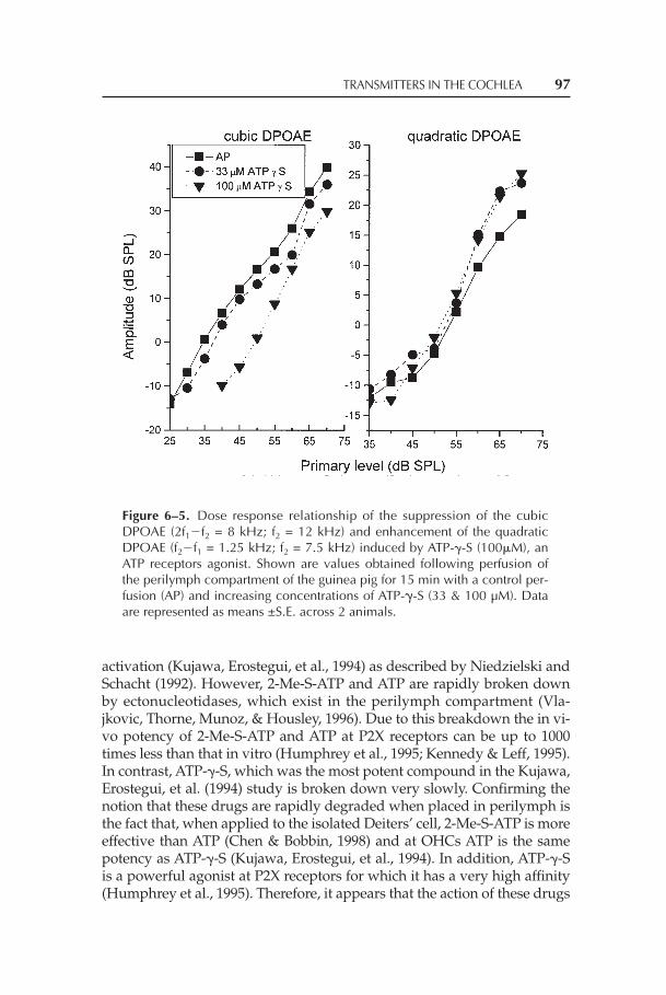
activation (Kujawa, Erostegui, et al., 1994) as described by Niedzielski andSchacht (1992). However, 2-Me-S-ATP and ATP are rapidly broken downby ectonucleotidases, which exist in the perilymph compartment (Vla-jkovic, Thorne, Munoz, & Housley, 1996). Due to this breakdown the in vi-vo potency of 2-Me-S-ATP and ATP at P2X receptors can be up to 1000times less than that in vitro (Humphrey et al., 1995; Kennedy & Leff, 1995).In contrast, ATP-�-S, which was the most potent compound in the Kujawa,Erostegui, et al. (1994) study is broken down very slowly. Confirming thenotion that these drugs are rapidly degraded when placed in perilymph isthe fact that, when applied to the isolated Deiters’ cell, 2-Me-S-ATP is moreeffective than ATP (Chen & Bobbin, 1998) and at OHCs ATP is the samepotency as ATP-�-S (Kujawa, Erostegui, et al., 1994). In addition, ATP-�-Sis a powerful agonist at P2X receptors for which it has a very high affinity(Humphrey et al., 1995). Therefore, it appears that the action of these drugs
TRANSMITTERS IN THE COCHLEA 97
Figure 6–5. Dose response relationship of the suppression of the cubicDPOAE (2f1�f2 = 8 kHz; f2 = 12 kHz) and enhancement of the quadraticDPOAE (f2�f1 = 1.25 kHz; f2 = 7.5 kHz) induced by ATP-�-S (100�M), anATP receptors agonist. Shown are values obtained following perfusion ofthe perilymph compartment of the guinea pig for 15 min with a control per-fusion (AP) and increasing concentrations of ATP-�-S (33 & 100 µM). Dataare represented as means ±S.E. across 2 animals.

in vivo may be due to the same P2X2 receptors activated in vitro and notdue to P2Y receptors as initially suggested by Kujawa, Erostegui, et al.(1994). On the other hand, at present there is no reason to eliminate an ac-tion of the drugs at P2Y ATP receptors and so one must be cautious in at-tributing the action of the drugs to either P2X or P2Y ATPRs until furtherstudies are carried out.
Additional support for the Deiters’ cell site-of-action hypothesis forthe action of these drugs placed in perilymph are the observations of Du-lon, Blanchet, and Laffon (1994). Dulon et al. (1994) demonstrated thatDeiters’ cells move and alter their tension in response to an increase in in-ternal calcium levels. Since ATP induced activation of P2X2 ATP receptorsresults in an inward current of Na+ and Ca2+ in Deiters’ cells (Figure 6–6;Ashmore & Ohmori, 1990; Dulon et al., 1993), ATP may also induce a stiff-
98 GENETICS AND HEARING LOSS
Figure 6–6. ATP-induced increase in intracellularCa2+ in a Deiters’ cell. Cell isolation and dye load-ing and measurements were carried out in theLSUMC calcium imaging facility utilizing a Noran’sOdyssey laser scanning confocal microscope anddata analysis software. Application of 100 µM ATPonto an indo-1/AM loaded Deiters’ cell was by apressure puff ejector via a glass pipette near the cell.
ness change in Deiters’ cells. This change in stiffness may alter cochlearmechanics (Kolston & Ashmore, 1996) and be reflected as a change inDPOAEs (Figure 6–5; Kujawa, Erostegui, et al., 1994). At the apex of thecochlea, Deiters’ cells hold the OHCs at their base and at their apical pole(Figure 6–7). In other turns of the cochlea, the Deiters’ cell holds one OHCat the base and then an adjacent ones at the apical pole (see Figure 1–2 onpage 13 in Brownell, 1996). An ATP-induced change in [Ca 2+] in Deiters’cells may alter the Deiters’ cells tension, which in turn may act as a “bow”to tighten or loosen the tension on the string (OHCs). Since the shorteningand lengthening of the OHCs appear to be responsible for the gain in thecochlear amplifier, then the alteration in stiffness of the Deiters’ cells mayact as a “set point adjustment” on the OHC motor to maintain an optimalgain. So the ATP agonists placed in perilymph and acting on P2X2 ATP re-ceptors on Deiters’ cells may increase the stiffness of Deiters’ cells to suchan extent that the cochlear amplifier is unable to function and the cubicDPOAE is reduced and the quadratic enhanced (Figure. 6–5; Kujawa,Erostegui, et al., 1994).
MEASURES OF THE FUNCTION OF ENDOGENOUSATP AND ATP RECEPTORS BATHED BY PERILYMPH
The Quadratic DPOAEs and SP
Frank and Kossl (1996) suggested that the quadratic DPOAE is a more sen-sitive indicator of the “set point” of the cochlear amplifier than the cubicDPOAE. van Emst (1996) suggested that the SP is also influenced by the“operating point of the apical transducer channel.” The effects of suraminand PPADs, ATP antagonists, may be evidence that endogenous ATP af-fects the set point of the cochlear amplifier. Both drugs reversibly affect thequadratic DPOAE and suppressed the negative SP when the drugs wereplaced in the perilymph compartment (Chen et al., 1998; Kujawa, Fallon,et al., 1994; Skellett et al., 1997). PPADS (1 mM), which is much more po-tent than suramin in blocking the actions of ATP, induced more than a 10dB suppression of the initial value of the time varying quadratic DPOAE(Chen et al., 1998). In addition, PPADS (1 mM) suppressed the quadraticDPOAE intensity function recorded immediately after a 15 min applica-tion of the drug in silence (Figure 6–8). This is in contrast to the little or nochange in the intensity function of the quadratic DPOAE reported by Chenet al. (1998). This is explained by the fact that Chen et al. (1998) recordedthe quadratic DPOAE intensity function 15 min after recording responsesto the primaries, at which point the quadratic DPOAE had recovered tobase line values. This indicates that these particular effects of PPADS onthe quadratic DPOAE were readily reversible. In addition, PPADS had an
TRANSMITTERS IN THE COCHLEA 99
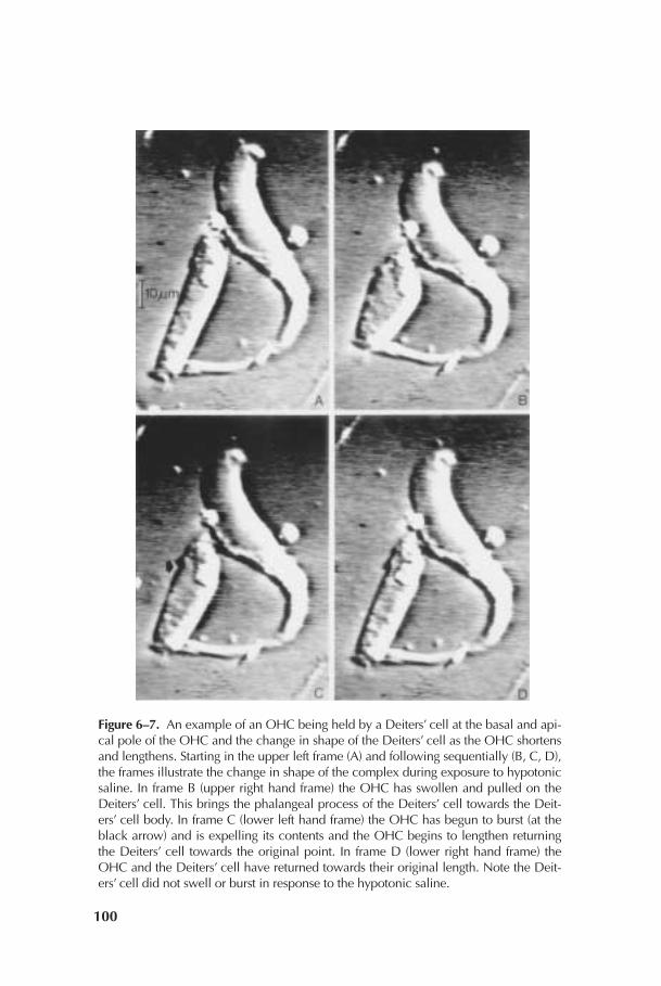
Figure 6–7. An example of an OHC being held by a Deiters’ cell at the basal and api-cal pole of the OHC and the change in shape of the Deiters’ cell as the OHC shortensand lengthens. Starting in the upper left frame (A) and following sequentially (B, C, D),the frames illustrate the change in shape of the complex during exposure to hypotonicsaline. In frame B (upper right hand frame) the OHC has swollen and pulled on theDeiters’ cell. This brings the phalangeal process of the Deiters’ cell towards the Deit-ers’ cell body. In frame C (lower left hand frame) the OHC has begun to burst (at theblack arrow) and is expelling its contents and the OHC begins to lengthen returningthe Deiters’ cell towards the original point. In frame D (lower right hand frame) theOHC and the Deiters’ cell have returned towards their original length. Note the Deit-ers’ cell did not swell or burst in response to the hypotonic saline.
100

even larger effect (up to @20 dB sound equivalents) on the high intensitynegative SP (scala vestibuli) that was readily reversible (Figure 6–9; Chenet al., 1998). These data are supportive of the speculation that these ATP re-ceptor antagonists may induce their effects by blocking the depolarizationand the increase in internal calcium in Deiters’ cells induced by endoge-nous ATP acting on P2X2 ATP receptors (Bobbin et al., 1998; Chen et al.,1998; Skellett et al., 1997).
Intense Sound Exposure
To further test the hypothesis that sound releases endogenous ATP to af-fect the function of the cochlea, the effect of PPADS on a moderately in-tense sound (95 dB SPL; 15 min; 6700 Hz) was tested. This level of sound
TRANSMITTERS IN THE COCHLEA 101
Figure 6–8. PPADS (1 mM) slightly enhances the growth functions for thecubic DPOAE (2f1�f2 = 8 kHz; f2 = 12 kHz) and suppresses the growthfunction for the quadratic DPOAE (f2�f1 = 1.25 kHz; f2 = 7.5 kHz).Shown are values obtained immediately following perfusion of the peri-lymph compartment in guinea pig with a control artificial perilymph per-fusion (AP2), PPADS (1 mM) and a wash (wash 1). Perfusions were carriedout in silence and recorded immediately after termination of the perfu-sion. The quadratic DPOAE values were collected first to avoid the reversalof the magnitude of the quadratic over time reported in Chen et al. (1998).

apparently induces only a temporary threshold shift as monitored by al-terations in cochlear mechanics via DPOAEs (Puel, Bobbin, & Fallon,1988). Therefore, it was reasoned that if endogenous ATP has an action onDeiters’ cells to alter their stiffness then ATP may have a role in this sound-induced alteration in mechanics.
The methods used were very similar to those used previously (Puel etal., 1988; Puel, Ruel, d’Aldin, & Pujol, 1998). Animals were exposed to thepure tone 10 min after the start of a perfusion with artificial perilymph (in-tense sound group) or 1 mM PPADS (intense sound + PPADS group) andthe tone was terminated 10 min before the end of perfusion. A third groupreceived PPADs alone (PPADS alone group). Cochlear potentials evokedby 10 kHz tone bursts were recorded from an electrode in basal turn scalavestibuli. As shown in recordings of the cochlear microphonics (Figure6–10) preliminary results indicate that the effects of the moderately intensesound combined with PPADS were larger than the effects induced by in-tense sound-alone or PPADS-alone. In addition, the enhanced effect wasreadily reversed with washing (Figure 6–10).
102 GENETICS AND HEARING LOSS
Figure 6–9. PPADS (1 mM) reversibly suppresses the negative SPrecorded from scala vestibuli in response to 10 kHz tone bursts.Shown are the magnitude of the negative SP as a function of intensityrecorded after predrug artificial perilymph perfusion (AP; 15 min), af-ter perfusion with 1.0 mM PPADS (35 min), and after postdrug washwith artificial perilymph (wash; 15 min). All perfusions were throughthe perilymph compartment. Data are represented as means ± S.E.across 5 animals.

These results are consistent with the hypothesis that endogenous ex-tracellular ATP acting on ATP receptors on Deiters’ cells increases the ten-sion these cells apply to the motion of the OHCs and subsequently to thecochlear partition (Bobbin et al., 1998; Dulon, 1995; Dulon et al., 1993, 1994;Skellett et al., 1997). The PPADs block of these effects of endogenous ATPwould indirectly decrease normal tension and so increase the motion in-duced by the moderately intense sound. This would in turn potentiate theeffects of the intense sound.
There are many alternative explanations of these results and of therole of P2X2 ATP receptors on Deiters’ cells. Deiters’ cells may play a rolein the uptake and removal of extracellular K+ that enters and exits theOHC as transduction current (Spicer, Smythe, & Schulte, 1999). An inwardrectifying potassium channel is usually assigned this task; however, ourlaboratory has failed to find electrophysiological evidence for such a chan-
TRANSMITTERS IN THE COCHLEA 103
Figure 6–10. Effects of treatment with a moderately intense soundalone (noise, 6.7 kHz, 95 dB SPL, 15 min), a combination of the in-tense sound with 1 mM PPADs (noise & ppads) and 1 mM PPADSalone (ppads) on 62 dB SPL 10 kHz tone burst-evoked CM recordedfrom the scala vestibuli. Shown are values obtained after pretreat-ment artificial perilymph perfusion (AP), after treatment (i.e., noiseexposure during AP perfusion; noise during ppads perfusion; ppadsperfusion alone) and after washes with artificial perilymph (wash).The perilymph compartment was perfused. Data are displayed asmeans ± S.E. across n = 5 animals per treatment condition. The as-terisk indicates a significant (p< 0.05) difference from AP and a sig-nificant (p< 0.05) difference from wash within treatment group.

nel in Deiters’ cells (Nenov, Chen, & Bobbin, 1998). Instead, extracellularATP and the P2X2 ATP receptors on Deiters’ cells may be intimately in-volved in this removal of K+. Activation of P2X2 ATP receptors on Deiters’cells results in an increase in intracellular Ca2+ and Na+ in a manner simi-lar to the activation of excitatory amino acid receptors in glia (Gallo & Rus-sell, 1995). By analogy with the relationship between neurons and glia(Gallo & Russell, 1995), this increase in Ca2+ and Na+ could activate K+ up-take through the efforts of a Na+/Ca2+ exchanger and a Na+/K+ pump. Inaddition, if [Ca2+] signals are propagated as a wave from the site of activityalong the supporting cells coupled by gap junctions in a manner similar toglia, then the activation of P2X2 ATP receptors may support spatial buffer-ing of K+ by drawing this ion away from the OHCs (as suggested for gliaby Dietzel, Heinemann, & Lux, 1989; Gallo & Russell, 1995). Thus, duringthe moderately intense sound exposure in the presence of PPADS, theremay have been an increase in extracellular K+ concentration between thehair cells and the Deiters’ cells due to a blockade of the K+ uptake processinvolving P2X2 ATP receptors in Deiters’ cells. An increase in K+ outsidethe basal end of the OHCs would produce a reduction in CM magnitude.Future experiments will determine if either or both the stiffness hypothesisand the K+ hypothesis suggested here are correct.
The release of a chemical or neuromodulator onto these supportingcells in vivo during sound exposure was suggested by Oesterle and Dallos(1990) to explain the slow “end dc shift” they observed in electrical record-ings from supporting cells during sound stimulation. Wangemann (1996)has shown release of ATP from an isolated whole organ of Corti into thefluid surrounding the tissue, presumably into the vicinity of the Deiters’cells. The source of the endogenous ATP acting on the ATP receptors onDeiters’ cells may be the nerve endings forming synapses with these cells(Burgess, Adams, & Nadol, 1997).
SUMMARY
In summary, different cells in the cochlea exhibit different phenotypical re-sponses to ATP. Preliminary evidence indicates that these phenotypes maybe due to different combinations of P2X2 splice variant subunits expressedin the different cells. In addition, it appears that extracellular ATP has sev-eral functions in the cochlea. One function in the endolymph compartmentmay be to regulate endolymph composition and the other function in theperilymph compartment may be to modulate cochlear mechanics or to en-sure uptake of K+.
Acknowledgments: This work was supported in part by a research grant from theNational Science Foundation, American Hearing Research Foundation, the Deaf-
104 GENETICS AND HEARING LOSS

ness Research Foundation, the National Organization for Hearing Research, Kam’sFund for Hearing Research, the National Institutes of Health, and the Louisiana Li-ons Eye Foundation.
REFERENCES
Ashmore, J. F., & Ohmori, H. (1990). Control of intracellular calcium by ATP in iso-lated outer hair cells of the guinea-pig cochlea. Journal of Physiology (London),428, 109–131.
Barnard, E. A., Simon, J., & Webb, T. E. (1997). Nucleotide receptors in the nervoussystem. Molecular Neurobiology, 15, 103–130.
Bledsoe, S. C., Jr., Bobbin, R. P., & Puel, J.-L. (1988). Neurotransmission in the innerear. In A. F. Jahn & J. R. Santos-Sacchi (Eds.), Physiology of hearing (pp. 385–406).New York: Raven Press.
Bobbin, R. P. (1996). Chemical receptors on outer hair cells and their molecularmechanisms. In C. I. Berlin (Ed.), Hair cells and hearing aids (pp. 29–55). SanDiego, CA: Singular Publishing Group.
Bobbin, R. P. (1997). Transmitters in the cochlea: The chemical machinery in the ear.In C. I. Berlin (Ed.), Neurotransmission and hearing loss: Basic science, diagnosis, andmanagement (pp. 25–46). San Diego, CA: Singular Publishing Group.
Bobbin, R. P., Bledsoe, S. C., Jr., Winbery, S. L., & Jenison, G. L. (1985). Actions ofputative neurotransmitters and other relevant compounds on Xenopus laevislateral line. In D. G. Drescher (Ed.), Auditory biochemistry (pp. 102–122). Spring-field, IL: Charles C. Thomas.
Bobbin, R. P., Chen, C., Nenov, A. P., & Skellett, R. A. (1998). Transmitters in thecochlea: The quadratic distortion product and its time varying response may re-flect the function of ATP in the cochlea. In C. I. Berlin (Ed.), Otoacoustic emissions(pp. 61–83). San Diego, CA: Singular Publishing Group.
Bobbin, R. P., Chu, S., Skellett, R. A., Campbell, J., & Fallon, M. (1997). Cytotoxicityand mitogenicity of adenosine triphosphate in the cochlea. Hearing Research,113, 155–164.
Bobbin, R. P., & LeBlanc, C. S. (1999). Apamin reduces but does not abolish the ef-fects of contralateral suppression of cubic DPOAEs. In C. I. Berlin (Ed.), The ef-ferent auditory system (pp. 61–71). San Diego, CA: Singular Publishing Group.
Bobbin, R. P., & Thompson, M. H. (1978). Effects of putative transmitters on affer-ent cochlear transmission. Annals of Otology, Rhinology, and Laryngology, 87,185–190.
Brake, A. J., Wagenbach, M. J., & Julius, D. (1994). New structural motif for ligand-gated ion channels defined by an ionotropic ATP receptor. Nature, 371, 519–523.
Brandle, U., Spielmanns, P., Osteroth, R., Sim, J., Surprenant, A., Buell, G., Ruppers-berg, J. P., Plinkert, P. K., Zenner, H. P., & Glowatzki, E. (1997). Desensitizationof the P2X(2) receptor controlled by alternative splicing. FEBS Letters, 404,294–298.
Brownell, W. E. (1996). Outer hair cell electromotility and otoacoustic emissions. InC. I. Berlin (Ed.), Hair cells and hearing aids (pp. 3–27). San Diego, CA: SingularPublishing Group.
TRANSMITTERS IN THE COCHLEA 105
Burgess, B. J., Adams, J. C., & Nadol, J. B., Jr. (1997). Morphologic evidence for in-nervation of Deiters’ and Hensen’s cells in the guinea pig. Hearing Research, 108,74–82.
Burnstock, G. (1990). Purinergic mechanisms. In G. R. Dubyak & J. S. Fedan (Eds.),Biological actions of extracellular ATP. Annals of the New York Academy of Science,603, 1–18.
Chen, C., & Bobbin, R. P. (1998). P2X receptors in cochlear Deiters’ cells. BritishJournal of Pharmacology, 124, 337–344.
Chen, C., LeBlanc, C., & Bobbin, R. P. (1997). Differences in the distribution of re-sponses to ATP and acetylcholine between outer hair cells of rat and guinea pig.Hearing Research, 110, 87–94.
Chen, C., Nenov, A., & Bobbin, R. P. (1995). Noise exposure alters the response ofouter hair cells to ATP. Hearing Research, 88, 215–221.
Chen, C., Nenov, A. P., Norris, C., & Bobbin, R. P. (1995). ATP modulation of L-typeCa2+ channel currents in guinea pig outer hair cells. Hearing Research, 86, 25–33.
Chen, C., Skellett, R. A., Fallon, M., & Bobbin, R. P. (1998). Additional pharmaco-logical evidence that endogenous ATP modulates cochlear mechanics. HearingResearch, 118, 47–61.
Dietzel, I., Heinemann, U., & Lux, H. D. (1989). Relations between slow extracellu-lar potential changes, glial potassium buffering, and electrolyte and cellular vol-ume changes during neuronal hyperactivity in cat brain. Glia, 2, 25–44.
Dubyak, G. R., & El-Moatassim, C. (1993). Signal transduction via P2-purinergic re-ceptors for extracellular ATP and other nucleotides. American Journal of Physiolo-gy, 265, C577–C606.
Dulon, D. (1995). Ca2+ signaling in Deiters’ cells of the guinea-pig cochlea activeprocess in supporting cells? In A. Flock, D. Ottoson, & M. Ulfendahl (Eds.), Ac-tive hearing (pp. 195–207). London: Elsevier Science Ltd.
Dulon, D., Blanchet, C., & Laffon, E. (1994). Photo-released intracellular Ca2+
evokes reversible mechanical responses in supporting cells of the guinea-pigorgan of Corti. Biochemical and Biophysical Research Communications, 201, 1263–1269.
Dulon, D. Moataz, R., & Mollard, P. (1993). Characterization of Ca2+ signals gener-ated by extracellular nucleotides in supporting cells of the organ of Corti. CellCalcium 14, 245– 254.
Dulon, D., Mollard, P., & Aran, J.-M. (1991). Extracellular ATP elevates cytosolicCa2+ in cochlear inner hair cells. NeuroReport 2, 69–72.
Emst, M. G.,van (1996). Origin of the cochlear summating potential in the guinea pig[Thesis]. Department of Otorhinolaryngology, Utrecht University, Utrecht,Germany.
Eybalin, M. (1993). Neurotransmitters and neuromodulators of the mammaliancochlea. Physiological Reviews, 73, 309–373.
Frank, G., & Kossl, M. (1996). The acoustic two-tone distortions 2f1–f2 and f2–f1 andtheir possible relation to changes in the operating point of the cochlear amplifi-er. Hearing Research, 98, 104–115.
Fredholm, B. B. (1995). Purinoceptors in the nervous system. Pharmacology and Toxi-cology, 76, 228–239.
Gallo, V., & Russell, J. T. (1995). Excitatory amino acid receptors in glia: Differentsubtypes for distinct functions. Journal of Neuroscience Research, 42, 1–8.
Henzi, V., & MacDermott, A. B. (1992). Characteristics and function of Ca2+- and in-ositol 1,4,5-triphosphate-releasable stores of Ca2+ in neurons. Neuroscience, 46,251–273.
106 GENETICS AND HEARING LOSS

Housley, G. D. (1997). Extracellular nucleotide signaling in the inner ear. MolecularNeurobiology, 16, 21–48.
Housley, G. D., Greenwood, D., & Ashmore, J. F. (1992). Localization of cholinergicand purinergic receptors on outer hair cells isolated from the guinea-pigcochlea. Proceedings of the Royal Society of London, Series B: Biological Sciences (Lon-don), 249, 265–273.
Housley, G. D., Greenwood, D., Bennett, T., & Ryan, A. F. (1995). Identification of ashort form of the P2xR1-purinoceptor subunit produced by alternative splicingin the pituitary and cochlea. Biochemical and Biophysical Research Communications,212, 501–508.
Housley, G. D., Kanjhan, R., Raybould, N. P., Greenwood, D., Salih, S. G., Jarlebark,L., Burton, L. D., Setz, V. C. M., Cannell, M. B., Soeller, C., Christie, D. L., Usami,S., Matsubara, A., Yoshie, H., Ryan, A. F., & Thorne, P. R. (1999). Expression ofthe P2X2 receptor subunit of the ATP-gated ion channel in the cochlea: Implica-tions for sound transduction and auditory neurotransmission. Journal of Neuro-science, 19, 8377–8388.
Housley, G. D., Luo, L., & Ryan, A. F. (1998). Localization of mRNA encoding theP2X2 receptor subunit of the adenosine 5'-triphosphate-gated ion channel in theadult and developing rat inner ear by in situ hybridization. Journal of Compara-tive Neurology, 393, 403–414.
Humphrey, P. P. A., Buell, G., Kennedy, I., Khakh, B. S., Michel, A. D., Surprenant,A., & Trezise, D. J. (1995). New insights on P2X purinoceptors. Naunyn-Schmiede-berg’s Archives of Pharmacology, 352, 585–596.
Ikeda, K., Saito, Y., Nishiyama, A., & Takasaka, T. (1991). Effect of neuroregulatorson the intracellular calcium level in the outer hair cell isolated from the guineapig. Otology, Rhinology, and Laryngology, 53, 78–81.
Kakehata, S., Nakagawa, T., Takasaka, T., & Akaike, N. (1993). Cellular mechanismof acetylcholine-induced response in dissociated outer hair cells of guinea-pigcochlea. Journal of Physiology (London), 463, 227–244.
Kemp, D. T. (1998). Otoacoustic emissions: Distorted echos of the cochlea’s travel-ing wave . In C. I. Berlin (Ed.), Otoacoustic emissions: Basic science and clinical ap-plications (pp. 1–60). San Diego, CA: Singular Publishing Group.
Kennedy, C., & Leff, P. (1995). How should P2X purinoceptors be classified pharma-cologically? Trends in Pharmacological Science, 16, 168–174.
King, B. F., Townsend-Nicholson, A., & Burnstock, G. (1998). Metabotropic recep-tors for ATP and UTP: Exploring the correspondence between native and re-combinant nucleotide receptors. Trends in Pharmacological Science, 19, 506–514.
King, M., Housley, G. D., Raybould, N. P., Greenwood, D., & Salih, S. G. (1998). Ex-pression of ATP-gated ion channels by Reissner’s membrane epithelial cells.NeuroReport, 9, 2467– 2474.
Kirk, D. L., & Yates, G. K. (1998). ATP in endolymph enhances electrically evokedoto-acoustic emissions from the guinea pig cochlea. Neuroscience Letters, 250,149–152.
Kolston, P. J., & Ashmore, J. F. (1996). Finite element micromechanical modeling ofthe cochlea in three dimensions. Journal of the Acoustical Society of America, 99,455–467.
Koshimizu, T., Tomic, M., Van Goor, F., & Stojilkovic, S. S. (1998). Functional role ofalternative splicing in pituitary P2X2 receptor-channel activation and desensiti-zation. Molecular Endocrinology, 12, 901–913.
TRANSMITTERS IN THE COCHLEA 107

Kujawa, S. G., Erostegui, C., Fallon, M., Crist, J., & Bobbin, R. P. (1994). Effects ofAdenosine 5'-triphosphate and related agonists on cochlear function. HearingResearch, 76, 87–100.
Kujawa, S. G., Fallon, M., & Bobbin, R. P. (1994). ATP antagonists cibacron blue,basilen blue and suramin alter sound-evoked responses of the cochlea and au-ditory nerve. Hearing Research, 78, 181–188.
Lin, X, Hume, R. I., & Nuttall, A. L. (1993). Voltage-dependent block by neomycinof the ATP-induced whole cell current of guinea-pig outer hair cells. Journal ofNeurophysiology, 70, 1593–1605.
Liu, J., Kozakura, K., & Marcus, D. (1995). Evidence for purinergic receptors investibular dark cell and strial marginal cell epithelia of the gerbil. Auditory Neu-roscience, 1, 331–340.
Mammano, F., Frolenkov, G. I., Lagostena, L., Belyantseva, I. A., Kurc, M., Dodane,V., Colavita, A., & Kachar, B. (1999). ATP-induced Ca2+ release in cochlear outerhair cells: Localization of an inositol triphosphate-gated Ca2+ store to the base ofthe sensory hair bundle. Journal of Neuroscience, 19, 6918–6929.
Mills, D. M. (1998). Origin and implications of two “components” in distortionproduct otoacoustic emissions. In C. I. Berlin (Ed.), Otoacoustic emissions: Basicscience and clinical applications (pp. 85–104). San Diego, CA: Singular PublishingGroup.
Moataz, R., Saito, T., & Dulon, D. (1992). Evidence for voltage sensitive Ca2+ chan-nels in supporting cells of the organ of Corti: Characterization by indo-1 fluo-rescence. Advances in the Biosciences, 83, 53–59.
Mockett, B. G., Bo, X., Housley, G. D., Thorne, P. R., & Burnstock, G. (1995). Autora-diographic labelling of P2 purinoceptors in the guinea pig cochlea. Hearing Re-search, 84, 177–193.
Mockett, B. G., Housley, G. D., & Thorne, P. R. (1994). Fluorescence imaging of ex-tracellular purinergic receptor sites and putative ecto-ATPases sites on isolatedcochlear hair cells. Journal of Neuroscience, 14, 6992–7007.
Munoz, D. J. B., Thorne, P. R., Housley, T. E., & Billett, T. E. (1995). Adenosine 5'-triphosphate (ATP) concentrations in the endolymph and perilymph of theguinea pig cochlea. Hearing Research, 90, 119–125.
Munoz, D. J. B., Thorne, P. R., Housley, T. E., Billett, T. E., & Battersby, J. M. (1995).Extracellular adenosine 5'-triphosphate (ATP) in the endolymphatic compart-ment influences cochlear function. Hearing Research, 90, 106–118.
Nakagawa, T., Akaike, N., Kimitsuki, T., Komune, S., & Arima, T. (1990). ATP-in-duced current in isolated outer hair cells of guinea pig cochlea. Journal of Neuro-physiology, 63, 1068–1074.
Nenov, A. P., Chen, C., & Bobbin, R. P. (1998). Outward rectifying potassium cur-rents are the dominant voltage activated currents present in Deiters’ cells. Hear-ing Research, 123, 168–182.
Niedzielski, A. S., & Schacht, J. (1992). P2 purinoceptors stimulate inositol phos-phate release in the organ of Corti. NeuroReport, 3, 273–275.
Nilles, R., Jarlebark, L., Zenner, H. P., & Heilbronn, E. (1994). ATP-induced cyto-plasmic [Ca2+] increases in isolated cochlear outer hair cells. Involved receptorand channel mechanisms. Hearing Research, 73, 27–34.
North, R. A., & Barnard, E. A. (1997). Nucleotide receptors. Current Opinion in Neu-robiology, 7, 346–357.
108 GENETICS AND HEARING LOSS

Oesterle, E. C., & Dallos, P. (1990). Intracellular recordings from supporting cells inthe guinea pig cochlea: DC potentials. Journal of Neurophysiology, 64, 617–636.
Ogawa, K., & Schacht, J. (1995). P2y purinergic receptors coupled to phosphoinosi-tide hydrolysis in tissues of the cochlear lateral wall. NeuroReport, 6, 1538–1540.
Parker, M. S., Bobbin, R. P., & Deininger, P. L. (1997). Guinea pig organ of Corti con-tains mRNA for ATP receptor type P2X2. Association for Research in Otolaryngolo-gy Abstracts, 20, 217.
Parker, M. S., Larroque, M. L., Campbell, J. M., Bobbin, R. P., & Deininger, P. L.(1998). Novel variant of the P2X2 ATP receptor from the guinea pig organ ofCorti. Hearing Research, 121, 62–70.
Partin, K. M., Fleck, M. W., & Mayer, M. L. (1996). AMPA receptor flip/flop mu-tants affecting deactivation, desensitization, and modulation by cyclothiazide,aniracetam, and thiocyanate. Journal of Neuroscience, 16, 6634–6647.
Puel, J. L. (1995). Chemical synaptic transmission in the cochlea. Progress in Neuro-biology, 47, 449–476.
Puel, J. L., Bobbin, R. P., & Fallon, M. (1988). The active process is affected first byintense sound exposure. Hearing Research, 37, 53–64.
Puel, J. L., Ruel, J., d’Aldin, C. G., & Pujol, R. (1998). Excitotoxicity and repair ofcochlear synapses after noise-trauma induced hearing loss. NeuroReport, 9,2109–2114.
Rios, E., & Brum, G. (1987). Involvement of dihydropyridine receptors in excita-tion-contraction coupling in skeletal muscle. Nature, 325, 717–720.
Simon, J., Kidd, E. J., Smith, F. M., Chessell, I. P., Murrell-Lagnado, R., Humphrey,P. P. A., & Barnard, E. A. (1997). Localization and functional expression of splicevariants of the P2X2 receptor. Molecular Pharmacology, 52, 237–248.
Skellett, R. A., Chen, C., Fallon, M., Nenov, A. P., & Bobbin, R. P. (1997). Pharmaco-logical evidence that endogenous ATP modulates cochlear mechanics. HearingResearch, 111, 42–54.
Spicer, S. S., Smythe, N., & Schulte, B. A. (1999). Distribution of canalicular reticu-lum in Deiters’ cells and pillar cells of gerbil cochlea. Hearing Research, 130, 7–18.
Sugasawa, M., Erostegui, C., Blanchet, C., & Dulon, D. (1996a). ATP activates non-selective cation channels and calcium release in inner hair cells of the guinea pigcochlea. Journal of Physiology (London), 491.3, 707–718.
Sugasawa, M., Erostegui, C., Blanchet, C., & Dulon, D. (1996b). ATP activates acation conductance and a Ca+2-dependent Cl– conductance in Hensen cells ofthe guinea-pig cochlea. American Journal of Physiology, 271, C1817–1827.
Suzuki, M., Ikeda, K., Sunose, H., Hozawa, K., Kusakari, C., Katori, Y., & Takasaka,T. (1995). ATP-induced increase in intracellular Ca+2 concentration in the cul-tured marginal cell of the stria vascularis of guinea-pigs. Hearing Research, 86,68–76.
Troyanovskaya, M., & Wackym, P. A. (1998). Evidence for three additional P2X2purinoceptor isoforms produced by alternative splicing in the adult rat vestibu-lar end-organs. Hearing Research, 126, 201–209.
Vlajkovic, S. M., Thorne, P. R., Munoz, D. J. B., & Housley, G. D. (1996). Ectonu-cleotidase activity in the perilymphatic compartment of the guinea pig cochlea.Hearing Research, 99, 31–37.
Wang, D., Huang, N., Heller, E. J., & Heppel, L. A. (1994). A novel synergistic stim-ulation of Swiss 3T3 cells by extracellular ATP and mitogens with opposite ef-fects on cAMP levels. Journal of Biological Chemistry, 269, 16648–16655.
TRANSMITTERS IN THE COCHLEA 109

Wangemann, P. (1996). Ca+2-dependent release of ATP from the organ of Corti mea-sured with a luciferin-luciferase bioluminescence assay. Auditory Neuroscience, 2,187–192.
White, P. N., Thorne, P. R., Housley, G. D., Mockett, B., Billett, T. E., & Burnstock, G.(1995). Quinacrine staining of marginal cells in the stria vascularis of theguinea-pig cochlea: A possible source of extracellular ATP? Hearing Research, 90,97–105.
Wilcox, E. R., & Fex, J. (1992). Construction of a cDNA library from microdissectedguinea pig organ of Corti. Hearing Research, 62, 124–126.
Xiang, Z., Bo, X., & Burnstock, G. (1999). P2X receptor immunoreactivity in the ratcochlea, vestibular ganglion and cochlear nucleus. Hearing Research, 128,190–196.
Yamashita, T., Amano, H., & Kumaawa, T. (1993). Efferent neurotransmitters andintracellular Ca+2 concentrations in inner hair cells of guinea pig. Otology, Rhi-nology, and Laryngology, 55, 201–204.
Zoeteweij, J. P., Van de Water, B., De Bont, H. J. G. M., & Nagelkerke, J. F., (1996).The role of a purinergic P2z receptors in calcium-dependent cell killing of isolat-ed rat hepatocytes by extracellular adenosine triphosphate. Hepatology, 23,858–865.
110 GENETICS AND HEARING LOSS

Communities have recently come to light in various parts of the world inwhich a fairly substantial deaf population has become integrated into the lifeof a larger populace. One of the most successful such adaptations occurredon Martha’s Vineyard, Massachusetts in the 19th century (Groce, 1985). Ac-cording to the memory of the oldest living inhabitants, a sign languageknown both to the deaf and the hearing citizens of the island enabled them tointeract with apparent ease. Another such community is Providence Island,Colombia (Washtabaugh, 1980). In this case, also, the presence of a mutuallyintelligible sign language allowed a degree of interaction between deaf andhearing people. There are additional communities in Latin America, Africa,Nepal, and elsewhere in which a local sign language made possible both aninteractive deaf community and accommodation to hearing people. Becausecommunities in which successful accommodations to hearing loss are rare,each of these situations allows a special opportunity to understand themeans by which hearing and deaf people can live together successfully.
Bengkala, Bali is another example of a successful accommodation of adeaf population to a community that has developed a unique sign languageshared by deaf and hearing residents. The preceding chapter in this volumeby Friedman et al., discussed the discovery of the community by two In-donesian physicians and the subsequent genetic research which has beencarried out on the causative DFNB3 gene. This chapter provides a summaryof the social and linguistic characteristics of the Bengkala deaf community.
7
Adaptation to Deafness in aBalinese Community
John T. Hinnant
Dr. Hinant did not present his work at the symposium, but it was sowell represented by Dr. Friedman’s comments, and the thought oflearning more about this unique culture was so intriguing, that weinvited Dr. Hinant to share his work and to share some videos of thisfascinating work on the accompanying CD-ROM.
111
THE VILLAGE OF BENGKALA, BALI
The village of Bengkala is located in the hill country of North Bali. AsFriedman notes in the preceding chapter, prasasti (metal plates inscribedwith proclamations from local kings, written in ancient Balinese script)associated with Bengkala are dated 1178 AD. They promulgate a legalcode and set the geographical boundaries for a community that was ap-parently already flourishing and very much in need of regulation by theking of that time. Quite likely, the village antedates the arrival of Hindureligion on Bali.
Bengkala is a poor village. Because of its altitude, easily accessiblegroundwater is not available for the ubiquitous Balinese wet rice farm-ing. Irrigation water, from Bali’s famous Subak aqueduct system (Lans-ing, 1991), is only available for part of the village. Consequently, a vari-ety of crops, centering on fruit production, are used to partially replacethe vital rice crop as a source of food and income.
THE DEAF POPULATION OF BENGKALA
It is not possible to know exactly when the first deaf person was born inthe village. Balinese Hindu ancestral beliefs cause a kind of genealogicalamnesia (Geertz & Geertz, 1975). Two generations after a person has died,all memory of his name is lost. This makes good sense. In Balinese Hindubelief, each individual is reincarnated, and so a family’s ancestors are nowits children. There is no need to remember the ancestral names of individ-uals who are reincarnated. Not surprisingly, the first deaf person whosename appears on our genealogical charts has only been dead for two gen-erations. Even if this person is not the first deaf individual, it is clear thatthere has been a steady increase, generation by generation, in the numberof deaf people in the village. We have identified a total of 62 deaf individu-als in the history of Bengkala, 48 of whom are still living. In families wheredeaf people have married deaf people, there are up to four generations ofliving deaf individuals present.
DEAF SOCIAL INTEGRATION IN BENGKALA
An understanding of the reasons Bengkala is one of the very few commu-nities worldwide in which deaf people are well integrated into the sociallife of a community requires an excursion into the nature of Balinese Hin-du society and into the organization of this particular village. Balinese vil-lages are kin based. Groups of patrilineally related kinsmen live in resi-dential clusters, or house yards. (Geertz & Geertz, 1975) In Bengkala, the
112 GENETICS AND HEARING LOSS
deaf people live interspersed among their hearing relatives in several ofthe house yards. Consequently, the deaf grow up among a large number ofhearing relatives. Small hearing and deaf children learn the unique signlanguage of the village together. Hearing children who grow up in this en-vironment seem to learn sign language before they learn to speak. Signingis both part of communications and part of play. For the deaf children, ex-posure to the sign language occurs at birth. There is no part of their devel-opment in which they are deprived of the potential for symbolic commu-nication. The crucial communicative contact between baby and mother isinsured. Wherever a deaf baby looks, there is signing.
LIFE CYCLE AND SOCIAL IDENTITY
In discussing the lives of deaf people, it is perhaps best to start at the pointwhich is, for us, the beginning—the birth of the baby. However, in BalineseHindu religion, birth is merely one moment in a chain of births, deaths, andrebirths. Since babies are considered to be reincarnations of their ancestors,they are therefore sacred. For the first 6 months of their lives, their feet maynot touch the profane earth. They must be carried everywhere by adults.
Reincarnation and Social Identity
When the baby is 1 month old, its father must go to a trance medium(balian) who contacts the ancestral realm to find out its former identity.Usually, the baby is identified as the reincarnation of one of its grandpar-ents or of a dead relative from the grandparents’ generation. On a few oc-casions, the trip to the trance medium can have genetic consequences. If,later in life, this baby-grown-to-adulthood has chronic misfortune, anothertrance medium may decide that the first medium was wrong. Instead ofbeing the child of a particular family and the reincarnation of a particularancestor, the individual in fact turns out to belong to an entirely differentkin group. To remedy this situation, the individual must go through anidentity change ritual, which involves a wayang kulit (leather puppet) per-formance. In Balinese conception, our world is a place of little importance.The realm of gods, temple spirits, and ancestors is the real world, whichcan only be seen through the screen of the puppet performance. (Lansing,1983) The puppets impersonate divinities, and during the performancesthey reenact the lives of gods portrayed in the great Hindu sagas, the Ma-habrata and Ramayana. The puppet master, the Dalang, is a man of enor-mous power, for it is he who mediates between this world and the worldof the gods by controlling the actions of the puppets and speaking with allof their voices. During the puppet performance, through a particular pup-
ADAPTATION TO DEAFNESS IN A BALINESE COMMUNITY 113

pet, the Dalang will indicate the new name and kinship affiliation of thedistressed individual.
I have enough faith in Western empiricism to believe that the geneticmaterial of the individual belongs to the first, culturally mistaken identity.During the process of preparing a comprehensive genealogy of Bengkala,I came across cases of individuals and their descendants being shifted en-tirely away from their biological parentage, and placed in the genealogiesof other clans. For purposes of genetic analysis, we relocated them in theiroriginal kin groups. This instance is an example of the generally slipperynature of kinship systems, which are actually culturally determined andtherefore variable. It also illustrates the fluid nature of personal identityin Bali.
The First Years of Life and Deafness
The general pattern of Balinese social maturation also applies to the deafpeople. For 6 months, they are considered to be divinities whose feet mustnever touch the earth. Occasionally, during this 6-month period of very in-tense interaction with adults, it becomes apparent that a baby cannot hear.The infant is then subjected to sharp noises to see if it reacts. If the baby’sparents are deaf, there is already the general expectation that the child willbe deaf. Only in cases of hearing parents giving birth to deaf children isthere any surprise.
There have been a number of situations in which a deaf baby is bornto hearing parents who are heterozygotes for the recessive gene causingdeafness in this community. The deaf members of the community, oncethey learn of such a child, visit and ensure that it learns to sign. Usually,the parents are already familiar with sign language and are able to teachthe baby. However in 1998, a deaf baby was born to hearing parents whodo not know the sign language. They are put in the awkward position ofhaving to learn to sign as adults.
Maturation, Voluntary Groups, and Deaf Social Networks
As deaf children mature, they will join organizations that are peculiar toBalinese society. These organizations are called sekeha, and are voluntarygroups that form around certain activities. When the activities are com-pleted, the sekeha disband. There are sekeha for planting and harvestingrice. There are sekeha for a range of other agricultural tasks. There are evensekeha for making music and drinking palm wine. The financial goal of thesekeha in Bengkala is to save money to buy pigs for pork distribution at thefestival of Galungan every 6 Balinese months. Additionally, for the deaf,the sekeha provide the opportunity to interact with hearing people in worksituations that are not necessarily kinship based.
114 GENETICS AND HEARING LOSS

Deaf Exclusion From Age Grades and the Adat System
There is one area of the life of the village, a form of age grade organization,in which deaf people do not participate. The first grade, called teruna/teruni(for male/female members), begins when boys and girls are around theage of puberty. The young teruna/teruni form one of the most active groupsin the village. They must always be present at temple ceremonies, andmust carry out most of the work of preparing the Hindu temples for ritu-als. They also prepare secular celebrations. This organization is obviouslyan important means of socializing young adults.
The promotion out of this age grade occurs at marriage. Young men,who, through marriage, have recently become heads of households, jointhe Desa Adat. This is the council of household heads of the village. In Bali-nese villages, there are two systems of law. One is the traditional, or Adat,system and the other is the Indonesian legal system. Members of the DesaAdat regularly meet to discuss village issues, particularly issues concerningreligious practice and the maintenance of the villages temples. Members ofthe Adat must contribute food and labor to major ceremonies, and they re-ceive a special and highly prized ritual food called lawar in return.
Deaf children do not join the teruna/teruni system, and young deafmen do not join the Desa Adat at marriage. Issues that the deaf people wishto present at Adat meetings are voiced by a hearing member on their be-half; decisions reached by the Adat are signed to the deaf people after themeetings. Also, while the deaf do not contribute ingredients for the mak-ing of the all-important lawar at temple festivals, they do receive the sameportion of the prepared ritual food as do members of the Adat.
Marriage
Throughout the fieldwork, issues centering around marriage, divorce, anddomestic relations were the dominant topic of discourse. The deaf peoplemarry at a fairly early age, even though finding a spouse is a difficultproblem. Generally, deaf people marry one another, although there havebeen some deaf-hearing marriages. Several of the deaf have had a succes-sion of spouses both hearing and deaf. Recently, some of the deaf peoplehave begun to marry deaf people from other villages in hopes that chil-dren produced by these intervillage unions may not be deaf. Reciprocally,deaf men from other villages in the vicinity (in which there are very fewdeaf people) have begun visiting Bengkala in hopes of finding a wife.
Adult Life and Subsistence
The deaf people are farmers like the rest of the village community, andthey inherit and use land like anyone else in the community. At present,many of the men are also employed as day laborers in the village and in
ADAPTATION TO DEAFNESS IN A BALINESE COMMUNITY 115

other communities. Several of the women are seamstresses. According tooral history collected from the deaf and hearing members of the communi-ty, employment opportunities for the deaf have greatly improved in recenttimes. In the past, there was little wage labor, and what there was centeredaround water. As mentioned earlier, the village, with its high altitude, hasa chronic water shortage. Deaf men received (very little) money from vil-lage households for carrying water in buckets from the river, which is lo-cated in a deep ravine at the edge of the village.
Today, there are two systems of piped-in water. The head of the vil-lage has continued the historical association between water and deaf peo-ple by placing them in charge of all of the water systems of the village.This involves continual effort and interaction with hearing people to keepthe water pipes mended and operating. Deaf people are also in charge ofwater for temple ceremonies and festivals.
RELIGIOUS PARTICIPATION
There is an additional all embracing dimension of Balinese culture, itsunique form of Hindu religion. It is with good reason that Margaret Meadin the 1940s wrote of the “the incredible busyness of the Balinese Hindus”(Bateson & Mead, 1942). On the village level, the Hinduism of Bali is amatter of continual ritual performance. Triggered by significant days inthe island’s ritual calendar, the more than 20,000 temples of Bali alternatein a continual cycle of elaborate celebration. Much of the effort of ritualpreparation falls to women. Offerings, called banten, are constructed bywomen from flowers, banana and palm leaves, fruit, and cooked meat.Men are also involved in preparing babi guling (roast suckling pig) and oth-er ritual essentials. Performances involve both sexes. Formal temple cere-monies require the recitation of mantras, which the deaf people do notknow, but they do participate in the rest of the temple ceremonies. Womenparticularly are able to perform as well as their hearing friends in thepreparation of ritual offerings.
There is a widespread belief in Bali that there are individuals capableof practicing magic, both good and bad. Other individuals, called leyak, arethought to be capable of transforming themselves at night into animals,traveling to people’s homes, and mystically consuming their bodies over acourse of many visits. In addition, there is a belief in hantus, the unquietghosts of those who have not been properly cremated. The deaf people areespecially aware of this realm of beliefs. I have sat with them manyevenings watching individuals sign stories of leyaks that they have seenand other extranormal events they claim to have witnessed. The evil doersare always hearing. This realm of belief seems to be more immediately realfor the deaf people than are the mantra-based temple ceremonies.
116 GENETICS AND HEARING LOSS

SIGN LANGUAGE AND DEAFNESS
Indonesia has an official sign language, which is taught in schools for thedeaf, but is not understood by the citizens of Bengkala. Bengkala sign lan-guage developed in the context of this one village and reflects the life ofthe village. It would be extremely difficult for an outsider to understandthe language without having lived in the village with the deaf people.
Although there are many specific signs that can be applied in differ-ent contexts, much of the signing involves a complex narration of events(and gossip) using signs created specifically to communicate those events.The narration not only indicates what happens, but by the use of facial ex-pressions and by body language, the whole feeling tone of the event isconveyed in ways more complete than mere speech allows. This narra-tional quality of the sign language poses a problem for a researcher fromoutside the village. By the end of each research trip, I have caught up onthe narration of events and can usually understand what is being dis-cussed. However, at the beginning of each new research trip, it becomesnecessary to learn what has been happening lately and how it is represent-ed in sign narration.
Bengkala sign has developed independently of writing. In Bali, thereare many written languages—Indonesian, three Balinese languages, threeJavanese languages, and Sanskrit. However, like many of the older hear-ing citizens of the village, the deaf people cannot read. Consequently, thereis no finger spelling of unfamiliar terms. Also, the deaf do not know thenames assigned them by their hearing relatives. Instead, they have an al-ternate system for identifying one another and their hearing compatriots.They use name signs. The unofficial leader of the deaf community has theprivilege of assigning a name sign to each individual, deaf and hearing.The name signs are based either on some distinctive physical trait (a scar, abig nose in my case, and so on) or some aspect of character. These signs arenot necessarily flattering or permanent. When some new and appealingaspect of an individual occurs to the deaf leader, he creates a new namesign and puts it into circulation.
Hearing loss is congenital. There is no evidence that the deaf peoplehave ever heard sound. Tests, using a portable audiometer, were conduct-ed with all of the deaf people. There was no evidence from the tests thatanyone of any age was able to hear. The deaf, therefore, have no directcontact with spoken language. Their only contact with speech is possiblyan indirect one. While the history of the development of the Bengkala signlanguage is not known, it seems reasonable to assume that hearing peoplewere involved in its development. A question for future analysis iswhether the grammatical structure of the sign language has parallels withthe structure of spoken Balinese. My best guess at this point is that the signlanguage, like languages in general, has developed its own unique gram-matical patterns.
ADAPTATION TO DEAFNESS IN A BALINESE COMMUNITY 117
Although the sign language centers on the use of hands, facial expres-sions, and body language, the deaf people are not silent when communi-cating. For example, an intensifier used to give emphasis involves makinga popping sound with the lips. There are also sounds intended to attractthe attention of hearing people. Each deaf person has his or her own dis-tinctive attention getting sound. Also, some deaf individuals emphasizetheir general signing with a variety of vocalizations, especially duringemotionally charged situations.
Although they are unable to hear in the usual sense, it is apparent thatthe deaf people are extremely sensitive to vibration. This first came to myattention when I was showing them a videotape I had made of them. Atthe same time, I was rewinding another videotape with a rather noisyrewinding machine. One of the deaf men turned around, puzzled, andseemed to be seeking the source of the sound. I showed him and thenasked how he knew about it. He indicated that he felt it in his body. Thissensitivity to vibration is particularly important for the involvement ofsome of the deaf people in a dance performance group.
MARTIAL ARTS AND DANCE
In the 1960s, two hearing men began independently teaching performanceskills to a group of deaf men. The first of these hearing men was a gymnastand martial arts practitioner. Over a lengthy period of time he taught thecomplex and difficult sport called pencak silat to eight of the deaf men.They, in turned, instructed virtually the entire deaf community. Today,even the very small deaf children practice the sport.
The second hearing individual began the process of teaching classicalBalinese dance to the same deaf men. In Bali, dance is a vital part of wor-ship. It is believed that the Balinese gods periodically descend on each ofthe temples of Bali in turn expecting to be entertained with beautiful cloth-ing, complex offerings, gamelan music, and dance. In any Balinese village,most people, male and female alike, have had some experience attemptingclassical Balinese dance. The instructor for the deaf dance group used adrum, rather than the usual gamelan orchestra, to provide the musical ac-companiment for the dance. The deaf performers received their cues fromthe base vibration of the drum and the movement of the instructor’s handswhile drumming. Also, unknown to the audience at performances, the in-structor signed instructions to the deaf performers between beats.
History of the Deaf Performance Group
The new found skills in both silat and dance were put to use when theeight deaf men formed a performance company under the direction of
118 GENETICS AND HEARING LOSS
their hearing dance teacher. In the 1960s, this troupe traveled from villageto village putting on performances before police groups (emphasizing silatcombat) and in village settings (emphasizing dance performance). Thetroupe was well known. People from other villages have told me that theyremember, as children, seeing the dancers from Bengkala perform. Afterbeing active for several years, the dance troupe disbanded because thehearing director and teacher became involved with commercial activitiesthat demanded all of his time. The troupe was revived in the mid 1990s, af-ter the Governor of Bali became interested in the deaf people. He assigneda professional dance teacher to prepare the troupe for public performanc-es. While the original teacher had emphasized classical Balinese dance, thenew instructor began lessons in dances of his own invention. He was ap-parently of the opinion that deafness is a great tragedy and that the deafpeople are indeed sad. Several of his dances have them lamenting theirdeafness. He also taught them to perform using vocalizations. There areBalinese dances in which vocalizations are used but they are rare. Underthis new teacher, a somewhat macabre, form of dance has recentlyemerged.
Both silat and dance skills have given the members of the deaf com-munity a new series of options. Silat is used not only for personal defenseand occasional performance, but also is the basis for membership by manyof the deaf men and teenagers in the village guard, or hancip. Eachevening, the hancip members, both hearing and deaf, gather and prepare tospend the evening sitting in the administrative offices at the center of thevillage. Occasionally they go out on patrol, but most of the time is spentexchanging stories and gossip. Sign language is used throughout, becausealmost always several of the deaf people participate in the rotating mem-bership of the village guard. This interaction provides yet another situa-tion for close communication between deaf and hearing people.
The Contemporary Importance of Performance
The combined silat and dance performances by the deaf people have putBengkala on the media map. The deaf group has performed for both Bali-nese and national Indonesian television groups. Newspaper reportershave come to the village and have written about the deaf people. The deafcan see themselves occasionally performing on television. There is a planfor the deaf troupe to perform at the great Festival of Bali, which is heldeach summer for an international audience. All of this has created a realsense of accomplishment and recognition among the deaf community ofBengkala.
The finely honed skills required by silat and dance performance am-ply demonstrate that there do not seem to be significant vestibular problemsin this deaf community in terms of body control. In some forms of deaf-
ADAPTATION TO DEAFNESS IN A BALINESE COMMUNITY 119

ness, individuals experience dizziness and other problems when attempt-ing to carry out activities requiring a refined sense of balance. However,when a number of deaf and hearing people in Bengkala were specificallytested by asking them to close their eyes and walk in a straight line, thesubjective state of several deaf individuals belied their obvious ability.They reported seeing stars behind their eyelids, dizziness, and a kind ofdrunken feeling. Many of the deaf people did not react in this manner, andnone of the hearing people did so, including the hearing parents (obligateheterozygotes) of deaf individuals.
ATTITUDES ABOUT DEAFNESS
The word kolok is used to refer to the deaf. It refers to inability to speak,more than to inability to hear. It is a somewhat derogatory word, implyinga lack of mental ability. It is generally used by people in the village whohave little contact with the deaf people and little or no ability to sign. Thekin, affines, and neighbors of deaf people generally refer to them by nameor relationship, just as they would to anyone else in the community. Theyattach no stigma to deafness.
When asked about their own attitudes toward deafness, the deaf indi-cated that they want to hear. They believe their deafness limits their em-ployment options. Several people, especially the teenagers, have signedtheir suspicion that hearing people laugh at them. Also, in a culture wheremusic of many types fills the air, several of the deaf men and boys havelamented their inability to hear it. There is one exception. One young boysigned that he likes being deaf because it makes him special. Given the at-tention the deaf people have received from the Governor of Bali, the In-donesian media, and various visiting geneticists and anthropologists, hisobservation is reasonable. He intends to marry a deaf woman somedayand wants to have deaf children. In 1998, he found an Indonesian sponsorwho has taken him to Denpasar, the capital, where he has begun attendinga school for the deaf.
DO THE DEAF HAVE A SEPARATE DEAF CULTURE?
One issue that should be discussed is whether there is a deaf communitywith a capital “D” having its own distinctive culture in Bengkala. The evi-dence is somewhat mixed. On several occasions, deaf people have told mehow happy they are to leave behind the hearing members of the village,go out into the gardens, and simply be with one another. There they cansign much more quickly and do not have to worry about explaining thecontext of their signs to hearing people. There are many gatherings in thegardens for the purpose of feasting, drinking palm wine, practicing silat
120 GENETICS AND HEARING LOSS
and other sports, and just gossiping about the hearing people. The deafmove about the village in one another’s company and generally knowmore about one another’s activities than they know about the activities ofhearing people.
On the other hand, several of the deaf people, particularly women, donot participate in activities with the deaf community. They live at homeand seldom appear with other deaf people. Also, when the deaf communi-ty throws a party in the gardens, inevitably a few of the particularly ablesigners among their hearing relatives are invited. Balinese people, hearingand deaf, are profoundly social. Connections among people based on kin-ship and marriage, along with membership in sekeha groups, templegroups, and so on, make it very difficult for any exclusive organization toexist among either the hearing or the deaf. The deaf people are immersedin the same complex social and cultural world as the hearing. They do notseem to have invented a distinct Deaf Culture that can be differentiatedfrom that of the hearing. However, they have highly distinctive interestsand experiences by virtue of deafness itself.
One thing is certain. Communication between the deaf and manyhearing members of the community is continuous. There is a saying by thehearing community that the deaf people have no secrets. Since they com-municate by signing and are forever signing with their hearing relativesand friends, events in their lives are widely known. Reciprocally, they careabout the hearing people and are extraordinarily keen observers of theirbehavior and their foibles.
ONGOING RESEARCH
Since interdisciplinary research on deaf communities is a relatively newendeavor, it is perhaps useful to mention the types of social data that havebeen collected. To facilitate the genetic research, a comprehensive genealo-gy of the entire village of 2285 living individuals and more than 800 deadpeople, was constructed. At the same time a database was created with arecord for each individual appearing in the genealogy. Using this informa-tion, we were able to connect 12 of the 13 kindreds originally collected bythe two Indonesian physicians who discovered the village into one com-prehensive picture of the deaf families. (See Figure 6–1 in the precedingchapter for an example of figures constructed from the geneologies.) Theconstruction of accurate geneologies is probably the most useful thing ananthropologist can do for human genetic research. Because systems of kin-ship and marriage often have unpleasant surprises built into them as far asgenetic research is concerned, it is also the job of the anthropologist to ac-quire a broad understanding of the culture and its impact on kinship andmarriage. The instance of finding a new personal and kin identity men-tioned earlier is one example.
ADAPTATION TO DEAFNESS IN A BALINESE COMMUNITY 121

In terms of the broader cultural and linguistic aspects of the research,I have so far spent almost 2 years living in the village with the deaf people,participating in events and watching what they do on a daily basis. Thistechnique is called “participant observation” by anthropologists. It seemslike no technique at all, but it is probably the most important one we have.In addition to daily observation, specific inquiries were made concerningthe traditional history of the village and beliefs about the origin of its citi-zens. Medical beliefs were examined with a special emphasis on under-standing the nature of deafness. The general economic and political life ofthe village was studied in order to evaluate the role of the deaf people. Ex-tensive autobiographies of almost all of the deaf people were videotaped,and then laboriously translated from sign. Biographies of the deaf peoplewere collected from many of their hearing neighbors and relatives. Nearly100 hours of videotape and thousands of photographs (many taken by thedeaf people themselves) were collected and will serve as one basis for fur-ther analysis of the language and social contexts of the deaf. And, ofcourse, observation of the continual performance of Balinese Hindu ritu-als, and of those who participate in them, has been a backdrop for all otheractivities in the village.
CONCLUSION
A full account of the language and lives of the deaf people of Bengkalamust await a book-length presentation, which is currently in preparation.As additional communities such as Bengkala are studied in detail, a clear-er picture of the factors that differentiate between successful and unsuc-cessful social adaptations to profound deafness should emerge. Evidencefrom communities already studied suggests that a sign language knownby deaf and hearing alike is vital. Social means for integrating deaf peopleinto the larger community are also essential. In Bengkala, co-residence inkin-based house yards, obligations among kinsmen and affines, sekeha vol-untary organization membership, civic responsibilities for water systemmaintenance and village guard duties, along with the endless cycle of reli-gious events provide ample social integration of the deaf into the largercommunity.
Acknowledgments: None of this research would have been possible without thehelp of the deaf and hearing citizens of Bengkala. From the beginning, the deafpeople have shared their language and their lives with us. While I was living inBengkala, deaf people were either gathered in my host family’s house yard, or Iwas in their houses or on some adventure in the gardens with them. Much of thestill photography of the deaf people was the work of two of the technologicallygifted deaf teenagers. Another major participant in the photography and in data
122 GENETICS AND HEARING LOSS

gathering in general has been I. Ketut Kanta, my research assistant throughout theresearch. Since 1994, he has worked tirelessly year round keeping track of events inthe lives of the deaf, including long periods when the demands of academic lifekept me in America.
The two Indonesians who found the village, Drs. I. Nyoman Arhya and S.Winata, invited us to participate in the research, and facilitated the entire period offield investigation. Permission for fieldwork came from LIPI, the Indonesian Scien-tific Organization, and from the Governor of Bali. The members of the localBengkala village administration provided hospitality and assistance throughoutthe fieldwork. Funding for the anthropological part of the research has been pro-vided by the Deafness Research Foundation and by the Hearing Research Center atMichigan State University.
Final thanks go to Dr. Thomas Friedman who guided all aspects of the re-search, and who had faith that anthropology might be of benefit in investigationsthat usually are confined to the natural science disciplines related to genetics.
REFERENCES
Bateson, G., & Mead, M. (1942). Balinese character, a photographic analysis. NewYork: The New York Academy of Sciences.
Geertz, H., & Geertz, C. (1975). Kinship in Bali. Chicago: University of ChicagoPress.
Groce, N. E. (1985). Everyone here spoke sign language: Hereditary deafness onMartha’s Vineyard. Cambridge, MA: Harvard University Press.
Lansing, J. S. (1983). The three worlds of Bali. New York: Praeger.Lansing, John Stephen (1991). Priests and programmers: Technologies of power in the
engineered landscape of Bali. Princeton, NJ: Princeton University Press.Washtabaugh, W. (1980). The organization and use of Providence Island sign lan-
guage. In W. C. Stokoe (Ed.), Sign language structure. Silver Spring, MD: Lin-stok Press.
ADAPTATION TO DEAFNESS IN A BALINESE COMMUNITY 123

Once we received the fascinating tapes from Dr. Hinnant on the Balinesesign language discussed in his chapter, we were content to leave that asthe sole video on the CD-ROM that accompanies this book. However,when we played some of this material to colleagues in medicine and sci-ence, they all came back with the same question: Is this the same sign lan-guage used all over the world? It was not clear that sign languages differaround the world; American Sign Language differs from English Sign Lan-guage and French or Australian Sign Languages, although they are usual-ly cross-interpretable with some effort.
We felt that a brief review and clarification of visual languages thatare helpful to the Deaf was appropriate, and we decided to include exam-ples of some of them. Of the languages displayed, please keep in mindthat ASL is not English on the hands; Cued Speech/Language, on theother hand, is a tool that puts any desired language on the hands for vi-sual decoding. This is not to imply that one is better than the other, justdifferent, with different purposes and different histories
AMERICAN SIGN LANGUAGE OR ASL
ASL, the most common of the sign languages used in America, is de-scribed as the “Natural Language of the Deaf” by its proponents and is thelingua franca of the Culturally Deaf Community in America. Its presenta-tion here in this book is owed to Ilene Miner, one of our highly respectedcolleagues who is a hearing social worker for the Deaf. Although her ex-pertise is widespread, she is especially honored and respected for herwork among patients and families with Usher syndrome. The segmenthere is part of an exchange between Ilene, a patient with Usher syndrome,and an audience of professionals many of whom were also Deaf.
The astute observer will note that the syntax and word order is notEnglish on the hands (e.g., time is expressed spatially in front of the bodyfor future occurrences and behind or over the shoulder for past occur-
125
A
Appendix
rences) and that the hand movements, although highly descriptive, rarelycarry phonologic information. The facial expressions and animation are es-pecially noteworthy in Ilene’s discourse. Note also that the interpretersometimes loses her place and or “lags” since ASL is not interpretableword-for-word but needs to be creatively handled in “chunks”. The visu-ally impaired speaker was not aware of the problem and needed to be“tapped” to get her attention.
A great strength of ASL is its universal acceptance among the cultural-ly Deaf, and its elegant and powerful linguistic structure. For those whodoubt its legitimacy as a biologically based language, one need only readof its dependency on the same brain structures as in spoken language (see,for example, Hickock et al., 1999, Brain and Language, pp. 233–248; Hickocket al, 1996, Nature, pp. 699–702; and Corina et al., 1999, Neuroimage, pp.570–581).
Its weaknesses involve the problems of hearing adults learning it as asecond language in order to teach it to their Deaf children. It is a difficultlanguage to learn (see Kemp, 1988, American Annals of the Deaf, pp255–259). Late learners will almost always “have an accent” and will learnthe language and its expression far better from a native Deaf person; in ad-dition, it is reportedly quite difficult to teach native users of ASL to readand write English idiomatically, because they cannot “eavesdrop” on it.(See for example, Galvan, 1999, American Annals of the Deaf, pp. 320–324, orFleetwood and Metzger, 1998, Cued Language.)
CUED LANGUAGE
(Originally Called Cued Speech)
Cued Speech was invented by Dr. Orin Cornett at Gallaudet University ina successful effort to make its practitioners literate in their native language.It uses four hand positions and eight hand shapes along with mouth posi-tions to identify all the phonemes of English (and any other language forthat matter) unambiguously. Its great strength is that it allows people whocannot hear to eavesdrop on the conversation of the users and absorb bothlanguage and culture; it does not require users to learn a new sign for eachword or concept—just cue the phonemes in their own language.
Dr. Cathy Quenin supplied the excerpts on Cued Speech/Languagefrom her dissertation. They show the effortless and virtually 100% accuracyof a deaf woman tracking a highly complex passage, in contrast to her hav-ing to lip-read and listen alone. Note especially the comfort and ease whichthe listener exhibits when cues are used, compared to the many errors andthe frustration she experienced when lip-reading alone was required.
126 GENETICS AND HEARING LOSS
CODE SWITCHING FROM ASL TOCUED LANGUAGE TO SPOKEN ENGLISH
As an added bonus, the CD-ROM includes an example of trilingual com-munication using spoken English, Cued Speech, and ASL. The scene isaround a dining room table. In the center is a girl with a cochlear implantwho is fluent in ASL. To her right is a deaf peer considering an implant. Atthe table is an adult male who also cues and is communicating with bothgirls. He cues to the girl in the center, she speaks back reasonably intelligi-bly and she communicates to the Deaf girl without speaking but usingsigns. In fact Cued Speech (better named Cued Language, see Fleetwoodand Metzger, 1998) does not conflict with sign languages of any sort be-cause they are so distinct, whereas various dialects of signs can and doconflict with one another.
APPENDIX A 127

AAAV. See Adeno-associated virusAcadians, 62
and gene for Usher syndrome, 63Ad vectors
first generation, 73second generation, 76
Adeno-associated virus (AAV), 76–79Adenoviral (ad) vectors, 74–76Adenoviruses, description of, 74–75Aequioria vitoria, 79Age grade organization in Bengkala,
115Alport syndrome, and deafness, 2Apical surface of hair cells, 12ATP and ATP receptors in cochlea,
function of, 96–99ATP receptors
in cochleafunction of, 96–99location of, 88–90
on cell surfaces exposed toendolymph, 96
on surfaces lining perilymphcompartment, 96–99
BBacterial artificial chromosomes
(BACs), 63–64BAC minimal tiling contig, 65contig, 49
Bengkala, Bali, deafness incommunity
attitudes about deafness, 120
conclusion, 122congenital and profound deafness
in, 32–41deaf culture, 120–121deaf population of, 112deaf social integration in, 112–113life cycle and social identity,
113–116adult life and subsistence,
115–116deaf exclusion from age grades
and Adat system, 115first years of life and deafness,
114marriage, 115maturation, voluntary groups
and deaf socialnetworks, 114
reincarnation and, 113–114marital arts and dance, 118–120
contemporary importance ofperformance, 119–120
history of deaf performancegroup, 118–119
ongoing research, 121–122 partial geneology of families in,
34–35religious participation, 116sign language and deafness,
117–118village of, 111–112
Bengkala sign language, 117Brain-derived neurotrophic factor
(BDNF), 80producing herpes vector, 80
Branchio-oto-renal syndrome, 2
129
I
Index
CCandidate genes
analysis, 64–69in dn region and homology to
DFNB7/11, 53–54Chemicals in cochlea, defining
function of, 87–88Chromosome 17 at p11.2, DFNB3
maps to, 33–36Chromosome 19 region, physical
mapping of, 52Cochlea
ATP and ATP receptors in, 96–99defining function of chemicals in,
87–88introduction of vectors to, 77location of ATP receptors in, 88–90transgene expression in, 78transmitters in. See Transmitters
Cochlear ductillustrations of, 21ionic homeostatis in, 19–23
Congenital hearing loss in Bengkala,117
DDance skills in Bengkala, 118–119Deaf culture in Bengkala, 120–121Deaf mouse mutants, value of, 5–6Deaf population of Bangkala, 112 Deafness and mouse models, 48–54
candidate genes in dn region andhomology to DFNB7/11, 53–54
deafness, dn mouse, 50–51physical mapping of dn region,
51–53Deafness in Bengkala, congenital and
profound, 32–41DFNB3 families in India, 36DFNB3 maps to chromosome 17 at
p11.2, 33–36functional cloning of shaker-2
mouse, 36–39hair cell stereocilia of shaker-2,
abnormal shortness of, 41mutations of human MYO15
causing deafness, 39–41
Myo15 expressed in sensory organsof inner ear, 39
Deafness in Bengkalaand sign language in, 117–118attitudes about, 120
Deafness in humans, 1–4Deiters’ cells, 96–98, 103
OHC being held by, 100site-of-action hypothesis, 98–99
Desa Adat system in Bengkala, 115DFNa15 human deafness gene, 49DFNB3 review
epidemiology of, 42families in India, 36maps to chromosome 17 at p11.2,
33–36DFNB7/11, candidate genes in dn
region and homology to, 53–54Dimethyldiotadecylammonium
bromide (DDAB), 81Disease genes, phsyical and genetic
maps used as molecular toolsto identify, 61–70
Dn mouse as model for humandeafness, 50–51
physical mapping of dn region,51–53
Dn/dn homozygous mouse, 51DNA sequencing of whole clones,
large-scale, 64, 66–67
EEar, inner, Myo15 expressed in
sensory organs of, 39Endogenous ATP and ATP receptors
bathed by perilymph, 99–104intense sound exposure, 101–104quadratic DPOAEs and SP, 99–101
Endolymph, ATP receptors on cellsurfaces exposed to, 96
Epidemiology of DFNB3, 42Exon trapping, 66
FFluorescent in situ hydridization
(FISH), 52–53
130 GENETICS AND HEARING LOSS

GGene therapy for treatment of hearing
disordersadeno-associated virus (AAV),
76–79adenoviral (ad) vectors, 74–76future of, 82–83herpes simplex virus (HSV) vectors,
79–80nonviral gene therapy, 81–82potential of, 73–74viral vectors, 74
Genetic and physical maps asmolecular tools to identifydisease genes, 61–70
Genetic deafnessdescription of, 1forms of, 1–4research on, future, 23–25
Glossary, 55–57, 69–70Green fluorescent protein (GFP), 79
HHair cell development and function
role of myosin VI molecule in, 17,18
role of MYO7A in, 10–14stereocilia of shaker-2, abnormal
shortness of, 41Herpes simplex virus (HSV) vectors,
79–80Hindu religion in Bengkala, 116HSV. See Herpes simplex virusHuman deafness
statistics on, 1–4MYO7A mutations in, 7–10research, mouse models and
progress in, 47–55
IIn vivo gene transfer. See Gene
therapyIndia, DFNB3 families in, 36Inner ear, Myo15 expressed in sensory
organs of, 39Intense sound exposure, 101–104Ionic homeostatis in cochlear duct,
19–23
KKinocilium and arrangement of
stereocilia bundle, relationshipbetween, 13
Kit mouse mutant gene, 20, 21
LLibrary screening and contig
constructions, 63–64Life cycle and social identity of deaf
in Bengkalaadult life and subsistence, 115–116deaf exclusion from age grades and
Adat system, 115first years of life and deafness, 114marriage, 115maturation, voluntary groups and
deaf social networks, 114reincarnation and, 113–114
MMaps. See Physical mapsMarriage in Bengkala, 115Martial arts and dance in Bengkala,
118–120contemporary importance of
performance, 119–120history of deaf performance group,
118–119Mfg mouse mutant, 20, 21Mice, use of to study deafness, 4–6Mouse models and progress in
human deafness researchand deafness, 48–54
candidate genes in dn region andhomology to DFNB7/11,53–54
deafness, dn mouse, 50–51physical mapping of dn region,
51–53glossary, 55–57mouse as excellent model system,
47–48mouse models and deafnesstransgenic mice, and future, 54–55
Mutations of human MYO15 causingdeafness, 39–41
INDEX 131

MYO7A human deafness gene, 48–49MYO7A mutations
finding of nonsyndromic andsyndromic forms of deafness, 9–10
MYO7A gene, 6in humans, 7–10
Myosin-15 (MYO15) molecular motor,review of
deafness in Bengkala, congenitaland profound, 32–41
DFNB3 families in India, 36DFNB3 maps to chromosome 17
at p11.2, 33–36functional cloning of shaker-2
mouse, 36–39hair cell stereocilia of shaker-2,
abnormal shortness of, 41mutations of human MYO15
causing deafness, 39–41Myo15 expressed in sensory
organs of inner ear, 39domain structure, 38early research, 31–32epidemiology of DFNB3, 42future studies, 43genomic structure of, 39
Myosins and deafnessdefinition of, 37MYO7A mutations in humans, 7–10role of MYO7A in hair cell
development and function,10–14
shaker 1 mutant mouse, 6–7Snell’s waltzer, 14–18
NNested PCR, 91Neuromotor in organ of Corti. See
TransmittersNeurotropism of HSV vectors to
infect cells, 80Nonviral gene therapy, 81–82Nonsyndromic deafness
causes of, 2features of, 2, 4form of deafness in people with
MYO7A mutations, 9molecules involved in, 3
OOHC of guinea pig, 94 Online Mendelian Inheritance in
Man, 2Organ of Corti, see also Transmitters in
cochleatypes of P2X receptors in, 90various cells in, 89–90
Ototoxic neomycin treatment throughcochleostomy, 80
Outer hair cells (OHCs), 87
PP2X receptors in, types of, 90–95Pendred syndrome, and deafness, 2Performance groups of deaf in
Bengkala, 118–119Perilymph compartment, ATP
receptors on surfaces lining,96–98
Physical and genetic maps asmolecular tools to identifydisease genes
Acadians, 62appendix, 69candidate gene analysis, 64–69glossary, 69–70library screening and contig
constructions, 63–64linkage analysis, 63Usher syndrome, 61–62
Pou3f4 mouse mutant, 20, 22Primer sequences and NCBI/TIGR
identification numbers forpotential exons, 67–68
QQuadratic DPOAEs and SP, 99–101
RReincarnation and social identity in
Bengkala, 113–114Religious participation of deaf in
Bengkala, 116RT-PCR products, analysis of, 92–93
132 GENETICS AND HEARING LOSS

SShaker 1 mutant mouse
alleles, 11mutations and resulting protein
levels, 6–7Shaker-2 mouse review, 42
functional cloning of, 36–39hair cell stereocilia of, abnormal
shortness of, 41Sign language and deafness in
Bengkala, 117–118Silat and dance skills in Bengkala,
118–119Snell’s waltzer mouse mutant, 14–18
cochlea, 15hair cells, 17fusion of membranes between
stereocilia, 15scanning electron micrographs of,
16Social identity of deaf in Bengkala,
113–116Social integration of deaf in Bengkala,
112–113Social networks of deaf in Bengkala,
114Steele-dickie mouse mutant, 20, 21Syndromic deafness
description of, 2molecules involved in, 3
TTransgene expression in cochlea, 78Transgenic mice
containing BAC 124J3, 55and future of deafness research,
54–55Transmitters in cochlea, ATP as
neuromodulator in organ ofCorti
defining function of chemicals incochlea, 87–88
function of ATP and ATP receptorsin cochlea, 96–99
ATP receptors on cell surfacesexposed to endolymph, 96
ATP receptors on surfaces liningperipymph compartment,96–99
location of ATP receptors incochlea, 88–90
measures of function ofendogenous ATP and ATPreceptors bathed byperilymph, 99–104
intense sound exposure, 101–104quadratic DPOAEs and SP,
99–101summary, 104types of P2X receptors in, 90–95
Treacher Collins syndrome, anddeafness, 2
UUsher syndrome, 61–62
and deafness, 2, 4gene for Acadian form of, 63mutations in, 9spectrum of MYO7A mutations
leading to, 8
VVaritint-waddler mouse mutant, 20, 22Viral vectors for in vivo gene delivery,
74
WWaardenburg syndrome, and
deafness, 2
YYeast artificial chromosome (YAC), 52
INDEX 133