
Ahmet ALANAY, MD Cagatay OZTURK, MD Meric ENERCAN, MD Ibrahim ORNEK, MD Mehmet TEZER, MD
Upload
nguyendienCategory
view
214download
0
Listen to this manuscript’s
audio summary by
JACC Editor-in-Chief
Dr. Valentin Fuster.
J O U R N A L O F T H E AM E R I C A N C O L L E G E O F C A R D I O L O G Y V O L . 6 8 , N O . 1 0 , 2 0 1 6
ª 2 0 1 6 B Y T H E AM E R I C A N C O L L E G E O F C A R D I O L O G Y F O UN DA T I O N I S S N 0 7 3 5 - 1 0 9 7 / $ 3 6 . 0 0
P U B L I S H E D B Y E L S E V I E R h t t p : / / d x . d o i . o r g / 1 0 . 1 0 1 6 / j . j a c c . 2 0 1 6 . 0 5 . 0 9 0
Genetic Screening of Anderson-FabryDisease in Probands Referred FromMultispecialty Clinics
Valentina Favalli, PHD,a Eliana Disabella, BD,a Mariadelfina Molinaro, BD,b Marilena Tagliani, BD,aAnna Scarabotto, BD,a Alessandra Serio, MD,a Maurizia Grasso, PHD,a Nupoor Narula, MD,a,c Carmela Giorgianni, BS,a
Clelia Caspani, BD,a Monica Concardi, BS,a Manuela Agozzino, MD,a Calogero Giordano, BD,a
Alexandra Smirnova, BD,a Takahide Kodama, MD,a,d Lorenzo Giuliani, BS,a Elena Antoniazzi, MD,e
Riccardo G. Borroni, MD,a,f Camilla Vassallo, MD,f Filippo Mangione, MD,g Laura Scelsi, MD,h Stefano Ghio, MD,h
Carlo Pellegrini, MD,i Marialuisa Zedde, MD,j Laura Fancellu, MD,k GianPietro Sechi, MD,k Antonello Ganau, MD,l
Stefania Piga, MD,l Annarita Colucci, MD,m Daniela Concolino, MD,n Maria Teresa Di Mascio, MD,o Danilo Toni, MD,o
Marina Diomedi, MD,p Claudio Rapezzi, MD,q Elena Biagini, MD,q Massimiliano Marini, MD,r Maurizia Rasura, MD,s
Maurizio Melis, MD,t Antonia Nucera, MD,u,v Donata Guidetti, MD,w Michelangelo Mancuso, MD,x
Umberto Scoditti, MD,y Pamela Cassini, BD,a Jagat Narula, MD,z Luigi Tavazzi, MD,aa Eloisa Arbustini, MDa
ABSTRACT
Fro
Un
Ma
Jap
da
Ma
IRC
BACKGROUND Anderson-Fabry disease (AFD) is a rare X-linked lysosomal storage disease, caused by defects of the
alpha-galactosidase A (GLA) gene. AFD can affect the heart, brain, kidney, eye, skin, peripheral nerves, and gastroin-
testinal tract. Cardiology (hypertrophic cardiomyopathy), neurology (cryptogenic stroke), and nephrology (end-stage
renal failure) screening studies suggest the prevalence of GLA variants is 0.62%, with diagnosis confirmation in 0.12%.
OBJECTIVES This study sought to expand screening from these settings to include ophthalmology, dermatology,
gastroenterology, internal medicine, pediatrics, and medical genetics to increase diagnostic yield and comprehensively
evaluate organ involvement in AFD patients.
METHODS In a 10-year prospective multidisciplinary, multicenter study, we expanded clinical, genetic, and biochemical
screening to consecutive patients enrolled from all aforementioned clinical settings. We tested the GLA gene and
a-galactosidase A activity in plasma and leukocytes. Inclusion criteria comprised phenotypical traits and absence of
male-to-male transmission. Screening was extended to relatives of probands harboring GLA mutations.
RESULTS Of 2,034 probands fulfilling inclusion criteria, 37 (1.8%) were carriers of GLA mutations. Cascade family
screening identified 60 affected relatives; clinical data were available for 4 affected obligate carriers. Activity of
a-galactosidase A in plasma and leukocytes was diagnostic in male subjects, but not in female subjects. Of the 101 family
members harboring mutations, 86 were affected, 10 were young healthy carriers, and 5 refused clinical evaluation. In the
86 patients, involved organs or organ systems included the heart (69%), peripheral nerves (46%), kidney (45%), eye
(37%), brain (34%), skin (32%), gastrointestinal tract (31%), and auditory system (19%). Globotriaosylceramide accu-
mulated in organ-specific and non-organ-specific cells in atypical and classic variants, respectively.
CONCLUSIONS Screening probands with clinically suspected AFD significantly increased diagnostic yield. The heart
was the organ most commonly involved, independent of the clinical setting in which the patient was first evaluated.
(J Am Coll Cardiol 2016;68:1037–50) © 2016 by the American College of Cardiology Foundation.
m the aCenter for Inherited Cardiovascular Diseases, Istituto di Ricovero e Cura a Carattere Scientifico (IRCCS) Foundation
iversity Hospital Policlinico San Matteo, Pavia, Italy; bPharmacokinetics, IRCCS Foundation University Hospital Policlinico San
tteo, Pavia, Italy; cInternal Medicine, Mayo Clinic, Rochester, Minnesota; dCardiovascular Center, Toranomon Hospital, Tokyo,
an; eOphthalmology, IRCCS Foundation University Hospital Policlinico San Matteo, Pavia, Italy; fDermatology, IRCCS Foun-
tion University Hospital Policlinico San Matteo, Pavia, Italy; gNephrology, IRCCS Foundation University Hospital Policlinico San
tteo, Pavia, Italy; hCardiology, IRCCS Foundation University Hospital Policlinico San Matteo, Pavia, Italy; iCardiac Surgery,
CS Foundation University Hospital Policlinico San Matteo, Pavia, Italy; jNeurology Unit, IRCCS-Arcispedale Santa Maria Nuova,
ABBR EV I A T I ON S
AND ACRONYMS
AFD = Anderson-Fabry disease
Gal = galactosidase A (enzyme)
Gb3 = globotriaosylceramide
HCM = hypertrophic
cardiomyopathy
LV = left ventricle
LVH = left ventricular
hypertrophy
PBMC = peripheral blood
mononuclear cell(s)
TIA = transient ischemic attack
WML = white matter lesions
Reggio Emi
UniversitynPediatrics
Policlinico
Italy; rCard
Azienda Os
Clinical Ne
cenza, ItalyzIcahn Scho
Villa Maria
funds from
Pavia—Rice
1145809; F
contributio
University
Italia, FUJI
received tr
grants from
Bayer Italia
entis. Dr. A
relationship
Manuscript
Favalli et al. J A C C V O L . 6 8 , N O . 1 0 , 2 0 1 6
Genetic Screening of Anderson-Fabry Disease S E P T E M B E R 6 , 2 0 1 6 : 1 0 3 7 – 5 0
1038
A nderson-Fabry disease (AFD) is anX-linked disorder caused by defi-ciency of the lysosomal enzyme
a-galactosidase A (a-Gal) (1), resulting inintracellular accumulation of globotriaosyl-ceramide (Gb3) and multiorgan system dam-age. AFD prevalence at birth ranges between1 in 40,000 and 1 in 110,000, based on levelof a-Gal activity (1–3). The diagnosis mostoften results from screening large series ofpatients with phenotypic traits commonlyobserved in AFD (4–6). In a recent systematicreview of screening studies of high-risk pop-ulations, the overall prevalence of individ-uals with alpha-galactosidase A (GLA) gene
variants was 0.62% (including genetic variants of un-known significance); prevalence of a definitive diag-nosis was 0.12% (7).
SEE PAGE 1051
Often associated with a delayed diagnosis, AFD isotherwise characterized by variability in age of onset,phenotype (including the early, severe classic phe-notypes as well as the later, more mild presentation)(3), and phenotype among carriers of the same mu-tations within families (3,8,9). Male subjects with theclassic form of AFD and low or absent enzymatic ac-tivity develop early signs and symptoms in childhoodor adolescence (acroparesthesias/neuropathic pain,angiokeratomas, gastrointestinal symptoms, cornealopacities) (10). Vascular complications, renal failure,thickening of the left ventricular (LV) wallsmimicking sarcomeric hypertrophic cardiomyopathy(HCM), cryptogenic stroke, or transient ischemic
lia, Italy; kNeurosciences, Neurological Clinic, University of Sassar
of Sassari, Sassari, Italy; mMedical Genetics, Azienda Ospedaliera
, University Magna Graecia, Catanzaro, Italy; oNeurology and Psych
Tor Vergata, Roma, Italy; qCardiology, Dipartimento di Medicina
iology, Santa Chiara Hospital, Trento, Italy; sStroke Unit, Azienda
pedaliera G. Brotzu, Cagliari, Italy; uStroke Unit, Neurology, Sain
urological Sciences, Western University, London, Ontario, Canad
; xNeurological Institute, University of Pisa, Pisa, Italy; yNeuro
ol of Medicine at Mount Sinai, New York, New York; and the aaS
Care and Research, Ettore Sansavini Health Science Foundation, C
the following: the Italian Ministry of Health to the IRCCS Foun
rca corrente Gruppo Interdisciplinare Malattia Anderson Fabry
ondazione Cariplo 20050010984; European Union INHERITANCE
ns for research activity on heritable cardiovascular diseases and
Hospital Policlinico San Matteo Pavia were received from SHIRE It
REBIO, Hitachi Medical System, Arrow, Toshiba Medical System,
avel reimbursement and conference registration fees from SHIR
SHIRE Italia. Dr. Toni has received Speakers Bureau and adviso
, and Pfizer Italia. Dr. Tavazzi has received support from Servier
rbustini has received economy travel grant from Shire Italy. A
s relevant to the contents of this paper to disclose. P.K. Shah, M
received May 18, 2016; accepted May 19, 2016.
attack (TIA) are features that may occur in adults(4–6,8). Patients with atypical variants may developlate-onset renal failure, left ventricular hypertrophy(LVH), cerebrovascular disease, or a combinationthereof (3,11). Female subjects experience later onsetof disease and, typically, exhibit heterogeneous andmilder phenotypes (3,8,9,11). The residual enzymeactivity in female heterozygotes may be normal,limiting the role of ascertaining a-Gal activity (12–14).Thus, genetic testing is necessary to confirm thediagnosis in female subjects. The integrated pheno-typic and genetic diagnosis provides the basis forenzyme replacement therapy, though evidence-basedclinical benefits when the heart, kidney, and brain areinvolved are limited by low numbers of randomizedcontrolled trials (15).
Our study was designed to screen patients from allclinical settings pertinent to AFD to increase the yieldof diagnoses of AFD in high-risk cohorts of patientspresenting with different but relevant phenotypictraits (isolated or combined) and accurately describeorgan involvement in patients/relatives (16).
METHODS
In 2004, we established a prospective multidisci-plinary and multicenter study, evaluating patientswith a high degree of suspicion for AFD presenting tocardiology, neurology, nephrology, pediatrics,ophthalmology, dermatology, gastroenterology, in-ternal medicine, and genetics clinics. These patientsunderwent comprehensive clinical evaluation, ge-netic counseling, pathologic analysis, and cascadefamily screening. Plasma and leukocyte enzyme
i, Sassari, Italy; lClinical and Experimental Medicine,
di Rilievo Nazionale San G. Moscati, Avellino, Italy;
iatry, Sapienza University, Roma, Italy; pStroke Unit,
Diagnostica e Sperimentale, University of Bologna,
Ospedaliera Sant’Andrea, Roma, Italy; tStroke Unit,
t Andrea Hospital, La Spezia, Italy; vDepartment of
a; wNeurology, Guglielmo da Saliceto Hospital, Pia-
logical Institute, University of Parma, Parma, Italy;
cientific Directorate, Maria Cecilia Hospital, Gruppo
otignola, Italy. This research has been supported by
dation University Hospital Policlinico San Matteo of
project and Ricerca Finalizzata n� RF-PSM-2008-
project 241924; and Magica Onlus charity. Liberal
related educational events to the IRCCS Foundation
alia, Roche Diagnostics Italia, Nikon MSD Italia, JEOL
DBA Italia SRL, and Bracco Suisse SA. Dr. Zedde has
E. Drs. Fancellu and Sechi have received research
ry board compensations from Boehringer Ingelheim,
, St. Jude Medical, CVIE Therapeutics, and Cardior-
ll other authors have reported that they have no
D, served as Guest Editor for this paper.

J A C C V O L . 6 8 , N O . 1 0 , 2 0 1 6 Favalli et al.S E P T E M B E R 6 , 2 0 1 6 : 1 0 3 7 – 5 0 Genetic Screening of Anderson-Fabry Disease
1039
activity assays and genetic testing were centralized in2 core labs, 1 for the former (overseen by M.M.) and 1for the latter (overseen by E.Ar.). Clinical evaluationincluded the following: physical examination toassess overt traits such as “Fabry face” and angio-keratomas; cardiological work-up with visit, electro-cardiography, echocardiography, and cardiacmagnetic resonance imaging (when accepted andpossible); neurologic evaluation with magnetic reso-nance imaging, according to the neurologist’s in-dications; nephrology evaluation, and relatedbiochemical and imaging investigation in patientswith otherwise unexplained proteinuria, increasedurinary albumin excretion, abnormal glomerularfiltration rate, and estimated glomerular filtrationrate, up to severe chronic renal failure; ophthalmo-logic evaluation for corneal deposits and other traitsassociated with AFD (e.g., retinal vessel tortuosityand cataracts); pediatric evaluation in children withacroparesthesia/neuropathic pain, gastrointestinaldisturbances after exclusion of diseases such as celiacdisease and inflammatory bowed disease; dermato-logic visit with dermoscopy and biopsy, when neededor feasible, of skin lesions; and psychological support,when accepted, by patients or their relatives.
The project was approved by the ethical commit-tees of all participating centers. Each specialistparticipating in the project proceeded with multidis-ciplinary evaluation upon identifying a tell-tale clin-ical trait suggestive of AFD. Table 1 shows majorinclusion criteria based on organ system (details inthe Online Appendix). A web-assisted database wasdeveloped to collect clinical information. Familyscreening was offered to all relatives of probands,including clinical and imaging evaluation, as well as
TABLE 1 Clinical Criteria for Enrollment
Clinical Setting n Age (yrs)MutatedProbands
Neurology 1,323 48 (40–55) 17 (1.28) Crypto
Cardiology 473 46 (33–59) 8 (1.69) HCM, c
Nephrology 72 51.5 (46–64) 2 (2.7) Chroni
Ophthalmology 23 43 (35–51) 3 (13) Noniat
Pediatrics 41 13 (8.5–15) 3 (7.3) Abdomacr
Internal medicine/gastroenterology
47 45 (36–53) 3 (6.3) Gastro
Medical genetics 28 40 (39–53) 2 (7.1) MultioX-l
Dermatology 27 45 (37–53) 0 Presenare
Values are n, median (interquartile range), or n (%). *Exclusion of most common causes
AFD ¼ Anderson-Fabry disease; CD ¼ Crohn’s disease; HCM ¼ hypertrophic cardiomhypertrophy; TIA ¼ transient ischemic attack; WML ¼ white matter lesions.
genetic testing and determination of enzyme levelassay. We used the MOGE(S) (morphofunctional, or-gan involvement, genetic or familial, etiology, stage)classification system (16) for genotype/phenotypedescription.
GENETIC TESTS AND ASSAYS. We sequenced codingand flanking regions of the GLA gene (OnlineAppendix) and performed multiple ligation-dependent probe amplification in male patients whodemonstrated low a-Gal activity in plasma or gran-ulocytes but tested normal at sequencing. The anal-ysis included intronic haplotypes potentiallyassociated with decreased enzyme activity (17). TheIVS4þ919 G>A mutation that has been reported ascommon in late onset Chinese and Taiwanese AFDpatients (18,19) was investigated by sequencing usingspecific primers. Mutations and variants of unknownsignificance have been defined according to guide-lines (20); criteria are in the Online Appendix.
In 8 patients with LVH (>15 mm), we additionallysequenced the HCM gene panel to exclude theconcomitant, coincidental presence of 2 differentgenetic diseases. In patients with cryptogenic strokeor TIA and carriers of the p.(Asp313Tyr) variant, wesequenced the mitochondrial deoxyribonucleic acidregion encompassing MT-TL1 and MT-RNR2 genescommonly associated with MELAS (mitochondrialencephalomyopathy, lactic acidosis, and stroke-likeepisodes) syndrome mutations (21).
We assessed a-Gal activity in blood plasma andperipheral blood leukocytes using fluorimetry and4-methylumbelliferyl–a-D-galactopyranoside as asubstrate in the presence of N-acetylgalactosamine(Online Appendix). We generated standards for
Inclusion Criteria / Common to All Disciplines*
genic stroke, TIA, migraine with/without aura with imaging showing WML
oncentric mild LVH, no LV outflow tract obstruction
c renal failure; proteinuria, increased urinary albumin excretion
rogenic cornea verticillata, juvenile cataract, retinal vessel tortuosity
inal crises of pain with diarrhea after exclusion of CD and IBD;oparesthesias/neuropathic pain, heat intolerance
intestinal and multiorgan/tissue disturbances/involvement
rgan/tissue involvement suspected for AFD with evaluation of possibleinked transmission in family pedigrees
ce of angiokeratomas not exclusively located in typical “bathing suit”as; labial and proximal nail fold telangiectasia
of similar phenotypes and of probands from families with male-to-male transmission.
yopathy; IBD ¼ irritable bowel disease; LV ¼ left ventricular; LVH ¼ left ventricular
Favalli et al. J A C C V O L . 6 8 , N O . 1 0 , 2 0 1 6
Genetic Screening of Anderson-Fabry Disease S E P T E M B E R 6 , 2 0 1 6 : 1 0 3 7 – 5 0
1040
reference values of plasma and granulocyte a-Galactivity in a control group in accordance with theStandards for Reporting of Diagnostic Accuracy (22).TISSUE BIOPSY AND IN VITRO CELLULAR STUDIES.
We performed tissue biopsies to evaluate intracellularaccumulation of Gb3. Anti-a-Gal and anti-Gb3 (CD77)antibodies were used for immunohistochemicalstudies, both by light and electron microscopy. Tissuebiopsies were performed in mutation carriers withlow a-Gal levels as follows: patients carrying novelmutations and patients with very mild LVH (#13 mm)before deciding about enzyme replacement therapyadministration; patients with symptoms such as se-vere gastrointestinal disturbances to exclude comor-bidities; and patients with severe LVH to confirm Gb3accumulation. In patients with predominant neuro-logic traits (stroke, TIA), we could not perform brainbiopsy, and determination of organ involvement wasimaging based. We therefore generated cell culturesfrom circulating peripheral blood mononuclear cells(PBMC) to test the presence of a-Gal and Gb3(Online Appendix). In fact, recent guidelines forinvestigating causality of sequence variants in humandisease indicate the need of experimental validationof the predicted damaging impact of candidate vari-ants using assays of patient-derived tissue or well-established cell systems (23).
STATISTICAL ANALYSIS. Descriptive statistics werecomputed as median and interquartile range forcontinuous variables and as counts and percentagesfor categorical variables. We used Mann-Whitney Utests for continuous variables and Fisher and chi-square tests for categorical variables. A p value <0.05was considered statistically significant. MEDCALC(version 14.10.2, MedCalc Software, Ostend, Belgium)was used for statistical evaluation. Receiver-operating characteristic analysis was used to findoptimal thresholds for plasma and leukocytes in bothmale and female carriers versus noncarriers of mu-tations. Receiver-operating characteristic areas underthe curve were calculated as a measure of diagnosticperformance, with differences calculated and testedaccording to the methods of Hanley and McNeil (24).We used cutoff points that maximized both sensi-tivity and specificity (25).
RESULTS
We enrolled 2,034 consecutive probands who pre-sented with 1 or more clinical traits typically associ-ated with AFD, after excluding known causes of thesame traits (Table 1, Central Illustration).
We identified GLA mutations in 37 of 2,034 pro-bands (1.8%) including 19 male and 18 female patients
(Online Table 1). Excluding the 8 proband carriers ofthe debated pseudodeficiency variant p.(Asp313Asn)and the carrier of the novel p.(Gln57Arg), the preva-lence was 1.3%. Overall, 133 relatives of 37 probandsunderwent clinical and genetic screening, 60 ofwhom were mutation carriers. We obtained detailedclinical information for 4 obligate carriers. Multipleligation-dependent probe amplification in 135 caseswith plasma enzyme activity in the low to normalranges did not show large gene rearrangements.Haplotypes I to IV that were reported as being asso-ciated with decreased enzyme activity levels (11) werefound in 517 of 2,034 patients and did not show sig-nificant association with enzyme plasma levels whencompared with cases without the same haplotypes(Online Table 2). Novel, unique intronic, synony-mous, and missense variants are reported in OnlineTable 3. The Taiwanese-Chinese IVS4þ919 G>A mu-tation (18,19) was not seen in our series. Sequencingof the HCM genes in 8 patients with LVH >15 mmidentified known and novel genetic variants of un-known significance (Online Table 3), in presence ofsubstantial tissue accumulation of Gb3 GLAp.(Phe113Leu), n ¼ 1 (Online Figure 1); p.(Tyr184Asp),n ¼ 1; and p.(Asn215Ser), n ¼ 4], in absence of segre-gation of the HCM variant with the cardiac phenotypein the family [GLA p.(Gln57Arg)], and in a boy withclassic AFD and carrier of the GLA p.(Phe337Ser).
Plasma a-Gal activity measurements were availablein 1,501 consecutive probands (714 were female, 787male) of the 2,034 genetically screened probands.Leukocyte a-Gal assay results were available in 865probands (421 were female, 444 male). Plasma me-dian values were similar in male and female subjectswho did not carry GLA mutations (Figures 1A and 1B),lower and diagnostic in male carriers of mutations,but lower and nondiagnostic in female subjects withGLA mutations (Online Table 4). The specificity,sensitivity, and area under the curve are presented inFigures 1C to 1F.ORGANS, TISSUES, AND CELLS. Organ system involve-ment was assessed in a multidisciplinary context.Application of the MOGE(S) system displayed theclinical profile (Online Table 5): 62 of 96 mutationcarriers (65%) demonstrated multiple organ involve-ment; 23 (24%) had 1 organ involvement only; 1(1%) complained of acroparesthesias; 10 (10%) werehealthy carriers. For 5 of 101 mutation carriers, wewere unable to perform multidisciplinary evaluation(Table 2).
We performed tissue biopsies in 17 patients(endomyocardial in 6, gastric in 1, skin in 10). Lightand electron microscopy of the endomyocardial bi-opsy demonstrated intracellular Gb3 deposits in

CENTRAL ILLUSTRATION Screening for AFD
Clinical Setting for Expanded Screening
Neurology
Heart68%
Kidney45%
Peripheral nerves45%
Eye38%
Skin34%
Brain34%
Gastro31%
Ear19%
Cardiology
Nephrology
Ophthalmology
Pediatrics
Gastro/internal medicine
Medical genetics
Dermatology
Typical Clinical Setting For Genetic Screening of Anderson-Fabry Disease
Organ Involvement (%)
0%
7.1%
6.3%
7.3%
13%
2.7%
1.69%
1.28% of screened patients were carriers of GLA mutations
Favalli, V. et al. J Am Coll Cardiol. 2016;68(10):1037–50.
This study was designed to demonstrate that screening high-risk patients from multiple clinical settings could increase the diagnostic yield for Anderson-Fabry disease
(AFD). Of the 8 clinical fields pertinent to AFD included, all produced positive cases except dermatology. In the 86 affected family members, the heart was the organ
with highest involvement.
J A C C V O L . 6 8 , N O . 1 0 , 2 0 1 6 Favalli et al.S E P T E M B E R 6 , 2 0 1 6 : 1 0 3 7 – 5 0 Genetic Screening of Anderson-Fabry Disease
1041
cardiac and classic AFD (Figures 2A and 2B). Immu-nohistochemistry with anti-Gb3 antibodies demon-strated specific immunostaining of the deposits(Figures 2C and 2D). In classic AFD, skin biopsiesdemonstrated Gb3 deposits in vascular and nonvas-cular smooth muscle cells, dermal fibroblasts, adipo-cytes, and endothelial cells (Figures 3A to 3E); myelinsheaths of sensitive neurons showed degenerativefeatures (Figure 3F). However, skin biopsies failed todemonstrate significant Gb3 accumulation in carriersof GLA cardiac variants (Online Figures 2 and 3).
Gastric biopsy in a carrier of the p.(Ser401X) mutationshowed positive anti-Gb3 immunostain in vascularsmooth muscle cells and endothelia as well as inter-stitial cells (Figure 4). Immunostain with anti-a-Galand anti-CD77 antibodies demonstrated attenuatedand normal expression of the enzyme in classic andatypical forms, respectively (Figure 5A), and Gb3presence (Figure 5B) in cultured PBMC from patientswith atypical variants.
The GLA p.(Asp313Tyr) pseudodeficiency allelewas classified as a genetic variant of unknown
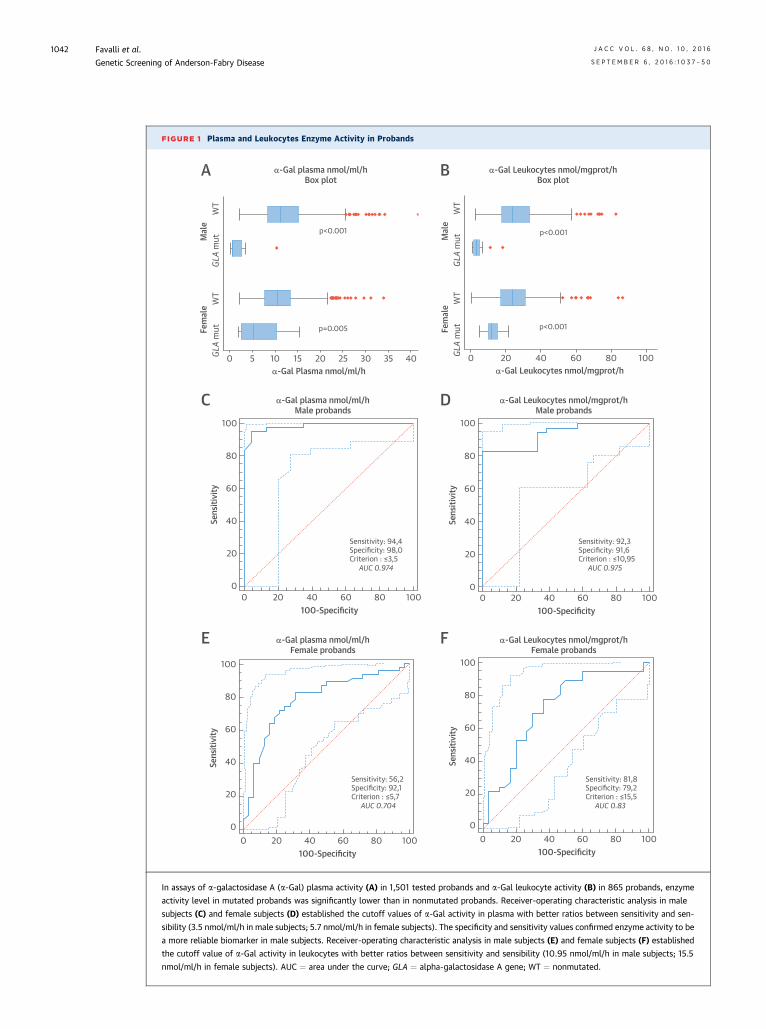
FIGURE 1 Plasma and Leukocytes Enzyme Activity in Probands
0 5 10 15 20 25 30 35 40α-Gal Plasma nmol/ml/h
GLA
mut
WT
GLA
mut
WT
Fem
ale
Mal
e
0 20 40 60 80 100α-Gal Leukocytes nmol/mgprot/h
GLA
mut
WT
GLA
mut
WT
Fem
ale
Mal
e
100
80
60
40
20
0
Sens
itivi
ty
0 20 40 60 80 100100-Specificity
100
80
60
40
20
0
Sens
itivi
ty
0 20 40 60 80 100100-Specificity
100
80
60
40
20
0
Sens
itivi
ty
0 20 40 60 80100-Specificity
100
100
80
60
40
20
0
Sens
itivi
ty
0 20 40 60 80100-Specificity
100
p<0.001
p=0.005
p<0.001
p<0.001
Sensitivity: 94,4Specificity: 98,0Criterion : ≤3,5
AUC 0.974
Sensitivity: 92,3Specificity: 91,6Criterion : ≤10,95
AUC 0.975
Sensitivity: 56,2Specificity: 92,1Criterion : ≤5,7
AUC 0.704
Sensitivity: 81,8Specificity: 79,2Criterion : ≤15,5
AUC 0.83
α-Gal plasma nmol/ml/hBox plot
α-Gal Leukocytes nmol/mgprot/hBox plot
α-Gal plasma nmol/ml/hMale probands
α-Gal Leukocytes nmol/mgprot/hMale probands
α-Gal plasma nmol/ml/hFemale probands
α-Gal Leukocytes nmol/mgprot/hFemale probands
A B
C D
E F
In assays of a-galactosidase A (a-Gal) plasma activity (A) in 1,501 tested probands and a-Gal leukocyte activity (B) in 865 probands, enzyme
activity level in mutated probands was significantly lower than in nonmutated probands. Receiver-operating characteristic analysis in male
subjects (C) and female subjects (D) established the cutoff values of a-Gal activity in plasma with better ratios between sensitivity and sen-
sibility (3.5 nmol/ml/h in male subjects; 5.7 nmol/ml/h in female subjects). The specificity and sensitivity values confirmed enzyme activity to be
a more reliable biomarker in male subjects. Receiver-operating characteristic analysis in male subjects (E) and female subjects (F) established
the cutoff value of a-Gal activity in leukocytes with better ratios between sensitivity and sensibility (10.95 nmol/ml/h in male subjects; 15.5
nmol/ml/h in female subjects). AUC ¼ area under the curve; GLA ¼ alpha-galactosidase A gene; WT ¼ nonmutated.
Favalli et al. J A C C V O L . 6 8 , N O . 1 0 , 2 0 1 6
Genetic Screening of Anderson-Fabry Disease S E P T E M B E R 6 , 2 0 1 6 : 1 0 3 7 – 5 0
1042
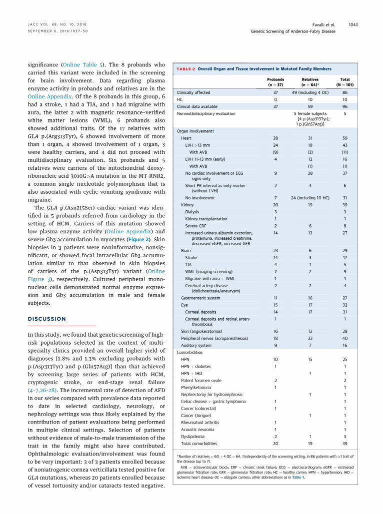
TABLE 2 Overall Organ and Tissue Involvement in Mutated Family Members
Probands(n ¼ 37)
Relatives(n ¼ 64)*
Total(N ¼ 101)
Clinically affected 37 49 (including 4 OC) 86
HC 0 10 10
Clinical data available 37 59 96
Nonmultidisciplinary evaluation 5 female subjects[4 p.(Asp313Tyr);1 p.(Gln57Arg)]
5
Organ involvement†
Heart 28 31 59
LVH >13 mm 24 19 43
With AVB (9) (2) (11)
LVH 11–13 mm (early) 4 12 16
With AVB (1) (1)
No cardiac involvement or ECGsigns only
9 28 37
Short PR interval as only marker(without LVH)
2 4 6
No involvement 7 24 (including 10 HC) 31
Kidney 20 19 39
Dialysis 3 3
Kidney transplantation 1 1
Severe CRF 2 6 8
Increased urinary albumin excretion,proteinuria, increased creatinine,decreased eGFR, increased GFR
14 13 27
Brain 23 6 29
Stroke 14 3 17
TIA 4 1 5
WML (imaging screening) 7 2 9
Migraine with aura þ WML 1 1
Cerebral artery disease(dolichoectasia/aneurysm)
2 2 4
Gastroenteric system 11 16 27
Eye 15 17 32
Corneal deposits 14 17 31
Corneal deposits and retinal arterythrombosis
1 1
Skin (angiokeratomas) 16 12 28
Peripheral nerves (acroparesthesias) 18 22 40
Auditory system 9 7 16
Comorbidities
HPN 10 15 25
HPN þ diabetes 1 1
HPN þ IHD 1 1
Patent foramen ovale 2 2
Phenylketonuria 1 1
Nephrectomy for hydronephrosis 1 1
Celiac disease þ gastric lymphoma 1 1
Cancer (colorectal) 1 1
Cancer (tongue) 1 1
Rheumatoid arthritis 1 1
Acoustic neuroma 1 1
Dyslipidemia 2 1 3
Total comorbidities 20 19 39
*Number of relatives ¼ 60 þ 4 OC ¼ 64. †Independently of the screening setting, in 86 patients with $1 trait ofthe disease (up to 7).
AVB ¼ atrioventricular block; CRF ¼ chronic renal failure; ECG ¼ electrocardiogram; eGFR ¼ estimatedglomerular filtration rate; GFR ¼ glomerular filtration rate; HC ¼ healthy carrier; HPN ¼ hypertension; IHD ¼ischemic heart disease; OC ¼ obligate carriers; other abbreviations as in Table 1.
J A C C V O L . 6 8 , N O . 1 0 , 2 0 1 6 Favalli et al.S E P T E M B E R 6 , 2 0 1 6 : 1 0 3 7 – 5 0 Genetic Screening of Anderson-Fabry Disease
1043
significance (Online Table 5). The 8 probands whocarried this variant were included in the screeningfor brain involvement. Data regarding plasmaenzyme activity in probands and relatives are in theOnline Appendix. Of the 8 probands in this group, 6had a stroke, 1 had a TIA, and 1 had migraine withaura, the latter 2 with magnetic resonance–verifiedwhite matter lesions (WML); 6 probands alsoshowed additional traits. Of the 17 relatives withGLA p.(Arg313Tyr), 6 showed involvement of morethan 1 organ, 4 showed involvement of 1 organ, 3were healthy carriers, and 4 did not proceed withmultidisciplinary evaluation. Six probands and 5relatives were carriers of the mitochondrial deoxy-ribonucleic acid 3010G>A mutation in the MT-RNR2,a common single nucleotide polymorphism that isalso associated with cyclic vomiting syndrome withmigraine.
The GLA p.(Asn215Ser) cardiac variant was iden-tified in 5 probands referred from cardiology in thesetting of HCM. Carriers of this mutation showedlow plasma enzyme activity (Online Appendix) andsevere Gb3 accumulation in myocytes (Figure 2). Skinbiopsies in 3 patients were noninformative, nonsig-nificant, or showed focal intracellular Gb3 accumu-lation similar to that observed in skin biopsiesof carriers of the p.(Asp313Tyr) variant (OnlineFigure 3), respectively. Cultured peripheral mono-nuclear cells demonstrated normal enzyme expres-sion and Gb3 accumulation in male and femalesubjects.
DISCUSSION
In this study, we found that genetic screening of high-risk populations selected in the context of multi-specialty clinics provided an overall higher yield ofdiagnoses [1.8% and 1.3% excluding probands withp.(Asp313Tyr) and p.(Gln57Arg)] than that achievedby screening large series of patients with HCM,cryptogenic stroke, or end-stage renal failure(4–7,26–28). The incremental rate of detection of AFDin our series compared with prevalence data reportedto date in selected cardiology, neurology, ornephrology settings was thus likely explained by thecontribution of patient evaluations being performedin multiple clinical settings. Selection of patientswithout evidence of male-to-male transmission of thetrait in the family might also have contributed.Ophthalmologic evaluation/involvement was foundto be very important: 3 of 3 patients enrolled becauseof noniatrogenic cornea verticillata tested positive forGLA mutations, whereas 20 patients enrolled becauseof vessel tortuosity and/or cataracts tested negative.

FIGURE 2 Endomyocardial Biopsies in Cardiac and Classic AFD
Mutations seen in endomyocardial biopsies from patients include the following: (A) a hematoxylin and eosin stain sample from a male carrier of
the p.(Asn215Ser) mutation in which the optically empty myocytes constitute a typical light microscopy marker of intracellular storage;
(B) electron micrograph showing typical osmiophilic bodies in the endomyocardial biopsies of a female carrier of the p.(Tyr184Asp) mutation;
(C) light immuno-microscopy of EMB samples from a young male carrier’s of the p.(Asn215Ser) mutation demonstrating early hypertrophic
cardiomyopathy; and (D) electron immuno-microscopy of EMB sample from a male patient carrier of the p.(Phe113Leu). AFD ¼ Anderson-Fabry
disease; EMB ¼ endomyocardial biopsy.
Favalli et al. J A C C V O L . 6 8 , N O . 1 0 , 2 0 1 6
Genetic Screening of Anderson-Fabry Disease S E P T E M B E R 6 , 2 0 1 6 : 1 0 3 7 – 5 0
1044
This supported evidence from other recent reports(29,30), in which noniatrogenic cornea verticillatawas a highly specific clinical marker.
Whereas severity scores showed that heart, kidney,and brain involvement are the major prognostic de-terminants (31,32) in AFD, diagnostic scoring systemscould better take advantage of objective markers ofhigh diagnostic specificity, regardless of clinical rele-vance. Traits such as acroparesthesias (33) or abdom-inal crises (3) are subjectively described, and thuschallenging to ascribe to AFD. Differential diagnosis inadults includes systemic amyloidosis with peripheralneuropathy in patients with cardiac and renalinvolvement. In children, the recurrence of abdominalcrises and acroneuropathic pain triggered by heat orfever increases clinical suspicion. Concomitant clini-cally overt involvement of the heart, kidneys, andbrain, which may contribute to the clinical suspicion,does not necessarily recur in all patients, especially
those with cardiac, renal, or nervous GLA variants.Mitochondrial diseases, such as MELAS, might showconcentric HCM that typically evolved through lateventricular dilation and dysfunction, renal failure thatmay necessitate hemodialysis, and recurrent crypto-genic stroke, but they might also demonstrate oculartraits that do not include corneal deposits. Diffuseangiokeratomas rarely bring a patient to clinicalattention. Phenotype heterogeneity within familiesdemonstrating classic forms of AFD or involvement ofthe same organ in those carrying atypical GLA variantsmight contribute to clinical suspicion. Overall, pa-tients with AFD should be evaluated as a whole, andwithin the context of family history, independent ofthe predominant organ involved, a strategy requiringa disease-oriented diagnostic mindset. The MOGE(S)nosology was recently generated to precisely describethe genotype-phenotype in cardiomyopathies (16)(Online Table 5).

FIGURE 3 Skin Biopsy in AFD
Electron micrographs of a skin biopsy sample from a male patient carrier of the p.Ala292_Met296del mutation show globotriaosylceramide
accumulation in (A) nonvascular smooth muscle cells; (B) endothelial cells; (C) dermal fibroblasts; (D) vascular smooth muscle cells;
and (E) adipocytes. (F) This sensory cutaneous nerve shows degenerative features. AFD ¼ Anderson-Fabry disease.
J A C C V O L . 6 8 , N O . 1 0 , 2 0 1 6 Favalli et al.S E P T E M B E R 6 , 2 0 1 6 : 1 0 3 7 – 5 0 Genetic Screening of Anderson-Fabry Disease
1045
DIAGNOSIS OF AFD. Definitive diagnosis should relyon genetic testing, enzyme activity, and tissuestudies demonstrating intracellular Gb3 accumula-tion. Absent pathological evaluation, interpretationof genetic testing results is supported by enzyme as-says in male subjects and by imaging or functionaltests sufficient to demonstrate tissue involvementotherwise. Confirmed pathologic mutations overallare sufficient for the diagnosis. For novel mutations
or atypical or provisional variants, in silico analysesmight contribute, but are not conclusive: a typicalexample is p.(Asn215Ser), which is benign in silico butassociated with severe intramyocyte accumulation ofGb3; or conversely, p.(Asp313Tyr), which is damagingin silico but considered to be a genetic variant ofunknown significance or single nucleotide poly-morphism. Recently, p.(Arg118Cys), originally inter-preted as pathologic, has been reported as a variant
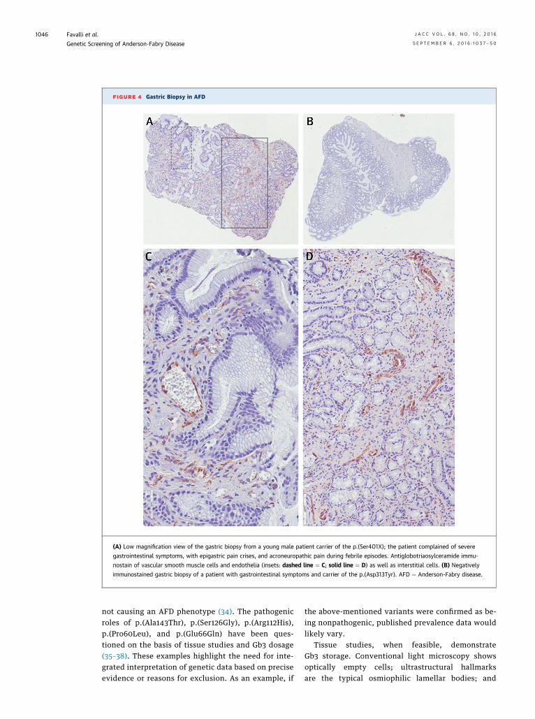
FIGURE 4 Gastric Biopsy in AFD
(A) Low magnification view of the gastric biopsy from a young male patient carrier of the p.(Ser401X); the patient complained of severe
gastrointestinal symptoms, with epigastric pain crises, and acroneuropathic pain during febrile episodes. Antiglobotriaosylceramide immu-
nostain of vascular smooth muscle cells and endothelia (insets: dashed line ¼ C; solid line ¼ D) as well as interstitial cells. (B) Negatively
immunostained gastric biopsy of a patient with gastrointestinal symptoms and carrier of the p.(Asp313Tyr). AFD ¼ Anderson-Fabry disease.
Favalli et al. J A C C V O L . 6 8 , N O . 1 0 , 2 0 1 6
Genetic Screening of Anderson-Fabry Disease S E P T E M B E R 6 , 2 0 1 6 : 1 0 3 7 – 5 0
1046
not causing an AFD phenotype (34). The pathogenicroles of p.(Ala143Thr), p.(Ser126Gly), p.(Arg112His),p.(Pro60Leu), and p.(Glu66Gln) have been ques-tioned on the basis of tissue studies and Gb3 dosage(35–38). These examples highlight the need for inte-grated interpretation of genetic data based on preciseevidence or reasons for exclusion. As an example, if
the above-mentioned variants were confirmed as be-ing nonpathogenic, published prevalence data wouldlikely vary.
Tissue studies, when feasible, demonstrateGb3 storage. Conventional light microscopy showsoptically empty cells; ultrastructural hallmarksare the typical osmiophilic lamellar bodies; and

FIGURE 5 In Vitro Study of AFD
(A) Cultured peripheral blood mononuclear cells immunostained with anti-a-Gal antibody from patients carriers of the p.(Asn215Ser),
p.(Phe113Leu), p.(Asp313Tyr) and p.Tyr184Asp mutation showed immunostaining similar to that of cells from control subjects (Ctrl). (B) In
cultured peripheral blood mononuclear cells of 3 patient carriers of p.(Phe113Leu), p.(Arg215Ser), and p.(Asp313Tyr), green particles indicated
anti-Gb3 immunoreactivity, which is highly represented in the 3 cell lines. DAPI ¼ 4’,6-diamidino-2-phenylindole; Gb3¼ globotriaosylceramide;
other abbreviations as in Figures 1 and 3.
J A C C V O L . 6 8 , N O . 1 0 , 2 0 1 6 Favalli et al.S E P T E M B E R 6 , 2 0 1 6 : 1 0 3 7 – 5 0 Genetic Screening of Anderson-Fabry Disease
1047
PERSPECTIVES
COMPETENCY IN MEDICAL KNOWLEDGE: A
rare, lysosomal disease, AFD affects multiple organ
systems. Precise diagnosis is essential for the effective
enzyme replacement therapy administration. The
heart is the most commonly involved organ, regard-
less of the clinical setting in which the patient pre-
sents, but screening patients in ophthalmology,
dermatology, gastroenterology, internal medicine,
pediatrics, and medical genetics clinics in addition to
high-risk patients in cardiology (HCM), neurology
(cryptogenic stroke, TIA, and migraine), and
nephrology (end-stage renal failure) clinics for de-
fects in the GLA gene, increases the diagnostic yield.
TRANSLATIONAL OUTLOOK: Future AFD studies
could benefit from use of the MOGE(S) nosology
system to provide patient-specific description of
multiorgan involvement, inheritance pattern, and the
associated mutation.
Favalli et al. J A C C V O L . 6 8 , N O . 1 0 , 2 0 1 6
Genetic Screening of Anderson-Fabry Disease S E P T E M B E R 6 , 2 0 1 6 : 1 0 3 7 – 5 0
1048
immuno-light and electron microscopy demonstratespecific labeling with anti-Gb3 antibodies. Our resultssupported a tissue-specific, mutation-dependent“affinity” for Gb3 storage and contributed toexplaining the cardiac, renal, and, probably, neuro-logic variants: in fact, Gb3 accumulated in myocytesbut not in skin or vascular cells of patients who carrythe cardiac variants [i.e., p.(Asn215Ser) andp.(Phe113Ser)]. However, classic, juvenile, and sys-temic forms have typically demonstrated involve-ment of interstitial and vascular smooth muscle cells[p.(Ala292_Met296del) in skin; p.(Ser401X) in gastrictissue] (Figures 3 and 4). Therefore, skin biopsy isuseful in patients with classic AFD but useless inatypical variants (Online Figures 2 and 3). In culturedPBMC from carriers of atypical cardiac variants, theimmunohistochemical expression of a-Gal wasnormal, confirming a functional impairment ratherthan decreased enzyme expression; the paradigmaticexample is p.(Asn215Ser), which causes the loss of a-Gal glycosylation at site 3, which is essential forenzyme solubility (39).
In our experience, the issue of the pseudodefi-ciency allele p.(Asp313Tyr) requires further discus-sion: all 8 probands in our series were fromneurologic settings; 7 also showed extraneurologictraits. Skin biopsies, similar to those in cardiac vari-ants, were not diagnostically helpful. Brain biopsiesare not possible, although imaging demonstratedtissue and vascular pathology. Autopsy or otherpathologic evaluations do not exist. Family segrega-tion studies (Online Figure 4) and in silico analysis donot help, based on both past in vitro studies demon-strating 60% of wild-type enzyme activity in vitro(40,41) and identification of 3 patients with a secondGLA mutation (42,43). Based on our data, thep.(Asp313Tyr) variant recurs in patients presentingwith cryptogenic stroke, TIA, WML identified by im-aging, and migraine with aura associated with WML.Cultured PBMC from mutation carriers showed fea-tures similar to those from carriers of confirmedpathologic mutations. Evidence, both for (44–46) andagainst (41,43), was nonconclusive for unambiguousassignment of disease causality or contribution forthis variant. In the large family reported by Lenderset al. (46), all 7 mutation carriers had WML but 2noncarriers did not. Neurologists face the difficultrole of managing these patients and excluding withcertainty this variant’s contribution to the complexneurological spectrum of signs/symptoms observed.
STUDY LIMITATIONS. Our study was designed morethan 10 years ago and did not include data on Gb3 andLyso-Gb3, which is emerging as a sensitive diagnostic
marker. The different prevalence of traits mimickingcardiac, neurologic, and renal disease (seen in AFD) inthe general population may help to explain the dif-ferences in numbers of cases enrolled per discipline.
CONCLUSIONS
Expanded screening of high-risk populations from thecardiology, neurology, and nephrology settings toophthalmology, gastroenterology, internal medicine,pediatrics, dermatology, and medical geneticsincreased the yield of diagnosis of AFD. The heart wasthe most commonly involved organ, regardless of theclinical setting in which the patient was first evalu-ated. Traits contributing to the diagnosis differedfrom those impacting prognosis, including non-iatrogenic cornea verticillata. Pathologic analysisconfirmed disease-causing mutations and alsocontributed to unraveling the role of novel and pro-visional variants identified in the past. The system-atic annotation of key genotype-phenotype data,such as with the MOGE(S) nosology system, helped tocollect precise clinical and genetic data for futurestudies.
REPRINT REQUESTS AND CORRESPONDENCE: Dr.Eloisa Arbustini, Centre for Inherited CardiovascularDiseases, IRCCS Foundation, University HospitalPoliclinico San Matteo, Piazzale Golgi 19,27100 Pavia,Italy. E-mail: [email protected].
J A C C V O L . 6 8 , N O . 1 0 , 2 0 1 6 Favalli et al.S E P T E M B E R 6 , 2 0 1 6 : 1 0 3 7 – 5 0 Genetic Screening of Anderson-Fabry Disease
1049
RE F E RENCE S
1. Desnick RJ, Ioannou YA, Eng ME. Alpha-galactosidase A deficiency: Fabry disease. In:Scriver CR, Beaudet AL, Sly WS, Valle D, editors.The Metabolic and Molecular Bases of InheritedDisease. 8th edition. New York, NY: McGraw-Hill,2001:3733–74.
2. Meikle PJ, Hopwood JJ, Clague AE, Carey WF.Prevalence of lysosomal storage disorders. JAMA1999;281:249–54.
3. El-Abassi R, Singhal D, England JD. Fabry’sdisease. J Neurol Sci 2014;344:5–19.
4. Wozniak MA, Kittner SJ, Tuhrim S, et al. Fre-quency of unrecognized Fabry disease amongyoung European-American and African-Americanmen with first ischemic stroke. Stroke 2010;41:78–81.
5. Palecek T, Honzikova J, Poupetova H, et al.Prevalence of Fabry disease in male patients withunexplained left ventricular hypertrophy in pri-mary cardiology practice: prospective Fabry car-diomyopathy screening study (FACSS). J InheritMetab Dis 2014;37:455–60.
6. van der Tol L, Svarstad E, Ortiz A, et al. Chronickidney disease and an uncertain diagnosis of Fabrydisease: approach to a correct diagnosis. MolGenet Metab 2015;114:242–7.
7. van der Tol L, Smid BE, Poorthuis BJ, et al.A systematic review on screening for Fabry dis-ease: prevalence of individuals with genetic vari-ants of unknown significance. J Med Genet 2014;51:1–9.
8. Nagueh SF. Anderson-Fabry disease and otherlysosomal storage disorders. Circulation 2014;130:1081–90.
9. Gambarin FI, Disabella E, Narula J, et al. Whenshould cardiologists suspect Anderson-Fabry dis-ease? Am J Cardiol 2010;106:1492–9.
10. Laney DA, Peck DS, Atherton AM, et al. Fabrydisease in infancy and early childhood: a system-atic literature review. Genet Med 2015;17:323–30.
11. Mehta A, Hughes DA. Fabry Disease. 2002 Aug5 [Updated 2013 Oct 17]. In: Pagon RA, Adam MP,Ardinger HH, et al., editors. GeneReviews�[Internet]. Seattle (WA): University of Washington,Seattle; 1993-2016. Available from: http://www.ncbi.nlm.nih.gov/books/NBK1292/. AccessedMarch 30, 2016.
12. Desnick RJ, Allen KY, Desnick SJ, Raman MK,Bernlohr RW, Krivit W. Fabry’s disease: enzymaticdiagnosis of hemizygotes and heterozygotes.Alpha-galactosidase activities in plasma, serum,urine, and leukocytes. J Lab Clin Med 1973;81:157–71.
13. Winchester B, Young E. Biochemical and ge-netic diagnosis of Fabry disease. In: Mehta A,Beck M, Sunder-Plassmann G, editors. Fabry Dis-ease Perspectives From 5 Years FOS. Oxford, UK:Oxford PharmaGenesis, 2006:169–81.
14. Pasqualim G, Simon L, Sperb-Ludwig F, et al.Fabry disease: a new approach for the screening offemales in high-risk groups. Clin Biochem 2014;47:657–62.
15. El Dib RP, Nascimento P, Pastores GM. Enzymereplacement therapy for Anderson-Fabry disease.Cochrane Database Syst Rev 2013;2:CD006663.
16. Arbustini E, Narula N, Dec GW, et al. TheMOGE(S) classification for a phenotype-genotypenomenclature of cardiomyopathy: endorsed bythe World Heart Federation. J Am Coll Cardiol2013;62:2046–72.
17. Zeevi DA, Hakam-Spector E, Herskovitz Y,Beeri R, Elstein D, Altarescu G. An intronic haplo-type in a galactosidase A is associated withreduced mRNA expression in males with crypto-genic stroke. Gene 2014;549:275–9.
18. Hsu TR, Sung SH, Chang FP, et al. Endomyo-cardial biopsies in patients with left ventricularhypertrophy and a common Chinese later-onsetFabry mutation (IVS4þ919G>A). Orphanet JRare Dis 2014;9:96.
19. Chien YH, Lee NC, Chiang SC, Desnick RJ,Hwu WL. Fabry disease: incidence of the commonlater-onset a-galactosidase A IVS4þ919G/Amutation in Taiwanese newborns—superiority ofDNA-based to enzyme-based newborn screeningfor common mutations. Mol Med 2012;18:780–4.
20. Richards S, Aziz N, Bale S, et al., for the ACMGLaboratory Quality Assurance Committee. Stan-dards and guidelines for the interpretation ofsequence variants: a joint consensus recommen-dation of the American College of Medical Ge-netics and Genomics and the Association forMolecular Pathology. Genet Med 2015;17:405–24.
21. MITOMAP: A human mitochondrial genomedatabase. Available at: http://www.mitomap.org/MITOMAP/. Accessed March 30, 2016.
22. Bossuyt PM, Reitsma JB, Standards forReporting of Diagnostic Accuracy. The STARDinitiative. Lancet 2003;361:71.
23. MacArthur DG, Manolio TA, Dimmock DP, et al.Guidelines for investigating causality of sequencevariants in human disease. Nature 2014;508:469–76.
24. Hanley JA, McNeil BJ. A method of comparingthe areas under receiver operating characteristiccurves derived from the same cases. Radiology1983;148:839–43.
25. Bland JM, Altman DG. Statistical methods forassessing agreement between two methods ofclinical measurement. Lancet 1986;1:307–10.
26. Elliott P, Baker R, Pasquale F, et al., for theACES Study Group. Prevalence of Anderson-Fabrydisease in patients with hypertrophic cardiomy-opathy: the European Anderson-Fabry Diseasesurvey. Heart 2011;97:1957–60.
27. Kilarski LL, Rutten-Jacobs LC, Bevan S, et al.,for the UK Young Lacunar Stroke DNA Study.Prevalence of CADASIL and Fabry disease in acohort of MRI defined younger onset lacunarstroke. PLoS One 2015;10:e0136352.
28. Kotanko P, Kramar R, Devrnja D, et al. Resultsof a nationwide screening for Anderson-Fabrydisease among dialysis patients. J Am Soc Neph-rol 2004;15:1323–9.
29. van der Tol L, Sminia ML, Hollak CE,Biegstraaten M. Cornea verticillata supports adiagnosis of Fabry disease in non-classical phe-notypes: results from the Dutch cohort and asystematic review. Br J Ophthalmol 2016;100:3–8.
30. Fledelius HC, Sandfeld L, Rasmussen ÅK,Madsen CV, Feldt-Rasmussen U. Ophthalmicexperience over 10 years in an observationalnationwide Danish cohort of Fabry patients withaccess to enzyme replacement. Acta Ophthalmol2015;93:258–64.
31. Whybra C, Kampmann C, Krummenauer F,et al. The Mainz Severity Score Index: a new in-strument for quantifying the Anderson–Fabry dis-ease phenotype, and the response of patients toenzyme replacement therapy. Clin Genet 2004;65:299–307.
32. Patel V, O’Mahony C, Hughes D, et al. Clinicaland genetic predictors of major cardiac events inpatients with Anderson-Fabry disease. Heart 2015;101:961–6.
33. Biegstraaten M, Binder A, Maag R, Hollak CE,Baron R, van Schaik IN. The relation between smallnerve fibre function, age, disease severity and painin Fabry disease. Eur J Pain 2011;15:822–9.
34. Ferreira S, Ortiz A, Germain DP, et al. Thealpha-galactosidase A p.Arg118Cys variant doesnot cause a Fabry disease phenotype: data fromindividual patients and family studies. Mol GenMetab 2015;114:248–58.
35. Terryn W, Vanholder R, Hemelsoet D, et al.Questioning the pathogenic role of the GLA p.Ala143Thr “mutation” in Fabry disease: implica-tions for screening studies and ERT. JIMD Rep2012;8:101–8.
36. Rombach SM, Dekker N, Bouwman MG, et al.Plasma globotriaosylsphingosine: diagnosticvalue and relation to clinical manifestations ofFabry disease. Biochim Biophys Acta 2010;1802:741–8.
37. Kobayashi M, Ohashi T, Fukuda T, et al. Noaccumulation of globotriaosylceramide in theheart of a patient with the E66Q mutation in thealpha-galactosidase A gene. Mol Genet Metab2012;107:711–5.
38. Togawa T, Tsukimura T, Kodama T, et al. Fabrydisease: biochemical, pathological and structuralstudies of the alpha-galactosidase A with E66Qamino acid substitution. Mol Genet Metab 2012;105:615–20.
39. Ioannou YA, Zeidner KM, Grace ME,Desnick RJ. Human a-galactosidase A: glycosyla-tion site 3 is essential for enzyme solubility.Biochem J 1998;332:789–97.
40. Froissart R, Guffon N, Vanier MT, Desnick RJ,Maire I. Fabry disease: D313Y is an alpha-galactosidase A sequence variant that causespseudodeficient activity in plasma. Mol GenetMetab 2003;80:307–14.
41. Niemann M, Rolfs A, Giese A, et al. Lyso-Gb3indicates that the alpha-galactosidase A mutationD313Y is not clinically relevant for Fabry disease.JIMD Rep 2013;7:99–102.

Favalli et al. J A C C V O L . 6 8 , N O . 1 0 , 2 0 1 6
Genetic Screening of Anderson-Fabry Disease S E P T E M B E R 6 , 2 0 1 6 : 1 0 3 7 – 5 0
1050
42. Yasuda M, Shabbeer J, Benson SD, Maire I,Burnett RM, Desnick RJ. Fabry disease: character-ization of alpha-galactosidase A double mutationsand the D313Y plasma enzyme pseudodeficiencyallele. Hum Mutat 2003;22:486–92.
43. Guffon N, Froissart R, Chevalier-Porst F,Maire I. Mutation analysis in 11 French patientswith Fabry disease. Hum Mutat 1998; Suppl 1:S288–90.
44. Baptista MV, Ferreira S, Pinho-E-Melo T, et al.,for the PORTuguese Young STROKE Investigators.
Mutations of the GLA gene in young patientswith stroke: the PORTYSTROKE study—screeninggenetic conditions in Portuguese young strokepatients. Stroke 2010;41:431–6.
45. Brouns R, Thijs V, Eyskens F, et al., for theBeFaS Investigators. Belgian Fabry study: preva-lence of Fabry disease in a cohort of 1000 youngpatients with cerebrovascular disease. Stroke2010;41:863–8.
46. Lenders M, Duning T, Schelleckes M, et al.Multifocal white matter lesions associated with
the D313Y mutation of the a-galactosidase A gene.PLoS One 2013;8:e55565.
KEY WORDS a-Gal, biochemical, familyscreening, GLA, MOGE(S) classification,multidisciplinary evaluation
APPENDIX For supplemental material,please see the online version of this article.


