Genetic Detection of Pseudomonas spp. in Commercial Amazonian Fish
13
Int. J. Environ. Res. Public Health 2013, 10, 3954-3966; doi:10.3390/ijerph10093954 International Journal of Environmental Research and Public Health ISSN 1660-4601 www.mdpi.com/journal/ijerph Article Genetic Detection of Pseudomonas spp. in Commercial Amazonian Fish Alba Ardura 1, *, Ana R. Linde 2 and Eva Garcia-Vazquez 1 1 Department of Functional Biology, University of Oviedo. C/Julian Claveria s/n, Oviedo 33006, Spain; E-Mail: [email protected] 2 Laboratory of Toxicology, National School of Public Health, Fundaçao Oswaldo Cruz, Rio de Janeiro 21041, Brazil; E-Mail: [email protected] * Author to whom correspondence should be addressed; E-Mail: [email protected]; Tel.: +34-985-102-726; Fax: +34-985-103-534. Received: 23 June 2013; in revised form: 23 July 2013 / Accepted: 15 August 2013 / Published: 29 August 2013 Abstract: Brazilian freshwater fish caught from large drainages like the River Amazon represent a million ton market in expansion, which is of enormous importance for export to other continents as exotic seafood. A guarantee of bacteriological safety is required for international exports that comprise a set of different bacteria but not any Pseudomonas. However, diarrhoea, infections and even septicaemia caused by some Pseudomonas species have been reported, especially in immune-depressed patients. In this work we have employed PCR-based methodology for identifying Pseudomonas species in commercial fish caught from two different areas within the Amazon basin. Most fish caught from the downstream tributary River Tapajòs were contaminated by five different Pseudomonas species. All fish samples obtained from the River Negro tributary (Manaus markets) contained Pseudomonas, but a less diverse community with only two species. The most dangerous Pseudomonas species for human health, P. aeruginosa, was not found and consumption of these fish (from their Pseudomonas content) can be considered safe for healthy consumers. As a precautionary approach we suggest considering Pseudomonas in routine bacteriological surveys of imported seafood. Keywords: Amazon River; Pseudomonas; molecular tests; commercial fish; food safety OPEN ACCESS
Transcript of Genetic Detection of Pseudomonas spp. in Commercial Amazonian Fish

Int. J. Environ. Res. Public Health 2013, 10, 3954-3966; doi:10.3390/ijerph10093954
International Journal of
Environmental Research and Public Health
ISSN 1660-4601 www.mdpi.com/journal/ijerph
Article
Genetic Detection of Pseudomonas spp. in Commercial Amazonian Fish
Alba Ardura 1,*, Ana R. Linde 2 and Eva Garcia-Vazquez 1
1 Department of Functional Biology, University of Oviedo. C/Julian Claveria s/n,
Oviedo 33006, Spain; E-Mail: [email protected] 2 Laboratory of Toxicology, National School of Public Health, Fundaçao Oswaldo Cruz,
Rio de Janeiro 21041, Brazil; E-Mail: [email protected]
* Author to whom correspondence should be addressed; E-Mail: [email protected];
Tel.: +34-985-102-726; Fax: +34-985-103-534.
Received: 23 June 2013; in revised form: 23 July 2013 / Accepted: 15 August 2013 /
Published: 29 August 2013
Abstract: Brazilian freshwater fish caught from large drainages like the River Amazon
represent a million ton market in expansion, which is of enormous importance for export to
other continents as exotic seafood. A guarantee of bacteriological safety is required for
international exports that comprise a set of different bacteria but not any Pseudomonas.
However, diarrhoea, infections and even septicaemia caused by some Pseudomonas
species have been reported, especially in immune-depressed patients. In this work we have
employed PCR-based methodology for identifying Pseudomonas species in commercial
fish caught from two different areas within the Amazon basin. Most fish caught from the
downstream tributary River Tapajòs were contaminated by five different Pseudomonas
species. All fish samples obtained from the River Negro tributary (Manaus markets)
contained Pseudomonas, but a less diverse community with only two species. The most
dangerous Pseudomonas species for human health, P. aeruginosa, was not found and
consumption of these fish (from their Pseudomonas content) can be considered safe for
healthy consumers. As a precautionary approach we suggest considering Pseudomonas in
routine bacteriological surveys of imported seafood.
Keywords: Amazon River; Pseudomonas; molecular tests; commercial fish; food safety
OPEN ACCESS

Int. J. Environ. Res. Public Health 2013, 10 3955
1. Introduction
Brazil contains a rich biodiversity of animal and plant taxa distributed at varied latitudes, and could
open a new international market for high quality food products, perhaps targeting delicatessen shops
and specialized restaurants. Such a market would be exclusive of Brazil because many species from
some regions, like the Amazon, are unique and endemic. Due to their enormous diversity [1,2],
Amazonian fishes represent a potentially interesting sector for export. Tools for labeling these fishes
are currently being developed [3], aimed at enabling Amazonian fish introduction in demanding
markets like the European one, where the normatives on traceability and food security control are strict
(e.g., European Directives CE-178/2002; CE-1759/2006).
Introduction of exotic species in a new market encompasses some potential risks. One of them is
introduction of parasites or pathogens endemic of their native region [4]. Control of such pathogens in
the importer country may not be required if they are normally absent from local food. An example
applicable to the fish trade could be Pseudomonas. These bacteria constitute a part of the normal fish
microbiota, but are opportunistic and may become infectious and spread diseases in stressed fish [5].
Pseudomonas can be a problem for human consumers too. They appear in processes of seafood
spoilage [6,7] and in ready-to-eat products [8]. In some conditions they can become human pathogens
and cause infection. Many pathogeneses of Pseudomonas in humans, generally caused by only one
species (most frequently P. aeruginosa), are health-care associated illnesses [9,10], and the risk
of disease by ingestion in healthy consumers has been considered generally low in developed
countries [11]. However, such risk exists and can be serious depending on the circumstances.
Contamination with enterotoxigenic Pseudomonas has been reported from food and drinking water
samples in some countries [12]. Jertborn and Svennerholm [13] have discovered enterotoxin-producing
Pseudomonas in Swedish travellers with diarrhoea, somewhat more frequently in travellers visiting
Africa, Asia and Latin America. In association with other bacteria they can cause severe cholera
symptoms in healthy adults [14]. Even when their enterotoxigenic activity is weak they can still
produce diarrhoea in immunodeficient individuals [15]. In addition they can cause skin problems; their
presence in cosmetics is considered a health threat in the U.S. [16], and have produced outbreaks of
skin infections [17]. Therefore there are infection risks when manipulating contaminated seafood.
Notwithstanding the information provided above, Pseudomonas species are not catalogued as a
foodborne pathogen in Europe and other regions. Control tests of imported fish and shellfish include
various bacterial species like Escherichia coli, C. botulinum, Listeria monocytogenes, Staphylococcus
aureus, Enterobacter sakazakii and Salmonella sp., but not Pseudomonas spp. (e.g., European
Council Directive 1991, 1995 and Council Directive 1998; Canadian Food Inspection
Agency [18], European Comission [19], American Food Safety and Inspection Service (FSIS) [20].
The aim of this study was to investigate the presence of Pseudomonas spp. in samples of
commercial fish sold in Brazilian markets from two different Amazonian states (Table 1): the River
Tapajós (Para) and the River Negro (Amazonas). The results may inform about the convenience of
including these bacteria in routine controls for Brazilian fish exports as well as in local markets. The
molecular tools used in this study were PCR-amplification with Pseudomonas-specific primers and
sequencing 16S rRNA genes. This type of methodology is highly sensitive and has been employed in
other surveys of foodborne bacteria [21,22].

Int. J. Environ. Res. Public Health 2013, 10 3956
Table 1. Samples analyzed, fish species with their common names and Pseudomonas
species identified.
Sample Origin Fish spp. Common name Pseudomonas species
T1 Tapajós Prochilodus nigricans Curimatá Pseudomonas psychrophila T2 Tapajós Cetopsis candiru Candiru Pseudomonas spp. T3 Tapajós Leporinus piau Piau Pseudomonas psychrophila T4 Tapajós Leporinus piau Piau Pseudomonas syringae T5 Tapajós Serrasalmus rhombeus Piranha Pseudomonas fragi T6 Tapajós Leporinus piau Piau Pseudomonas fluorescens T7 Tapajós Ageneiosus brevifilis Bocudo Pseudomonas fluorescens T8 Tapajós Leporinus piau Piau Pseudomonas psychrophila T9 Tapajós Leporinus piau Piau Pseudomonas syringae T10 Tapajós Leporinus piau Piau Pseudomonas fluorescens T11 Tapajós Leporinus piau Piau Pseudomonas spp. T12 Tapajós Leporinus piau Piau Pseudomonas psychrophila T13 Tapajós Prochilodus nigricans Curimatá Pseudomonas psychrophila T14 Tapajós Leporinus piau Piau Pseudomonas fragi T15 Tapajós Leporinus piau Piau Pseudomonas psychrophila T16 Tapajós Leporinus piau Piau Pseudomonas putida T17 Tapajós Leporinus piau Piau - T18 Tapajós Leporinus piau Piau - M1 Negro Chaetobranchopsis orbicularis Acará branco Pseudomonas putida M2 Negro Astonotus ocellatus Acará-açú Pseudomonas putida M3 Negro Astonotus ocellatus Acará-açú Pseudomonas putida M4 Negro Astonotus ocellatus Acará-açú Pseudomonas putida M5 Negro Osteoglossum bicirrhosum Aruanà Pseudomonas putida M6 Negro Brachypatystoma rousseauxii Dourada Pseudomonas putida M7 Negro Semaprochilodus insignis Jaraquí Pseudomonas putida M8 Negro Plagioscion squamosissimus Pescada Pseudomonas putida M9 Negro Phractocephalus hemioliopterus Pirarara Pseudomonas psychrophila
M10 Negro Pseudoplatystoma fasciatum Surubim Pseudomonas putida M11 Negro Cichla temensis Tucunaré Pseudomonas putida M12 Negro Cichla temensis Tucunaré Pseudomonas putida
2. Experimental Section
2.1. Sampling
The 30 fish samples analyzed (Table 1) were obtained from two different tributaries within the
Amazon basin (Figure 1): the River Negro (Manaus markets; n = 12) and the River Tapajós (n = 18),
and were directly purchased from fishermen in local harbors and markets. All fish specimens were
morphologically identified in situ by visual inspection and taxonomically classified employing
standard taxonomic guides. After cleaning the fish surface with ethanol, samples of muscle (the edible
tissue) were excised in situ with sterilized blades and tweezers and immediately stored in absolute
ethanol. Ethanol-preserved samples were transported in coolers to the laboratory for genetic analysis.

Int. J. Environ. Res. Public Health 2013, 10 3957
Figure 1. A map with proportions of different Pseudomonas species found in each
sampling site: Manaus and Tapajós.
2.2. Genetic Analyses
DNA extraction and PCR amplification were carried out in sterile conditions to prevent
cross-contamination of samples during the process. Total DNA was extracted from a small piece
(approximately 5 mg) of alcohol-preserved fish tissue by the standard protocol of Estoup et al. [23],
using Chelex® resin (Bio-Rad Laboratories, Hercules, CA, USA). Chelex® is a chelating material used
to purify other compounds from a tissue via ion exchange. It is often used for DNA extraction in
preparation for PCR. Polar resin beads bind polar cellular components after breaking open cells, while
DNA and RNA remain suspended in water solution above the Chelex®. The tissue was introduced in
an Eppendorf tube with 500 µL of Chelex® resin (10%) and 7 µL of Proteinase K (20 mg/mL). It was
incubated at 55 °C for 90 min. The DNA was dissolved in the aqueous solution. Finally, it was
introduced in an oven at 100 °C during 20 min for inactivating the enzyme. The tube was stored at
4 °C or frozen at −20 °C for long-time preservation.
A fragment of the 16S rRNA gene was amplified by polymerase chain reaction (PCR), employing
the Pseudomonas genus specific primers PA-GS-F (5′-GACGGGTGAGTAATGCCTA-3′) and
PA-GS-R (5′-CACTGGTGTTCCTTCCTATA-3′) described by Spilker et al. [24] They amplify a
DNA region of 618 nucleotides located between the sites 113 and 712, position and size relative to 16S

Int. J. Environ. Res. Public Health 2013, 10 3958
rDNA sequence of Pseudomonas aeruginosa AT2 (AB091760) [24]. The amplification reaction was
performed in a total volume of 40 µL, including Promega (Madison, WI, USA) Buffer 1X, 2.5 mM
MgCl2, 0.25 mM dNTPs, 20 pmol of each primer, 20 ng of template DNA, and 1 U of DNA Taq
polymerase (Promega). The PCR conditions were the following: an initial denaturation at 95 °C for
5 min, 10 cycles at 94 °C for 15 s, annealing at 53 °C for 30 s and elongation at 72 °C for 45 s.
This was repeated for 25 cycles, increasing the elongation step at 72 °C by 5 s every cycle. The final
extension phase was at 72 °C for 10 min.
PCR products were visualized in 2% agarose gels with 3 µL of 10 mg/mL ethidium bromide.
Stained bands were excised from the gel, and DNA was purified with an Eppendorf PerfectPrep Gel
CleanUp® kit prior to sequencing. After that, amplified and purified products were precipitated using
standard 2-propanol precipitation and re-suspended in formamide.
Sequencing was performed in an ABI PRISM 3100 Genetic Analyzer (Applied Biosystems, Foster
City, CA, USA) with BigDye 3.1 terminator system, at the Sequencing Unit of the University of
Oviedo (Oviedo, Spain).
2.3. Sequence Edition and Phylogenetic Analysis
Sequences obtained from the 16S rRNA gene amplicons were visualized and edited employing the
BioEdit Sequence Alignment Editor software [25]. Sequences were aligned with the ClustalW
application [26] included in BioEdit.
The phylogenetic analysis was performed with the software MEGA 4.0 [27]. This software was
employed to reconstruct the phylogenetic trees of the Pseudomonas species found in fish samples from
16S rDNA sequences. The methodology chosen was the neighbor-joining (NJ), the standard method of
phylogenetic inference in DNA barcoding studies [28] because it allows to rapid analysis of
large species assemblages [29]. The molecular substitution model was chosen using the software
jModeltest [30] to determine the best suited model of sequence evolution and accompanying
evolutionary parameter values for the data. Robustness of the NJ topology was assessed using
2,000 bootstrap replicates.
Pseudomonas species identification was made by comparing generated 16S rDNA sequences with
reference sequences present in the GenBank database by means of BLAST online program [31].
2.4. Pseudomonas Diversity Estimates
Pseudomonas species diversity in each Amazonian location was estimated by means of ecological
index (Shannon, H) using PRIMER 6 (Software package from the Plymouth Marine Laboratory,
Lutton, Ivybridge, UK). The number of haplotypes (h) and nucleotide diversity (π) were calculated
with the ARLEQUIN software [32,33].
2.5. Statistics
To compare the proportion of contaminated fish between locations, chi-square statistics was
employed. Analysis was carried out using the SPSS 13.0 software (SPSS Inc., Chicago, IL, USA).
Int. J. Environ. Res. Public Health 2013, 10 3959
3. Results and Discussion
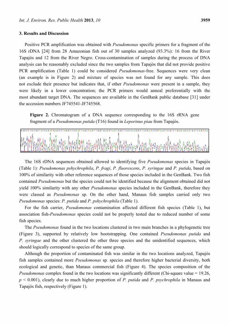
Positive PCR amplification was obtained with Pseudomonas specific primers for a fragment of the
16S rDNA [24] from 28 Amazonian fish out of 30 samples analyzed (93.3%): 16 from the River
Tapajòs and 12 from the River Negro. Cross-contamination of samples during the process of DNA
analysis can be reasonably excluded since the two samples from Tapajòs that did not provide positive
PCR amplification (Table 1) could be considered Pseudomonas-free. Sequences were very clean
(an example is in Figure 2) and mixture of species was not found for any sample. This does
not exclude their presence but indicates that, if other Pseudomonas were present in a sample, they
were likely in a lower concentration; the PCR primers would anneal preferentially with the
most abundant target DNA. The sequences are available in the GenBank public database [31] under
the accession numbers JF745541-JF745568.
Figure 2. Chromatogram of a DNA sequence corresponding to the 16S rRNA gene
fragment of a Pseudomonas putida (T16) found in Leporinus piau from Tapajós.
The 16S rDNA sequences obtained allowed to identifying five Pseudomonas species in Tapajós
(Table 1): Pseudomonas pshychrophila, P. fragi, P. fluorescens, P. syringae and P. putida, based on
100% of similarity with other reference sequences of those species included in the GenBank. Two fish
contained Pseudomonas but the species could not be identified because the alignment obtained did not
yield 100% similarity with any other Pseudomonas species included in the GenBank, therefore they
were classed as Pseudomonas sp. On the other hand, Manaus fish samples carried only two
Pseudomonas species: P. putida and P. pshychrophila (Table 1).
For the fish carrier, Pseudomonas contamination affected different fish species (Table 1), but
association fish-Pseudomonas species could not be properly tested due to reduced number of some
fish species.
The Pseudomonas found in the two locations clustered in two main branches in a phylogenetic tree
(Figure 3), supported by relatively low bootstrapping. One contained Pseudomonas putida and
P. syringae and the other clustered the other three species and the unidentified sequences, which
should logically correspond to species of the same group.
Although the proportion of contaminated fish was similar in the two locations analyzed, Tapajós
fish samples contained more Pseudomonas sp. species and therefore higher bacterial diversity, both
ecological and genetic, than Manaus commercial fish (Figure 4). The species composition of the
Pseudomonas complex found in the two locations was significantly different (Chi-square value = 19.26,
p < 0.001), clearly due to much higher proportion of P. putida and P. psychrophila in Manaus and
Tapajós fish, respectively (Figure 1).

Int. J. Environ. Res. Public Health 2013, 10 3960
Figure 3. Neighbour-Joining tree constructed based on 16S rDNA Pseudomonas
sequences found in this survey. Bootstrap values (in percent).
P. psychrophila
P. fragi
Pseudomonas spp.
P. fluorescens
P. putida
P. syringae
Pseudomonas sp.

Int. J. Environ. Res. Public Health 2013, 10 3961
Figure 4. Diversity parameters of fishborne Pseudomonas communities from the
Amazonian Tapajòs and Negro tributaries. Metagenetic h and π parameters, and
Shannon index.
The results presented here, although based on small sample sizes, suggest that Pseudomonas are
endemically present in Amazonian fish sold in local markets since most analyzed fish yielded positive
PCR amplification for these bacteria. Pseudomonas aeruginosa, the most dangerous species for
human health [9,10,17] and especially for consumers [13,15], was not detected. Therefore
consumption of these fish can be considered generally safe for healthy people, at least from their
Pseudomonas content.
The characteristics of the Pseudomonas species found from Brazilian fish samples (Table 2) may
suggest the origin of the contamination. Fish infection in some Tapajós samples was suggested by the
presence of the well-known fish pathogen P. fluorescens, which is considered as opportunistic
pathogenic species in aquaculture [34,35], responsible for bacterial septicemia in fish. This species was
present in three (16.7%) samples from Tapajós, but in none from Manaus (Figure 1). Pseudomonas
infections in fish are promoted by different stressors [6,36]. Environmental stress produced by mining
has been reported in the River Tapajós [37], and could contribute to facilitate fish infection by
opportunistic Pseudomonas.
P. putida was found in most Manaus samples (Figure 1) and in only one sample from Tapajós.
Different Pseudomonas species have been associated with seafood (including chilled fish) spoilage, for
example P. fragi [6], therefore the likely origin of contamination of these samples could be seafood
manipulation, long time of storage before selling or simply opportunistic growth of these bacteria on
fish exposed without protection in the open-door local markets. P. putida infections have also been
reported in fish species, for example in farmed rainbow trout [38], also associated to stress, therefore
this last possibility cannot be totally ruled out. P. psychrophila grows in cold conditions [39,40],
unusual in the natural tropical Amazonian environment; they could be an opportunistic colonizer
during the storage in cold rooms previous to selling in the market. Finally, P. syringae is a plant
0
1
2
3
4
5
6
7
Tapajos Manaos
h
π (*100)
Shannon Index

Int. J. Environ. Res. Public Health 2013, 10 3962
pathogen which can infect a wide range of plant species; more than any mineral or other organism is
responsible for the surface frost damage in plants exposed to the environment [41]; like
P. psychrophila tends to be favored by wet and cool conditions [41], being more probable that appear
like an opportunistic colonizer during the storage in cool rooms.
Table 2. Characteristics of the Pseudomonas species found from commercial Amazonian
fish and potential risk for humans.
Pseudomonas species Characteristics Pathogenesis reported for humans
P. fluorescens Opportunistic pathogen in fish [42] Oncology patients [43]
P. fragi Seafood spoilage [6,44]
Opportunistic microbiota [6] No published data about this
P. psychrophila No *
P. putida Seafood spoilage [44]
Cosmopolitan opportunist [46] Immunodepressed patients [45]
Nosocomial infections [47] P. syringae No *
* They cannot survive at temperatures above 32 °C [39,41], and therefore cannot grow in humans where
normal body temperature is 37 °C.
From the phylogenetic point of view, the tree obtained grouped the identified species consistently
with previous phylogenetic studies of the genus [47,48]. The same marker, 16S rRNA gene, was used
together with other three genes, since although this is a powerful tool for genus assignments, it does
not discriminate sufficiently at the inter-species level [49]. In this case the discrimination level of
16S rRNA gene is enough to determine the contamination present in fishes with different species
of Pseudomonas.
Although we have not found the most dangerous species, the Pseudomonas found in our study
could be potentially harmful for vulnerable or immunodepressed consumers (Table 2). Infections by
P. fluorescens and P. putida had been reported in old studies [43–45,50] (and references therein), and
were confirmed later. P. fluorescens is a potential pathogen due to their capacity of adhesion to nerves [50],
and outbreaks in oncology patients have been discovered [43]. On the other hand, P. putida bacteremia
seemed to be infrequent and affect mainly immunocompromised patients, with a good prognosis since
most cases were cured [45]; however, recent emergent multidrug-resistant and carbapenem-resistant.
P. putida isolates cause difficult-to-treat nosocomial infections in seriously ill patients [51]. In brief,
these species could cause problems in vulnerable people and do not represent a serious threat for
healthy consumers [52], but using a precautionary approach it could be wise to start considering them
for future seafood tests. The presence of these pathogens in the products tested here does not mean that
they are a risk for consumers; in general Pseudomonas sp. represents a hazard for the health when its
number exceeds 106–107 CFU/g of product [11,12,15,17] but CFU has not been quantified here. Rather
these results could be considered an exploratory work on presence/absence of Pseudomonas. If routine
surveys were undertaken they should include quantification of the bacteria concentration. RT-PCR
based methods could be employed since they can estimate the number of DNA molecules present in a
sample. These methods are relatively cheap nowadays and the sequencing cost per sample in our study
was approximately 3€ (real cost). However, the analysis of foodborne bacteria is being revolutionized
with new sequencing technologies such as NGS [53], and prospects are of better prices for large-scale

Int. J. Environ. Res. Public Health 2013, 10 3963
analysis. On the other hand, this type of PCR-based methods allow to detecting extremely low number
of microorganisms based on the production of specific gene copies of a microorganism in question, but
it does not distinguish living bacteria from dead cells. Since PCR methodology is rapid (a few hours),
additional tests based on the count of total viable microorganisms could be used after initial detection
and identification by PCR. Examples are Standard Plate Count [54], determination of most probable
number of viable bacteria [55], methods based on fluorescence techniques [56] or direct counting at
the microscope [57].
4. Conclusions
The possible presence of Pseudomonas in fish and seafood should be considered when food
imports arrive from countries or areas with Pseudomonas endemism and high prevalence of
enterotoxigenic-derived diseases. We suggest that routine tests for Pseudomonas could be included in
the battery of tests aimed at controlling the bacteriological quality of imported fish. PCR-based
methodologies, like those employed in this study, are easy and fast and could be considered as a
complementary tool to bacterial cultivation.
Acknowledgments
This study has been funded by the Spanish Agency of International Cooperation for Development
(AECID) Project D/023514/09. We are grateful to Vanessa Gomes, Ione Ginuino, Gema E. Adan and
Eduardo del Rosal for collaboration in sampling. Ivan G. Pola helped with laboratory work.
Conflicts of Interest
The authors declare no conflict of interest.
References
1. Saint-Paul, U.; Zuanon, J.; Villacorta-Correa, M.A.; García, M.; Fabré, N.N.; Berger, U.; Junk, W.J.
Fish communities in central amazonian white- and blackwater floodplains. Environ. Biol. Fishes
2000, 57, 235–250.
2. Fernandes, C.C.; Podos, J.; Lundberg, J.G. Amazonian ecology: Tributaries enhance the diversity
of electric fishes. Science 2004, 305, 1960–1962.
3. Ardura, A.; Pola, I.G.; Linde, A.R.; Garcia-Vazquez, E. DNA-based methods for species
authentication of Amazonian commercial fish. Food Res. Int. 2010, 43, 2295–2302.
4. FAO/WHO: Food and Agriculture Organization of the United Nations/ World Health
Organization. Microbiological Risk Assessment Series. In Risk Characterization of
Microbiological Hazards in Food. Guidelines; WHO: Geneva, Switzerland, 2009; Volume 17, p. 116.
5. Sakata, T. Microflora of Healthy Animals. In Methods for the Microbiological Examination of
Fish and Shellfish Chichester; Austin, B., Austin, D.A., Eds.; Ellis Horwood Ltd.: England, UK,
1989; pp. 141–163.

Int. J. Environ. Res. Public Health 2013, 10 3964
6. Gram, L.; Huss, H.H. Fresh and Processed Fish and Shellfish. In The Microbiological Safety and
Quality of Foods; Lund, B.M., Baird-Parker, A.C., Gould, G.W., Eds.; Chapman & Hall: London,
UK, 2000; pp. 472–506.
7. Gram, L.; Ravn, L.; Rasch, M.; Bruhn, J.B.; Christensen, A.B.; Givskov, M. Food
spoilage-interactions between food spoilage bacteria. Int. J. Food Microbiol. 2002, 78, 79–97.
8. Nyenje, M.E.; Odjadjare, C.E.; Odjadjare, L.; Tanih, N.F.; Green, E.; Ndip, R.N. Foodborne
pathogens recovered from ready-to-eat foods from roadside cafeterias and retail outlets in Alice,
Eastern Cape Province, South Africa: Public health implications. Int. J. Environ. Res. Public
Health 2012, 9, 2608–2619.
9. Bagshaw, S.M.; Laupland, K.B. Epidemiology of intensive care unit-acquired urinary tract
infections. Curr. Opin. Infect. Dis. 2006, 19, 67–71.
10. Zilberberg, M.D.; Shorr, A.F. Epidemiology of healthcare-associated pneumonia (HCAP).
Semin. Respir. Crit. Care Med. 2009, 30, 10–15.
11. Mena, K.D.; Gerba, C.P. Risk assessment of Pseudomonas aeruginosa in water. Rev. Environ.
Contam. Toxicol. 2009, 201, 71–115.
12. Jiwa, S.F.H.; Krovacek, K.; Wadstrom, T. Enterotoxigenic bacteria in food and water from an
ethiopian community. Appl. Environ. Microbiol. 1981, 41, 1010–1019.
13. Jertborn, M.; Svennerholm, A.M. Enterotoxin-producing bacteria isolated from Swedish travellers
with diarrhoea. Scand. J. Infect. Dis. 1991, 23, 473–479.
14. Bockemühl, J.; Fleischer, K.; Bednarek, I. A cholera-like illness in a traveller due to a mixed
infection with enterotoxigenic Escherichia coli, Vibrio parahaemolyticus and Pseudomonas
aeruginosa. Infection 1983, 11, 272–274.
15. Adlard, P.A.; Kirov, S.M.; Sanderson, K.; Cox, G.E. Pseudomonas aeruginosa as a cause of
infectious diarrhoea. Epidemiol. Infect.1998, 121, 237–241.
16. Wong, S.; Street, D.; Delgado, S.I.; Klontz, K.C. Recalls of foods and cosmetics due to microbial
contamination reported to the U.S. Food and Drug Administration. J. Food Prot. 2000, 63,
1113–1116.
17. Craun, G.F.; Brunkard, J.M.; Yoder, J.S.; Roberts, V.A.; Carpenter, J.; Wade, T.; Calderon, R.L.;
Roberts, J.M.; Beach, M.J.; Roy, S.L. Causes of outbreaks associated with drinking water in the
United States from 1971 to 2006. Clin. Microbiol. Rev. 2010, 23, 507–528.
18. Product Inspection of Imported Fish. Csnsdian Food Inspection Agency. 2013. Available online:
http://www.inspection.gc.ca/english/fssa/fispoi/import/pol/procprode.shtml (accessed on 26
August 2013).
19. Microbiological Criteria. Health and Consumers. European Commission. 2011. Available online:
http://ec.europa.eu/food/food/biosafety/salmonella/microbio_en.htm (accessed on 26 August 2013).
20. Food Safety and Inspection Service. United States Department of Agriculture. Available online:
http://www.fsis.usda.gov/ (accessed on 26 August 2013).
21. Bennett, A.R.; Greenwood, D.; Tennant, C.; Banks, J.G.; Betts, R.P. Rapid and definitive
detection of Salmonella in foods by PCR. Lett. Appl. Microbiol. 1998, 26, 437–441.
22. Wang, R.-F.; Cao, W.W.; Cerniglia, C.E. A universal protocol for PCR detection of 13 species of
foodborne pathogens in foods. J. Appl. Microbiol. 1997, 83, 727–736.

Int. J. Environ. Res. Public Health 2013, 10 3965
23. Estoup, A.; Largiader, C.R.; Perrot, E.; Chourrout, D. Rapid one-tube DNA extraction for reliable
PCR detection of fish polymorphic markers and transgenes. Mol. Mar. Biol. Biotechnol. 1996, 5,
295–298.
24. Spilker, T.; Coenye, T.; Vandamme, P.; LiPuma, J.J. PCR-based assay for differentiation of
Pseudomonas aeruginosa from other Pseudomonas species recovered from cystic fibrosis
patients. J. Clin. Microbiol. 2004, 42, 2074–2079.
25. Hall, T.A. BioEdit: A user-friendly biological sequence alignment editor and analysis program for
Windows 95/98/NT. Nucleic Acids Symp. Ser. 1999, 41, 95–98.
26. Thompson, J.D.; Higgins, D.G.; Gibson, T.J. Clustal-W, Improving the sensitivity of progressive
multiple sequence alignment trough sequence weighting, position-specific gap penalties and
weight matrix choice. Nucleic Acids Res. 1994, 22, 4673–4680.
27. Tamura, K.; Dudley, J.; Nei, M.; Kumar, S. MEGA4: Molecular Evolutionary Genetics Analysis
(MEGA) software version 4.0. Mol. Biol. Evolut. 2007, 24, 1596–1599.
28. Hebert, P.; Cywinska, A.; Ball, S.; deWaard, J. Biological identification through DNA barcodes.
Proc. R. Soc. B: Biol. Sci. 2003, 270, 313–321.
29. Kumar, S.; Gadadkar, S. Efficiency of the neighbour-joining method in reconstructing deep and
shallow evolutionary relationships in large phylogenies. J. Mol. Evolut. 2000, 51, 544–553.
30. Posada, D. jModelTest: Phylogenetic model averaging. Mol. Biol. Evolut. 2008, 25, 1253–1256.
31. National Center for Biotechnology Information. Available online: http://www.ncbi.nlm.nih.gov/
(accessed on 26 August 2013).
32. Nei, M. Molecular Evolutionary Genetics; Columbia University Press: New York, NY, USA,
1987; p. 512.
33. Excoffier, L.; Laval, G.; Schneider, S. Arlequin (version 3.0): An integrated software package for
population genetics data analysis. J. Evol. Bioinform. Online 2005, 1, 47–50.
34. Shiose, J.; Wakabayashi, H.; Tominaga, M.; Egusa, S. A report on a disease of cultured carp due
to a capsulated Pseudomonas. Fish Pathol. 1974, 9, 79–83.
35. Alderman, D.J.; Polglase, J.L. Pathogens, Parasites and Commensals. In Freshwater Crayfish—
Biology, Management and Exploitation; Holdich, D.M., Lowry, R.S., Eds.; Timber Press:
Portland, OR, USA, 1998; pp. 168–187.
36. Kusuda, R.; Toyoshima, R. Characteristics of a pathogenic Pseudomonas isolated from cultured
yellowtail. Fish Pathol. 1976, 1, 133–139.
37. Uryu, Y.; Malm, O.; Thornton, I.; Payne, I.; Cleary, D. Mercury contamination of fish and its
implications for other wildlife of the Tapajós Basin, Brazilian Amazon. Conserv. Biol. 2001, 15,
438–446.
38. Altinok, I.; Kayisa, S.; Capkin, E. Pseudomonas putida infection in rainbow trout. Aquaculture
2006, 261, 850–855.
39. Yumoto, I; Kusano, T.; Shingyo, T.; Nodasaka, Y.; Matsuyama, H.; Okuyama, H. Assignment of
Pseudomonas sp. strain E-3 to Pseudomonas psychrophila spp. nov., a new facultatively
psychrophilic bacterium. Extremophiles 2001, 5, 343–349.
40. Morita, R.Y. Psychrophilic bacteria. Bacteriol. Rev.1975, 39, 144–167.
41. Hirano, S.S.; Upper, C.D. Population biology and epidemiology of Pseudomonas syringae.
Annu. Rev. Phytopathol. 1990, 28, 155–177.
Int. J. Environ. Res. Public Health 2013, 10 3966
42. Bruno, D.W.; Ellis, A.E. Salmonid Disease Management. Dev. Aquac. Fish. Sci. 1996, 29, 729–824.
43. Hsueh, P.R.; Teng, L.J.; Pan, H.J.; Chen, Y.C.; Sun, C.C.; Ho, S.W.; Luh, K.T. Outbreak of
Pseudomonas fluorescens bacteremia among oncology patients. J. Clin. Microbiol. 1998, 36,
2914–2917.
44. Von Graevenitz, A.; Weinstein, J. Pathogenic significance of Pseudomonas fluorescens and
Pseudomonas putida. Yale J. Biol. Med. 1971, 44, 265–273.
45. Yoshino, Y.; Kitazawa, T.; Kamimura, M.; Tatsuno, K.; Ota, Y.; Yotsuyanagi, H. Pseudomonas
putida bacteremia in adult patients: five case reports and a review of the literature. J. Infect.
Chemother. 2011, 17, 278–282.
46. Timmis, K.N. Pseudomonas putida: A cosmopolitan opportunist par excellence.
Environ. Microbiol. 2002, 4, 779–781.
47. Yamamoto, S.; Kasai, H.; Arnold, D.L.; Jackson, R.W.; Vivian, A.; Harayama, S. Phylogeny of
the genus Pseudomonas: Intrageneric structure reconstructed from the nucleotide sequences of
gyrB and rpoD genes. Microbiology 2000, 146, 2385–2394.
48. Franzetti, L.; Scarpellini, M. Characterisation of Pseudomonas spp. isolated from foods.
Ann. Microbiol. 2007, 57, 39–47.
49. Mulet, M.; Lalucat, J.; García-Valdés, E. DNA sequence-based analysis of the Pseudomonas
species. Environ. Microbiol. 2010, 12, 1513–1530.
50. Picot, L.; Mezghani-Abdelmoula, S.; Merieaua, A.; Lerouxb, P.; Cazina, L.; Orangea, N.;
Feuilloley, M.G.J. Pseudomonas fluorescens as a potential pathogen: Adherence to nerve cells.
Microbes Infect. 2001, 3, 985–995.
51. Kim, S.E.; Park, S.H.; Park, H.B.; Park, K.H.; Kim, S.H.; Jung, S.I.; Shin, J.H.; Jang, H.C.;
Kang, S.J. Nosocomial Pseudomonas putida bacteremia: High rates of carbapenem resistance and
mortality. Chonnam Med. J. 2012, 48, 91–95.
52. Gilardi, G.L. Infrequently encountered Pseudomonas species causing infection in humans.
Ann. Int. Med. 1972, 77, 211–215.
53. Wilson, M.R.; Allard, M.W.; Brown, E.W. The forensic analysis of foodborne bacterial pathogens
in the age of whole-genome sequencing. Cladistics 2013, 29, 449–461.
54. LeChevallier, M.W.; Seidler, R.J.; Evans, T.M. Enumeration and characterization of standard
plate count bacteria in chlorinated and raw water supplies. Appl. Environ. Microbiol. 1980, 40,
922–930.
55. Hihgsmith, A.K.; Abshire, R.L. Evaluation of most-probable-number technique for the
enumeration of Pseudomonas aeruginosa. Appl. Microbiol. 1975, 30, 596–601.
56. Breeuwer, P.; Abee, T. Assessment of viability of microorganisms employing fluorescence
techniques. Int. J. Food Microbiol. 2000, 55, 193–200.
57. Kogure, K.; Simidu, U.; Taga, N. A tentative direct microscopic method for counting living
marine bacteria. Can. J. Microbiol. 1979, 25, 415–420.
© 2013 by the authors; licensee MDPI, Basel, Switzerland. This article is an open access article
distributed under the terms and conditions of the Creative Commons Attribution license
(http://creativecommons.org/licenses/by/3.0/).