GENETIC APPROACHES FOR THE DIAGNOSIS AND …
91
GENETIC APPROACHES FOR THE DIAGNOSIS AND TREATMENT OF CONGENITAL TOOTH AGENESIS A Dissertation by JOHN C. BONDS Submitted to the Office of Graduate and Professional Studies of Texas A&M University in partial fulfillment of the requirements for the degree of DOCTOR OF PHILOSOPHY Chair of Committee, Gabriele Mues Committee Members, Rena D’Souza Kathy Svoboda Chunlin Qin Jerry Feng Head of Department, Paul Dechow August 2014 Major Subject: Biomedical Sciences Copyright 2014 John C. Bonds
Transcript of GENETIC APPROACHES FOR THE DIAGNOSIS AND …

GENETIC APPROACHES FOR THE DIAGNOSIS AND TREATMENT OF
CONGENITAL TOOTH AGENESIS
A Dissertation
by
JOHN C. BONDS
Submitted to the Office of Graduate and Professional Studies of Texas A&M University
in partial fulfillment of the requirements for the degree of
DOCTOR OF PHILOSOPHY
Chair of Committee, Gabriele Mues
Committee Members, Rena D’Souza Kathy Svoboda Chunlin Qin Jerry Feng Head of Department, Paul Dechow
August 2014
Major Subject: Biomedical Sciences
Copyright 2014 John C. Bonds

ii
ABSTRACT
Congenital tooth agenesis is the most common developmental anomaly in man.
More severe forms of tooth agenesis (> 5 missing teeth) demand lengthy and expensive
treatment approaches such as bone augmentation surgeries and placement of multiple
implants. Tooth agenesis is caused by mutations in genes responsible for early tooth
development; and ever since it had been shown that timely injections of functional
recombinant gene products can overcome the corresponding, mutation-based
developmental disorder, such new therapeutic strategies for the prevention of tooth
agenesis should be attempted.
In this research project I have pursued two objectives:
1.) Basic research into the molecular genetics and therapeutics of the tooth
agenesis gene PAX9. Since PAX9 is an intra-cellular transcription factor which cannot
be replaced directly, suitable downstream targets for therapy have to be identified by
comparing wild type and Pax9 deficient tooth organs.
2.) Clinically oriented research into the molecular diagnostics of human tooth
agenesis. We use candidate gene sequencing in large numbers of people with tooth
agenesis to identify the majority of human tooth agenesis genes and to determine the
molecular cause of tooth agenesis in individuals.
In the first study I identify the genes and pathways that are affected by Pax9
deficiency using microarray and q-PCR technology, and find that the Fgf, Shh and Wnt

iii
pathways are more affected than Bmp4 which had previously been considered the main
target of Pax9 in tooth development.
The next study shows that it is possible to apply therapeutic approaches to
unravel the complexity of molecular signaling within the developing craniofacial
complex. Using small molecule Wnt therapies we are able to rescue palatal clefting in
Pax9-deficient mice.
Our third study presents a clinical aspect of human molecular genetics where we
establish that tooth agenesis does not predispose women to ovarian cancer, as had been
previously suggested.
The last study shows that mutations in WNT10A, but not in WNT10B or WNT6,
are highly prevalent in populations with tooth agenesis. We also suggest that there must
be some kind of heterozygous advantage to retaining mutations in Wnt10a. However,
that advantage has not been identified.

iv
DEDICATION
To my loving and beautiful wife and our amazing children.

v
ACKNOWLEDGEMENTS
I would like to thank my committee chair, Dr. Mues, for her awesome support,
guidance, unlimited patience, and frequent inspirational kicks. Also thanks to my
committee members, Dr. D’Souza, Dr. Svoboda, Dr. Qin, and Dr. Feng, for their
guidance and support throughout the course of this research.
Thanks also goes to my friends and colleagues Anika, Priyam, Wendy, Maria,
and Leslie, and the department faculty and staff including Nancy Anthony, Marge
Palma, and Jeanne Santa-Cruz, and the ARU staff Priscilla Hooks and Gerald Hill, for
making my time at Texas A&M University a great experience. I also want to extend my
gratitude to Rulang Jiang for supplying the Pax9-deficient mice used in this study.
Finally, thanks to my mother and father for their encouragement and to my wife
and children for their patience and love.

vi
TABLE OF CONTENTS
Page
ABSTRACT ....................................................................................................................... ii DEDICATION .................................................................................................................. iv ACKNOWLEDGEMENTS ............................................................................................... v TABLE OF CONTENTS .................................................................................................. vi LIST OF FIGURES ......................................................................................................... viii LIST OF TABLES ............................................................................................................ ix CHAPTER I INTRODUCTION AND LITERATURE REVIEW .................................... 1 Overview .................................................................................................... 1 The Molecular Genetics of Pax9 Deficiency.............................................. 3 The Clinical Genetics of Tooth Agenesis................................................... 6 Summary .................................................................................................. 11 CHAPTER II PAX9 DEFICIENCY IN TOOTH DEVELOPMENT AFFECTS MULTIPLE GENETIC PATHWAYS OTHER THAN BMP4 ....................................... 13 Synopsis ................................................................................................... 13 Introduction .............................................................................................. 13 Materials and Methods ............................................................................. 16 Results ...................................................................................................... 19 Discussion ................................................................................................ 23 CHAPTER III A BOOST IN WNT SIGNALING RESCUES PALATE FORMATION .................................................................................................................. 28 Synopsis ................................................................................................... 28 Introduction .............................................................................................. 28 Materials and Methods ............................................................................. 29 Results ...................................................................................................... 31 Discussion ................................................................................................ 32

vii
CHAPTER IV IS THERE A LINK BETWEEN OVARIAN CANCER AND TOOTH AGENESIS? ..................................................................................................................... 34 Synopsis ................................................................................................... 34 Introduction .............................................................................................. 35 Materials and Methods ............................................................................. 38 Results ...................................................................................................... 40 Discussion ................................................................................................ 43 CHAPTER V THE WNT10A GENE IN ECTODERMAL DYSPLASIAS AND SELECTIVE TOOTH AGENESIS .................................................................................. 47 Synopsis ................................................................................................... 47 Introduction .............................................................................................. 48 Materials and Methods ............................................................................. 50 Results ...................................................................................................... 52 Discussion ................................................................................................ 54
CHAPTER VI CONCLUSION ........................................................................................ 57 REFERENCES ................................................................................................................. 62 APPENDIX A FIGURES ................................................................................................. 71 APPENDIX B TABLES .................................................................................................. 77

viii
LIST OF FIGURES
Page
Figure 2-1 RT-qPCR data versus microarray data for E14.5. .................................... 71 Figure 2-2 Relative gene expression pattern in Pax9-/- tooth organs at E13.5 and
E14.5 ........................................................................................................ 73 Figure 3-1 Rescue of cleft palate in Pax9-/- E18.5 embryos ....................................... 74 Figure 5-1 WNT10A mutations found in our tooth agenesis patient cohort ............... 75 Figure 5-2 Incomplete penetrance and phenotypic variability of WNT10A ............. 76

ix
LIST OF TABLES
Page Table 2-1 DEGs with a >1.5-fold change between wild-type and Pax9/Osr2
knock-in mice at E14.5 (microarray data) ................................................ 77 Table 4-1 Sequence variants found in ovarian cancer patients with or without
tooth agenesis ........................................................................................... 78 Table 4-2 All sequence variants and their allele frequencies in selected study
groups ....................................................................................................... 79 Table 4-3 Detailed analysis of significant allele frequency differences in all study
groups ....................................................................................................... 81 Table 5-1 WNTA10 mutation allele frequencies ...................................................... 82

1
CHAPTER I
INTRODUCTION AND LITERATURE REVIEW
OVERVIEW
Teeth, like all other organs of the body, are formed and maintained as the result
of temporally and spatially organized expression patterns of genes (Vaahtokari et al.,
1996). Disruption of this process results in tooth agenesis, the congenital absence of one
or more permanent teeth, which is the most common inherited disorder in humans,
affecting up to 10% of the population even when third molars are excluded (Mattheeuws
et al., 2004; Shapiro and Farrington, 1983). Third molar agenesis is the most common
with an incidence of 20% (JM, 1956; Lavelle et al., 1970); but it is usually disregarded
in the missing tooth count and does not require any intervention.
Traditionally, tooth agenesis can be separated into three main categories: a small
number (1 to 5) of congenitally missing teeth is commonly referred to as hypodontia and
consists typically of missing mandibular second premolars or maxillary lateral incisors
with a prevalence of 3-4% and 2%, respectively. Many cases of this form of tooth
agenesis may be satisfactorily treated with conventional dentistry. Missing greater than 5
teeth is termed oligodontia and is less common with a prevalence of about 0.1%. A much
rarer occurrence of congenitally missing all teeth is known as anodontia. Oligodontia
and anodontia patients would greatly benefit from innovative approaches.
Tooth agenesis can arise as an isolated trait and is then also referred to as
selective or non-syndromic; or tooth agenesis can present as part of a syndrome. The

2
syndromic form of tooth agenesis most often affects additional ectodermal appendages
such as hair, nails, glands, and the skin itself. Both syndromic and non-syndromic tooth
agenesis normally fall within the hypodontia or oligodontia category (Kapadia et al.,
2007; Nieminen, 2009). Although tooth agenesis does not present a life-threatening
condition, it has a significant and long-term effect on oral health and well-being because
it affects mastication, speech, and esthetic appearances for patients. Current treatment
protocols for patients with tooth agenesis can impose significant functional, emotional
and financial burdens on patients and their families and includes not only orthodontic
and conventional procedures, but also expensive therapies such as bone augmentation
surgeries and the placement of multiple implants, the latter of which can result in peri-
implantitis in 20% of patients or may not integrate into the bone resulting in implant and
possible bone loss (Callan, 2007; Mombelli et al., 2012). Restorative features such as
fixed partial dentures (bridges), crowns, or removable partial dentures have been shown
to be sufficient for long-term use (Bartlett, 2007), however these are not without their
negative outcomes as well.
The obvious best approach for treating tooth agenesis patients would be
prevention based on knowledge about the genetic origin of the disorder. Understanding
the molecular genetics will most likely play an important role in the diagnosis,
prognosis, and treatment of dental disorders such as caries, periodontal disease,
mineralization defects, and especially tooth agenesis as it already does in the medical
field. A thorough understanding on the molecular level would allow researchers to find
suitable targets for supplementation with exogenous proteins or for modification by

3
pharmacologically active small chemicals. Since most tooth agenesis-causing gene
mutations lead to haploinsufficiency, a small increase in active gene product can make a
big difference in outcome (Das et al., 2002). Also, the permanent dentition is much more
frequently affected than the primary dentition, which would probably allow for
recombinant protein (or small molecule drug) replacement therapy after birth, perhaps
even in the form of a local instead of systemic application. The successful use of
recombinant Ectodysplasin-A (EDA) protein to prevent ectodermal dysplasia symptoms
including tooth agenesis in affected mice and dogs has proven that such a molecular
genetics approach is indeed feasible. The molecular interactions during development are
so complex and difficult to dissect that it may be more revealing to make informed
guesses and test directly using suspected gene products in experimental animals.
In the following research project I pursued a molecular genetics approach
towards the diagnosis of human tooth agenesis and possible treatment options of tooth
agenesis caused by PAX9 mutations.
THE MOLECULAR GENETICS OF PAX9 DEFICIENCY
One focus of this research project is on the function of the transcription factor
Pax9, specifically the exploration of the molecular mechanisms that lead to tooth
developmental arrest resulting from Pax9 deficiency. Pax9 is a member of the Pax
(paired box) gene family that contains a paired type homeodomain with DNA binding
properties (Neubuser et al., 1995; Wang et al., 2009b). In humans, a severe tooth
agenesis phenotype can be caused by the mutation of just one PAX9 allele (Stockton et

4
al., 2000) and since the disorder is transmitted as an autosomal dominant trait with a
50% chance of transmission, affected members are fairly common in families with a
Pax9 mutation (Goldenberg et al., 2000). In mice, both copies of the Pax9 gene have to
be disabled to produce a tooth agenesis phenotype and the affected embryos also suffer
from cleft palate, polydactyly, lack of thymus and parathyroid glands, and postnatal
death.
Pax9 expression during the different stages of tooth development
Embryonic tooth development is a continuous process but is usually divided into
several stages, each representing specific attributes and signaling events between the
odontogenic epithelial and mesenchymal tissue layers. In mice (and also in humans),
Pax9 is expressed very early in the presumptive tooth mesenchyme independent from
epithelial signals around embryonic day E11.5, after the formation of the dental lamina
around the dental placode stage. Mechanical compaction of mesenchymal cells may be
the driving force for the initiation of Pax9 expression (Mammoto and Ingber, 2010).
Pax9 expression becomes stronger during the next stage of tooth development,
the bud stage. This stage is characterized by the advancement of the lamina/placode into
the underlying mesenchyme of the first branchial arch (Thesleff, 2006) and the shift of
odontogenic potential from the dental epithelium to the dental mesenchyme. During this
process, mesenchymal cells gather around the tooth bud and form the dental papilla,
which later will differentiate into the tooth pulp and dentin-secreting odontoblasts
(Kollar and Baird, 1970).

5
Pax9 continues to be expressed in dental mesenchyme when the bud stage
progresses into the cap stage at E14.5 and the enamel knot begins to form (Thesleff et
al., 2001). Mesenchymally expressed gene products are involved in the induction of the
enamel knot, which acts as a transient signaling center in the tooth epithelium and
further drives the morphogenesis of the crown of the tooth (Aberg et al., 1997).
With Pax9 deficiency the advancement from the bud to the cap stage does not
occur and the tooth does not form.
Pax9 function during tooth development
Pax9 is a paired box transcription factor and its main task is the regulation of
expression of other genes in the dental mesenchyme. Studies in knockout mice (Peters et
al., 1998b) have revealed by in situ hybridization that Pax9 deficiency causes down-
regulation of Msx1, another homeodomain transcription factor, which occasionally can
also act as a transcriptional repressor. Also down-regulated are lymphoid enhancer factor
1 (Lef1) and the Tgfβ family member bone morphogenic protein 4 (Bmp4). Lef1 is a
transcription factor in the Wnt pathway and is important in the dental epithelium, but
less so in the mesenchyme. Bmp4 on the other hand has been considered the most
important signaling factor for the progression of tooth morphogenesis, therefore the
main function of Pax9 was thought to be the transcriptional activation of mesenchymal
Bmp4.
In vitro investigations showed that Pax9 can activate a proximal Bmp4 promoter
fragment, strengthening this hypothesis. In these experiments it was shown that Msx1

6
could potentiate the activity of Pax9 on the Bmp4 promoter, although it could not
activate the promoter by itself (Ogawa et al., 2006). The conclusion was that Pax9 and
Msx1 form a positive feedback loop for activation of mesenchymal Bmp4. Tooth
development arrest in Pax9- and also in Msx1-deficient individuals was caused by the
lack of Bmp4.
However, some doubt arose about this theory when it was shown that tooth
agenesis-causing Msx1 mutations do not ameliorate the in vitro Bmp4 activation (Kong
et al., 2011; Wang et al., 2011). Later it was also shown that some tooth types develop
without any mesenchymal Bmp4 (Jia et al., 2013).
One goal of this project is to better understand the role of Pax9 during tooth
development and to gain more insight into its downstream signaling targets. Because
mutations in Pax9 can cause severe agenesis of posterior teeth, they are a desirable target
for molecular therapies which can be tested in Pax9-deficient mice. Since Pax9 is an
intracellular transcription factor, which cannot be replaced directly, suitable targets for
therapy must be sought after among the downstream effector genes of Pax9.
THE CLINICAL GENETICS OF TOOTH AGENESIS
Another goal of my project is to identify the known genes or even find new genes
responsible for tooth agenesis in individual patients. There are currently only six genes
(MSX1, AXIN2, EDA, EDAR, EDARADD, and WNT10A) other than PAX9 that cause
selective (non-syndromic) tooth agenesis in humans. All seven together account for

7
about half of all cases worldwide. The causative genes for the other half remain to be
determined.
Known tooth agenesis genes
Most of these genes have been identified studying large tooth agenesis families
with linkage analysis and gene sequencing.
EDA Pathway (EDA, EDAR, EDARADD; X-linked detected in 1996 (Kere et al.,
1996); Selective discovered in 2006 (Tao et al., 2006))
Mutations in Ectodysplasin A (EDA) cause X-linked hypohidrotic ectodermal
dysplasia (XHED) or selective tooth agenesis (STHAGX1). Males are strongly affected,
females are not or only mildly affected. Different sets of mutations cause either the
XHED syndrome or the non-syndromic tooth agenesis.
EDA pathway genes Ectodysplasin-A receptor (EDAR) and EDAR-associated
death domain (EDARADD) also cause HED, which is often autosomal recessive or they
cause selective tooth agenesis, which is present in about 50% of heterozygotes. Primary
and permanent dentitions are both affected, with a predilection for incisor agenesis.
Perinatal treatment with recombinant EDA protein is curative in mice and dogs with
EDA mutations. Clinical trials have started to test the efficacy of this protein in humans
with EDA mutations as well. Testing hypodontia patients for EDA mutations may
become mandatory in the near future.

8
MSX1 (detected in 1996 (Vastardis et al., 1996))
Msx1 is a homeodomain transcription factor which can inhibit or activate
transcription. It is expressed in tooth bud mesenchyme as well as in the developing heart,
limb, and other craniofacial tissue. Inheritance of MSX1-associated phenotypes is
autosomal dominant – a mutation in one allele is sufficient to cause tooth agenesis in
humans. MSX1 mutations affect only the permanent human dentition. Mice, which have
only one dentition, require the loss of both Msx1 alleles to develop a missing tooth
phenotype. Humans with mutations in Msx1 normally fail to develop premolars and
third molars (Kim et al., 2006) and these mutations are normally located in the
homeodomain.
MSX1 mutations can also be found in cleft lip/palate patients and Witkop
syndrome.
PAX9 (detected in 2000 (Stockton et al., 2000))
PAX9 encodes a paired box transcription factor expressed in tooth bud
mesenchyme, thymus, parathyroid glands, and limb buds. In vitro, PAX9 cooperates
with MSX1 to induce BMP4, a key signaling factor in tooth development.–The mutation
of one PAX9 allele is sufficient to cause tooth agenesis in humans with autosomal
dominant inheritance. Most PAX9 mutations affect the permanent dentition while some
affect both primary and permanent dentitions. Molars are the predominantly missing
tooth group followed by premolars and in rare cases incisors. Functional studies have

9
shown that loss of DNA-binding is the most common cause for dysfunction of mutant
proteins (Wang et al., 2009b).
AXIN2 (detected in 2004 (Lammi et al., 2004)).
AXIN2 is an inhibitor in the Wnt pathway. The colon cancer gene APC
(adenomatous polyposis coli) is part of the same pathway. AXIN2-caused hypodontia
has been found in 6 independent cases. AXIN2 mutations cause mild to severe tooth
agenesis with a mixed distribution pattern. In one family with AXIN2 mutation
oligodontiawas inherited together with a predisposition for colon cancer, therefore all
patients with severe mixed tooth agenesis should be tested for AXIN2 mutations.
WNT10A (detected as selective tooth agenesis gene in 2009 (Bohring et al., 2009) and
in 2012 (van den Boogaard et al., 2012)).
WNT10A is in the Wnt family of signaling factors that play major roles in
development and oncogenesis. WNT10A mutations have a very high prevalence in
human tooth agenesis (van den Boogaard et al., 2012). They can also cause ectodermal
dysplasia syndromes and in fact WNT10A was initially discovered as the cause of the
rare Odonto-onycho-dermal dysplasia (Adaimy et al., 2007) and Schopf-Schulz-Passarge
syndromes (Nagy et al., 2010).
Heterozygous and homozygous mutations cause mild or severe tooth agenesis
phenotypes, respectively. The phenotype is similar to the EDA phenotype with incisors
being affected in mild forms while a mixed agenesis pattern is seen in severe forms.

10
Surprisingly, one single mutation (Phe228Ile) is the cause of most cases in European
populations.
Suspected tooth agenesis genes
BMP4, MSX2, and PITX2 mutations have been found in single patients with
relatively mild tooth agenesis (own observations and (Huang et al., 2013)).
Detection of new tooth agenesis genes
Linkage analysis, the classical method for finding causative genes for genetic
disorders, requires large families with many affected individuals. Such large families
have become rare in Western societies and we will have to approach gene identification
using new means. The latest approach is exome sequencing which is perfectly suited for
the detection of the genetic cause of rare, apparently genetic, disorders even when a
large family is not available. However, at this point this method is still expensive and not
suitable for large-scale screening applications.
In principle, all genes that are expressed during tooth development are candidates
for investigation in patients with tooth agenesis. However, some of these genes are more
likely candidates than others. For example, genes that also cause tooth agenesis in
knockout mice or genes that are downstream of a known tooth agenesis gene, like the
downstream genes EDAR and EDARADD in the EDA pathway or genes that cause
syndromic tooth agenesis such as EDA and WNT10A, are more likely to be candidate
genes for selective tooth agenesis.

11
For large-scale mutation screening of tooth agenesis samples, a candidate gene
approach with direct sequencing of dozens of independent samples is still a useful
approach.
Because I am pursuing a DDS/PhD degree, I have engaged in both basic science
and clinical research around the common theme of diagnosis and future therapy of tooth
agenesis. This project reflects both aspects of scientific research which was tailored to
provide me with a more rounded scientific training.
SUMMARY
The two objectives of my research are:
1) Basic research into the molecular genetics and therapeutics of the tooth agenesis
gene Pax9.
Mutations in this gene can cause severe agenesis of posterior teeth and are
therefore a desirable target for molecular therapies which can be tested in Pax9 deficient
mice. Since Pax9 is an intracellular transcription factor which cannot be replaced
directly, suitable targets for therapy have to be identified among downstream effector
genes of Pax9.
12
2) Clinical approaches to study and diagnose the genetic causes of tooth agenesis in
human populations.
In order to provide as many tooth agenesis patients as possible with the new
therapies it is mandatory to find as many human tooth agenesis genes as possible. This
can be done by candidate gene sequencing in large numbers of people with tooth
agenesis. Candidate gene sequencing is also used for the molecular diagnosis of tooth
agenesis in individual families.
Chapter II of this dissertation presents a basic science approach to understanding
the downstream effects of Pax9. Using microarray technology and quantitative PCR we
identify the genes and pathways affected by Pax9 deficiency.
Chapter III provides evidence that an informed guess about important Pax9 target
genes as derived from the results in Chapter II can lead to useful choices for replacement
therapies. Using replacement therapies we show that we are able to rescue palatal
clefting in Pax9-deficient mice.
Chapter IV presents a clinical aspect of human molecular diagnostics where we
establish that tooth agenesis is not likely to predispose women to ovarian cancer, as had
been previously suggested.
Chapter V is a study about the prevalence of Wnt10a, Wnt10b and Wnt6 in
populations with tooth agenesis.

13
CHAPTER II
PAX9 DEFICIENCY IN TOOTH DEVELOPMENT AFFECTS MULTIPLE GENETIC
PATHWAYS OTHER THAN BMP4
SYNOPSIS
The molecular mechanisms involved in tooth development have been thoroughly
studied in recent years, leading to the discovery of all the major signaling pathways that
contribute to tooth formation. However, many details are still missing about the exact
role of genes that, when mutated, are associated with human tooth agenesis. Deficiency
of PAX9, a paired domain transcription factor in tooth bud mesenchyme, leads to
congenitally missing teeth in both humans and mice which could be partly attributed to
the down-regulation of Bmp4, a signaling factor required for several critical steps in
tooth development. To learn more about the activities of the Pax9 protein, we studied
Pax9-dependent gene expression levels in the developing murine tooth using microarray
and quantitative PCR. Our findings suggest that Bmp4 activation may not be the most
prevalent activity of Pax9 since Bmp4 showed only modest down-regulation in Pax9-
deficient tooth anlagen when compared to other signaling and transcription factors such
as Fgf, Shh, Tcfap2, Foxf1, Egr, and several others.
INTRODUCTION
Tooth development has been studied since the late 1930’s, but only in the last
twenty years have many of the underlying molecular mechanisms been discovered,

14
notably the transcription factors and signaling pathways that are the primary driving
forces responsible for this process which involves reciprocal genetic interactions
between epithelial and mesenchymal layers of the dental lamina. Other studies
(Mammoto and Ingber, 2010) have shown that mechanical forces induced by
condensation and cell compaction also play a role in tooth formation by directly
activating the expression of genes, such as the paired domain transcription factor Pax9 in
tooth bud mesenchyme.
In mice, the Pax9 gene is prominently expressed in the mesenchymal layer of the
early tooth anlage. Lack of Pax9 expression leads not only to tooth agenesis, but also to
missing thymus and parathyroid glands, cleft secondary palate, and supernumerary digits
of the hind limb (Peters et al., 1998b). In situ hybridization studies with Pax9 knockout
mice revealed a decreased expression of homeodomain transcription
factor/transcriptional suppressor Msx1, Tgfβ-related signaling factor Bmp4, and Wnt
pathway transcription factor Lef1 in tooth bud mesenchyme. Bmp4 had been previously
identified as an important signaling factor in tooth development and thus the reduction of
Bmp4 expression was considered the most likely cause of the tooth agenesis seen in
Pax9-deficient mice and humans. This notion was strengthened by in vitro
investigations suggesting that Pax9 can activate both Msx1 and Bmp4 promoters and
that Msx1 cooperates with Pax9 to induce Bmp4 expression in tooth organ mesenchyme
although Msx1 by itself cannot activate the Bmp4 promoter (Ogawa et al., 2006).
However, later studies suggested that mesenchymal expression of Bmp4 requires
additional factors besides Pax9 and Msx1 and that Pax9 and Msx1 may contribute to the

15
activation of regulatory networks other than the Bmp4 signaling pathway (Nakatomi et
al., 2010; Zhao et al., 2008; Zhou et al., 2011). Therefore, we hypothesized that Pax9
and Msx1 do not regulate Bmp4 to the extent previously thought, and that Bmp4 down-
regulation may contribute, but not be the central component that leads to tooth agenesis
in Pax9-deficient tooth germs.
Knowledge about the main downstream targets of Pax9 in tooth development is
central for the discovery of new therapeutic approaches for the prevention of tooth
agenesis in humans, which is caused by haploinsufficiency of Pax9 and presents with
severe agenesis of predominantly posterior teeth. Ever since it had been shown that a
few, well-timed injections of recombinant ectodysplasin A can rescue hair, gland and
tooth formation in animals with ectodermal dysplasia (Gaide and Schneider, 2003), this
“missing protein replacement” approach has become a potentially viable alternative to
current tooth replacement therapies. Since the intracellular transcription factor Pax9
cannot easily be replaced, substitution of downstream extracellular targets of Pax9 will
be necessary to overcome developmental arrest of the tooth germ. Alternatively, small
molecule chemicals with the property of pathway activators or inhibitors could be
employed once the critical downstream pathways have been discovered.
In this study we evaluated gene expression differences between Pax9-deficient
and wild-type (WT) embryonic tooth anlagen of mice using expression microarrays and
quantitative PCR to uncover new downstream effector genes of Pax9 in the developing
tooth.

16
MATERIALS AND METHODS
Mouse line
We used a C57/B6 mouse strain in which the first exon and half of the second
exon of Pax9 was replaced by an FRT-flanked neo expression cassette followed by Myc-
Osr2 cDNA segment, referred to as Pax9-/-. This strain was kindly provide by Dr.
Rulang Jiang, Cincinnati’s Children’s Hospital, and was shown to be functionally Pax9-
null in homozygous embryos. Since homozygotes (Pax9-/-) die at birth, heterozygous
(Pax9+/-) mice were mated and embryos from multiple litters were harvested at either
embryonic day E13.5 and E14.5 according to IACUC standards. Immediately after
harvesting, mandibular 1st molar tooth anlagen were micro-dissected using a stereo
microscope and stored separately in RNAlater (Ambion). After genotyping, RNA
extraction was performed on these tissues using an RNeasy Mini Kit (Qiagen). Tooth
bud RNA from 15-20 homozygous (Pax9-/-) embryos and from a similar number of wild-
type littermates was pooled separately and stored at -800C.
Data acquisition/analysis
Expression microarrays were performed by the UT Southwestern Medical Center
microarray core facility and included Whole Mouse Genome 430 2.0 arrays and a Mouse
Exon 1.0 ST array (Affymetrix). For the Whole Genome array the statistical analysis,
performed by Ingenuity’s iReport, identified 73 significantly differentially expressed
genes (DEGs) (I think that there were more in Hannah’s 430 and exon arrays and the
E13.5 array – Hanna’s exon array showed 74, her 430 array showed 176; mine was 179;

17
this number was obtained using the iReport software, not looking at fold change in
particular. We can change to say that we identified DEGs as having a fold change
greater than 1.5 instead) out of approximately 39,000 transcripts (representing about
14,000 different genes) with a fold change cutoff value of 1.5 or greater. The Whole
Mouse Genome array DEGs were clustered based on molecular function. From all the
genes affected by Pax9 deficiency, known mesenchymal transcription regulators and
signaling molecules were chosen as the most important candidates for further analysis,
followed by epithelially expressed genes with large expression differences. Genes with
poorly understood function were also chosen as candidate genes if they showed large
expression differences between Pax9-/- and wild-type. The localization of the transcripts
of these DEGs were determined using the Eurexpress database: “A Transcriptome Atlas
Database for Mouse Embryo” that uses in situ hybridization on sagittal sections of E14.5
mouse embryos to depict the expression pattern of most genes down to the single cell
level (Diez-Roux et al., 2011).
Real-time quantitative PCR
RNA was quantified with a Nanodrop spectrophotometer (Thermo), and equal
amounts of Pax9-/- and WT RNA were used. Two step Reverse Transcriptase
quantitative PCR (RT-qPCR) was performed using the GoScript Reverse Transcription
System (Promega) with both random and oligo(dT) primers and GoTaq qPCR Master
Mix (Promega). cDNA was prepared from total RNA using wild-type versus Pax9-/-
mouse tooth organ tissue. RT-qPCR was performed following Minimum Information
18
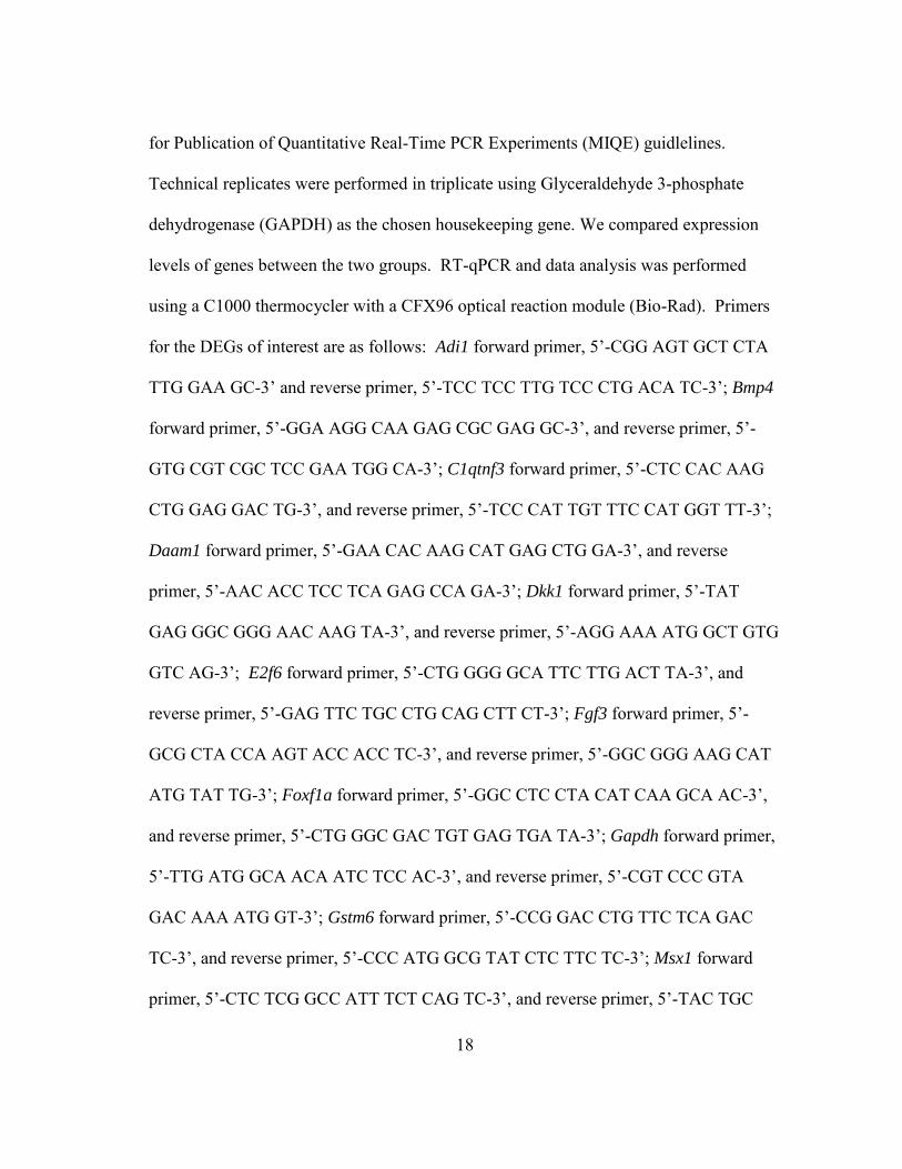
for Publication of Quantitative Real-Time PCR Experiments (MIQE) guidlelines.
Technical replicates were performed in triplicate using Glyceraldehyde 3-phosphate
dehydrogenase (GAPDH) as the chosen housekeeping gene. We compared expression
levels of genes between the two groups. RT-qPCR and data analysis was performed
using a C1000 thermocycler with a CFX96 optical reaction module (Bio-Rad). Primers
for the DEGs of interest are as follows: Adi1 forward primer, 5’-CGG AGT GCT CTA
TTG GAA GC-3’ and reverse primer, 5’-TCC TCC TTG TCC CTG ACA TC-3’; Bmp4
forward primer, 5’-GGA AGG CAA GAG CGC GAG GC-3’, and reverse primer, 5’-
GTG CGT CGC TCC GAA TGG CA-3’; C1qtnf3 forward primer, 5’-CTC CAC AAG
CTG GAG GAC TG-3’, and reverse primer, 5’-TCC CAT TGT TTC CAT GGT TT-3’;
Daam1 forward primer, 5’-GAA CAC AAG CAT GAG CTG GA-3’, and reverse
primer, 5’-AAC ACC TCC TCA GAG CCA GA-3’; Dkk1 forward primer, 5’-TAT
GAG GGC GGG AAC AAG TA-3’, and reverse primer, 5’-AGG AAA ATG GCT GTG
GTC AG-3’; E2f6 forward primer, 5’-CTG GGG GCA TTC TTG ACT TA-3’, and
reverse primer, 5’-GAG TTC TGC CTG CAG CTT CT-3’; Fgf3 forward primer, 5’-
GCG CTA CCA AGT ACC ACC TC-3’, and reverse primer, 5’-GGC GGG AAG CAT
ATG TAT TG-3’; Foxf1a forward primer, 5’-GGC CTC CTA CAT CAA GCA AC-3’,
and reverse primer, 5’-CTG GGC GAC TGT GAG TGA TA-3’; Gapdh forward primer,
5’-TTG ATG GCA ACA ATC TCC AC-3’, and reverse primer, 5’-CGT CCC GTA
GAC AAA ATG GT-3’; Gstm6 forward primer, 5’-CCG GAC CTG TTC TCA GAC
TC-3’, and reverse primer, 5’-CCC ATG GCG TAT CTC TTC TC-3’; Msx1 forward
primer, 5’-CTC TCG GCC ATT TCT CAG TC-3’, and reverse primer, 5’-TAC TGC

19
TTC TGG CGG AAC TT-3’; Odam forward primer, 5’-AGC CAG ACC TCT CTC
AGC AG-3’, and reverse primer, 5’-AAA TAG CTG CTG CCC TGT GT-3’; Osr2
(exon1) forward primer, 5’-CAA CAC GCT CGC TCT TTA CA-3’, and reverse primer,
5’-GCA CAG CTT GGA AAG GTC AT-3’; Osr2 (exons 2-3) forward primer, 5’-AGT
TTT GCG GCA GAC ACT TT-3’, and reverse primer, 5’-TCC TTT CCC ACA CTC
CTG AC-3’; Pax9 forward primer, 5’-CCA AGG GCA ACA GTC ACC-3’, and reverse
primer, 5’-GGC GGC TCA GTC TAT CAC TC-3’; Shh forward primer, 5’-GCC ATC
TCT GTG ATG AAC CA-3’, and reverse primer, 5’-CCA CGG AGT TCT CTG CTT
TC-3’; Tfap2b forward primer, 5’-CCA AGA AGT GGG CTC AGA AG-3’, and reverse
primer, 5’-TGG CAT CTT CAA CTG ACT GC-3’; Trp63 forward primer, 5’-TTT GAT
GCC CTC TCT CCA TC-3’, and reverse primer, 5’-CTT CGC AAT CTG GCA GTA
CA-3’.
RESULTS
In this study we investigated the Pax9-dependent gene expression pattern in
mouse embryonic day 14.5 (E14.5) tooth organs and their surrounding mesenchymal
layer by microarray in order to obtain the overall pattern of gene expression changes
around the time of developmental arrest (Table 2-1). We chose to pool tooth bud RNA
from 20 to 30 embryos from different litters for each array to avoid sampling bias and
the need for cDNA amplifications. Also, although Pax9 is only expressed in the
mesenchymal layer of the tooth bud, we chose to isolate whole tooth bud RNA from
mandibular first molars for our arrays instead of separating the mesenchymal from the

20
epithelial layer which could have resulted in greater loss or degradation of material. We
adopted this strategy because the spatiotemporal expression pattern of the majority of
potential target genes was either well-known or verifiable by consulting the Eurexpress
data base and because we were interested in exploring indirect targets of Pax9 as well as
direct ones since both are potentially suitable targets for replacement therapies. We
confirmed our E14.5 results using RT-qPCR (Figure 2-1) and followed up our analysis
with arrays using E13.5 tooth bud RNA, which were added later for the assessment of
temporal changes in expression patterns (Figure 2-2).
Our results suggest that Pax9 has a direct or indirect impact on the up- and down-
regulation of many diverse genes in the mesenchymal and sub-mesenchymal layer; in
addition the differential expression of a large number of transcripts was detected in the
epithelial layer and enamel knot, including several that had not been prominently
associated with tooth formation. The array data were used mainly as a guide for the more
accurate quantitation of expression differences by quantitative PCR.
As expected, Pax9 levels were significantly decreased in Pax9-deficient tooth
organ tissue, but surprisingly, the microarray and RT-qPCR data obtained from a
comparison of Pax9-/- and wild-type tooth organs suggested that Bmp4 expression was
less than 1.5 fold down-regulated by Pax9 deficiency at both E13.5 and E14.5. Bmp4 did
not even once appear as a significantly differentially expressed gene in any of the four
arrays even if a cut-off value of 1.3 fold expression difference was chosen; and only
rarely was a larger expression difference encountered in RT-qPCR investigations of
Pax9-deficient and wild-type cDNAs.

21
Instead, several other genes showed more impressive differential regulation;
among these were a few signaling factors such as mesenchymally expressed fibroblast
growth factor 3 (Fgf3) along with Fgf4, Fgf20, Lymphotoxin B (Ltb) and sonic
hedgehog (Shh), which are expressed in the epithelial enamel knot signaling center. In
fact, Shh was the most strongly down-regulated gene in the E14.5 arrays; but it may not
be the sole cause of the tooth developmental arrest because Shh mutations in humans are
not associated with absence of posterior teeth like seen in Pax9 deficiency; instead they
feature a solitary, upper central incisor (Roessler et al., 1996). The exact molecular
mechanism of Shh expression dependence on Pax9 is not yet clear, but probably
involves Fgf and Wnt pathways. Fgf3 and Fgf4, which are strongly down-regulated in
Pax9-/- tooth buds at E14.5 have previously been shown to be dependent on epithelial
Wnt signaling and their lack of expression in Lef1-/- mice was associated with strongly
reduced Shh expression in the enamel knot (Kratochwil et al., 2002). Notably in humans,
Fgf3 mutations are associated with microtia and microdontia (Alsmadi et al., 2009);
furthermore association studies have implied a connection between the Fgf3 gene and
tooth agenesis (Kuchler et al., 2013). Fgf and Shh pathway components should be
amenable to substitution therapies as a potential future treatment for PAX9-associated
tooth agenesis.
Also significantly differentially expressed were many transcription factors such
as early growth response 3 (Egr3), transcription factor ap2 (Tcfap2), odd skipped related
2 (Osr2), distal-less 2 (Dlx2), lymphoid enhancer factor 1 (Lef1), Sp6, forkhead box f1a
(Foxf1a), t-box transcription factor 1 (Tbx1), paired-like homeodomain transcription

22
factor 2 (Pitx2) and others. Receptors, signal transduction proteins, structural proteins
and pathway inhibitors were affected as well. Among the transcription factors, Tcfap2b
was found to be one of the more strongly down regulated genes at both E13.5 and E14.5
in Pax9-/- mice where it is strongly and exclusively expressed in molar dental
mesenchyme. Tcfap2b is a member of the AP2 family of transcription factors that are
involved in many developmental processes. Mutations in Tcfap2b are responsible for
Char syndrome (Satoda et al., 2000), which is a condition that affects the development of
the face including teeth, heart, and limbs. Egr3, which is also strongly and fairly
exclusively expressed in tooth bud mesenchyme at E14.5, is substantially down-
regulated; but nothing is currently known about its functional significance in tooth
development. Several Keratin transcripts, most significantly Krt17, were down
regulated.
The results for Osr2 were at first confusing because, contrary to expectation, it
was up-regulated in the array obtained with Pax9-/- tooth bud RNA. This led to our
suspicion that the Osr2 transgene which was inserted into the Pax9 locus and can be
transcribed but not translated into protein, may have been responsible for this outcome.
Indeed, when we performed RT-qPCR using primers to amplify the part of the Osr2
cDNA not present in the transgene insert, we could show that the expression of
endogenous Osr2 was actually down-regulated by more than 3-fold.
Of the inhibitors that affect the Wnt and Bmp4 pathways, Sostdc1, Dkk4, Sfrp4
and Apcdd1 are down-regulated while Dkk1 and Chrdl1 show modest up-regulation at
E14.5 (Ahn et al., 2010; Fedi et al., 1999; Hsieh et al., 1999; Leyns et al., 1997; Sakuta

23
et al., 2001), suggesting that Pax9 may have a role in fine-tuning Wnt-induced activation
of the morphogenetic process; possibly to allow for mesenchymal growth before final
differentiation. Some of these extracellular pathway inhibitors may also be useful for
replacement therapies.
Several of the differentially expressed genes were found in the sub-mesenchymal
layer (C1qtnf3, Sfrp4, and others), indicating that the activities of Pax9 are not limited to
the tooth bud but may include supporting structures.
DISCUSSION
Human trials for the substitution of recombinant EDA for a curative treatment of
hypohidrotic ectodermal dysplasia, including its associated tooth agenesis have started.
These trials were born from experiments with EDA deficient mice and dogs which
showed that treating affected embryos via maternal injection or the affected newborn
mice and dog pups with recombinant Eda could prevent the phenotypic effects of the
gene mutation (Gaide and Schneider, 2003). This success inspired the pursuit of similar
approaches for the treatment of other genetically linked developmental disorders.
Pax9 deficiency causes tooth developmental arrest at bud stage in both humans
and mice, yet the molecular mechanisms that lead to this defective phenotype have
remained poorly understood. We pursued this knowledge gap with microarray studies
from murine embryonic tooth organs. Only around 200 genes were shown to be
differentially expressed greater than 1.5 fold. Using bioinformatics approaches and real-
time quantitative PCR we narrowed our results to even fewer genes in which we were

24
confident were differentially expressed and also located in the developing tooth organ
region.
Unexpectedly Bmp4 expression was not significantly altered in our study of
Pax9-deficient tooth buds versus wild-type tooth buds. This result requires careful
scrutiny since it does not agree with prior investigations. In wild-type mice, Bmp4 had
been repeatedly shown by in situ hybridization to be prominently expressed in tooth bud
mesenchyme at E13.5 and E14.5; at E14.5 Bmp4 was shown to be additionally
expressed in the emerging enamel knot signaling center in tooth bud epithelium. In
Pax9-deficient mice, however, Bmp4 was reported to be neither detectable in the
mesenchymal nor in the epithelial layer by in situ hybridization. Therefore we had
expected a significant difference of Bmp4 expression in our RNA samples from E14.5
WT and Pax9-deficient whole tooth germs but to our disappointment this was not the
case. We did not observe a similar discrepancy between previously reported in situ
hybridization results and our array or qPCR data with respect to Msx1, Lef1, Osr2, Shh
and Fgfs. At the present it is not clear what caused this disagreement; to our support it
should be noted that it has also been shown recently that Bmp4 expression in Msx1
deficient mice is by far not as extensively down-regulated as had previously been
assumed (our unpublished results and (Jia et al., 2013; O'Connell et al., 2012)).
Since Bmp4 expression seems largely unchanged in Pax9-/- deficiency; a slight
delay in developmental timing should have allowed for the accumulation of sufficient
Bmp4 to proceed with tooth morphogenesis (Miletich et al., 2011). Bmp4 mutations in
humans have been described to affect the development of eyes but not teeth; only one

25
paper (Huang et al., 2013) suggests that a Bmp4 prodomain mutation may be associated
with relatively mild premolar agenesis. In summary, our data suggest that BMP4 is not
the most important factor in the development of Pax9 related tooth agenesis.
Endogenous Osr2 is significantly down regulated in Pax9-/- tooth bud tissue as
expected, since it had been previously described that expression of Osr2 in tooth bud
mesenchyme is dependent on Pax9 (Zhou et al., 2011). Osr2 however must be an
inhibitor of tooth development because its removal leads to an additional row of teeth or
to significant rescue of the normal row of teeth in mice lacking all mesenchymal Bmp4
(Jia et al., 2013). Therefore the down regulation of Osr2 cannot be responsible for the
tooth developmental arrest resulting from Pax9 deficiency. In contrast to the situation in
Pax9-/- mice, experimental Osr2 down regulation in Msx1-/- tooth germs seems to restore
tooth development (Zhang et al., 2009b). This demonstrates quite clearly that the
complexity of gene regulatory events during tooth development requires further
research.
According to our results Shh and Fgf3 are the most strongly down-regulated
genes at E14.5. Also substantially reduced is the expression of two other Fgf genes, Fgf4
and Fgf20, that are well-known for their contribution to odontogenesis. Fgfs and Shh
have been previously shown to be integrated into an epithelial-mesenchymal signaling
loop which is initiated by epithelial Wnt signaling and mediated by Lef1, a transcription
factor in the canonical Wnt pathway (Kratochwil et al., 2002). More specifically, this
signaling loop probably starts with Wnt 10 (a or b) around bud stage leading to Lef1
induced Fgf4 expression in the epithelial enamel knot area. Fgf4 then signals to the

26
dental mesenchyme resulting in mesenchymal Fgf3 activation which in turn is required
for subsequent Shh expression in the enamel knot signaling center. This signaling loop is
interrupted in Lef1 deficient mice leading to tooth developmental arrest at bud stage
similar to the situation in Pax9-deficient mice. In Lef1 deficiency, tooth development
can be completely rescued by the application of Fgf4 (also Fgfs 7, 8a, 9 or 10) but not by
application of Bmp4 or Shh. Our results suggest that Pax9 is somewhere involved in this
Wnt-Fgf signaling loop. Additional support for this theory is provided by the finding that
constitutional activation of Wnt signaling can override the Pax9-associated tooth
developmental arrest leading to an abundance of irregular teeth (O'Connell et al., 2012).
The importance of the Wnt pathway in odontogenesis is furthermore underscored by the
fact that mutation in WNT10a appear to be the most common cause of human tooth
agenesis (Bohring et al., 2009; Mues et al., 2014; van den Boogaard et al., 2012).
The question now arises at which stage and how Pax9 becomes involved in this
Wnt-Fgf-Shh signaling loop. Pax9 starts being expressed quite early (before E12) in the
condensing mesenchyme and Fgf3 cannot be its most important target because selective
Fgf3 deficiency leads only to smaller, not to missing teeth. Similarly, a reduction of Shh
cannot be the sole cause of Pax9-associated tooth agenesis since human Shh mutations
produce a much less severe and also different dental phenotype than Pax9, featuring
only a solitary maxillary central incisor instead of the molar and premolar agenesis
encountered in patients with PAX9 mutations (Das et al., 2002; Roessler et al., 1996).
The modulation of some aspects of Wnt signaling itself may be another factor
contributing to the Pax9-associated tooth agenesis. This can be inferred from the down-

27
regulation of the Wnt pathway inhibitors Sostdc1, Dkk4, Sfrp4 and Apcdd1 and the up-
regulation of Dkk1 at E14.5. All these inhibitors show different expression profiles and
only Dkk1 seems to be exclusively located in the dental mesenchyme. Dkk1 (Dickkopf-
related protein 1) is a potent Wnt antagonist and other studies have shown that over-
expression of Dkk1 in a 2.3-kb Col1a-Dkk1 transgenic mouse leads to malformed
second molars and loss of third molars (Han et al., 2011). It would be of great interest to
test the effect of these Wnt inhibitors on the craniofacial development of Pax9-deficient
mouse embryos with or without the addition of Fgfs and Shh.
Another important role of Pax9 (and especially Msx1) may be the promotion of
mesenchymal growth, probably mediated by Fgfs like Fgf3 coupled with a delay of
premature differentiation, which could be achieved through temporary global inhibition
of epithelial Wnt signaling.
Lately it is becoming apparent that mesenchymal Bmp4 activation is mainly
achieved through Wnt signaling, but Pax9 and Msx1 may contribute slightly through
positive regulation of mesenchymal Lef1 expression (Behrens et al., 1996). On the other
hand, the importance of mesenchymal Bmp4 expression has been diminished by
showing that its expression is not required to the extent previously thought (Jia et al.,
2013)- possibly a lack of mesenchymal Bmp4 can be compensated for by epithelial
Bmp4.

28
CHAPTER III
A BOOST IN WNT SIGNALING RESCUES PALATE FORMATION
SYNOPSIS
In this study, we found that a few maternal injections of a small molecule Wnt
pathway activator prevents cleft palate formation in Pax9-/- mouse embryos without
affecting other associated phenotypes such as tooth and thymus agenesis, or hind limb
polydactyly. No overt adverse effects of this treatment were detected in mother or
normal littermates suggesting that small molecule signaling pathway modulators may be
effective for the prevention of developmental abnormalities.
INTRODUCTION
Pax9 is a paired box transcription factor which is required for craniofacial, tooth,
and limb development. Mice without a functional Pax9 gene die at birth and have a
missing thymus, missing parathyroid glands, cleft secondary palate, tooth agenesis, and
supernumerary digits of the hind limb while heterozygous mice are completely normal
(Peters et al., 1998b). In humans only heterozygous pathogenic mutations have been
found in the PAX9 gene, all of which cause severe non-syndromic tooth agenesis of
mostly posterior teeth (Wang et al., 2009b). Concordantly, a possible association of the
PAX9 gene with orofacial clefting (Ichikawa et al., 2006; Lee et al., 2012; Song et al.,
2013) in humans has also been described.

29
The molecular and pathophysiological mechanisms leading to cleft palate in
Pax9-deficient mice have been reported to involve down-regulation of Bmp4, Fgf10,
Shh and Osr2 resulting in malformed palatal shelves which fail to elevate, a defect which
could be partially rescued by restoring Osr2 expression (Zhou et al., 2013). Pax9 has
also been described to play a role in palatal fusion when studied in TGF-β3 null mice
(Sasaki et al., 2007), however, others have shown that Pax9-deficient palates can fuse in
vitro when placed next to each other suggesting that clefting is primarily due to lack of
proper palate morphogenesis and elevation.
In order to gain a better understanding about Pax9 target genes in tooth
development, we previously evaluated the gene expression differences between Pax9-
deficient (Pax9-/-) and wild-type (Pax9+/+) mouse embryonic tooth bud tissue by
expression microarray and qPCR analysis. Unexpectedly, among all the gene expression
changes we found several genes involved in the Wnt signaling pathway, which had so
far not been described as a Pax9 target although it had been shown that constitutively
activated Wnt signaling can overcome the tooth developmental arrest in Pax9 deficient
mice (O'Connell et al., 2012).
MATERIALS AND METHODS
To determine if the Wnt pathway does in fact play a role in the pathogenesis of
the Pax9-/- phenotype we chose to modulate the Wnt pathway in vivo by injecting a small
molecule Dickkopf-related protein 1 (Dkk1) inhibitor (Pelletier et al., 2009) (WAY-
262611, Enzo Life Sciences) into pregnant Pax9+/- mice which had been mated with

30
Pax9+/- males. Pax9-/- mice were kindly provided by Rulang Jiang and are previously
described (Zhou et al., 2013). Briefly, the Pax9 locus contains a frt-flanked neo
expression cassette followed by an unexpressed Myc-Osr2A cDNA cassette which
replaces exon 2 of the Pax9 gene. Pax9-/- mice resulting from the mating of heterozygous
parents of this strain display phenotypes identical to a previously described Pax9-
deficient mouse model (Peters et al., 1998b) such as missing teeth, cleft palate, thymus
and parathyroid gland abnormalities, and hind limb polydactyly, all of which are
completely penetrant.
The drug used for increasing Wnt signaling, (1-(4-(naphthalen-2-yl)pyrimidin-2-
yl)piperidin-4-yl) methanamine, also known as WAY-262611, has been described
previously (Pelletier et al., 2009) and was shown to potentiate the Wnt β-catenin cellular
signaling pathway through the inhibition of the potent Wnt inhibitor Dkk1. WAY-
262611 was dissolved in DMSO and diluted 1:10 with PBS. Three consecutive doses of
12.5 mg/kg were injected in the tail veins of Pax9+/- pregnant mice at embryonic days
E12.5, E13.5 and E14.5 (n=15; 6 pregnant females with 15 homozygous recessive
embryos total). The injection of vehicle (10% DMSO in PBS) alone at E12.5, E13.5 and
E14.5 did not rescue palate formation in the Pax9-/- embryos (n=7; 3 pregnant females
with 7 homozygous recessive embryos total). All mouse pups were inspected
immediately after birth (P0) and their genotype was determined. The palate phenotype of
the Pax9-/- pups was observed both visually with a stereo dissecting microscope and
histologically using H&E staining. Wild type and heterozygous Pax9+/- littermates of the
Pax9-/- pups as well as their mothers were observed for 12 weeks).

31
All experiments complied with all relevant institutional and national animal
welfare laws, guidelines, and policies.
RESULTS
To determine if the Wnt pathway does in fact play a role in the pathogenesis of
the Pax9-/- phenotype we chose to modulate the Wnt pathway in vivo by injecting the
small molecule Dkk1 inhibitor (WAY-262611, Enzo Life Sciences) into pregnant Pax9+/-
mice which had been mated with Pax9+/- males. Since Dkk1 is a potent Wnt inhibitor,
expressed coordinately with Pax9 in dental mesenchyme, we expected WAY-262611 to
be effective in increasing Wnt signaling activity and reversing any phenotypic symptoms
of the Pax9-/- mouse embryos that were caused by down regulation of Wnt activity. We
found that WAY-262611 could prevent the cleft palate phenotype of Pax9-/- mouse
embryos (Figure 3-1), but not any of the other developmental malformations such as
missing teeth, lack of pharyngeal pouch derivatives, or polydactyly. Thirteen out of a
total of 15 Pax9-/- pups from 6 litters showed rescued palate fusion. However, the palatal
fusion did not prevent postnatal death of the pups, indicating that other Pax9 target
organs, such as parathyroid glands or thymus, may be more instrumental in perinatal
lethality.
Additionally, the WAY-262611 injections had no negative effects on the
wellbeing of the mother or the health of the Pax9-/+ or Pax9+/+ littermates, both of which
were followed for more than twelve weeks. The injection of vehicle (10% DMSO in

32
PBS) alone at E12.5, E13.5 and E14.5 did not rescue palate formation in Pax9-/- embryos
(n=7).
DISCUSSION
In terms of preventing tooth agenesis, it is likely that Pax9 affects additional
pathways besides just Wnt and therefore a therapeutic approach that targets several
different pathways would probably be needed to be effective. Alternatively, a stronger
boost in Wnt signaling or different Wnt pathway activator may be required.
Wnt genes are known to regulate many developmental processes, including
craniofacial development, but they are rarely mentioned as contributors to clefting
disorders: One report describes a family with homozygous nonsense mutations in WNT3
causing orofacial clefts as part of a Tetra-amelia syndrome (Niemann et al., 2004); and a
few association studies also suggest that WNT3 may contribute to an increased risk for
cleft lip/palate in humans (Menezes et al., 2010; Mostowska et al., 2012). In mice,
investigators have shown that the cleft palate following retinoic acid administration
during pregnancy is at least partly caused by inhibition of canonical Wnt signaling (Hu
et al., 2013). Other investigators found that cleft lip with cleft palate caused by
inactivation of Pbx genes also involves Wnt down-regulation; they succeeded in
rescuing cleft lip but not cleft palate formation by ectopic ectodermal Wnt expression
(Ferretti et al., 2011).
The implications of our findings are the following: 1) The Wnt pathway seems to
have a significant role in orofacial cleft development. 2) The paired domain transcription

33
factor Pax9 is involved in the regulation of Wnt pathway activity during the
development of some craniofacial tissues. 3) Small molecule modulators of major
signaling pathways have the potential to become effective drugs for the prevention of
developmental malformations. 4) The critical targets of any developmentally active
transcription factor are likely to differ from tissue to tissue requiring combination
replacement therapy. 5) Temporally restricted administration of these agents may be
tolerated without causing severe adverse effects.
Finally, the possibility exists that WAY-262611 or Dkk1 have a wider spectrum
of molecular activity besides affecting only Wnt signaling. Further investigation of this
drug and the function of Dkk1is warranted to understand the exact mechanism that is
driving their role in palatal growth, morphogenesis, and/or fusion.
Acknowledgements
We thank Dr. Rulang Jiang, Cincinnati Children’s Hospital Medical Center, for
kindly providing the Pax9-/- mice. This project is supported by the US National Institutes
of Health grant R01DE019471-01.

*Reprinted with permission from “Is There a Link Between Ovarian Cancer and Tooth Agenesis?” by Bonds J, Pollan-White S, Xiang L, Mues G, D’Souza R, 2014, Eur J Med
Genet, 57(5):235-9, Copyright 2014 by Elsevier B.V.
34
CHAPTER IV
IS THERE A LINK BETWEEN OVARIAN CANCER AND TOOTH AGENESIS?*
SYNOPSIS
An epidemiologic study from the year 2008 found a highly significant increase of
congenital tooth agenesis in women with ovarian cancer suggesting that a common
genetic etiology may predispose women to both conditions. The finding was reminiscent
of a previously described family harboring an AXIN2 mutation which could be shown to
segregate with both the tooth agenesis and the predisposition to colon cancer transmitted
in this family. Since tooth agenesis as a marker for susceptibility to ovarian cancer
would be of great relevance to both oncologists and women with inborn missing teeth,
the relationship between the two disorders requires a thorough assessment. We examined
DNA samples from the ovarian cancer patients who participated in the original study, to
look for a possible genetic connection between their ovarian malignancies and tooth
agenesis. MSX1, PAX9, AXIN2, EDA, WNT10A, BARX and BRCA1 genes were selected
for sequence analysis as they may cause tooth agenesis, are expressed in the female
reproductive system, and/or are involved in tumorigenesis in general or specifically in
the ovary.
_________________

35
Our study revealed evidence that one half of the dually affected patients had an
independent causation of the two conditions, thus reducing the previously estimated
ovarian cancer risk for women with congenital tooth agenesis quite significantly.
INTRODUCTION
In 2008, Chalothorn et al. described an increased prevalence of congenitally
missing teeth in women with epithelial ovarian cancer (Chalothorn et al., 2008). Twenty
percent of women with neoplastic ovarian disease reported one or two missing teeth,
versus three percent in a cancer-free control sample. Surprisingly, ten ovarian cyst
patients (unpublished) displayed an even greater prevalence of hypodontia: forty percent.
These observations suggested that there may be common genetic factors affecting both
tooth development and susceptibility to the formation of epithelial tumors or cysts of the
ovary, similar to the sequence variant in the AXIN2 gene which causes both tooth
agenesis and colorectal cancer (Lammi et al., 2004).
Early detection of epithelial ovarian cancer is difficult and as a result, the
mortality rate is unacceptably high. If a link were found between tooth agenesis and
ovarian cancer, semi-annual screening could become the standard of care for women
with tooth agenesis to increase the early detection rate (van Nagell et al., 2007). This
would not only provide a new diagnostic tool but also open up new biological insight
into epithelial ovarian cancer which, according to latest findings, may actually originate
in the fallopian tube epithelium since gene expression patterns in these two tissues
resemble each other closely (Kurman and Shih Ie, 2011).

36
The lifetime risk for neoplastic ovarian disease is only about a fifth of that for
tooth agenesis, which occurs in approximately 3% to 9% of the population even when 3rd
molars are excluded (Mattheeuws et al., 2004; Shapiro and Farrington, 1983). So far,
sequence variants in WNT10A, MSX1, PAX9, AXIN2 and EDA pathway genes have been
shown to cause about 50% of selective tooth agenesis in humans (Bergendal et al., 2011;
Bohring et al., 2009; Lammi et al., 2004; Stockton et al., 2000; Tao et al., 2006;
Vastardis et al., 1996). Several of these genes are also expressed in tumor cells of the
female reproductive system, suggesting a possible mechanism for the relationship
between ovarian disorders and hypodontia.
The homeobox gene BARX2 is expressed in maxillary and mandibular arches
(Jones et al., 1997 ) and in the developing tooth and is frequently dysregulated in
epithelial ovarian cancer (Sellar et al., 2001; Sellar et al., 2002). BARX1, a related
homebox gene, plays an important role in molar morphogenesis (Gould and Walter,
2000).
Overexpression of Msx1 inhibits ovarian carcinoma cell proliferation by
inducing apoptosis through interaction with the tumor suppressor p53 (Park et al., 2005).
MSX1 deficiency has been seen in human ovarian cancer cells (Park et al., 2001 ) and
other malignancies (Peters and Balling, 1999). Sequence variants in MSX1 have been
shown repeatedly to cause oligodontia of premolars, molars and incisors similar to
PAX9, a paired box transcription factor which is thought to control mesenchymal Bmp4
signaling during odontogenesis (Peters et al., 1998a). PAX9 expression was also found in
five of six epithelial ovarian cancer cell lines examined (Muratovska et al., 2003).

37
AXIN2 is a member of the Wnt signaling pathway, which is highly conserved in
evolution and controls many events during embryogenesis such as morphogenesis,
proliferation, motility and cell fate. Individuals with AXIN2 sequence variants can have
both tooth agenesis and a strong predisposition for developing colorectal cancer (Lammi
et al., 2004; Mostowska et al., 2006). WNT10A is the most commonly altered gene in
tooth agenesis (Bohring et al., 2009) with a large number of missing teeth in
homozygotes, and a few missing teeth in about 50% of heterozygotes. Sequence variants
in BRCA1 are the best-known causes of breast and ovarian cancer and, although the gene
is also expressed in the developing tooth, it has never been implicated in tooth agenesis.
The EDA gene is not associated with ovarian cancer or development, however,
sequence variants of the EDA gene cause the syndrome X-linked Hypohidrotic
Ectodermal Dysplasia (Li et al., 2008) and also non-syndromic tooth agenesis. Carrier
females may present with one or two missing or malformed teeth. Therefore an EDA
sequence variant found in any of the ovarian cancer samples would signify coincidental
tooth agenesis.
The primary goal of our study was to investigate if the five well-established tooth
agenesis genes WNT10A, EDA, PAX9, MSX1, and AXIN2 as well as the BARX1, 2 and
BRCA1 genes show any evidence of involvement in the tooth agenesis/ovarian cancer
association by doing a thorough sequence analysis of these candidate genes in the
original patient sample from the study by Chalothorn.

38
MATERIALS AND METHODS
Subject recruitment
The original protocol was IRB approved by the University of Kentucky. Fifty
subjects with ovarian cancer and another ten with ovarian cystic disease, each with or
without tooth agenesis, were recruited from the University of Kentucky Ovarian
Screening Clinic. Inclusion criteria were peri- or post-menopausal, ages 45 or older and
no obvious signs of a syndrome. Additional unrelated patients with tooth agenesis and
no personal or family history of ovarian disease were recruited under a separate IRB
approval from Texas A&M University Baylor College of Dentistry. All participants
were Caucasians except one who was of Japanese ancestry.
The final patient cohorts consisted of four groups: 1) 10 patients with agenesis of
1 to 2 teeth and ovarian cancer, 2) 40 patients with only ovarian cancer, 3) 35 patients
with agenesis of 1-8 teeth without ovarian disorders, and 4) the 10 patients with ovarian
cysts of whom 4 had mild tooth agenesis.
The wild-type sequence and allele frequencies of common variants in control
populations were obtained from the NCBI SNP database and the NHLBI Exome
sequencing project (ESP). Wild-type reference sequences obtained from NCBI are as
follows: MSX1 (NM_002448.3), PAX9 (NM_006194.3), AXIN2 (NM_004655.3), EDA
(NM_001399.4), WNT10A (NM_025216.2), BARX1 (NM_021570.3), BARX2
(NM_003658.4) and BRCA1 (NM_007294.3).
Informed consent was obtained and a thorough patient history and dental exam
was performed. If a patient was unsure of history, the patient’s dentist was consulted to

39
confirm etiology of any missing teeth. DNA samples were obtained using BuccalAmp
swabs and sent to Texas A&M University Baylor College of Dentistry for analysis.
DNA extraction from buccal swabs
Since buccal swabs may not have yielded sufficient material for the analysis of
eight to ten genes, the samples were amplified by Whole Genome Amplification (WGA)
with the GenomiPhi WGA system (GE Healthcare). Successful genome amplification
was verified by gel electrophoresis of amplified samples together with a quantitation
marker.
Polymerase chain reaction and sequencing of products
The exons of each gene were PCR amplified with GoTaq reagents (Promega)
using a 96-well plate format for the 95 samples and one negative control. Several of the
amplicons were very GC-rich and required PCR optimization and the use of 5% DMSO.
Quality and quantity of PCR products was confirmed by gel-electrophoresis, followed
by treatment with ExoSapIt (Affymetrix) and then addition of specifically designed
sequencing primers. Automated dideoxy chain terminator sequencing was done by
Seqwright, TX and GenScript, NJ.
Analysis of sequencing results
All sequences were visually inspected for heterozygous base changes and
compared to the corresponding wild-type sequences previously mentioned using the

40
NCBI BLAST program. Once a nucleotide change was found, the SNP (single
nucleotide polymorphism) database was consulted to determine if the SNP is a common
polymorphism. For common SNPs, the allele frequencies were compared 1) between the
different experimental groups and 2) between experimental groups and Caucasian
population controls reported in NCBI and NHLBI databases. For MSX1 polymorphisms,
the Caucasian control allele frequencies from a study by Jezewski et al. (supplement)
were also employed (Jezewski et al., 2003). Chi-square statistics was used for the
determination of statistical significance of allele frequency differences.
RESULTS
Sequence analysis of the EDA, WNT10A and BRCA1 genes yielded several
interpretable results (Table 4-1):
1) One of the ten patients with combined ovarian cancer/tooth agenesis was a
carrier of the known p.Arg69Leu sequence variant in EDA (rs132630309), explaining
the tooth agenesis, and she also had a frameshift sequence variant in BRCA1,
p.Gln1096_Ser1097=fs (rs80357686), most probably responsible for the ovarian cancer.
2) Another tooth agenesis/ovarian cancer patient had the BRCA1 frameshift
sequence variant, p.Lys679Ter (rs80357082), which was also present in two other
ovarian cancer patients without tooth agenesis, indicating that this patient’s ovarian
cancer and her tooth agenesis have different roots and also that the investigated
population shared common ancestors since the frequency of this sequence variant seems

41
to be quite high, but no population allele frequencies are available (presumably due to
gene patent issues).
3) Three of the remaining eight tooth agenesis/ovarian cancer patients had the
WNT10A p.Phe228Ile sequence variant (rs121908120), which in heterozygous form is
probably the most common sequence variant encountered in mild tooth agenesis while
causing severe tooth agenesis when homozygously inherited. There were also three
patients with this sequence variant in the “ovarian cancer only” group, however about
50% of people with this sequence variant normally do not present with tooth agenesis.
These results taken together suggest that at least half of the samples from the
combined ovarian cancer/tooth agenesis patients identified in the epidemiological study
by Chalothorn et al. show evidence for independent causation of the two conditions. In
the ten ovarian cyst patients we found sequence variants neither in BRCA1 nor in the
tooth agenesis genes WNT10A or EDA.
Sequence analysis results from the other investigated genes are presented in
Tables 4-2 and 4-3 and are briefly presented below. These results were calculated for the
whole group, including ovarian cancer and ovarian cyst groups.
In BARX1, the missense sequence variant Ala48Thr was detected in 17 samples,
but no significant allele frequency differences between the groups were found. The other
five polymorphisms in this gene were also insignificant.
In BARX2, we found four heterozygous p.Ser64Pro sequence variants; three of
them in individuals with tooth agenesis, with and without ovarian cancer. p.Ser64Pro is
fairly conserved among species suggesting its importance in protein function; however

42
since the control allele frequency reported in the SNP database is similar to the one
found in this study, it may not play a role in tooth agenesis. On the other hand, we do not
know if the control population used for SNP data was screened for missing teeth. The
synonymous p.Pro203= polymorphism in exon 2 occurred in the ovarian cancer cohort
in 50% of those with tooth agenesis versus 33% without tooth agenesis; however, the
difference was not statistically significant. The remaining polymorphisms found for
BARX2 did not demonstrate allele patterns consistent with an association with a disease
phenotype.
A substantial number of polymorphisms were found in the MSX1 gene including
p.Ala40Gly (formerly p.Ala34Gly). The minor allele frequency of this polymorphism
was significantly higher in all groups with tooth agenesis (regardless of ovarian disease
status) compared to the NHLBI ESP Caucasian control group (n> 2,000). However using
the allele frequency values for 154 Caucasian controls which were reported by Jezewsky
et al. in their study about MSX1 sequence variants in cleft lip/palate patients (Jezewski et
al., 2003), the differences were not statistically significant. The allele frequencies of the
MSX1 polymorphisms c.*6C>T (rs8670) and c.-18G>A (rs186861426) were also
significantly higher in most groups with tooth agenesis using the NHLBI ESP (Table 4-
3), but not with the Jezewski control frequencies; we also noticed that these SNPs appear
to be syntenic with p.Ala40Gly.
In AXIN2 we found the missense sequence variant p.Ser762Asn in a single
ovarian cancer patient without tooth agenesis. This sequence variant may thus contribute
to cancer susceptibility but not to tooth agenesis. We also found the previously reported

43
c.1994_1995insG sequence variant in one patient with combined ovarian cysts and tooth
agenesis; however, we suspect that it is a PCR artifact since its appearance was
dependent on PCR conditions and it occurs after a run of seven G nucleotides.
Sequencing of PAX9 revealed five known and one new polymorphism (Table 4-
2). One of these, a C to T transition adjacent to the p.Ala240Pro polymorphism, lead to a
synonymous histidine codon which was significantly more frequent in samples from
tooth agenesis patients (Table 4-3).
DISCUSSION
A genetic connection between development and cancer is intuitively
understandable in that both processes are characterized by rapid cell growth which often
involves the same signaling pathways. However it is less intuitively understandable that
a developmental deficit like tooth agenesis should be associated with the uncontrolled
expansion of cell growth found in cancer. The finding that a nonsense sequence variant
in AXIN2, which should lead to increased WNT signaling, can cause both severe tooth
agenesis and a predisposition for colon cancer in humans (Lammi et al., 2004) is
therefore somewhat surprising because increased WNT signaling leads to supernumerary
teeth in experimental animal models (Jarvinen et al., 2006) and Axin2 promotes
oncogenicity in colon cancer by the upregulation of Snail1 (Wu et al., 2012). Thus, only
a gain-of function sequence variant, not a nonsense sequence variant, in Axin2 would be
compatible with its double role as suppressor of tooth development and promoter of
colon cancer. The double role is easier to fathom in the case of sequence variants in

44
MSX1, a protein that is able to interact with both DNA and other proteins and can act as
a transcriptional suppressor or as an activator depending on cellular context.
Since a causal linkage between tooth agenesis and ovarian cancer would have
major ramifications for the large number of women with minor tooth agenesis, the issue
requires quite urgent clarification. Therefore, we undertook this investigation and
approached the question of common genetic factors in tooth agenesis and ovarian cancer
by screening known tooth agenesis causing genes for sequence variants as suggested in
the original study by Chalothorn and also added the BRCA1 gene and the newly
attributed tooth agenesis gene WNT10A.
We did not find a single candidate gene or sequence variant that could explain
the increased co-occurrence of tooth agenesis with ovarian cancer or ovarian cysts with
the exception of the controversial c.1994_1995insG sequence variant in Axin2. But we
did find BRCA1 sequence variants in two of the ten cases of ovarian cancer/tooth
agenesis patients, explaining the ovarian cancer. Additionally, one of these two also had
an EDA sequence variant, clearly demonstrating separate origins of ovarian cancer and
tooth agenesis. Furthermore, we found well known tooth agenesis-causing WNT10A
sequence variants in three additional members of the ovarian cancer/tooth agenesis
group arguing for independent causation of the tooth agenesis in these patients. Our
findings do not prove that the two conditions arose independently from each other but
they make it seem quite likely for at least half of the cases.
The observation by Chalothorn et al. that their 10 ovarian cyst patients had an
even higher prevalence of hypodontia than the ovarian cancer cohort reinforces this

45
notion since so far no genetic link has been detected between epithelial ovarian cancer
and ovarian cysts making it unlikely that a single gene may be responsible for all 3
disorders.
Additional evidence for independent causation comes from a recent study which
did find increased rates of (self-reported) cancer in patients with tooth agenesis (Kuchler
et al., 2013). However, ovarian cancer, in contrast to prostate, breast, and nervous
system malignancies, was not among the significant results. We also have been enrolling
a few hundred subjects in our own “Missing Tooth Study” over the past 10 years and
have not noted any case with ovarian cancer or cysts in her/his family’s medical history;
however, the reason for this could be the higher prevalence of tooth agenesis and
incomplete recall of family health issues.
On the other hand, we know that the investigated genes represent only a minority
of all the possible candidates that may be involved in hypodontia and it would certainly
be worthwhile to investigate additional genes which are commonly associated with
ovarian cancer such as BRCA2, BRCA-interacting protein, ErbB2 and p53, for example,
which are also quite strongly expressed in the tooth bud of developing mouse embryos
(Diez-Roux et al., 2011).
In conclusion we propose that a larger epidemiological study should be
conducted to confirm any link between ovarian cancer and tooth agenesis and challenge
the currently held notion of a predisposition of developing cancer in tooth agenesis
patients. Ideally, a large family with both ovarian cancer and tooth agenesis may be
recruited for linkage analysis.

46
Acknowledgements
We thank all patients who participated in this study and Drs. Chalothorn and
Beeman from the University of Kentucky for providing the samples. The study was
supported by NIH/NIDCR awards R03DE019554-01 to GM and R01DE019471-01 to
RDS. The authors declare no potential conflict of interest with respect to the research,
authorship and or publication of this article.

*Reprinted with permission from “The Wnt10a Gene in ectodermal dysplasias and selective tooth agenesis” by Mues G, Bonds J, Xiang L, Vieira A, Seymen F, Klein O, and D’Souza R, 2014, Am J Med Genet A, Copyright 2014 by John Wiley & Sons, Inc.
47
CHAPTER V
THE WNT10A GENE IN ECTODERMAL DYSPLASIAS AND SELECTIVE TOOTH
AGENESIS*
SYNOPSIS
Mutations in the WNT10A gene were first detected in the rare syndrome odonto-
onycho-dermal dysplasia (OODD, OMIM257980) but have now also been found to
cause about 35–50% of selective tooth agenesis (STHAG4, OMIM150400), a common
disorder that mostly affects the permanent dentition. In our random sample of tooth
agenesis patients, 40% had at least one mutation in the WNT10A gene.
TheWNT10A Phe228Ile variant alone reached an allele frequency of 0.21 in the tooth
agenesis cohort, about 10 times higher than the allele frequency reported in large SNP
databases for Caucasian populations. Patients with bi-allelic WNT10A mutations have
severe tooth agenesis while heterozygous individuals are either unaffected or have a
mild phenotype. Mutations in the coding areas of the WNT10B gene, which is co-
expressed with WNT10A during odontogenesis, and the WNT6 gene which is located at
the same chromosomal locus as WNT10A in humans, do not contribute to the tooth
agenesis phenotype.
__________________

48
INTRODUCTION
Adaimy et al. (2007) performed autozygosity mapping in three consanguineous
Lebanese families with the rare Odonto-Onycho-Dermal-Dysplasia syndrome (OODD,
MIM 257980) which had been previously characterized phenotypically in the same
population (Fadhil et al., 1983). They found that all affected family members were
homozygous for the same nonsense mutation in the WNT10A gene leading to the
phenotypic features of severe hypodontia, onychodysplasia, smooth tongue as well as
palmar and plantar hyperhidrosis and hyperkeratosis. The phenotype of heterozygous
family members was not recorded. Two years later Bohring et al. (2009) reported that
WNT10A mutations are not restricted to the rare OODD syndrome but also found in
other Ectodermal Dysplasia entities like the Schöpf-Schulz-Passarge syndrome (MIM
224750) which additionally features eyelid cysts and predisposition to adnexal skin
tumors. Bohring et al. also described a high prevalence of apparently non-syndromic
tooth agenesis among their homozygous patients as well as mild, predominantly dental
symptoms in about half of the heterozygous family members. Three further reports about
the high prevalence of WNT10A mutations in Ectodermal Dysplasia syndromes and in
non-syndromic tooth agenesis followed in 2011 (Cluzeau et al.), in 2012 (van den
Boogaard et al.), and in 2013 (Plaisancie et al.).
The expression of Wnt10a along with Wnt10b, Shh, Bmps2 and 4 and other
developmentally active gene products during mouse odontogenesis had been
investigated as early as 1998 (Dassule and McMahon). They detected Wnt10a at around
mouse embryonic day 12 (E12) by in situ hybridization in the inner epithelial/ enamel

49
knot area of the tooth bud, where it was co-expressed with Wnt10b. Since Wnt10b
expression was recognizable a little earlier and more prominently than Wnt10a
expression, further investigations in this study focused on the Wnt10b molecule. At later
stages of tooth development (mouse E14 to E18), Wnt10a can also be found in the
mesenchymal preodontoblast layer where it contributes to or initiates odontoblast
differentiation, possibly through the up-regulation of dentin sialophosphoprotein (Dspp)
expression (Yamashiro et al., 2007).
WNT10A, which is located adjacent to WNT6 at 2q35 in humans, is also active
during the development of hair follicles and limbs, and in hematopoiesis. In adult tissues
it is expressed in lymph nodes, blood, adrenal gland, prostate, testis, ovary, retina, brain,
lung and kidney; and may also play a role in several neoplastic disorders, notably
ameloblastomas, keratocystic odontogenic tumors, lymphomas and leukemias but is also
found up-regulated in several cancers. Functional studies showed that Wnt10a activates
the canonical Wnt pathway and regulates mesenchymal cell fate in that it inhibits
adipogenesis and stimulates osteoblastogenesis (Cawthorn et al., 2012).
The general role of canonical Wnt signaling during tooth development has been
explored in more detail by stabilization of β-catenin or depletion of Apc, a positive and a
negative regulator of canonical Wnt signaling respectively. Both procedures
constitutively activate Wnt signaling in tooth bud epithelium leading to the formation of
many accessory tooth buds sprouting from the original tooth anlage. The resulting
supernumerary teeth are often small but otherwise completely normal (Jarvinen et al.,
2006; Wang et al., 2009a). Interestingly, the expression of Pax9 and Msx1, two normally

50
essential transcription factors in tooth bud mesenchyme, are not required for the
formation of these supernumerary teeth. Furthermore, the inactivation of the Wnt
secretion facilitator Wntless (Wls) was recently shown to prevent intraepithelial Wnt
signaling leading to tooth developmental arrest (Zhu et al., 2013).
When we sequenced the WNT10A gene in the random collection of tooth
agenesis patients who participate in our “missing tooth” study, we also found a large
number of WNT10A mutations in our samples confirming the importance of WNT10A
in tooth development.
MATERIALS AND METHODS
Patient recruitment
Tooth agenesis study participants were recruited via website and through
collaboration with Drs. Alexandre Vieira (University of Pittsburgh) and Ophir Klein
(University of California at San Francisco), following IRB approved protocols. People of
all ages with any number of missing teeth except third molars were included, and only
patients with overt ectodermal dysplasia symptoms were excluded. The final cohort
consisted of 90 unrelated samples; half of them were from Caucasian Americans and the
other half from patients from Turkey which are considered to be mostly of
Mediterranean- European ancestry. Cheek swab or saliva samples were collected for the
isolation of genomic DNA. The wild type sequence and allele frequencies of common
variants in control populations were obtained from the NCBI SNP data bases as well as
the NHLBI Exome sequencing project (ESP).

51
DNA extraction from buccal swabs
DNA extraction was performed with the Puregene Buccal Cell Kit (Qiagen).
Since buccal swabs do not yield sufficient material for the analysis of multiple genes, the
samples were amplified by Whole Genome Amplification (WGA) with the REPLI-g
WGA system (Qiagen) Successful genome amplification was verified by gel
electrophoresis of amplified samples together with a quantitation marker. DNA samples
received from our collaborators were also amplified by WGA.
Polymerase chain reaction and sequencing of products
Exons of the WNT10a, WNT10b and WNT6 genes were PCR amplified with
GoTaq reagents (Promega) using a 96-well plate format for the 90 samples and the
controls. Quality and quantity of PCR products was confirmed by gel-electrophoresis,
followed by treatment with ExoSapIt (USB) and addition of the sequencing primers.
Automated dideoxy chain terminator sequencing was done by GenScript, Piscataway,
NJ.
Analysis of sequencing results
All sequences were visually inspected for heterozygous base changes and
compared to the corresponding wild type sequences using the ‘BLAST’ program. Once a
nucleotide change was found, the SNP (single nucleotide polymorphism) data base was
consulted to determine if the SNP is a common polymorphism. For appropriate SNPs,
the allele frequencies were compared between the experimental groups consisting of

52
Caucasian and Turkish samples; and between the experimental and the Caucasian
population control groups reported in NCBI and NHLBI data bases.
RESULTS
Wnt10A but neither WNT6 nor WNT10B contribute to tooth agenesis
About forty percent of our random group of tooth agenesis patients had at least
one missense, nonsense or frameshift mutation in the WNT10A gene. The different
mutations encountered in our patient samples are shown in Figure 5-1. Most common
was the mono- or bi-allelic Phe228Ile mutation with a prevalence of about 31 percent
and an allele frequency of 0.21 compared to an allele frequency of about 0.02 in large
Caucasian control populations and only 0.007 in African American controls (Table 5-1).
The allele frequency of Phe228Ile was also calculated separately for our Caucasian
(0.216) and Turkish participants (0.20) to exclude any influence that ethnic background
differences could have had on the allele frequency.
Since Phe228Ile is so much more common in Caucasian tooth agenesis patients,
it either is the causative factor or is closely linked to the causative mutation. Since
WNT10A is located only a few kb telomeric of WNT6 on chromosome 2q35 we included
the latter in our analysis but did not find any mutations or polymorphisms that were
syntenic with the nucleotide change leading to WNT10A Phe228Ile. The common
WNT6 variant Pro155Arg occurred in our tooth agenesis population at a frequency
similar to normal control populations and only 1 of the 10 patients who had the WNT6
Pro155Arg variant also had WNT10A Phe228Ile.

53
Since the WNT10A and WNT10B proteins are co-expressed in the inner dental
epithelium of developing teeth and share 62 percent identity, it was conceivable that
WNT10B mutations may also cause missing teeth. But sequencing of the whole coding
area of the WNT10B gene did not reveal any nucleotide changes that could possibly be
implicated in the tooth agenesis phenotype.
Phenotypes associated with WNT10A mutations
We did not receive any reports about missing primary teeth although some
patients remembered having relatively small deciduous teeth. The number of missing
teeth in the permanent dentition depended strongly on whether the affected individual
was heterozygous or homozygous/ compound heterozygous for WNT10A mutations.
Heterozygous patients were missing up to 6 permanent teeth while homozygous patients
were generally missing from 6 to 26, most often 16 teeth.
While all patients with bi-allelic mutations had oligodontia, many heterozygous
relatives of study participants were not affected suggesting incomplete penetrance
(Figure 5-2). Syndromic ectodermal dysplasia manifestations were not encountered in
our study population because they constituted exclusion criteria for participation in the
study. One study participant however had a history of benign skin tumors, possibly
bearing some resemblance to the Schöpf-Schulz-Passarge syndrome and another one
reported mild heat intolerance.
The tooth agenesis pattern in heterozygous patients parallels that of common
mild tooth agenesis with a predominant absence of lower second premolars and upper

54
lateral incisors; but mandibular incisors are also frequently absent, occasionally even a
canine or a first premolar. The pattern is similar to EDA pathway associated selective
tooth agenesis in that the anterior teeth are more often affected (Mues et al., 2010; Mues
et al., 2009; Tarpey et al., 2007). Patients with homozygous WNT10a mutations have
also posterior tooth agenesis similar to syndromic EDA pathway mutation phenotypes
(Lexner et al., 2007).
DISCUSSION
Traditionally we distinguished between syndromic and non-syndromic tooth
agenesis. Syndromic tooth agenesis was most often encountered as part of an Ectodermal
Dysplasia phenotype while non-syndromic tooth agenesis, also called isolated or
selective tooth agenesis/hypodontia (STHAG1-6 and X1 in OMIM), should only affect
the dentition without any systemic manifestations. Inherent in the syndromic versus non-
syndromic classification was the assumption that the two disorders had fundamentally
different genetic causations.
However, the more we learn about the pathogenesis of tooth agenesis the more
we realize that there is an extensive overlap between the genetic basis of syndromic and
non-syndromic forms of tooth agenesis (Nieminen, 2009) and the phenotypic distinction
may not have been helpful for the search of additional tooth agenesis genes. From a
genetic point of view, non-syndromic and syndromic tooth agenesis are often caused by
the same genes, but the development of some teeth seems to be more sensitive to gene

55
dosage and thus are the first organs to be affected, while other ectodermal appendices
may still form normally (Mues et al., 2010).
The distinction between syndromic and non-syndromic tooth agenesis may also
be problematic with respect to the new, biologically based diagnostic and therapeutic
approaches for which dental professionals who are traditionally the only health care
providers for non-syndromic tooth agenesis patients, may still be little prepared.
Classification of the STHAGs as ectodermal dysplasia entities may therefore be
desirable from a clinical point of view.
The high prevalence of WNT10A mutations is truly astounding, especially since
this gene is hardly ever mentioned in the extensive literature about the molecular
genetics of tooth development. Even more surprising is the large number of Caucasian
tooth agenesis patients with one particular mutation, WNT10A Phe228Ile. Pathogenic
mutations are usually lost from the gene pool of a population unless they have some kind
of survival advantage like for example heterozygous mutations in the hemoglobin genes
which are of benefit in areas with high malaria incidence. The survival advantage is
often lost in individuals with homozygous mutations like in sickle cell disease, but since
homozygously affected individuals are quite rare, the mutation has an overall positive
effect on population growth (Fleming et al., 1979). It will certainly be interesting to find
a cause for the high prevalence of WNT10A Phe228Ile mutations.
Interesting is also that the tooth agenesis pattern of patients with WNT10A
mutations resembles that of EDA pathway associated hypodontia. Both WNT and EDA
pathways are known to operate predominantly in the epithelial layer of the developing

56
tooth and repeated interactions between the two pathways have been observed during
ectodermal appendage formation and in vitro (Durmowicz et al., 2002; Laurikkala et al.,
2001; Zhang et al., 2009a). It is therefore possible that the new, biological replacement
therapies for EDA pathway mutation associated disorders may also be of value in the
much more common WNT10A disorder. It would certainly be worth testing this
possibility because EDA replacement therapies have shown great efficacy in
ameliorating disease symptoms in animals and are currently tested in humans, while the
generation of WNT10A specific therapeutics would be quite complicated not only from
a chemical engineering point of view but also because of the lack of a one to one
correspondence of ligands and receptors in the WNT pathway, potentially leading to
severe adverse effects, and even more importantly, a lack of a Wnt10a-deficient,
diphyodont animal model for the testing of these therapeutics.
Acknowledgements
We thank all patients for their participation in this study which was supported by
NIH/NIDCR awards R03DE019554-01 and R01DE019471-01 and-02. At the Pittsburgh
site, we would like to thank Kathleen Deeley, Megan Weber, and Jacqueline Noel for
providing support in sample handling and internal quality control. Some of the samples
were collected with the support of Asli Patir and Mine Yildirim. The University of
Pittsburgh Dental Registry and DNA Repository is supported by the University of
Pittsburgh School of Dental Medicine and NIH Grant UL1 RR024153.

57
CHAPTER VI
CONCLUSION
In this project I investigated both the basic science and clinical aspects of the
molecular genetics of tooth agenesis in order to:
A) Characterize those target genes of the transcription factor Pax9 which mediate
the tooth developmental arrest observed with Pax9 deficiency. These target genes have
not only the potential to serve as therapeutics for the rescue of the tooth developmental
arrest but may also be tooth agenesis causing genes themselves deserving to be screened
for mutations in tooth agenesis patients.
B) Identify the particular mutations in known or suspected tooth agenesis genes
that are responsible for the tooth agenesis in each individual of a large group of patients
with inborn missing teeth. An accurate molecular diagnosis is required before any
molecular therapeutics can be used and the majority of the as yet unknown tooth
agenesis genes should be discovered.
The initial project in which I studied the downstream effects of Pax9 revealed
that the regulatory mechanisms surrounding tooth agenesis were not as straightforward
as previously thought. The most intriguing aspect of this study is that the gene that was
presumed to be the central downstream effector of Pax9 on tooth formation, Bmp4, had
mRNA levels that were relatively unchanged in our Pax9-deficient model during early
tooth formation. This coupled with the fact that many other signaling pathways were
much more heavily affected indicates that tooth agenesis cannot be easily attributed to

58
the loss of a single gene or gene product downstream of the causative gene and may
therefore require treatment with multiple replacement factors if the mutated gene product
itself is not suitable for replacement.
The signaling pathways that appear to be markedly affected by Pax9 deficiency,
Wnt, Fgf and Shh, were previously thought to be relatively minor targets of Pax9 or the
result of diminished Bmp4 activity. The latter seems unlikely in light of our finding of
barely changed Bmp4 expression.
The impact of Pax9 on Wnt pathway regulators is of special interest because
recent clinical studies indicate that mutations in one Wnt pathway member, the
WNT10A gene, may be responsible for approximately 40% of all congenital human
tooth agenesis. In concord with this clinical finding, many basic research studies have
also detected the importance of the Wnt pathway which is gradually being revealed to be
the master regulator of tooth formation, much like it is a major player in limb formation.
Constitutively activated Wnt signaling has even been shown to overcome the tooth
developmental arrest in Pax9 deficient murine tooth buds (O'Connell et al., 2012).
To test the effects of a temporary and physiologically more tolerable increase in
Wnt signaling in our Pax9-deficient mouse model we used a small molecule Wnt agonist
(an antagonist of the Wnt inhibitor Dkk1 which was upregulated in Pax9-/- mice at
E14.5). Injection of small amounts of this molecule into the maternal tail vein at discreet
times during embryonic development led to the surprising finding that we could rescue
the cleft palate phenotype of the Pax-/- embryos but the tooth phenotype was unchanged.

59
Nevertheless, another fortunate finding was that there were no apparent deleterious
effects of using this molecule on normal and healthy wild-type embryos or the mother.
Originally it was assumed that the cleft palate was to blame for the postnatal
death of Pax9-deficient pups. However, pups with rescued palate still died at birth, with
the cause now most likely being the lack of development of other missing pharyngeal
pouch derivatives. Hind limb polydactyly was also unchanged, presumably because the
drug was given after the start of limb and digit formation. It would be interesting to see
if this polydactyly would be corrected by earlier application of the drug. Most likely,
however, at this earlier stage of development, around E10, the treatment would produce
more undesirable side effects. Since we could rescue the cleft palate but no other
phenotype manifestations of Pax9 deficiency, we suggest that combination therapy may
be needed to totally restore Pax9-deficient mice, for example adding recombinant Fgf3,
4 and/or an Shh agonist like the Smoothened Agonist, SAG.
We also tested the effect of injecting the Bmp4 agonist isoliquiritigenin (not
published). This molecule was shown to act as a Bmp4 agonist in cell culture and in
zebrafish; however, it had not yet been tested in mammals. Our results showed that there
was no effect of this treatment suggesting that either this molecule does not perform well
in mammals or that Bmp4 deficiency is not a major cause of the Pax9-deficient
phenotype. Based on our microarray and RT-qPCR results it is most likely the latter.
The other aspect of this dissertation focuses on clinical approaches to diagnose
the molecular genetic causes of tooth agenesis in individual patients. In order to find new
tooth agenesis genes we screened several of our newly discovered Pax9 downstream

60
target genes (Fgf3, Fgf4, Tcfap2b, Sostdc1, Lef1 and others) for mutations in 90
independent DNA samples from patients with tooth agenesis. None of the chosen genes
showed mutations in our tooth agenesis samples except for one rare polymorphism in
Lef1 which may have contributed to the tooth agenesis phenotype of the donor of that
sample.
Our study also investigated the alleged association of ovarian cancer with tooth
agenesis and showed that there was essentially no increased risk for women to acquire
ovarian cancer if they had missing teeth because we found through candidate gene
sequence analysis that the tooth agenesis and the ovarian cancer most probably arose
independently from each other. The importance of our study is obvious since tooth
agenesis is the most common developmental abnormality in humans and this high
prevalence would lead to unnecessary anxiety in many women who, in reality, are not at
an increased risk at all of developing a malignant neoplasm. Studies such as this suggest
the need to further investigate issues where clinical associations suggest causality when
in fact there is only a correlation.
We also showed through diagnostic gene sequencing in 90 tooth agenesis
samples that all known Wnt mutations that affect tooth formation reside within the
WNT10A gene and not within other Wnt genes such as WNT6 which is located adjacent
to WNT10a on chromosome 2 or Wnt10b which is co-expressed with WNT10A during
tooth development. Also of note is that a single mutation, Phe228Ile, is astonishingly
common in Caucasian populations and yet there does not seem to be an advantage to this
mutation. Usually harmful mutations are lost from the gene pool unless they have

61
heterozygote advantage such as mutations in hemoglobin in individuals with sickle cell
anemia who gain resistance to malaria. It would seem that the great unsolved question is
what kind of heterozygote advantage a reduced Wnt10a activity could have.
Our overall conclusion is that it should be possible to rescue genetically caused
developmental disorders by substitution of the crucial gene products at the correct
developmental time interval. The substitution therapy may consist of recombinant
proteins or small molecule genetic pathway regulators and may require only one factor
in the case that the dysfunctional gene product can be replaced directly, or it may require
the replacement of several factors if downstream targets of the dysfunctional gene
product have to be used. In any case the genetic cause of the developmental disorder has
to be diagnosed before the appropriate replacement therapy can be determined.

62
REFERENCES
Aberg T, Wozney J, Thesleff I (1997). Expression patterns of bone morphogenetic proteins (Bmps) in the developing mouse tooth suggest roles in morphogenesis and cell differentiation. Dev Dyn 210(4):383-396. Adaimy L, Chouery E, Megarbane H, Mroueh S, Delague V, Nicolas E et al. (2007). Mutation in WNT10A is associated with an autosomal recessive ectodermal dysplasia: the odonto-onycho-dermal dysplasia. Am J Hum Genet 81(4):821-828. Ahn Y, Sanderson BW, Klein OD, Krumlauf R (2010). Inhibition of Wnt signaling by Wise (Sostdc1) and negative feedback from Shh controls tooth number and patterning. Development 137(19):3221-3231. Alsmadi O, Meyer BF, Alkuraya F, Wakil S, Alkayal F, Al-Saud H et al. (2009). Syndromic congenital sensorineural deafness, microtia and microdontia resulting from a novel homoallelic mutation in fibroblast growth factor 3 (FGF3). Europ J Hum Genet
17(1):14-21. Bartlett D (2007). Implants for life? A critical review of implant-supported restorations. Journal of dentistry 35(10):768-772. Behrens J, von Kries JP, Kuhl M, Bruhn L, Wedlich D, Grosschedl R et al. (1996). Functional interaction of beta-catenin with the transcription factor LEF-1. Nature
382(6592):638-642. Bergendal B, Klar J, Stecksen-Blicks C, Norderyd J, Dahl N (2011). Isolated oligodontia associated with mutations in EDARADD, AXIN2, MSX1, and PAX9 genes. Am J Med
Genet A 155A(7):1616-1622. Bohring A, Stamm T, Spaich C, Haase C, Spree K, Hehr U et al. (2009). WNT10A mutations are a frequent cause of a broad spectrum of ectodermal dysplasias with sex-biased manifestation pattern in heterozygotes. Am J Hum Genet 85(1):97-105. Callan DP (2007). Dental implant design and oral and systemic health. Compend Contin
Educ Dent 28(9):482-484, 486-490, 492. Cawthorn WP, Bree AJ, Yao Y, Du B, Hemati N, Martinez-Santibanez G et al. (2012). Wnt6, Wnt10a and Wnt10b inhibit adipogenesis and stimulate osteoblastogenesis through a beta-catenin-dependent mechanism. Bone 50(2):477-489.

63
Chalothorn LA, Beeman CS, Ebersole JL, Kluemper GT, Hicks EP, Kryscio RJ et al. (2008). Hypodontia as a risk marker for epithelial ovarian cancer: a case-controlled study. J Am Dent Assoc 139(2):163-169. Cluzeau C, Hadj-Rabia S, Jambou M, Mansour S, Guigue P, Masmoudi S et al. (2011). Only four genes (EDA1, EDAR, EDARADD, and WNT10A) account for 90% of hypohidrotic/anhidrotic ectodermal dysplasia cases. Hum Mutat 32(1):70-72. Das P, Stockton DW, Bauer C, Shaffer LG, D'Souza RN, Wright T et al. (2002). Haploinsufficiency of PAX9 is associated with autosomal dominant hypodontia. Hum
Genet 110(4):371-376. Dassule HR, McMahon AP (1998). Analysis of epithelial-mesenchymal interactions in the initial morphogenesis of the mammalian tooth. Dev Biol 202(2):215-227. Diez-Roux G, Banfi S, Sultan M, Geffers L, Anand S, Rozado D et al. (2011). A high-resolution anatomical atlas of the transcriptome in the mouse embryo. PLoS Biol
9(1):e1000582. Durmowicz MC, Cui CY, Schlessinger D (2002). The EDA gene is a target of, but does not regulate Wnt signaling. Gene 285(1-2):203-211. Fadhil M, Ghabra TA, Deeb M, Der Kaloustian VM (1983). Odontoonychodermal dysplasia: a previously apparently undescribed ectodermal dysplasia. Am J Med Genet
14(2):335-346. Fedi P, Bafico A, Nieto Soria A, Burgess WH, Miki T, Bottaro DP et al. (1999). Isolation and biochemical characterization of the human Dkk-1 homologue, a novel inhibitor of mammalian Wnt signaling. J Biol Chem 274(27):19465-19472. Ferretti E, Li B, Zewdu R, Wells V, Hebert JM, Karner C et al. (2011). A conserved Pbx-Wnt-p63-Irf6 regulatory module controls face morphogenesis by promoting epithelial apoptosis. Dev Cell 21(4):627-641. Fleming AF, Storey J, Molineaux L, Iroko EA, Attai ED (1979). Abnormal haemoglobins in the Sudan savanna of Nigeria. I. Prevalence of haemoglobins and relationships between sickle cell trait, malaria and survival. Ann Trop Med Parasitol
73(2):161-172. Gaide O, Schneider P (2003). Permanent correction of an inherited ectodermal dysplasia with recombinant EDA. Nat Med 9(5):614-618.

64
Goldenberg M, Das P, Messersmith M, Stockton DW, Patel PI, D'Souza RN (2000). Clinical, radiographic, and genetic evaluation of a novel form of autosomal-dominant oligodontia. J Dent Res 79(7):1469-1475. Gould D, Walter M (2000). Cloning, Characterization, Localization, and Mutational Screening of the Human BARX1 Gene. Genomics 68(336-342. Han XL, Liu M, Voisey A, Ren YS, Kurimoto P, Gao T et al. (2011). Post-natal effect of overexpressed DKK1 on mandibular molar formation. J Dent Res 90(11):1312-1317. Hsieh JC, Kodjabachian L, Rebbert ML, Rattner A, Smallwood PM, Samos CH et al. (1999). A new secreted protein that binds to Wnt proteins and inhibits their activities. Nature 398(6726):431-436. Hu X, Gao J, Liao Y, Tang S, Lu F (2013). Retinoic acid alters the proliferation and survival of the epithelium and mesenchyme and suppresses Wnt/beta-catenin signaling in developing cleft palate. Cell Death Dis 4(e898). Huang Y, Lu Y, Mues G, Wang S, Bonds J, D'Souza R (2013). Functional evaluation of a novel tooth agenesis-associated bone morphogenetic protein 4 prodomain mutation. Eur J Oral Sci 121(4):313-318. Ichikawa E, Watanabe A, Nakano Y, Akita S, Hirano A, Kinoshita A et al. (2006). PAX9 and TGFB3 are linked to susceptibility to nonsyndromic cleft lip with or without cleft palate in the Japanese: population-based and family-based candidate gene analyses. J Hum Genet 51(1):38-46. Jarvinen E, Salazar-Ciudad I, Birchmeier W, Taketo MM, Jernvall J, Thesleff I (2006). Continuous tooth generation in mouse is induced by activated epithelial Wnt/beta-catenin signaling. Proc Natl Acad Sci U S A 103(49):18627-18632. Jezewski PA, Vieira AR, Nishimura C, Ludwig B, Johnson M, O'Brien SE et al. (2003). Complete sequencing shows a role for MSX1 in non-syndromic cleft lip and palate. J
Med Genet 40(6):399-407. Jia S, Zhou J, Gao Y, Baek JA, Martin JF, Lan Y et al. (2013). Roles of Bmp4 during tooth morphogenesis and sequential tooth formation. Development 140(2):423-432. JM C (1956). Congenital dental anomalies occurring in 3,557 children. J Dent Child
23(206-218. Jones F, Kioussi C, Copertino DW, Kallunki P, Holst BD, Edelman GM (1997 ). Barx2, a new homeobox gene of the Bar class, is expressed in neural and craniofacial structures during development. Proc Natl Acad Sci U S A 94(6):2632-2637.

65
Kapadia H, Mues G, D'Souza R (2007). Genes affecting tooth morphogenesis. Orthod
Craniofac Res 10(4):237-244. Kere J, Srivastava AK, Montonen O, Zonana J, Thomas N, Ferguson B et al. (1996). X-linked anhidrotic (hypohidrotic) ectodermal dysplasia is caused by mutation in a novel transmembrane protein. Nat Genet 13(4):409-416. Kim JW, Simmer JP, Lin BP, Hu JC (2006). Novel MSX1 frameshift causes autosomal-dominant oligodontia. J Dent Res 85(3):267-271. Kollar EJ, Baird GR (1970). Tissue interactions in embryonic mouse tooth germs. II. The inductive role of the dental papilla. J Embryol Exp Morphol 24(1):173-186. Kong H, Wang Y, Patel M, Mues G, D'Souza RN (2011). Regulation of bmp4 expression in odontogenic mesenchyme: from simple to complex. Cells Tissues Organs
194(2-4):156-160. Kratochwil K, Galceran J, Tontsch S, Roth W, Grosschedl R (2002). FGF4, a direct target of LEF1 and Wnt signaling, can rescue the arrest of tooth organogenesis in Lef1(-/-) mice. Genes Dev 16(24):3173-3185. Kuchler EC, Lips A, Tannure PN, Ho B, Costa MC, Granjeiro JM et al. (2013). Tooth agenesis association with self-reported family history of cancer. J Dent Res 92(2):149-155. Kurman RJ, Shih Ie M (2011). Molecular pathogenesis and extraovarian origin of epithelial ovarian cancer--shifting the paradigm. Hum Pathol 42(7):918-931. Lammi L, Arte S, Somer M, Jarvinen H, Lahermo P, Thesleff I et al. (2004). Mutations in AXIN2 cause familial tooth agenesis and predispose to colorectal cancer. Am J Hum
Genet 74(5):1043-1050. Laurikkala J, Mikkola M, Mustonen T, Aberg T, Koppinen P, Pispa J et al. (2001). TNF signaling via the ligand-receptor pair ectodysplasin and edar controls the function of epithelial signaling centers and is regulated by Wnt and activin during tooth organogenesis. Dev Biol 229(2):443-455. Lavelle CL, Ashton EH, Flinn RM (1970). Cusp pattern, tooth size and third molar agenesis in the human mandibular dentition. Archives of oral biology 15(3):227-237. Lee JK, Park JW, Kim YH, Baek SH (2012). Association between PAX9 single-nucleotide polymorphisms and nonsyndromic cleft lip with or without cleft palate. J
Craniofac Surg 23(5):1262-1266.

66
Lexner MO, Bardow A, Hertz JM, Nielsen LA, Kreiborg S (2007). Anomalies of tooth formation in hypohidrotic ectodermal dysplasia. Int J Paediatr Dent 17(1):10-18. Leyns L, Bouwmeester T, Kim SH, Piccolo S, De Robertis EM (1997). Frzb-1 is a secreted antagonist of Wnt signaling expressed in the Spemann organizer. Cell
88(6):747-756. Li S, Li J, Cheng J, Zhou B, Tong X, Dong X et al. (2008). Non-syndromic tooth agenesis in two Chinese families associated with novel missense mutations in the TNF domain of EDA (ectodysplasin A). PLoS ONE 3(6):e2396. Mammoto T, Ingber DE (2010). Mechanical control of tissue and organ development. Development 137(9):1407-1420. Mattheeuws N, Dermaut L, Martens G (2004). Has hypodontia increased in Caucasians during the 20th century? A meta-analysis. Eur J Orthod 26(1):99-103. Menezes R, Letra A, Kim AH, Kuchler EC, Day A, Tannure PN et al. (2010). Studies with Wnt genes and nonsyndromic cleft lip and palate. Birth defects research Part A,
Clinical and molecular teratology 88(11):995-1000. Miletich I, Yu WY, Zhang R, Yang K, Caixeta de Andrade S, Pereira SF et al. (2011). Developmental stalling and organ-autonomous regulation of morphogenesis. Proc Natl
Acad Sci U S A 108(48):19270-19275. Mombelli A, Muller N, Cionca N (2012). The epidemiology of peri-implantitis. Clinical
oral implants research 23 Suppl 6(67-76. Mostowska A, Biedziak B, Jagodzinski PP (2006). Axis inhibition protein 2 (AXIN2) polymorphisms may be a risk factor for selective tooth agenesis. J Hum Genet
51(3):262-266. Mostowska A, Hozyasz KK, Biedziak B, Wojcicki P, Lianeri M, Jagodzinski PP (2012). Genotype and haplotype analysis of WNT genes in non-syndromic cleft lip with or without cleft palate. Eur J Oral Sci 120(1):1-8. Mues G, Tardivel A, Willen L, Kapadia H, Seaman R, Frazier-Bowers S et al. (2010). Functional analysis of Ectodysplasin-A mutations causing selective tooth agenesis. Eur J
Hum Genet 18(1):19-25. Mues G, Bonds J, Xiang L, Vieira AR, Seymen F, Klein O et al. (2014). The WNT10A gene in ectodermal dysplasias and selective tooth agenesis. Am J Med Genet A.

67
Mues GI, Griggs R, Hartung AJ, Whelan G, Best LG, Srivastava AK et al. (2009). From ectodermal dysplasia to selective tooth agenesis. Am J Med Genet A 149A(9):2037-2041. Muratovska A, Zhou C, He S, Goodyer P, Eccles MR (2003). Paired-Box genes are frequently expressed in cancer and often required for cancer cell survival. Oncogene
22(39):7989-7997. Nagy N, Wedgeworth E, Hamada T, White JM, Hashimoto T, McGrath JA (2010). Schopf-Schulz-Passarge syndrome resulting from a homozygous nonsense mutation in WNT10A. J Dermatol Sci 58(3):220-222. Nakatomi M, Wang XP, Key D, Lund JJ, Turbe-Doan A, Kist R et al. (2010). Genetic interactions between Pax9 and Msx1 regulate lip development and several stages of tooth morphogenesis. Dev Biol 340(2):438-449. Neubuser A, Koseki H, Balling R (1995). Characterization and developmental expression of Pax9, a paired-box-containing gene related to Pax1. Dev Biol 170(2):701-716. Niemann S, Zhao C, Pascu F, Stahl U, Aulepp U, Niswander L et al. (2004). Homozygous WNT3 mutation causes tetra-amelia in a large consanguineous family. Am
J Hum Genet 74(3):558-563. Nieminen P (2009). Genetic basis of tooth agenesis. Journal of experimental zoology
Part B, Molecular and developmental evolution 312B(4):320-342. O'Connell DJ, Ho JW, Mammoto T, Turbe-Doan A, O'Connell JT, Haseley PS et al. (2012). A Wnt-bmp feedback circuit controls intertissue signaling dynamics in tooth organogenesis. Sci Signal 5(206):ra4. Ogawa T, Kapadia H, Feng JQ, Raghow R, Peters H, D'Souza RN (2006). Functional consequences of interactions between Pax9 and Msx1 genes in normal and abnormal tooth development. J Biol Chem 281(27):18363-18369. Park J, Park K, Kim S, Lee JH (2001 ). Msx1 gene overexpression induces G1 phase cell arrest in human ovarian cancer cell line OVCAR3. Biochem Biophys Res Commun
281(5):1234-1240. Park K, Kim K, Rho SB, Choi K, Kim D, Oh SH et al. (2005). Homeobox Msx1 interacts with p53 tumor suppressor and inhibits tumor growth by inducing apoptosis. Cancer Res 65(3):749-757.

68
Pelletier JC, Lundquist JTt, Gilbert AM, Alon N, Bex FJ, Bhat BM et al. (2009). (1-(4-(Naphthalen-2-yl)pyrimidin-2-yl)piperidin-4-yl)methanamine: a wingless beta-catenin agonist that increases bone formation rate. J Med Chem 52(22):6962-6965. Peters H, Neubüser A, Balling R (1998a). Pax genes and organogenesis: Pax9 meets tooth development. Eur J Oral Sci 106 (Suppl 1):38-43. Peters H, Neubuser A, Kratochwil K, Balling R (1998b). Pax9-deficient mice lack pharyngeal pouch derivatives and teeth and exhibit craniofacial and limb abnormalities. Genes Dev 12(17):2735-2747. Peters H, Balling R (1999). Teeth: Where and how to make them. Trends Genet 15(59-65. Plaisancie J, Bailleul-Forestier I, Gaston V, Vaysse F, Lacombe D, Holder-Espinasse M
et al. (2013). Mutations in WNT10A are frequently involved in oligodontia associated with minor signs of ectodermal dysplasia. Am J Med Genet A 161A(4):671-678. Roessler E, Belloni E, Gaudenz K, Jay P, Berta P, Scherer SW et al. (1996). Mutations in the human Sonic Hedgehog gene cause holoprosencephaly. Nat Genet 14(3):357-360. Sakuta H, Suzuki R, Takahashi H, Kato A, Shintani T, Iemura S et al. (2001). Ventroptin: a BMP-4 antagonist expressed in a double-gradient pattern in the retina. Science 293(5527):111-115. Sasaki Y, O'Kane S, Dixon J, Dixon MJ, Ferguson MW (2007). Temporal and spatial expression of Pax9 and Sonic hedgehog during development of normal mouse palates and cleft palates in TGF-beta3 null embryos. Arch Oral Biol 52(3):260-267. Satoda M, Zhao F, Diaz GA, Burn J, Goodship J, Davidson HR et al. (2000). Mutations in TFAP2B cause Char syndrome, a familial form of patent ductus arteriosus. Nat Genet
25(1):42-46. Sellar GC, Li L, Watt KP, Nelkin BD, Rabiasz GJ, Stronach EA et al. (2001). BARX2 induces cadherin 6 expression and is a functional suppressor of ovarian cancer progression. Cancer Res 61(19):6977-6981. Sellar GC, Watt KP, Li L, Nelkin BD, Rabiasz GJ, Porteous DJ et al. (2002). The homeobox gene BARX2 can modulate cisplatin sensitivity in human epithelial ovarian cancer. Int J Oncol 21(5):929-933. Shapiro SD, Farrington FH (1983). A potpourri of syndromes with anomalies of dentition. Birth Defects Orig Artic Ser 19(1):129-140.

69
Song T, Wu D, Wang Y, Li H, Yin N, Zhao Z (2013). SNPs and interaction analyses of IRF6, MSX1 and PAX9 genes in patients with nonsyndromic cleft lip with or without palate. Molecular medicine reports 8(4):1228-1234. Stockton DW, Das P, Goldenberg M, D'Souza RN, Patel PI (2000). Mutation of PAX9 is associated with oligodontia. Nat Genet 24(1):18-19. Tao R, Jin B, Guo SZ, Qing W, Feng GY, Brooks DG et al. (2006). A novel missense mutation of the EDA gene in a Mongolian family with congenital hypodontia. J Hum
Genet 51(5):498-502. Tarpey P, Pemberton TJ, Stockton DW, Das P, Ninis V, Edkins S et al. (2007). A novel Gln358Glu mutation in ectodysplasin A associated with X-linked dominant incisor hypodontia. Am J Med Genet A 143(4):390-394. Thesleff I, Keranen S, Jernvall J (2001). Enamel Knots as Signaling Centers Linking Tooth Morphogenesis and Odontoblast Differentiation. Advances in Dental Research
15(1):14-18. Thesleff I (2006). The genetic basis of tooth development and dental defects. American
Journal of Medical Genetics Part A 140A(23):2530-2535. Vaahtokari A, Aberg T, Jernvall J, Keranen S, Thesleff I (1996). The enamel knot as a signaling center in the developing mouse tooth. Mechanisms of development 54(1):39-43. van den Boogaard MJ, Creton M, Bronkhorst Y, van der Hout A, Hennekam E, Lindhout D et al. (2012). Mutations in WNT10A are present in more than half of isolated hypodontia cases. J Med Genet 49(5):327-331. van Nagell JR, Jr., DePriest PD, Ueland FR, DeSimone CP, Cooper AL, McDonald JM
et al. (2007). Ovarian cancer screening with annual transvaginal sonography: findings of 25,000 women screened. Cancer 109(9):1887-1896. Vastardis H, Karimbux N, Guthua SW, Seidman JG, Seidman CE (1996). A human MSX1 homeodomain missense mutation causes selective tooth agenesis. Nat Genet
13(4):417-421. Wang XP, O'Connell DJ, Lund JJ, Saadi I, Kuraguchi M, Turbe-Doan A et al. (2009a). Apc inhibition of Wnt signaling regulates supernumerary tooth formation during embryogenesis and throughout adulthood. Development 136(11):1939-1949.

70
Wang Y, Groppe JC, Wu J, Ogawa T, Mues G, D'Souza RN et al. (2009b). Pathogenic mechanisms of tooth agenesis linked to paired domain mutations in human PAX9. Hum
Mol Genet 18(15):2863-2874. Wang Y, Kong H, Mues G, D'Souza R (2011). Msx1 mutations: how do they cause tooth agenesis? J Dent Res 90(3):311-316. Wu ZQ, Brabletz T, Fearon E, Willis AL, Hu CY, Li XY et al. (2012). Canonical Wnt suppressor, Axin2, promotes colon carcinoma oncogenic activity. Proc Natl Acad Sci U
S A 109(28):11312-11317. Yamashiro T, Zheng L, Shitaku Y, Saito M, Tsubakimoto T, Takada K et al. (2007). Wnt10a regulates dentin sialophosphoprotein mRNA expression and possibly links odontoblast differentiation and tooth morphogenesis. Differentiation 75(5):452-462. Zhang Y, Tomann P, Andl T, Gallant NM, Huelsken J, Jerchow B et al. (2009a). Reciprocal requirements for EDA/EDAR/NF-kappaB and Wnt/beta-catenin signaling pathways in hair follicle induction. Dev Cell 17(1):49-61. Zhang Z, Lan Y, Chai Y, Jiang R (2009b). Antagonistic actions of Msx1 and Osr2 pattern mammalian teeth into a single row. Science 323(5918):1232-1234. Zhao H, Oka K, Bringas P, Kaartinen V, Chai Y (2008). TGF-beta type I receptor Alk5 regulates tooth initiation and mandible patterning in a type II receptor-independent manner. Dev Biol 320(1):19-29. Zhou J, Gao Y, Zhang Z, Zhang Y, Maltby KM, Liu Z et al. (2011). Osr2 acts downstream of Pax9 and interacts with both Msx1 and Pax9 to pattern the tooth developmental field. Dev Biol 353(2):344-353. Zhou J, Gao Y, Lan Y, Jia S, Jiang R (2013). Pax9 regulates a molecular network involving Bmp4, Fgf10, Shh signaling and the Osr2 transcription factor to control palate morphogenesis. Development 140(23):4709-4718. Zhu X, Zhao P, Liu Y, Zhang X, Fu J, Ivy Yu HM et al. (2013). Intra-epithelial requirement of canonical Wnt signaling for tooth morphogenesis. J Biol Chem
288(17):12080-12089.

71
APPENDIX A
FIGURES
Figure 2-1. RT-qPCR data versus microarray data for E14.5.
-2.5 -0.5 1.5 3.5 5.5 7.5 9.5
ADI1
BMP4
C1QTNF3
DAAM1
DKK1
E2F6
FGF3
FOXF1A
GSTM6
MSX1
ODAM
OSR2
PAX9
SHH
TFAP2B
TRP63
Fold Change
Microarray Value
Average RT-qPCR Value

72
Figure 2-1 Continued.
FGF3, SHH, and TFAP2B all showed a >9.5-fold change (E14.5). Most microarray gene
expression values align with their RT-qPCR counterparts except for DAAM1 and
ODAM. DAAM1 showed overexpression in the array while RT-qPCR showed very
slight under-expression. ODAM’s relative fold change was no different from wild-type
values when performing RT-qPCR.

73
Figure 2-2: Relative gene expression pattern in Pax9-/-
tooth organs at E13.5 and
E14.5
Pax9-deficient gene expression levels in E13.5 and E14.5 relative to wild-type
expression. Note that Bmp4 levels are only slightly reduced in the Pax9-deficient tooth
organs while Fgf3 and Shh are greatly reduced. Dkk1, a potent Wnt antagonist, has an
increased expression level which suggests reduced Wnt activity.

74
Figure 3-1. Rescue of cleft palate in Pax9-/-
E18.5 embryos.
a.) E18.5 Pax9-/- untreated embryo. b.) E18.5 wild-type (Pax9+/+) embryo. Note normal
development of palate and tooth organs. c.) E18.5 Pax9-/- treated with 12.5 mg/kg WAY-
262611 at E12.5, E13.5, and E14.5. Arrows indicate dental lamina arrested at early bud
stage (a and c) and normal tooth organs (b); *** indicates secondary cleft palate; white
arrowhead indicates fused palate.

75
Figure 5-1. WNT10A mutations found in our tooth agenesis patient cohort.
e4=exon4 of WNT10A, fs=frame shift, F=Phenylalanine, I=Isoleucine, W=Tryptophan,
N=Asparagine, R=Arginine, V=Valine, M=Methionine, H=Histidine, C=Cysteine,
G=Glycine, S=Serine.

76
Figure 5-2. Incomplete penetrance and phenotypic variability of WNT10A
Two pedigrees showing incomplete penetrance and variability of the phenotype
depending on the number of WNT10A alleles affected. The fifth generation of the
pedigree on the left is still too young for phenotype evaluation. An additional feature in
this family is that the husband of the homozygous index patient (arrow) also carries the
F228I variant, a 1 in 50 chance. The pedigree on the right also shows that two different
tooth agenesis genes may contribute to the phenotypes, because the daughter has several
missing teeth but no F228I mutation while her two homozygous brothers have severe
tooth agenesis. F=Phenylalanine, I=Isoleucine

77
APPENDIX B
TABLES
Table 2-1. DEGs with a >1.5-fold change between wild-type and Pax9/Osr2 knock-
in mice at E14.5 (microarray data).
Function Fold Change
1.5-2.0 2.0-3.0 3.0+
Transcriptional Regulator
FOXF1A, TRP63, EGR2, MSX1
OSR2, TFAP2C PAX9, EGR3
Other DKK1, LEF1 DAAM1, GSTM6,
G2E3, HAS3, KRT17, DKK4
ADI1, ODAM, SHH
Unknown DLX1AS, VWA2 VEPH1 C1QTNF3
Red indicates a down-regulated gene and blue indicates an up-regulated gene. All genes
show a positive signal in the E14.5 tooth bud according to the Eurexpress database.

78
Table 4-1. Sequence variants found in ovarian cancer patients with or without tooth
agenesis.
Pt# BRCA1
(NM_007294.3)
EDA
(NM_001399.4)
WNT10A
(NM_025216.2)
AXIN2
(NM_004655.3) TOOTH AGENESIS PHENOTYPE
2 p.Phe228Ile
3 p.Ser762Asn
5 p.Lys679stop
7 p.Lys894fs
10 p.Lys527fs
13 p.Lys894fs
14 1 upper lateral incisors
16 p.Phe228Ile 1 upper 2nd premolar
19 p.Met1652Ile
20 p.Phe228Ile
27 1 lower 2nd premolar
29 2 upper lateral incisors
30 p.Phe228Ile 2 upper lateral incisors
32 p.Phe228Ile 1 upper 2nd premolar
33 p.Gln1096fs p.R69L upper lateral peg
34 p.Phe228Ile
35 p.Lys894fs 2 upper lateral incisors
36 1 upper 2nd molar
43 1 upper lateral incisor
56 c.1994_1995insG 1 upper lateral incisor, 1 upper 1st premolar
Ovarian Cancer patients with/without tooth agenesis who were found to have sequence
variants in the breast/ovarian cancer gene BRCA1 and the tooth agenesis genes EDA and
WNT10A. Bold entries represent patients with combined ovarian cancer/tooth agenesis;
the others have ovarian cancer without tooth agenesis. Patient numbers 1-50 are ovarian
cancer patients; #56 is an ovarian cyst patient. Phenotypes were previously reported by
Chalothorn.

79
Table 4-2. All sequence variants and their allele frequencies in selected study
groups. SEQUENCE VARIANT ALLELE FREQUENCY (MINOR ALLELE)
REMARKS Gene rs# Location Total
(n=95) OV±TA (n=60)
TA±OV (n=49)
OV+TA (n=14)
Control
PAX9
(NM_006194.3)
rs12881240 p.His239= 0.30’ 0.24 0.31’ 0.18 0.20 Significant NHLBI rs4904210 p.Ala240Pro 0.31 0.34 0.26 0.29 0.36 rs61734510 p.K172= 0.011 0.008 0.01 0.008 rs17104965 c.*259C>T 0.032 0.033 0.031 0.036 0.02 rs17766200 c.*429T>C 0.126 0.075 0.163 0.036 0.093 c.*209G>T 0.005 0.01
MSX1
(NM_002448.3)
c.-243G>A 0.01 0.008 0.01 Promoter region rs186861426 c.-18G>A 0.12’’ 0.075’ 0.15’’ 0.036 0.025 Significant NHLBI c.-7G>T 0.005 0.01 rs36059701 p.Ala40Gly 0.22’’ 0.217’ 0.255’’ 0.32’’ 0.143 Significant NHLBI rs34165410 p.Gly116= 0.026 0.0167 0.04 0.036 0.04 p.Thr39= 0.005 0.01 rs8670 c.*6C>T 0.29’’ 0.24 0.34’’ 0.286 0.2 Significant NHLBI rs1095 c.*68C>T 0.005 0.008 0.036 0.1171 rs2229262 c.*70C>T 0.005 0.01 0.0191 rs12532 c.*276A>G 0.3 0.31 0.28 0.286 0.24 rs33929633 c.470-23delT 0.1 See below2
AXIN2
(NM_004655.3)
CI002733 c.1994_1995insG 0.005 0.008 0.01 0.036 PCR artifact? rs35415678 p.Leu688= 0.016 0.008 0.03 0.036 0.04 rs117688560 p.Ser762Asn 0.005 0.008 0.0014 rs9906513 c.1907+39T>A N/D 0.03 0.036 0.04 49TA samples rs186861426 c.-18G>A 0.12’’ 0.075’ 0.15’’ 0.036 0.025 Significant NHLBI rs36059701 p.Ala40Gly 0.22’’ 0.217’ 0.255’’ 0.32’’ 0.143 Significant NHLBI rs34165410 p.Gly116= 0.026 0.0167 0.04 0.036 0.04 rs8670 c.*6C>T 0.29’’ 0.24 0.34’’ 0.286 0.2 Significant NHLBI rs1095 c.*68C>T 0.005 0.008 0.036 0.1171 rs2229262 c.*70C>T 0.005 0.01 0.0191 rs12532 c.*276A>G 0.3 0.31 0.28 0.286 0.24 rs33929633 c.470-23delT 0.1 See below2
BARX1
(NM_021570.3)
rs191789925 p.Ala48Thr 0.096 0.087 0.095 0.036 0.066 rs12684081 c.600+30C>T 0.295 0.306 0.274 0.25 0.27 rs149299341 c.600+50C>G 0.006 0.01 0.008 rs11793856 p.Pro240= 0.26 0.25 0.276 0.267 rs2297835 c.*17G>T 0.005 0.008 0.01 rs59397123 c.*108G>A 0.06 0.05 0.06 0.092 Low cover
BARX2
(NM_003658.4)
rs117979658 c.187+46G>T 0.017 Ov±TA only rs142822705 p.Ser64Pro 0.02 0.017 0.02 0.02 rs149011396 p.Ile93= 0.005 0.01 0.0014 rs10791010 c.574-5C>T 0.18 0.25 0.2 Ov±TA only3
rs10791011 p.Pro203= 0.18 0.25 0.2 Ov±TA only3
rs60183112 c.*128_*129insC 0.23 0.286 0.23 Ov±TA only3
EDA
(NM_001399.4)
rs132630309 c.206G>T 0.036 0.011 Ov+TA only rs2274469 c.526+27T>A 0.11 0.125 Ov+TA only
rs11426919 c.527-34_527-33insA
0.5 Artifact
rs10579679 c.706+12delTinsCT
0.36 Ov+TA only
rs140058036 c.741+47G>C 0.07 0.06 Ov+TA only rs62604271 c.741+125G>T 0.07 Ov+TA only rs2296765 c.742-11C>T 0.25 0.37 Ov+TA only rs201036606 c.*35C>G 0.036 Ov+TA only
OV±TA: All patients with ovarian disease (cancer and cysts) with or without tooth
agenesis. TA±OV: All patients with tooth agenesis regardless of ovarian disease status.
OV+TA: All patients with both ovarian disease and tooth agenesis. 1ethnically
unmatched control; 2Homozygotes are not recognizable in the sequencing reaction;

80
Table 4-2 Continued.
3rs10791010, rs10791011 and rs60183112 appear to be syntenic, the latter also as a
separate polymorphism. ’=significant, ’’=highly significant difference compared to
NHLBI Caucasian control population but not in comparison with the (smaller) control
group described by Jezewski, et al. [supplement].

81
Table 4-3. Detailed analysis of significant allele frequency differences in all study
groups.
SEQUENCE VARIANT ALLELE FREQUENCY (MINOR ALLELE)
REMARKS
Gene rs# Location Total
(n=95)
OVnoTA
(n=46)
OV±TA
(n=60)
TA±OV
(n=49)
TAnoOV
(n=35)
OV+TA
(n=14)
Contr-
ol
PAX9 rs12881240 p.H239= 0.30’ 0.25 0.24 0.31’ 0.36’’ 0.18 0.20 Significant
MSX1
rs186861426 c.-18G>A 0.12’’ 0.087’ 0.075’ 0.15’’ 0.2’’ 0.036 0.025 Significant
rs36059701 p.Ala40Gly 0.22’’ 0.185 0.217’ 0.255’’ 0.23’ 0.32’’ 0.143 Significant
rs8670 c.*6C>T 0.29’’ 0.23 0.24 0.34’’ 0.37’’ 0.286 0.2 Significant
OVnoTA: All patients with ovarian disease only. OV±TA: All patients with ovarian
disease with or without tooth agenesis. TA±OV: All patients with tooth agenesis
regardless of ovarian disease status. TAnoOV: All patients with tooth agenesis, no
ovarian disease. OV+TA: All patients with both ovarian disease and tooth agenesis.
’=significant, ’’=highly significant when compared to the NHLBI Caucasian control
population but not in comparison with the (smaller) control group described in Jezewski
et al. [supplement]. Genotype frequency in the NHLBI Caucasian control population
displayed Hardy-Weinberg Equilibrium for all Msx1 SNPs. The Pax9 SNP CAC>CAT
deviated modestly from HWE probably due to its dependence on the adjacent SNP
GCG>CCG, creating a potential CpG dinucleotide, which has a tendency to either alter
the third position C of the CAC codon to a T or the first position G of the adjacent GCG
to a C. rs36059701 and rs8670 are syntenic. NCBI reference sequences for PAX9 and
MSX1 are NM_006194.3 and NM_002448.3, respectively.

82
Table 5-1. WNTA10 mutation allele frequencies.
Source Ethnicity/
study group Number of detected
alleles F228I allele
frequency Our Tooth Agenesis (TA) study
Caucasians with TA
127 Phe and 33 IIle 0.206
NCBI SNP data base Caucasians 3348 Phe and 67 Ile 0.02
NHLBI Exome Sequencing Project (ESP)
Caucasians 8389 Phe and 209 Ile 0.024
NHLBI Exome Sequencing Project (ESP)
African Americans
4370 Phe and 32 Ile 0.007
Bohring et al. 2009 Am J Hum Genet 85: 97
Caucasians without TA
396 Phe and 2 Ile 0.005
The WNT10A mutation Phe228Ile is unlikely to be a common polymorphism since its
allele frequency is about 10 times higher in the tooth agenesis group than it is in several
large control groups.