Genetic and Metabolic Markers for the Development of...
73
Genetic and Metabolic Markers for the Development of Diabetes after Gestational Diabetes Mellitus Ekelund, Magnus 2010 Link to publication Citation for published version (APA): Ekelund, M. (2010). Genetic and Metabolic Markers for the Development of Diabetes after Gestational Diabetes Mellitus. Department of Clinical Sciences, Lund University. General rights Copyright and moral rights for the publications made accessible in the public portal are retained by the authors and/or other copyright owners and it is a condition of accessing publications that users recognise and abide by the legal requirements associated with these rights. • Users may download and print one copy of any publication from the public portal for the purpose of private study or research. • You may not further distribute the material or use it for any profit-making activity or commercial gain • You may freely distribute the URL identifying the publication in the public portal Take down policy If you believe that this document breaches copyright please contact us providing details, and we will remove access to the work immediately and investigate your claim.
Transcript of Genetic and Metabolic Markers for the Development of...

LUND UNIVERSITY
PO Box 117221 00 Lund+46 46-222 00 00
Genetic and Metabolic Markers for the Development of Diabetes after GestationalDiabetes Mellitus
Ekelund, Magnus
2010
Link to publication
Citation for published version (APA):Ekelund, M. (2010). Genetic and Metabolic Markers for the Development of Diabetes after Gestational DiabetesMellitus. Department of Clinical Sciences, Lund University.
General rightsCopyright and moral rights for the publications made accessible in the public portal are retained by the authorsand/or other copyright owners and it is a condition of accessing publications that users recognise and abide by thelegal requirements associated with these rights.
• Users may download and print one copy of any publication from the public portal for the purpose of private studyor research. • You may not further distribute the material or use it for any profit-making activity or commercial gain • You may freely distribute the URL identifying the publication in the public portalTake down policyIf you believe that this document breaches copyright please contact us providing details, and we will removeaccess to the work immediately and investigate your claim.

i
Genetic and Metabolic Markers for the Development of Diabetes
after Gestational Diabetes Mellitus
Doctoral Thesis
Magnus Ekelund
Lund University
Department of Clinical Sciences
Diabetes and Endocrinology
Skåne University Hospital
With the permission of the Medical Faculty of Lund University, to be presented for public examination in the CRC Lecture Hall at the Clinical
Research Centre, Entrance 72, Skåne University Hospital Malmö, on May 20, 2010, at 1:00 pm.
Faculty Opponent
Associate Professor Elisabeth Mathiesen
Department of Endocrinology Rigshospitalet, University of Copenhagen
Copenhagen, Denmark

iii
Genetic and Metabolic Markers for the Development of Diabetes
after Gestational Diabetes Mellitus
Doctoral Thesis
Magnus Ekelund
Lund University
Department of Clinical Sciences
Diabetes and Endocrinology
Skåne University Hospital

iv

v
To Charlotte, Caroline and Sophie

vi

vii
ACKNOWLEDGEMENTS
I would like to thank everyone who has helped me by contributing to this work or by providing much-appreciated support in other ways. I would especially like to thank the following people: Docent Kerstin Berntorp, my supervisor, for all her time, effort, advice and enthusiasm. Professor Leif Groop, my assistant supervisor, for all his support and constructive criticism. Dr Nael Shaat and Dr Jianping Weng, my co-authors and close collaborators, for their help and support, especially with the genetic aspects of the studies. Peter Almgren for helping me with the statistics. My other co-authors: Eva Anderberg, Dr Göran Ekberg, Dr Anders Frid, Professor Sten Ivarsson, Professor Mona Landin-Olsson, Dr Markku Lehto, Professor Åke Lernmark, Dr Haiyan Li, Docent Valeriya Lyssenko, Dr Anna Nilsson, Roland Perfekt and Dr Anders Åberg. Docent Per Katzman and Docent Thomas Kjellström, my mentors, for their encouragement and inspiration, and for introducing me to this field of research. Vera Gunnarsson and Ylva Wessman for technical assistance and help. Docent Olle Melander and Docent Pål Wölner-Hansen for valuable comments. My colleagues in Helsingborg: Dr Johan Kalén, Dr Peter Kalén, Dr Jerker Nederman, Dr Helene Nilsson and Dr Henrik Olsen for their friendship, support and understanding. All other colleagues, past and present, in Helsingborg and Malmö for their friendship and support. And last, but not least, my family: Charlotte, Caroline and Sophie. This research was financed by grants from Thelma Zoega’s Foundation, Stig and Ragna Gorthon’s Foundation and the County of Skåne.

viii

ix
PAPERS
This thesis is based on the following papers, which will be referred to in the text by their Roman numerals. The papers are appended at the end of the thesis.
I. Screening for MODY mutations, GAD antibodies, and type 1 diabetes-associated HLA genotypes in women with gestational diabetes mellitus J Weng*, M Ekelund*, M Lehto, H Li, G Ekberg, A Frid, A Åberg, LC Groop, K Berntorp Diabetes Care: Jan;25(1):68-71 (2002) *These authors contributed equally.
II. Genotypic and phenotypic differences between Arabian and
Scandinavian women with gestational diabetes mellitus N Shaat, M Ekelund, Å Lernmark, S Ivarsson, A Nilsson, R Perfekt, K Berntorp, L Groop Diabetologia: 45:878-884 (2004)
III. Prediction of postpartum diabetes in women with gestational
diabetes mellitus M Ekelund, N Shaat, P Almgren, L Groop, K Berntorp Diabetologia: 53:452-457 (2010)
IV. The FTO rs8050136 variant predicts postpartum diabetes in women
with gestational diabetes mellitus M Ekelund*, N Shaat*, P Almgren, E Anderberg, M Landin-Olsson, V Lyssenko, L Groop, K Berntorp
Manuscript *These authors contributed equally.
Paper I is reprinted with permission from The American Diabetes Association. Papers II and III are reprinted with permission from Springer-Verlag.

x

xi
ABBREVIATIONS
BMI Body mass index CDKN2A/2B Cyclin-dependent kinase inhibitor-2A and -2B gene CI Confidence interval CRP C-reactive protein CV Coefficient of variation DM Diabetes mellitus EASD European Association for the Study of Diabetes FTO Fat mass and obesity associated gene GAD65Abs Glutamic acid decarboxylase 65 antibodies GCK Glucokinase GDM Gestational diabetes mellitus GIGT Gestational impaired glucose tolerance GNGT Gestational normal glucose tolerance HLA Human leukocyte antigen HNF1A Hepatocyte nuclear factor 1 homeobox A gene HOMA Homeostasis model assessment HOMA-IR Homeostasis model assessment of insulin resistance HR Hazard ratio IA-2Abs Tyrosine phosphatase antibodies IFG Impaired fasting glucose IGT Impaired glucose tolerance INS VNTR Insulin gene, variable number of tandem repeats IPF1 Insulin promoter factor 1 gene KCJN11 Potassium inwardly rectifying channel, subfamily J,
member 11 gene MODY Maturity onset diabetes of the young NGT Normal glucose tolerance OGTT Oral glucose tolerance test OR Odds ratio
Pc Corrected p−value

xii
PPARG Peroxisome proliferator activated receptor, gamma 2 gene
SD Standard deviation SEM Standard error of the mean SNP Single nucleotide polymorphism TCF7L2 Transcription factor 7-like 2 gene

xiii
CONTENTS
POPULÄRVETENSKAPLIG SAMMANFATTNING PÅ SVENSKA ......................................................................................... 1
1. BACKGROUND AND DEFINITION OF GDM ............................... 3
1.1 History ................................................................................. 3
1.2 Definition of GDM ................................................................ 3
1.3 Prevalence .......................................................................... 4
2. PATHOPHYSIOLOGY .................................................................. 5
2.1 Insulin resistance ................................................................. 5
2.2 Insufficient beta cell function ............................................... 6
3. GENETICS AND GDM .................................................................. 7
3.1 Introduction .......................................................................... 7
3.2 Type 1 diabetes genes and GDM ........................................ 7
3.3 Type 2 diabetes genes and GDM ........................................ 8
3.4 MODY and GDM ................................................................. 9
4. THE CONSEQUENCES OF GDM ............................................... 11
4.1 For the child… ................................................................... 11
4.2 For the mother... ................................................................ 12
5. SCREENING AND DIAGNOSIS ................................................. 15
5.1 To screen or not to screen ................................................ 15
5.2 Screening tests .................................................................. 15
5.3 OGTT for screening and diagnosis.................................... 16
5.4 Screening in Skåne ........................................................... 19
6. AIMS OF THIS WORK ................................................................ 21

xiv
7. SUBJECTS AND METHODS ...................................................... 23
7.1 Subjects ............................................................................. 23
7.2 Methods ............................................................................. 24
8. RESULTS .................................................................................... 29
8.1 Paper I. Screening for MODY mutations, GAD antibodies, and type 1 diabetes associated HLA genotypes in women with gestational diabetes mellitus ................................ 29
8.2 Paper II. Genotypic and phenotypic differences between Arabian and Scandinavian women with gestational diabetes mellitus .......................................................................... 31
8.3 Paper III. Prediction of postpartum diabetes in women with gestational diabetes mellitus ................................................ 32
8.4 Paper IV. The FTO rs8050136 variant predicts postpartum diabetes in women with gestational diabetes mellitus ......................................................................................... 39
9. DISCUSSION .............................................................................. 43
9.1 Prevalence of diabetes after GDM .................................... 43
9.2 Prevention and prediction of diabetes after GDM .............. 43
9.3 GDM and genetics ............................................................. 45
10. CONCLUSIONS ........................................................................ 47
REFERENCES ................................................................................ 49

1
POPULÄRVETENSKAPLIG SAMMANFATTNING PÅ SVENSKA
Graviditetsdiabetes (GDM) definieras som ett diabetestillstånd som upptäcks under graviditet. Ofta orsakas detta tillstånd av de hormonförändringar som uppträder under graviditet vilka medför en ökad insulinresistens, dvs. ett ökat motstånd mot insulinets effekter. Diagnosen GDM utesluter dock inte diabetes av annan orsak som upptäcks under graviditet. Efter förlossning försvinner oftast GDM eftersom hormonförändringarna då går tillbaka. Det är dock ett välkänt faktum att GDM innebär en kraftigt ökad risk för kvinnan att senare i livet drabbas av typ 2 diabetes, s.k. vuxendiabetes. Denna risk uppgick i vår studie till 30% inom 5 år. GDM medför också risker för barnet. Obehandlad GDM eller annan diabetes med högt blodsocker hos modern, ger i sin tur högt blodsocker hos fostret. Detta medför en ökad insulininsöndring hos fostret vilket ger risk för alltför kraftig fostertillväxt och förlossningskomplikationer. Det är också visat att GDM för barnets del medför risk för försämrad utveckling under uppväxten. GDM och typ 2 diabetes har ofta likartade uppkomstmekanismer, dvs. insulinresistens och bristande insulinproduktion. Släktskapet mellan GDM och typ 2 diabetes är därför tydligt. Maturity Onset Diabetes of the Young (MODY) betecknar andra diabetestillstånd som är monogent nedärvda, har stark ärftlighet och ofta debut före 25 års ålder. I vår studie påvisades MODY-mutationer hos 5% av GDM kvinnor med diabetes i släkten. Arabiska kvinnor med GDM befanns i vår studie ha en högre grad av insulinresistens och en lägre grad av kompensatorisk ökning av insulinproduktionen jämfört med skandinaviska kvinnor med GDM, vilka å andra sidan uppvisade vissa drag som är gemensamma med typ 1 diabetes, s.k. ungdomsdiabetes. Eftersom GDM medför risk för framtida diabetes, särskilt typ 2 diabetes, hos drabbade kvinnor vore det önskvärt att finna markörer för framtida diabetesutveckling för att kunna ge individuella råd angående grad av risk och rekommendationer om livsstil. Vår studie visade att högt fasteblodglukos under graviditet, komplicerad av GDM, är förenat med ökad risk för framtida diabetes,

2
vilket andra grupper tidigare också har visat. Vad som däremot var ett nytt fynd i vår studie var att HbA1c, glykosylerat hemoglobin, som används som ett mått på långtidsblodsockret, kan användas på likartat vis. Både fasteblodglukos och HbA1c är lättillgängliga och relativt billiga test, som således skulle kunna tjäna som markörer för framtida utveckling av manifest diabetes efter GDM. FTO-genen har tidigare visats ha samband med typ 2 diabetes. Vår avslutande studie påvisade en association mellan varianter i FTO-genen och utveckling av diabetes efter GDM, sannolikt beroende på FTO-genens effekt att ge ökad övervikt. Genetisk testning kan alltså komma att bli ett sätt att identifiera kvinnor med särskilt hög risk att utveckla diabetes efter GDM.

3
1. BACKGROUND AND DEFINITION OF GDM
1.1 History The first recorded case of gestational diabetes mellitus (GDM) was described in 1823 by the German physician Heinrich Bennewitz, who presented his thesis, “De Diabete Mellito Graviditatis Symptomate”, in Berlin the following year. He described symptoms of diabetes, i.e. thirst and polyuria, in a pregnant woman. These symptoms disappeared after pregnancy, and Dr Bennewitz therefore believed that her diabetes was actually the result of pregnancy [1]. Increased risk of foetal death in pregnancies complicated by diabetes was first reported in 1882 [2], but the concept of GDM as a prediabetic state was not proposed until the 1940s. Studies during the 1940s also reported significantly increased perinatal mortality in infants born to mothers who subsequently developed diabetes [3-6]. The first prospective study of glucose metabolism during pregnancy was initiated in 1954 in Boston, USA, using an oral glucose tolerance test (OGTT). The glucose concentration in blood was measured 1 hour after the ingestion of 50 g glucose. This test has since been widely adopted in the USA to screen for diabetes during pregnancy [7]. The name gestational diabetes mellitus was introduced by O’Sullivan in 1961 [8].
1.2 Definition of GDM When we talk about GDM, we actually mean hyperglycaemia due to the pregnancy itself. It is, however, often impossible to know whether the patient already had diabetes before pregnancy or not. For practical reasons, GDM is therefore defined as: “Carbohydrate intolerance of varying severity with onset or first recognition during pregnancy” [9]. This definition encompasses pre-existing diabetes revealed during pregnancy, and does not take into account whether hyperglycaemia persists after pregnancy or not [10].

4
1.3 Prevalence The prevalence of GDM is about 2% in Sweden [11], but reported values vary between 1 and 17% worldwide, depending on the population studied and the diagnostic tests and criteria that have been used. In certain high-risk Arabian populations, a prevalence of almost 40% has been reported [12-18]. The glucose tolerance of women with GDM usually reverts to normal after delivery. However, GDM often returns in subsequent pregnancies, with recurrence rates between 17 and 70% [12, 19-27]. The considerable differences in prevalence, as well as in recurrence rates may, to some extent, reflect dissimilarities between populations; but are also the result of a lack of consensus regarding screening, i.e. which patients should be screened, and which diagnostic methods and diagnostic criteria should be used.

5
2. PATHOPHYSIOLOGY
2.1 Insulin resistance
2.1.1 What happens during normal pregnancy?
In early human pregnancy, insulin secretion increases while insulin sensitivity may be unchanged, decreased or increased. Adipose tissue accretion is promoted during this phase. In late gestation, maternal adipose tissue depots decline, while postprandial free fatty acid levels increase and glucose tolerance deteriorates [28]. Kühl studied women without GDM during early, mid- and late pregnancy and also after delivery [29]. He observed no significant changes in the fasting serum glucose concentration during pregnancy, but the fasting serum insulin level gradually increased. The mean glucose concentration following an OGTT was not changed until the second half of pregnancy, when the level was significantly elevated. The serum insulin response to glucose was significantly increased at all stages of gestation, indicating a reduction in insulin sensitivity during pregnancy. These results were supported by the findings reported by Buchanan et al. who also studied women without GDM, and found that their insulin sensitivity was reduced to only one third of that in nonpregnant women matched for age and weight. They also reported that pregnant women showed a threefold increase in first- and second-phase insulin responses compared with nonpregnant women in order to compensate for the increased insulin resistance [30]. Catalano et al. performed prospective longitudinal studies in which a hyper-insulinaemic-euglycaemic clamp model was used to investigate obese and non-obese women with normal glucose tolerance (NGT) before and during planned pregnancies [31-33]. Insulin sensitivity showed a 47% decrease in obese women and a 56% decrease in non-obese women during pregnancy. The result of these physiological changes is that glucose tolerance gradually deteriorates, even during a pregnancy that is not complicated by GDM [29, 32]. In a review, Catalano concluded that women with NGT show a 30% increase in basal hepatic glucose production, a progressive decrease in insulin sensitivity, and an associated 3.0- to 3.5-fold increase in insulin response by weeks 34 to 36 of pregnancy [34].

6
2.1.2 Why do these changes occur?
The process of gradually deteriorating glucose tolerance starts during mid-pregnancy, and progresses through the third trimester due to the increased production of several insulin antagonising hormones from the placenta, i.e. human placental growth hormone and human placental lactogen. Corticotropin-releasing hormone is also produced in the placenta, which leads to increased production of cortisol. The increased production of these hormones results in increased insulin resistance at postreceptor level in skeletal muscle [30, 32, 35-41].
2.1.3 What are the special characteristics of GDM?
The traditional, and most often reported, risk factors for GDM are previous delivery of a macrosomic infant, high maternal age, weight and parity, and a family history of diabetes [12]. As mentioned above, insulin resistance increases during pregnancy, even in women who remain normoglycaemic, whereas women who develop GDM also often have a more chronic form of insulin resistance, present already before pregnancy. This insulin resistance deteriorates during pregnancy due to the physiological changes mentioned above [33]. Most women with GDM thus have a combination of acquired and chronic insulin resistance, and are therefore, as a group, slightly more insulin resistant than pregnant women without GDM [39]. Women with a history of GDM also show evidence of subclinical inflammation, with elevated levels of inflammatory markers, e.g. tumour necrosis factor-α. Tumour necrosis factor-α and other proinflammatory mediators suppress the transcription of adiponectin, which is a key insulin sensitizing hormone produced by adipose tissue [42-45]. Subclinical inflammation may thus, in addition to the above mentioned hormonal changes, contribute to the increased insulin resistance in women with GDM [28].
2.2 Insufficient beta cell function Compared with normoglycaemic women, those with GDM exhibit lower insulin secretion for their degree of insulin resistance, mainly due to impaired early-phase insulin secretion [30, 32, 46-50]. Available evidence suggests that beta cell defects in GDM result from autoimmune disease, monogenic causes and insulin resistance, i.e. the same factors that cause beta cell defects in general [51]. In summary, the cause of GDM is in most cases considered to be inadequate insulin secretion that cannot compensate for the physiologically increased insulin resistance that occurs during pregnancy; commonly associated with chronic insulin resistance, starting during mid-pregnancy and progressing through the third trimester.

7
3. GENETICS AND GDM
3.1 Introduction GDM often represents diabetes in evolution, which provides an excellent opportunity to study the pathogenesis of the disease. Most women who develop GDM show signs of beta cell dysfunction related to chronic insulin resistance, but an important minority does not [39]. Some of these women appear to have autoimmune beta cell dysfunction [39]. A pregnancy can also reveal maturity onset diabetes of the young (MODY) [52]. All these, and other cases, are covered by the broad definition of GDM if the diabetic state is first detected during pregnancy [9]. A genetic component in GDM is strongly suggested since women with a family history of diabetes have an increased risk of GDM, and since GDM clusters in families [53-56]. The genetics of GDM have been described in review articles by Watanabe et al. [57] and by Shaat and Groop from our own group who, in their systematic review concluded that GDM shares genetic features with type 1 diabetes, type 2 diabetes and MODY which will be dicussed in this chapter [14]. Apart from the factors discussed below, there have been reports indicating possible associations between GDM and a number of other genes including a certain genotype of the insulin gene, variable number of tandem repeats (INS VNTR) [58] and the genes encoding: sulphonylurea receptor 1 [59], insulin receptor and insulin-like growth factor 2 [60], leptin [61], visfatin [62, 63], lectin [64], ectonucleotide pyrophosphatase/phosphodiesterase 1 [65], insulin receptor substrate 1 [66-68], glucose transporter 4 [69], adiponectin [42, 70-73], mannose-binding calpain-10 [66, 74], haemochromatosis [75] and beta 3-adrenergic receptor [67, 76-78]. There have also been similar reports regarding GDM and mutations in mitochondrial DNA [79, 80]. Some of these studies have reported contradictory results, and the findings have not always been reproducible.
3.2 Type 1 diabetes genes and GDM The HLA (Human Leukocyte Antigen) region on chromosome 6 was early identified as a major group of susceptibility genes for type 1 diabetes [81-85]. The possible association between GDM and HLA has also been studied, but with

8
somewhat contradictory results. Many studies have failed to show increased frequencies of any HLA II alleles in GDM patients [86-88]; whereas the HLA-DR3 and HLA-DR4 alleles were found more often in GDM patients in another study, but only in a subgroup of black patients [89]. Associations between HLA-DR3, HLA-DR4 and HLA-DQB1*0302 and auto-antibodies have been demonstrated in GDM women [86, 90]. A study in Skåne the southernmost region of Sweden, showed that GDM women testing positive for at least one autoantibody, had a significantly higher frequency of HLA-DR3-DQ2/X, or DR4, or DR4-DQ/X genotypes compared with healthy controls, whereas antibody negative GDM patients in the same study exhibited an increased frequency of HLA-DR7-DQ2/X, DR9-DQ9/X and DR14-DQ5/X genotypes [91]. Our own group has previously examined 764 women with GDM, negative for glutamic acid decarboxylase 65 antibodies (GAD65Abs), tyrosine phosphatase antibodies (IA-2Abs) and insulin autoantibodies, in a population-based diabetes prediction study in Skåne [92]. The frequency of type 1 diabetes high-risk HLA-DQ alleles (DQB1*0201, DQB1*0302) did not differ between women with GDM and 1191 randomly selected non-diabetic control mothers, also testing negative for these autoantibodies. In contrast, the low-risk DQB1*0602 allele was less prevalent in women with GDM than in controls. It was therefore suggested that this allele may offer protection not only from type 1 diabetes but also from GDM [92].
3.3 Type 2 diabetes genes and GDM The mechanisms underlying type 2 diabetes are complex [93], and the traditional genetic detection process employing linkage analyses and candidate gene studies was slow until 2007. Only the three genes peroxisome proliferator activated receptor, gamma 2 (PPARG), potassium inwardly rectifying channel, subfamily J, member 11 (KCJN11) and transcription factor 7-like 2 (TCF7L2) could be consistently associated with type 2 diabetes [94]. The situation then changed dramatically in 2007 with the introduction of whole genome-wide association studies (WGAS), resulting in the detection of many novel genes for type 2 diabetes [95-103]. The transcription factor gene TCF7L2 was found to be at the top of the list in every WGAS [94]. It is also noteworthy that a common denominator for several of the recently discovered genes, i.e. KCJN11, TCF7L2 and many others, is that many of them seem to be involved in the secretion of insulin [93]. These findings emphasize the importance of beta cell dysfunction in the pathogenesis of type 2 diabetes. Beta cell dysfunction, resulting in an inability
9
to compensate for the increased insulin demand during pregnancy, is also a distinctive feature of GDM [14]. GDM and type 2 diabetes also share some traditional risk factors, such as age, obesity and high-fat diet [12, 104]. These findings, together with the fact that GDM is a well-known risk factor for developing type 2 diabetes later in life [105], support the theory that in many cases GDM is type 2 diabetes in evolution, and that the two conditions share the same background. In agreement with this, GDM has been shown to be associated with genetic variants connected to type 2 diabetes susceptibility loci. In a study by our group, published in 2005, 588 Scandinavians with GDM and 1,189 pregnant controls without diabetes were examined for polymorphisms in genes that had previously been associated with type 2 diabetes. It was concluded that the E23K polymorphism of KCNJ11 seemed to predispose Scandinavian women to GDM [66]. However, it has been difficult to replicate many genetic variants associated with type 2 diabetes susceptibility loci in GDM studies, apart from the TCF7L2 variants, which have been shown to be the genetic variants most strongly associated with GDM in most populations studied so far [106-108]. On the other hand, to the best of the author’s knowledge only one study has been conducted to investigate the ability of these genetic variants to predict type 2 diabetes postpartum. This study by Lauenborg et al., showed an association between the variant rs10811661 in the cyclin-dependent kinase inhibitor-2A and -2B (CDKN2A/2B) gene and the rs10010131 variant in the Wolfram syndrome 1 (WFS1) gene with incident type 2 diabetes up to 2 years postpartum [109].
3.4 MODY and GDM MODY is the term used to describe monogenic autosomal dominant forms of diabetes with impaired beta cell function presenting usually before the age of 25 years. The genes underlying susceptibility to MODY types 1 to 6 have been identified. All forms of MODY, except MODY 2, are caused by mutations in transcription factors [110-113]. MODY 2 is caused by mutations in the gene encoding the glycolytic enzyme glucokinase (GCK), and leads to mild chronic hyperglycaemia [52, 112, 114, 115]. MODY 2 accounts for approximately 20 to 30% of all MODY subtypes [116, 117]. Beta cell dysfunction makes MODY genes potential candidate genes also for GDM, since both conditions are characterized by impaired beta cell function [111, 112]. Several studies have identified mutations in GCK genes in GDM patients, with an estimated prevalence of about 5% in women with GDM [118-120]. Our own group has shown that common variants in MODY genes, i.e. GCK− and HNF1A (Hepatocyte nuclear factor 1 homeobox A) genes, increase the risk of GDM [121].

10

11
4. THE CONSEQUENCES OF GDM
4.1 For the child… It has long been known that manifest diabetes is associated with significant risks for the foetus. In 1952, Jörgen Pedersen postulated that diabetic foetopathy in a maternal diabetic state is caused by increased foetal insulin secretion, which in turn is caused by foetal hyperglycaemia due to maternal hyperglycaemia [122]. GDM is also associated with increased risk of obstetric complications due to the high birth weight of the infant, with increased risk of complications both at delivery and later [123-126]. Despite the fact that it has long been clear that maternal hyperglycaemia is harmful to the foetus, the maternal glucose level at which the risk of adverse pregnancy outcome increases is still controversial. In a Swedish study conducted in Skåne, the rate of caesarean delivery and infant macrosomia was found to be increased in the group with capillary blood glucose values of >7.8 mmol/l 2 hours after the ingestion of 75 g glucose [127]. The international Hyperglycaemia and Adverse Pregnancy Outcome (HAPO) study was initiated in 1999, aimed at determining the degree of glucose intolerance, short of diabetes, that leads to a clinically important risk of adverse perinatal outcome. The study was blinded and 25,505 pregnant women at 15 centres in nine countries underwent 75 g OGTTs. The results showed strong continuous relations between maternal glucose levels, even below those diagnostic of diabetes, and increased birth weight and increased cord-blood serum C-peptide levels [128]. It has thus now been clearly demonstrated that GDM increases the risk to the child, but does treating the mother reduce these risks? In a large randomized Australian study, Crowther et al. showed that treating GDM significantly reduced serious neonatal morbidity compared with routine prenatal care [129]. These results have been supported by a retrospective study by Langer et al., who compared pregnancy outcome between treated and non-treated women with GDM [126]. More recently, in a randomized multicentre study by Landon et al., it was shown that treatment of even milder forms of GDM improved the outcome of pregnancy [130].

12
4.2 For the mother... From the mother’s point of view, GDM is a well-known risk factor for developing diabetes, especially type 2 diabetes, later in life. The cumulative incidence of diabetes increases markedly within the first five years after delivery, but then appears to plateau [131]. However, the reported prevalence of postpartum diabetes after GDM varies considerably due to differences in diagnostic criteria, follow-up time and the population studied [12, 105, 131-138]. Kjos and Buchanan reported a 17 to 63% risk of type 2 diabetes developing 1 to 16 years after GDM [139]. Löbner et al. found an 8-year postpartum diabetes risk of 52.7%. In women testing positive for GAD65Abs and/or IA-2Abs the risk was 96% by 8 years. Furthermore, the risk also increased in women who required insulin during pregnancy, had a body mass index (BMI) >30 kg/m2 or had had more than two prior pregnancies [134]. The progression to diabetes following GDM appears to be even more rapid among ethnic groups with a high background prevalence of type 2 diabetes [13]. The prevalence of the metabolic syndrome has also been shown to be three times higher in women previously treated for GDM through diet, than in age-matched control subjects in a Danish study [140]. According to a systematic review by Golden et al. published in 2009, elevated levels of fasting glucose, 2-hour OGTT glucose and the OGTT glucose area under the curve were predictors of subsequent type 2 diabetes after GDM [141]. The prevalence of type 2 diabetes after GDM has also been described in a recent systematic review and meta-analysis by Bellamy et al., who investigated the development of type 2 diabetes 6 weeks or more after the end of the index pregnancy. After the exclusion of studies without a control group, 20 prospective or retrospective cohort studies from 1960 to 2009 that fulfilled the authors’ criteria remained. The meta-analysis showed an at least seven-fold increased risk of developing type 2 diabetes in women with previous GDM, compared with women who had been normoglycaemic during pregnancy. GDM and type 2 diabetes share many risk factors, i.e. family history of diabetes, increased BMI, increased age and Asian or black ethnic origin, suggesting an overlapping cause between GDM and type 2 diabetes [105]. One of the studies included in the meta-analysis was the study conducted by our own group, in which Åberg et al. concluded that maternal age over 40, a high 2-hour OGTT glucose value during pregnancy and insulin treatment during pregnancy predicted impaired glucose tolerance (IGT) or diabetes at follow-up one year after GDM [132]. Progression to diabetes may be even more rapid today than previously, because of the increased prevalence of obesity and physical inactivity. Lauenborg et al.

13
retrospectively studied risk factors for the incidence of diabetes in 2 cohorts of women in Denmark with diet-treated GDM, who were delivered from 1978 to 1985 and from 1987 to 1996. The incidence of diabetes had more than doubled over ten years, from 18.3% to 40.9%, and seemed to be due to a significant increase in BMI [142]. It has also been shown that among women with a family history of type 2 diabetes, those with prior GDM were even more likely, not only to have risk factors for cardiovascular disease, including metabolic syndrome and type 2 diabetes, but also to have experienced cardiovascular events, which also occurred at a younger age [143].

14

15
5. SCREENING AND DIAGNOSIS
5.1 To screen or not to screen Despite the increasing amount of evidence, from both randomized and retrospective studies, that untreated GDM is associated with risks to the child, and that GDM signifies a future risk of diabetes in the mother, controversy remains regarding screening tests, diagnostic tests and the level of hyperglycaemia that is diagnostic of GDM [144]. Some have recommended universal screening of all pregnant women, while others are of the opinion that, although universal screening maximizes screening sensitivity, it might be less cost-effective than selective screening focusing on women with the most often reported risk factors for GDM, such as older maternal age, family history of diabetes, previous delivery of a macrosomic infant, high prepregnancy weight, high pregnancy weight gain, high parity and high-risk ethnicity [12, 136, 139, 145-147].
5.2 Screening tests Many of the tests traditionally used for screening for GDM suffer from considerable weaknesses.
• Anamnestic criteria have often been used. However, a history of previous GDM, macrosomic infant, stillbirth or diabetes mellitus in the family only reveals some GDM cases [147]. A study by Östlund and Hanson at Uppsala University in Sweden showed that traditional anamnestic risk factors indicating an OGTT identified less than half of the total GDM group. The sensitivity increased when ethnicity was introduced as a risk factor [148].
• Glucosuria may occur during pregnancy, even in the absence of diabetes, due to an increase in glomerular filtration rate and a decrease in the tubular reabsorptive capacity for glucose [149].
• Fasting glucose level does not detect mild forms of GDM [150]. However, in another Swedish low-risk population, fasting capillary glucose measurements were found to be an acceptable and useful screening test for GDM [151].

16
• Repeated random blood glucose measurements during pregnancy were suggested during the early 1980s as a cheap and efficient screening method for GDM [149, 152]. Stangenberg et al. performed a study in which a blood glucose value ≥6.5 mmol/l was used as the selection criterion for OGTT [152]. Based on these results Persson and Hanson initiated routine screening for the detection of GDM in Sweden in the middle of the 1980s, by random measurements of blood glucose levels 4 to 6 times during pregnancy. Later results have also shown that repeated random blood glucose measurements give useful information and also increase the sensitivity of the screening procedure [153, 154].
5.3 OGTT for screening and diagnosis Diagnostic tests differ from the above mentioned screening tests. GDM is usually diagnosed using some kind of OGTT. O’Sullivan and Mahan proposed the first criteria for the diagnosis of GDM in 1964 [155]. These early criteria have been widely used, especially in the USA, and were later modified by Carpenter and Coustan [156]. The mother’s postpartum risk of developing diabetes mellitus was considered the main issue during this period, since at that time it was not obvious that GDM was associated with adverse perinatal events. Evidence for this was found later [124, 125, 127, 145, 157, 158]. The details of the different diagnostic criteria discussed in this section are given in Table 1. The criteria recommended by the National Diabetes Data Group (NDDG) or the American Diabetes Association (ADA) are most commonly used in the USA today [159, 160]. The ADA recommendations are based on the Carpenter-Coustan 100 g OGTT criteria [160]. The ADA also supports a 75 g OGTT [161], as well as a 50 g OGTT. The 50 g OGTT is, however, only recommended for screening, but is used for this purpose very frequently in the USA. In 1990, more than 75% of all pregnant women in the USA were screened for GDM [162]. The Diabetic Pregnancy Study Group of the European Association for the Study of Diabetes (EASD) previously suggested a 75 g OGTT, but with different diagnostic values from the ADA criteria mentioned above, and the criteria proposed by the World Health Organization (WHO) mentioned below. According to these EASD criteria, GDM is confirmed by a 2-hour capillary blood glucose concentration ≥9 mmol/l, which corresponds to the 95th percentile [163]. This criterion was accepted in Sweden and Denmark, but in no other countries [164, 165]. Since this was generally adopted in clinical praxis in Sweden at the time of this study, the

17
diagnosis of GDM in the present papers is based entirely on a 2-hour capillary blood glucose concentration ≥9 mmol/l. The WHO criteria from 1985 are widely used throughout the world for the diagnosis of GDM [166]. In 1999 the WHO suggested new criteria for the diagnosis of GDM, recommending the diagnostic limit of IGT in nonpregnant women as diagnostic of GDM [167]. These later criteria issued by the WHO mean a significant lowering of the cut-off value for diagnosing GDM compared with many other criteria, which seems appropriate since results, including those of our group, emphasize the significance of even moderately increased 2-hour OGTT blood glucose values (7.8 to 8.9 mmol/l) [127]. Based on the results of the HAPO study mentioned above [168], new recommend-ations were suggested at the ADA meeting in New Orleans, USA, in June 2009 (Buchanan, Kitzmiller and Metzger). Fasting glucose, 1-hour and 2-hour levels were all considered important. If the fasting glucose level exceeded the limit given in Table 1 the risk of adverse pregnancy outcome is 8%. The risk increases to 14% with 1-hour glucose levels above the limit given, and is 16% if the 2-hour value is also exceeded.

18
Tab
le 1
C
rite
ria
used
for
scr
eeni
ng a
nd d
iagn
osis
of
GD
M.
The
cri
teri
a us
ed t
hrou
ghou
t th
e pr
esen
t w
ork
are
give
n in
col
umn
5,
(EA
SD
199
1) a
nd s
ugge
stio
ns f
or n
ew c
rite
ria
base
d on
the
resu
lts
of th
e H
AP
O s
tudy
are
giv
en in
the
last
col
umn.
AD
A
1995
-199
8
ND
DG
19
79
WH
O
1985
W
HO
19
99
EA
SD
19
91
Ne
w
reco
mm
end-
atio
ns, A
DA
,Ju
ne 2
009
O
GT
T,
g gl
ucos
e 10
0 75
50
a
100
75
75
75
75
Ove
rnig
ht fa
stin
g
Yes
Y
es
No
Yes
Y
es
Yes
Y
es
Yes
Fas
ting
gluc
ose
leve
l m
mol
/l (m
g/dl
) ≥5
.3 (
95)
≥5.3
(95
)
≥7
.8 *
(14
0)
≥5.1
1-ho
ur g
luco
se le
vel
mm
ol/l
(mg/
dl)
≥10.
0 (1
80)
≥1
0.0
(18
0)
≥7.8
(14
0)
≥10.
6 (1
90)
- -
≥10.
0
2-ho
ur g
luco
se le
vel
mm
ol/l
(mg/
dl)
≥8.6
(15
5)
≥8.6
(15
5)
- ≥9
.2 (
165
) ≥1
1.1*
(20
0)
≥7.8
(14
0)
≥9.0
(16
2)
≥8.5
3-ho
ur g
luco
se le
vel
mm
ol/l
(mg/
dl)
≥7.8
(14
0)
- -
≥8.1
(14
5)
-
- -
Typ
e of
sam
ple
Ven
ous
plas
ma
Ven
ous
plas
ma
Ven
ous
plas
ma
Ven
ous
plas
ma
Ven
ous
plas
ma
Ven
ous
plas
ma
Ca
pill
ary
bl
ood
Ven
ous
plas
ma
a For
scr
eeni
ng p
urpo
ses
only
. *
Fas
ting
and
/or
2-ho
ur v
alue
s sh
ould
be
met
or
exce
eded
for
the
diag
nosi
s of
GD
M, a
ccor
ding
to th
e W
HO
, 198
5.
18

19
5.4 Screening in Skåne Physicians in the county of Skåne questioned the screening procedure based on random glucose measurements around 1990. A prerequisite for such a method is that the random measurements are truly random with respect to the intake of food and drink. That would not be the case if the woman, for example, always had her antenatal appointment at the same time of day. Åberg et al. found that infants previously born of women who were subsequently diagnosed as having GDM in a later pregnancy were heavier than the controls. The rate of intrauterine deaths was also significantly higher than in the control group. They concluded that GDM might have been undetected in previous pregnancies [123]. Another possible shortcoming of a screening method based on random glucose measurements is that a pathological random glucose value must be followed by another, diagnostic, test. Many women could be unnecessarily worried by this, since many pathological values obtained with random glucose tests would be followed by a normal OGTT result. A Danish study has shown that a selective screening method based on pre-screening including an interview to identify clinical risk factors, two fasting capillary blood glucose measurements and a urine test for glucosuria, failed to predict GDM and adverse foetal outcome [169]. The traditional method of screening for GDM, by offering an OGTT only in selected cases, has thus also been questioned in Denmark. Members of our research group, initiated a screening programme offering direct diagnostic OGTTs to all pregnant women in order to overcome the above mentioned problems. The programme started in 1991 in the district around the Skåne University Hospital in Lund, with a population of approximately 200,000. In 1995, the procedure was extended to the whole county of Skåne, with approximately 1.2 million inhabitants. The OGTT programme is now part of the clinical routine in the official antenatal care programme, with a compliance of more than 95% [170]. Since data at the time the programme started had shown that fasting glucose did not increase during normal pregnancies, the initial fasting glucose measurement has been omitted [11, 171], and a standard 2-hour, 75 g OGTT is performed after an overnight fast. All pregnant women in Skåne are thus offered this kind of OGTT in week 27 or 28 of their pregnancy. For women with first-degree family history of diabetes, or GDM in previous pregnancies, an OGTT is also performed in weeks 10 to 12 of pregnancy and, if normal, repeated in week 28.

20

21
6. AIMS OF THIS WORK
The overall aims of the present work were to determine the prevalence of postpartum diabetes mellitus among GDM women in a prospective study, 5 years postpartum; to characterize these women according to insulin secretion, insulin sensitivity, autoimmunity and genetics; and to identify possible markers and predictors for the development of manifest diabetes. The specific aims of the individual studies are given below.
I. To investigate whether genetic susceptibility to type 1 diabetes or MODY increases susceptibility to GDM.
II. To determine whether autoimmunity and genetic variations affecting
insulin secretion and action contribute to the development of GDM, and whether the pathogenesis of GDM differs between women of Scandinavian and Arabian descent.
III. To study the prevalence of postpartum diabetes 5 years after GDM, and
to investigate biochemical and clinical predictors of postpartum diabetes.
IV. To ascertain whether genetic variants that predispose individuals to
type 2 diabetes could predict the development of diabetes in women with GDM after a median follow-up of 59 months postpartum.

22

23
7. SUBJECTS AND METHODS
7.1 Subjects The subjects were recruited from among pregnant women in the region given an OGTT as part of the clinical routine in antenatal care. The OGTT was performed at local antenatal clinics, in week 27 or 28 of pregnancy. If the patient had previously had GDM, or had a first-degree family history of diabetes, the risk of GDM was considered particularly high. These women were therefore offered OGTTs already in weeks 10 to 12 of their pregnancy. If these early OGTTs showed normal values, a second OGTT was offered in week 27 or 28. The study populations were recruited from the following four groups.
1. Women from the region of Malmö, who were pregnant in 1996 to 1999. Extended evaluation including biochemical and genetic testing was performed during pregnancy. OGTTs were offered to the women in this group 1, 2 and 5 years postpartum.
2. Women from the region of Lund, who were pregnant in 1996 to 2000 and from the region of Malmö who were pregnant in 1995 and 2000. Women in this group were offered genetic testing, but not the extensive clinical follow-up programme.
3. Women from four of five maternity departments in Skåne, who were delivered in 2002 to 2005, were invited to participate. Extended evaluation including biochemical and genetic testing, and OGTT after 1 to 2 and 5 years postpartum was offered to women in this group.
4. Women participating in the Diabetes Prediction in Skåne study.
7.1.1 Paper I
The study population was recruited from Group 1. 110 consecutive cases of GDM (women of Swedish descent) were recruited in the period March 1996 to June 1999. To increase the likelihood of finding MODY mutations, family history of diabetes was used as an additional selection criterion. 66 of the 110 GDM subjects (60%), aged 31.4 ±4.5 years (mean ±SD) had at least one first- or second-degree relative with diabetes. These 66 subjects thus formed the study population for the study presented in Paper I.

24
7.1.2 Paper II
The study population was recruited from Groups 1, 2 and 4. 500 consecutive GDM cases (400 women of Scandinavian and 100 women of Arabian descent) and 550 non-diabetic pregnant controls (428 of Scandinavian and 122 of Arabian descent) were recruited. The Arabian women were immigrants from most of the Arab countries (Iraq, Lebanon, Morocco, Palestine, Syria, etc.). The reason for the different sample sizes was the limited number of women of Arabian descent living in the region. 7.1.3 Paper III
The study population was recruited from Group 1. 182 consecutive women with GDM agreed to participate. However, 8 of them were not included in the study because OGTTs could not be performed on more than one occasion. The study population used for the study presented in Paper III thus consists of 174 women with GDM.
7.1.4 Paper IV
The study population was recruited from Groups 1, 2 and 3. 174 women were recruited from Group 1 and 350 women from Group 2. 164 women from Group 3, fulfilling the same diagnostic criteria for GDM as Groups 1 and 2 were also recruited. Furthermore, 324 women with glucose levels within the lower range of IGT were recruited from Group 3. This category was designated gestational impaired glucose tolerance (GIGT) in order to differentiate it from women fulfilling the criteria for GDM, and from those with gestational normal glucose tolerance (GNGT). Finally, 174 women with GNGT were recruited from Group 3 to form a control group.
7.2 Methods
7.2.1 OGTT
A simplified 75 g OGTT, omitting the initial fasting glucose measurement, was performed after an overnight fast. Seventy-five grams anhydrous glucose was given, dissolved in 300 ml water. After 2 hours, duplicate blood samples were taken for the determination of glucose concentrations. Five μl capillary blood was collected in each microcuvette and immediately analysed in a Hemocue device

25
(HemoCue, Ängelholm, Sweden). If the difference between the two samples exceeded 0.3 mmol/l a third sample was taken. If the difference again exceeded 0.3 mmol/l the results of the test were rejected, and a second OGTT was offered [11, 171]. In the earlier part of these studies, the clinical routine in Sweden was to analyse glucose levels in whole blood, however, in 2004 the recommendation was changed to plasma levels; these values were therefore used during the later part of the work. The relation: plasma glucose = 1.11 x blood glucose has been used when required to allow comparisons [172]. The recommendations of the WHO (1999) regarding the definition, diagnosis and classification of diabetes mellitus have been used throughout this work, and are summarized in Table 2 [167].

26
Table 2 Values used for the diagnosis of diabetes mellitus and other kinds of hyperglycaemia, according to the WHO (1999). Glucose concentration mmol/l (mg/dl)
Venous blood
Capillary blood
Venous plasma
Capillary plasma
Diabetes mellitus (DM)
≥6.1 (≥110)
≥6.1 (≥110)
≥7.0 (≥126)
≥7.0 (≥126)
Fasting
or 2 h after 75 g glucose load
≥10.0 (≥180)
≥11.1 (≥200)
≥11.1 (≥200)
≥12.2 (≥220)
Impaired glucose tolerance (IGT)
<6.1 (<110)
<6.1 (<110)
<7.0 (<126)
<7.0 (<126)
Fasting (if measured) and 2 h after 75 g glucose load
≥6.7
(≥120)
≥7.8
(≥140)
≥7.8
(≥140)
≥8.9
(≥160)
Impaired fasting glucose (IFG)
≥5.6 (≥100)
and <6.1
(<110)
≥5.6 (≥100)
and <6.1
(<110)
≥6.1 (≥110)
and <7.0
(<126)
≥6.1 (≥110)
and <7.0
(<126)
Fasting and, if measured 2 h after 75 g glucose load
<6.7
(<120)
<7.8
(<140)
<7.8
(<140)
<8.9
(<160)

27
7.2.2 Metabolic measurements
BMI was calculated as weight/height² (kg/m²). Insulin resistance was estimated as the homeostasis model assessment (HOMA) of insulin resistance (HOMA-IR) index: (fasting serum insulin x fasting plasma glucose)/22.5. Beta cell function was evaluated by both the HOMA-beta cell index ([fasting serum insulin (μU/ml) x 20] / [fasting plasma glucose (mmol/l)-3.5]) [173], and the insulinogenic index, defined as the ratio of the increment in insulin to the increment in glucose during the first 30 minutes of the OGTT ([insulin 30 min–insulin 0 min]/[glucose 30 min–glucose 0 min]) [174]. The disposition index was used to adjust insulin secretion for the degree of insulin resistance (insulinogenic index/HOMA-IR).
7.2.3 Assays
Blood glucose was analysed using a HemoCue device (HemoCue, Ängelholm, Sweden; coefficient of variation (CV) =3.1 to 3.7% [11]), or a glucose oxidation method on a Beckman Glucose Analyzer II (Beckman Instruments, Fullerton, CA, USA; CV <1%). Serum insulin was measured using an enzyme-linked immunosorbent assay (Dako, Glostrup, Denmark; interassay CV =7%). C-peptide concentrations were measured with a radioimmunoassay (Linco Research, St. Charles, MO, USA; interassay CV =9%). HbA1c was measured by ion-exchange chromatography [175]; normal range 3.6 to 5.0%. Cholesterol, HDL cholesterol, triacylglycerol and urate were measured on a Beckman Coulter LX20 Analyzer (Beckman Counter, Brea, CA, USA). LDL cholesterol concentration was calculated using the Friedewald formula [176]. The presence of GAD65Abs was determined by radioimmunoassay [177].
7.2.4 Statistical analyses
• In Paper I, the results are presented as means ±SEM. The significance of differences between group frequencies was tested with the χ2 or Fisher’s exact test, and inter-group means were tested with the Mann−Whitney test or Kruskal−Wallis test.
• In Paper II, the results are presented as means ±SEM. The significance of
differences between group means was tested by ANOVA or the Mann−Whitney test. Logarithmic transformation was used for data with a right-skewed distribution. The HOMA-IR index was adjusted for BMI or PPARG genotype using ANCOVA. Group frequencies were compared using the χ2 or Fisher’s exact test.

28
• In Paper III, the results are presented as means ±SD, or as medians and interquartile ranges. ANOVA was used to test for differences between group means (symmetrically distributed variables), and the Kruskal−Wallis test was used to identify differences between medians (non-symmetrically distributed variables). χ2 tests (Pearson) or Fisher’s exact test were used for comparisons of group frequencies. Multivariate logistic regression with backward elimination and a retention p−value of <0.05 was used to identify possible correlations between the development of future manifest diabetes and the risk factors assessed during the pregnancy. These risk factors were elevated levels of fasting blood glucose and HbA1c, high BMI, first-degree family history of diabetes, high-risk ethnicity, previous pregnancies, and older age. Odds ratios (ORs) are expressed per standard deviation (SD) increment in the respective continuous covariate.
• In Paper IV, the variables are presented as medians (and interquartile
ranges). χ2 tests were used for comparisons of group frequencies. Survival analysis was used to estimate the effect of genetic variants (risk and non-risk genotypes defined from previous studies) on the risk of developing postpartum diabetes and illustrated by Kaplan−Meier survival curves. The risk of developing postpartum diabetes was expressed as a hazard function (the negative slope divided by the survival curve) using an age- and ethnicity-adjusted Cox proportional hazards regression model [178]. The hazard function is the (conditional) probability of the development of postpartum diabetes during a time interval divided by the length of that time interval, for an individual who does not have diabetes at the start of the time interval. The relative effect is presented as the ratio between the hazard functions (hazard ratio [HR]) of the two groups. HRs quantify the size of the effect of discrete variables (carriers versus non-carriers). The genetic models were predefined (an additive and a dominant model) to test the effect of the genetic variants on postpartum development of diabetes. Individuals with missing data were excluded from the analyses. The Bonferroni correction was used to correct for multiple testing, where significant p−values were multiplied by 13 (i.e. the number of tested single nucleotide polymorphisms (SNPs). A two-sided p−value <0.05 was considered statistically significant. Power calculations were performed using the Genetic Power Calculator [179]. The prevalence of GDM was assumed to be 2%. The present study has, under a multiplicative model, >80% power to detect an effect size of 1.35 in Europeans and 2.1 in non-Europeans (as measured in terms of genotypic relative risk) when the frequency of the predisposing allele equals 30% (for α =0.05).

29
8. RESULTS
8.1 Paper I. Screening for MODY mutations, GAD antibodies, and type 1 diabetes associated HLA genotypes in women with gestational diabetes mellitus 8.1.1 Clinical characteristics From a group of 110 Swedish women with GDM, those at highest risk of having MODY mutations were identified by using family history of diabetes as a selection criterion. Mutations in MODY genes 1 to 4, the presence of GAD65Abs, and HLA–DQB1 risk genotypes were studied in the 66 GDM patients who had a family history of diabetes. An OGTT was repeated in 43 women, 1 year postpartum. 8.1.2 Genetics No increase in type 1 diabetes associated HLA-DQB1 alleles or GAD65Abs was found when compared with a group of patients with type 2 diabetes (n=82) or healthy control subjects (n=86). Mutations in known MODY genes were identified in 3 of the 66 subjects: 1 MODY 2, A303R (CGG-TGG) in the GCK gene, 1 MODY 3, A203H (CGT-CAT) in the HNF1A gene, and 1 MODY 4, P239Q (CCG-CAG) in the insulin promoter factor 1 (IPF1) gene (see Figure 1). Of the 46 GDM subjects followed up, 2 had diabetes and 17 had IGT 1 year postpartum (see Figure 2). Of the two subjects who developed manifest diabetes, one carried a MODY 3 mutation (A203H) in the HNF1A gene. No increase in high-risk HLA alleles or GAD65Abs was found in the women who had manifest diabetes or IGT 1 year postpartum.

30
110 womenwith GDM
44 withoutfamily history
of DM
66 with family history
of DM
Pregnant again,needs insulin
NGT
Manifest DM
1 MODY 2
1 MODY 3
1 MODY 4
Clinical follow-up
1 year
Manifest DM
Clinical follow-up
later
Figure 1. Screening for MODY genes 1 to 4 showed mutations in 3 out of 66 patients with a family history of diabetes.
66 with family history
of DM
43 examined1 year
postpartum
16 IGT. AllGAD-negative.
2 DM. BothGAD-negative.
1 MODY 3.
25 NGT.1 GAD-positive.
Figure 2. Clinical data at the 1-year follow-up.

31
8.2 Paper II. Genotypic and phenotypic differences between Arabian and Scandinavian women with gestational diabetes mellitus 8.2.1 Clinical characteristics
400 Scandinavian and 100 Arabian women with GDM were studied. 428 Scandinavian and 122 Arabian pregnant women without diabetes served as ethnicity-matched control groups. The women with GDM and the controls were investigated with respect to GAD65Abs, HLA-DQB1 risk genotypes, INS VNTR, mitochondrial tRNAleu A3243G mutation, and PPARG Pro12Ala polymorphism. The Arabian GDM women had a higher HOMA-IR index (3.2 ±0.3 vs. 2.2 ±0.2, p=0.02) and a lower disposition index than Scandinavian GDM women after adjustment for BMI, i.e. the Arabian GDM women were more insulin resistant (50%). They also showed impaired beta cell compensation for their degree of insulin resistance. 8.2.2 Autoimmunity The presence of GAD65Abs was associated with GDM among both Scandinavian and Arabian women. The frequency of GAD65Abs was 4.2% among Scandinavian women with GDM, compared with 0.9% in the Scandinavian controls (p=0.008). Similar frequencies were observed in Arabians, where 4.6% of GDM women tested positive for GAD65Abs compared with none (0.0%) in the corresponding control group (p=0.03). 8.2.3 Genetics The frequency of HLA-DQB1 risk genotypes was slightly higher in Scandinavian women with GDM (46.3%) than in the Scandinavian controls (38.8%), p=0.03, whereas no significant differences were found between the Arabian women with GDM (47%) and the Arabian controls (51.6%), p=0.47. There were no significant differences in the frequency of the INS VNTR alleles or PPARG-Pro12Ala polymorphism between GDM women and controls, in the Arabian or the Scandinavian groups.

32
8.3 Paper III. Prediction of postpartum diabetes in women with gestational diabetes mellitus
8.3.1 Clinical characteristics 174 women with GDM were given OGTTs during pregnancy, as well as 1, 2 and 5 years postpartum. Women who developed impaired fasting glucose (IFG), IGT or diabetes were compared with women who remained normoglycaemic at 5 years. The insulinogenic index, disposition index and HOMA beta cell index were used to assess beta cell function, and insulin resistance was estimated by HOMA-IR. Baseline characteristics of the participants at 5 years and non-participants are presented in Table 3. A flow chart of the study population and the results is presented in Figure 3. Table 3 Baseline characteristics during pregnancy comparing participants and non-participants at 5 years. Participants Non-participants p-value
n 144 30 Age (years) 31.4 (5.2) 33.2 (5.7) 0.0998 BMI during pregnancy (kg/m²) 28.1 (25.9-31.8) 28.9 (25.9-31.4) 0.9947 Diagnostic B-glucose (2 h) at OGTT (mmol/l) 9.5 (9.2-10.2) 9.7 (9.3-10.3) 0.3702 Diagnosed early in pregnancy 26 (18) 5 (17) 1.000 HbA1C (%) 4.2 (3.9-4.6) 4.1 (3.8-4.4) 0.4894 GAD65Abs-positive 5 (3) 2 (7) 0.336 Swedish descent 67 (47) 16 (53) 0.550 First-degree relative(s) with diabetes 81 (56) 17 (57) 1.000 Insulin treatment during pregnancy 19 (15) 1 (3) 0.128 Previous pregnancies None 55 (39) 15 (52) 0.177 1 39 (27) 7 (24) 2 17 (12) 5 (17) >2 32 (22) 2 (7)
Data are given as n (%), means (SD) or median (interquartile range). The percentages were calculated from all non-missing data for each phenotype within each group. Differences in means (medians) were tested using a t-test (Mann−Whitney test).

33
1 ye
arfo
llow
-up
123
out
of 1
74 p
atie
nts
rep
eate
dO
GT
T
Stu
dyp
opul
atio
n17
4 pa
tient
s re
peat
edO
GT
T a
t in
clus
ion
8 pa
tient
s ex
clud
edd
ueto
lack
of
rep
eat
OG
TT
2 ye
arfo
llow
-up
85 o
utof
rem
aini
ng15
9 p
atie
nts
repe
ate
dO
GT
T
5 ye
arfo
llow
-up
112
out
of r
emai
ning
152
pat
ient
s re
peat
ed
OG
TT
182
patie
nts
agre
ed
to p
artic
ipat
e
15 p
atie
nts
with
DM
1 ye
arpp
7 pa
tient
sw
ith D
M
2 ye
ars
pp
14 p
atie
nts
with
DM
5 y
ear
spp
27 p
atie
nts
with
IG
T 5
yea
rspp
4 pa
tient
s w
ith I
FG
5 y
ears
pp
67 p
atie
nts
with
NG
T 5
yea
rspp
7 pa
tient
s D
M a
ccor
ding
tore
cord
s/re
gist
ers
5 ye
ars
pp
3 pa
tient
sN
GT
acc
ordi
ngto
clin
ical
data
5ye
ars
pp
30
pat
ient
s la
cked
5-y
ear
dat
a
43
pa
tien
ts
of
144
dia
gno
sed
with
DM
du
rin
g
5 y
ea
rsp
ostp
art
um
18 o
f th
ese
follo
we
du
p1
and/
or 2
yea
rspp
9 pa
tient
sIG
T a
t 1
and/
or2
year
spp
Figu
re 3
. Flo
w c
hart
of
the
stud
y po
pula
tion
and
res
ults
reg
ardi
ng d
istu
rban
ces
in g
luco
se m
etab
olis
m d
urin
g th
e 5-
year
fol
low
-up.
pp =
pos
tpar
tum
33

34
Differences in frequency were tested using Fisher’s exact test. The study population was divided into three groups with respect to glucose tolerance after 5 years: (1) NGT, (2) IGT and/or IFG, and (3) diabetes. The characteristics of the women during pregnancy are presented in Table 4. A comparison of baseline data was made between the NGT group and the diabetes group, as well as between the NGT group and IGT and/or IFG group. Women who developed diabetes during follow-up had higher fasting glucose and HbA1c levels at inclusion than those who remained normoglycaemic. They also had a lower HOMA beta cell index, insulinogenic index and disposition index, and required insulin more frequently during pregnancy than those who remained normoglycaemic (30% vs. 1%). There were more women of non-Nordic than Swedish origin (65% vs. 41%) in the groups with abnormal glucose tolerance. Multivariate logistic regression analysis showed that the variables independently associated with diabetes postpartum were: previous pregnancies (OR 4.3, p=0.018), first-degree family history of diabetes (OR 4.1, p=0.025), HbA1c (OR 2.6, p=0.012) and fasting glucose level (OR 2.1, p=0.038) during pregnancy. In addition to a model including only the main effects, the possibility of an interaction between HbA1c and fasting glucose level was investigated. However, this interaction was non-significant (OR 1.0, p=0.985). Previous pregnancies (OR 4.1, p=0.025) and ethnicity (OR 3.1, p=0.047) were the variables independently associated with IGT and/or IFG postpartum. The women were grouped into quartiles according to HbA1c and fasting glucose levels during pregnancy. The medians (range) of HbA1c (%) in the respective quartiles were: 3.8 (2.6-3.9) (n=48), 4.1 (4-4.2) (n=27), 4.4 (4.3-4.6) (n=29) and 5.0 (4.7-5.4) (n=32). The corresponding values for fasting blood glucose level (mmol/l) were: 3.8 (3-3.95) (n=30), 4.2 (4-4.35) (n=29), 4.7 (4.4-5.1) (n=26) and 5.45 (5.15-7.4) (n=28). A logistic regression analysis testing the value of HbA1c and fasting glucose quartiles in predicting diabetes 5 years later showed that individuals with HbA1c or fasting glucose in quartile 4 had a 4.8-fold (p=0.00014) and 6.8-fold (p=0.00002) increased risk of developing postpartum diabetes, respectively, compared with women in quartiles 1 to 3, see Figure 4.

35
Table 4 Comparison of baseline characteristics in relation to glucose tolerance at 5-year follow-up. Values 5 years postpartum p−value
NGT IGT-IFG DM n 70 31 43 Age at delivery, (years) 31.0 (4.6) 32.0 (5.9) 31.6 (5.8) 0.6 BMI during pregnancy, (kg/m2) 27.0
(25.8-29.9) 29.3 (26.2-32.0)
30.9 (27.1-32.9)
0.02
Swedish descent, n (%) 41 (59) 8 (26) 18 (42) 0.008 First-degree relative(s) with diabetes, n (%) 34 (49) 17(55) 30 (70) 0.09 One or more previous pregnancies, n (%) 33 (47) 24 (80) 31 (72) 0.002 Diagnosis early in pregnancy, n (%) 3 (4) 2 (6) 21 (49) <0.0005 HbA1c, (%) 4.1 (0.48) 4.2 (0.52) 4.5 (0.51) 0.0001 Infant birth weight, (kg) 3.6 (0.62) 3.7 (0.51) 3.7 (0.56) 0.4 Pregnancy >37 weeks, n (%) 62 (89) 30 (97) 36 (84) 0.064 Insulin treatment during pregnancy, n (%) 1 (1) 5 (16) 13 (30) <0.0005 GAD65Abs-positive at any time, n (%) 4 (6) 2 (6) 2 (5) 1 Diagnostic OGTT, 2 h glucose (mmol/l) 9.4
(9.1-9.8) 9.5 (9.2-10.1)
9.8 (9.3-10.8)
0.004
Repeat OGTT glucose (mmol/l) Fasting glucose 4.3
(3.9-4.7) 4.3 (3.9-4.9)
5.2 (4.4-5.6)
0.0004
1-h glucose 9.2 (8.2-10.1)
9.2 (8.1-10)
9.9 (8.8-10.8)
0.19
2-h glucose 7.8 (6.9-9.2)
7.9 (7.4-8.7)
8.9 (8-10.1)
0.005
Repeat OGTT serum insulin (pmol/l) Fasting glucose 54
(39-81) 62 (38-114)
61 (39-72)
0.76
1-h glucose 402 (288-492)
369 (272-455)
246 (204-312)
0.0004
2-h glucose 457 (272-564)
456 (329-608)
324 (263-450)
0.09
HOMA beta cell 143.4
(104.2-245.6) 134.6 (98.9-219.1)
92.4 (66.2-135.5)
0.0012
HOMA-IR 1.8 (1.4-3.0)
2.4 (1.3-3.8)
2.4 (1.5-3.2)
0.45
Insulinogenic index 4.4 (3.0-6.0)
2.9 (2.1-4.7)
2.6 (1.7-3.4)
0.0012
Disposition index 2.2 (1.1-3.4)
1.8 (1.0-2.4)
1.0 (0.7-1.7)
0.01
Data are given as n (%), means (SD) or medians (interquartile range).

36
a) DM(%)
0102030405060708090
100
2.6-3.9 4.0-4.2 4.3-4.6 4.7-5.4
HbA1c
(%)
b) DM(%)
0102030405060708090
100
3.0-3.95 4.0-4.35 4.4-5.1 5.15-7.4 F-glc
(mmol/l) Figure 4. HbA1c (a) and fasting glucose (F-glc) (b) in the upper quartile during pregnancy predicts postpartum diabetes.

37
Results from the 1- and 2-year follow-up are given in Table 5. Women with abnormal glucose tolerance after 5 years had a higher BMI and waist to hip ratio than those with NGT. The HOMA beta cell index did not differ between the groups, but the insulinogenic index was lower among women who had developed diabetes after 5 years, especially when adjusted for degree of insulin resistance. Cholesterol levels were similar in all three groups, while urate concentrations were higher in the diabetes group than in the other groups. The median (interquartile range) BMI at the end of the study was 24.3 (21.4-28.1) in the NGT group, 26.6 (23.6-30.7) in the IGT and/or IFG group and 28.7 (24.2-33.1) in the diabetes group (p=0.0053).

38
Table 5 Comparison of follow-up data 1 and 2 years postpartum in relation to glucose tolerance 5 years postpartum. Values 5 years postpartum p−value
NGT IGT-IFG DM n 1 year 57 23 30 2 years 41 24 19 BMI (kg/m2) 1 year 24.6 (4.6) 27.2 (4.6) 29.1 (4.8) 0.0001 2 years 23.0
(20.8-25.7) 27.0 (23.1-30.5)
29.8 (25.8-34.2)
0.0001
WHR 1 year 0.80 (0.74-0.83)
0.81 (0.77-0.85)
0.84 (0.79-0.89)
0.033
2 years 0.78 (0.06) 0.81 (0.06) 0.85 (0.07) 0.0009
HbA1c 1 year 4.1 ( 0.26) 4.2 (0.40) 4.8 (0.56) <0.00005
2 years 4.1 (0.33) 4.4 (0.37) 4.6 (0.46) <0.00005
S-Cholesterol (mmol/l) 1 year 4.3 (3.6-5.0) 4.4 (4.1-5.1) 4.8 (4.2-5.5) 0.06
2 years 4.4 (4.1-4.6) 3.9 (3.6-5.0) 4.8 (4.5-5.2) 0.06
HDL (mmol/l) 1 year 1.3 (1.1-1.5) 1.2 (1.1-1.4) 1.2 (1.1-1.5) 0.67
2 years 1.3 (0.2) 1.2 (0.2) 1.2 (0.3) 0.09
LDL (mmol/l) 1 year 2.8 (0.7) 2.8 (0.7) 3.0 (0.9) 0.53
2 years 2.7 (0.7) 2.8 (0.9) 3.1 (0.6) 0.25
Triacylglycerol (mmol/l) 1 year 0.8 (0.6-1.1) 1.0 (0.7-1.4) 1.1 (0.8-1.8) 0.04
2 years 0.8 (0.6-1.0) 0.8 (0.7-1.2) 1.4 (1.0-2.0) 0.0008
Urate (μmol/l) 1 year 258 (221-278)
229 (206-282)
276 (240-333)
0.02
2 years 261 (225-288)
245 (225-285)
303 (287-324)
0.002
Fasting glucose (mmol/l) 1 year 5.0 (0.48) 5.2 (0.60) 5.8 (1.0) <0.00005
2 years 5.0 (4.8-5.1) 5.5 (4.85-5.8) 5.7 (5.4-6.2) 0.0001
2-h glucose (mmol/l) 1 year 6.1 (1.4) 7.0 (1.2) 9.8 (3.3) <0.00005
2 years 6.2 (1.3) 6.5 (1.1) 10.1 (3.7) <0.00005
IGT and/or IFG 1 year 15/3 16/2 11/0
2 years 13/1 11/5 3/1
HOMA beta cell 1 year 56 (42-86) 62 (49-104) 70 (46-107) 0.62
2 years 64.8 (44.4-99.8)
83.4 (39.2-102.0)
72.1 (48.4-96.4)
0.96
HOMA-IR 1 year 1.6 (1.1-2.1) 2.0 (1.4-2.9) 2.6 (1.7-3.7) 0.0003
2 years 1.7 (1.1-2.2) 3.2 (1.5-4.4) 3.8 (2.3-4.3) 0.03
Insulinogenic index 1 year 9.1 (4.9-13.8) 8.3 (5.1-16.8) 5.5 (4.3-7.7) 0.08
2 years 9.2 (6.3-15.7) 10.8 (5.5-15.2) 5.5 (3.8-8.5) 0.02 Disposition index 1 year 6.1 (3.7-8.4) 3.9 (3.4-5.1) 1.7 (1.2-4.7) 0.0003
2 years 6.1 (3.7-8.2) 3.5 (2.4-6.6) 1.7 (1.0.2.3) 0.0001
Data are given as n, means (SD) or median (interquartile range). Differences in means (medians) were tested using ANOVA (Kruskal−Wallis test).

39
8.4 Paper IV. The FTO rs8050136 variant predicts postpartum diabetes in women with gestational diabetes mellitus 8.4.1 Clinical characteristics A total of 514 women (68% Europeans and 32% non-Europeans) with GDM were included. Of these, 471 (92%) were followed up with postpartum OGTTs for a median period of 59 months (interquartile range 14 to 62 months). Based on the results of the OGTTs, diabetes was diagnosed in 23%, IGT in 22% and NGT in 56% of the cases. For further details see Table 6. Table 6 Clinical characteristics of the study population in Paper IV. Characteristic Malmö
1995-2000 Skåne 2002-2005
Diagnosis during pregnancy GDM GDM GIGT GNGT n
350 164 324 174
Ethnicity
European
234 (67%)
118 (72%)
271 (84%)
154 (89%)
Non-European
114 (33%)
46 (28%)
53 (16%)
20 (11%)
Unknown
2 (<1%)
OGTT postpartum
Time postpartum, months
28 (12-61)
61 (31-63)
44 (14-61)
17 (14-24)
DM
84 (24%)
22 (13%)
23 (7%)
4 (2%)
IGT
61 (17%)
42 (26%)
53 (16%)
28 (16%)
NGT
165 (47%)
97 (59%)
246 (76%)
139 (80%)
Missing clinical follow-up
40 (11%)
3 (2%)
2 (<1%)
3 (2%)
Data are given as n (%) and medians (interquartile range).

40
8.4.2 Genetics The HR for developing postpartum diabetes in women with the FTO rs8050136 variant was 1.53 (95% CI 1.15-2.03, p=0.003; corrected p-value [Pc] for multiple testing =0.039) for the A-allele under the additive model, and 1.77 (95% confidence interval 1.14–2.76, p=0.011; Pc=0.14) under the dominant model after adjustment for age and ethnicity [see Figure 5]. After adjustment for postpartum BMI and correction for multiple testing, the significance did not persist (p=0.055; Pc=0.715 under the dominant model and p=0.008; Pc=0.104 under the additive model). The overall increase in risk could be mainly attributed to the women of non-European descent, who had a HR of 2.2 (95% CI 1.37–3.5, p=0.001; Pc=0.013) for the development of postpartum diabetes. The other SNPs studied were not associated with a significant increase in risk of developing postpartum diabetes. The HR for developing diabetes in different FTO allele carriers obtained from Cox proportional hazards regression adjusted for age and ethnicity is given in Table 7. 5a) Additive model
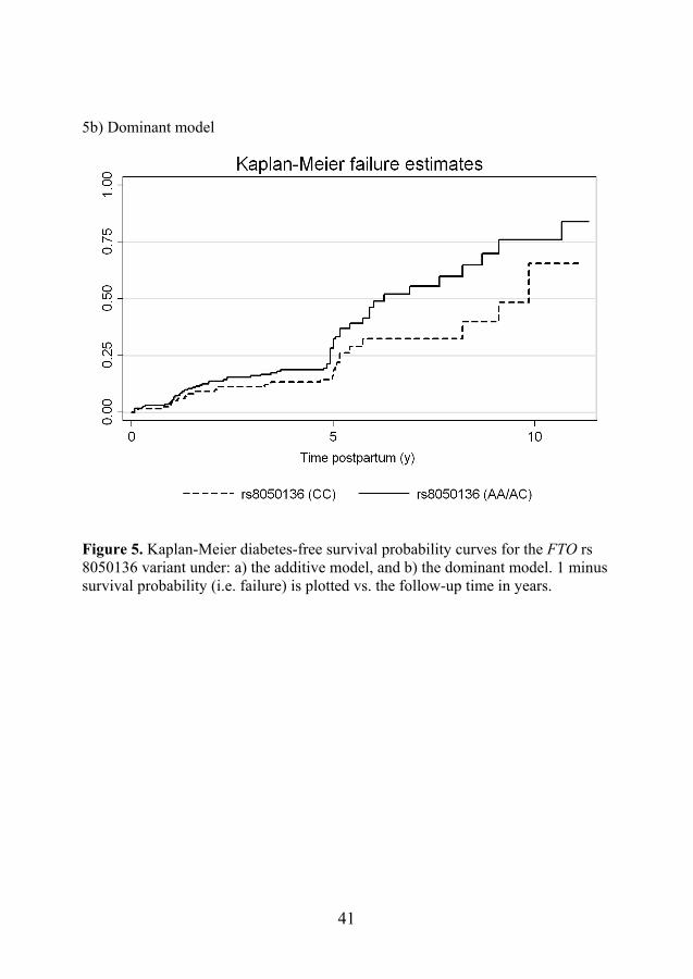
41
5b) Dominant model
Figure 5. Kaplan-Meier diabetes-free survival probability curves for the FTO rs 8050136 variant under: a) the additive model, and b) the dominant model. 1 minus survival probability (i.e. failure) is plotted vs. the follow-up time in years.

42
Table 7 The effect of the genetic variants studied on the risk of developing postpartum diabetes.
SNP Dominant model Additive model
HR (95% CI) for development of
postpartum diabetes
p-value HR (95% CI) for development of
postpartum diabetes
p−value
CDKAL1 rs7754840
0.94 (0.63–1.40) 0.77 0.98 (0.75–1.30) 0.91
CDKAL1 rs7756992
0.94 (0.64-1.40) 0.77 0.94 (0.72-1.24) 0.68
CDKN2A/2B rs10811661
0.55 (0.08-4.02) 0.56 1.14 (0.70-1.86) 0.60
HHEX rs1111875
1.07 (0.61-1.88) 0.80 1.26 (0.94-1.68) 0.12
IGF2BP2 rs1470579
1.16 (0.77-1.74) 0.48 1.27 (0.94-1.71) 0.12
IGF2BP2 rs4402960
1.02 (0.67-1.53) 0.94 1.14 (0.84-1.55) 0.41
TCF7L2 rs7903146
1.42 (0.95-2.14) 0.09 1.23 (0.94-1.62) 0.13
PPARG rs1801282
0.51 (0.12-2.10) 0.35 0.92 (0.58-1.47) 0.74
SLC30A8 rs13266634
0.75 (0.38-1.51) 0.43 1.16 (0.82-1.63) 0.40
GCK rs1799884
1.36 (0.90-2.07) 0.15 1.25 (0.90-1.74) 0.19
HNF1A rs1169288
1.04 (0.67-1.60) 0.86 0.99 (0.75-1.32) 0.97
KCNJ11 rs5219
0.95 (0.62-1.44) 0.81 1.09 (0.81-1.45) 0.58
FTO rs8050136
1.77 (1.14-2.76) 0.01 (Pc=0.14)
1.53 (1.15-2.03) 0.003 (Pc=0.039)
43
9. DISCUSSION
9.1 Prevalence of diabetes after GDM The prevalence of diabetes 5 years postpartum was 30% in the study presented in Paper III, which is in line with findings by other groups. It is, however, important to bear in mind that the prevalence of diabetes after GDM is closely related to the cut-off value applied for the diagnosis of GDM. A lower cut-off value includes those who do not exhibit major disturbance in glucose metabolism, thus, the prevalence of manifest diabetes postpartum can be assumed to be lower. Ethnic factors are also of great importance in the prevalence of postpartum diabetes. The prevalence of GDM and type 2 diabetes are correlated in a given population, and the progression to diabetes following GDM appears to be more rapid among ethnic groups with a high background prevalence of type 2 diabetes [12, 13].
9.2 Prevention and prediction of diabetes after GDM
To date, a number of prospective studies have indicated that it is possible to lower the incidence, or delay the onset, of diabetes in individuals at high risk of developing the disease by changing their lifestyle and/or pharmacological treatment. Changes in lifestyle have been found to be effective in preventing diabetes in patients with IGT in several previous studies [180-183]. Other studies have shown that pharmacological treatment with troglitazone or pioglitazone have diabetes-preventing or diabetes-delaying effects in women with previous GDM [184-188]. In the Troglitazone in Prevention of Diabetes (TRIPOD) study, treatment with troglitazone reduced the incidence of diabetes by 55% in women with prior GDM [188]. Beneficial effects were also found in the Diabetes Prevention Program (DPP), where both lifestyle changes and treatment with metformin were found to be highly effective in delaying or preventing diabetes in women with IGT and a history of GDM. Metformin was also found to be more effective in women with a history of GDM than in those without [189].

44
Women with GDM represent a high-risk group, showing signs of diabetes in evolution already at childbearing age. They are thus an ideal group for primary diabetes prevention, which should preferably be introduced while they are still pregnant, highly motivated and under close medical care. Recommendations concerning increased physical activity, maintenance of a normal weight and healthy diet should be included. The risk of developing diabetes following GDM is high. However, not every woman with GDM develops diabetes. The ability to prospectively identify women with GDM who are at the highest risk of developing diabetes would be important from a preventive point of view. Genetic, as well as clinical and biochemical, variables have therefore been examined in GDM patients. It would be particularly interesting if markers for postpartum diabetes could be easily determined during pregnancy, at a reasonable cost. In the study presented in Paper III, it was concluded that fasting blood glucose ≥5.2 mmol/l and HbA1c ≥4.7% according to the Mono S-method (equivalent to ≥5.7% according to the method by the National Glycohemoglobin Standardization Program) are associated with a 4- to 6-fold increase in the risk of developing diabetes within 5 years after GDM. High fasting glucose level has been extensively studied as a predictor of diabetes after GDM. In a systematic review by Kim et al., an elevated fasting glucose level during pregnancy was found to be the risk factor most commonly associated with future risk of type 2 diabetes, in addition to measures of pancreatic beta cell function [131]. Schaefer-Graf et al. found that the highest fasting plasma glucose level during pregnancy was the best predictor of diabetes in early puerperium [190]. Similarly, Metzger et al. reported that in Chicago approximately three-quarters of women with GDM developed diabetes within 6 months of delivery, when their fasting plasma glucose level during pregnancy was >7.2 mmol/l, compared with 10% of women who had a fasting glucose level <5.8 mmol/l [191]. In the present study, a fasting blood glucose concentration ≥5.2 mmol/l, corresponding to a plasma glucose level of 5.8 mmol/l [172], was associated with a 6-fold increase in the risk of postpartum diabetes. To the best of the author’s knowledge, this is the first report of HbA1c as a possible predictor of diabetes after GDM. This finding is supported by a recent study by Kwong et al., where postpartum hyperglycaemia was also significantly associated with higher HbA1c [192]. HbA1c has long been a valuable test in monitoring the effects of treatment of diabetes, but its use has previously not been advocated for diagnostic purposes due to a lack of standardization of its measurement. These problems have, however, now been addressed, and programmes for the standardization of HbA1c assays have been established [193].

45
9.3 GDM and genetics GDM often represents diabetes in evolution, which provides an excellent opportunity to study the pathogenic mechanisms underlying the development of diabetes. A genetic component of GDM is strongly suggested, and GDM shares genetic features with type 1 diabetes, type 2 diabetes and MODY [14, 57]. The studies presented here have been performed over several years. In the study described in Paper I, which was carried out in 2002, an association between GDM and MODY was demonstrated. Approximately 5% of the studied GDM subjects with a family history of diabetes carried a MODY mutation, in line with other reports [118-120]. No correlations between GDM and autoimmunity or high-risk HLA genotypes were found in this study, which, however, was quite small. It is not surprising to find correlations between GDM and MODY since beta cell dysfunction characterizes both conditions. Such correlations have also been found by others. Mutations in the GCK gene (MODY 2) have been studied by Ellard et al., who detected these mutations in 80% of GDM patients selected on the basis of clinical phenotype. They therefore suggested that patients with GDM who have a persistently elevated fasting glucose level, but show a small increment during OGTT, require insulin in pregnancy and have a first-degree relative with mild hyperglycaemia, should be considered for mutation analysis of the GCK gene [194]. In the study presented in Paper II Scandinavian and Arabian women were tested and compared regarding variations in genes affecting insulin secretion and action. An association was found between GAD65Abs and HLA-DQB1 risk genotypes in Scandinavian women, indicating that GDM might share some genetic features with type 1 diabetes. Arabian women with GDM were more insulin resistant than Scandinavian women with the same BMI. The findings in the studies presented in Paper II, as well as Paper IV, support previous evidence that pathogenic mechan-isms may differ between different ethnic groups. The primary aim of the study presented in Paper IV was to investigate whether genetic variants that predispose individuals to type 2 diabetes could predict the development of diabetes postpartum in women with GDM. The key finding was that the FTO rs8050136 variant is associated with an increased risk of developing postpartum diabetes. Adjusting for BMI clearly attenuated the effect of the FTO variant on the risk of postpartum diabetes, suggesting that most of its effect is mediated through its effect on BMI. The effect was also mainly attributed to non-European ethnicity. These results are supported by previous studies showing that the common FTO rs9939609 variant increased the risk of diabetes through an effect on BMI [195], and that FTO rs8050136 polymorphism is associated with

46
measures of obesity [196]. However, in a meta-analysis in an Asian population the A-allele of rs805136 was found to be associated with an increased risk of type 2 diabetes, independent of measures of BMI and obesity [197]. It is interesting to note that the rs9939609 variant is in strong linkage disequilibrium with the rs8050136 and rs7193144 variants in the FTO gene [198]. The finding that the FTO rs8050136 variant increases the risk of postpartum diabetes is not surprising, since most women with GDM are obese, which is also the case in type 2 diabetes. Interestingly, a recent study has shown that the FTO rs8050136 variant is associated with obese type 2 diabetes, whereas the TCF7L2 rs7901695 variant was found to be the predominant genetic risk factor in non-obese type 2 diabetes subjects [199]. A Japanese study has shown that the FTO rs1121980 variant was strongly associated with the plasma immunoreactive insulin level and HOMA-IR, even after adjusting for age, gender and BMI. Moreover, a haplotype of three SNPs, rs9939609, rs1121980 and rs1558902 in the FTO gene, has also been reported to be significantly associated with plasma immunoreactive insulin and HOMA-IR [200]. The mechanism by which FTO polymorphism increases the risk of postpartum diabetes is thus thought to be its effect on BMI and insulin resistance. This is consistent with the pathogenesis of GDM and type 2 diabetes, where insulin resistance is one of the predominant features of both disorders [12]. Only a few studies have been conducted to specifically investigate the effect of FTO variants in women with GDM. Lauenborg et al. examined 11 established type 2 diabetes susceptibility variants in women with GDM, and showed that the CDKN2A/2B rs10811661 and the WFS1 rs10010131 polymorphisms were associated with incident type 2 diabetes up to 2 years postpartum [109]. However, their study revealed no effect of the FTO rs9939609 variant, either on the risk of GDM or postpartum diabetes. The short follow-up period (up to 2 years) could explain the discrepancy between their study and the present one. Interestingly, the effect of the FTO rs8050136 polymorphism on the risk of postpartum diabetes in our study was mainly attributed to non-European ethnicity. This is in agreement with previous observations that non-European ethnicity is a risk factor for GDM, as well as for diabetes [12]. Further examination of other genetic variants will hopefully increase our understanding of GDM. Genetic testing may become an appropriate approach to identifying women at high risk of developing postpartum diabetes after GDM.

47
10. CONCLUSIONS
The conclusions drawn from the results of this work are summarized below.
• The prevalence of MODY mutations in the study population was 5% among women with GDM and a family history of diabetes.
• Arabian women were more insulin resistant, than Scandinavian women
with the same BMI, and exhibited impaired beta cell compensation for their degree of insulin resistance.
• In Scandinavian women, GDM may have genetic features in common with
type 1 diabetes.
• The prevalence of manifest diabetes 5 years postpartum in women with GDM was 30%.
• Fasting glucose level and HbA1c are possible predictors of diabetes
following GDM. These tests are easy to perform during pregnancy and relatively cheap. They could help identify women at particularly high risk of developing diabetes postpartum. Counselling and education regarding the risk of future diabetes and treatment could then be initiated during pregnancy in high-risk individuals.
• Variants of the FTO gene were associated with an increased risk of
developing postpartum diabetes, probably due to its effect of increasing obesity. Thus, genetic testing could be an appropriate means of identifying pregnant women at high risk of developing postpartum diabetes.

48
49
REFERENCES
1. Bennewitz, H., De diabete mellito, graviditatis symptomate, in University of Berlin. 1824. 2. Duncan, M., On puerperal diabetes. Trans Obstet Soc London, 1882. 24: p. 256-285. 3. Miller, H., The effect of the prediabetic state on the survival of the fetus and the
birthweight of the newborn infant. N Engl J Med, 1945. 233: p. 376-8. 4. D Hurwitz, D.J., Carbohydrate metabolism in normal pregnancy. N Engl J Med, 1946.
234: p. 327-9. 5. Gilbert, J.A. and D.M. Dunlop, Diabetic fertility, maternal mortality, and foetal loss rate.
Br Med J, 1949. 1(4592): p. 48-51. 6. Jackson, W.P., Studies in pre-diabetes. Br Med J, 1952. 2(4786): p. 690-6. 7. Wilkerson, H.L. and Q.R. Remein, Studies of abnormal carbohydrate metabolism in
pregnancy; the significance of impaired glucose tolerance. Diabetes, 1957. 6(4): p. 324-9. 8. O'Sullivan, J.B., Gestational diabetes. Unsuspected, asymptomatic diabetes in pregnancy.
N Engl J Med, 1961. 264: p. 1082-5. 9. Freinkel, N., Josimovich J, Conference Planning Committee, American Diabetes
Association Workshop-Conference on Gestational Diabetes. Summary and recommendations. Diabetes Care, 1980. 3: p. 499-501.
10. Metzger, B.E., Summary and recommendations of the Third International Workshop-Conference on Gestational Diabetes Mellitus. Diabetes, 1991. 40 Suppl 2: p. 197-201.
11. Anderberg, E., et al., A simplified oral glucose tolerance test in pregnancy: compliance and results. Acta Obstet Gynecol Scand, 2007. 86(12): p. 1432-6.
12. Ben-Haroush, A., Y. Yogev, and M. Hod, Epidemiology of gestational diabetes mellitus and its association with Type 2 diabetes. Diabet Med, 2004. 21(2): p. 103-13.
13. Hunt, K.J. and K.L. Schuller, The increasing prevalence of diabetes in pregnancy. Obstet Gynecol Clin North Am, 2007. 34(2): p. 173-99, vii.
14. Shaat, N. and L. Groop, Genetics of gestational diabetes mellitus. Curr Med Chem, 2007. 14(5): p. 569-83.
15. Hadden, D.R., Geographic, ethnic, and racial variations in the incidence of gestational diabetes mellitus. Diabetes, 1985. 34 Suppl 2: p. 8-12.
16. Agarwal, M.M., et al., Gestational diabetes screening of a multiethnic, high-risk population using glycated proteins. Diabetes Res Clin Pract, 2001. 51(1): p. 67-73.
17. Yue, D.K., et al., Why does ethnicity affect prevalence of gestational diabetes? The underwater volcano theory. Diabet Med, 1996. 13(8): p. 748-52.
18. Dornhorst, A., et al., High prevalence of gestational diabetes in women from ethnic minority groups. Diabet Med, 1992. 9(9): p. 820-5.
19. Acosta, I., et al., Prevalence of diabetes mellitus among pregnant women receiving health services at the Puerto Rico University Hospital, Puerto Rico, 1997-1998. P R Health Sci J, 2001. 20(2): p. 165-70.
20. Dong, Z.G., et al., Value of early glucose tolerance testing in women who had gestational diabetes in their previous pregnancy. Aust N Z J Obstet Gynaecol, 1993. 33(4): p. 350-7.
21. Foster-Powell, K.A. and N.W. Cheung, Recurrence of gestational diabetes. Aust N Z J Obstet Gynaecol, 1998. 38(4): p. 384-7.
50
22. Gaudier, F.L., et al., Recurrence of gestational diabetes mellitus. Obstet Gynecol, 1992. 80(5): p. 755-8.
23. MacNeill, S., et al., Rates and risk factors for recurrence of gestational diabetes. Diabetes Care, 2001. 24(4): p. 659-62.
24. Major, C.A., et al., Recurrence of gestational diabetes: who is at risk? Am J Obstet Gynecol, 1998. 179(4): p. 1038-42.
25. Nohira, T., et al., Recurrence of gestational diabetes mellitus: rates and risk factors from initial GDM and one abnormal GTT value. Diabetes Res Clin Pract, 2006. 71(1): p. 75-81.
26. Philipson, E.H. and D.M. Super, Gestational diabetes mellitus: does it recur in subsequent pregnancy? Am J Obstet Gynecol, 1989. 160(6): p. 1324-9; discussion 1329-31.
27. Spong, C.Y., et al., Recurrence of gestational diabetes mellitus: identification of risk factors. Am J Perinatol, 1998. 15(1): p. 29-33.
28. Barbour, L.A., et al., Cellular mechanisms for insulin resistance in normal pregnancy and gestational diabetes. Diabetes Care, 2007. 30 Suppl 2: p. S112-9.
29. Kuhl, C., Glucose metabolism during and after pregnancy in normal and gestational diabetic women. 1. Influence of normal pregnancy on serum glucose and insulin concentration during basal fasting conditions and after a challenge with glucose. Acta Endocrinol (Copenh), 1975. 79(4): p. 709-19.
30. Buchanan, T.A., et al., Insulin sensitivity and B-cell responsiveness to glucose during late pregnancy in lean and moderately obese women with normal glucose tolerance or mild gestational diabetes. Am J Obstet Gynecol, 1990. 162(4): p. 1008-14.
31. Catalano, P.M., et al., Longitudinal changes in glucose metabolism during pregnancy in obese women with normal glucose tolerance and gestational diabetes mellitus. Am J Obstet Gynecol, 1999. 180(4): p. 903-16.
32. Catalano, P.M., et al., Longitudinal changes in insulin release and insulin resistance in nonobese pregnant women. Am J Obstet Gynecol, 1991. 165(6 Pt 1): p. 1667-72.
33. Catalano, P.M., et al., Carbohydrate metabolism during pregnancy in control subjects and women with gestational diabetes. Am J Physiol, 1993. 264(1 Pt 1): p. E60-7.
34. Catalano, P.M., Carbohydrate metabolism and gestational diabetes. Clin Obstet Gynecol, 1994. 37(1): p. 25-38.
35. Ryan, E.A., M.J. O'Sullivan, and J.S. Skyler, Insulin action during pregnancy. Studies with the euglycemic clamp technique. Diabetes, 1985. 34(4): p. 380-9.
36. Kuhl, C., Insulin secretion and insulin resistance in pregnancy and GDM. Implications for diagnosis and management. Diabetes, 1991. 40 Suppl 2: p. 18-24.
37. Fasching, P., et al., [Monitoring daily insulin needs--an important follow-up parameter in late pregnancy in diabetic mothers?]. Geburtshilfe Frauenheilkd, 1992. 52(10): p. 596-601.
38. Rizza, R.A., L.J. Mandarino, and J.E. Gerich, Cortisol-induced insulin resistance in man: impaired suppression of glucose production and stimulation of glucose utilization due to a postreceptor detect of insulin action. J Clin Endocrinol Metab, 1982. 54(1): p. 131-8.
39. Metzger, B.E., et al., Summary and recommendations of the Fifth International Workshop-Conference on Gestational Diabetes Mellitus. Diabetes Care, 2007. 30 Suppl 2: p. S251-60.
40. Handwerger, S. and M. Freemark, The roles of placental growth hormone and placental lactogen in the regulation of human fetal growth and development. J Pediatr Endocrinol Metab, 2000. 13(4): p. 343-56.
41. Brelje, T.C., et al., Effect of homologous placental lactogens, prolactins, and growth hormones on islet B-cell division and insulin secretion in rat, mouse, and human islets: implication for placental lactogen regulation of islet function during pregnancy. Endocrinology, 1993. 132(2): p. 879-87.
42. Winzer, C., et al., Plasma adiponectin, insulin sensitivity, and subclinical inflammation in women with prior gestational diabetes mellitus. Diabetes Care, 2004. 27(7): p. 1721-7.
43. Heitritter, S.M., et al., Subclinical inflammation and vascular dysfunction in women with previous gestational diabetes mellitus. J Clin Endocrinol Metab, 2005. 90(7): p. 3983-8.
51
44. Bruun, J.M., et al., Regulation of adiponectin by adipose tissue-derived cytokines: in vivo and in vitro investigations in humans. Am J Physiol Endocrinol Metab, 2003. 285(3): p. E527-33.
45. Fasshauer, M., et al., Adiponectin gene expression and secretion is inhibited by interleukin-6 in 3T3-L1 adipocytes. Biochem Biophys Res Commun, 2003. 301(4): p. 1045-50.
46. Damm, P., et al., A longitudinal study of plasma insulin and glucagon in women with previous gestational diabetes. Diabetes Care, 1995. 18(5): p. 654-65.
47. Swinn, R.A., et al., Excessive secretion of insulin precursors characterizes and predicts gestational diabetes. Diabetes, 1995. 44(8): p. 911-5.
48. Xiang, A.H., et al., Multiple metabolic defects during late pregnancy in women at high risk for type 2 diabetes. Diabetes, 1999. 48(4): p. 848-54.
49. Brunzell, J.D., et al., Relationships between fasting plasma glucose levels and insulin secretion during intravenous glucose tolerance tests. J Clin Endocrinol Metab, 1976. 42(2): p. 222-9.
50. Kuhl, C. and J.J. Holst, Plasma glucagon and the insulin:glucagon ratio in gestational diabetes. Diabetes, 1976. 25(1): p. 16-23.
51. Buchanan, T.A., et al., What is gestational diabetes? Diabetes Care, 2007. 30 Suppl 2: p. S105-11.
52. Hattersley, A.T., et al., Linkage of type 2 diabetes to the glucokinase gene. Lancet, 1992. 339(8805): p. 1307-10.
53. Kieffer, E.C., et al., Obesity and gestational diabetes among African-American women and Latinas in Detroit: implications for disparities in women's health. J Am Med Womens Assoc, 2001. 56(4): p. 181-7, 196.
54. Solomon, C.G., et al., A prospective study of pregravid determinants of gestational diabetes mellitus. JAMA, 1997. 278(13): p. 1078-83.
55. Williams, M.A., et al., Familial aggregation of type 2 diabetes and chronic hypertension in women with gestational diabetes mellitus. J Reprod Med, 2003. 48(12): p. 955-62.
56. Martin, A.O., et al., Frequency of diabetes mellitus in mothers of probands with gestational diabetes: possible maternal influence on the predisposition to gestational diabetes. Am J Obstet Gynecol, 1985. 151(4): p. 471-5.
57. Watanabe, R.M., et al., Genetics of gestational diabetes mellitus and type 2 diabetes. Diabetes Care, 2007. 30 Suppl 2: p. S134-40.
58. Litou, H., et al., Increased prevalence of VNTR III of the insulin gene in women with gestational diabetes mellitus (GDM). Diabetes Res Clin Pract, 2007. 76(2): p. 223-8.
59. Rissanen, J., et al., Sulfonylurea receptor 1 gene variants are associated with gestational diabetes and type 2 diabetes but not with altered secretion of insulin. Diabetes Care, 2000. 23(1): p. 70-3.
60. Ober, C., et al., Increased risk for gestational diabetes mellitus associated with insulin receptor and insulin-like growth factor II restriction fragment length polymorphisms. Genet Epidemiol, 1989. 6(5): p. 559-69.
61. Kaufmann, R.C., et al., An animal model of gestational diabetes. Am J Obstet Gynecol, 1981. 141(5): p. 479-82.
62. Krzyzanowska, K., et al., Increased visfatin concentrations in women with gestational diabetes mellitus. Clin Sci (Lond), 2006. 110(5): p. 605-9.
63. Chan, T.F., et al., Decreased plasma visfatin concentrations in women with gestational diabetes mellitus. J Soc Gynecol Investig, 2006. 13(5): p. 364-7.
64. Megia, A., et al., Mannose-binding lectin gene polymorphisms are associated with gestational diabetes mellitus. J Clin Endocrinol Metab, 2004. 89(10): p. 5081-7.
65. Shao, J., et al., Decreased insulin receptor tyrosine kinase activity and plasma cell membrane glycoprotein-1 overexpression in skeletal muscle from obese women with gestational diabetes mellitus (GDM): evidence for increased serine/threonine phosphorylation in pregnancy and GDM. Diabetes, 2000. 49(4): p. 603-10.
52
66. Shaat, N., et al., Association of the E23K polymorphism in the KCNJ11 gene with gestational diabetes mellitus. Diabetologia, 2005. 48(12): p. 2544-51.
67. Fallucca, F., et al., Polymorphisms of insulin receptor substrate 1 and beta3-adrenergic receptor genes in gestational diabetes and normal pregnancy. Metabolism, 2006. 55(11): p. 1451-6.
68. Tok, E.C., et al., Association of insulin receptor substrate-1 G972R variant with baseline characteristics of the patients with gestational diabetes mellitus. Am J Obstet Gynecol, 2006. 194(3): p. 868-72.
69. Garvey, W.T., et al., Multiple defects in the adipocyte glucose transport system cause cellular insulin resistance in gestational diabetes. Heterogeneity in the number and a novel abnormality in subcellular localization of GLUT4 glucose transporters. Diabetes, 1993. 42(12): p. 1773-85.
70. Cseh, K., et al., Plasma adiponectin and pregnancy-induced insulin resistance. Diabetes Care, 2004. 27(1): p. 274-5.
71. Williams, M.A., et al., Plasma adiponectin concentrations in early pregnancy and subsequent risk of gestational diabetes mellitus. J Clin Endocrinol Metab, 2004. 89(5): p. 2306-11.
72. Tsai, P.J., et al., Maternal plasma adiponectin concentrations at 24 to 31 weeks of gestation: negative association with gestational diabetes mellitus. Nutrition, 2005. 21(11-12): p. 1095-9.
73. Ranheim, T., et al., Adiponectin is reduced in gestational diabetes mellitus in normal weight women. Acta Obstet Gynecol Scand, 2004. 83(4): p. 341-7.
74. Leipold, H., et al., Calpain-10 haplotype combination and association with gestational diabetes mellitus. Obstet Gynecol, 2004. 103(6): p. 1235-40.
75. Cauza, E., et al., Increased C282Y heterozygosity in gestational diabetes. Fetal Diagn Ther, 2005. 20(5): p. 349-54.
76. Festa, A., et al., Trp64Arg polymorphism of the beta3-adrenergic receptor gene in pregnancy: association with mild gestational diabetes mellitus. J Clin Endocrinol Metab, 1999. 84(5): p. 1695-9.
77. Alevizaki, M., et al., Study of the Trp64Arg polymorphism of the beta3-adrenergic receptor in Greek women with gestational diabetes. Diabetes Care, 2000. 23(8): p. 1079-83.
78. Tsai, P.J., et al., Lack of relationship between beta3-adrenergic receptor gene polymorphism and gestational diabetes mellitus in a Taiwanese population. Metabolism, 2004. 53(9): p. 1136-9.
79. Chen, Y., et al., Mitochondrial gene mutations in gestational diabetes mellitus. Diabetes Res Clin Pract, 2000. 48(1): p. 29-35.
80. Yanagisawa, K., et al., Mutation in the mitochondrial tRNA(leu) at position 3243 and spontaneous abortions in Japanese women attending a clinic for diabetic pregnancies. Diabetologia, 1995. 38(7): p. 809-15.
81. Barbosa, J., et al., Analysis of linkage between the major histocompatibility system and juvenile, insulin-dependent diabetes in multiplex families. Reanalysis of data. J Clin Invest, 1978. 62(2): p. 492-5.
82. Trucco, M. and J.S. Dorman, Immunogenetics of insulin-dependent diabetes mellitus in humans. Crit Rev Immunol, 1989. 9(3): p. 201-45.
83. Noble, J.A., et al., The role of HLA class II genes in insulin-dependent diabetes mellitus: molecular analysis of 180 Caucasian, multiplex families. Am J Hum Genet, 1996. 59(5): p. 1134-48.
84. Nerup, J., et al., HLA, islet cell antibodies, and types of diabetes mellitus. Diabetes, 1978. 27 Suppl 1: p. 247-50.
85. Cudworth, A.G. and J.C. Woodrow, Genetic susceptibility in diabetes mellitus: analysis of the HLA association. Br Med J, 1976. 2(6040): p. 846-8.

53
86. Ferber, K.M., et al., Predictive value of human leukocyte antigen class II typing for the development of islet autoantibodies and insulin-dependent diabetes postpartum in women with gestational diabetes. J Clin Endocrinol Metab, 1999. 84(7): p. 2342-8.
87. Kuhl, C., Etiology and pathogenesis of gestational diabetes. Diabetes Care, 1998. 21 Suppl 2: p. B19-26.
88. Vambergue, A., et al., Gestational diabetes mellitus and HLA class II (-DQ, -DR) association: The Digest Study. Eur J Immunogenet, 1997. 24(5): p. 385-94.
89. Freinkel, N., et al., Gestational diabetes mellitus. Heterogeneity of maternal age, weight, insulin secretion, HLA antigens, and islet cell antibodies and the impact of maternal metabolism on pancreatic B-cell and somatic development in the offspring. Diabetes, 1985. 34 Suppl 2: p. 1-7.
90. Rubinstein, P., et al., HLA antigens and islet cell antibodies in gestational diabetes. Hum Immunol, 1981. 3(3): p. 271-5.
91. Torn, C., et al., Different HLA-DR-DQ and MHC class I chain-related gene A (MICA) genotypes in autoimmune and nonautoimmune gestational diabetes in a Swedish population. Hum Immunol, 2004. 65(12): p. 1443-50.
92. Papadopoulou, A., et al., The type 1 diabetes protective HLA DQB1*0602 allele is less frequent in gestational diabetes mellitus. Diabetologia, 2009. 52(7): p. 1339-42.
93. Ridderstrale, M. and L. Groop, Genetic dissection of type 2 diabetes. Mol Cell Endocrinol, 2009. 297(1-2): p. 10-7.
94. Groop, L. and V. Lyssenko, Genetic basis of beta-cell dysfunction in man. Diabetes Obes Metab, 2009. 11 Suppl 4: p. 149-58.
95. Scott, L.J., et al., A genome-wide association study of type 2 diabetes in Finns detects multiple susceptibility variants. Science, 2007. 316(5829): p. 1341-5.
96. Lyssenko, V. and L. Groop, Genome-wide association study for type 2 diabetes: clinical applications. Curr Opin Lipidol, 2009. 20(2): p. 87-91.
97. Lyssenko, V., et al., Clinical risk factors, DNA variants, and the development of type 2 diabetes. N Engl J Med, 2008. 359(21): p. 2220-32.
98. Saxena, R., et al., Genome-wide association analysis identifies loci for type 2 diabetes and triglyceride levels. Science, 2007. 316(5829): p. 1331-6.
99. Zeggini, E., et al., Meta-analysis of genome-wide association data and large-scale replication identifies additional susceptibility loci for type 2 diabetes. Nat Genet, 2008. 40(5): p. 638-45.
100. Zeggini, E., et al., Replication of genome-wide association signals in UK samples reveals risk loci for type 2 diabetes. Science, 2007. 316(5829): p. 1336-41.
101. Lyssenko, V., et al., Common variant in MTNR1B associated with increased risk of type 2 diabetes and impaired early insulin secretion. Nat Genet, 2009. 41(1): p. 82-8.
102. Prokopenko, I., et al., Variants in MTNR1B influence fasting glucose levels. Nat Genet, 2009. 41(1): p. 77-81.
103. Kathiresan, S., et al., Polymorphisms associated with cholesterol and risk of cardiovascular events. N Engl J Med, 2008. 358(12): p. 1240-9.
104. Zimmet, P., K.G. Alberti, and J. Shaw, Global and societal implications of the diabetes epidemic. Nature, 2001. 414(6865): p. 782-7.
105. Bellamy, L., et al., Type 2 diabetes mellitus after gestational diabetes: a systematic review and meta-analysis. Lancet, 2009. 373(9677): p. 1773-9.
106. Watanabe, R.M., et al., Transcription factor 7-like 2 (TCF7L2) is associated with gestational diabetes mellitus and interacts with adiposity to alter insulin secretion in Mexican Americans. Diabetes, 2007. 56(5): p. 1481-5.
107. Cho, Y.M., et al., Type 2 diabetes-associated genetic variants discovered in the recent genome-wide association studies are related to gestational diabetes mellitus in the Korean population. Diabetologia, 2009. 52(2): p. 253-61.
108. Shaat, N., et al., A variant in the transcription factor 7-like 2 (TCF7L2) gene is associated with an increased risk of gestational diabetes mellitus. Diabetologia, 2007. 50(5): p. 972-9.

54
109. Lauenborg, J., et al., Common type 2 diabetes risk gene variants associate with gestational diabetes. J Clin Endocrinol Metab, 2009. 94(1): p. 145-50.
110. Horikawa, Y., et al., Mutation in hepatocyte nuclear factor-1 beta gene (TCF2) associated with MODY. Nat Genet, 1997. 17(4): p. 384-5.
111. Fajans, S.S., G.I. Bell, and K.S. Polonsky, Molecular mechanisms and clinical pathophysiology of maturity-onset diabetes of the young. N Engl J Med, 2001. 345(13): p. 971-80.
112. Owen, K. and A.T. Hattersley, Maturity-onset diabetes of the young: from clinical description to molecular genetic characterization. Best Pract Res Clin Endocrinol Metab, 2001. 15(3): p. 309-23.
113. Shih, D.Q. and M. Stoffel, Molecular etiologies of MODY and other early-onset forms of diabetes. Curr Diab Rep, 2002. 2(2): p. 125-34.
114. Polonsky, K.S., Lilly Lecture 1994. The beta-cell in diabetes: from molecular genetics to clinical research. Diabetes, 1995. 44(6): p. 705-17.
115. Froguel, P., et al., Familial hyperglycemia due to mutations in glucokinase. Definition of a subtype of diabetes mellitus. N Engl J Med, 1993. 328(10): p. 697-702.
116. Frayling, T.M., et al., beta-cell genes and diabetes: molecular and clinical characterization of mutations in transcription factors. Diabetes, 2001. 50 Suppl 1: p. S94-100.
117. Pruhova, S., et al., Genetic epidemiology of MODY in the Czech republic: new mutations in the MODY genes HNF-4alpha, GCK and HNF-1alpha. Diabetologia, 2003. 46(2): p. 291-5.
118. Saker, P.J., et al., High prevalence of a missense mutation of the glucokinase gene in gestational diabetic patients due to a founder-effect in a local population. Diabetologia, 1996. 39(11): p. 1325-8.
119. Stoffel, M., et al., Identification of glucokinase mutations in subjects with gestational diabetes mellitus. Diabetes, 1993. 42(6): p. 937-40.
120. Zouali, H., et al., Linkage analysis and molecular scanning of glucokinase gene in NIDDM families. Diabetes, 1993. 42(9): p. 1238-45.
121. Shaat, N., et al., Common variants in MODY genes increase the risk of gestational diabetes mellitus. Diabetologia, 2006. 49(7): p. 1545-51.
122. Pedersen, J., Diabetes and pregnancy. Blood sugar of newborn infants. 1952. 123. Aberg, A., et al., Impaired glucose tolerance during pregnancy is associated with
increased fetal mortality in preceding sibs. Acta Obstet Gynecol Scand, 1997. 76(3): p. 212-7.
124. Aberg, A. and L. Westbom, Association between maternal pre-existing or gestational diabetes and health problems in children. Acta Paediatr, 2001. 90(7): p. 746-50.
125. Aberg, A., L. Westbom, and B. Kallen, Congenital malformations among infants whose mothers had gestational diabetes or preexisting diabetes. Early Hum Dev, 2001. 61(2): p. 85-95.
126. Langer, O., et al., Gestational diabetes: the consequences of not treating. Am J Obstet Gynecol, 2005. 192(4): p. 989-97.
127. Aberg, A., H. Rydhstroem, and A. Frid, Impaired glucose tolerance associated with adverse pregnancy outcome: a population-based study in southern Sweden. Am J Obstet Gynecol, 2001. 184(2): p. 77-83.
128. Group, H.S.C.R., et al., Hyperglycemia and adverse pregnancy outcomes.[see comment]. New England Journal of Medicine, 2008. 358(19): p. 1991-2002.
129. Crowther, C.A., et al., Effect of treatment of gestational diabetes mellitus on pregnancy outcomes. N Engl J Med, 2005. 352(24): p. 2477-86.
130. Landon, M.B., et al., A multicenter, randomized trial of treatment for mild gestational diabetes. N Engl J Med, 2009. 361(14): p. 1339-48.
131. Kim, C., K.M. Newton, and R.H. Knopp, Gestational diabetes and the incidence of type 2 diabetes: a systematic review. Diabetes Care, 2002. 25(10): p. 1862-8.

55
132. Aberg, A.E., et al., Predictive factors of developing diabetes mellitus in women with gestational diabetes. Acta Obstet Gynecol Scand, 2002. 81(1): p. 11-6.
133. Damm, P., Gestational diabetes mellitus and subsequent development of overt diabetes mellitus. Dan Med Bull, 1998. 45(5): p. 495-509.
134. Lobner, K., et al., Predictors of postpartum diabetes in women with gestational diabetes mellitus. Diabetes, 2006. 55(3): p. 792-7.
135. O'Sullivan, J.B., Diabetes mellitus after GDM. Diabetes, 1991. 40 Suppl 2: p. 131-5. 136. Dornhorst, A. and M. Rossi, Risk and prevention of type 2 diabetes in women with
gestational diabetes. Diabetes Care, 1998. 21 Suppl 2: p. B43-9. 137. Damm, P., et al., Predictive factors for the development of diabetes in women with
previous gestational diabetes mellitus. Am J Obstet Gynecol, 1992. 167(3): p. 607-16. 138. Kjos, S.L., et al., Predicting future diabetes in Latino women with gestational diabetes.
Utility of early postpartum glucose tolerance testing. Diabetes, 1995. 44(5): p. 586-91. 139. Kjos, S.L. and T.A. Buchanan, Gestational diabetes mellitus. N Engl J Med, 1999.
341(23): p. 1749-56. 140. Lauenborg, J., et al., The prevalence of the metabolic syndrome in a danish population of
women with previous gestational diabetes mellitus is three-fold higher than in the general population. J Clin Endocrinol Metab, 2005. 90(7): p. 4004-10.
141. Golden, S.H., et al., Antepartum glucose tolerance test results as predictors of type 2 diabetes mellitus in women with a history of gestational diabetes mellitus: a systematic review. Gend Med, 2009. 6 Suppl 1: p. 109-22.
142. Lauenborg, J., et al., Increasing incidence of diabetes after gestational diabetes: a long-term follow-up in a Danish population. Diabetes Care, 2004. 27(5): p. 1194-9.
143. Carr, D.B., et al., Gestational diabetes mellitus increases the risk of cardiovascular disease in women with a family history of type 2 diabetes. Diabetes Care, 2006. 29(9): p. 2078-83.
144. Yogev, Y., B.E. Metzger, and M. Hod, Establishing diagnosis of gestational diabetes mellitus: Impact of the hyperglycemia and adverse pregnancy outcome study. Seminars In Fetal & Neonatal Medicine, 2009. 14(2): p. 94-100.
145. O'Sullivan, J.B., et al., Screening criteria for high-risk gestational diabetic patients. Am J Obstet Gynecol, 1973. 116(7): p. 895-900.
146. Hanna, F.W. and J.R. Peters, Screening for gestational diabetes; past, present and future. Diabet Med, 2002. 19(5): p. 351-8.
147. Weeks, J.W., et al., Gestational diabetes: does the presence of risk factors influence perinatal outcome? Am J Obstet Gynecol, 1994. 171(4): p. 1003-7.
148. Ostlund, I. and U. Hanson, Occurrence of gestational diabetes mellitus and the value of different screening indicators for the oral glucose tolerance test. Acta Obstet Gynecol Scand, 2003. 82(2): p. 103-8.
149. Lind, T. and J. Anderson, Does random blood glucose sampling outdate testing for glycosuria in the detection of diabetes during pregnancy? Br Med J (Clin Res Ed), 1984. 289(6458): p. 1569-71.
150. Perucchini, D., et al., Using fasting plasma glucose concentrations to screen for gestational diabetes mellitus: prospective population based study. Bmj, 1999. 319(7213): p. 812-5.
151. Fadl, H., et al., Fasting capillary glucose as a screening test for gestational diabetes mellitus. Bjog, 2006. 113(9): p. 1067-71.
152. Stangenberg, M., B. Persson, and E. Nordlander, Random capillary blood glucose and conventional selection criteria for glucose tolerance testing during pregnancy. Diabetes Res, 1985. 2(1): p. 29-33.
153. Ostlund, I. and U. Hanson, Repeated random blood glucose measurements as universal screening test for gestational diabetes mellitus. Acta Obstet Gynecol Scand, 2004. 83(1): p. 46-51.

56
154. Berg, M., et al., Early random capillary glucose level screening and multidisciplinary antenatal teamwork to improve outcome in gestational diabetes mellitus. Acta Obstet Gynecol Scand, 2007. 86(3): p. 283-90.
155. O'Sullivan, J.B. and C.M. Mahan, Criteria for the Oral Glucose Tolerance Test in Pregnancy. Diabetes, 1964. 13: p. 278-85.
156. Carpenter, M.W. and D.R. Coustan, Criteria for screening tests for gestational diabetes. Am J Obstet Gynecol, 1982. 144(7): p. 768-73.
157. Gabbe, S.G., et al., Management and outcome of class A diabetes mellitus. Am J Obstet Gynecol, 1977. 127(5): p. 465-9.
158. O'Sullivan, J.B., et al., Gestational diabetes and perinatal mortality rate. Am J Obstet Gynecol, 1973. 116(7): p. 901-4.
159. Classification and diagnosis of diabetes mellitus and other categories of glucose intolerance. National Diabetes Data Group. Diabetes, 1979. 28(12): p. 1039-57.
160. Metzger, B.E. and D.R. Coustan, Summary and recommendations of the Fourth International Workshop-Conference on Gestational Diabetes Mellitus. The Organizing Committee. Diabetes Care, 1998. 21 Suppl 2: p. B161-7.
161. Sacks, D.A., et al., Toward universal criteria for gestational diabetes: the 75-gram glucose tolerance test in pregnancy. Am J Obstet Gynecol, 1995. 172(2 Pt 1): p. 607-14.
162. Landon, M.B., S.G. Gabbe, and L. Sachs, Management of diabetes mellitus and pregnancy: a survey of obstetricians and maternal-fetal specialists. Obstet Gynecol, 1990. 75(4): p. 635-40.
163. Lind, T. and P.R. Phillips, Influence of pregnancy on the 75-g OGTT. A prospective multicenter study. The Diabetic Pregnancy Study Group of the European Association for the Study of Diabetes. Diabetes, 1991. 40 Suppl 2: p. 8-13.
164. Jensen, D.M., et al., Proposed diagnostic thresholds for gestational diabetes mellitus according to a 75-g oral glucose tolerance test. Maternal and perinatal outcomes in 3260 Danish women. Diabet Med, 2003. 20(1): p. 51-7.
165. Nord, E., U. Hanson, and B. Persson, Blood glucose limits in the diagnosis of impaired glucose tolerance during pregnancy. Relation to morbidity. Acta Obstet Gynecol Scand, 1995. 74(8): p. 589-93.
166. Diabetes mellitus. Report of a WHO Study Group. World Health Organ Tech Rep Ser, 1985. 727: p. 1-113.
167. Definition, Diagnosis and Classification of Diabetes Mellitus and its Complications. Part 1: diagnosis and classification of diabetes mellitus Report of a WHO consultation. 1999, World Health Organization.
168. Metzger, B.E., et al., Hyperglycemia and adverse pregnancy outcomes. N Engl J Med, 2008. 358(19): p. 1991-2002.
169. Schytte, T., et al., The clinical impact of screening for gestational diabetes. Clin Chem Lab Med, 2004. 42(9): p. 1036-42.
170. Hakansson, A., et al., Antenatal care in southern Sweden. A population-based prospective study describing the diagnostic panorama of pregnancy. Acta Obstet Gynecol Scand, 1991. 70(7-8): p. 531-8.
171. Agardh, C.D., A. Aberg, and N.E. Norden, Glucose levels and insulin secretion during a 75 g glucose challenge test in normal pregnancy. J Intern Med, 1996. 240(5): p. 303-9.
172. Burnett, R.W., et al., IFCC recommendation on reporting results for blood glucose. Clin Chim Acta, 2001. 307(1-2): p. 205-9.
173. Matthews, D.R., et al., Homeostasis model assessment: insulin resistance and beta-cell function from fasting plasma glucose and insulin concentrations in man. Diabetologia, 1985. 28(7): p. 412-9.
174. Phillips, D.I., et al., Understanding oral glucose tolerance: comparison of glucose or insulin measurements during the oral glucose tolerance test with specific measurements of insulin resistance and insulin secretion. Diabet Med, 1994. 11(3): p. 286-92.
57
175. Jeppsson, J.O., et al., Measurement of hemoglobin A1c by a new liquid-chromatographic assay: methodology, clinical utility, and relation to glucose tolerance evaluated. Clin Chem, 1986. 32(10): p. 1867-72.
176. Friedewald, W.T., R.I. Levy, and D.S. Fredrickson, Estimation of the concentration of low-density lipoprotein cholesterol in plasma, without use of the preparative ultracentrifuge. Clinical Chemistry, 1972. 18(6): p. 499-502.
177. Tuomi, T., et al., Clinical and genetic characteristics of type 2 diabetes with and without GAD antibodies. Diabetes, 1999. 48(1): p. 150-7.
178. Klein, J.M., ML, Survival analysis: Techniques for censored and truncated data. 2003, Springer: New York. p. 536.
179. Purcell, S., S.S. Cherny, and P.C. Sham, Genetic Power Calculator: design of linkage and association genetic mapping studies of complex traits. Bioinformatics, 2003. 19(1): p. 149-50.
180. Eriksson, K.F. and F. Lindgarde, Prevention of type 2 (non-insulin-dependent) diabetes mellitus by diet and physical exercise. The 6-year Malmo feasibility study. Diabetologia, 1991. 34(12): p. 891-8.
181. Pan, X.R., et al., Effects of diet and exercise in preventing NIDDM in people with impaired glucose tolerance. The Da Qing IGT and Diabetes Study. Diabetes Care, 1997. 20(4): p. 537-44.
182. Tuomilehto, J., et al., Prevention of type 2 diabetes mellitus by changes in lifestyle among subjects with impaired glucose tolerance. N Engl J Med, 2001. 344(18): p. 1343-50.
183. Knowler, W.C., et al., Reduction in the incidence of type 2 diabetes with lifestyle intervention or metformin. N Engl J Med, 2002. 346(6): p. 393-403.
184. Berkowitz, K., et al., Effect of troglitazone on insulin sensitivity and pancreatic beta-cell function in women at high risk for NIDDM. Diabetes, 1996. 45(11): p. 1572-9.
185. Xiang, A.H., et al., Effect of pioglitazone on pancreatic beta-cell function and diabetes risk in Hispanic women with prior gestational diabetes. Diabetes, 2006. 55(2): p. 517-22.
186. Xiang, A.H., et al., Effect of pioglitazone on progression of subclinical atherosclerosis in non-diabetic premenopausal Hispanic women with prior gestational diabetes. Atherosclerosis, 2008. 199(1): p. 207-14.
187. Azen, S.P., et al., TRIPOD (TRoglitazone In the Prevention Of Diabetes): a randomized, placebo-controlled trial of troglitazone in women with prior gestational diabetes mellitus. Control Clin Trials, 1998. 19(2): p. 217-31.
188. Buchanan, T.A., et al., Preservation of pancreatic beta-cell function and prevention of type 2 diabetes by pharmacological treatment of insulin resistance in high-risk hispanic women. Diabetes, 2002. 51(9): p. 2796-803.
189. Ratner, R.E., et al., Prevention of diabetes in women with a history of gestational diabetes: effects of metformin and lifestyle interventions. J Clin Endocrinol Metab, 2008. 93(12): p. 4774-9.
190. Schaefer-Graf, U.M., et al., Clinical predictors for a high risk for the development of diabetes mellitus in the early puerperium in women with recent gestational diabetes mellitus. Am J Obstet Gynecol, 2002. 186(4): p. 751-6.
191. Metzger, B.E., et al., Prepregnancy weight and antepartum insulin secretion predict glucose tolerance five years after gestational diabetes mellitus. Diabetes Care, 1993. 16(12): p. 1598-605.
192. Kwong, S., et al., Postpartum diabetes screening: adherence rate and the performance of fasting plasma glucose versus oral glucose tolerance test. Diabetes Care, 2009. 32(12): p. 2242-4.
193. Hoelzel, W., et al., IFCC reference system for measurement of hemoglobin A1c in human blood and the national standardization schemes in the United States, Japan, and Sweden: a method-comparison study. Clinical Chemistry, 2004. 50(1): p. 166-74.
194. Ellard, S., et al., A high prevalence of glucokinase mutations in gestational diabetic subjects selected by clinical criteria. Diabetologia, 2000. 43(2): p. 250-3.

58
195. Frayling, T.M., et al., A common variant in the FTO gene is associated with body mass index and predisposes to childhood and adult obesity. Science, 2007. 316(5826): p. 889-94.
196. Pecioska, S., et al., Association between type 2 diabetes loci and measures of fatness. PLoS One. 5(1): p. e8541.
197. Liu, Y., et al., Meta-analysis Added Power to Identify Variants in FTO Associated With Type 2 Diabetes and Obesity in the Asian Population. Obesity. 2010: p. 7.
198. Andreasen, C.H., et al., Low physical activity accentuates the effect of the FTO rs9939609 polymorphism on body fat accumulation. Diabetes, 2008. 57(1): p. 95-101.
199. Timpson, N.J., et al., Adiposity-related heterogeneity in patterns of type 2 diabetes susceptibility observed in genome-wide association data. Diabetes, 2009. 58(2): p. 505-10.
200. Shimaoka, I., et al., Association of gene polymorphism of the fat-mass and obesity-associated gene with insulin resistance in Japanese. Hypertens Res. 2010: p. 15.