GENETIC AND BIOCHEMICAL ANALYSIS OF THE … AND BIOCHEMICAL ANALYSIS OF THE ENDORIBONUCLEASE E/G...
205
GENETIC AND BIOCHEMICAL ANALYSIS OF THE ENDORIBONUCLEASE E/G FAMILY IN RNA METABOLISM IN ESCHERICHIA COLI K-12 by DAE HWAN CHUNG (Under the Direction of Sidney R. Kushner) ABSTRACT Degradation of mRNA and maturation of stable RNAs provide important mechanisms for controlling gene expression at post-transcriptional level. In Escherichia coli, the RNase E/G endoribonuclease family plays a central role in the initiation of both processes. This dissertation research was an attempt to broaden our understanding of physiological roles and functional relationships of RNase E and RNase G by using a combination of genetic and biochemical analysis. The rng-219 and rng-248 alleles, comprising single amino acid substitutions within the predicted RNase H domain of RNase G, are able to support cell viability in the total absence of RNase E when present at physiologically relevant protein levels. These observations suggest that the difference in biological activities between the two enzymes is governed by their RNase H domains to some extent. The in vivo characterization of rne∆1018/rng-219 and rne∆1018/rng-248 double mutants allowed critical examination of the distinct physiological roles of RNase E and RNase G in E. coli RNA metabolism. The degradation of certain mRNAs and the processing of some tRNA precursors are absolutely dependent on RNase E activity. In contrast, 9S rRNA processing is effectively restored by the altered RNase G proteins in the absence of RNase E.
Transcript of GENETIC AND BIOCHEMICAL ANALYSIS OF THE … AND BIOCHEMICAL ANALYSIS OF THE ENDORIBONUCLEASE E/G...

GENETIC AND BIOCHEMICAL ANALYSIS OF THE ENDORIBONUCLEASE E/G FAMILY IN
RNA METABOLISM IN ESCHERICHIA COLI K-12
by
DAE HWAN CHUNG
(Under the Direction of Sidney R. Kushner)
ABSTRACT
Degradation of mRNA and maturation of stable RNAs provide important mechanisms for
controlling gene expression at post-transcriptional level. In Escherichia coli, the RNase E/G
endoribonuclease family plays a central role in the initiation of both processes. This dissertation
research was an attempt to broaden our understanding of physiological roles and functional
relationships of RNase E and RNase G by using a combination of genetic and biochemical
analysis.
The rng-219 and rng-248 alleles, comprising single amino acid substitutions within the
predicted RNase H domain of RNase G, are able to support cell viability in the total absence of
RNase E when present at physiologically relevant protein levels. These observations suggest
that the difference in biological activities between the two enzymes is governed by their RNase
H domains to some extent.
The in vivo characterization of rne∆1018/rng-219 and rne∆1018/rng-248 double mutants
allowed critical examination of the distinct physiological roles of RNase E and RNase G in E.
coli RNA metabolism. The degradation of certain mRNAs and the processing of some tRNA
precursors are absolutely dependent on RNase E activity. In contrast, 9S rRNA processing is
effectively restored by the altered RNase G proteins in the absence of RNase E.

We also examined the biochemical properties of purified RNase E, RNase G and Rng-
219 proteins. The purified RNase G and Rng-219 proteins cleave structured RNA substrates,
such as 9S rRNA and tRNAs, at identical sites as RNase E. Although, both RNase E/G prefer
RNA substrates with 5’-monophosphate termini, the presence of a 5’-triphosphate affects the
efficiency of RNase E much more than RNase G. A surprising result is the greater catalytic
activity of RNase G and Rng-219 proteins in the presence of Mn2+ than Mg2+.
INDEX WORDS: RNase E, RNase G, mRNA degradation, 9S rRNA processing,
tRNA precursor, Escherichia coli

GENETIC AND BIOCHEMICAL ANALYSIS OF THE ENDORIBONUCLEASE E/G FAMILY IN
RNA METABOLISM IN ESCHERICHIA COLI K-12
by
DAE HWAN CHUNG
B. S., Ajou University, SOUTH KOREA, 1997
M. S., University of Philippines, Philippines, 1999
A Dissertation Submitted to the Graduate Faculty of The University of Georgia in Partial
Fulfillment of the Requirements for the Degree
DOCTOR OF PHILOSOPHY
ATHENS, GEORGIA
2008

© 2008
DAE HWAN CHUNG
All Rights Reserved

GENETIC AND BIOCHEMICAL ANALYSIS OF THE ENDORIBONUCLEASE E/G FAMILY IN
RNA METABOLISM IN ESCHERICHIA COLI K-12
by
DAE HWAN CHUNG
Major Professor: Sidney R. Kushner
Committee: Anna Glasgow Karls Michael McEachern Michael Terns Richard Meagher
Electronic Version Approved: Maureen Grasso Dean of the Graduate School The University of Georgia August 2008

iv
DEDICATION
To my parents, sisters, wife, and my son.

v
ACKNOWLEDGEMENTS
I would like to express my heartfelt gratitude to my major advisor Dr. Sidney R. Kushner,
for providing insightful advice with constant encouragement, strong support, and great patience.
I want to thank the other members of my advisory committee, Dr. Anna Glasgow-Karls, Dr.
Michael McEachern, Dr. Michael Terns, and Dr. Richard Meagher, for their help in directing my
research and providing constructive criticism. I also wish to thank all the past and present
members of the Kushner lab for all their help. A special thank to Dr. Bijoy Mohanty and Dr.
Tariq Perwez, for their answering millions questions, fruitful guiding, and enthusiastic
encouragement.
I appreciate many kind and friendly fellow graduate students, visitors, postdoctoral
researchers, staffs and faculty members in the department of Genetics. I would like to thank my
wife, YOON KYUNG OH, for taking care of me, and my lovely son, ALEXANDER CHUNG, for
being great joy and inspiration for us. Finally, in a very inadequate manner, I wish to thank my
dear father, mother, and sisters. Their love, support and understanding are the primary power
leading my every success.

vi
TABLE OF CONTENTS
Page
ACKNOWLEDGEMENTS ...........................................................................................................v
CHAPTER
1 Introduction and Literature Review ............................................................................1
2 Single amino acid changes in the predicted RNase H domain of E. coli RNase G lead
to the complementation of RNase E deletion mutants .........................................54
3 Biochemical analysis of RNase G reveals significant overlap in substrate specificity
and cleavage site selection with its essential homologue, RNase E .................108
4 Conclusion.............................................................................................................172
APPENDICES
A Single amino acid changes in the predicted RNase H domain of E. coli RNase G lead
to complementation of RNase E mutants ..........................................................181

CHAPTER I
Introduction and Literature Review

2
INTRODUCTION
Ribonucleases involved in mRNA degradation and RNA maturation (or processing) play
important roles in many aspects of RNA metabolism. For example, messenger RNA (mRNA)
degradation is a fundamental aspect of the control of gene expression in Escherichia coli by
helping regulate the steady-state level of transcripts. The inherent instability of mRNAs (1,2)
and the wide variation of their half-lives from a few seconds to more than 30 minutes (3-5) is
thought to play an important role in cellular adaptation to a variety of growth conditions and
environmental changes (6-9). mRNA instability is principally caused by the activity of several
endo- and exoribonucleases involved in their degradation, although certain important structural
features of mRNA molecules can also affect their stability (10-14).
In contrast, stable RNAs, primarily ribosomal RNAs (rRNAs) and transfer RNAs (tRNAs),
are typically synthesized as parts of functionally inactive large precursor molecules that must be
subsequently processed to their mature forms. Maturation of rRNAs and tRNAs in bacteria is
initiated by separating individual RNA units from larger precursor RNAs by endonucleolytic
cleavages. These larger precursor RNA molecules include individual rRNAs and tRNAs within
the seven ribosomal RNA operons, tRNAs associated with mRNAs in multifunctional transcripts,
tRNAs existing as a monocistronic transcripts, or tRNAs present in polycistronic precursors
(15,16). Endonucleolytic cleavage events release smaller precursors that are matured into
16S, 23S, and 5S rRNAs, and tRNAs. The final processing steps are carried out by a
combination of additional endonucleolytic and 3’ 5’ exonucleolytic cleavag→ e reactions (16-19).
In E. coli, mRNA degradation, tRNA and rRNA maturation start with endonucleolytic
cleavages followed by exonucleolytic processing of the fragments. There is significant overlap
among the endoribonucleases involved in the decay of mRNAs and the processing of structural
RNAs (rRNAs and tRNAs). Many of the endoribonucleases (RNase E, RNase G, RNase III,
RNase P, and perhaps others) involved in mRNA degradation also participate in the maturation

3
of stable RNAs (15,16,20-22). In contrast, the exoribonucleases involved in mRNA
degradation are different from those in RNA maturation. In mRNA degradation, RNase II,
PNPase (polynucleotide phosphorylase), RNase R, and oligoribonuclease are used to degrade
the decay intermediates (23-28). Interestingly, the exoribonucleolytic trimming of 3’ ends in the
maturation of the stable RNAs is accomplished by a combination of RNase PH, RNase D,
RNase BN, RNase T and RNase II (18,29-32).
This review summarizes the salient features of the E. coli ribonucleases involved in
mRNA degradation and stable RNA maturation, and in particular, the RNase E/G
endoribonuclease family which are involved in numerous post-transcriptional mechanisms in
bacteria.
RIBONUCLEASES
Close to twenty RNases have already been identified in Escherichia coli (16). The
ribonucleases involved in RNA metabolism in E. coli fall into two major classes according to
their mode of action. Endoribonucleases cleave RNAs internally by cleaving either side of the
phosphodiester linkage depending on the reaction mechanism, while exoribonucleases digest
RNAs from the ends.
Exoribonucleases involved in E. coli RNA metabolism
Exoribonucleases digest RNA molecules one nucleotide at a time from either 3’ or 5’
terminus. Currently, eight 3’ to 5’ exoribonucleases have been characterized in E. coli (Table
1.1) (15,19). No evidence of a 5’ to 3’ exoribonuclease activity has yet been found in E. coli.
Six of the exoribonucleases release nucleoside 5 monophosphates by hydrolytic cleavage′ s
(RNase II, RNase BN, RNase D, RNase R, RNase T, and oligoribonuclease). The remaining

4
two, generate nucleoside diphosphates by employing a phosphorolytic mechanism (polynucleotide
phosphorylase, and RNase PH).
A. RIBONUCLEASE II (RNase II)
RNase II is encoded by the rnb gene, which is located at 29 minutes on the E. coli
genetic map (33,34). The gene contains two functional promoters and is transcribed as a
monocistronic mRNA (35). The protein contains 644 amino acids with a calculated molecular
mass of 72.5 kDa and is not associated with other macromolecular components (36,37).
Recent studies have shown that the post-translational levels of RNase II are partly regulated by
a protein called Gmr (Gene modulating RNase II) (38).
RNase II is a Mg2+ and K+ dependent hydrolytic 3’→5’ exoribonuclease, specific for
single-stranded RNA, releasing ribonucleoside 5’ monophosphates (37,39). It exhibits high
processivity, but is sensitive to RNA secondary structure. In vitro studies employing a synthetic
RNA substrate suggest that the catalytic activity of RNase II is significantly impeded as it
approaches within ~10 unpaired residues of a double-stranded region (37,40). Although
RNase II can degrade relatively short single-stranded RNAs, its activity is limited to substrates
longer than 3-5 nucleotides (41,42).
RNase II is responsible for 90% of the exonucleolytic activity in crude extracts of E. coli
(43). The primary physiological role of RNase II appears to be the degradation of mRNA, and
it is essential for cell viability in the absence of PNPase (polynucleotide phosphorylase) (23).
However, RNase II can paradoxically act to protect some mRNAs and antisense RNAs from
further degradation by blocking the access of other 3’ 5’ → exonucleases where it rapidly
degrades poly (A) tails and remains bound to the substrate (44-48). In the absence of other
exoribonucleases, RNase II also functions in the maturation of tRNAs and other small, stable
RNAs (18,19).

5
B. POLYNUCLEOTIDE PHOSPHORYLASE (PNPase)
Polynucleotide phosphorylase (PNPase) is a homotrimer of a 711 amino acid
polypeptide encoded by the pnp gene. It maps at 71.3 min on the E. coli chromosome and lies
downstream of the rpsO gene as a part of a dicistronic operon (49,50). PNPase catalyzes
three reactions in vitro: the processive 3’ to 5’ phosphorolytic degradation of single-stranded
RNA releasing nucleoside diphosphates; the synthesis of RNA using nucleoside diphosphates
as substrates; and, an exchange reaction between free phosphate and the β-phosphate of
ribonucleoside diphosphates (51-53). In vivo, PNPase is the 3’ 5’ exoribonuclease involved →
in processive phosphorolytic degradation of poly(A) tails, mRNAs and certain defective tRNAs
(23,48,54,55). PNPase also functions as a second poly(A) polymerase activity in E. coli by
adding heteropolymeric 3’ tails (56).
In E. coli, PNPase is found mostly in the cytoplasm (57) with a major fraction being a
constituent of the degradosome, a multiprotein complex formed by RNase E (which provides the
scaffold to the entire structure), enolase, RhlB (an RNA helicase) and PNPase (58-60). While
PNPase is inhibited by secondary structures (40), the presence of an RNA helicase in the
degradosome (61,62), as well as the activity of poly(A) polymerase (46), enables PNPase to
eventually proceed to degrade mRNAs containing secondary structures such as Rho
independent transcription terminators.
During E. coli cell growth at low temperatures, PNPase plays an important role in cold
shock adaptation. The level of PNPase is increased by about seven-fold after cold shock (63)
and the enzyme is required to repress the production of cold shock proteins at the end of the
acclimation phase by selective degradation of CSP (cold shock protein) mRNAs at 15°C (64).
However, PNPase does not seem to be essential to E. coli viability under normal growth
conditions, unless either RNase II or RNase R, the other main exoribonucleases in the cell, are
also defective (23,42,65,66). Strains carrying a mutations in both the pnp and the rph gene

6
(encoding the exoribonuclease RNase PH) show extreme cold sensitivity at 31°C and are
unable to assemble 50S ribosomes at this temperature (67). These results imply that PNPase
contains a high degree of functional overlap in specific processes with other E. coli
exoribonucleases.
C. OLIOGORIBONUCLEASE (ORNase)
Escherichia coli oligoribonuclease (ORNase) was initially identified as an RNase
specific for short oligoribonucleotides (24,68). ORNase is encoded by orn gene, which maps
at 94 minutes on the E. coli chromosome. It is the smallest among the eight known E. coli
exoribonucleases with a reported molecular weight of 20.7 kDa (34,69). Its ability to digest
RNA oligonucleotides 5 nt and shorter is required to complete the RNA degradation process
(24), since these small molecules are not substrates for PNPase, RNase II and RNase R
(42,53). In a conditional orn mutant, small fragments (2-5 nt in length) derived from mRNAs
accumulate to high levels. Oligoribonuclease is the only exoribonuclease that is by itself
essential for cell viability (24).
D. RIBONUCLEASE T (RNase T)
Escherichia coli RNase T, encoded by the rnt gene, maps at 37.2 min on the E. coli
chromosome and encodes a 23.5 kDa polypeptide that is active as an α2 dimer form both in vivo
and in vitro (70,71). This enzyme appears to be the unique exoribonuclease in E. coli that is
capable of efficiently removing residues close to a duplex structure without unwinding the
double helix. Thus, RNase T plays an important role for the 3’ end maturation of many stable
RNAs (29). Its exoribonuclease activity is essential for generating the mature 3’ ends of 5S

7
and 23S rRNAs (72,73) and tRNA 3’ end processing (18). Interestingly, RNase T also displays
strong DNA exonuclease activity (74,75).
E. RIBONUCLEASE (RNase D)
Escherichia coli RNase D with a molecular mass of 42.7 kDa is encoded by the rnd
gene, maps at 40.6 minutes on the E. coli chromosome (76,77). The enzyme is a 3’→5’
hydrolytic exoribonuclease that displays a high degree of specificity for certain tRNA–related
molecules. In vitro, RNase D is active on denatured tRNA molecules and on tRNAs containing
additional residues following the mature 3’ terminus (76,78). Subsequent in vivo studies have
shown that RNase D participates in the 3’ end maturation of tRNAs (19), but strains lacking
RNase D do not exhibit any noticeable growth defects (79). RNase D does become essential
for cell viability when other exoribonucleases, such as RNase II, BN, T, and PH, are eliminated.
This property of RNase D suggests that it can function as a backup enzyme when the primary
nucleases employed for tRNA maturation are missing (18,29,30).
F. RIBONUCLEASE R (RNase R)
RNase R, encoded by rnr, was originally discovered as a residual hydrolytic exoribonuclease
activity in strains lacking RNase II (39,80). While RNase II accounts for more than 90% of the
poly(A) RNA degrading activity in crude cell extracts, RNase R contributes only residual activity
against this substrate. RNase R shares structural properties, including ~60% of sequence
similarity with RNase II (65,81). In vitro studies by Deutscher’s group strongly suggest that
RNase R is the only known E. coli exoribonuclease that is able to degrade RNA with secondary
structures without the aid of a helicase activity (42).

8
RNase R mutants have no growth phenotype in rich medium. However, a double
mutant strain missing of both RNase R and PNPase activity is inviable (65). Fragments of 16S
and 23S rRNA accumulate at high levels in a temperature-sensitive double mutant (66).
RNase R also plays a role in the degradation of defective stable RNAs (55) and structured
regions of mRNAs, such as REP (repeated extragenic palindrome) elements (28).
G. RIBONUCLEASE PH (RNase PH)
RNase PH, encoded by rph, is the member of the PDX exoribonuclease family, which
catalyzes a Pi-dependent degradation of RNA leading to the release of nucleoside diphosphates
rather than monophosphates (82,83). This enzyme is involved in 3’ end maturation of tRNAs
and other small, stable RNAs in E. coli. (19,84,85). Like PNPase, the other phosphorolytic
exoribonuclease in E. coli, RNase PH is also able to carry out a synthetic reaction in which
nucleoside diphosphates are substrates for the addition of nucleotides onto the 3’ termini of a
variety of RNAs (86). rph single mutants grow normally and exhibit no defect in tRNA
maturation. However, RNase Tˉ RNase PHˉ double-mutant strains exhibit a significant growth
defect and high accumulation of tRNA precursors (18,87). Furthermore, the RNase PHˉ
PNPaseˉ double mutant is cold-sensitive and shows a defect in 50S ribosomal subunit
assembly (67).
H. RIBONUCLEASE BN (RNase BN)
RNase BN has been renamed as RNase Z, which is an endoribonuclease. It will be
discussed in more detail in the section on RNase Z.

9
Endoribonucleases involved in mRNA degradation and stable RNA maturation other than
RNase E/G endoribonuclease family
Endoribonucleases are hydrolytic enzymes which cleave internal phosphodiester
linkages within ribonucleic acids. The biochemical behaviors and functional roles of five well-
characterized endoribonucleases in E. coli are presented below. These are RNases I, III, P,
LS, and Z (Table 1.2).
A. RIBONUCLEASE III (RNase III)
RNase III was discovered in 1967 as the third RNase activity of E. coli (88). It is the
only known double-stranded RNA (dsRNA) specific endoribonuclease in E. coli (89,90). E. coli
RNase III, encoded by the rnc gene, is active as a homodimer of two identical polypeptides of
226 amino acids with a calculated molecular mass of 25.6 kDa (91,92). The bacterial RNase
III proteins, including E. coli RNase III and Aquifex aeolicus RNase III, are composed of an N-
terminal endonuclease domain (endoND) and a C-terminal dsRNA-binding domain (dsRBD)
(93-95). This enzyme can alter gene expression either by cleaving dsRNA or by binding to
RNAs without subsequent cleavage (96). As a processing enzyme, RNase III cleaves regions
of 11 or more base pairs of RNA–RNA duplex structure, creating 5’- phosphate and 3’ hydroxyl
termini with an overhang of 2 nucleotides length (15,93,97). It also acts as a regulatory protein
that binds and stabilizes certain substrates, thus suppressing the expression of some genes
(98,99).
The primary functional role of RNase III is the maturation of the ribosomal RNAs. The
enzyme cleaves in the double-stranded regions bracketing the 16S and 23S rRNAs in the
primary 30S transcript of the rRNA operons, providing shorter intermediates for further processing
(17,100). The inactivation of RNase III activity in E. coli leads to accumulation of the

10
incompletely processed 23S and some 30S rRNA species but only exhibits a mild effect on cell
growth (101,102). RNase III is also involved in the maturation or degradation of some tRNAs
and mRNAs (15,90,93). Although the half-lives of total pulse-labeled RNA were not
significantly affected in an RNase III mutant, degradation of some mRNAs is initiated by RNase
III cleavages (103,104). The direct involvement of RNase III in mRNA degradation was
observed for several mRNAs, including rnc (105), pnp (106), dicB (107), and metY (108).
B. RIBONUCLEASE I/I* (RNase I/RNase I*)
RNase I is a nonspecific endoribonuclease that mainly resides in the periplasmic space
of E. coli (109,110). Unlike other E. coli ribonucleases, it does not require a divalent cation for
cleavage activity and generates cleavage products with 3’-phosphates (111). The physiological
role of RNase I remains unclear. RNase I deficient strains show no obvious phenotype and no
major metabolic defect (110,112). However, a number of studies have suggested that RNase I
may be involved in stable RNA degradation under certain stress conditions (113-115).
Furthermore, the identification and characterization of RNase I*, an isoform of RNase I found in
the bacterial inner membrane, has suggested that RNase I might play some role in the terminal
step in mRNA degradation (116).
C. RIBONUCLEASE P (RNase P)
RNase P is an essential endoribonuclease, which was initially discovered in attempts to
elucidate the biosynthesis of tRNA precursors in E. coli (117). It is a ribonucleoprotein complex
and has been found in all three domains of life (Archaea, Bacteria and Eukarya) as well as in
mitochondria and chloroplasts (118,119). E. coli RNase P consists of a catalytic RNA subunit
(MI RNA) 377 nt in length and a small basic protein subunit (C5 protein) 13.8 kDa in mass

11
(120,121). The activity of RNase P generates cleavage products with 5’-phosphate and 3’-OH
termini in a reaction that requires Mg2+, but other cations can also be utilized (122). In vitro, the
MI RNA can slowly cleave tRNA precursors in the absence of C5 protein. However, the
addition of C5 protein significantly increases the efficiency of cleavage under physiological
conditions and the C5 protein subunit is required for RNase P activity and cell viability in vivo
(123,124).
RNase P catalyses the hydrolytic removal of the 5’ leader sequences from tRNA
precursors to produce the mature 5’ terminus. This cleavage reaction is essential to generate
functional tRNA molecules (118,125). It is also involved in maturation of the 5’ end of p4.5S
RNA, several small RNAs, and in the processing of a limited number of polycistronic mRNAs
(126,127) and tRNAs (128,129). A global survey of RNase P function identified cleavage sites
within the intercistronic regions of the tna, secG, rbs, and his operons, indicating that RNase P
plays a very limited role in mRNA turnover (127).
D. RIBONUCLEASE LS (RNase LS)
RNase LS (Late-gene Silencing in bacteriophage T4) is encoded by rnlA (formerly
named yfjN), which maps at 59.6 min on the E. coli chromosome (130,131). It preferentially
cleaves RNA 3’ to pyrimidines with broad sequence specificity (132,133). The primary physical
role of E. coli RNase LS seems to be as an antagonist of bacteriophage T4 (134). RNase LS
also plays some role in E. coli mRNA decay. The examination of the chemical decay of
individual transcripts in RNase LS mutant allele revealed that the decay rate of rpsO and bla
was decreased compare to wild type (131). In addition, the involvement of RNase LS in the
degradation of stable RNA, a 307 nucleotide fragment possessing an internal sequence of 23S
rRNA, has been suggested (131).

12
E. RIBONUCLEASE Z (RNase Z)
RNase Z is required for the maturation of the 3’-end of tRNA precursors lacking a
chromosomally encoded CCA determinant in eukaryotic cells and Bacillus subtilis (135,136).
Genes encoding RNase Z homologues have been found in eukaryotes, archaea, and about half
of the sequenced eubacterial genomes (137,138). E. coli RNase Z is encoded by rnz/elaC,
mapping at 51.3 min on the E. coli chromosome (139,140). However, no obvious function was
initially ascribed to E. coli RNase Z, since all of the 86 tRNAs in E. coli contain chromosomally
encoded a CCA triplet and are not substrates for RNase Z (137,141). However, recent study
has clearly demonstrated that this enzyme is involved in mRNA decay as an endonuclease and
has no appreciable activity on E. coli tRNA precursors under physiological conditions (140).
Ezraty et al. (2005) have claimed that the E. coli elaC gene encodes RNase BN, a
previously characterized 3 to 5 exonuclease ′ ′ involved in 3’ end maturation of tRNAs (87,142).
They observed only limited exonucleolytic cleavage activity under conditions of high Co2+
(0.2 mM) and microgram levels of protein (141). In contrast, RNase Z-specific endonucleolytic
cleavages were observed at nanogram amounts of protein. Additionally, optimal RNase BN
reaction conditions completely abolished the endonucleolytic activity of RNase Z on the trnD-
Leu2 transcript (140). These results suggest that the functional role of E. coli RNase Z under
physiological conditions is in mRNA degradation as an endoribonuclease.
The RNase E/G endoribonuclease family
The RNase E/G endoribonuclease family plays central role in both RNA degradation
and processing in E. coli (26,143-145). Proteins in this endoribonuclease family contain an
evolutionarily conserved amino terminal domain which contains an S1 RNA binding domain,
which is a highly distributed and ancient single-stranded RNA-binding motif (146-148). Genes

13
encoding RNase E/G-like proteins have been found in many bacteria, cyanobacteria, red and
green algae, plastid genomes and the nuclear genomes of several higher plants (149-151).
A. RIBONUCLEASE E (RNase E)
RNase E (rne) is an essential endoribonuclease in E. coli that was initially discovered
as an rRNA processing enzyme required for converting 9S rRNA precursors into 126-residue
pre-5S rRNA (152,153). This multifunctional enzyme was independently identified as ams-1
(altered mRNA stability), a thermo-labile mutation that led to increased chemical half-life of total
RNA at elevated temperatures (154,155). The subsequent cloning and sequencing of the ams
gene as well as ams-1 and rne-3071 mutants revealed that ams and rne encode the same
protein (156-159). The ams-1 allele has been renamed rne-1 to more accurately reflect its
biochemical function. E. coli RNase E is a 1061 amino acid protein with a calculated molecular
weight of 118 kDa, but which migrates on SDS-PAGE gels with an apparent size of 180 kDa
(Fig. 1.1) (58,160,161).
Various studies have demonstrated that RNase E plays a significant role in many
aspects of RNA metabolism in E. coli. It has been shown to be involved in the maturation of
tRNAs (162-164), the processing of 16S rRNA precursors (165,166), the maturation of the M1
subunit of RNase P (164,167), the regulation of DNA synthesis by ColE1-type plasmids (168),
the tmRNA mediated control of mRNA quality (169), the decay of small noncoding regulatory
RNAs (170,171), and in the degradation of many, if not most mRNAs (155,172-175). In addition,
RNase E activity is essential to maintain a proper cellular ratio of the FtsZ and FtsA proteins,
which are involved in cell division process in E. coli (176,177).
RNase E has been shown to be composed of at least three functionally and structurally
distinct domains (Fig. 1.1). The N-terminal half of the protein contains the highly conserved
catalytic domain, which is associated with endonucleolytic activity and appears to be sufficient

14
for cell viability (164,174,178,179). The X-ray crystallographic structure of RNase E revealed
that the N-terminal 529-amino acid segment forms a catalytically active homotetramer held
together by a pair of Zn ions (180,181). The catalytic core of each RNase E protomer contains
five subdomains; an RNase H domain, a S1 RNA binding domain, a 5 sensor region, a DNase ′
1 domain, and a Zn2+ link (180). Mutations in the S1 RNA binding domain such as rne-1 (at
residue 66) and rne-3071 (at residue 68) alleles result in thermolabile RNase E activity (Fig. 1.1)
(146). A truncated RNase E protein, which consists of only the first 395 amino acids and lacks
the Zn2+ link region, is sufficient to maintain the cell viability (179). This result indicates that
tetrameric quaternary structure is not essential for the core enzymatic function of RNase E.
A central region of the RNase E protein contains an arginine-rich RNA-binding site
(ARRBS), which is located between amino acid residues 597 and 684 (Fig. 1.1) (182). This
segment of the protein contains proline-rich regions that have been shown to responsible for the
slower-than-expected migration when analyzed by SDS/PAGE (178). Although this region has
a strong RNA binding activity in vitro, it does not show any noticeable effect on endonucleolytic
cleavage activity of RNase E in vivo (178,182). The deletion of the ARRBS from the RNase E
protein yields strains that exhibit almost identical growth properties and only a slight defect on
mRNA decay compared with the full length protein (164,174).
The rest of the C-terminal region is an exceptionally long stretch of unstructured protein,
highly divergent across bacterial species that serves as a scaffold for the formation of a multi-
component complex call the degradosome (Fig. 1.1) (60,147,183-185). Extensive biochemical
and immunological experiments have revealed that RNase E based degradosome contains the
3’ 5’ → phosphorolytic exoribonuclease polynucleotide phosphorylase (PNPase), a DEAD-box
ATP-dependent RNA helicase (RhlB), and the glycolytic pathway enzyme enolase (Fig. 1.1)
(58-60,186-188). RNase E is also co-purified and co-precipitated with other associated
proteins, present in substoichiometric amounts relative to major components, including

15
polyphosphate kinase (PPK), which catalyzes the reversible polymerization of polyphosphate
from ADP, and the chaperon proteins DnaK and GroEL (187,189,190). RNase E is also able
to form variable ribonucleoprotein complexes with Hfq/small RNAs in the absence of PNPase,
RhIB, and enolase, through its C-terminal scaffold region. This RNase E-Hfq complex, distinct
from the RNA degradosome, appears to be necessary for the degradation of mRNAs targeted
by small RNAs (191).
The physical association of an endoribonuclease (RNase E), an exoribonuclease
(PNPase), and an RNA helicase (RhlB) in a single multiprotein complex makes the degradosome
ideally suited for breakdown of specific transcripts (175). In degradosome-dependent mRNA
decay, RhlB is required to unwind structured RNA molecules so that they become suitable
substrates for the associated ribonucleases (RNase E and PNPase) (59). In addition,
endonucleolytic cleavage activity of RNase E can remove 3’ stem-loops and release RNA
fragments containing single-stranded 3’ ends, providing substrates for PNPase (188). The role
of enolase, an enzyme that catalyses the reversible dehydration of 2-phosphoglycerate to
phosphoenolpyruvate, in the degradosome is still unclear (183). Recently, enolase was
proposed to play a regulatory role in the degradation of ptsG mRNA in response to phosphosugar
stress (192).
In E. coli, the degradosome assembly is not essential for cell growth, rRNA processing,
and normal mRNA decay (174,193,194). However, it is required to sense intracellular poly (A)
level (195). In addition, the degradosome has been shown to be important for degradation of
mRNA fragments containing REP (repeated extragenic palindrome) elements and untranslated
mRNAs (196,197).
A recent study, based on in vivo fluorescence tagging of proteins, shows that the major
components of the RNA degradosome are co-localized as helical filamentous structures that coil
around the length of the cell (198) . The authors indicated that the RNA degradosome exists

16
as a cytoskeletal structure in E. coli, thereby compartmentalizing RNA degradative and
processing activities within the cell (198,199).
Variants of the RNA degradosome complex have been found in other bacteria such as
Streptomyces coelicolor and Rhodobacter capsulatus, although PNPase was not identified in
the complex found in R. capsulatus (149,200). The widespread existence of degradosome-like
multiprotein complexes among phylogenetically distant organisms indicates that the physical
and functional associations of ribonucleases, helicases, and cofactors ensure a tight control of
mRNA processing and decay that cannot be efficiently achieved by free enzymes (145,201).
RNase E cleaves a single-stranded RNA with a preference for A/U-rich sequences, and
generates products with a free 3’-hydroxyl group and a 5’-monophosphate (202-204). It
requires either Mn2+ or Mg2+ and a monovalent cation such as Na+, K+, or NH4 + for catalytic
activity (153). In addition, RNase E seems to be a 5′ end-dependent endoribonuclease
(205,206), although under certain circumstances, the enzyme can cleave at internal sites
without any interaction with the 5'-terminus (207). Subsequently, biochemical characterization
of RNase E has revealed that it preferentially cleaves substrates with a 5’-monophosphate over
those with a 5’-OH or triphosphate group (205,208,209) and degrades the substrates
processively or quasi-processively in the 3’ to 5’ scanning mode (210).
The crystal structure of the homotetrameric catalytic domain of RNase E in complex
with 5’-monophosphate RNA substrate provides a molecular explanation for the basis of 5′-end
selectivity and a plausible catalytic mechanism for this enzyme (180). A number of specific
interactions were observed between the 5’-terminal phosphate and residues of a 5’-sensor
pocket, which appears too small to entirely accommodate 5 ends that are tri′ -phosphorylated or
double-stranded RNA (180,211). A ‘mouse-trap’-like mechanism of RNase E has been
proposed whereby binding of the 5’-monophosphate by the 5’-sensor pocket induces the

17
neighboring S1 domain to clamp down on the RNA downstream, increasing the catalytic power
of the enzyme (180,209).
RNase E autoregulates its own synthesis by controlling the degradation rate of its
mRNA (rne), such that its half-life varies inversely with cellular RNase E activity (212,213).
Feedback inhibition of rne gene expression is, in part, mediated in cis by a 351 nucleotide
5’ untranslated region (UTR), which contains an evolutionarily well-conserved RNA stem-loop
structures (214,215). The C-terminal region of RNase E has also been implicated in the
autoregulation of the protein level (174,215). In addition, the synthesis of RNase E is also
affected by the cellular poly(A) levels and composition of growth medium (9,195). Furthermore,
two recently discovered protein inhibitors of RNase E, RraA and RraB (Regulators of ribonuclease
activity A and B), inhibit RNase E endonucleolytic activity and appear to interact with the non-
catalytic region of RNase E (216,217). Thus, the cellular level and activity of RNase E in E.
coli is tightly controlled at many different levels.
B. RIBONUCLEASE G (RNase G)
Escherichia coli RNase G was originally identified as the product of the cafA gene
(cytoplasmic axial filament), overproduction of which causes significant morphological changes,
including the formation of anucleated chained cells containing long axial filaments and mini cells.
These findings suggested that CafA might have a role in chromosome segregation and cell
division (218). Interestingly, the elongated cell phenotype associated with the over-expression
of CafA protein was also observed when RNase E was inactivated by temperature elevation in
strains carrying an rne temperature-sensitive mutation (i.e. rne-3071 and rne-1) (154,219,220).
Subsequently, CafA has been demonstrated to have endoribonucleolytic activity both in vivo
(165,166,211,221) and in vitro (215,222). Accordingly, it has been renamed RNase G (cafA to
rng) to more accurately reflect its biochemical properties.

18
RNase G has been shown to be an endoribonuclease involved in maturation of the
5 terminus of 16S rRNA in ′ E. coli (165,166). A strain carrying the rng::cat mutation accumulates
a precursor form of 16S rRNA, called 16.3S rRNA, which contains 66 extra 5’ nucleotides and a
mature 3’-terminus (165,166). In addition, it is also involved in the degradation of the adhE and
eno mRNAs (223,224). Microarray analysis of the E. coli transcriptome revealed that the
steady-state level of only 18 mRNAs was significantly affected (at least 1.5 fold increase) by the
inactivation of RNase G (225). These results suggest that RNase G has a very limited role in
mRNA degradation in E. coli. Two independent research groups isolated and characterized
novel RNase G mutants that were defective in degradation of adhE and eno mRNA transcripts,
while the maturation of 16.3S rRNA appears to be normal in cells containing these mutants
(211,223). These findings suggest that substrate recognition or catalysis of RNase G is quite
different on rRNA and mRNA molecules.
A series of in vitro and in vivo studies of RNase G have demonstrated that its rate of
cleavage of RNA is dependent on the nature of 5’-end of the substrates (211,215,222). It
cleaves RNA molecules at single-stranded sites within A/U rich regions, and has a preference
for 5’-monophosphorylated substrates rather than 5’-triphosphrylated or 5’-hydroxylated ones
(215,222). In contrast, the 3’-phosphorylation status did not affect the cleavage activity of
RNase G at all (222). The RNA binding studies and Michaelis–Menten analysis of RNase G
demonstrated that the presence of a 5’ monophosphate enhanced the binding of RNase G to
RNA substrates (211).
RNase G, encoded by the rng/cafA gene located at 73 min on the E. coli genetic map
(226), is 489 amino acids in length with a predicted molecular mass of 55 kDa (Fig. 1.1) (227).
Velocity sedimentation and oxidative cross-linking analysis employing purified RNase G protein
indicated that RNase G exists principally as dimers in equilibrium with a small pool of monomers,
tetramers and higher multimers (227). Dimerization is necessary for full catalytic activation of

19
RNase G (227). The prevention of dimer formation by multiple mutations in serine residues,
which are involved in inter- and intramolecular interaction (227), or by employing protein
chimeras, which contain Maltose binding protein (MBP) domain at amino terminus (222), results
in a partial loss of its activity and a shift in the distribution of RNase G multimers toward the
monomeric form.
C. Structural and functional relationship between RNase E and RNase G
The best characterized members of RNase E/G endoribonuclease family are
Escherichia coli RNase E and RNase G, polypeptides of 1061 and 489 amino acids, respectively
(Fig. 1.1) (148,165,228). The RNase E/G–like proteins have been classified into four
subgroups based on the position and sequences of their putative catalytic domains, and the
presence or absence of auxiliary regions containing ‘scaffold’ sequences (149). According to this
classification, E. coli RNase E is a Type I enzyme, which contains the catalytic domain near the
N-terminal end and the ‘scaffold’ is in the C-terminal half. In contrast, E. coli RNase G
resembles type IV enzymes, which contain only the highly conserved catalytic domain and have
sizes ranging from 375 to 515 amino acids in length (149).
There is 49.5% sequence similarity and 34.1% sequence identity between the N-terminal
domain of RNase E (residues 1-489) and the entire length of RNase G (146,178). While
RNase G shares a high degree of homology with the N-terminal domain of RNase E, the highly
conserved region associated with endonucleolytic activity, it completely lacks an Arginine rich
binding site and a C-terminal domain, which is largely unstructured and poorly conserved across
the bacterial species (Fig. 1.1) (60,178,188). More extensive sequence similarity between the
two proteins is found in two regions, residues 39-61 and 317-351 of RNase G (Fig. 1.1)
(146,229). The high similarity region 1 (HSR 1, residues 39-61) contains a consensus motif for
nucleotide binding and is a part of the S1 RNA binding domain (180,230). Two independently

20
isolated rne temperature-sensitive mutations, rne-1 and rne-3071 alleles, are mapped within this
region (146). The HSR 2 (residues 317-351) appears to be part of the DNase I-like domain
which contains the catalytic site of this endoribonuclease family (178,180).
In view of the sequence similarity and structural homology of RNase G to the catalytic
domain of RNase E, it is not surprising that these two enzymes have a number of common in
vitro properties. Both enzymes have a propensity to cleave the AU-rich single-stranded
segments and a preference for substrates with a 5′ monophosphate over a 5′ triphosphate or
5′ hydroxyl ones (204,205,215,222). Furthermore, in vitro analysis of RNase E/G homologues
from both Gram-Negative and Gram-Positive bacteria strongly suggests that the 5′-end
dependence is apparently a common feature of this endoribonuclease family, and may be an
ancient and evolutionally conserved mechanism (231,232). However, their precise substrate
specificities and scanning mode of enzymatic action in cleavage site selection are not identical.
A purified RNase G preparation was able to cleave two RNase E substrates in vitro (RNA I and
the 5’ UTR of ompA) at sites somewhat different from those recognized by RNase E, and it
exhibited slower cleavage rate than purified RNase E. In addition, it cleaved a 9S rRNA
precursor only at high enzyme levels (222). In contrast, RNase E degrades substrates quasi-
processively in a 3’ to 5’ scanning mode, whereas RNase G exhibits a nondirectional and
distributive mode of action on the cleavage of the same substrates (210). Moreover, RNase G
appears to cleave more promiscuously within a given sequence in vitro than does RNase E
(222). The formation of either RNase E or RNase G multimers (predominantly tetramers and
dimers, respectively) is required for the full functioning of these enzymes (180,209,227,233).
Some functional homology between RNase E and RNase G was also suggested, albeit
only partially (229,234). The conditional lethality associated with the rne-1 allele was partially
suppressed by the introduction of the wild-type rng allele on a high copy number plasmid. On

21
the other hand, introduction of the rng:cat mutation into the rne-1 mutant strain exacerbated its
temperature sensitivity (234,235).
Although RNase E and RNase G share considerable sequence homology, catalytic
properties and some functional overlap, RNase G has very limited biological functions. Unlike
RNase E, which is an essential endoribonuclease and plays central roles in RNA degradation
and processing (155,162-164,236), RNase G is dispensable and its inactivation does not result
in any discernable effect on cell growth under normal physiological conditions (165,166,234).
Both RNase G and RNase E are required for a two step, sequential maturation of the 5’ terminus
of 16S rRNA. However, RNase G is responsible for generating the mature 5’ end of 16S rRNA
(165,166,237). Recent in vivo studies demonstrated that although RNase G inefficiently
participates in mRNA degradation and 9S rRNA processing, RNase G does not involved in
either tRNA maturation or M1 RNA processing (225,235,238).
The limited physiological role of RNase G in E. coli is possibly explained by
observations that its cellular concentration is < 4% that of RNase E (225). Therefore, RNase E
is the primary source of total cellular RNase E/G activity in normal physiological conditions, and
is an essential endoribonuclease in E. coli. In fact, over-expression of the wild type RNase G
did not rescue an rne mutant phenotype in E. coli (215,234,235,238). In contrast, high level
expression of altered forms of RNase G, which contain either six additional amino acids at
its N-terminus or six histidine residues at C-terminus, resulted in apparent complementation the
RNase E deficiency (177,225,238).
My dissertation research has attempted to broaden our understanding of physiological
roles and functional relationships of RNase E/G endoribonuclease family in RNA metabolism in
Escherichia coli. Although extensive studies have been performed over the past 30 years to
obtain a better understanding of the biological functions and biochemical mechanisms of RNase
E, these studies have had major limitations. In particular, these investigations employed either

22
temperature sensitive or various kinds of truncation mutation proteins, in which strains retained
residual RNase E activity. Furthermore, what specific features of the two proteins are
responsible for the differences in the biological activities between RNase E and its homolog
RNase G, still remains as an unanswered question.
In this dissertation, isolation and characterization of altered RNase G proteins (Rng-219
and Rng-248), which are able to support cell viability in the total absence of RNase E when
present at levels comparable to normal physiological amount of RNase E, has provided a
unique opportunity to investigate more carefully important questions outlined. As described in
Chapter 2, our molecular genetics and computer modeling analysis data suggest that the
RNase H domain region in two proteins is responsible for at least some of differences between
their in vivo biological activities. Additionally, in vivo analysis of the rng-219 and rng-248
alleles in the complete absence of RNase E has provided interesting new insights into the
distinct roles of RNase E and RNase G in tRNA maturation, mRNA decay and 9S rRNA
processing. Purification and biochemical characterizations of Rne, Rng and Rne-219 proteins
are presented in Chapter 3. These data show that in vitro the RNase E and RNase G actually
have surprisingly similar substrate specificities and that RNase G is much more active in the
presence of Mn2+ than Mg2+.
REFERENCES
1. Brenner, S., Jacob, F. and Meselson, M. (1961) An unstable intermediate carrying
information from genes to ribosomes for protein synthesis. Nature, 190, 576-581.
2. Gros, F., Hiatt, H., Gilbert, W., Kurland, C.B., Risebrough, R.W. and Watson, J.D. (1961)
Unstable ribonucleic acid revealed by pulse labelling of Escherichia coli. Nature, 190,
581-585.
3. Pedersen, S., Reeh, S. and Friesen, J.D. (1978) Functional mRNA half-lives in E. coli.

23
Mol. Gen. Genet., 166, 329-336.
4. Bäga, M., Gäransson, M., Normark, S. and Uhlin, B.E. (1988) Processed mRNA with
differential stability in the regulation of E. coli pilin gene expression. Cell, 52, 197-206.
5. Goldenberg, D., Azar, I. and Oppenheim, A.B. (1996) Differential mRNA stability of the
cspA gene in the cold-shock response of Escherichia coli. Mol. Microbiol., 19, 241-248.
6. Nilsson, G., Belasco, J.G., Cohen, S.N. and von Gabain, A. (1984) Growth-rate
dependent regulation of mRNA stability in Escherichia coli. Nature, 312, 75-77.
7. Resnekov, O., Rutberg, L. and von Gabain, A. (1990) Changes in the stability of specific
mRNA species in response to growth stage in Bacillus subtilis. Proc. Natl. Acad. Sci.
USA, 87, 8355-8359.
8. Klug, G. (1991) Endonucleolytic degradation of puf mRNA in Rhodobacter capsulatus is
influenced by oxygen. Proc. Natl. Acad. Sci. USA, 88, 1765-1769.
9. Le Derout, J., Regnier, P. and Hajnsdorf, E. (2002) Both temperature and medium
composition regulate RNase E processing efficiency of the rpsO mRNA coding for
ribosomal protein of S15 of Escherichia coli. J. Mol. Biol., 319, 341-349.
10. Belasco, J.G., Nilsson, G., von Gabain, A. and Cohen, S.N. (1986) The stability of E. coli
gene transcripts is dependent on determinants localized to specific mRNA segments.
Cell, 46, 245-251.
11. Mackie, G.A. (1987) Posttranscriptional regulation of ribosomal protein S20 and stability
of S20 mRNA species. J. Bacteriol., 169, 2697-2701.
12. Plamann, M.D. and Stauffer, G.V. (1990) E. coli glyA mRNA decay: The role of 3'
secondary Structure and the effects of the pnp and rnb mutations. Mol. Gen. Genet., 220,
301-306.
13. Emory, S.A., Bouvet, P. and Belasco, J.G. (1992) A 5'-terminal stem-loop Structure can
stabilize mRNA in Escherichia coli. Genes Dev., 6, 135-148.

24
14. Diwa, A. and Belasco, J.G. (2002) Critical features of a conserved RNA stem-loop
important for feedback regulation of RNase E synthesis. J. Biol. Chem., 277, 20415-
20422.
15. Nicholson, A.W. (1999) Function, mechanism and regulation of bacterial ribonucleases.
FEMS Microbiol. Rev., 23, 371-390.
16. Deutscher, M.P. (2006) Degradation of RNA in bacteria: comparison of mRNA and
stable RNA. Nucleic Acids Res., 34, 659-666.
17. Srivastava, A.K. and Schlessinger, D. (1990) Mechanisms and regulation of bacterial
ribosomal RNA processing. Annu. Rev. Microbiol., 44, 105-129.
18. Li, Z. and Deutscher, M.P. (1996) Maturation pathways for E.coli tRNA precursors: a
random multienzyme process in vivo. Cell, 86, 503-512.
19. Li, Z., Pandit, S. and Deutscher, M.P. (1998) 3' Exoribonucleolytic trimming is a common
feature of the maturation of small, stable RNAs in Escherichia coli. Proc. Natl. Acad. Sci.
USA, 95, 2856-2861.
20. Kushner, S.R. (1996) In Neidhardt, F. C., Curtiss III, R., Ingraham, J. L., Lin, E. C. C.,
Low, J., K.B., Magasanik, B., Reznikoff, W. S., Riley, M., Schaechter, M. and Umbarger,
H. E. (eds.), Escherichia coli and Salmonella: Cellular and Molecular Biology, Second
Edition. ASM Press, Washington, DC, Vol. 1, pp. 849-860.
21. Condon, C. (2007) Maturation and degradation of RNA in bacteria. Curr. Opin. Microbiol.,
10, 271-278.
22. Worrall, J.A.R. and Luisi, B.F. (2007) Information available at cut rates: Structure and
mechanism of ribonucleases. Curr. Opin. Struct. Biol., 17, 128-137.
23. Donovan, W.P. and Kushner, S.R. (1986) Polynucleotide phosphorylase and
ribonuclease II are required for cell viability and mRNA turnover in Escherichia coli K-12.
Proc. Natl. Acad. Sci. USA, 83, 120-124.
25
24. Ghosh, S. and Deutscher, M.P. (1999) Oligoribonuclease is an essential component of
the mRNA decay pathway. Proc. Natl. Acad. Sci. USA, 96, 4372-4377.
25. Steege, D.A. (2000) Emerging features of mRNA decay in bacteria. RNA, 6, 1079-1090.
26. Kushner, S.R. (2002) mRNA decay in Escherichia coli comes of age. J. Bacteriol., 184,
4658-4665.
27. Mohanty, B.K. and Kushner, S.R. (2003) Genomic analysis in Escherichia coli
demonstrates differential roles for polynucleotide phosphorylase and RNase II in mRNA
abundance and decay. Mol. Microbiol., 50, 645-658.
28. Cheng, Z.-F. and Deutscher, M.P. (2005) An important role for RNase R in mRNA decay.
Mol. Cell, 17, 313-318.
29. Kelly, K.O. and Deutscher, M.P. (1992) The presence of only one of five
exoribonucleases is sufficient to support the growth of Escherichia coli. J. Bacteriol., 174,
6682-6684.
30. Reuven, N.B. and Deutscher, M.P. (1993) Multiple exoribonucleases are required for the
3' processing of Escherichia coli tRNA precursors in vivo. FASEB J., 7, 143-148.
31. Callahan, C. and Deutscher, M.P. (1996) Identification and characterization of the
Escherichia coli rbn gene encoding the tRNA processing enzyme RNase BN. J.
Bacteriol., 178, 7329-7332.
32. Zuo, Y. and Deutscher, M.P. (2002) The physiological role of RNase T can be explained
by its unusual substrate specificity. J. Biol. Chem., 277, 29654-29661.
33. Zilhao, R., Cairrao, F., Régnier, P. and Arraiano, C.M. (1995) Precise physical mapping
of the Escherichia coli rnb gene, encoding ribonuclease II. Mol. Gen. Genet., 248, 242-
246.
34. Rudd, K.E. (1998) Linkage map of Escherichia coli K-12, Edition 10: the physical map.
Microbiol. Molec. Biol. Rev., 62, 985-1019.
26
35. Zilhao, R., Plumbridge, J., Hajnsdorf, E., Régnier, P. and Arraiano, C.M. (1996)
Escherichia coli RNase II: Characterization of the promoters involved in the transcription
of rnb. Microbiol., 142, 367-375.
36. Deutscher, M.P., Marlor, C.W. and Zaniewski, R. (1984) Ribonuclease T: new
exoribonuclease possibly involved in end-turnover of tRNA. Proc. Natl. Acad. Sci. USA,
81, 4290-4293.
37. Coburn, G.A. and Mackie, A.G. (1996) Overexpression, purification and properties of
Escherichia coli ribonuclease II. J. Biol. Chem., 271, 1048-1053.
38. Cairrao, F., Chora, A., Zilhao, R., Carpousis, A.J. and Arraiano, C.M. (2001) RNase II
levels change according to the growth conditions: characterization of gmr, a new
Escherichia coli gene involved in the modulation of RNase II. Mol. Microbiol., 39, 1550-
1561.
39. Gupta, R.S., Kasai, T. and Schlessinger, D. (1977) Purification and some novel
properties of Escherichia coli RNase II. J. Biol. Chem., 252, 8945-8949.
40. Spickler, C. and Mackie, G.A. (2000) Action of RNase II and polynucleotide
phosphorylase against RNAs containing stem-loops of defined Structure. J. Bacteriol.,
182, 2422-2427.
41. Cannistraro, V.J. and Kennell, D. (1999) The reaction mechanism of ribonuclease II and
its interaction with nucleic acid secondary structures. Biochim. Biophys. Acta, 1433, 170-
187.
42. Cheng, Z.F. and Deutscher, M.P. (2002) Purification and characterization of the
Escherichia coli exoribonuclease RNase R. Comparison with RNase II. J. Biol. Chem.,
277, 21624-21649.
43. Deutscher, M.P. and Reuven, N.B. (1991) Enzymatic basis for hydrolytic versus
phosphorolytic mRNA degradation in Escherichia coli and Bacillus subtilis. Proc. Natl.

27
Acad. Sci. USA, 88, 3277-3280.
44. Hajnsdorf, E., Steier, O., Coscoy, L., Teysset, L. and Régnier, P. (1994) Roles of RNase
E, RNase II and PNPase in the degradation of the rpsO transcripts of Escherichia coli:
stabilizing function of RNase II and evidence for efficient degradation in an ams pnp rnb
mutant. EMBO J., 13, 3368-3377.
45. Cruz, A.A., Marujo, P.E., Newbury, S.F. and Arraiano, C.M. (1996) RNase E can inhibit
the decay of some degradation intermediates: Degradation of Desulfovibrio vulgaris
cytochrome c-3 mRNA in E. coli. Biochimie, 78, 227-235.
46. Coburn, G.A. and Mackie, G.A. (1998) Reconstitution of the degradation of the mRNA
for ribosomal protein S20 with purified enzymes. J. Mol. Biol., 279, 1061-1074.
47. Marujo, P.E., Hajnsdorf, E., Le Derout, J., Andrade, R., Arraiano, C.M. and Regnier, P.
(2000) RNase II removes the oligo(A) tails that destabilize the rpsO mRNA of
Escherichia coli. RNA, 6, 1185-1193.
48. Mohanty, B.K. and Kushner, S.R. (2000) Polynucleotide phosphorylase, RNase II and
RNase E play different roles in the in vivo modulation of polyadenylation in Escherichia
coli. Mol. Microbiol., 36, 982-994.
49. Portier, C. (1975) Quatenary Structure of polynucleotide phosphorylase from Escherichia
coli: Evidence of a complex between two types of polypeptide chains. Eur. J. Biochem.,
55, 573-582.
50. Regnier, P., Grunberg-Manago, M. and Portier, C. (1987) Nucleotide sequence of the
pnp gene of Escherichia coli encoding polynucleotide phosphorylase. J. Biol. Chem.,
262, 63-68.
51. Thang, M.N., Guschlbauer, W., Zachau, H.G. and Grunberg-Manago, M. (1967)
Degradation of transfer ribonucleic acid by polynucleotide phosphorylase. I. Mechanism
of phosphorylsis and Structure of tRNA. J. Mol. Biol., 26, 403-421.

28
52. Godefroy, T., Cohn, M. and M., G. (1970) Kinetics of polymerization and phosphorolysis
reactions of E. coli polynucleotide phosphorylase. Role of oligonucleotides in
polymerization. Eur. J. Biochem., 12, 236-249.
53. Littauer, U.Z. and Soreq, H. (1982) In Boyer, P. D. (ed.), The Enzymes. Academic Press,
New York, Vol. 15, pp. 517-553.
54. O'Hara, E.B., Chekanova, J.A., Ingle, C.A., Kushner, Z.R., Peters, E. and Kushner, S.R.
(1995) Polyadenylylation helps regulate mRNA decay in Escherichia coli. Proc. Natl.
Acad. Sci. USA, 92, 1807-1811.
55. Li, Z., Reimers, S., Pandit, S. and Deutscher, M.P. (2002) RNA quality control:
degradation of defective transfer RNA. EMBO J., 21, 1132-1138.
56. Mohanty, B.K. and Kushner, S.R. (2000) Polynucleotide phosphorylase functions both
as a 3' - 5' exonuclease and a poly(A) polymerase in Escherichia coli. Proc. Natl. Acad.
Sci. USA, 97, 11966-11971.
57. Liou, G.-G., Jane, W.-N., Cohen, S.N., Lin, N.-S. and Lin-Chao, S. (2001) RNA
degradosomes exist in vivo in Escherichia coli as multicomponent complexes associated
with the cytoplasmic membrane via the N-terminal region of ribonuclease E. Proc. Natl.
Acad. Sci. USA, 98, 63-68.
58. Carpousis, A.J., Van Houwe, G., Ehretsmann, C. and Krisch, H.M. (1994) Copurification
of E. coli RNAase E and PNPase: Evidence for a specific association between two
enzymes important in RNA processing and degradation. Cell, 76, 889-900.
59. Py, B., Higgins, C.F., Krisch, H.M. and Carpousis, A.J. (1996) A DEAD-box RNA
helicase in the Escherichia coli RNA degradosome. Nature, 381, 169-172.
60. Vanzo, N.F., Li, Y.S., Py, B., Blum, E., Higgins, C.F., Raynal, L.C., Krisch, H.M. and
Carpousis, A.J. (1998) Ribonuclease E organizes the protein interactions in the
Escherichia coli RNA degradosome. Genes Dev., 12, 2770-2781.

29
61. Liou, G.-G., Chang, H.-Y., Lin, C.-S. and Lin-Chao, S. (2002) DEAD box RhlB RNA
helicase physically associates with exoribonuclease PNPase to degrade double-
stranded RNA independent of the degradosome-assembling region of RNase E. J. Biol.
Chem., 277, 41157-41162.
62. Lin, P.-H. and Lin-Chao, S. (2005) RhlB helicase rather than enolase is the Β-subunit of
the Escherichia coli polynucleotide phosphorylase (PNPase)-exoribonucleolytic complex.
Proc. Natl. Acad. Sci. USA, 102, 16590-16595.
63. Jones, P.G., VanBogelen, R.A. and Neidhardt, F.C. (1987) Induction of proteins in
response to low temperature in Escherichia coli. J Bacteriol. , 169, 2092-2095.
64. Yamanaka, K. and Inouye, M. (2001) Selective mRNA degradation by polynucleotide
phosphorylase in cold shock adaptation in Escherichia coli. J. Bacteriol., 183, 2808-2816.
65. Cheng, Z.F., Zuo, Y., Li, Z., Rudd, K.E. and Deutscher, M.P. (1998) The vacB gene
required for virulence in Shigella flexneri and Escherichia coli encodes the
exoribonuclease RNase R. J. Biol. Chem., 273, 14077-14080.
66. Cheng, Z.F. and Deutscher, M.P. (2003) Quality control of ribosomal RNA mediated by
polynucleotide phosphorylase and RNase R. Proc. Natl. Acad. Sci. USA, 100, 6388-
6393.
67. Zhou, Z. and Deutscher, M.P. (1997) An essential function for the phosphate-dependent
exoribonucleases RNase PH and polynucleotide phosphorylase. J. Bacteriol., 179,
4391-4395.
68. Niyogi, S.K. and Datta, A.K. (1975) A novel oligoribonuclease of Escherichia coli I.
Isolation and properties. J. Biol. Chem., 250, 7307-7312.
69. Zhang, X., Zhu, L. and Deutscher, M.P. (1998) Oligoribonuclease is encoded by a highly
conserved gene in the 3'-5' exonuclease superfamily. J. Bacteriol., 180, 2779-2781.
70. Deutscher, M.P., Marlor, C.W. and Zaniewski, R. (1985) RNase T is responsible for the

30
end-turnover of tRNA in Escherichia coli. Proc. Natl. Acad. Sci. USA, 82, 6427-6430.
71. Huang, S. and Deutscher, M.P. (1992) Sequence and transcriptional analysis of the
Escherichia coli rnt gene encoding RNase T. J. Biol. Chem., 267, 25609-25613.
72. Li, Z. and Deutscher, M.P. (1995) The tRNA processing enzyme RNase T is essential for
maturation of 5S RNA. Proc. Natl. Acad. Sci. USA, 92, 6883-6886.
73. Li, Z., Pandit, S. and Deutscher, M.P. (1999) Maturation of 23S ribosomal RNA requires
the exoribonuclease RNase T. RNA, 5, 139-146.
74. Viswanathan, M., Lanjuin, A. and Lovett, S.T. (1999) Identification of RNase T as a high-
copy suppressor of the UV sensitivity associated with single-strand DNA exonuclease
deficiency in Escherichia coli. Genetics, 151, 929-934.
75. Zuo, Y. and Deutscher, M.P. (1999) The DNase activity of RNase T and its application to
DNA cloning. Nucleic Acids Res., 27, 4077-4082.
76. Cudny, H., Zaniewski, R. and Deutscher, M.P. (1981) Escherichia coli RNase D.
Catalytic properties and substrate specificity. J. Biol. Chem., 256, 5633-5637.
77. Zhang, J. and Deutscher, M.P. (1988) Escherichia coli RNase D: sequencing of the rnd
structural gene and purification of the overexpressed protein. Nucleic Acids Res., 16,
6265-6278.
78. Ghosh, R.K. and Deutscher, M.P. (1978) Identification of an Escherichia coli nuclease
acting on structurally altered transfer RNA molecules. J. Biol. Chem., 253, 997-1000.
79. Blouin, R.T., Zaniewski, R. and Deutscher, M.P. (1983) Ribonuclease D is not essential
for the normal growth of Escherichia coli or bacteriophage T4 or for the biosynthesis of a
T4 suppressor tRNA. J. Biol. Chem., 258, 1423-1426.
80. Kasai, T., Gupta, R.S. and Schlessinger, D. (1977) Exoribonucleases in wild type
Escherichia coli and RNase II-deficient mutants. J. Biol. Chem., 252, 8950-8956.
81. Deutscher, M.P. (1993) Promiscuous exoribonucleases of Escherichia coli. J. Bacteriol.,
31
175, 4577-4583.
82. Cudny, H. and Deutscher, M.P. (1988) 3' Processing of tRNA precursors in
ribonuclease-deficient Escherichia coli. J. Biol. Chem., 263, 1518-1523.
83. Kelly, K.O. and Deutscher, M.P. (1992) Characterization of Escherichia coli RNase PH. J.
Biol. Chem., 267, 17153-17158.
84. Deutscher, M.P., Marshall, G.T. and Cudny, H. (1988) RNase PH: A new phosphate-
dependent nuclease distinct from polynucleotide phosphorylase. Proc. Natl. Acad. Sci.
USA, 85, 4710-4714.
85. Li, Z. and Deutscher, M.P. (1994) The role of individual exoribonucleases in processing
at the 3' end of Escherichia coli tRNA precursors. J. Biol. Chem., 269, 6064-6071.
86. Ost, K.A. and Deutscher, M.P. (1990) RNase PH catalyzes a synthetic reaction, the
addition of nucleotides to the 3' end of RNA. Biochimie, 72, 813-818.
87. Kelly, K.O., Reuven, N.B., Li, Z. and Deutscher, M.P. (1992) RNase PH is essential for
tRNA processing and viability in RNase-deficient Escherichia coli cells. J. Biol. Chem.,
267, 16015-16018.
88. Robertson, H.D., Webster, R.E. and Zinder, N.D. (1967) A nuclease specific for double-
stranded RNA. Virology, 12, 718-719.
89. Robertson, H.D., Webster, R.E. and Zinder, N.D. (1968) Purification and properties of
ribonuclease III from Escherichia coli. J. Biol. Chem., 243, 82-91.
90. Conrad, C. and Rauhut, R. (2002) Ribonuclease III: new sense from nuisance. Int. J.
Biochem. Cell Biol., 34, 116-129.
91. March, P.E., Ahnn, J. and Inouye, M. (1985) The DNA sequence of the gene (rnc)
encoding ribonuclease III of Escherichia coli. Nucleic Acids Res., 13, 4677-4685.
92. Nashimoto, H. and Uchida, H. (1985) DNA sequencing of the Escherichia coli
ribonuclease III gene and its mutations. Mol. Gen. Genet., 201, 25-29.

32
93. Court, D. (1993) In Belasco, J. and Brawerman, G. (eds.), Control of messenger RNA
stability. Academic Press, New York, pp. 71-117.
94. Li, H. and Nicholson, A.W. (1996) Defining the enzyme binding domain of a ribonuclease
III processing signal. Ethylation interference and hydroxyl radical footprinting using
catalytically inactive RNase III mutants. EMBO J., 15, 1421-1433.
95. Blaszczyk, J., Tropea, J.E., Bubunenko, M., Routzahn, K.M., Waugh, D.S., Court, D.L.
and Ji, X.H. (2001) Crystallographic and modeling studies of RNase III suggest a
mechanism for double-stranded RNA cleavage. Structure (Cambridge), 9, 1225-1236.
96. Gan, J., Tropea, J.E., Austin, B.P., Court, D.L., Waugh, D.S. and Ji, X. (2006) Structural
Insight into the Mechanism of Double-Stranded RNA Processing by Ribonuclease III.
Cell, 124, 355-366.
97. Robertson, H.D. (1982) Escherichia coli ribonuclease III cleavage sites. Cell, 30, 669-
672.
98. Dasgupta, S., Fernandez, L., Kameyama, L., Inada, T., Nakamura, Y., Pappas, A. and
Court, D.L. (1998) Genetic uncoupling of the dsRNA-binding and RNA cleavage
activities of the Escherichia coli endoribonuclease RNase III--the effect of dsRNA
binding on gene expression. Mol. Microbiol., 28, 629-640.
99. Calin-Jageman, I. and Nicholson, A.W. (2003) RNA Structure-dependent uncoupling of
substrate recognition and cleavage by Escherichia coli ribonuclease III. Nucleic Acids
Res., 31, 2381-2392.
100. Bram, R.J., Young, R.A. and Steitz, J.A. (1980) The ribonuclease III site flanking 23S
sequences in the 30S ribosomal precursor RNA of Escherichia coli. Cell, 19, 393-401.
101. King, T.C., Sirdeshmukh, R. and Schlessinger, D. (1984) RNase III cleavage is obligate
for maturation but not for function of Escherichia coli pre-23S rRNA. Proc. Natl. Acad.
Sci. USA, 81, 185-188.

33
102. Babitzke, P., Granger, L. and Kushner, S.R. (1993) Analysis of mRNA decay and rRNA
processing in Escherichia coli multiple mutants carrying a deletion in RNase III. J.
Bacteriol., 175, 229-239.
103. Grunberg-Manago, M. (1999) Messenger RNA stability and its role in control of gene
expression in bacteria and phages. Annu. Rev. Gen., 33, 193-227.
104. Wilson, H.R., Yu, D., Peters, H.K., 3rd, Zhou, J.G. and Court, D.L. (2002) The global
regulator RNase III modulates translation repression by the transcription elongation
factor N. EMBO J., 21, 4154-4161.
105. Bardwell, J.C.A., Regnier, P., Chen, S.-M., Nakamura, Y., Grunberg-Manago, M. and
Court, D.L. (1989) Autoregulation of RNase III operon by mRNA processing. EMBO J. ,
8, 3401-3407.
106. Portier, C., Dondon, L., Grunberg-Manago, M. and Regnier, P. (1987) The first step in
the functional inactivation of the Escherichia coli polynucleotide phosphorylase
messenger is ribonuclease III processing at the 5' end. EMBO J., 6, 2165-2170.
107. Faubladier, M., Cam, K. and Bouche, J.P. (1990) Escherichia coli cell division inhibitor
dicF-RNA of the dicB operon. J. Mol. Biol., 32, 461-471.
108. Regnier, P. and Grunberg-Manago, M. (1989) Cleavage by RNase III in the transcripts of
the metY-nus-infB operon of Escherichia coli releases the tRNA and initiates the decay
of the downstream mRNA. J. Mol. Biol., 210, 293-302.
109. Neu, H.C. and Heppel, L.A. (1964) Some observations on the "latent" ribonuclease of
Escherichia coli. Biochemistry, 51, 1267-1274.
110. Zhu, L., Gangopadhyay, T., Padmandabha, K.P. and Deutscher, M.P. (1990)
Escherichia coli rna gene encoding RNAse I: Cloning, overexpression, subcellular
distribution of the enzyme, and use of an rna deletion to identify additional RNases. J.
Bacteriol., 172, 3146-3151.

34
111. Meador III, J., Cannon, B., Cannistraro, V.J. and Kennell, D. (1990) Purification and
characterization of Escherichia coli RNase I. Comparisons with RNase M. Eur. J.
Biochem., 187, 549-553.
112. Gesteland, R.F. (1966) Isolation and characterization of ribonuclease I mutants of
Escherichia coli. J. Mol. Biol., 16, 67-84.
113. Beppu, T. and Arima, K. (1969) Induction by mercuric ion of extensive degradation of
cellular ribonucleic acid in Escherichia coli. J Bacteriol. , 98, 888-897.
114. Kaplan, R. and Apirion, D. (1974) The involvement of ribonuclease I, ribonuclease II, and
polynucleotide phosphorylase in the degradation of stable ribonucleic acid during carbon
starvation in E. coli. J. Biol. Chem., 249, 149-151.
115. Ito, R. and Ohnishi, Y. (1983) The roles of RNA polymerase and RNAase I in stable RNA
degradation in Escherichia coli carrying the srnB+ gene. Biochim. Biophys. Acta, 739,
27-34.
116. Cannistraro, V.J. and Kennell, D. (1991) RNase I*, a form of RNase I, and mRNA
degradation in Escherichia coli. J. Bacteriol., 173, 4653-4659.
117. Robertson, H.D., Altman, S. and Smith, J.D. (1972) Purification and Properties of a
Specific Escherichia coli Ribonuclease which Cleaves a Tyrosine Transfer Ribonucleic
Acid Precursor. J. Biol. Chem., 247, 5243-5251.
118. Frank, D.N. and Pace, N.R. (1998) Ribonuclease P: unity and diversity in a tRNA
processing ribozyme. Annu. Rev. Biochem., 67, 153-180.
119. Schon, A. (1999) Ribonuclease P: the diversity of a ubiquitous RNA processing enzyme.
FEMS Microbiol. Rev. , 23, 391-406.
120. Guerrier-Takada, C., Gardiner, K., Marsh, T., Pace, N. and Altman, S. (1983) The RNA
moiety of ribonuclease P is the catalytic subunit of the enzyme. Cell, 35, 849-857.
121. Brown, J.W. (1998) The ribonuclease P database. Nucleic Acids Res., 26, 351-352.
35
122. Smith, D., Burgin, A.B., Haas, E.S. and Pace, N.R. (1992) Influence of metal ions on the
ribonuclease P reaction. Distinguishing substrate binding from catalysis. J. Biol. Chem.,
267, 2429-2436.
123. Altman, S. (1990) Ribonuclease P. Postscript. J. Biol. Chem., 265, 20053-20056.
124. Pace, N.R. and Smith, D. (1990) Ribonuclease P: function and variation. J. Biol. Chem.,
265, 3587-3590.
125. Altman, S., Kirsebom, L. and Talbot, S. (1995) In Soll, D. and RajBhandary (eds.), tRNA:
Structure and Function. American Society for Microbiology Press, Washington, DC, pp.
67-78.
126. Peck-Miller, K.A. and Altman, S. (1991) Kinetics of the processing of the precursor to
4.5S RNA, a naturally occurring substrate for RNase P from Escherichia coli. J. Mol.
Biol., 221, 1-5.
127. Li, Y. and Altman, S. (2003) A specific endoribonuclease, RNase P, affects gene
expression of polycistronic operon mRNAs. Proc. Natl. Acad. Sci. USA, 100, 13213-
13218.
128. Mohanty, B.K. and Kushner, S.R. (2007) Ribonuclease P processes polycistronic tRNA
transcripts in Escherichia coli independent of ribonuclease E. Nucleic Acids Res., 35,
7614-7625.
129. Mohanty, B.K. and Kushner, S.R. (2008) Rho-independent transcription terminators
inhibit RNase P processing of the secG leuU and metT tRNA polycistronic transcripts in
Escherichia coli. Nucleic Acids Res., 36, 364-375.
130. Otsuka, Y., Ueno, H. and Yonesaki, T. (2003) Escherichia coli endoribonucleases
involved in cleavage of bacteriophage T4 mRNAs. J. Bacteriol., 185, 983-990.
131. Otsuka, Y. and Yonesaki, T. (2005) A novel endoribonuclease, RNase LS, in
Escherichia coli. Genetics, 169, 13-20.

36
132. Kai, T. and Yonesaki, T. (2002) Multiple mechanisms for degradation of bacteriophage
T4 soc mRNA. Genetics, 160, 5-12.
133. Kanesaki, T., Hamada, T. and Yonesaki, T. (2005) Opposite roles of the dmd gene in the
control of RNase E and RNase LS activities. Genes Genet. Syst., 80, 241-249.
134. Yamanishi, H. and Yonesaki, T. (2005) RNA cleavage linked with ribosomal action.
Genetics, 171, 419-425.
135. Schiffer, S., Rosch, S. and Marchfelder, A. (2002) Assigning a function to a conserved
group of proteins: the tRNA 3'processing enzymes. EMBO J., 21, 2769-2677.
136. Pellegrini, O., Nezzar, J., Marchfelder, A., Putzer, H. and Condon, C. (2003)
Endonucleolytic processing of CCA-less tRNA precursors by RNase E in Bacillus subtilis.
EMBO J., 22, 4534-4543.
137. Minagawa, A., Takaku, H., Takagi, M. and Nashimoto, M. (2004) A novel
endonucleolytic mechanism to generate the CCA 3'-terminai of tRNA molecules in
Thermotoga maritima. J. Biol. Chem., 279, 15688-15697.
138. de la Sierra-Gallay, I.L., Pellegrini, O. and Condon, C. (2005) Structural basis for
substrate binding, cleavage and allostery in the tRNA maturase RNase Z. Nature, 433,
657-661.
139. Rudd, K.E. (2000) EcoGene: a genome sequence database for Escherichia coli K-12.
Nucleic Acids Res., 28, 60-64.
140. Perwez, T. and Kushner, S.R. (2006) RNase Z in Escherichia coli plays a significant role
in mRNA decay. Mol. Microbiol., 60, 723-737.
141. Ezraty, B., Dahlgren, B. and Deutscher, M.P. (2005) The RNase Z homologue encoded
by Escherichia coli elaC gene is RNase BN. J. Biol. Chem., 280, 16542-16545.
142. Schmidt, F.J. and McClain, W.H. (1978) An Escherichia coli ribonuclease which removes
an extra nucleotide from a biosynthetic intermediate of bacteriophage T4 proline transfer

37
RNA. Nucleic Acids Res., 5, 4129-4139.
143. Cohen, S.N. and McDowall, K.J. (1997) RNase E: still a wonderfully mysterious enzyme.
Mol. Microbiol., 23, 1099-1106.
144. Coburn, G.A. and Mackie, G.A. (1999) Degradation of mRNA in Escherichia coli: An old
problem with some new twists. Prog. Nucleic Acid Res., 62, 55-108.
145. Regnier, P. and Arraiano, C.M. (2000) Degradation of mRNA in bacteria: emergence of
ubiquitous features. Bioessays, 22, 235-244.
146. McDowall, K.J., Hernandez, R.G., Lin-Chao, S. and Cohen, S.N. (1993) The ams-1 and
rne-3071 temperature-sensitive mutations in the ams gene are in close proximity to each
other and cause substitutions within a domain that resembles a product of the
Escherichia coli rne locus. J. Bacteriol., 175, 4245-4249.
147. Kaberdin, V.R., Miczak, A., Jakobsen, J.S., Lin-Chao, S., McDowall, K.J. and von
Gabain, A. (1998) The endoribonucleolytic N-terminal half of Escherichia coli RNase E is
evolutionarily conserved in Synechocystis sp. and other bacteria but not the C-terminal
half, which is sufficient for degradosome assembly. Proc. Natl. Acad. Sci. USA, 95,
11637-11642.
148. Condon, C. and Putzer, H. (2002) The phylogenetic distribution of bacterial
ribonucleases. Nucleic Acids Res., 30, 5339-5346.
149. Lee, k. and Cohen, S.N. (2003) A Streptomyces coelicolor functional orthologue of
Escherichia coli RNase E shows shuffling of catalytic and PNPase-binding domains. Mol.
Microbiol., 48, 349-360.
150. Bollenbach, T.J., Schuster, G. and Stern, D.B. (2004) Cooperation of endo- and
exoribonucleases in chloroplast mRNA turnover. Prog. Nucleic Acid Res., 78, 305-337.
151. Schein, A., Sheffy-Levin, S., Glaser, F. and Schuster, G. (2008) The RNase E/G-type
endoribonuclease of higher plants is located in the chloroplast and cleaves RNA similarly

38
to the E. coli enzyme. RNA, 14, 1057-1068.
152. Ghora, B.K. and Apirion, D. (1978) Structural analysis and in vitro processing to p5
rRNA of a 9S RNA molecule isolated from an rne mutant of E. coli. Cell, 15, 1055-1066.
153. Misra, T.K. and Apirion, D. (1979) RNase E, an RNA processing enzyme from
Escherichia coli. J. Biol. Chem., 254, 11154-11159.
154. Kuwano, M., Ono, M., Endo, H., Hori, K., Nakamura, K., Hirota, Y. and Ohnishi, Y.
(1977) Gene affecting longevity of messenger RNA: a mutant of Escherichia coli with
altered mRNA stability. Mol. Gen. Genet., 154, 279-285.
155. Ono, M. and Kuwano, M. (1979) A conditional lethal mutation in an Escherichia coli
strain with a longer chemical lifetime of mRNA. J. Mol. Biol., 129, 343-357.
156. Mudd, E.A., Krisch, H.M. and Higgins, C.F. (1990) RNase E, an endoribonuclease, has a
general role in the chemical decay of Escherichia coli mRNA: evidence that rne and ams
are the same genetic locus. Mol. Microbiol., 4, 2127-2135.
157. Babitzke, P. and Kushner, S.R. (1991) The Ams (altered mRNA stability) protein and
ribonuclease E are encoded by the same structural gene of Escherichia coli. Proc. Natl.
Acad. Sci. USA, 88, 1-5.
158. Melefors, Ö. and von Gabain, A. (1991) Genetic studies of cleavage-initiated mRNA
decay and processing of ribosomal 9S RNA show that the Escherichia coli ams and rne
loci are the same. Mol. Microbiol., 5, 857-864.
159. Taraseviciene, L., Miczak, A. and Apirion, D. (1991) The gene specifying RNase E (rne)
and a gene affecting mRNA stability (ams) are the same gene. Mol. Microbiol., 5, 851-
855.
160. Casarégola, S., Jacq, A., Laoudj, D., McGurk, G., Margarson, S., Tempête, M., Norris, V.
and Holland, I.B. (1992) Cloning and analysis of the entire Escherichia coli ams gene.
ams is identical to hmp-1 and encodes a 114 kDA protein that migrates as a 180 kDa

39
protein. J. Mol. Biol., 228, 30-40.
161. Cormack, R.S., Genereaux, J.L. and Mackie, G.A. (1993) RNase E activity is conferred
by a single polypeptide: Overexpression, purification, and properties of the
ams/rne/hmp1 gene product. Proc. Natl. Acad. Sci. USA, 90, 9006-9010.
162. Ray, B.K. and Apirion, D. (1981) Transfer RNA precursors are accumulated in
Escherichia coli in the absence of RNase E. Eur. J. Biochem., 114, 517-524.
163. Li, Z. and Deutscher, M.P. (2002) RNase E plays an essential role in the maturation of
Escherichia coli tRNA precursors. RNA, 8, 97-109.
164. Ow, M.C. and Kushner, S.R. (2002) Initiation of tRNA maturation by RNase E is
essential for cell viability in Escherichia coli. Genes Dev., 16, 1102-1115.
165. Li, Z., Pandit, S. and Deutscher, M.P. (1999) RNase G (CafA protein) and RNase E are
both required for the 5' maturation of 16S ribosomal RNA. EMBO J., 18, 2878-2885.
166. Wachi, M., Umitsuki, G., Shimizu, M., Takada, A. and Nagai, K. (1999) Escherichia coli
cafA gene encodes a novel RNase, designated as RNase G, involved in processing of
the 5' end of 16S rRNA. Biochem. Biophys. Res. Comm., 259, 483-488.
167. Lundberg, U. and Altman, S. (1995) Processing of the precursor to the catalytic RNA
subunit of RNase P from Escherichia coli. RNA, 1, 327-334.
168. Lin-Chao, S. and Cohen, S. (1991) The rate of processing and degradation of antisense
RNA I regulates the replication of ColE1-type plasmids in vivo. Cell, 65, 1233-1242.
169. Lin-Chao, S., Wei, C.-L. and Lin, Y.-T. (1999) RNase E is required for the maturation of
ssrA and normal ssrA RNA peptide-tagging activity. Proc. Natl. Acad. Sci. USA, 96,
13406-12411.
170. Masse, R., Escorcia, F.E. and Gottesman, S. (2003) Coupled degradation of a small
regulatory RNA and its mRNA targets in Escherichia coli. Genes Dev., 17, 2374-2383.
171. Zhang, A., Wassarman, K.M., Rosenow, C., Tjaden, B.C., Storz, G. and Gottesman, S.

40
(2003) Global analysis of small RNA and mRNA targets of Hfq. Mol. Microbiol., 50,
1111-1124.
172. Arraiano, C.M., Yancey, S.D. and Kushner, S.R. (1988) Stabilization of discrete mRNA
breakdown products in ams pnp rnb multiple mutants of Escherichia coli K-12. J.
Bacteriol., 170, 4625-4633.
173. Hajnsdorf, E., Braun, F., Haugel-Nielsen, J., Le Derout, J. and Régnier, P. (1996)
Multiple degradation pathways of the rpsO mRNA of Escherichia coli. RNase E interacts
with the 5' and 3' extremities of the primary transcript. Biochimie, 78, 416-424.
174. Ow, M.C., Liu, Q. and Kushner, S.R. (2000) Analysis of mRNA decay and rRNA
processing in Escherichia coli in the absence of RNase E-based degradosome assembly.
Mol. Microbiol., 38, 854-866.
175. Bernstein, J.A., Lin, P.-H., Cohen, S.N. and Lin-Chao, S. (2004) Global analysis of
Escherichia coli RNA degradosome function using DNA microarrays. Proc. Natl. Acad.
Sci. USA, 101, 2748-2763.
176. Takada, A., Nagai, K. and Wachi, M. (2005) A decreased level of FtsZ is responsible for
inviability of RNase E-deficient cells. Genes Cells, 10, 733-741.
177. Tamura, M., Lee, K., Miller, C.A., Moore, C.J., Shirako, Y., Kobayashi, M. and Cohen,
S.N. (2006) RNase E maintenance of proper FtsZ/FtsA ratio required for nonfilamentous
growth of Escherichia coli cells but not for colony-forming ability. J. Bacteriol., 188, 5145-
5152.
178. McDowall, K.J. and Cohen, S.N. (1996) The N-terminal domain of the rne gene product
has RNase E activity and is non-overlapping with the arginine-rich RNA-binding motif. J.
Mol. Biol., 255, 349-355.
179. Caruthers, J.M., Feng, Y., McKay, D.B. and Cohen, S.N. (2006) Retention of core
catalytic functions by a conserved minimal ribonuclease E peptide that lacks the domain

41
required for tetramer formation. J. Biol. Chem., 281, 27046-27051.
180. Callaghan, A.J., Marcaida, M.J., Stead, J.A., McDowall, K.J., Scott, W.G. and Luisi, B.F.
(2005) Structure of Escherichia coli RNase E catalytic domain and implications for RNA
turnover. Nature, 437, 1187-1191.
181. Callaghan, A.J., Redko, Y.U., Murphy, L.M., Grossman, J.G., Yates, D., Garman, E., Ilag,
L.L., Robinson, C.V., Symmons, M.F., McDowall, K.J. et al. (2005) "Zn-Link": a metal
sharing interface that organizes the quaternary Structure and catalytic site of
endoribonuclease, RNase E. Biochemistry, 44, 4667-4775.
182. Taraseviciene, L., Bjork, G.R. and Uhlin, B.E. (1995) Evidence for a RNA binding region
in the Escherichia coli processing endoribonuclease RNase E. J. Biol. Chem., 270,
26391-26398.
183. Coburn, G.A., Miao, X., Briant, D.J. and Mackie, G.A. (1999) Reconstitution of a minimal
RNA degradosome demonstrates functional coordination between a 3' exonuclease and
a DEAD-box RNA helicase. Genes Dev., 13, 2594-2603.
184. Tompa, P. (2002) Intrinsically unstructured proteins. Trends Biochem. Sci. , 27, 527-533.
185. Callaghan, A.J., Aurikko, J.P., Ilag, L.L., Grossman, J.G., Chandran, V., Kuhnel, K.,
Poljak, L., Carpousis, A.J., Robinson, C.V., Symmons, M.F. et al. (2004) Studies on the
RNA degradosome-organizing domain of the Escherichia coli ribonuclease RNase E. J.
Mol. Biol., 340, 965-979.
186. Py, B., Causton, H., Mudd, E.A. and Higgins, C.F. (1994) A protein complex mediating
mRNA degradation in Escherichia coli. Mol. Microbiol., 14, 717-729.
187. Miczak, A., Kaberdin, V.R., Wei, C.-L. and Lin-Chao, S. (1996) Proteins associated with
RNase E in a multicomponent ribonucleolytic complex. Proc. Natl. Acad. Sci. USA, 93,
3865-3869.
188. Carpousis, A.J. (2002) The Escherichia coli degradosome: Structure, function and
42
relationship to other ribonucleolytic multienzyme complelxes. Biochem. Soc. Trans.,
30(Part 2), 150-156.
189. Blum, E., Py, B., Carpousis, A.J. and Higgins, C.F. (1997) Polyphosphate kinase is a
component of the Escherichia coli RNA degradosome. Mol. Microbiol., 26, 387-398.
190. Regonesi, M.E., Del Favero, M., Basilico, F., Briani, F., Benazzi, L., Tortora, P., Mauri, P.
and Deho, G. (2006) Analysis of the Escherichia coli RNA degradosome composition by
a proteomic approach. Biochimie, 88, 151-161.
191. Morita, T., Maki, K. and Aiba, H. (2005) RNase E-based ribonucleoprotein complexes:
mechanical basis of mRNA destabilization mediated by bacterial noncoding RNAs.
Genes Dev., 19, 2276-2186.
192. Morita, T., Kawamoto, H., Mizota, T., Inada, T. and Aiba, H. (2004) Enolase in the RNA
degradosome plays a crucial role in the rapid decay of glucose transporter mRNA in the
response to phosphosugar stress in Escherichia coli. Mol. Microbiol., 54, 1063-1075.
193. Kido, M., Yamanaka, K., Mitani, T., Niki, H., Ogura, T. and Hiraga, S. (1996) RNase E
polypeptides lacking a carboxyl-terminal half suppress a mukB mutation in Escherichia
coli. J. Bacteriol., 178, 3917-3925.
194. Lopez, P.J., Marchand, I., Joyce, S.A. and Dreyfus, M. (1999) The C-terminal half of
RNase E, which organizes the Escherichia coli degradosome, participates in mRNA
degradation but not rRNA processing in vivo. Mol. Microbiol., 33, 188-199.
195. Mohanty, B.K. and Kushner, S.R. (2002) Polyadenylation of Escherichia coli transcripts
plays an integral role in regulating intracellular levels of polynucleotide phosphorylase
and RNase E. Mol. Microbiol., 45, 1315-1324.
196. Leroy, A., Vanzo, N.F., Sousa, S., Dreyfus, M. and Carpousis, A.J. (2002) Function in
Escherichia coli of the non-catalytic part of RNase E: role in the degradation of
ribosome-free mRNA. Mol. Microbiol., 45, 1231-1243.

43
197. Khemici, V. and Carpousis, A.J. (2004) The RNA degradosome and poly(A) polymerase
of Escherichia coli are required in vivo for the degradation of small mRNA decay
intermediates containing REP-stabilizers. Mol. Microbiol., 51, 777-790.
198. Taghbalout, A. and Rothfield, L. (2007) RNase E and the other constituents of the RNA
degradosome are components of the bacterial cytoskeleton. Proc. Natl. Acad. Sci. USA,
104, 1667-1672.
199. Taghbalout, A. and Rothfield, L. (2008) RNaseE and RNA Helicase B Play Central Roles
in the Cytoskeletal Organization of the RNA Degradosome. J. Biol. Chem., 283, 13850-
13855.
200. Jaeger, S., Fuhrmann, O., Heck, C., Hebermehl, M., Schiltz, E., Rauhut, R. and Klug, G.
(2001) An mRNA degrading complex in Rhodobacter capsulatus. Nucleic Acids Res., 29,
4581-4588.
201. Carpousis, A.J., Vanzo, N.F. and Raynal, L.C. (1999) mRNA degradation. A tale of
poly(A) and multiprotein machines. Trends Genet., 15, 24-28.
202. Ehretsmann, C.P., Carpousis, A.J. and Krisch, H.M. (1992) Specificity of Escherichia coli
endoribonuclease RNase E: In vivo and in vitro analysis of mutants in a bacteriophage
T4 mRNA processing site. Genes Dev., 6, 149-159.
203. Lin-Chao, S., Wong, T.-T., McDowall, K.J. and Cohen, S.N. (1994) Effects of nucleotide
sequence on the specificity of rne-dependent and RNase E-mediated cleavages of RNA
I encoded by the pBR322 plasmid. J. Biol. Chem., 269, 10797-10803.
204. McDowall, K.J., Lin-Chao, S. and Cohen, S.N. (1994) A + U content rather than a
particular nucleotide order determines the specificity of RNase E cleavage. J. Biol.
Chem., 269, 10790-10796.
205. Mackie, G.A. (1998) Ribonuclease E is a 5'-end-dependent endonuclease. Nature, 395,
720-723.

44
206. Mackie, G.A. (2000) Stabilization of circular rpsT mRNA demonstrates the 5'-end
dependence of RNase E action in vivo. J. Biol. Chem., 275, 25069-25072.
207. Baker, K.E. and Mackie, G.A. (2003) Ectopic RNase E sites promote bypass of 5'-end-
dependent mRNA decay in Escherichia coli. Mol. Microbiol., 47, 75-88.
208. Redko, Y., Tock, M.R., Adams, C.J., Kaberdin, V.R., Grasby, J.A. and McDowall, K.J.
(2003) Determination of the catalytic parameters of the N-terminal half of Escherichia
coli ribonuclease E and the identification of critical functional groups in RNA substrates.
J. Biol. Chem., 278, 44001-44008.
209. Jiang, X. and Belasco, J.G. (2004) Catalytic activation of multimeric RNase E and
RNase G by 5'-monophosphorylated RNA. Proc. Natl. Acad. Sci. USA, 101, 9211-9216.
210. Feng, Y., Vickers, T.A. and Cohen, S.N. (2002) The catalytic domain of RNase E shows
inherent 3' to 5' directionality in cleavage site selection. Proc. Natl. Acad. Sci. USA, 99,
14746-14751.
211. Jourdan, S.S. and McDowall, K.J. (2008) Sensing of 5' monophosphate by Escherichia
coli RNase G can significantly enhance association with RNA and simulate the decay of
functional mRNA transcripts in vivo. Mol. Microbiol., 67, 102-115.
212. Mudd, E.A. and Higgins, C.F. (1993) Escherichia coli endoribonuclease RNase E:
autoregulation of expression and site-specific cleavage of mRNA. Mol. Microbiol., 3,
557-568.
213. Jain, C. and Belasco, J.G. (1995) RNase E autoregulates its synthesis by controlling the
degradation rate of its own mRNA in Escherichia coli: unusual sensitivity of the rne
transcript to RNase E activity. Genes Dev., 9, 84-96.
214. Diwa, A., Bricker, A.L., Jain, C. and Belasco, J.G. (2000) An evolutionarily conserved
RNA stem-loop functions as a sensor that directs feedback regulation of RNase E gene
expression. Genes Dev., 14, 1249-1260.

45
215. Jiang, X., Diwa, A. and Belasco, J.G. (2000) Regions of RNase E important for 5'-end-
dependent RNA cleavage and autoregulated synthesis. J. Bacteriol., 182, 2468-2475.
216. Lee, K., Zhan, X., Gao, J., Feng, Y., Meganathan, R., Cohen, S.N. and Georgiou, G.
(2003) RraA: a protein inhibitor of RNase E activity that globally modulates RNA
abundance in E. coli. Cell, 114, 623-634.
217. Gao, J.J., Lee, K., Zhao, M., Qiu, J., Zhan, X.M., Saxena, A., Moore, C.J., Cohen, S.N.
and Georgiou, G. (2006) Differential modulation of E. coli mRNA abundance by inhibitory
proteins that alter the composition of the degradosome. Mol. Microbiol., 61, 394-406.
218. Okada, Y., Wachi, M., Hirata, A., Suzuki, K., Nagai, K. and Matsuhashi, M. (1994)
Cytoplasmic axial filaments in Escherichia coli cells: possible function in the mechanism
of chromosome segregation and cell division. J. Bacteriol., 176, 917-922.
219. Goldblum, K. and Apirion, D. (1981) Inactivation of the ribonucleic acid-processing
enzyme ribonuclease E blocks cell division. J. Bacteriol., 146, 128-132.
220. Wang, M. and Cohen, S.N. (1994) ard-1: a human gene that reverses the effects of
temperature-sensitive and deletion mutations in the Escherichia coli rne gene and
encodes an activity producing RNase E-like cleavages. Proc. Natl. Acad. Sci. USA, 91,
10591-10595.
221. Umitsuki, G., Wachi, M., Takada, A., Hikichi, T. and Nagia, K. (2001) Involvement of
RNase G in in vivo mRNA metabolism in Escherichia coli. Genes Cells, 6, 403-410.
222. Tock, M.R., Walsh, A.P., Carroll, G. and McDowall, K.J. (2000) The CafA protein
required for the 5'-maturation of 16 S rRNA is a 5'- end-dependent ribonuclease that has
context-dependent broad sequence specificity. J. Biol. Chem., 275, 8726-8732.
223. Wachi, M., Naoko, K., Umitsuki, G., Clarke, D.P. and Nagai, K. (2001) A novel RNase G
mutant that is defective in degradation of adhE mRNA but proficient in the processing of
16S rRNA precursor. Biochem. Biophys. Res. Comm., 289, 1301-1306.

46
224. Kaga, N., Umitsuki, G., Nagai, K. and Wachi, M. (2002) RNase G-dependent
degradation of the eno mRNA encoding a glycolysis enzyme enolase in Escherichia coli.
Biosci. Biotechnol. Biochem., 66, 2216-2220.
225. Lee, K., Bernstein, J.A. and Cohen, S.N. (2002) RNase G complementation of rne null
mutation identified functional interrelationships with RNase E in Escherichia coli. Mol.
Microbiol., 43, 1445-1456.
226. Wachi, M., Doi, M., Ueda, T., Ueki, M., Tsuritani, K., Nagai, K. and Matsuhashi, M.
(1991) Sequence of the downstream flanking region of the shape-determining genes
mreBCD of Escherichia coli. Gene, 106, 135-136.
227. Briant, D.J., Hankins, J.S., Cook, M.A. and Mackie, G.A. (2003) The quartenary
Structure of RNase G from Escherichia coli. Mol. Microbiol., 50, 1381-1390.
228. Marcaida, M.J., DePristo, M.A., Chandran, V., Carpousis, A.J. and Luisi, B.F. (2006) The
RNA degradosome: life in the fast lane of adaptive molecular evolution. Trends Biochem.
Sci., 31, 359-365.
229. Wachi, M. and Nagai, K. (2001) Escherichia coli ribonuclease G. Methods Enzymol., 342,
55-63.
230. Bycroft, M., Hubbard, T.J.P., Proctor, M., Freund, S.M.V. and Murzin, A.G. (1997) The
solution Structure of the S1 RNA binding domain: a member of an ancient nucleic acid-
binding fold. Cell, 88, 235-242.
231. Kaberdin, V.R. and Bizebard, T. (2005) Characterization of Aquifex aeolicus RNase E/G.
Biochem. Biophys. Res. Comm., 327, 382-392.
232. Zeller, M.E., Csanadi, A., Miczak, A., Rose, T., Bizebard, T. and Kaberdin, V.R. (2007)
Quarternary Structure and biochemical properties of mycobacterial RNase E/G.
Biochemical J. , 403, 207-215.
233. Callaghan, A.J., Grossman, J.G., Redko, Y.U., Ilag, L.L., Moncrieffe, M.C., Symmons,

47
M.F., Robinson, C.V., McDowall, K.J. and Luisi, B.F. (2003) Quaternary Structure and
catalytic activity of the Escherichia coli ribonuclease E amino-terminal catalytic domain.
Biochemistry, 42, 12848-13855.
234. Wachi, M., Umitsuki, G. and Nagai, K. (1997) Functional relationship between
Escherichia coli RNase E and the CafA protein. Mol. Gen. Genet., 253, 515-519.
235. Ow, M.C., Perwez, T. and Kushner, S.R. (2003) RNase G of Escherichia coli exhibits
only limited functional overlap with its essential homologue, RNase E. Mol. Microbiol., 49,
607-622.
236. Apirion, D. and Lassar, A.B. (1978) Conditional lethal mutant of Escherichia coli which
affects processing of ribosomal RNA. J. Biol. Chem., 253, 1738-1742.
237. Masaaki, W., Genryou, U., Miwa, S., Ayako, T. and Kazuo, N. (1999) Escherichia coli
cafA gene encodes a novel RNase, designated as RNase G, involved in processing of
the 5' end of 16S rRNA. Biochem. Biophys. Res. Comm., 259, 483-488.
238. Deana, A. and Belasco, J.G. (2004) The function of RNase G in Escherichia coli is
constrained by its amino and carboxyl termini. Mol. Microbiol., 51, 1205-1217.

48
Table 1.1. 3’ to 5’ exoribonucleases in E. coli.
Enzyme Gene
Map
position
(min)
Subunit
MW
(kDa)
Catalytic
mechanisms Suggested Function(s)
RNase II rnb 29 72.5 Hydrolytic cleavage,
a processive exoribonuclease
mRNA degradation,
stable RNA maturation
RNase D rnd 40.6 42.7 Hydrolytic cleavage,
a distributive exoribonuclease Stable RNA maturation
RNase R rnr 94.9 92.1 Hydrolytic cleavage,
a processive exoribonuclease
Degradation of both mRNAs
and defective stable RNAs
RNase T rnt 37.2 23.5 Hydrolytic cleavage,
a distributive exoribonuclease Stable RNA maturation
Oligoribonuclease orn 94.6 20.7 Hydrolytic cleavage,
a processive exoribonuclease
Completion of mRNA
and stable RNA degradation
PNPase pnp 71.3 77.1 Phosphorolytic cleavage,
a processive exoribonuclease
Degradation of both mRNAs
and defective stable RNAs

49
RNase PH rph 82.2 25.5 Phosphorolytic cleavage,
a processive exoribonuclease Stable RNA maturation
RNase BN* rbn
(rnz) 87.8 32.8
Hydrolytic cleavage,
a distributive exoribonuclease Stable RNA maturation
*RNase BN same as RNase Z (see the Table 1.2).

50
Table 1.2. Endoribonucleases in E. coli that are involved in either mRNA degradation or stable RNA maturation.
Enzyme Gene
Map
position
(min)
Subunit
MW
(kDa)
Catalytic
mechanisms Suggested Function(s)
RNase E rne 24.6 118.2 Activated by 5’ monophosphate
ssRNA specific endoribonuclease
mRNA degradation,
stable RNA maturation
RNase G rng 73.2 55.2 Activated by 5’ monophosphate
ssRNA specific endoribonuclease
mRNA degradation,
stable RNA maturation
RNase I rna 13.9 27.2 Active in the absence of divalent cation
a nonspecific endoribonuclease
Degradation of both mRNAs
and defective stable RNAs
RNase III rnc 58.2 25.6 Recognizes specific stem-loop structures
dsRNA specific endoribonuclease
mRNA degradation,
stable RNA maturation
RNase LS rnlA/yfjN 59.6 39.9 Hydrolytic endoribonuclease
Preferentially cleaves RNA 3’ to pyrimidines
Degradation of both mRNAs
and defective stable RNAs
RNase P
rnpA
a rnpB
b
83.7
70.4
13.8
130
Hydrolytic endoribonuclease
Mg2+
ion dependent
mRNA degradation,
stable RNA maturation

51
RNase Zc
rnz/elaC 51.3 32.8
Hydrolytic endoribonuclease
Zn2+
ion dependent
mRNA degradation,
stable RNA maturation
arnpA encodes for C5 protein subunit of RNase P.
brnpB encodes for MI RNA subunit of RNase P. RNase P is unique in E. coli in that the RNA subunit contains the catalytic site.
c RNase Z same as RNase BN (see the Table 1.1).

52
Figure 1.1. Comparison of the E. coli RNase E and RNase G proteins.
A schematic representation of 1061 amino acid long RNase E is presented showing the N-
terminal catalytic region (amino acid 1 - 529), ARRBS (arginine-rich RNA binding site, amino
acid 597 - 684), and the degradosome scaffolding region (amino acid 734 - 1045) including the
3’ to 5’ exoribonuclease PNPase (polynucleotide phosphorylase), the RNA helicase RhlB, and
enolase binding regions. The six subdomains in the N-terminal catalytic region as identified by
the crystallographic analysis (Callaghan et. al., 2005) are color coded: S1 RNA binding domain,
a 5 sensor region, an RNase H domain, a DNase 1 domain, a Zn′ 2+ link, and a small domain.
There is 34.1% of sequence identity between RNase G and RNase E over the first 488 aa of
both proteins. T he HSR 1 (amino acid 39 - 61) and HSR 2 (amino acid 317 - 351), regions are
59% and 73% identical, respectively, between the two proteins. Drawing is not to scale.

53

CHAPTER 2
Single amino acid changes in the predicted RNase H domain of E. coli
RNase G lead to complementation of RNase E deletion mutants1
1 Dae-hwan Chung, Zhao Min, Bi-Cheng Wang, and Sidney R. Kushner. Submitted to Nucleic Acids
Research, 06/02/2008

55
ABSTRACT
The endoribonuclease RNase E of Escherichia coli is an essential enzyme that plays a
major role in all aspects of RNA metabolism. In contrast, its homologue, RNase G, seems to
have more limited functions. It is involved in the processing of the 5’ terminus of 16S rRNA and
the initiation of decay of a limited number of mRNAs, but is not required for cell viability, does
not appear to participate in tRNA maturation, and cannot substitute for RNase E under normal
physiological conditions. Using a combination of molecular genetics and computer modeling,
we show that two different spontaneously occurring single amino acid substitutions (rng-219 and
rng-248) within the predicted RNase H domain of RNase G result in complementation of the
growth defect associated with various RNase E mutants, suggesting that this region of the two
proteins may help distinguish their in vivo biological activities. In addition, analysis of
rne∆1018/rng-219 and rne∆1018/rng-248 double mutants has provided interesting insights into
the distinct roles of RNase E and RNase G in mRNA decay and tRNA maturation.
INTRODUCTION
Endoribonuclease E (RNase E) of Escherichia coli, encoded by the rne gene, is
essential for cell viability and plays a major role in mRNA decay (1,2), rRNA processing (3-5),
tRNA maturation (6-8), and a variety of other aspects of RNA metabolism (9-11). In contrast,
RNase G, a protein that is 34% identical to the amino terminal catalytic region of RNase E
(amino acids 1-489) (12,13), is not required for cell viability, is present in low abundance, and
under normal physiological conditions, cannot complement RNase E mutations (13-15).
While both enzymes employ a 5’-end-dependent mechanism for degrading RNA
molecules (16,17), in vivo they appear to have significantly different substrate specificities. For
example, RNase E is required for the processing of many tRNA precursors (7,8), but these

56
molecules are not effective substrates for RNase G (14,18). In addition, although both proteins
are involved in generating the mature 5’ terminus of the 16S rRNA in vivo, they cleave the
precursor at distinct sites (4,5).
Based on these data, it was not surprising that increased expression (>4-fold, see
below) of the native RNase G protein, achieved by changing the copy number of the rng locus,
did not lead to complementation of RNase E mutants (13,14,18). However, higher level
expression (174-1440-fold, see below) of an extended form of RNase G [containing either six
additional chromosomally encoded amino acids at its amino terminus or six histidine at its
carboxy terminus] either from its own mutationally altered promoter (18), a lacZ promoter
(15,18) or a pBAD arabinose promoter (19) resulted in some growth in various rne mutants.
However, none of these experiments addressed what features distinguished the
observed differences in the biological activities among the native RNase G, the extended form
of RNase G, and RNase E. The solution of the X-ray crystallographic structure of the first 529
amino acids of RNase E (20) has provided a potential means to answer this question. In
particular, Callaghan et al. (20) determined the presence of five distinct subregions (S1, 5’
sensor, RNase H, DNase I, Zn-link) within the first 415 amino acids that presumably all play a
role in the activity of RNase E (Fig. A1.1). In fact, it has been shown that only 415 N-terminal
amino acids are sufficient for the essential biological function of RNase E, since the rne∆645
allele supports cell viability at both 30oC and 44oC (8). In addition, it has been demonstrated
that the Zn-link motif helps promote the multimerization of the protein in vitro (21,22).
Furthermore, the isolation of temperature-sensitive mutants with substitutions at amino acids 66
and 68 (rne-1 and rne-3071) has implicated the importance of the S1 RNA binding domain for
biological activity (3,23,24). It has also been argued that the DNase I subdomain appears to
contain the catalytic site of the protein (20).

57
The computer generated model for RNase G presented here predicts that the protein
has a three-dimensional structure that is remarkably similar to that of RNase E (Fig. A1.1).
Even so, a >30-fold increase in the cellular levels of either the native or the extended form of
RNase G or chimeric proteins (containing various combinations of the S1, 5’ sensor, RNase H
and DNase I subdomains from RNase E and RNase G) did not complement either the rne-1 or
rne∆1018 alleles. However, we describe spontaneously arising single amino acid substitutions
(rng-219 and rng-248) within the predicted RNase H domain of RNase G that lead to
complementation of the growth defect associated with rne deletion and point mutations at
physiologically relevant protein levels. Interestingly, an rne deletion mutant growing in the
presence of either altered RNase G protein still exhibited dramatic defects in the decay of some
mRNAs and the processing of tRNA precursors, while 9S rRNA maturation took place at almost
wild-type levels. We also present a comparison in the same genetic background of the RNase
G-expressing plasmids described in this study with those used by Lee et al. (15) and Deanna
and Belasco (18).
RESULTS
Overexpression of either the wild-type (489 aa) or the extended form (495 aa) of RNase G
does not complement rne mutants
We have previously shown that expression of the wild-type RNase G protein from its
own promoter in a high-copy number plasmid (30-50 copies/cell) did not complement an E. coli
rne-1 mutant (14). Accordingly, for the experiments described here we replaced the rng
regulatory region with the three promoters and ribosome binding site derived from RNase E (25)
such that the rng locus would be transcribed at a level comparable to the rne gene. To ensure
that only the native form of RNase G (489 aa) would be synthesized, the upstream GUG codon

58
was changed to CUG and a canonical ribosome binding site was inserted seven nt upstream of
the AUG start codon (pDHK23, Fig. 2.1A). Additionally, in a second construct we changed the
upstream GUG start codon to AUG to improve translation of the extended form of RNase G
(495 aa; pDHK11, Fig. 2.1A), in order to try and reproduce the results of Lee et al. (15) and
Deana and Belasco (18). Both plasmids led to at least an ∼30-fold increase in the level of the
RNase G protein (Fig. 2.1B, lanes 3 and 4 and Table 2.1). The protein produced by pDHK11
had a higher molecular weight, as expected (Fig. 2.1B, lane 3).
Subsequently, we transformed either pDHK11 or pDHK23 into an rne-1 recA56 strain
(SK6610). While both the transformants [SK3475 (rne-1/pDHK11) and SK3500 (rne-1/ pDHK23)]
grew at 30oC when streaked on a Luria agar plate (Fig. 2.2A), no growth was detected after 48
hr at 44oC (Fig. 2.2B). If a culture of the untransformed parent (SK6610) was plated at 44oC, we
obtained approximately one temperature-resistant revertant for every 108 cells (Table 2.2).
These colonies grew when restreaked at 44oC and have subsequently been shown to contain
intragenic second-site suppressor mutations (Perwez, et al., submitted). In contrast, there was
a ∼200-fold increase in the frequency of temperature-resistant survivors in both the pDHK11 or
pDHK23 transformants (Table 2.2). However, these isolates did not grow when restreaked at
44oC (Fig. 2.2). In addition, when plasmid DNA was obtained from eight independent colonies
picked from the 44oC plates and retransformed into the rne-1 recA56 strain (SK6610), the
frequency of survivors at 44oC was identical to that shown in Table 2.2 (data not shown).
To confirm that neither pDHK11 (Kmr) nor pDHK23 (Kmr) could stably complement the
loss of RNase E activity, we attempted to displace a plasmid carrying the wild-type rne gene
[pSBK1 (rne+ Cmr)] from an rne∆1018::bla deletion strain [SK9714, (26)] with both of these
plasmids. However, after growing Kmr transformants obtained with either pDHK11 or pDHK23
for over 200 generations in the absence of Cm selection, no Kmr Cms transformants were
detected amongst the many thousands of colonies tested (data not shown).

59
Two independent single amino acid changes in RNase G lead to stable complementation
of RNase E mutants
Since increased expression of either the native or extended form of RNase G failed to
substitute for RNase E in the experiments described above, we designed a protocol to
determine if we could isolate spontaneously arising RNase G mutants that could complement a
complete rne deletion (rne∆1018) (26). We took advantage of the fact that an rne∆610
truncation allele supports cell viability in the rne∆1018 genetic background at 37oC but not at
44oC (26). Thus, in an rne∆1018 strain carrying both the rne∆610 (Cmr) and rng+ (Kmr) on
separate plasmids with identical origins of DNA replication, Kmr Cms survivors at 44oC
presumably would only contain a mutated rng gene.
Accordingly, we transformed a rne∆1018::bla/rne∆610 Cmr strain (SK9957) with pDHK23
(rng+ Kmr) (Fig. 2.1A) at 37oC. Kmr Cmr transformants were subsequently grown for several
hundred generations at 44oC in the presence of only Km. When 1000 individual colonies were
tested, ∼150 were Kmr and Cms (See Materials and Methods). However, unlike what we
observed in the experiments described in Table 2.2, these temperature-resistant survivors grew
when restreaked at 44oC (Fig. 2.2, pDHK28 and pDHK26). In addition, plasmid DNA isolated
from six independent isolates displaced pSBK1 (rne+) from an rne∆1018 deletion strain
(SK9714) and complemented the temperature-sensitive growth associated with the rne-1 allele
in SK6610 (rne-1), in contrast to what as observed with either pDHK11 or pDHK23 (Fig. 2.2A,B,
and data not shown). Furthermore, western blot analysis showed no detectable RNase E
protein in rne∆1018 strains carrying either pDHK26 (rng-248) or pDHK28 (rng-219) (data not
shown).
We sequenced the entire rng insert, including the rne promoter region, from six
independent plasmid isolates in order to determine what, if any, mutational changes had

60
occurred in pDHK23. In two of the plasmids (one of which was named pDHK28), a G → T
transversion mutation in the rng coding sequence was observed in the first base-pair of the
codon for aa 219, resulting in a Val to Phe substitution (rng-219). In the other four plasmids
(one of which was named pDHK26), there was a G → A transition in the first base pair of the
codon for amino acid 248, causing a Glu to Lys substitution (rng-248). In the RNase E protein,
the amino acids at these two corresponding positions are Ala and Leu, respectively.
To confirm that the observed complementation resulted from these specific amino acid
substitutions, we created pDHK32 (rng-219) and pDHK33 (rng-248) using site-directed
mutagenesis. Both of these plasmids behaved identically to the original plasmids (pDHK26)
and (pDHK28) (data not shown). Furthermore, pDHK32 (rng-219) and pDHK33 (rng-248)
restored normal 16S rRNA processing in an rng::cat genetic background (data not shown).
Complementation of the growth defect associated with RNase E deficient strains is
dependent on the intracellular level of the Rng-219 and Rng-248 proteins
Although we did not obtain complementation of rne mutants by overexpressing either the
wild-type or extended form of RNase G (Table 2.2, Fig. 2.2), we wanted to find out if the growth
observed in an rne∆1018 strain carrying either the rng-219 or the rng-248 alleles resulted simply
from a further increase in the expression of the mutant proteins compared to what was obtained
with the wild-type Rng protein from pDHK23 (rng+). Western blot analysis demonstrated that
cells carrying pDHK23 (rng+), pDHK26 (rng-248) or pDHK28 (rng-219) produced comparable
levels of RNase G protein (Fig. 2.1B, lanes 4 and 5 and Table 2.1).
We next compared the growth properties of strains carrying the rng-219 or rng-248
alleles on either 6-8 copy (pDHK26 and pDHK28) or single copy (pDHK29 and pDHK30)
number plasmids (Table 2.3). As a control for these experiments, a strain with the rne∆645
allele was included because this truncated RNase E protein supports cell viability at 30oC, 37oC,

61
and 44oC (8). Although strains containing 6-8 copies of either the rng-248 (SK3541) or rng-219
(SK3543) mutations grew significantly slower at 37oC than strains carrying either the rne+, rne-1
or the rne∆645 alleles, we were surprised that the generation times of the rne+, rne∆645, rng-
219 and rng-248 strains were similar at 44oC (Table 2.3).
When the rng-219 and rng-248 alleles were present on single-copy plasmids, Western
blot analysis showed an 3.2-fold reduction in the amount of RNase G protein compared to 6-8
copies/cell (Table 2.1). Under these conditions the rng-219 and rng-248 alleles still supported
cell viability in the rne∆1018 genetic background at 30oC and 37oC, but the mutants had
significantly longer generation times compared to strains carrying the rne+, rne-1, and rne∆645
alleles (Table 2.3). In addition, the rng-219 and rng-248 mutants ceased growing after shift to
44oC (Table 2.3). In fact, the rng-219 strain displayed a more rapid cessation of growth than the
rne-1 strain (Table 2.3). Analysis of cell viability showed reproducible differences between the
rne∆1018/rne-1 and rne∆1018/rng-219 strains (Fig. 2.3). Cell viability of the rne∆1018/rne-1
strain remained largely unchanged after shift to 44oC followed by a gradual decrease after 60
min. In contrast, there was actually a 10-fold increase in cell viability in the rne∆1018/rng-219
strain for the first 120 min after the temperature shift (Fig. 2.3).
These data indicated that the complementation of the rne deletion was dependent on the
cellular level of the mutant RNase G protein. To provide further support for this conclusion we
used site-directed mutagenesis to introduce the rng-219 and rng-248 alleles into an rng gene
carried on the 6-8 copy number plasmid pUGK24 (14) that contained its native promoter and
ribosome binding site present in to generate pDHK34 and pDHK35, respectively. Based on
Western blot analysis, we determined that there was an ∼23-fold reduction in RNase G protein
levels with these constructions compared to what was obtained with pDHK26 and pDHK28
(Table 2.1). Under these conditions, neither of these plasmids displaced pSBK1 (rne+) from
SK9714 at either 30oC or 37oC or complemented an rne-1 mutant at 44oC (data not shown).

62
pRNG3 and pRNG1200 yield more RNase G protein than either pDHK28 ( rng-219) or
pDHK26 (rng-248) but support poorer growth in the rne-1 genetic background
The results described above differ from those of Lee et al. (15) and Deana and Belasco
(18), who observed growth of RNase E mutants overexpressing of an extended form of RNase
G that contained six extra amino acids at the amino terminus. Since their experiments were
carried out in different genetic backgrounds, we obtained pRNG3, which expresses an amino
terminal extended form of RNase G that also has six additional histidines at the carboxy
terminus from a lacZ promoter in a plasmid with a pSC101 origin of DNA replication (15) and
pRNG1200, which expresses an amino terminal extended form of RNase G from a modified
RNase G promoter in a pUC plasmid, and transformed them into SK6610 (rne-1 recA56).
Individual purified transformants were patched onto a Luria agar master plate along with
rne-1 derivatives carrying pDHK11 (rng extended form), pDHK23 (rng wild-type), pDHK28
(rng-219), pDHK26 (rng-248), an untransformed rne-1 strain and an isogenic wild type control
(Fig. 2.4). Following overnight growth of the master plate at 30oC, replicas were made and
grown at 44oC. As shown in Fig. 2.4B, after 24 hr there was no growth in the rne-1 control or
rne-1 transformants carrying pDHK11 and pDHK23. Very spotty growth was observed with
pRNG1200 and with pRNG3 in the presence of IPTG. In contrast, in strains carrying either
rng-219 (pDHK28) or rng-248 (pDHK26) alleles uniform growth was obtained (Fig. 2.4). After
48 hours of growth at 44oC more growth was observed with pRNG3, but it was still spotty and
much less than observed with either pDHK28 (rng-219) or pDHK26 (rng-248) (Fig. 2.4B). No
further improvement in growth was observed with pRNG1200 after 48 hr (Fig. 2.4B).
In a separate experiment, liquid cultures of wild type, rne-1, rne-1/pRNG3, rne-1/pRNG1200
and rne-1/pDHK28 (rng-219) were grown to ∼108 cells/ml at 30oC and then shifted to 44oC.
After 24 hrs of shaking, viable cells/ml were determined by plating aliquots on Luria agar plates
at 30oC. The number of viable cells in an rne-1 strain carrying either pRNG3 or pRNG1200 was

63
19- and 21-fold lower, respectively, than the wild type control. In contrast, in the rne-1/pDHK28
strain there was only a 4-fold reduction. By comparison there was a 29-fold decrease in the rne-1
strain that did not carry any plasmid (data not shown).
Western blot analysis of RNase G protein levels of various strains showed that the rne-
1/pDHK11, rne-1/pDHK23, and rne-1/pDHK28 strains had comparable levels of RNase G
protein (Fig. 2.1B). In contrast, the total protein samples from rne-1/pRNG3 (in the presence of
100 µM IPTG) and the rne-1/pRNG1200 strains had to be reduced 2- and 20-fold, respectively,
in order to avoid the overloading the gel (Fig. 2.1B). Taken together, the data in Fig. 2.1B and
Table 2.1 indicate that there was a 174- and 1440-fold increase in the amount of RNase G
protein in the pRNG3 and pRNG1200 strains, respectively.
Both amino acid changes are located within the predicted RNase H domain of RNase G
The solution of the crystal structure of the catalytic region of RNase E (20) identified five
distinguishable subdomains within the catalytic portion of the protein (5’ sensor, S1 RNA binding
region, RNase H and DNase I domains and a Zn-link). Based on the overall 34.1% sequence
identity between RNase G and RNase E proteins within the catalytic region, we used the
program Geno 3D (http://geno3d-pbil-ibcp.fr), an online homology modeling program (27) to
obtain a predicted 3-D structure for RNase G (Fig. A1.1B). This analysis showed that RNase G
can be folded, with a high degree of certainty, into the same five distinctive subdomains as
RNase E (Fig. A1.1A). Of the five subdomains, the predicted catalytic site contained within the
DNase I domain was the most structurally conserved region (45.5% sequence identity), while
the RNase H region showed the most divergence (26.6% sequence identity) (Fig. A1.1A and
A1.1B). Interestingly, both the rng-219 and rng-248 mutations occurred in the predicted RNase
H domain. Detailed analysis of the mutationally altered RNase H domains showed very subtle

64
changes in the side groups extending from two distinct α-helices associated with the RNase H
domain in the two RNase G mutants (Fig. A2.2A, A2.2B).
Domain swaps between RNase E and RNase G generate proteins that are not biologically
active
Based on the high degree of predicted structural similarity between RNase E and RNase
G (Fig. A1.1), we hypothesized that a domain swapping approach, as successfully used with
poly(A) polymerase and tRNA nucleotidyltransferase (28), might help distinguish important
features of these two homologues. To test this idea directly, we made three chimeric constructs:
amino acids 213-281 of the RNase H domain in RNase G were replaced with the same region
derived from RNase E; amino acids 1-280 of RNase G were replaced by the S1 binding region,
5’ sensor region and the complete RNase H domain from RNase E; and, amino acids 280-489
of RNase G were replaced with the DNase I domain and Zn link from RNase E (amino acids
280-418).
While all three chimeric proteins were expressed at levels comparable to those observe
with pDHK26 and pDHK28 (data not shown), none of them could complement either the rne-1
or rne∆1018 alleles, neither did these proteins improve the maturation of 5S rRNA, or convert
the 16.3 S rRNA precursor into its mature 16S form (data not shown). In fact, 5S rRNA
processing actually decreased in strains carrying the chimeric plasmids compared to rne-1 and
rne-1 carrying the native RNase G protein controls (data not shown).
The Rng-219 and Rng-248 proteins effectively restore 9S rRNA processing at 44 oC
RNase E was initially discovered based on its role in the processing of the 9S rRNA
precursor into the mature 5S rRNA (3). Subsequent analysis of this reaction showed that while

65
rne-1 mutants are quite defective in 9S rRNA processing (26), truncated RNase E proteins
cleaved this substrate quite efficiently (26,29). Furthermore, it has been observed that
increased levels of wild-type RNase G partially restored 9S rRNA processing in an rne-1 mutant
at 44oC (14). However, the ability of the Rng-219 and Rng-248 proteins to support cell viability
in an rne∆1018 strain provided an opportunity to examine 9S rRNA processing in the complete
absence of RNase E.
Initially, we measured 9S rRNA processing in strains carrying the various rne and rng
alleles in 6-8 copies/cell. We observed that 9S rRNA processing was less efficient in the rng
mutants at 30oC compared to wild-type, rne-1 and rne∆645 strains, but reproducibly improved at
44oC (Fig. A1.3A). In particular, at 30oC the processed fractions (PF, defined as the fraction of
mature 5S rRNA relative all of 5S rRNA containing species) of 5S rRNA were nearly identical in
the wild-type, rne-1 and rne∆645 strains (Fig. A1.3A, lanes 1-3) in agreement with previous
results (8). However, the PF was reduced to ∼0.70 in the rng mutants (Fig. A1.3A, lanes 4, 5).
Upon shift to 44oC, the PF in the rne-1 strain decreased to 0.48 (Fig. A1.3A, lane 7), while the
PF for the rng-248 and rng-219 alleles actually increased to 0.82 and 0.84 (Fig. A1.3A, lanes 9,
10), respectively.
When the same experiment was performed with strains carrying either the rng-219 or
rng-248 alleles in single copy, 9S rRNA processing in the two rng mutants was comparable at
both 30oC and 44oC (Fig. A1.3B, lanes, 14, 15, 19, 20), even though the cells ceased growing at
the elevated temperature. Interestingly, the PFs of the two rng mutants were more than 2-fold
higher than what was observed with the rne-1 strain at 44oC (Fig. A1.3B, lanes 17, 19, 20). In
all cases, the pattern of processing intermediates obtained in the rne-1, rng-219 and rng-248
strains was nearly identical (Fig. A1.3A, A1.3B) and generally agreed with the results of Lee
et al. (15).

66
The absence of RNase E differentially affects the initiation of decay of specific mRNAs
Although experiments using various rne alleles (i.e., rne-1, rne131, rne∆610 and
rne∆645) from different laboratories have shown that the endonucleolytic decay of specific
mRNAs is initiated by RNase E (26,30-32), recent work has suggested that both RNase G and
RNase Z can also participate in the decay of certain transcripts (14,15,33-36). The
rne∆1018/rng-219 and rne∆1018/rng-248 strains provided a means to test if the initiation of
endonucleolytic decay of specific mRNAs was completely dependent on the presence of RNase
E. For our analysis, we determined the half-lives of four mRNAs previously demonstrated to
decay in an RNase E-dependent fashion (rpsO, rpsT, cspA and cspE) (30,36,37). In agreement
with previously published results (14,26), the half-lives of all four transcripts increased
significantly in the rne∆1018/rne-1 strain when the allele was present in 6-8 copies/cell (Table
2.4). In addition, with the exception of cspE, the half-lives increased further when the rne-1
allele was in single copy (Table 2.4).
Strikingly different results were obtained for the four mRNAs in strains carrying the rng-
219 and rng-248 alleles in 6-8 copies/cell. The half-lives of both rpsO transcripts were
dramatically longer compared to the rne-1 strain (11.6- and 3.0-fold increases in the half-lives of
the P1-RIII and P1-t1 species, respectively, in the rng mutants versus the rne-1 strain, Table 2.4).
Furthermore, when the mutant alleles were present in single copy, the half-lives of both species
increased even further (Table 2.4, Fig. 2.5A), indicating that RNase E was primarily responsible
for initiating the endonucleolytic decay of this transcript. Similarly, the half-life of the cspA
mRNA increased significantly in the rng-219 and rng-248 mutants compared to rne-1 (Table 2.4)
suggesting that its decay was also highly dependent on RNase E, in agreement with the recent
report of Hankins et al. (38). Interestingly, in the rne∆1018/rng-219 and rne∆1018/rng-248
mutants numerous decay intermediates of cspA were present that were not observed in the

67
rne∆610/rne-1 strain (Fig. 2.5C). It should be noted that the half-life of the cspA mRNA
obtained here in wild-type E. coli (1-2 min, Table 2.4) was significantly longer than has been
reported previously (38,39).
In contrast with what was observed with rpsO and cspA, the rpsT and cspE mRNAs had
comparable half-lives in the rne-1, rng-219 and rng-248 strains when the alleles were present in
either single copy/cell or 6-8 copies/cell (Table 2.4). In fact, the half-life of rpsT in the
rne∆1018/rng-219 and rne∆1018/rng-248 strains was 2.6 and 2.7 min, respectively, when the
alleles were present in either 6-8 copies/cell. Since an 8.9 min half-life was observed in an
rne-1 rng::cat double mutant (14), it would appear that either mutant RNase G protein was able
to substitute for RNase E in the initiation of the decay of this mRNA. This idea was supported
by the presence of major new rpsT decay intermediates in the rng-219 and rng-248 strains that
were not present in either wild-type or rne-1 strains (Fig. 2.5B). Interestingly, even when the
rng-219 and rng-248 alleles were present in single copy, the rpsT half-life (between 6-7 min,
Table 2.4) was still shorter than that observed in the rne-1 rng::cat double mutant.
The maturation of tRNA Cys, tRNAHis, and tRNA Pro is completely dependent on RNase E
RNase E is critical for the maturation of many tRNA precursors in E. coli (6-8). In fact, it
was proposed that the essential function of RNase E involves its role in the initiation of tRNA
maturation (7,8), although Deana and Belasco (18) have challenged this hypothesis. However,
all the published experiments examining the role of RNase E in tRNA maturation up to now
have been carried out using strains that either contained some RNase E activity (rne∆610,
rne∆645 or rne under the control of a lac promoter) or have residual activity even at the
nonpermissive temperature (rne-1 and rne-3071) (40). Thus, the rne∆1018/rng-219 and
rne∆1018/rng-248 mutants permitted the examination of tRNA maturation in the complete
absence of RNase E activity.

68
We compared the processing of four tRNA species (tRNAHis, tRNAPro, tRNACys and
tRNAAsn) that have been studied extensively (7,8,14,18) in wild-type, rne-1, and rng strains. As
an additional control we included the rne∆645 allele, since the truncated RNase E protein it
encodes has been shown to support cell viability at both 30oC and 44oC (8). tRNAHis, tRNACys
and tRNAPro were chosen because their maturation is highly dependent on RNase E (7,8). In
contrast, tRNAAsn was included because each of its four precursors appeared to be processed
relatively efficiently in rne-1 mutants (8).
tRNA maturation was first tested with rne+, rne-1, rne∆645, rng-219 and rng-248 alleles
present on a single-copy plasmid in a rne∆1018 deletion strain (Fig. 2.6A-D). The efficiency of
tRNA maturation was quantified by determining the processed fraction (PF) of mature tRNA
relative to its total transcript level in each strain. Interestingly, at 30oC, a condition where all the
strains were viable, the PF values of all four tRNA species were 2-10-fold lower in the rng-219
and rng-248 mutants than in the rne+, rne∆645, and rne-1 strains (Fig. 2.6A-D). Furthermore,
the processing of tRNAHis, tRNACys and tRNAPro, as expected, was more dramatically affected
than that of tRNAAsn. For example, the PF of tRNAHis (0.05) decreased over 10-fold in the rng-
219 and rng-248 strains at 30oC (Fig. 2.6A) compared to only an ∼2-fold reduction for tRNAAsn
(Fig. 2.6C).
In experiments carried out after a two-hour shift to 44oC, the PF values of all four tRNAs
decreased significantly in the rne-1 strain compared to the wild-type and rne∆645 controls (Fig.
2.6A-D). In contrast, the PF values for all four tRNA species actually increased 1.3-2.1-fold in
the rng-219 and rng-248 strains compared to what was observed at 30oC but were still less than
or equal to those obtained with the rne-1 allele (Fig. 2.6A-D).
When the experiments were repeated with each allele present in 6-8 copies/cell, the
efficiency of tRNA maturation in the rne-1, rng-219 and rng-248 strains improved 1.2-2.0 fold for
all four tRNA species at 30oC and 44oC compared to what was observed when the alleles were

69
present in single copy (Figs. 2.6 and Fig. A1.4). However, in all cases except for tRNAPro, the
PF values in the rng-219 and rng-248 strains at 44oC were significantly less than in the rne-1
strain (compare .56 for tRNACys in the rne-1 strain to 0.20 in either rng mutant, Appendix 1, Fig.
A1.4D), even though under these conditions the rng-219 and rng-248 strains were viable, while
the rne-1 mutant was not.
There was a direct correlation between generation time (Table 2.3) and the efficiency of
tRNA maturation (Figs. 2.6A-D; A1.4A-D). For example, the growth rate of the rne∆645 strain at
44oC did not differ significantly from the wild-type control (37 vs. 29 mins, Table 2.3) and the
PFs of the four tRNAs were comparable (Fig. 2.6A-D). In contrast, at 30oC the generation times
of the rng-219 and rng-248 strains increased from 57 min to 140 min along with a 2-10-fold drop
in the processed fractions of the four tRNAs tested (Fig. 2.6A-D).
DISCUSSION
The results reported here demonstrate that even when RNase G levels were increased
∼30-fold (Fig. 2.1B, Table 2.1) by employing pDHK23, the native protein did not complement the
loss of RNase E activity (Fig. 2.2). Furthermore, in contrast to previous reports (15,18), the
presence of six extra amino acids at the amino terminus, a situation that does not occur in vivo
(41), did not improve the ability of RNase G to substitute for RNase E under our experimental
conditions (Figs. 2.2, 2.4).
Furthermore, the data Figs. 2.1B, 2.4 and Table 2.1 provide insights into the differences
between the results reported here and the experiments of Lee et. al. (15) and Deana and
Belasco (18). First, both pRNG3 (15) and pRNG1200 (18) lead to the synthesis of between
5-43-fold more RNase G protein, respectively, than was observed with pDHK28 (Fig. 2.1B,
Table 2.1), resulting in intracellular levels of RNase G that are 5-43-fold higher than normal
physiological levels of RNase E (Table 2.1). Yet even under these conditions, the extended

70
form of RNase G only weakly supported cell growth of an rne-1 strain at 44oC compared to the
Rng-219 and Rng-248 proteins (Fig. 2.4). In contrast, when the extended form of RNase G was
expressed at levels comparable to either Rng-219 or Rng-248, no growth at 44oC was observed
(Fig. 2.4).
Since the sequences of the pRNG3 and pRNG1200 plasmids did not reveal any rng
mutations (data now shown), it appears that the weak cell growth observed with pRNG3 and
pRNG1200 at 44oC (Fig. 2.4) arose primarily from the very high levels of RNase G. In addition,
since the survivors obtained with pRNG1200 did not grow when restreaked at 44oC, it seems
that the very high a level of RNase G present in these cells may actually be toxic at this
temperature, a result also previously observed with artificially increased levels of RNase E
(42,43). Furthermore, the ability of the extended form of RNase G to yield survivors compared
to the native protein in the experiments of Deana and Belasco (18) could arise from a slight
modification of the protein’s catalytic activity associated with the extra six amino acids that
reduces its toxicity at 37oC but not at 44oC. In contrast, the complementation of the rne∆1018
and rne-1 alleles observed with the rng-219 and rng-248 encoded RNase G occurs over a range
of mutant protein concentrations that is less than or equal to the normal physiological level of
RNase E (Table 2.1), suggesting a possible gain of function for the two altered RNase G
proteins.
While it was quite surprising that subtle changes in the predicted RNase H domain of
RNase G, a region with the least apparent 3-dimensional similarity to RNase E (Figs. A1.1,
A1.2), led to proteins that complemented the absence of RNase E, this result indicates that at
least some of the biological differences between the two enzymes are governed by this domain.
In fact, these mutants indicate a significant role for the RNase H subdomain in the activity of
both proteins. Whether the RNase H domain is critical for initial binding of various RNA
molecules or is involved in promoting phosphodiester bond cleavages remains to be determined.

71
Although the experiments reported here do not provide a definitive explanation for the
differences in the catalytic activities of RNase E and RNase G, the ability of the altered Rng
proteins to support cell viability at physiological relevant levels in an rne deletion strain has
provided an opportunity to critically examine to role of RNase E in mRNA decay and tRNA
maturation. Since the half-lives of both major rpsO transcripts increased dramatically in the
complete absence of RNase E (Table 2.4) and no decay intermediates were observed (Fig.
2.5A), it would appear that the rpsO mRNAs are not substrates for RNase Z, RNase LS (44),
RNase G, and the altered forms of RNase G. Although there might be some concern that the
Rng-219 and Rng-248 proteins could have a dominant negative phenotype, as has been
observed with certain RNase E mutants (45), the ability of both mutant proteins to support cell
viability in an rne∆1018 rng::cat double mutant (data not shown) suggests that this is not the
case.
It is also noteworthy that the half-life of the cspA mRNA increased significantly in the
absence of RNase E (Table 2.4). However, the presence of a number of minor decay
intermediates (Fig. 2.6C) suggested that the altered RNase G proteins can very inefficiently
cleave this mRNA. The results with both the rpsO and cspA mRNAs also indicate that the Rne-
1 protein retains a significant amount of residual activity at the nonpermissive temperature, a
conclusion also supported by Mohanty and Kushner (46).
In contrast, the half-lives of the two major rpsT transcripts (rpsTP1 and rpsTP2) did not
change significantly among the rne∆1018/rne-1, rne∆1018/rng-219 and rne∆1018/rng-248
strains under any condition tested (Table 2.4), supporting previous results that other
ribonucleases such as RNase G and RNase Z can initiate the decay of these transcripts (14,36).
Furthermore, the change in the decay pattern of the rpsT transcripts in the rne∆1018/rng-219
and rne∆1019/rng-248 strains (Fig. 2.5B) demonstrated that the altered RNase G proteins
probably cleaved the transcripts at locations different from those recognized by RNase E, and

72
that the resulting decay intermediates required either RNase E or a functional degradosome for
their further degradation. The cspE mRNA also appeared to be susceptible to decay by other
ribonucleases in the absence of RNase E (Table 2.4). Thus, it appears that some mRNAS
(rpsO and cspA) are much more dependent on RNase E for their decay than others (rpsT and
cspE) (Table 2.4).
The data on tRNA maturation were of equal interest. While RNase E is required for
tRNA maturation (7,8), it processes some tRNA precursors more efficiently than others (8).
The results presented in Figs. 2.6 and A1.4 confirm that for some tRNAs (notably tRNAHis,
tRNAPro, and tRNACys) maturation of the polycistronic transcripts containing these species
appears to be absolutely dependent on RNase E under normal physiological conditions. For
example, when the rng-219 and rng-248 alleles were present on single copy plasmids, the PFs
of these three tRNAs at 30oC were 0.05-0.12 (Fig. 2.6). When the level of the mutant RNase G
proteins was increased ∼3.2-fold (Table 2.1), the processed fractions increased to between
0.10-0.18 at the same temperature (Fig. A1.4). These data indicate that the small amount of
these three tRNAs arising in the RNase E deletion mutant were a direct result of inefficient
processing by the Rng-219 and Rng-248 proteins. Thus, it appears that RNase E is absolutely
required to process the polycistronic operons that contain tRNAHis, tRNACys, and tRNAPro.
Whether the loss of cell viability in the absence of RNase E is the direct result of this defect in
tRNA maturation still remains to be determined.
MATERIALS AND METHODS
Bacterial strains
The E. coli K-12 strains used in this study were all derived from MG1693 (rph-1 thyA715)
(2) and are listed in Table 2.5. Strains SK3475, SK3500, SK3538, SK3546, SK3541, SK3543,

73
SK3563, SK3564, SK5065, and SK5067, were constructed either by standard plasmid
transformation protocols or plasmid displacement as previously described (25,26).
Isolation of the rng-219 and rng-248 alleles
SK9957 [rne∆1018::bla thyA715 rph-1 recA56 srlD300::Tn10 Tcr/pMOK15 (rne∆610
Cmr)] (26) was transformed with pDHK23 (rng+, Kmr) and Kmr transformants were selected at
37oC. Individual transformants were grown overnight at 37oC with shaking in Luria broth
containing thymine (50 µg/ml) and kanamycin (25 µg/ml). The cultures were then diluted into
fresh Luria broth containing thymine and kanamycin and grown at 44°C for several hundred cell
divisions. Since both pMOK15 and pDHK23 contained the same pSC101 origin of DNA
replication, and the Rne∆610 protein encoded by rne∆610 cannot support cell viability at 44oC
(26), it was expected that any Cms Kmr survivors would contain an altered form of RNase G that
could complement the rne∆1018 allele. Approximately 150 Kmr Cms colonies were identified
after replica plating of 1000 individual survivors. Plasmid DNA isolated from six independent
survivors displaced pSBK1 (rne+ Cmr) from SK9714 (rne∆1018::bla thyA715 rph-1 recA56 srl-
300::Tn10 Tcr/ pSBK1 [rne+ Cmr]) (26). Accordingly, the 2.2 kb insert in each of the six plasmids
was sequenced on both strands using an automated sequencer (Applied Biosystems 3730xl
DNA analyzer).
Growth and viability studies
Overnight standing cultures of various strains in Luria broth supplemented with thymine
(50 µg/ml) and kanamycin (25 µg/ml), chloramphenicol (20 µg/ml) or spectinomycin (20 µg/ml),
where appropriate, were diluted 1:1000 into prewarmed fresh medium and shaken at 30oC or
37oC. Cultures were monitored with a Klett-Summerson Colorimeter (no. 42 green filter). For

74
the 44oC growth curves, cultures were initially grown with shaking at 37oC until they reached 40
Klett units above background and then were shifted to 44oC. For determination of cell viability,
aliquots were removed at the times indicated and plated on Luria agar medium at 30oC.
Colonies were counted after 48 hrs of growth.
Oligonucleotide primers
The sequences of the various oligonucleotides used in these experiments are provided
in Appendix 1, Table A1.2.
Plasmid constructions
Details of all the plasmid constructions are provided in the Appendix 1.
Site-directed mutagenesis
Experimental details are provided in the Appendix 1.
Western analysis
Western blot analysis of RNase E, RNase G and the RNase E/G chimeric proteins was
performed as described by Ow et al. (25,26). Protein concentrations were determined by the
Bio-Rad protein assay with bovine serum albumin as the standard. Protein samples (10 µg for
RNase E and 2-100 µg for RNase G) were electrophoresed in an 8% SDS–polyacrylamide gels
and electrotransferred to PVDF membranes (ImmobilonTM-P; Millipore) using a Bio-Rad Mini-
Protean 3 electrophoretic apparatus. The membranes were then probed with either RNase E
(1:2000 dilution) or RNase G (1:10000 dilution) antibodies using the ECL PlusTM Western
Blotting Detection Kit (GE Healthcare) as specified by the manufacturer. The RNase G antibody

75
was kindly provided by G. Mackie (41) and was pre-incubated with a one mg of protein extract
from RNase G-deficient E. coli cells [SK2538 rng::cat, (14)] prior to use. The RNase E MAP
antibody was raised against the first 20 amino acids of RNase E (26). Protein bands were
quantified using a Storm 840TM PhosphoImager (GE Healthcare) equipped with ImageQuant
v.5.2 software (Molecular Dynamics).
Northern analysis
Total RNA extraction and mRNA Northern blot analysis were done according to the
procedures described by O'Hara et al. (47) and Burnett (48). The steady-state RNAs used for
the 5S rRNA and tRNA Northerns were obtained from exponential cultures grown either at 30oC
or after shift to 44oC for 120 min. Northern analysis oftRNAs and 5S rRNA (5 µg/lane) were
done as described by Ow et al. (25,26). Probes for the 5S rRNA (PB5SRNA)(49) and tRNAs
(hisR, cysT, proM, asn) were oligonucleotides (8) complementary to the mature species and
were end-labeled with 32P using T4 polynucleotide kinase. For quantification, the RNA samples
were run on a 1.25% agarose gel and probed for 16S rRNA using the 32P-5'- end-labeled primer
16S1586 (26). mRNA half-lives were calculated using least squares linear regression analysis.
Computer modeling of RNase G structure
The protein sequence of RNase G was compared against the PDB database by PSI-
BLAST (50). Three PDB entries, 2bx2, 2c4r, and 2c0b, were selected as homologous
templates for the next step of molecular modeling. All three sequences were derived from the
catalytic domain of E. coli RNase E. The sequence identity between RNase G and the catalytic
domain of RNase E was 34.1%. To generate the model shown in Fig. A1.1, the inter/intra
restraints ratio was set to 0.9. The margins in the distance restraints and angle restraints were

76
0.5 Å and 1.0 Å, respectively. The maximum number of distance restraints was 20,000. Three
models were generated using Geno3D (http://geno3d-pbil.ibcp.fr), an online homology modeling
tool (27). The model with the lowest energy is presented in Fig. A1.1. In this model, 0.9% of
the residues occupied disallowed regions of the Ramachandran plot. The root mean square
deviation (RMSD) between this model and the RNase E templates range 1.48-1.66 Å. The
model of RNase G and the three dimensional structure of RNase E were analyzed and viewed
with PyMol. (http://pymol.sourceforge.net/).
ACKNOWLEDGEMENTS
The authors wish to thank J. Shivas for excellent technical assistance and B. Mohanty
and T. Perwez for reading the manuscript. pRNG3 and pRNG120 were graciously provided by
Dr. S. Cohen and Dr. J. G. Belasco, respectively. This work was supported in part by a grant
(GM57220) from the National Institute of General Medical Sciences to S.R.K.
REFERENCES
1. Kuwano, M., Ono, M., Endo, H., Hori, K., Nakamura, K., Hirota, Y. and Ohnishi, Y.
(1977) Gene affecting longevity of messenger RNA: a mutant of Escherichia coli with
altered mRNA stability. Mol. Gen. Genet., 154, 279-285.
2. Arraiano, C.M., Yancey, S.D. and Kushner, S.R. (1988) Stabilization of discrete mRNA
breakdown products in ams pnp rnb multiple mutants of Escherichia coli K-12. J.
Bacteriol., 170, 4625-4633.
3. Apirion, D. and Lasser, A.B. (1978) A conditional lethal mutant of Escherichia coli which
affects the processing of ribosomal RNA. J. Biol. Chem., 253, 1738-1742.

77
4. Li, Z., Pandit, S. and Deutscher, M.P. (1999) RNase G (CafA protein) and RNase E are
both required for the 5' maturation of 16S ribosomal RNA. EMBO J., 18, 2878-2885.
5. Wachi, M., Umitsuki, G., Shimizu, M., Takada, A. and Nagai, K. (1999) Escherichia coli
cafA gene encodes a novel RNase, designated as RNase G, involved in processing of
the 5' end of 16S rRNA. Biochem. Biophys. Res. Comm., 259, 483-488.
6. Ray, B.K. and Apirion, D. (1981) Transfer RNA precursors are accumulated in
Escherichia coli in the absence of RNase E. Eur. J. Biochem., 114, 517-524.
7. Li, Z. and Deutscher, M.P. (2002) RNase E plays an essential role in the maturation of
Escherichia coli tRNA precursors. RNA, 8, 97-109.
8. Ow, M.C. and Kushner, S.R. (2002) Initiation of tRNA maturation by RNase E is
essential for cell viability in Escherichia coli. Genes Dev., 16, 1102-1115.
9. Lundberg, U. and Altman, S. (1995) Processing of the precursor to the catalytic RNA
subunit of RNase P from Escherichia coli. RNA, 1, 327-334.
10. Lin-Chao, S., Wei, C.-L. and Lin, Y.-T. (1999) RNase E is required for the maturation of
ssrA and normal ssrA RNA peptide-tagging activity. Proc. Natl. Acad. Sci. USA, 96,
13406-12411.
11. Masse, R., Escorcia, F.E. and Gottesman, S. (2003) Coupled degradation of a small
regulatory RNA and its mRNA targets in Escherichia coli. Genes Dev., 17, 2374-2383.
12. Okada, Y., Wachi, M., Hirata, A., Suzuki, K., Nagai, K. and Matsuhashi, M. (1994)
Cytoplasmic axial filaments in Escherichia coli cells: possible function in the mechanism
of chromosome segregation and cell division. J. Bacteriol., 176, 917-922.
13. Wachi, M., Umitsuki, G. and Nagai, K. (1997) Functional relationship between
Escherichia coli RNase E and the CafA protein. Mol. Gen. Genet., 253, 515-519.

78
14. Ow, M.C., Perwez, T. and Kushner, S.R. (2003) RNase G of Escherichia coli exhibits
only limited functional overlap with its essential homologue, RNase E. Mol. Microbiol., 49,
607-622.
15. Lee, K., Bernstein, J.A. and Cohen, S.N. (2002) RNase G complementation of rne null
mutation identified functional interrelationships with RNase E in Escherichia coli. Mol.
Microbiol., 43, 1445-1456.
16. Mackie, G.A. (1998) Ribonuclease E is a 5'-end-dependent endonuclease. Nature, 395,
720-723.
17. Tock, M.R., Walsh, A.P., Carroll, G. and McDowall, K.J. (2000) The CafA protein
required for the 5'-maturation of 16 S rRNA is a 5'- end-dependent ribonuclease that has
context-dependent broad sequence specificity. J. Biol. Chem., 275, 8726-8732.
18. Deana, A. and Belasco, J.G. (2004) The function of RNase G in Escherichia coli is
constrained by its amino and carboxyl termini. Mol. Microbiol., 51, 1205-1217.
19. Tamura, M., Lee, K., Miller, C.A., Moore, C.J., Shirako, Y., Kobayashi, M. and Cohen,
S.N. (2006) RNase E maintenance of proper FtsZ/FtsA ratio required for nonfilamentous
growth of Escherichia coli cells but not for colony-forming ability. J. Bacteriol., 188, 5145-
5152.
20. Callaghan, A.J., Marcaida, M.J., Stead, J.A., McDowall, K.J., Scott, W.G. and Luisi, B.F.
(2005) Structure of Escherichia coli RNase E catalytic domain and implications for RNA
turnover. Nature, 437, 1187-1191.
21. Callaghan, A.J., Redko, Y.U., Murphy, L.M., Grossman, J.G., Yates, D., Garman, E., Ilag,
L.L., Robinson, C.V., Symmons, M.F., McDowall, K.J. et al. (2005) "Zn-Link": a metal
sharing interface that organizes the quaternary Structure and catalytic site of
endoribonuclease, RNase E. Biochemistry, 44, 4667-4775.

79
22. Jiang, X. and Belasco, J.G. (2004) Catalytic activation of multimeric RNase E and
RNase G by 5'-monophosphorylated RNA. Proc. Natl. Acad. Sci. USA, 101, 9211-9216.
23. McDowall, K.J., Hernandez, R.G., Lin-Chao, S. and Cohen, S.N. (1993) The ams-1 and
rne-3071 temperature-sensitive mutations in the ams gene are in close proximity to each
other and cause substitutions within a domain that resembles a product of the
Escherichia coli rne locus. J. Bacteriol., 175, 4245-4249.
24. Ono, M. and Kuwano, M. (1979) A conditional lethal mutation in an Escherichia coli
strain with a longer chemical lifetime of mRNA. J. Mol. Biol., 129, 343-357.
25. Ow, M.C., Liu, Q., Mohanty, B.K., Andrew, M.E., Maples, V.F. and Kushner, S.R. (2002)
RNase E levels in Escherichia coli are controlled by a complex regulatory system that
involves transcription of the rne gene from three promoters. Mol. Microbiol., 43, 159-171.
26. Ow, M.C., Liu, Q. and Kushner, S.R. (2000) Analysis of mRNA decay and rRNA
processing in Escherichia coli in the absence of RNase E-based degradosome assembly.
Mol. Microbiol., 38, 854-866.
27. Combet, C., Jambon, M., Deleage, G. and Geourjan, C. (2002) Geno3D: Automatic
comparative molecular modelling of protein. Bioinformatics, 18, 213-214.
28. Betat, H., Rammelt, C., Martin, G. and Mori, M. (2004) Exchange of regions between
bacterial poly(A) polymerase and the CCA-adding enzyme generates altered specificities.
Mol. Cell, 15, 389-398.
29. Lopez, P.J., Marchand, I., Joyce, S.A. and Dreyfus, M. (1999) The C-terminal half of
RNase E, which organizes the Escherichia coli degradosome, participates in mRNA
degradation but not rRNA processing in vivo. Mol. Microbiol., 33, 188-199.
30. Mackie, G.A. (1991) Specific endonucleolytic cleavage of the mRNA for ribosomal
protein S20 of Escherichia coli requires the products of the ams gene in vivo and in vitro.
J. Bacteriol., 173, 2488-2497.

80
31. Hajnsdorf, E. and Regnier, P. (1999) E. coli rpsO mRNA decay: RNase E processing at
the beginning of the coding sequence stimulates poly(A)-dependent degradation of the
mRNA. J. Mol. Biol., 286, 1033-1043.
32. Celesnik, H., Deana, A. and Belasco, J.G. (2007) Initiation of RNA decay in Escherichia
coli by 5' pyrophosphate removal. Mol. Cell, 27, 79-90.
33. Kaga, N., Umitsuki, G., Nagai, K. and Wachi, M. (2002) RNase G-dependent
degradation of the eno mRNA encoding a glycolysis enzyme enolase in Escherichia coli.
Biosci. Biotechnol. Biochem., 66, 2216-2220.
34. Wachi, M., Naoko, K., Umitsuki, G., Clarke, D.P. and Nagai, K. (2001) A novel RNase G
mutant that is defective in degradation of adhE mRNA but proficient in the processing of
16S rRNA precursor. Biochem. Biophys. Res. Comm., 289, 1301-1306.
35. Umitsuki, G., Wachi, M., Takada, A., Hikichi, T. and Nagia, K. (2001) Involvement of
RNase G in in vivo mRNA metabolism in Escherichia coli. Genes Cells, 6, 403-410.
36. Perwez, T. and Kushner, S.R. (2006) RNase Z in Escherichia coli plays a significant role
in mRNA decay. Mol. Microbiol., 60, 723-737.
37. Hajnsdorf, E., Steier, O., Coscoy, L., Teysset, L. and Régnier, P. (1994) Roles of RNase
E, RNase II and PNPase in the degradation of the rpsO transcripts of Escherichia coli:
stabilizing function of RNase II and evidence for efficient degradation in an ams pnp rnb
mutant. EMBO J., 13, 3368-3377.
38. Hankins, J.S., Zappavigna, C., Prud'homme-Genereux, A. and Mackie, G.A. (2007) Role
of RNA Structure and susceptibility to RNase E in regulation of a cold shock mRNA,
cspA mRNA. J. Bacteriol., 189, 4353-5358.
39. Goldenberg, D., Azar, I. and Oppenheim, A.B. (1996) Differential mRNA stability of the
cspA gene in the cold-shock response of Escherichia coli. Mol. Microbiol., 19, 241-248.

81
40. Mohanty, B.K. and Kushner, S.R. (2008) Rho-independent transcription terminators
inhibit RNase P processing of the secG leuU and metT tRNA polycistronic transcripts in
Escherichia coli. Nucleic Acids Res., 36, 364-375.
41. Briant, D.J., Hankins, J.S., Cook, M.A. and Mackie, G.A. (2003) The quartenary
Structure of RNase G from Escherichia coli. Mol. Microbiol., 50, 1381-1390.
42. Claverie-Martin, F., Diaz-Torres, M.R., Yancey, S.D. and Kushner, S.R. (1991) Analysis
of the altered mRNA stability (ams) gene from Escherichia coli: nucleotide sequence,
transcription analysis, and homology of its product to MRP3, a mitochondrial ribosomal
protein from Neurospora crassa. J. Biol. Chem., 266, 2843-2851.
43. Jain, C. and Belasco, J.G. (1995) RNase E autoregulates its synthesis by controlling the
degradation rate of its own mRNA in Escherichia coli: unusual sensitivity of the rne
transcript to RNase E activity. Genes Dev., 9, 84-96.
44. Otsuka, Y. and Yonesaki, T. (2005) A novel endoribonuclease, RNase LS, in
Escherichia coli. Genetics, 169, 13-20.
45. Briegel, K.J., Baker, A. and Jain, C. (2006) Identification and analysis of Escherichia coli
ribonuclease E dominant-negative mutants. Genetics, 172, 7-15.
46. Mohanty, B.K. and Kushner, S.R. (2007) Ribonuclease P processes polycistronic tRNA
transcripts in Escherichia coli independent of ribonuclease E. Nucleic Acids Res., 35,
7614-7625.
47. O'Hara, E.B., Chekanova, J.A., Ingle, C.A., Kushner, Z.R., Peters, E. and Kushner, S.R.
(1995) Polyadenylylation helps regulate mRNA decay in Escherichia coli. Proc. Natl.
Acad. Sci. USA, 92, 1807-1811.
48. Burnett, W.V. (1997) Northern blotting of RNA denatured in glyoxal without buffer
recirculation. Biotechniques, 22, 668-671.

82
49. Babitzke, P., Granger, L. and Kushner, S.R. (1993) Analysis of mRNA decay and rRNA
processing in Escherichia coli multiple mutants carrying a deletion in RNase III. J.
Bacteriol., 175, 229-239.
50. Altschul, S.F., Madden, T.L., Schaffer, A.A., Zhang, J., Zhang, Z., Miller, W. and Lipman,
D.J. (1997) Gapped BLAST and PSI-BLAST: a new generation of protein database
search programs. Nucleic Acids Res., 25, 3389-3402.
51. Hajnsdorf, E., Braun, F., Haugel-Nielsen, J., Le Derout, J. and Régnier, P. (1996)
Multiple degradation pathways of the rpsO mRNA of Escherichia coli. RNase E interacts
with the 5' and 3' extremities of the primary transcript. Biochimie, 78, 416-424.
52. Claverie-Martin, F., Diaz-Torres, M.R., Yancey, S.D. and Kushner, S.R. (1989) Cloning
of the altered mRNA stability (ams) gene of Escherichia coli K-12. J. Bacteriol., 171,
5479-5486.
53. Wang, R.F. and Kushner, S.R. (1991) Construction of versatile low-copy-number vectors
for cloning, sequencing and expression in Escherichia coli. Gene, 100, 195-199.
54. Deana, A., Ehrlich, R. and Reiss, C. (1996) Synonymous codon selection controls in vivo
turnover and amount of mRNA in Escherichia coli bla and ompA genes. J. Bacteriol.,
178, 2718-2720.

83
Table 2.1. Relative intracellular levels of RNase G.
Genotype Composition of RNase G Copies of rng/cell Relative Amount1 Ratio of RNase
G/RNase E2
rng+ Wild type 1 1 0.032
rne+/pUGK24 (rng+)3 Wild type 6-8 1.6 0.05
rne+/pUGK31 (rng+)3 Wild type 30-50 4.2 0.13
rne+/ pDHK11(*rng+)4 5’ extended form 6-8 31.8 0.95
rne+/pDHK23 (rng+)5 Wild type 6-8 33.5 1.0
rne+/pDHK26 (rng-248)5 Rng-248 6-8 36.9 1.1
rne+/pDHK28 (rng-219)5 Rng-219 6-8 36.5 1.1
rne+/pDHK29 (rng-248)6 Rng-248 1-2 12 0.36
rne+/pDHK30 (rng-219)6 Rng-219 1-2 11.9 0.36
rne-1/pRNG3 (*rng-his6)7 5’ extended form with six
histidines at carboxy terminus 6-8 174 5.2
rne-1/ pRNG1200 (*rng+)8 5’ extended form >100 1440 43

84
1The amount of RNase G detected in MG1693 by Western blotting (Materials and Methods) was set at 1. In order to visualize the
protein in wild type cells it was necessary to load 100 µg of protein. As a control 100 µg of an extract of SK2538 (rng::cat) was run
on the same gel.
2The ratio of RNase G protein to RNase E protein in vivo is based on the data of Lee et al. (15). They estimated that RNase G was
less than 4% of the amount of RNase E. However, for the calculations presented here we used a more conservative estimate of 3%.
3pUGK24 is a pSC101 derivative encoding wild type rng with its own promoter and ribosome binding site. pUGK31 is a ColE1
derivative carrying wild type rng with its own promoter and ribosome binding site. Both plasmids have been described elsewhere (14).
4pDHK11 is a pSC101 derivative encoding the 5’ extended form of rng that is transcribed from the three promoters of rne (See
Materials and Methods).
5pDHK23, pDHK26 and pDHK28 carry rng loci that do not encode the extra six amino acids at the amino terminus. Their isolation
and construction are described in Materials and Methods.
6pDHK29 and pDHK30 encode the rng inserts derived from pDHK26 and pDHK28 cloned into the single copy vector pMOK40 (8).
7pRNG3 encodes the 5’ extended form of RNase G as well as six histidine residues at the carboxy terminus (15) and is transcribed
from an inducible lacZ promoter (100 µM IPTG).
8pRNG1200 encodes both the 5’ extended form and native form of RNase G (see Fig. 2.1B, lane 7) transcribed from a mutated rng
promoter cloned into a pUC high copy number plasmid (18) In fact, both the extended form and native form of RNase G are
synthesized from this construct (Fig. 2.1B, lane 7).

85
Table 2.2. Frequency of apparent temperature-resistant revertants in the rne-1 genetic
background.
Strain Genotype Temperature-resistant
revertants/108 cellsa
SK6610 rne-1 recA56 0.8 ± 1.2b
SK3475 rne-1 recA56/pDHK11 (rng*) 212 ± 39c
SK3500 rne-1 recA56/ pDHK23 (rng+) 247 ± 24c
rng*, RNase G with six extra amino acids at the amino terminus; rng+, native form of RNase G.
aAverage of at least four independent determinations.
bRevertants obtained from SK6610 represent intragenic second-site suppressor mutations
within the rne coding sequence (Perwez, et al.,submitted).
cRevertants obtained from SK3475 and SK3500 did not grow when restreaked at 44oC.

86
Table 2.3. Generation times of strains carrying the rng-219 and rng-248 alleles.
Generation Time (min)a
Strain Genotype 30oC 37oC 44oC
Alleles present in 6-8 copies
SK9714 rne∆1018/rne+ NDb 35.2 ± 0.3 29.0 ± 3.0
SK9937 rne∆1018/rne-1 NDb 36.0 ± 0.7 tsc
SK9987 rne∆1018/rne∆645 NDb 48.0 ± 2.1 33.7 ± 2.0
SK3543 rne∆1018/rng-219 NDb 66.0 ± 5.0 35.8 ± 0.3
SK3541 rne∆1018 rng-248 NDb 68.7 ± 0.4 34.6 ± 0.3
Alleles present in single copy
SK10143 rne∆1018/rne+ 57.2 ± 0.8 34.5 ± 0.3 29.4 ± 1.0
SK10144 rne∆1018/rne-1 57.0 ± 1.6 36.4 ± 0.6 tsc
SK2685 rne∆1018/rne∆645 70.0 ± 2.7 43.5 ± 0.8 37.3 ± 0.2
SK3564 rne∆1018 rng-219 140 ± 2.0 100 ± 2.0 tsd
SK3563 rne∆1018/rng-248 140 ± 10.0 92 ± 2.0 tsc
aRepresents an average of three independent growth curves.
bND, not determined.
c ts, temperature sensitive. Growth stopped by 150 min after shift to 44oC.
d ts, temperature sensitive. Growth stopped by 120 min after shift to 44oC.

87
Table 2.4. Half-lives of mRNAs in various strainsa.
Transcript Half-life (min)b
rne+ rne-1 rng-248 rng-219
Alleles present in 6-8 copies/cell
rpsOP1-RIII 0.9 ± 0.1 1.2 ± 0.1 13.9 ± 0.5 13.7 ± 3.0
rpsOP1-t1 1.4 ± 0.2 5.5 ± 0.5 16.8 ± 2.0 16.8 ± 2.0
rpsTP1 1.5 ± 0.2 4.6 ±0.6 2.6 ± 0.3 2.7 ± 0.1
rpsTP2 1.8 ± 0.1 2.5 ± 0.6 2.4 ± 0.2 2.4 ± 0.1
cspA 1.1 ± 0.1 2.0 ± 0.2 3.8 ± 0.6 4.3 ± 0.3
cspE 1.1 ± 0.1 13.1 ± 0.1 10.3 ± 2.4 9.8 ± 0.6
Alleles present in single copy/cell
rpsOP1-RIII 1.2 ± 0.2 2.0 ± 0.3 17.2 ± 2.0 21.2 ± 3.8
rpsOP1-t1 2.6 ± 0.2 10.0 ± 2.0 >30 >30
rpsTP1 1.6 ± 0.2 7.6 ± 0.9 7.0 ± 1.0 6.6 ± 0.9
rpsTP2 1.9 ± 0.4 5.6 ± 0.4 5.6 ± 0.4 6.2 ± 0.9
cspA 2.2 ± 0.2 4.6 ± 0.5 9.6 ± 1.5 11.4 ± 0.6
cspE 2.4 ± 0.2 10.4 ± 1.2 14.5 ± 2.5 13.2 ± 1.2
All mRNA half-lives were measured immediately after shift to 44oC as described in Materials
and Methods. Each strain carries an rne∆1018 allele on the chromosome. The rpsOP1-RIII and
rpsOP1-t1 transcripts are those described by Hajnsdorf et al.(51). The rpsTP1 and rpsTP2 represent
transcripts from the gene’s two promoters (30).

88
aIn all of these strains, the chromosomal copy of the rne gene has been deleted (rne∆1018) and
the alleles indicated are present on either a 6-8 copy number plasmid or a single copy plasmid.
bRepresents the average of at least four independent determinations.

89
Table 2.5. Bacterial strains and plasmids DNA used in this study.
Strain Genotype Source of reference
MG1693 rph-1 thyA715 E.coli Genetic Stock Center
SK2538 rng::cat rph-1 thyA715 Cmr (14)
SK2685 rne∆1018::bla thyA715 rph-1 recA561 pDHK6 (rne∆645 Smr/Spr)/pWSK129 (Kmr) (8)
SK3475 rne-1 rph-1 thyA715 recA561/pDHK112(rng+ Kmr) This study
SK3500 rne-1 rph-1 thyA715 recA561/pDHK23(rng+ Kmr) This study
SK3538 rne-1 rph-1 thyA715 recA561/pDHK26(rng-248/ Kmr) This study
SK3540 rne-1 rph-1 thyA715 recA561/pDHK28(rng-219/ Kmr) This study
SK3541 rne∆1018::bla thyA715 rph-1 recA561/pDHK26 (rng-248 Kmr) This study
SK3543 rne∆1018::bla thyA715 rph-1 recA561/pDHK28 (rng-219 Kmr) This study
SK3548 rne∆1018::bla thyA715 rph-1 recA561/pUGK24 (rng+/Kmr) (14)
SK3549 rne∆1018::bla thyA715 rph-1 recA561/pUGK31 (rng+/Kmr) (14)
SK3563 rne∆1018::bla thyA715 rph-1 recA561/pDHK29 (rng-248 Smr/Spr)/pWSK129 (Kmr) This study

90
SK3564 rne∆1018::bla thyA715 rph-1 recA561/pDHK30 (rng-219 Smr/Spr)/pWSK129 (Kmr) This study
SK5065 rne-1 rph-1 thyA715 recA561/pRNG3 (placZrng+ Apr)3 This study
SK5067 rne-1 rph-1 thyA715 recA561/pRNG1200 (rng+ Kmr)4 This study
SK6610 rne-1 rph-1 thyA715 recA561 (52)
SK9714 rne∆1018::bla thyA715 rph-1 recA561/pSBK1(rne+ Cmr) (26)
SK9937 rne∆1018::bla thyA715 rph-1 recA561/pMOK13 (rne-1 Cmr) (26)
SK9957 rne∆1018::bla thyA715 rph-1 recA561/pMOK15 (rne∆610 Cmr) (26)
SK9987 rne∆1018::bla thyA715 rph-1 recA561/pMOK29 (rne∆645 Smr/Spr) (8)
SK10143 rne∆1018::bla thyA715 rph-1 recA561/pMOK44 (rne+ Smr/Spr)/pWSK129 (Kmr) (8)
SK10144 rne∆1018::bla thyA715 rph-1 recA561/pMOK45 (rne-1 Smr/SpR)/pWSK129 (Kmr) (8)
Plasmids
pDHK6 Single-copy plasmid with rne∆645 Smr/Spr (8)
pDHK11 6 – 8 copy plasmid with rng+ [along with 6 additional amino acids (MRKGIN) at 5’
–end and the regulatory region and ribosome binding site of rng have been
This study

91
replaced with those derived from rne] Kmr (Fig. 2.1)
pDHK23 6 – 8 copy plasmid with rng+ (rng-native form with the regulatory region and
ribosome binding site of rng has been replaced with those from rne and a
canonical ribosome binding site) Kmr (Fig. 2.1)
This study
pDHK26 6 – 8 copy plasmid with rng-248 Kmr This study
pDHK28 6 – 8 copy plasmid with rng-219 Kmr This study
pDHK29 Single-copy plasmid with rng-248 Smr/Spr This study
pDHK30 Single-copy plasmid with rng-219 Smr/Spr This study
pDHK32 6 – 8 copy plasmid with rng-219 Kmr (derived by PCR site-directed mutagenesis) This study
pDHK33 6 – 8 copy plasmid with rng-248 Kmr (derived by PCR site-directed mutagenesis) This study
pDHK34 6 – 8 copy plasmid with rng-219 Kmr (contains rng regulatory sequences) This study
pDHK35 6 – 8 copy plasmid with rng-248 Kmr (contains rng regulatory sequences) This study
pDHK38 6 – 8 copy plasmid with rne-rng Chimera 1 (amino acids 213-281 of RNase G
were replaced with those from RNase E) Kmr
This study
pDHK39 6 – 8 copy plasmid with rne-rng Chimera 2 (amino acids from 1-280 of RNase G This study

92
were replaced with those from RNase E) Kmr
pDHK40 6 – 8 copy plasmid with rne-rng Chimera 3 (amino acids 280-489 of RNase G
were replaced with amino acids 280-418 from RNase E ) Kmr
This study
pMOK13 6 – 8 copy plasmid with rne-1 Cmr (26)
pMOK15 6 – 8 copy plasmid with rne∆610 Cmr (26)
pMOK21 6 – 8 copy plasmid with rne∆645 Smr/Spr (8)
pMOK40 Single copy plasmid with Smr/Spr (8)
pMOK44 Single copy plasmid with rne+ Smr/Spr (8)
pMOK45 Single copy plasmid with rne-1 Smr/Spr (8)
pRNG3 6-8 copy plasmid with extended form of rng+ and six histidines at the carboxy
terminus under the control of the lacZ promoter Apr
(15)
pRNG1200 High copy number pUC plasmid with extended form of rng+ with a modified rng
promoter Kmr
(18)2
pSBK1 6 – 8 copy plasmid with rne+ Cmr (26)
pUGK24 6 – 8 copy plasmid with rng+ Kmr (14)

93
pUGK31 30-50 copy plasmid with rng+ Kmr (14)
pWSK129 6 – 8 copy plasmid with Kmr (53)
1All strains containing the recA56 allele also carry the srlD300::Tn10 Tcr insertion mutation.
2pDHK11 encodes an RNase G protein with six additional amino acids at its amino terminus.
3Plasmid pRNG3 carries rng+ [along with 6 additional amino acids (MRKGIN) at 5’ –end and 6 histidines at the 3’ end] under the
control of the lacZ promoter (15).
4Plasmid pRNG1200 carries rng+ [along with 6 additional amino acids (MRKGIN) at 5’ –end] under the control of a modified rng
promoter (54).

94
Figure 2.1. (A) Diagrammatic representation of the rng constructs in pDHK11 and pDHK23.
The chromosomal rng sequence is shown at the top. The native translation start, identified by
sequencing of the protein purified from E. coli (41) is shown as +1. The translation start site
employed by Lee et al. (15) and Deana and Belasco (18) is indicated as -18. The upstream
regulatory region of RNase E (shown to the left in blue), including its three promoters, as
identified by Ow et al. (25), was used to express rng in both pDHK11 and pDHK23. In pDHK11,
the GTG translation start codon was changed to ATG and the RNase E ribosome binding site
was inserted to increase translation efficiency. In pDHK23, the upstream GTG codon was
changed to CTG to block potential translation initiation and a canonical ribosome binding site
(underlined) was inserted seven nt upstream of the RNase G native translation start codon
(ATG). Altered nucleotides are shown in bold. Rightward black arrows indicate translation start
codons for the two constructs. Rightward blue arrows indicate the transcription start sites
associated with the three RNase E promoters. (B) Western blot analysis of RNase G in various
strains using either 40 (lanes 1-5), 20 (lane 6) or 2 µg of total cell protein. Lane 1, MG1693;
Lane 2, SK6610; Lane 3, SK3475; Lane 4, SK3500; Lane 5, SK3540; Lane 6, SK5065; and
Lane 7, SK5067. The Relative Quantities (RQ) of RNase G are reported in Table 2.1. pDHK11
and pRNG1200 encode an RNase G protein the contains an extra six amino acids at the amino
terminus (Fig. 2.1A). Based on the construction of pRNG1200 (18) both the native and
extended form of the protein will be synthesized (lane 7). pRNG3 encodes the extended form of
RNase G along with six histidine residues at the carboxy terminus (15).

95
GTG AGA AAA GGG ATA AAC ATG ACG GCT--------
-18 (putative start codon) +1 (native start)
rng
pDHK11 TG AGA AAA GGG ATA AAC ATG ACG GCT--------
A
TG AGA A GGG ATA AAC ATG ACG GCT--------
C GGpDHK23
rng
P3 P2 P1
P3 P2 P1
(A)

96
(B)
rn
e-1/
pR
NG
1200
wild
-typ
e
rne-
1/p
DH
K23
rne-
1/p
RN
G3
rne-
1/p
DH
K28
rne-
1/p
DH
K11
rne-
1
Lanes 1 2 3 4 5 6 7

97
Figure 2.2. Growth properties of strains carrying wild-type and mutant forms of RNase G. All
the strains were freshly streaked on L agar plates containing 50 µg/ml thymine and incubated
for 72 hrs at 30oC (A) or for 48 hrs at 44oC (B). The genotypes of the strains are depicted in (C).
The strain designations are as follows: rne+ (MG1693); rne-1 (SK6610); rne-1/pDHK23 rng+
(SK3500); rne-1/pDHK11 rng* (SK3475, contains six extra amino acids at the N-terminus of
RNase G); rne-1/rne-219 (SK3540); rne-1/rng-248 (SK3538); rne∆1018/rng-219 (SK3543); and
rne∆1018/rng-248 (SK3541).

98
(A) 30˚C (B) 44˚C
(C)

99
Figure 2.3. Measurement of cell viability in strains carrying single copies of the rne+ (□,
SK10143), rne-1 (◊, SK10144), and rng-219 (o, SK3564) alleles in an rne∆1018 deletion
background. Cell viability was determined as described in Materials and Methods.

100

101
Figure 2.4. Comparison of the growth properties by replica plating on Luria agar medium of an
rne-1 mutant carrying various rng plasmids. Individual colonies of the various strains as
indicated in (A) were patched onto a Luria agar master plate and grown overnight at 30oC.
Replicas were then incubated for either 24 or 48 hours at 44oC.

102

103
Figure 2.5. mRNA decay profiles of the rpsO (A), rpsT (B), and cspA (C) mRNAs. Total RNA (5
µg/lane) was isolated from cultures of SK10143 (wild-type), SK10144 (rne-1), SK3563 (rng-248),
and SK3564 (rng-219) at various times (min) as described in Materials and Methods.

104
(A) rpsO
rne∆∆∆∆1018/rne+ rne∆∆∆∆1018/rne-1 rne∆∆∆∆1018/rng-248 rne∆∆∆∆1018/rng-219
min 0 1 2 4 8 16 0 1 2 4 8 16 0 2 4 8 16 32 0 2 4 8 16 32
(B) rpsT
rne∆∆∆∆1018/rne+ rne∆∆∆∆1018/rne-1 rne∆∆∆∆1018/rng-248 rne∆∆∆∆1018/rng-219
min 0 1 2 4 8 16 0 1 2 4 8 16 0 2 4 8 16 32 0 2 4 8 16 32

105
(C) cspA
rne∆∆∆∆1018/rne+ rne∆∆∆∆1018/rne-1 rne∆∆∆∆1018/rng-248 rne∆∆∆∆1018/rng-219 min 0 1 2 4 8 16 0 1 2 4 8 16 0 2 4 8 16 32 0 2 4 8 16 32

106
Figure 2.6. Analysis of tRNA maturation in rne∆1018/rng-219 and rne∆1018/rng-248 mutants
when the rng alleles were present in single copy. Steady-state RNA (5 µg/lane) isolated from
cultures grown either at 30oC or after shift to 44oC was analyzed as described in Materials and
Methods. Oligonucleotides specific for tRNAHis (A), tRNAPro (B), tRNAAsn (C), and tRNACys (D)
were used as probes. PF denotes the processed fraction, which is defined as the amount of a
given mature tRNA divided by the total amount of tRNA transcribed (processed and
unprocessed. The numbers represent the average of 3-4 independent determinations. The
standard deviations from the mean are presented in Appendix 1, Table A1.1. Arrows in the right
margins indicate the position of each mature tRNA. RNA levels were normalized as described
in Materials and Methods.

107

CHAPTER 3
Biochemical Analysis of E. coli RNase G reveals significant overlaps in substrate
specificity and cleavage site selection with its essential homologue, RNase E1
1 Dae-hwan Chung and Sidney R. Kushner. To be Submitted to Journal of Molecular Biology

109
ABSTRACT
In Escherichia coli the endoribonucleases RNase E and RNase G are homologues that
are involved in various aspects of RNA metabolism. RNase E is essential for cell viability and
plays important roles in mRNA decay, tRNA maturation, rRNA processing and the degradation
of small regulatory RNAs and their target mRNAs. In contrast, RNase G is less abundant, is
not required for cell viability, and acts on a much more limited set of RNA molecules. While
both enzymes employ a 5’-end-dependent reaction mechanism in vivo, they cleave the 5’ leader
region of the immature 16S rRNA at different locations. Furthermore, under normal
physiological conditions RNase G is not able to complement the loss of RNase E activity.
Recently, we isolated single amino acid mutations in RNase G (rng-219 and rng-248) that lead
to complementation of various RNase E mutants. However, while the rng-219 and rng-248
alleles complemented the growth defect associated the loss of RNase E activity, mRNA decay
and tRNA maturation remained highly defective. Here we show, surprisingly, that in vitro the
purified RNase G and Rng-219 proteins have, overall, very similar cleavage site specificity to
RNase E, particularly on tRNA substrates. The major difference between the RNase G and
Rng-219 proteins appears to be increased cleavage specificity such that the Rng-219 protein
more closely resembles RNase E. In addition, RNase G is ∼50-fold more active in the presence
of Mn2+ than Mg2+.
INTRODUCTION
Escherichia coli contains two members of the RNase E/G endoribonuclease family,
which is widely distributed in eubacteria 1. On the one hand, RNase E, is essential for cell
viability and is involved in many aspects of RNA metabolism, including mRNA decay, rRNA

110
processing and tRNA maturation 2; 3; 4; 5; 6; 7; 8; 9; 10; 11. In contrast, inactivation of RNase G does
not lead to any obvious growth phenotype and the enzyme is only involved in the decay of a
limited number of mRNAs and the generation of the mature 5’ end of the 16S rRNA 10; 11; 12; 13; 14; 15.
Although the N-terminal catalytic domain of RNase E is 34.1% identical to the sequence
of RNase G 16, under normal physiological conditions RNase G cannot substitute for RNase E
14; 17; 18. This result is somewhat surprising since both proteins employ a similar 5’-end-
dependent mechanism for the cleavage of RNA substrates within A/U rich single-stranded
regions 19; 20; 21; 22; 23. In fact, the two enzymes cleave the 17S precursor of 16S rRNA at
different sites in vivo 10; 11. In contrast, in vitro experiments have suggested that both enzymes
can process the 9S precursor of 5S rRNA at similar sites 20. Additional biochemical and
structural studies have indicated that both RNase G and RNase E are more active in multimeric
form 21; 24; 25; 26; 27; 28 and that multimerization is promoted by the presence of a 5’
phosphomonoester terminus on the substrate 21; 24; 25.
Studies by Lee et al. 15 and Deana and Belasco 18 indicated that over-expression of an
extended form of RNase G containing an extra six amino acids at its 5’ terminus could lead to
the growth of RNase E mutants. Although the extra amino acids are encoded in the rng mRNA,
analysis of the purified protein demonstrated that the extended form of the protein is not
normally synthesized in vivo 28. These studies raised the interesting question of how RNase G
substituted for RNase E under these conditions. In trying to answer this question, Chung et al. 29
isolated two spontaneously arising mutations in the structural gene for RNase G (rng-219 and
rng-248) that led to the complementation of RNase E deletion mutants at physiologically
relevant levels of the mutant RNase G proteins. In these strains, it was observed that 9S rRNA
processing was restored to almost wild-type levels, while mRNA decay and tRNA maturation
remained very defective 29. Furthermore, the processing of the 5’ terminus of 16S rRNA was
also normal 29. It was also determined that the growth observed in the presence of the

111
extended form of RNase G 15; 18 resulted only after the extended protein was overproduced at
174-1000-fold higher levels than found in wild type E. coli 29.
Based on these observations, we attempted to determine if there were significant
biochemical differences between wild type RNase G and the protein encoded by the rng-219
allele. Derivatives of both proteins, tagged with six histidine residues at their carboxy termini,
where constructed and shown to encode biologically active proteins. The RNase G and Rng-219
proteins were purified to greater than 98% homogeneity from a strain deficient in polynucleotide
phosphorylase, a major 3’ 5’ exonuclease→ . Both purified RNase G proteins were subsequently
tested, along with purified RNase E on a variety of rRNA, tRNA and mRNA substrates. We
show here that RNase G is ∼50-fold more active in the presence of Mn2+ than Mg2+. In addition,
all three enzymes cleave a variety of tRNA precursors at identical locations, even though they
are not substrates for RNase G in vivo 14; 18. The data indicate that an important difference
between wild type RNase G and the Rng-219 protein is a significant decrease in the nonspecific
cleavages associated with the mutant enzyme. Accordingly, the cleavage specificity of Rng-219
is more similar to RNase E than RNase G, even though overall the enzyme is less active than
RNase G on the majority of the substrates tested.
RESULTS
Purification of the Rng + and Rng-219 Proteins.
The rng+ and rng-219 encoded RNase G proteins were purified as C-terminally His-
tagged proteins. Prior to purification of either RNase G (His)6 or Rng-219 (His)6 each IPTG-
inducible plasmid was transformed into SK2538 (rng::cat)14 and the ability of both modified
RNase G proteins to complete the 5’ end maturation of the 16S rRNA was experimentally

112
verified (data not shown). Initially, RNase G (His)6 was purified from MG1693 4, a wild type
strain of E. coli, as described in the Materials and Methods. Although the purified RNase G
looked to be greater than 98% pure based silver staining of a polyacrylamide gel (data not
shown), Western blot analysis showed the presence of small amounts of both polynucleotide
phosphorylase (PNPase) and RNase E (data not shown).
Since we suspected that the small amount of both contaminating ribonucleases arose
from their association in the multiprotein degradosome 30; 31, both plasmids were subsequently
transformed into SK10019 (pnp∆684::str/spc rph-1 thyA715) 32. The subsequent purification
procedure as described in Materials and Methods involved ammonium sulfate precipitation,
Ni2+-NTA agarose chromatography and Mono Q anion exchange chromatography. Interestingly,
the Rng-219 protein eluted at a lower salt concentration from the Mono Q column than wild-type
RNase G (data not shown). The two proteins were judged greater than 98% pure based on a
combination of coomassie blue and silver staining and western blot analysis with anti-RNase G
antibodies (Fig. 3.1 and data not shown). Each preparation contained a very small amount of
RNase G proteolysis products (Fig. 3.1C). More importantly, neither protein preparation
contained any detectable RNase E, PNPase or RNase II as determined by western blot analysis
(data not shown).
Optimization of RNase G activity.
In order to optimize the activity of RNase G, we generated an uniformly P32-labeled 254
nt RNA fragment from the 5’ end of the 16S rRNA (Fig. 3.2). Based on in vivo experiments 10; 11,
the 5’ terminus of this substrate is two nt downstream of an RNase E cleavage site and contains
a single RNase G cleavage that was predicted to yield two fragments, 67 and 187 nt in length
(Fig. 3.2A). Previously, RNase G had been assayed in the presence of 5 mM MgCl2 and 60 mM

113
KCl 24; 28. However, when we assayed our purified RNase G under these conditions, we saw
only very weak activity (Fig. 3.2B, lanes 17, 18). While activity improved if the 60 mM KCl was
omitted from the reaction mixtures (Fig. 3.2B, lanes 9-12), there were numerous discrete
species of which none had the expected sizes. Substitution of MnCl2 for MgCl2 led to the
production of a single major species of ∼155 nt and a second larger band at ∼207 nt (Fig. 3.2B,
lanes 3-8). Addition of 60 mM KCl inhibited RNase G activity in the presence of MnCl2 (Fig 3.2B,
13-16). Based on the quantification of the 155 nt species, there was a ∼50-fold increase in
activity in the presence of 5 mM Mn2+ versus 5 mM Mg2+. The increase in activity was a ∼40-
fold in the presence of 1 mM Mn2+. Optimal activity in the presence of Mn2+ was observed at
pH 7.6 and Zn2+ had no affect on activity (data not shown). It should be noted that the cleavage
pattern with this substrate was similar in the presence of either cation (Fig. 3.2B, data not
shown).
Since neither of the two major rRNA cleavage products appeared to arise from the
known in vivo site at +1 (Fig. 3.2A), we used primer extension analysis to map the 5’ termini of
the 155 and 207 nt species. We determined that the major 5’ terminus was located at +32 to
generate the 155 nt species, while the larger 207 nt fragment arose from an RNase G cleavage
at - 20 (Fig. 3.2A). The 99 nt fragment observed in Fig. 3.2B (lanes 3-8) represents the
upstream product derived from cleavage at the G1 site based on Northern blot analysis (data
not shown).
Analysis of 16S rRNA processing by RNase G, Rng-219, and RNase E.
Briant et al. 28 used a 671 nt 16S rRNA fragment was used to analyze RNase G activity
and showed that RNase G cut the fragment at the same location in vitro and in vivo.
Accordingly, for the comparative analysis of RNase G, Rng-219 and RNase E, we made a

114
different run-off transcript that started at the upstream RNase III cleavage site of the rrnB 16S
rRNA precursor (-154), yielding a fragment of 344 nt (Fig. 3.3A). Again based on in vivo data 24;
28, we expected fragments of 157 and 187 from cleavage with RNase G compared to fragments
of 91 and 253 nt with RNase E (Fig. 3.3A). To our surprise, all three enzymes cleaved the 344
nt substrate to generate a 155 nt fragment at the same site (verified by primer extension
analysis, data not shown) that was obtained with the shorter substrate used in Fig. 3.2B (Fig.
3.3B). The secondary cleavages by the three proteins, however, were significantly different.
Thus there were two additional RNase E cleavages at -39 and -34 (Fig. 3.3B, lanes 8-10), as
well as an Rng-219 cleavage at -20 that was not evident with the RNase G protein (Fig. 3.3B,
lanes 2-7). When taking into account that the RNase G reactions contained 10-fold more
protein than the RNase E digestion, we estimated an overall five-fold difference in the activity of
RNase G versus RNase E on this substrate. The composition of the smaller cleavage products
denoted by the � in Fig. 3.3B was not determined.
RNase G, Rng-219 and RNase E process the 9S rRNA precursor at identical sites.
RNase E was first identified based on its ability to process a 9S rRNA precursor to
generate a pre-5S form that has three extra nt at both its 5’ and 3’ termini 2; 33. Interestingly,
Tock et al. 20 found that their RNase G preparation did not cleave at the upstream RNase E
cleavage site (Fig. 3.4A). However, in vivo data has suggested that RNase G can substitute for
RNase E in the maturation of 5S rRNA 14; 29. Accordingly, we generated at 288 nt 9S rRNA
precursor molecule from the rrnD rRNA operon (Fig. 3.4A). As shown in Fig. 3.4B, all three
proteins cleaved the 288 nt 9S rRNA precursor to yield a 126 nt product, that had an extra three
nt at the 5’ terminus (based on primer extension analysis, data not shown) indicating that the
enzymes were cleaving at the sites observed in vivo. Based on the amount of each protein

115
added, the activity of RNase G was less reduced by ∼two-fold compared to RNase E. It also
appeared that RNase G had more nonspecific activity on this RNA molecule than the Rng-219
protein in that the 126 nt species began to disappear at longer time points (Fig. 3.4B and data
not shown). In addition, all three enzymes appeared to have multiple cleavage sites within the
upstream and downstream sequences, since neither the 96 nt upstream cleavage product nor
the 63 nt downstream fragment were detected in any significant amounts (data not shown).
RNase G and Rng-219 cleave the rpsT mRNA at unique sites compared to RNase E.
A variety of studies have shown that RNase E is involved in the decay of a large
number in specific mRNAs4; 15; 34, while RNase G appears to have a much more limited role in
mRNA decay12; 15; 35. In fact, it has been suggested that RNase G plays a backup role in the
decay of a variety of E. coli mRNAs14. To compare the activities of RNase G, Rng-219 and
RNase E on specific mRNA substrates, we chose the rpsT and cspA mRNAs. Both have been
shown in vivo to be dependent on RNase E for their decay 6; 36. However, It has also been
demonstrated that RNase G can also degrade the rpsT mRNA in the absence of RNase E 14. A
450 nt rpsT mRNA was generated that included the entire 5’ UTR starting from the P1 promoter
transcription start site and included the Rho-independent transcription terminator (Fig. 3.5A). As
expected from previous work 6, there was a major RNase E cleavage site that resulted in
removal of the Rho-independent transcription terminator when the substrate had a 5’
phosphomonoester terminus, as well as a nearby second site that produced a slightly shorter
species at longer time points (Fig. 3.5B, lanes 10-13). There were also a number of smaller
fragments and it appeared that that the initial cleavage product was degraded further as a
function time. In the case of both RNase G and the Rng-219 protein, the cleavage pattern was
almost completely different with RNase E (Fig 3.5B, lanes 2-9). There appeared to be a primary

116
cleavage site slightly upstream of the major RNase E cleavage site as well as a large number of
additional sites that led to a large number of smaller degradation products (Fig. 3.5B, lanes 2-9).
Furthermore, the fragment pattern obtained with the Rng-219 protein was slightly different than
that derived with wild type RNase G (Fig. 3.5B & 3.5C, lanes 2-9). Overall, it appeared as if the
Rng-219 protein was less active on the rpsT mRNA than RNase G.
Since both enzymes have been shown to be 5’-end-dependent and to be inhibited by the
presence of a 5’ triphosphate, we repeated the experiment without pretreatment of the run-off
transcript with TAP (See Materials and Methods). The presence of the 5’ triphosphate reduced
the activity of RNase G approximately two-fold but interestingly, did not significantly change the
overall degradation profile (compare Fig. 3.5B, lanes 2-5 and Fig. 3.5C, lanes 2-5). In the case
of the Rng-219 enzyme, its activity was also decreased by a factor of ∼two-fold (Fig. 3.5B, lanes
6-9 and Fig. 3.5C, lanes 6-9). With RNase E, there was four-five-fold inhibition of catalytic
activity, in general agreement with the results of Mackie 23 along with a significant change in the
profile of degradation products (Fig. 3.5B, lanes 10-13 and Fig. 3.5C, lanes 10-13).
RNase G, Rng-219 and RNase E generate recognize the same major cleavage site in the
cspA mRNA.
Hankins et al. 36 have demonstrated a single major RNase E cleavage site within the
cspA mRNA located 34 nt upstream of the Rho-independent transcription termination site. In
the experiments described in Fig. 3.6, we used a PCR DNA fragment that generated two distinct
run-off transcripts. A 461 nt transcript that contained the entire 5’ UTR as well as an extra 32 nt
downstream of the Rho-independent transcription terminator and a 429 nt species corresponding
to the in vivo derived mRNA (Fig. 3.6A). In agreement with the previously reported results 36,
RNase E cleaved both species once to generate a 395 nt product that accumulated over the

117
course of the reaction (Fig. 3.6B, lanes 8-10). Surprisingly, the reaction was equally efficient in
the presence or absence of the 5’ terminal triphosphate (Fig. 3.6B, lanes 8-10; 18-20). The
cleavage profile generated with the Rng-219 protein looked very similar to that obtained with
RNase E although the reaction was less efficient (Fig. 3.6B, lanes 5-7; 15-17). In the case of
RNase G, both transcripts were rapidly degraded, in the presence or absence of the 5’ terminal
triphosphate, without the appearance of a significant amount of the 395 nt decay intermediate
(Fig. 3.6B, lanes 2-4; 12-14). When the Northern blots were reprobed with an oligonucleotide
specific for the terminator region of the cspA transcript (Fig. 3.6A, oligo b), both a 66 nt (derived
from the 461 nt transcript) and a 34 nt (derived from the 429 nt transcript) species accumulated
in the RNase E digestion (Fig. 3.6C, lanes 8-10, 18-20). In contrast, the 66 nt species was only
weakly observed in the Rng-219 digestion and was barely detected with RNase G (Fig. 3.6C,
lanes 2-7, 12-17).
RNase G, Rng-219 and RNase E process the monocistronic pheU precursor tRNA at the
same site.
It has been previously shown in vivo that the pheU tRNA transcript is a substrate for
RNase E but not for RNase G 9; 14; 18. Accordingly, we expected to see a significant difference
between the ability of the two RNase G proteins and RNase E to process a primary pheU tRNA
transcript. For these experiments, we used a pheU DNA template that contained an extra G at
the 5’ mature terminus, the 76 nt mature pheU tRNA and 63 downstream nt that included the
Rho-independent transcription terminator (Fig. 3.7A). This template gave three distinct
transcription products with T7 RNA polymerase (Fig. 3.7B and 3.7C, lane 1). The unlabeled
pheU run-off transcript was digested with either RNase G, Rng-219 or RNase E over a time
course of 35 min and was subsequently analyzed on Northern blots using either probe a

118
(mature tRNA) or probe b (downstream sequences) (Fig. 3.7A). To our surprise, all three
enzymes generated a primary processing intermediate of 78-79 nt, which arose from cleavage
either 1-2 downstream of the encoded CCA determinate (Fig. 3.7B). Furthermore, neither of the
RNase G proteins were inhibited more than 2.7-fold by the presence of the 5’ triphosphate (Fig.
3.7B, lanes 2-9 and Fig. 3.7C, lanes 2-9 and Table 3.1). In contrast, the activity of RNase E
was inhibited by 15-fold (Fig. 3.7B, lanes 10-13 and Fig. 3.7C, lanes 10-13 and Table 3.1). In
addition, the Rng-219 protein had five-fold less activity on this substrate than the RNase G
protein (Fig. 3.7B, 3.7C).
Analysis of the processing of polycistronic tRNA transcripts.
Since the analysis of the monocistronic pheU tRNA transcript demonstrated that it was
cleaved efficiently by RNase G, we decided to test two additional polycistronic transcripts (argX
hisR leuT proM and glyW cysT leuZ) that have been shown to be processed efficiently in vivo
by RNase E 8; 9 but are not substrates for RNase G 14; 18. Unlabeled argX operon (argX hisR
leuT proM) transcripts were generated from a PCR fragment that produced two species. A 481
nt transcript derived from termination at the encoded Rho-independent terminator and a 505 nt
species that arose from run-off transcription of the entire template fragment (Fig. 3.8A).
Digestion products generated by treatment with RNase G, Rng-219 and RNase E were
analyzed using Northern blot analysis with the oligonucleotide probes shown in Fig. 3.8A. In the
first experiment, we used probe a to determine the nature of the pre-tRNAArg that was cleaved
from the polycistronic transcript. As shown in Fig. 3.8B, all three enzymes generated a species
of 140-141 nt that represented the 5’ leader, argX and 3’ trailer sequences up to positions of
+140 or 141. In the case of RNase E, there was a second major species of 228-229 nt that
represented an argX hisR fragment (Fig. 3.8B, lanes 12-16). Both RNase G proteins produced

119
some additional processing intermediates that were not observed with RNase E (Fig. 3.8B,
lanes 2-11). The RNase G protein was ∼four-fold more active than the Rng-219 protein.
Compared to RNase E, the ability of RNase G to release the pre-argX tRNA from the argX
polycistronic transcript was only reduced by a factor of two. The precise location of the E1/G1
cleavage sites shown in Fig. 3.8A were determined using primer extension analysis (Fig. 3.9B,
lanes 6-11). All three enzymes produced two distinct primer extension products located at +140
and +141 (Fig. 3.9B, lanes 6-11).
When the Northern analysis was repeated with a probe specific for hisR (probe c, Fig.
3.8A) the major species observed was ∼86 nt in length (Fig. 3.8D). As observed with argX,
RNase G was considerable more active than the Rng-219 protein (Fig. 3.8D, lanes 2-11). Both
RNase G proteins generated a series of minor processing intermediates that were not observed
with RNase E. In addition, RNase G also produced a product that was smaller than 86 nt (Fig.
3.8D, lanes 3-6). Primer extension analysis (Fig. 3.9B) revealed all three enzymes cleaved the
argX precursor at either position +228 or 229 with almost equal affinity (Fig. 3.9C, lanes 6-11).
In the case of RNase G, additional cleavages were also observed at positions +234, 235, and
242 (Fig. 3.9C, lanes 6-7), but were either greatly diminished in the presence of Rng-219 (Fig.
3.9C, lanes 8-9), or absent with the RNase E protein (Fig. 3.9C, lanes 10-11).
Reprobing the membrane with an oligonucleotide specific for leuT (Fig. 3.8A, oligo e)
resulted in a distinct pattern of processing intermediates with a major species of ∼107 nt arising
from cleavages at E2/G2 and E3/G3 (Fig. 3.8A and 3.8F, lanes 2-16). As seen with the argX
and hisR probes, the RNase G protein produced more processing intermediates than were
observed with RNase E (Fig. 3.8F, lanes 2-6). In addition, RNase G appeared to further
degrade the 107 nt species at later points in the digestion, as was also observed with the argX
species (Fig. 3.8B). Primer extension analysis also showed a cleavage site at +370, three nt
upstream for the mature 5’ terminus (E4/G4 for all three enzymes and well as a weaker

120
cleavage site at position +380 for wild type RNase G (G11, Fig. 3.8A and data not shown).
Cleavage at the +380 position, which falls within the mature proM was greatly diminished with
the Rng-219 protein (data not shown). As seen with both argX and hisR, there was significantly
less activity with the Rng-219 protein compared to RNase G along with some modification of the
cleavage profile (see the 206 nt species in Fig. 3.8F, lanes 2-11).
In the final experiment with the 5’ monophosphorylated argX precursor, the membrane
was reprobed with oligo g, which was specific for the mature proM tRNA (Fig. 3.8A). In this
case, the primary species was ∼84 nt length and represented the proM tRNA that had been
cleaved at both the 5’ and 3’ ends by either RNase G, Rng-219 or RNase E (Fig. 3.8A E4/G4
and E5/G5; Fig. 3.8E). Some additional cleavage products, such as a 110 nt species that
retained the Rho independent transcription terminator were specific for the two RNase G
proteins were also observed (Fig. 3.8H, lanes 2-11). As observed with the other three tRNAs in
the operon, RNase G was ∼four-fold more active than the Rng-219 protein. Additional probings
with oligonucleotides specific for intercistronic and terminator regions (probes b, d, f, h and I, Fig.
3.8A) were used to help establish the identity of the various fragments shown in Fig. 3.8 (data
not shown).
Based on our observation that the processing of the pheU tRNA precursor was
significantly affected by the presence of a 5’ triphosphate (Fig. 3.7, Table 3.1), we repeated the
experiments described in Fig. 3.8B, D, F, and H, using a 5’ triphosphate terminated argX
polycistronic transcript. As seen with pheU, RNase E activity on this polycistronic transcript was
reduced between 10- 25-fold depending on the individual pre-tRNA species in the presence of a
5’ triphosphate (Table 3.1). In contrast, the decrease in the activity of RNase G varied between
1.5-5.6- fold for the wild type protein and 3.7-12-fold for Rng-219 (Table 3.1). Furthermore,
while the presence of a triphosphate led to reduced catalytic activity for all three enzymes, it did
not change the profile of processing intermediates for any of the four tRNAs tested (compare

121
Fig. 3.8B, C; 3.8D, E; 8F, G; and 3.8H, I). The presence of the triphosphate did appear to
reduce the nonspecific cleavages associated with RNase G (Fig. 3.8C and 3.8G).
As a final experiment we also examined the processing of the glyW cysT leuZ
polycistronic transcript, which has also been shown to undergo RNase E dependent processing
to generate pre-tRNAs 8; 9. Initially, we used a uniformly labeled transcript and examined the
pattern of processing intermediates. While the overall processing profiles looked quite similar
(Fig. 3.10), an approximately 142 nt species (Fig. 3.10B, lanes 7, 8) was missing with both the
RNase G and Rng-219 proteins. When the experiment was repeated with an unlabeled glyW
cysT leuZ substrate and probed with a variety of oligonucleotides, it became clear that RNase
G cleavage only occurred at a site (E2/G1) that was four nt upstream of cysT (Fig. 3.10A)
generating a fragment of 142 nt. In contrast, digestion with RNase E produced a 113 nt
fragment that arose from cleavage 2-3 nt downstream of the glyW CCA determinant (E1) (Fig.
3.10A, data not shown). The glyW cysT leuZ transcript also differed in on other important
aspect, namely the inhibition of enzyme activity in the presence of a 5’ triphosphate. Unlike
what was observed with the pheU and argX transcripts, the highest level of inhibition was less
than two-fold for RNase E (Table 3.1).
RNase E and RNase G process polycistronic tRNA transcripts in the 5’ 3’ direction. →→→→
In order to determine if RNase G, Rng-219 and RNase E processed the polycistronic
tRNA transcripts in the 5’ → 3’ as would be expected based on the 5’ end dependence of both
ribonucleases 19; 20; 22, we examined the fraction of full-length tRNA precursor processed into
individual pre-tRNAs as a function of digestion time. In the case of the argX hisR leuT proM
polycistronic transcript, an indication of 5’ → 3’ directionality would be the initial appearance of
the pre-argX precursor, followed by pre-hisR and subsequently pre-leuT and pre-proM.

122
Separation of the last two tRNAs would be predicted to occur with the same reaction kinetics.
As shown in Fig. 3.11, these predictions were reflected in the appearance of the four pre-tRNAs
in the order of argX, hisR, and (leuT, proM).
DISCUSSION
While RNase E and RNase G are homologues 17 and act on RNA molecules employing
a similar 5’ end-dependent reaction mechanism 20; 22, in vivo they seem to have very different
substrate specificities. It thus of considerable interest that single amino acid substitutions in the
predicted RNase H domain of RNase G led to the complementation of the growth defects
associated with RNase E mutations 29. In the experiments reported here we have compared the
catalytic activities of purified RNase G, Rng-219 and RNase E on a variety of rRNA, mRNA and
tRNA substrates in vitro. The data provide some fascinating insights into the complex world of
these two related ribonucleases.
In the first place, RNase G is over 50-fold more active in the presence of Mn2+ than Mg2+
(Fig. 3.2), suggesting that there could be considerably more RNase G activity in vivo than
previously thought if the Mn2+ cofactor is tightly bound to the protein. This would be a required
since the in vivo the Mn2+ concentration is predicted to be in the micromolar range with most of it
being bound to proteins rather than being free in solution (Simon Silver, personal communication).
Thus it is possible that besides there being fewer RNase G protein molecules, relative to RNase
E 15, if Mn2+ is not available, the enzyme is probably only active on a very limited number of
substrates, including the17S rRNA precursor 10; 11 accounting for its inability to substitute for
RNase E. Since 16S rRNA is a very abundant in the cell and is matured very efficiently, it would
seem that RNase G may in fact be more active than previously envisioned.

123
This is a very important issue because on the hypotheses regarding the essential
function of RNase E in vivo involves its role in the processing of tRNA precursors such as pheU
and the argX and glyW polycistronic operons 8; 9. As shown here, it turns out that in vitro
RNase G and RNase E, overall, process these tRNA precursors at the same major cleavage
sites (Figs 3.7-3.10) to generate identical pre-tRNAs for further processing. The only apparent
exception is the failure of RNase G to cleave immediately downstream of glyW (Fig. 3.10), but
a slightly downstream cleavage by RNase G (Fig. 3.10) would still generate a pre-glyW tRNA
that could be exonucleolytic processed into its mature form. The biggest problem, however, is
that while the pre-tRNAs are generated by RNase E are relatively stable to further degradation
in vitro, that is not the case with RNase G. Thus with the argX operon at the later time points
the major species begin to disappear in the RNase G digestions but build up in the RNase E
reactions (Fig. 3.8). In fact, a number of RNase G cleavage sites were mapped within the
mature hisR and proM tRNAs (Fig. 3.8A). The fact that the Rng-219 protein has less
nonspecific activity on the tRNA substrates, suggests that if there were sufficient levels of the
Rng-219 protein, sufficient pre-tRNAs could be produced to generate to yield enough functional
tRNAs needed for cell survival. In contrast, high levels of the wild type RNase G would tend to
produce nonfunctional tRNA processed products.
The observed overall lack of cleavage specificity on the part of RNase G observed here
(Figs. 3.2-3.8) suggests that if it is using Mn2+ as a cofactor in vivo the enzyme breaks down the
initial cleavage products into many smaller fragments. While it is possible that the preparation of
RNase G used in these experiments is contaminated with another ribonuclease, its purification
from a PNPase deficient strains and a failure to show the presence of any RNase II or RNase E
protein, tends to suggest that this is an inherent property of RNase G. This lack of specificity
provides a possible explanation for the observations of Lee et al.15 and Deana and Belasco18, in
which they observed some growth of RNase E mutants in the presence of 174-1000-fold

124
increase in the level of a modified form of RNase G 29 that contained an extra six amino acids at
the amino terminus. Specifically, such a large increase in RNase G protein levels, whether it
was using Mn2+ or Mg2+ as its cofactor in vivo, would lead to a great deal of nonspecific
processing of tRNA precursors (Figs. 3.7, 3.8, and 3.10). Thus only a very small fraction of cells
might contain enough functional tRNAs, for example, to be able to survive. In fact, as was
observed by Chung et al.,29 in the case of pRNG1200 18, the survivors obtained in the
temperature sensitive rne-1 genetic background did not survive when restreaked at 44oC 29.
In contrast, the levels of the rng-219 and rng-248 encoded RNase G proteins were able
to support cell viability at protein concentrations that did not exceed that normal intracellular
RNase E levels 29. This observation is very significant since the in vitro data with the Rng-219
protein shows that its cleavage specificity has been increased somewhat for a number of
substrates, particularly tRNAs, even though its overall catalytic activity has been reduced (Figs.
3.7, 3.8, 3.10). Since it is still not clear why RNase E is an essential enzyme, what simply may
account for the ability of the rng-219 and rng-248 to complement rne mutations while the wild
enzyme cannot is the slight increase in substrate specificity that results from subtle changes in
the predicted RNase H domain of the protein. If the processing, maturation or decay of only a
limited number of substrates needs to be improved marginally to support cell viability, a minor
modification in the activity of RNase G might be all that is required.
In contrast to the data derived with the tRNA precursors, the picture with the mRNA
substrates tested here was significantly different. In one case, cspA the two enzymes had the
same major cleavage site immediate downstream of the translation stop site (Fig. 3.6A), but
again the RNase G protein cleaved the initial product at many additional locations (Fig. 3.6B,
lanes 2-4). The 395 nt primary cleavage product was more stable in the Rng-219 digestion and
very stable in the presence of RNase E (Fig. 3.6B, lanes 5-10). Furthermore, the downstream
cleavage products derived from the RNase E cleavage were also stable (Fig. 3.6C, lanes 8-10),

125
while they were only barely detectable in the RNase G digestion and weakly visible with Rng-
219. These data were very distinct from the observations with the rpsT mRNA where the
cleavage patterns were essentially completely different (Fig. 3.5). Thus unlike what was seen
with tRNAs, the two homologues can have very distinct specificities on the same RNA substrate.
The fact that the activity of RNase G and RNase E can vary widely on mRNA substrates
provides another alternative explanation for the failure of RNase G to substitute for RNase E,
even when there are increased levels of the protein in the cell. Recent work by Perwez et al.
(manuscript in preparation) with intragenic suppressors of temperature sensitive RNase E
mutations has suggested that normal mRNA decay is not essential for cell viability, it cannot be
ruled out that the decay or processing of a particular mRNA might be essential for the cell to
survive. If it happens to be a mRNA which is cleaved in a completely different manner by
RNase E versus RNase G, there would be no way for the two enzymes to substitute for each
other. What was observed for rpsT (Fig. 3.5) would be an example of this type of behavior. A
similar type of situation could exist for the processing or decay of a small regulatory RNA.
Finally, it is important to note that none of the three enzymes cleaved a shortened form
of a 16S rRNA precursor at the observed in vivo sites (Figs. 3.2 and 3.3). It is thus important to
realize that the choice of a substrate for in vitro analysis must be carefully chosen and the
cleavage pattern obtained verified in vivo if the data is to have any real significance. Clearly,
there is still much to be learned about overlapping and non overlapping functions of these two
homologues.

126
MATERIALS AND METHODS
Construction of His-tagged Rng and Rng-219.
The rng gene segment (1.5 kb) was amplified via PCR using oligonucleotides RNG-
His(up) (5’-GGAGGGGGATCCATGACGGCTGAATTGTTAG -3’) and RNG-His(down) (5’-
CCAGCGAATTCTTAGTGATGGTGATGGTGATGCATCATTACGACGTCAAACTG-3’), Pfu DNA
polymerase (Stratagene) and pUGK31 (rng+, Apr) plasmid DNA14 as a template. The PCR
product was cleaved with BamH1 and EcoR1 and cloned into pBMK31 [a regulated expression
vector containing a pBR322 origin of DNA replication that was derived from cloning the lacIq
gene from pAG70237 into pLAC1138] digested with BglII and EcoRI to generate pJSK1 (rng+,
Apr). To construct pJSK3 (rng-219, Apr), the experiment was identical to that describe above for
pJSK1, except that pDHK28 (rng-219, Kmr) 29, which contains the Val to Phe substitution at 219
amino acid position in rng gene, used as a template. DNA sequencing demonstrated that the
cloned rng and rng-219 genes were in frame with the six histidine codons followed by a
translation stop codon. Western blot analysis using anti-(His)6 antibody confirmed that induction
of pJSK1 and pJSK3 with IPTG led to the synthesis of the expected 56 kDa of C-terminal His-
tagged RNase G protein derivatives. We confirmed that the His-tagged Rng and Rng-219 were
biologically active in vivo by showing that each protein could complement the defect in 16S
rRNA processing in SK2538 (rng::cat)14 (data not shown).
Pur ification of His-tagged Rng, Rng-219 and Rne.
Purification of His-tagged Rng and Rng-219 was similar to the method described by
Briant et al. 28. In outline, two L cultures of SK10019 (thyA715 rph-1 pnp∆683::str/spcr) cells

127
harboring either pJSK1 and pJSK3 were grown at 37°C in LB medium containing ampicilin (200
µg/ml) to Klett 50 (No. 42 green filter) and then further incubated 2.5 hours in the presence of
0.2 mM IPTG. Cells were harvested at 3000 x g, and resuspended in Lysis Buffer [10 mM Tris-
HCl, pH 7.6, 1 mM EDTA, 0.1 mM DTT, 10 mM imidazole, 0.1 mM phenylmethylsulphonyl
fluoride (PMSF), 0.1 % Tween 20, 5 % glycerol, 1 mg/ml lysozyme (Sigma), and protease
inhibitor (Complete, EDTA-free from Roche)]. The suspension was sonicated (20 W, three 20 s
bursts with intermittent 60 s cooling by using Sonifier cell disruptor, Model W185) followed by
centrifugation at 15000 x g for 30 min at 4°C. Each supernatant fraction was p recipitated with
ammonium sulfate (∼40% saturation). The pellets were suspended in 5 ml of sterile water and
dialyzed immediately against Dialysis Buffer (10 mM Tris-HCl, pH 7.6, 1 mM EDTA, 0.1 mM
DTT, 0.1 % Tween 20, 5 % Glycerol, and 0.1 mM PMSF). The dialyzed protein sample was
mixed with 0.5 ml of Ni2+-NTA agarose resin (Qiagen) pre-equilibrated with Lysis buffer (without
Lysozyme). The lysate/resin mix was incubated for 1 h at 4°C with gentle agitation and the
entire suspension was poured into a column. The mixed resin was washed twice with 20 ml
each of Wash Buffer (10 mM Tris-HCl, pH 7.6, 1 mM EDTA, 0.1 mM DTT, 0.1 % Tween 20, 0.1
mM PMSF, and 20 mM imidazole). Proteins were eluted in two steps with 12 ml each of Elution
Buffer (10 mM Tris-HCl, pH 7.6, 1 mM EDTA, 0.1 mM DTT, 0.1 % Tween 20, and 0.1 mM
PMSF) containing either 50 mM or 300 mM imidazole, respectively. Elution fractions from the
300 mM imidazole wash were pooled and further purified by anion-exchange chromatography
on a one ml Mono Q column (GE Healthcare), eluted with a salt gradient (50 to 600 mM NaCl
gradient in Dialysis buffer). The peak fractions were pooled and dialyzed overnight against
Dialysis Buffer and stored at -70˚C. Under these conditions the purified proteins were stable for
over six months. His-tagged (carboxy terminus) RNase E was purified from SK10154
(ompT1000::kan) grown in LB broth as described previously 39.

128
Protein aliquots were diluted with 1X reaction buffer just before their use in cleavage
assays. Protein concentrations were determined by the Bio-Rad protein assay using bovine
serum albumin as the standard. Protein purity was verified using both Coomassie brilliant blue
G-250 as described by Sedmak and Grossberg 40 and Silver staining as described in Sambrook
and Russell 41. Based on the SDS-PAGE analysis, the protein purity was estimated at <98%.
Further analysis of the purified RNase G proteins for the presence of trace amounts of RNase E,
RNase II or polynucleotide phosphorylase was carried out using Western blot analysis. Three
µg of purified Rng and Rng-219 were electrophoresed in an 8% SDS–polyacrylamide gel and
electrotransferred to a PVDF membrane (ImmobilonTM-P; Millipore) using a Bio-Rad Mini-Protein
3 electrophoretic apparatus. The membranes were then probed with either polyclonal anti-
RNase G, polyclonal anti-RNase II or a MAP-anti-RNase E antibody 4 using the ECL PlusTM
Western Blotting Detection Kit (Amersham Biosciences) as specified by the manufacturer. The
RNase G antibody was kindly provided by George A. Mackie and was pre-incubated with a 1
mg of protein extract from RNase G-deficient E. coli cells [SK2538 (rng::cat),14] prior to use.
The anti-RNase II antibody was kindly provided by Cecelia Arraiano.
Preparation of RNA substrates.
Both internally 32P-labeled and unlabeled rRNAs, mRNAs, and tRNAs were prepared by
in vitro transcription of DNA templates using T7 RNA polymerase (Promega). The templates
were generated using PCR with Taq DNA polymerase (Sigma) along with an upstream primer
that in each case contained a T7 RNA polymerase promoter sequence. The DNA template for
the 254 nt 16S rRNA in vitro run-off transcript encoding three Gs, 64 nt of the 5′ leader
sequence and part of the 16S rRNA coding sequence (up to + 187 nt) from the rrnB operon was
made by PCR amplification using primers 16S (G)-up (5′-TAATACGACTCACTATAGGGACCA

129
AGTCTCAAGAGTGAACA -3’), 16S rRNA-Down (5′-CGACGTTATGCGGTATTAGCT -3’), and
MG1693 genomic DNA as a template. The DNA template for the 344 nt 16S rRNA in vitro run-
off transcript encoding three Gs, 154 nt of 5′ leader sequence, and part of the 16S rRNA coding
sequence (up to + 187 nt) in the rrnB operon was made by PCR amplification using primers 16S
rRNA-up (5′ -TAATACGACTCACTATAGGGAGAAGCGGCACTGCTCTTTAACA - 3’), 16S
rRNA-Down (5′-CGACGTTATGCGGTATTAGCT-3’), and MG1693 genomic DNA as a template.
The 344 nt 16S (b) rRNA substrate was the same as the 16S (a) rRNA substrate except that it
contained 90 additional nt from the 5’ leader region, which contained the known in vivo RNase E
cleavage site at -66 nt position 11. The DNA template for the 288 nt 9S rRNA in vitro run-off
transcript encoding three Gs, 99 nt of 5′ leader sequence, 120 nt of 9S rRNA whole coding
sequence, and 66 nt of 3′ trailer region in the rrnD operon was made by PCR amplification using
primers 5S rRNA-up (5′- TAATACGACTCACTATAGGGATAACCTTACAA CGCCGAAGC-3’),
5S rRNA-Down (5′- ACTGGGGACCTCACCCTTAC-3’), and MG1693 genomic DNA as a
template. The DNA template for the 450 nt rpsT mRNA was amplified using primers PT7-RPST
(5′-TAATACGACTCACTATAGGGAGAGCCATCA CTACGTAACGAGTG-3’), RPST-ANTI (5′-
CGCATCACAAAAGCAGCAGGC - 3’), and MG1693 genomic DNA as a template, while the
DNA template for the 461 nt cspA mRNA in vitro run off transcript was amplified using primers
cspA - 5’ UP (5′- TAATACGACTCACTATAGAACGGTTTGACGTAC AGACC -3’), cspA - 3’
Down (5′- CGCGATCGATTATTTATT TCCTG -3’), and MG1693 genomic DNA as a template.
The DNA template for the 140 nt pheU tRNA in vitro run-off transcript encoded three Gs, the
entire pheU coding sequence and 63 nt of downstream sequence was amplified using primers
PT-7 pheU (5’-TAATACGACTCACTATAGGCCCGGATAGCTCAGTC GG-3’), pheU antisense II
(5’- CGCTTA AATCGTGGCGTCCTG-3’), and MG1693 genomic DNA as a template. The DNA
template for the 505 nt argX hisR leuT proM in vitro run-off transcript was amplified using
primers ArgX 5-end (5′-TAATA CGACTCACTATAGGCAACGGCGCTAAG CGCCCGTA-3’),

130
ProM-ter (5′-AGGGTGACGAAATGCACAGA-3’), and MG1693 genomic DNA as a template.
The DNA template for the 452 nt glyW-cysT-leuZ in vitro run off transcript was amplified using
primers GlyW 5’ (5’-TAATACGACTCACTATAGGAACGGCGGCACTGATT GCC-3’), GlyW 3’
(5’- ACGACAAGTTGCAGGCAC AT), and MG1693 genomic DNA as a template. In vitro
transcription reactions were carried out at 30°C fo r 2-4 hours in 50 µl reaction volumes using 0.5
µg of DNA template, 500 µM each of GTP, ATP and UTP, 20 µM of CTP, 50 µCi [α-32P]-CTP,
20 U RNasin (Promega), and 20 U T7 RNA polymerase (Promega) in a buffer containing 40 mM
Tris-Cl pH 7.9, 6 mM MgCl2, 10 mM NaCl, 10 mM DTT, 2 mM spermidine. Unlabeled run-off
transcripts were transcribed as described above except that only non-radiolabeled nucleotides
were added. All run-off transcripts (5’-triphosphorylated RNA) were extracted from 6%
polyacrylamide/ 7M urea gels with phenol–chloroform–isoamyl alcohol (20:19:1), and then
precipitated with ethanol. To generate 5’-monophosphorylated RNA, the 5’-triphosphorylated
RNA was treated with Tobacco Acid Pyrophosphatase according to the manufacturer’s
instructions (Epicentre Technologies, Madison, WI, USA) as described previously 42.
RNase E and RNase G Cleavage Assays and Northern blot analysis
RNA cleavage assays with purified Rng and Rng-219 were carried out at 37˚C in 60 µl
reaction buffer (25 mM Tris-HCl pH 7.6, 100 mM NH4Cl, 5 mM MnCl2, 0.1 mM DTT, 0.1 mM
EDTA, and 5% Glycerol). RNase E cleavage reactions were performed as described previously 39.
Approximately 1 × 10⁵-2 × 10⁵ Cerenkov counts of a uniformly labeled RNA substrate or 300-
400 ng of non-radiolabelled RNA substrates were used per assay. The amount of purified
proteins (RNase E, RNase G, and Rng-219) in each cleavage reaction is noted in each figure
legend. For endonuclease activity assays, RNA transcripts were heated in the appropriate
reaction buffer for 2 min at 50˚C, 10 min at 37˚C and chilled on ice prior to adding either purified

131
RNase E or RNase G protein preparations. Eight µl samples were withdrawn at various time
points and mixed with equal volume of 2 X RNA gel-loading buffer (95% Formamide, 18mM
EDTA, and 0.025% SDS, Xylene Cyanol, and Bromophenol Blue). Samples were denatured at
95°C for 5 min and chilled on ice prior to loading onto either 6% or 8% polyacrylamide/7 M urea
gels depending on the size of the particular substrate. For Northern blot analysis, the cleavage
products were separated electrophoretically on a 6 % or 8% polyacrylamide/7 M urea gel and
electroblotted to a Biotrans+ membrane. All oligonucleotide probes were 5'-end-labeled with [γ-
32P]-ATP using T4 polynucleotide kinase. The membranes were scanned with a
PhosphorImager scanner (Storm 840 PC, Amersham Biosciences) and quantified using
ImageQuant 5.2 software.
Primer extension analysis.
The unlabeled argX hisR leuT proM or glyW cysT leuZ run-off transcripts containing a 5’-
monophosphate were prepared as described in above. Approximately 400 ng of the RNA
substrate was used in cleavage reactions with purified Rng (60 ng), Rng-219 (60 ng), or Rne
(12 ng). Reaction conditions were same as described above. Cleavage reactions were done in
60 µl reaction volumes at 37°C. Aliquots were removed at 4 and 16 min, phenol extracted and
precipitated with ethanol prior to primer extension. Primers ‘c’ (HisR), ‘e’ (LeuT), and ‘g’ (ProM)
in Fig. 3.8, were end-labelled with [γ-32P] ATP using T4 polynucleotide kinase (BioLabs). Primer
extensions were carried out using ThermoScriptTM Reverse Transcriptase (Invitrogen) at 60˚C
according to the manufacturer’s specification. The substrate RNA was degraded by adding
RNase A and each reaction mixture was phenol-extracted before running on 6%
polyacrylamide/7 M urea sequencing gels. Sequencing reactions performed with the same set
of primers were electrophoresed along with the respective primer extension products to map the

132
cleavage sites. Similar reactions were carried out with the unlabeled glyW cysT leuZ run-off
transcript.
Oligonucleotide probes and primers.
The sequences of the primers used in the experiments reported here are available on request.
ACKNOWLEDGEMENTS
We thank Bijoy Mohanty and Tariq Perwez for their help in preparation of this manuscript.
This work was supported in part by a grant from the National Institutes of Health to S.R. K.
(GM57220).
REFERENCES
1. Condon, C. & Putzer, H. (2002). The phylogenetic distribution of bacterial ribonucleases.
Nucleic Acids Res. 30, 5339-46.
2. Apirion, D. & Lassar, A. B. (1978). Conditional lethal mutant of Escherichia coli which
affects processing of ribosomal RNA. J. Biol. Chem. 253, 1738-1742.
3. Ono, M. & Kuwano, M. (1979). A conditional lethal mutation in an Escherichia coli strain
with a longer chemical lifetime of mRNA. J. Mol. Biol. 129, 343-357.
4. Ow, M. C., Liu, Q. & Kushner, S. R. (2000). Analysis of mRNA decay and rRNA
processing in Escherichia coli in the absence of RNase E-based degradosome assembly.
Mol. Microbiol. 38, 854-866.

133
5. Arraiano, C. M., Yancey, S. D. & Kushner, S. R. (1988). Stabilization of discrete mRNA
breakdown products in ams pnp rnb multiple mutants of Escherichia coli K-12. J.
Bacteriol. 170, 4625-4633.
6. Mackie, G. A. (1991). Specific endonucleolytic cleavage of the mRNA for ribosomal
protein S20 of Escherichia coli requires the products of the ams gene in vivo and in vitro.
J. Bacteriol. 173, 2488-2497.
7. Regnier, P. & Hajnsdorf, E. (1991). Decay of mRNA encoding ribosomal protein S15 of
Escherichia coli is initiated by an RNase E-dependent endonucleolytic cleavage that
removes the 3' stabilizing stem and loop Structure. J. Mol. Biol. 187, 23-32.
8. Li, Z. & Deutscher, M. P. (2002). RNase E plays an essential role in the maturation of
Escherichia coli tRNA precursors. RNA 8, 97-109.
9. Ow, M. C. & Kushner, S. R. (2002). Initiation of tRNA maturation by RNase E is essential
for cell viability in Escherichia coli. Genes Dev. 16, 1102-1115.
10. Wachi, M., Umitsuki, G., Shimizu, M., Takada, A. & Nagai, K. (1999). Escherichia coli
cafA gene encodes a novel RNase, designated as RNase G, involved in processing of
the 5' end of 16S rRNA. Biochem. Biophys. Res. Comm. 259, 483-488.
11. Li, Z., Pandit, S. & Deutscher, M. P. (1999). RNase G (CafA protein) and RNase E are
both required for the 5' maturation of 16S ribosomal RNA. EMBO J. 18, 2878-85.
12. Umitsuki, G., Wachi, M., Takada, A., Hikichi, T. & Nagia, K. (2001). Involvement of
RNase G in in vivo mRNA metabolism in Escherichia coli. Genes Cells 6, 403-410.
13. Wachi, M., Naoko, K., Umitsuki, G., Clarke, D. P. & Nagai, K. (2001). A novel RNase G
mutant that is defective in degradation of adhE mRNA but proficient in the processing of
16S rRNA precursor. Biochem. Biophys. Res. Comm. 289, 1301-1306.

134
14. Ow, M. C., Perwez, T. & Kushner, S. R. (2003). RNase G of Escherichia coli exhibits
only limited functional overlap with its essential homologue, RNase E. Mol. Microbiol. 49,
607-622.
15. Lee, K., Bernstein, J. A. & Cohen, S. N. (2002). RNase G complementation of rne null
mutation identified functional interrelationships with RNase E in Escherichia coli. Mol.
Microbiol. 43, 1445-1456.
16. Okada, Y., Wachi, M., Hirata, A., Suzuki, K., Nagai, K. & Matsuhashi, M. (1994).
Cytoplasmic axial filaments in Escherichia coli cells: possible function in the mechanism
of chromosome segregation and cell division. J. Bacteriol. 176, 917-922.
17. Wachi, M., Umitsuki, G. & Nagai, K. (1997). Functional relationship between Escherichia
coli RNase E and the CafA protein. Mol. Gen. Genet. 253, 515-519.
18. Deana, A. & Belasco, J. G. (2004). The function of RNase G in Escherichia coli is
constrained by its amino and carboxyl termini. Mol. Microbiol. 51, 1205-1217.
19. Jiang, X., Diwa, A. & Belasco, J. G. (2000). Regions of RNase E important for 5'-end-
dependent RNA cleavage and autoregulated synthesis. J. Bacteriol. 182, 2468-75.
20. Tock, M. R., Walsh, A. P., Carroll, G. & McDowall, K. J. (2000). The CafA protein
required for the 5'-maturation of 16 S rRNA is a 5'- end-dependent ribonuclease that has
context-dependent broad sequence specificity. J. Biol. Chem. 275, 8726-8732.
21. Jourdan, S. S. & McDowall, K. J. (2008). Sensing of 5' monophosphate by Escherichia
coli RNase G can significantly enhance association with RNA and simulate the decay of
functional mRNA transcripts in vivo. Mol. Microbiol. 67, 102-115.
22. Mackie, G. A. (1998). Ribonuclease E is a 5'-end-dependent endonuclease. Nature 395,
720-723.
23. Mackie, G. A. (2000). Stabilization of circular rpsT mRNA demonstrates the 5'-end
dependence of RNase E action in vivo. J. Biol. Chem. 275, 25069-25072.

135
24. Jiang, X. & Belasco, J. G. (2004). Catalytic activation of multimeric RNase E and RNase
G by 5'-monophosphorylated RNA. Proc. Natl. Acad. Sci. USA 101, 9211-9216.
25. Callaghan, A. J., Grossman, J. G., Redko, Y. U., Ilag, L. L., Moncrieffe, M. C., Symmons,
M. F., Robinson, C. V., McDowall, K. J. & Luisi, B. F. (2003). Quaternary Structure and
catalytic activity of the Escherichia coli ribonuclease E amino-terminal catalytic domain.
Biochemistry 42, 12848-13855.
26. Callaghan, A. J., Redko, Y. U., Murphy, L. M., Grossman, J. G., Yates, D., Garman, E.,
Ilag, L. L., Robinson, C. V., Symmons, M. F., McDowall, K. J. & Luisi, B. F. (2005). "Zn-
Link": a metal sharing interface that organizes the quaternary Structure and catalytic site
of endoribonuclease, RNase E. Biochemistry 44, 4667-4775.
27. Callaghan, A. J., Marcaida, M. J., Stead, J. A., McDowall, K. J., Scott, W. G. & Luisi, B. F.
(2005). Structure of Escherichia coli RNase E catalytic domain and implications for RNA
turnover. Nature 437, 1187-1191.
28. Briant, D. J., Hankins, J. S., Cook, M. A. & Mackie, G. A. (2003). The quartenary
Structure of RNase G from Escherichia coli. Mol. Microbiol. 50, 1381-1390.
29. Chung, D.-H., Min, Z., Wang, B.-C. & Kushner, S. R. (2008). Single amino acid changes
in the predicted RNAse H domain of E. coli RNase G lead to the complementation of
RNase E mutants. Nucleic Acids Res., submitted.
30. Carpousis, A. J., Van Houwe, G., Ehretsmann, C. & Krisch, H. M. (1994). Copurification
of E. coli RNAase E and PNPase: Evidence for a specific association between two
enzymes important in RNA processing and degradation. Cell 76, 889-900.
31. Py, B., Higgins, C. F., Krisch, H. M. & Carpousis, A. J. (1996). A DEAD-box RNA
helicase in the Escherichia coli RNA degradosome. Nature 381, 169-172.

136
32. Mohanty, B. K. & Kushner, S. R. (2003). Genomic analysis in Escherichia coli
demonstrates differential roles for polynucleotide phosphorylase and RNase II in mRNA
abundance and decay. Mol. Microbiol. 50, 645-658.
33. Ghora, B. K. & Apirion, D. (1978). Structural analysis and in vitro processing to p5 rRNA
of a 9S RNA molecule isolated from an rne mutant of E. coli. Cell 15, 1055-1066.
34. Bernstein, J. A., Lin, P.-H., Cohen, S. N. & Lin-Chao, S. (2004). Global analysis of
Escherichia coli RNA degradosome function using DNA microarrays. Proc. Natl. Acad.
Sci. USA 101, 2748-2763.
35. Kaga, N., Umitsuki, G., Nagai, K. & Wachi, M. (2002). RNase G-dependent degradation
of the eno mRNA encoding a glycolysis enzyme enolase in Escherichia coli. Biosci.
Biotechnol. Biochem. 66, 2216-2220.
36. Hankins, J. S., Zappavigna, C., Prud'homme-Genereux, A. & Mackie, G. A. (2007). Role
of RNA Structure and susceptibility to RNase E in regulation of a cold shock mRNA,
cspA mRNA. J. Bacteriol. 189, 4353-5358.
37. Mohanty, B. K. & Kushner, S. R. (1999). Analysis of the function of Escherichia coli
poly(A) polymerase I in RNA metabolism. Mol. Microbiol. 34, 1094-1108.
38. Warren, J. W., Walker, J. R., Roth, J. R. & Altman, E. (2000). Construction and
characterization of a highly regulable expression vector, pLAC11, and its multipurpose
derivatives, pLAC22 and pLAC33. Plasmid 44, 138-151.
39. Perwez, T. & Kushner, S. R. (2006). RNase Z in Escherichia coli plays a significant role
in mRNA decay. Mol. Microbiol. 60, 723-737.
40. Sedmak, J. J. & Grossberg, S. E. (1977). A rapid, sensitive, and versatile assay for
protein using Coomassie brilliant blue G250. Anal. Biochem. 79, 544-552.
41. Sambrook, J. & Russell, D. W. (2001). Molecular cloning: A laboratory manual. 3rd edit,
Cold Spring Harbor Laboratory Press.

137
42. Mohanty, B. K. & Kushner, S. R. (2008). Rho-independent transcription terminators
inhibit RNase P processing of the secG leuU and metT tRNA polycistronic transcripts in
Escherichia coli. Nucleic Acids Res. 36, 364-375.
43. Roy, M. K., Singh, B., Ray, B. K. & Apirion, D. (1983). Maturation of 5S rRNA:
Ribonuclease E cleavages and their dependence on precursor sequences. Eur. J.
Biochem. 131, 119-127.

138
Table 3.1 Relative activity of RNase G, Rng-219 and RNase E on tRNA transcripts terminated
with either a 5’ phosphomonoester or 5’ triphosphate.
Ratio of Activity on 5’ P versus 5’ PPP terminated substrate1
Enzyme/Transcript RNase G Rng-219 RNase E
pheU 2.4 2.7 15.5
argX 1.5 3.7 10.1
hisR 5.6 12 25.7
leuT 5.6 10.0 22.3
proM 4.0 8.1 14.3
glyW 0.9 1.2 1.1
cysT 1.3 1.2 0.9
leuZ 1.8 1.8 1.9
rpsT 2.7 2.4 4.9
1Ratios were determined from the data presented in Figures 3.7-3.9 and other experiments with
the glyW operon that are not shown.

139
Figure 3.1. Purification of His-tagged Rng and Rng-219 proteins.
Three µg of purified His-tagged Rng and Rng-219 derived from the pooled Mono Q column
fractions (Materials and Methods) were electrophoresed in an 8% SDS–polyacrylamide gel. (A)
Coomassie blue stained SDS-PAGE gel. (B) Silver stained SDS-PAGE gel. (C) Western blot
analysis of purified His-tagged RNase G protein derivatives probed with polyclonal anti-RNase
G antibody as described in Materials and Methods. Lane M contains size standards, which are
indicated on the left. The ~56 kDa of purified His-tagged RNase G protein derivatives are
indicated by an arrow on the right.

140
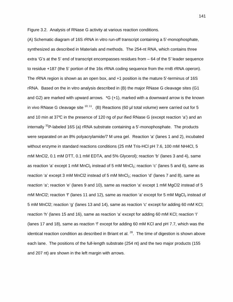
141
Figure 3.2. Analysis of RNase G activity at various reaction conditions.
(A) Schematic diagram of 16S rRNA in vitro run-off transcript containing a 5’-monophosphate,
synthesized as described in Materials and methods. The 254-nt RNA, which contains three
extra ‘G’s at the 5’ end of transcript encompasses residues from – 64 of the 5′ leader sequence
to residue +187 (the 5’ portion of the 16s rRNA coding sequence from the rrnB rRNA operon).
The rRNA region is shown as an open box, and +1 position is the mature 5’-terminus of 16S
rRNA. Based on the in vitro analysis described in (B) the major RNase G cleavage sites (G1
and G2) are marked with upward arrows. *G (+1), marked with a downward arrow is the known
in vivo RNase G cleavage site 10; 11. (B) Reactions (60 µl total volume) were carried out for 5
and 10 min at 37°C in the presence of 120 ng of pur ified RNase G (except reaction ‘a’) and an
internally 32P-labeled 16S (a) rRNA substrate containing a 5’-monophosphate. The products
were separated on an 8% polyacrylamide/7 M urea gel. Reaction ‘a’ (lanes 1 and 2), incubated
without enzyme in standard reactions conditions (25 mM Tris-HCl pH 7.6, 100 mM NH4Cl, 5
mM MnCl2, 0.1 mM DTT, 0.1 mM EDTA, and 5% Glycerol); reaction ‘b’ (lanes 3 and 4), same
as reaction ‘a’ except 1 mM MnCl2 instead of 5 mM MnCl2; reaction ‘c’ (lanes 5 and 6), same as
reaction ‘a’ except 3 mM MnCl2 instead of 5 mM MnCl2; reaction ‘d’ (lanes 7 and 8), same as
reaction ‘a’; reaction ‘e’ (lanes 9 and 10), same as reaction ‘a’ except 1 mM MgCl2 instead of 5
mM MnCl2; reaction ‘f’ (lanes 11 and 12), same as reaction ‘a’ except for 5 mM MgCl2 instead of
5 mM MnCl2; reaction ‘g’ (lanes 13 and 14), same as reaction ‘c’ except for adding 60 mM KCl;
reaction ‘h’ (lanes 15 and 16), same as reaction ‘a’ except for adding 60 mM KCl; reaction ‘I’
(lanes 17 and 18), same as reaction ‘f’ except for adding 60 mM KCl and pH 7.7, which was the
identical reaction condition as described in Briant et al. 28. The time of digestion is shown above
each lane. The positions of the full-length substrate (254 nt) and the two major products (155
and 207 nt) are shown in the left margin with arrows.

142

143
Figure 3.3. Processing of the 16S rRNA by RNase G, Rng-219 and RNase E.
(A) Schematic diagram of 16S rRNA in vitro run-off transcript. A 344 nt RNA, which contained
three extra ‘G’s at the 5’ end of transcript and encompassed residues from – 154 of 5′ leader
sequence (including in vivo cleavage sites of RNase E and RNase G 11) to residue +187 (the 5’
portion of the 16S rRNA coding sequence). The rRNA region is shown as an open box with the
+1 position being the mature 5’-terminus of 16S rRNA. Based on the in vitro analysis, the
overlapping RNase G and RNase E cleavage site that generates the 155 nt produce is
designated at E/G1 (upward arrow). Other RNase E and RNase G cleavage sites mapped by
primer extension analysis (data not shown) are marked by upward arrows. marked at the top
with arrows, The known in vivo RNase E and RNase G cleavage sites [*E (-66) and *G (+1)]
are marked with downward arrows. (B) Internally 32P-labeled 344 nt 16S rRNA substrate
containing a 5’-monophosphate, was incubated without enzyme (lane 1), with 60 ng of RNase G
(lanes 2 to 4), with 60 ng of Rng-219 (lanes 5 to 7), and with 6 ng of RNase E (lanes 8 to 10),
the products were separated on a 6 % polyacrylamide/7 M urea gel (see Materials and
Methods). The time of digestion is shown above each lane. The positions of the full length
substrate (344 nt), and the major products derived by RNase E and RNase G cleavage activity
are designated by leftward arrows. The composition of the bands are indicated by the star were
not determined.

144

145
Figure 3.4. In vitro analysis of the processing of 9S rRNA.
(A) Schematic diagram of 9S rRNA in vitro run-off transcript. The 288 nt RNA, which represents
the 9S rRNA processing intermediate contains 3 extra ‘G’s at the 5’ end of the transcript. The
mature 5S rRNA is shown as an open rectangle and the extra sequences are shown as lines.
Based on our in vitro analysis, which is in agreement with previous studies 43, the RNase E and
RNase G cleavage sites [E1/G1 (located at 3 nt upstream from 5’ mature end of 5S rRNA) and
E2/G2 (located at 3 nt downstream from 3’ mature end of 5S rRNA)] are marked with upward
arrows. (B) Internally 32
P-labeled 9S rRNA substrate with a 5’-monophosphate terminus derived
by treatment with TAP (see Materials and Methods) was incubated without enzyme (lane 1),
with 60 ng of RNase G (lanes 2 to 5), with 60 ng of Rng-219 (lanes 6 to 9), and with 12 ng of
RNase E (lanes 10 to 13). The products were separated on an 8% polyacrylamide/ 7 M urea
gel (see Materials and Methods). The time of digestion is shown above each lane. The
positions of the full-length substrate (288 nt) and the p5S product (126 nt) derived from either
RNase E or RNase G cleavage activity are marked in the right margin. The presence of the
extra nt at the 5’ end of the 126 nt fragment was determined by primer extension analysis (data
not shown).

146

147
Figure 3.5. In vitro analysis of the processing of the rpsT mRNA.
(A) Schematic diagram of the rpsT mRNA in vitro transcript used in this study. The rpsT mRNA
coding region is indicated as an open rectangle and the extra sequences are shown as lines.
The 450 nt in vitro generated mRNA substrate contains three extra ‘G’s at the 5’ end of
transcript along with the P1 promoter initiation site (-133) and ends at the encoded Rho
independent transcription terminator. The internally 32P-labeled transcript was synthesized as
described in Materials and Methods. (B and C) The rpsT mRNA terminated with either a 5’-
monophosphate (B) or a 5’-triphosphate (C) was incubated without enzyme (lane 1), with 120
ng of RNase G (lanes 2 to 5), with 120 ng of Rng-219 (lanes 6 to 9), and with 24 ng of RNase E
(lanes 10 to 13), the products were separated on a 6 % polyacrylamide/7 M urea gel (see
Materials and Methods). The time of digestion is shown above each lane. The primary RNase
E digestion products as predicted from earlier work 6; 39 are marked in the right margin. The full-
length transcript is marked in the left margin.

148

149
Figure 3.6. In vitro analysis of the processing of the cspA mRNA.
(A) Schematic diagram of the cspA mRNA in vitro transcripts. A schematic of the two cspA
transcripts are shown. They contain one extra ‘G’ at the 5’ terminus, 160 nt from the in vivo
encoded 5’ UTR, the cspA ORF (open rectangle) and 268 downstream nt that contains the Rho
independent transcription terminator. Based on our in vitro analysis and the work of 36, the
primary RNase E/G cleavage site (located 20 nt downstream of the translation terminator
codon) with a downward arrow. The 429 nt transcripts arose from termination at the encoded
Rho independent transcription terminator, while the 461 nt occurred through run-off transcription
to the end of the DNA template. The probes used for the Northern analyses are positioned
below the transcript. (B and C) Unlabeled cspA mRNA substrates containing either 5’-
triphosphate or 5’-monophosphate was incubated without enzyme (lanes 1 and 11), with 120 ng
of RNase G (lanes 2 to 4, and 12 to 14), with 120 ng of Rng-219 (lanes 5 to 7, and 15 to 17),
and with 18 ng of RNase E (lanes 8 to 10, and 18 to 20). The reactions products were
separated electrophoretically on an 8% polyacrylamide/7 M urea gel (see Materials and
Methods). The time (min) of digestion is shown above each lane. The same membrane was
used for probing with each oligonucleotide after stripping following the recommendations of the
membrane manufacturer. The positions of the two full-length substrates and major cleavage
products are shown in the right margin of each gel picture. (B) Probe a. (C) Probe b.

150

151

152
Figure 3.7. In vitro analysis of processing of the pheU tRNA substrate.
(A) Schematic diagram of the pheU tRNA in vitro run-off transcript. The DNA template used for
the in vitro transcription contained one extra ‘G’ upstream of the mature pheU tRNA, the 76 nt
mature pheU sequence (open rectangle) and 63 downstream nt including the Rho independent
transcription terminator. Transcription with T7 RNA polymerase (see Materials and Methods)
generated a full-length 140 nt species as well as two slight smaller premature termination
products. The probes used for the analysis are also positioned below the transcript. The
major RNase E/G [E/G (located at 1-2 nt downstream from the encoded CCA element)]
cleavage site is marked with a downward arrow. (B and C) Northern analysis was done using
the same 5’-end-labeled probe a. Unlabeled pheU tRNA transcripts terminated with either a 5’-
monophosphate (B) or a 5’-triphosphate (C) were incubated without enzyme (lanes 1), or with
150 ng of RNase G (lanes 2 to 5), 150 ng of Rng-219 (lanes 6 to 9), or 30 ng of RNase E (lanes
10 to 13). The products were separated electrophoretically on an 8 % polyacrylamide/7 M urea
gel (see Materials and Methods). The time of each digestion is shown above lanes. The
positions of the full-length substrate (140 residues) and major product are shown in the left
margin. The composition of the bands (�) generated by RNase G and Rng-219 was not
determined.

153

154
Figure 3.8. In vitro analysis of the processing of the argX hisR leuT proM polycistronic transcript.
(A) Schematic drawing of the argX hisR leuT proM operon DNA template used for in vitro
transcription. The fragment contains one extra ‘G’ upstream of the in vivo transcription start site
as well as 24 extra nt downstream of the Rho independent transcription terminator.
Transcription using this DNA template resulted in two species, a 505 nt run-off transcript and a
481 nt species arising from termination at the encoded Rho independent transcription terminator.
Based on our in vitro analysis the overlapping RNase E/G cleavage sites are marked with
downward blue arrows. Additional RNase G specific cleavage sites are marked with downward
black arrows. The locations of the probes used for the analysis are positioned below the
transcript. (B - I) The unlabeled argX hisR leuT proM polycistronic transcripts containing either
a 5’-monophosphate terminus (B, D, F, H) or a 5’ triphosphate terminus (C, E, G, I) were
incubated without enzyme (lanes 1), with 30 ng of RNase G (lanes 2 to 6), with 30 ng of Rng-
219 (lanes 7 to 11), and with 6 ng of RNase E (lanes 12 to 16). The products were separated
electrophoretically on a 6 % polyacrylamide/7 M urea gel (see Materials and Methods). The two
membranes (one for the 5’ monophosphate substrate and one for the 5’ triphosphate substrate)
used for the four Northern blots shown in this figure were sequentially probed by following the
recommendations of the manufacturer for membrane stripping. The composition of the major
cleavage products are shown in the right margins. The time of digestion is shown above each
lane. (B) 5’ monophosphate species probed for argX (probe a). (C) 5’ triphosphate species
probed for argX (probe a). (D) 5’ monophosphate species probed for hisR (probe c). (E) 5’
triphosphate species probed for hisR (probe c). (F) 5’ monophosphate species probed for leuT
(probe e). (G) 5’ triphosphate species probed for leuT (probe e). (H) 5’ monophosphate
species probed for proM (probe g). (I) 5’ triphosphate species probed for proM (probe g).

155

156

157

158

159

160

161

162

163

164
Figure 3.9. Primer extension analysis to map RNase G, Rng-219 and RNase E cleavage sites
within the argX hisR leuT proM polycistronic transcript.
(A) Schematic diagram of the major RNase G, Rng-219 and RNase E cleavage sites generated
within the argX hisR leuT proM polycistronic transcript. The 480 nt argX operon transcript is
identical to that described in Fig. 3.8. Important co-ordinate positions are indicated below the
vertical lines. The oligonucleotides used as primers for primer extension analysis are indicated
as asterisk capped horizontal lines. Positions of cleavage sites are shown with vertical straight-
line arrows (E/G – both RNase G and RNase E) and vertical dashed-line arrows (G – RNase G
only). The in vitro generated substrate, which was synthesized as described in Fig. 3.8, is
shown as a thick black horizontal arrow at the bottom. (B) Results with hisR primer. (C)
Results with leuT primer. An unlabeled argX hisR leuT proM polycistronic run-off transcript
substrate with a 5’-monophosphate was digested with RNase G (lanes 6 and 7), Rng-219 (lanes
8 and 9) or RNase E (lanes 10 and 11), or without enzyme (lane 5), at 37°C for the times
indicated. The reaction products were subsequently used in primer extension reactions as
described in Materials and Methods. The CTAG sequencing reactions were carried out using
the same primers as those used for each reverse transcription (lanes 1 to 4). Arrows indicate 5’
termini derived from the time-dependent RNase E and RNase G endonucleolytic cleavages.

165

166
Figure 3.10. In vitro analysis of processing of the glyW cysT leuZ polycistronic transcript.
(A) Schematic diagram of the glyW cysT leuZ polycistronic transcript used for in vitro analysis.
The fragment contains one extra ‘G’ upstream of the in vivo transcription start site as well as 19
extra nt downstream of the Rho independent transcription terminator. Transcription of this PCR-
generated DNA fragment generated a 452 nt RNA. Based on our in vitro experiments including
Northern and primer extension analysis the major E/G cleavage sites are marked with
downward arrows. It should be noted that RNase G does not cleave immediately downstream
of the glyW tRNA where there is an RNase E cleavage site. (B) An internally 32
P-labeled 452
nt substrate terminated with a 5’-monophosphate was incubated without enzyme (lanes 1 and 2),
with 60 ng of RNase G (lanes 3 and 4), with 60 ng of Rng-219 (lanes 5 and 6), or with 12 ng of
RNase E (lanes 7 and 8). The products were separated on a 6% polyacrylamide/7 M urea gel
(see Materials and Methods). The time of digestion is shown above each lane. The positions of
the full length substrate (452 nt), and the major products derived by RNase E and RNase G
cleavage activity are shown in the left margin. The compositions of the various processing
intermediates, if determined, are indicated in the right margin.

167

168
Figure 3.11. Release of pre-tRNAs from argX polycistronic transcript.
Data are derived from experiments shown in Fig. 3.8B, 8D, 8F, and 8H using a 5’
monophosphate terminated transcript (◇; pre-argX, ◯; pre-hisR, △; pre-leuT, □; pre-proM).
(A) RNase E. (B) RNase G. (C) Rng-219.

169
0
0.05
0.1
0.15
0.2
0.25
0.3
0 2 4 6 8 10 12 14 16
time (min)
Fra
ctio
n of
full
leng
th tr
ansc
ript
proc
esse
d to
pre
-tR
NA
(A)

170
0
0.1
0.2
0.3
0.4
0.5
0.6
0.7
0.8
0.9
0 2 4 6 8 10 12 14 16
time (min)
Fra
ctio
n of
full
leng
th tr
ansc
ript
pro
cess
ed to
pre
-tR
NA
(B)

171
0
0.05
0.1
0.15
0.2
0.25
0.3
0.35
0.4
0 2 4 6 8 10 12 14 16
time (min)
Fra
ctio
n of
full
leng
th tr
ansc
ript
proc
esse
d to
pre
-tR
NA
(C)

CHAPTER 4
Conclusion

173
In addition to the efficiency of transcription and the initiation of translation, mRNA
degradation and stable RNA maturation provide additional levels of post-transcriptional
regulation in the control of gene expression in Escherichia coli. The inherent instability of
mRNAs and their wide-range longevity provide prokaryotic organisms with mechanisms to
adjust their metabolic processes in response to continuously changing growth conditions. In
the case of stable RNAs, rRNAs and tRNAs, there is a direct correlation between growth rate
and the efficiency of tRNA and rRNA maturation (Chapter 2, Ow & Kushner, 2002, Ow et al.,
2003). Traditionally, these two events have been considered to be distinct processes.
However, there exists considerable overlap between the endoribonucleases involved in mRNA
degradation and stable RNA maturation (Deutscher, 2006, Kushner, 2007). It is now clear that
there are close interrelationships between degradation and maturation of RNA molecules.
The identification and extensive investigation of RNase E, an essential
endoribonuclease in E. coli, suggests that it plays a central role in mRNA decay (Ono & Kuwano,
1979, Arraiano et al., 1988, Ow et al., 2000), rRNA maturation (Ghora & Apirion, 1978, Li et al.,
1999), initiation of tRNA maturation (Li & Deutscher, 2002, Ow & Kushner, 2002), as well as
many other aspects of RNA metabolism. These findings have provided important advances in
the understanding of RNA metabolism in prokaryotes. E. coli encodes a related
endoribonuclease, RNase G, that shares a high degree of sequence homology and a number of
biochemical properties (McDowall & Cohen, 1996, Jiang et al., 2000, Tock et al., 2000).
However, RNase G is dispensable for cell viability and appears to have limited biological
functions compared to RNase E.
The results of Chapter 2 have directly addressed the question of whether over-
production of either native or extended forms of RNase G (containing either six additional amino
acids at its N-terminus or six histidines at its C-terminus) can complement RNase E deficiency
in E. coli. Our data clearly indicates that neither native nor extended forms of RNase G are

174
able to complement the loss of RNase E activity under normal physiological conditions. We
could get stable complementation of the rne-1 and rne△1018 alleles only through a
combination of mutational changes within the rng coding sequence and over-expression of the
altered RNase G protein to a comparable level as RNase E (Chapter 2). Based on these
observations, we conclude that the biological functions of the two enzymes differ not only
because of the low abundance of RNase G, but also because of the differences in their intrinsic
properties.
The rng-219 and rng-248 alleles, which are able to complement the growth defect
associated with various RNase E mutants at physiologically relevant protein levels, have
provided insights into what features of the two proteins are responsible for differences in their
biological functions (Chapter 2). Both the rng-219 and rng-248 mutations, comprising single
amino acid substitutions at residues 219 and 248 respectively, occurred in the predicted RNase
H domain which exhibits the most sequence divergence (only 26.6% identity) and the least
apparent 3-dimensional similarity to RNase E. These observations strongly imply that the
different substrate specificities and cleavage site selections of RNase E and RNase G are
controlled to some extent by their RNase H domains.
In addition, in vivo characterization of the altered RNase G proteins (Rng-219 and Rng-
248) in the total absence of RNase E (Chapter 2), has allowed us to investigate more critically
the distinct physiological roles of RNase E and RNase G in RNA metabolism in E. coli. The
analysis of tRNA processing efficiency of some tRNAs (tRNAHis, tRNACys, and tRNAPro)
demonstrates that the initiation of tRNA maturation of the polycistronic transcripts containing
these species appears to be absolutely dependent on RNase E under normal physiological
conditions (Chapter 2).
While RNase E appears to be the primary enzyme involved in the initiation of mRNA
decay (Bernstein et al., 2004), other ribonucleases such as RNase G (Li et al., 1999, Wachi et

175
al., 1999), RNase Z (Perwez & Kushner, 2006), RNase LS (Otsuka & Yonesaki, 2005), RNase
III and RNase P (Alifano et al., 1994) can initiate the decay of particular transcripts. The
mRNA half-life experiments employing rne∆1018/rng-219 and rne∆1018/rng-248 double
mutant strains allowed us to analyze the complete dependence of degradation initiation of
certain transcripts on RNase E. For example, while rpsO transcripts were dramatically
stabilized in both the double mutant strains and no decay intermediates were observed,
suggesting that RNase E was the only ribonuclease initiating its decay (Chapter 2); on the other
hand, the stability of rpsT transcripts was not significantly affected by the absence of RNase E
activity, and also new decay intermediates appeared that were not present in wild type cells.
These observations strongly imply that other ribonucleases besides RNase E are able to initiate
the decay of rpsT transcripts, and the altered RNase G proteins (Rng-219 and Rng-248) cleave
these transcripts at different locations than RNase E. The in vitro analysis employing purified
RNase E, Rng-219 and RNase G proteins has confirmed the different cleavage site selections
between RNase E and the two RNase G proteins on rpsT transcripts (Chapter 3).
In contrast, there was significant in vivo functional overlap between RNase E and
RNase G in 9S rRNA and 16S rRNA processing. rne-1 dependent defects in both rRNA’s
processing were restored to almost wild type levels by overproduction of either native or altered
forms of RNase G at physiologically comparable level of protein. Additionally, in the rne△1018
genetic background, Rng-219 and Rng-248 almost completely substituted for the function of
RNase E in the maturation of 9S rRNA and 16S rRNA (Chapter 2, Chung and Kushner,
unpublished results).
The in vitro assays of purified RNase E, Rng-219 and RNase G proteins employing
various full-length RNA substrates (9S rRNA, mRNAs and tRNAs) have provided a number of
new insights into the biochemical properties of the three proteins under conditions that resemble
the in vivo situation (Chapter 3). Both the Rng-219 and RNase G proteins site-specifically

176
cleaved structured RNAs (9S rRNA and tRNA substrates) at locations identical to those
obtained with RNase E. However, the Rng-219 and RNase G proteins also cleaved at
additional sites on mRNAs and tRNAs substrates. Interestingly, both RNase G protein
derivatives were much more active in the presence of Mn2+ than Mg2+.
The altered RNase G protein (Rng-219) clearly acts on substrates differently than wild
type RNase G (Chapter 3). The single amino acid substitution within the predicted RNase H
domain in RNase G caused a reduction in the non-specific cleavage activities of RNase G so
that it cleaved RNA molecules more like RNase E, and may remove the toxicity of RNase G
(Okada et al., 1994). This conclusion is supported by the observations that the RNase G
protein cleaves the region located within tRNAHis, tRNALeu and tRNAPro coding sequences, and
the cleavage products containing the 5S rRNA were less stable in the RNase G in vitro reaction
preparation than the Rng-219 preparation (Chapter 3). In addition, the cleavage profiles of the
cspA transcript in the RNase G cleavage assay suggest another possible explanation. RNase
G may completely degrade some transcripts which encode essential proteins that are required
to support cell viability and/or degrade one of the many small regulatory RNAs resulting in
inhibition of an essential pathway.
Both RNase E and RNase G have been shown to be 5’-end-dependent
endoribonucleases (Jiang et al., 2000, Tock et al., 2000) and the residues that make contact
with the 5’ end of substrates are conserved between two enzymes (Callaghan et al., 2005,
Jourdan & McDowall, 2008). Recently, the crystallographic structure of RNase E revealed that
the discrimination between 5’-monophosphate and 5’-triphosphate terminus of a substrate is
based on the size of the binding pocket and the net charge of the 5’ terminal groups. Although,
both RNase E and RNase G proteins exhibit a propensity to cleave substrates with a 5’
monophosphate over 5’ triphosphate ones, the cleavage activity on mRNAs and tRNAs of
RNase E is 2∼8 times more affected by the 5’ phosphorylation status than RNase G. These

177
data suggest that the size of the binding pocket and/or the key residues associated in
interactions with 5’ end of substrates of RNase G is actually different from RNase E.
Another hypothesis is that RNase G is able to cleave the RNA molecules much more
efficiently by ‘internal entry pathway’ mechanism (Joyce & Dreyfus, 1998, Baker & Mackie,
2003). The cleavage site of cspA mRNA is located at a significant distance from the 5’ end
and it has a protective 5’ stem-loop structure that might antagonize the binding ability of RNase
E and RNase G (Hankins et al., 2007). Thus the cleavage on cspA mRNA substrate would
constitute an example of the internal entry pathway mechanism by RNase E and RNase G.
The complete degradation of cspA transcript in RNase G cleavage assay (Chapter 3) strongly
implies a greater proficiency of RNase G in cleaving the substrates without interacting with their
5’ end than RNase E. These results could explain the broader sequence specificity and
distributive mode of cleavage action of RNase G as compared to RNase E (Feng et al., 2002).
An important question that still remains is how the subtle changes in the predicted
RNase H domain directly affect the biochemical activity of RNase G. Unfortunately, the use of
molecular modeling (Chapter 2) was not sufficient to accurately predict the nature of
conformational change in Rng-219 and Rng-248. In principle, the changes in biochemical
properties of altered RNase G proteins could be a consequence of differences in initial binding
with RNA substrates and/or in catalytic activation of the protein. These two possible
explanations can be distinguished by comparing the Michaelis–Menten kinetic parameters Kcat
and Km between wild type and altered RNase G proteins. However, the crystallization of
RNase G, Rng-219 and/or Rng-248 would be invaluable. Obtaining the crystal structure of
these proteins will not only help in addressing this question but also provide clues to better
understanding the features responsible for the observed differences in the physiological
properties and biochemical activities between RNase E and RNase G.

178
Finally, the construction of rne△1018/rng-219 and rne△1018/rng-248 double mutant
strains would be able to provide a useful means to achieve further comprehensive
understanding of the important features of RNase E by employing a recently developed high
density tiling microarray, which can analyze the gene expression profile on a transcriptome
scale. For example, we could identify tRNA species that are highly dependent on RNase E as
well as those independent of RNase E and new RNA substrates cleaved by RNase E, and
investigate critically the global effect of inactivation of RNase E in RNA metabolism in E. coli.
Such microarray analysis is currently under investigation (Mark B. Stead, Sarah Marshburn, and
Sidney R. Kushner).
REFERENCES
Alifano, P., F. Rivellini, C. Piscitelli, C. M. Arraiano, C. B. Bruni & M. S. Carlomagno, (1994)
Ribonuclease E provides substrates for ribonuclease P-dependent processing of a
polycistronic mRNA. Genes Dev. 8: 3021-3031.
Arraiano, C. M., S. D. Yancey & S. R. Kushner, (1988) Stabilization of discrete mRNA
breakdown products in ams pnp rnb multiple mutants of Escherichia coli K-12. J.
Bacteriol. 170: 4625-4633.
Baker, K. E. & G. A. Mackie, (2003) Ectopic RNase E sites promote bypass of 5'-end-dependent
mRNA decay in Escherichia coli. Mol. Microbiol. 47: 75-88.
Bernstein, J. A., P.-H. Lin, S. N. Cohen & S. Lin-Chao, (2004) Global analysis of Escherichia
coli RNA degradosome function using DNA microarrays. Proc. Natl. Acad. Sci. USA 101:
2748-2763.
Callaghan, A. J., M. J. Marcaida, J. A. Stead, K. J. McDowall, W. G. Scott & B. F. Luisi, (2005)
Structure of Escherichia coli RNase E catalytic domain and implications for RNA
turnover. Nature 437: 1187-1191.

179
Deutscher, M. P., (2006) Degradation of RNA in bacteria: comparison of mRNA and stable RNA.
Nucleic Acids Res. 34: 659-666.
Feng, Y., T. A. Vickers & S. N. Cohen, (2002) The catalytic domain of RNase E shows inherent
3' to 5' directionality in cleavage site selection. Proc. Natl. Acad. Sci. USA 99: 14746-
14751.
Ghora, B. K. & D. Apirion, (1978) Structural analysis and in vitro processing to p5 rRNA of a
9S RNA molecule isolated from an rne mutant of E. coli. Cell 15: 1055-1066.
Hankins, J. S., C. Zappavigna, A. Prud'homme-Genereux & G. A. Mackie, (2007) Role of RNA
Structure and susceptibility to RNase E in regulation of a cold shock mRNA, cspA mRNA.
J. Bacteriol. 189: 4353-5358.
Jiang, X., A. Diwa & J. G. Belasco, (2000) Regions of RNase E important for 5'-end-dependent
RNA cleavage and autoregulated synthesis. J. Bacteriol. 182: 2468-2475.
Jourdan, S. S. & K. J. McDowall, (2008) Sensing of 5' monophosphate by Escherichia coli
RNase G can significantly enhance association with RNA and simulate the decay of
functional mRNA transcripts in vivo. Mol. Microbiol. 67: 102-115.
Joyce, S. A. & M. Dreyfus, (1998) In the absence of translation, RNase E can bypass 5' mRNA
stabilizers in Escherichia coli. J. Mol. Biol. 282: 241-254.
Kushner, S. R., (2007) Messenger RNA decay. In: Escherichia coli and Salmonella: cellular and
molecular biology. A. Böck, R. Curtis III., C. A. Gross, J. B. Kaper, F. C. Neidhardt, T.
Nyström, K. E. Rudd & S. C. L. (eds). Washington, DC: American Society for
Microbiology Press, pp. http://www.ecosal.org.
Li, Z. & M. P. Deutscher, (2002) RNase E plays an essential role in the maturation of
Escherichia coli tRNA precursors. RNA 8: 97-109.
Li, Z., S. Pandit & M. P. Deutscher, (1999) RNase G (CafA protein) and RNase E are both
required for the 5' maturation of 16S ribosomal RNA. EMBO J. 18: 2878-2885.

180
McDowall, K. J. & S. N. Cohen, (1996) The N-terminal domain of the rne gene product has
RNase E activity and is non-overlapping with the arginine-rich RNA-binding motif. J. Mol.
Biol. 255: 349-355.
Okada, Y., M. Wachi, A. Hirata, K. Suzuki, K. Nagai & M. Matsuhashi, (1994) Cytoplasmic axial
filaments in Escherichia coli cells: possible function in the mechanism of chromosome
segregation and cell division. J. Bacteriol. 176: 917-922.
Ono, M. & M. Kuwano, (1979) A conditional lethal mutation in an Escherichia coli strain with a
longer chemical lifetime of mRNA. J. Mol. Biol. 129: 343-357.
Otsuka, Y. & T. Yonesaki, (2005) A novel endoribonuclease, RNase LS, in Escherichia coli.
Genetics 169: 13-20.
Ow, M. C. & S. R. Kushner, (2002) Initiation of tRNA maturation by RNase E is essential for cell
viability in Escherichia coli. Genes Dev. 16: 1102-1115.
Ow, M. C., Q. Liu & S. R. Kushner, (2000) Analysis of mRNA decay and rRNA processing in
Escherichia coli in the absence of RNase E-based degradosome assembly. Mol.
Microbiol. 38: 854-866.
Ow, M. C., T. Perwez & S. R. Kushner, (2003) RNase G of Escherichia coli exhibits only limited
functional overlap with its essential homologue, RNase E. Mol. Microbiol. 49: 607-622.
Perwez, T. & S. R. Kushner, (2006) RNase Z in Escherichia coli plays a significant role in
mRNA decay. Mol. Microbiol. 60: 723-737.
Tock, M. R., A. P. Walsh, G. Carroll & K. J. McDowall, (2000) The CafA protein required for the
5'-maturation of 16 S rRNA is a 5'- end-dependent ribonuclease that has context-
dependent broad sequence specificity. J. Biol. Chem. 275: 8726-8732.
Wachi, M., G. Umitsuki, M. Shimizu, A. Takada & K. Nagai, (1999) Escherichia coli cafA gene
encodes a novel RNase, designated as RNase G, involved in processing of the 5' end of
16S rRNA. Biochem. Biophys. Res. Comm. 259: 483-488.

APPENDIX A
Single amino acid changes in the predicted RNase H domain of E. coli
RNase G lead to complementation of RNase E deletion mutants1
1 Dae-hwan Chung, Zhao Min, Bi-Cheng Wang, and Sidney R. Kushner. Submitted to Nucleic Acids
Research as a Supplementary Information, 06/02/2008

182
MATERIALS AND METHODS
Plasmid constructions
The plasmids described below were generated using overlapping PCR techniques and
the high fidelity Pfu DNA polymerase (Stratagene). For the construction of pDHK11 (Fig. 2.1A),
a 2.2 kb fusion DNA fragment was synthesized that contained the regulatory region, ribosome
binding site, and ATG start codon of rne along with the rng+ sequence that included 15 bp
encoding an additional five amino acids (RKGIN) upstream of the native RNase G translation
start codon (1). The ATG from rne replaced the GTG from rng such that a total of six extra
amino acids were synthesized at the amino terminus of the RNase G protein derived from
pDHK11. First, an rne gene fragment (640 bp) was amplified by PCR using the primers RNE-UP
and RNE-OE1 and pQLK26 plasmid DNA (2) as template. In addition, an rng gene fragment
(1579 bp) was amplified by PCR using the primers RNG-OE1 and RNG-antisense and pUGK24
plasmid DNA (3) as template. The RNE-UP and RNG-antisense primers were engineered to
contain EcoRI and XbaI sites, respectively. Subsequently, overlap extension PCR with primers
RNE-OE1 and RNG-OE1 was used to generate a 2.2 kb fusion DNA fragment containing the
rne gene fragment (640 bp) and rng gene fragments (1579 bp). The amplified fusion DNA
fragment was cloned into pWSK129 (Wang and Kushner, 1991), which had been digested with
EcoRI and XbaI, to generate pDHK11.
Plasmid pDHK23 (encoding the native form of RNase G, Fig. 2.1A) is identical to
pDHK11 except for the GTG (putative upstream start codon derived from rng) was changed to
CTG to prevent translation initiation from this location and a canonical ribosome binding site
(AGGAGG) was inserted seven nt upstream of the ATG start codon to produce the native form
of RNase G (Fig. 2.1A). To make these changes, a 658 bp rne DNA fragment was amplified by
PCR using the primers RNE-UP and RNE-upstream and pQLK26 plasmid DNA as template

183
along with a rng gene fragment (1561 bp) that was amplified by using the primers RNG-RBS
(and RNG-antisense and pUGK24 plasmid DNA as template. The RNE-upstream and RNG-RBS
primer sequences were used for overlapping extension PCR to generate a 2.2 kb fusion DNA
fragment that was subsequently cloned into pWKS129 as described for pDHK11.
To generate pDHK31, a 2.2 kb EcoRI/NotI DNA fragment containing the complete
rne-rng fusion from pDHK23 was cloned into the single copy vector pMOK40 (4) that had been
digested with EcoRI and NotI.
pDHK29 (rng-248 Smr/Spr) and pDHK30 (rng-219 Smr/Spr) were made by cloning the
2.2 kb EcoRI/NotI DNA fragments from pDHK26 and pDHK28, respectively, into the EcoRI/NotI
sites of pMOK40 (4). Plasmids pDHK29, pDHK30 and pDHK31 all contained the single-copy
mini-F origin of DNA replication.
Plasmid pDHK38 [containing two RNase G fragments, one from the N-terminus to
amino acid 213 (including the S1 and 5’ sensor regions) and one including the C-terminal rng
coding sequence for amino acids 281-488 (DNase I domain and Zn-link) along with the rne
coding sequences for amino acids 213-281 (RNase H domain)] was constructed by employing a
two-step overlap extension PCR procedure. The regulatory region for this chimeric construct
was derived from RNase E as described for pDHK23. To generate the N-terminal 1.5 kb DNA
fragment containing rne/rng sequences (1.3 kb) and the 206 bp from rne, a 1.3 kb rne/rng DNA
fragment was amplified using primers RNE-UP and RNG-1260 down and plasmid pDHK23 as a
template. In addition, a 206 bp rne DNA fragment was amplified using primers RNE-1280up
and RNE-1486down and plasmid pQLK26 (2) as a template. Overlap extension PCR was then
used to generate an N-terminal fragment of 1.5 kb. The C-terminal 715 bp rng fragment was
amplified using the prime RNG1486up and RNG-antisense along with pUGK24 as the template.
The second overlap extension PCR was performed to generate a 2.2 kb fusion DNA fragment

184
containing the 1.5 kb rne/rng/rne DNA fragment and the downstream 715 bp rng fragment. The
amplified chimeric rne/rng gene was cloned into pWSK129 (5) as described for pDHK11.
Plasmid pDHK39 [containing the regulatory region and first 281 amino acids of RNase
E (S1, 5’ sensor and RNase H subdomains) fused to the rng coding sequence for amino acids
282-488] was constructed by first amplifying a 1.5 kb rne DNA fragment using primers RNE-UP
and RNE-1486down and pQLK26 plasmid DNA as a template. In addition a 715 bp rng DNA
fragment was amplified using primer RNG-1486up and RNG-antisense and pUGK24 plasmid
DNA as template. The RNE-1486down andRNA-1486up primers were used for the overlap
PCR reaction. The fusion fragment was cloned into pWSK129 as described for pDHK11.
Plasmid pDHK40 [containing the regulatory region of RNase E, the first 280 amino
acids of RNase G (S1, 5’ sensor and RNase H subdomains) along with amino acids 281-418
from RNase E (DNase I subdomain and Zn-link)] was constructed by first amplifying a 1.5 kb
rne/rng DNA fragment using primers RNE-UP and RNG+278down and pDHK23 plasmid DNA
as a template. In addition, a 450 bp rne DNA fragment was amplified using primers
RNE+279up and RNE+417down and pMOK21 (rne∆645) plasmid DNA (3) as a template. The
RNE+417down primer contained a XbaI site. The fusion fragment, generated using the
RNG+278down and RNE+279up primer sequences, was subsequently cloned into pWSK129 as
described for pDHK11.
All the plasmid constructions were verified using a combination of DNA sequencing and
western blot analysis. Western blot analysis of RNase E/G Chimeric proteins 1 and 3 employed
a polyclonal anti-RNase G antibody that was kindly provided by G. Mackie (1). RNase E/G
Chimeric protein 2 was detected using a MAP antibody raised against the first 20 amino acids of
RNase E (2). To determine if the RNase G proteins were biologically active, steady-state RNA
was isolated from independent transformants in an rng::cat genetic background (SK2538) and

185
the presence or absence of the 16.3S rRNA precursor was determined as described by Wachi
et al. (6).
Site-directed mutagenesis
Overlapping PCR was used to individually introduce the two different single nucleotide
changes in pDHK23. The G → T transversion at the first base-pair of the codon encoding aa
219 and the G → A transition at the first base-pair of aa 248 resulted in plasmids pDHK32 and
pDHK33, respectively. Plasmids pDHK34 (rng-219 Kmr) and pDHK35 (rng-248 Kmr) were
constructed to determine the effect of the two independent single amino acid substitutions on
RNase G when the proteins were synthesized from the native rng regulatory region instead of
from rne promoters. In this case, the experiment was identical to that described above for
pDHK32 and pDHK33, except that pUGK24 (rng+ Kmr) (3) plasmid DNA was used as a template.
Specific experimental details are available upon request. The presence of the predicted point
mutations in the four plasmids was confirmed by manual plasmid DNA sequencing using fmol
DNA Cycle Sequencing System Kit (Promega) as instructed by the manufacturer.
REFERENCES
1. Briant, D.J., Hankins, J.S., Cook, M.A. and Mackie, G.A. (2003) The quartenary structure
of RNase G from Escherichia coli. Mol. Microbiol., 50, 1381-1390.
2. Ow, M.C., Liu, Q. and Kushner, S.R. (2000) Analysis of mRNA decay and rRNA
processing in Escherichia coli in the absence of RNase E-based degradosome assembly.
Mol. Microbiol., 38, 854-866.

186
3. Ow, M.C., Perwez, T. and Kushner, S.R. (2003) RNase G of Escherichia coli exhibits
only limited functional overlap with its essential homologue, RNase E. Mol. Microbiol., 49,
607-622.
4. Ow, M.C. and Kushner, S.R. (2002) Initiation of tRNA maturation by RNase E is
essential for cell viability in Escherichia coli. Genes Dev., 16, 1102-1115.
5. Wang, R.F. and Kushner, S.R. (1991) Construction of versatile low-copy-number vectors
for cloning, sequencing and expression in Escherichia coli. Gene, 100, 195-199.
6. Wachi, M., Umitsuki, G., Shimizu, M., Takada, A. and Nagai, K. (1999) Escherichia coli
cafA gene encodes a novel RNase, designated as RNase G, involved in processing of
the 5' end of 16S rRNA. Biochem. Biophys. Res. Comm., 259, 483-488.
7. Callaghan, A.J., Marcaida, M.J., Stead, J.A., McDowall, K.J., Scott, W.G. and Luisi, B.F.
(2005) Structure of Escherichia coli RNase E catalytic domain and implications for RNA
turnover. Nature, 437, 1187-1191.
8. Ow, M.C., Liu, Q., Mohanty, B.K., Andrew, M.E., Maples, V.F. and Kushner, S.R. (2002)
RNase E levels in Escherichia coli are controlled by a complex regulatory system that
involves transcription of the rne gene from three promoters. Mol. Microbiol., 43, 159-171.

187
Table A1.1. Processed fractions for tRNA maturation experiments1
A. Alleles present in single copy
30oC
rne+ rne-1 rne∆645 rng-219 rng-248
tRNAHis 0.57 ± 0.11 0.40 ± 0.1 0.53 ± 0.13 0.05 ± 0.01 0.05 ± 0.0
tRNAPro 0.70 ± 0.13 0.59 ± 0.14 0.69 ± 0.12 0.12 ± 0.03 0.11 ± 0.02
tRNAAsn 0.95 ± 0.03 0.91 ± 0.02 0.96 ± 0.01 0.40 ± 0.05 0.42 ± 0.04
tRNACys 0.59 ± 0.12 0.44 ± 0.11 0.54 ± 0.10 0.11 ± 0.02 0.13 ± 0.01
44oC
rne+ rne-1 rne∆645 rng-219 rng-248
tRNAHis 0.64 ± 0.09 0.11 ± 0.1 0.43 ± 0.08 0.10 ± 0.0 0.11 ± 0.01
tRNAPro 0.74 ± 0.11 0.15 ± 0.02 0.54 ± 0.14 0.15 ± 0.03 0.15 ± 0.06
tRNAAsn 0.96 ± 0.02 0.64 ± 0.02 0.96 ± 0.03 0.53 ± 0.03 0.53 ± 0.06
tRNACys 0.64 ± 0.08 0.29 ± 0.03 0.59 ± 0.04 0.19 ± 0.03 0.23 ± 0.02

188
B. Alleles present in 6-8 copies
30oC
rne+ rne-1 rne∆645 rng-219 rng-248
tRNAHis 0.65 ± 0.05 0.61 ± 0.12 0.48 ± 0.03 0.10 ± 0.01 0.11 ± 0.02
tRNAPro 0.73 ± 0.03 0.74 ± 0.02 0.64 ± 0.09 0.15 ± 0.03 0.21 ± 0.05
tRNAAsn 0.97 ± 0.02 0.96 ± 0.02 0.97 ± 0.03 0.40 ± 0.03 0.46 ± 0.04
tRNACys 0.81 ± 0.11 0.74 ± 0.09 0.66 ± 0.04 0.19 ± 0.02 0.16 ± 0.01
44oC
rne+ rne-1 rne∆645 rng-219 rng-248
tRNAHis 0.74 ± 0.06 0.28 ± 0.03 0.43 ± 0.03 0.16 ± 0.01 0.18 ± 0.02
tRNAPro 0.70 ± 0.05 0.23 ± 0.06 0.60 ± 0.03 0.21 ± 0.02 0.29 ± 0.03
tRNAAsn 0.98 ± 0.03 0.90 ± 0.03 0.97 ± 0.04 0.64 ± 0.07 0.58 ± 0.06
tRNACys 0.71 ± 0.09 0.56 ± 0.12 0.59 ± 0.11 0.20 ± 0.03 0.20 ± 0.04
1Represents the average and the standard deviations from the mean of either three or four
independent determinations for each condition listed. Processed fraction is as described in
Figures 2.6 and A1.4.

189
Table A1.2. Oligonucleotides used for Northern analysis and automatic sequencing.
Primer Sequence (5’→ 3’)
PB5S CGCTACGGCGTTTCACTTCT
asn GACTCGAACCAGTGACATAC
cysT ACGGATTTGCAATCCGCTAC
hisR CCCACGACAACTGGAATCAC
proM CGGCGAGAGAGGATTCGAAC
pWSK 29-F CGCGCGTAATACGACTCACT
pWSK 29-R CGCGCAATTAACCCTCACTA
rne +456 CGCTGTTTTTCCGCATGAA
rng +440 CGACATCTCGGAACTGGTT
rng +874 GCCTGACTTACGAAGCGTT
rng +1322 GGTGGAGATGACGCGTAAA
rne (B) +549 CCTCGTGTCTAGTCGCGT
rng(B) +551 GCGAGAAGGGAGCGTGATA
rng(B) +942 GCTCCAGCTTGCTTGTCAT
rng(B) +1391 GCAGGTTGGGCATTCGTTA’

190
Figure A1.1. Stereo views of the crystal structure of the catalytic domain of E. coli RNase E and
the predicted structure of its homologue RNase G. (A) The three dimensional ribbon structure
of the catalytic domain of RNase E is based on the coordinates published by Callaghan et al. (7).
(B) The predicted structure of E. coli RNase G bound to RNA as described in Materials and
Methods. The RNA is shown in stick representation. Figures are prepared in PyMOL. The S1,
5’ sensor, RNase H and DNase I subdomains are indicated as well as the approximate locations
of the rng-219 and rng-248 alleles.

191

192
Figure A1.2. Expanded view of the RNase H subdomain of the RNase E, RNase G, Rng-219
and Rng-248 proteins. The structures were prepared as described in Experimental Procedures
and are viewed in PyMOL. The adjacent 5’ sensor and DNase I subdomains are indicated. (A)
Top view. (B) Bottom view. Cyan, RNase E; yellow, model of RNase G; green, model of Rng-
219; and magenta, model of Rng-248.
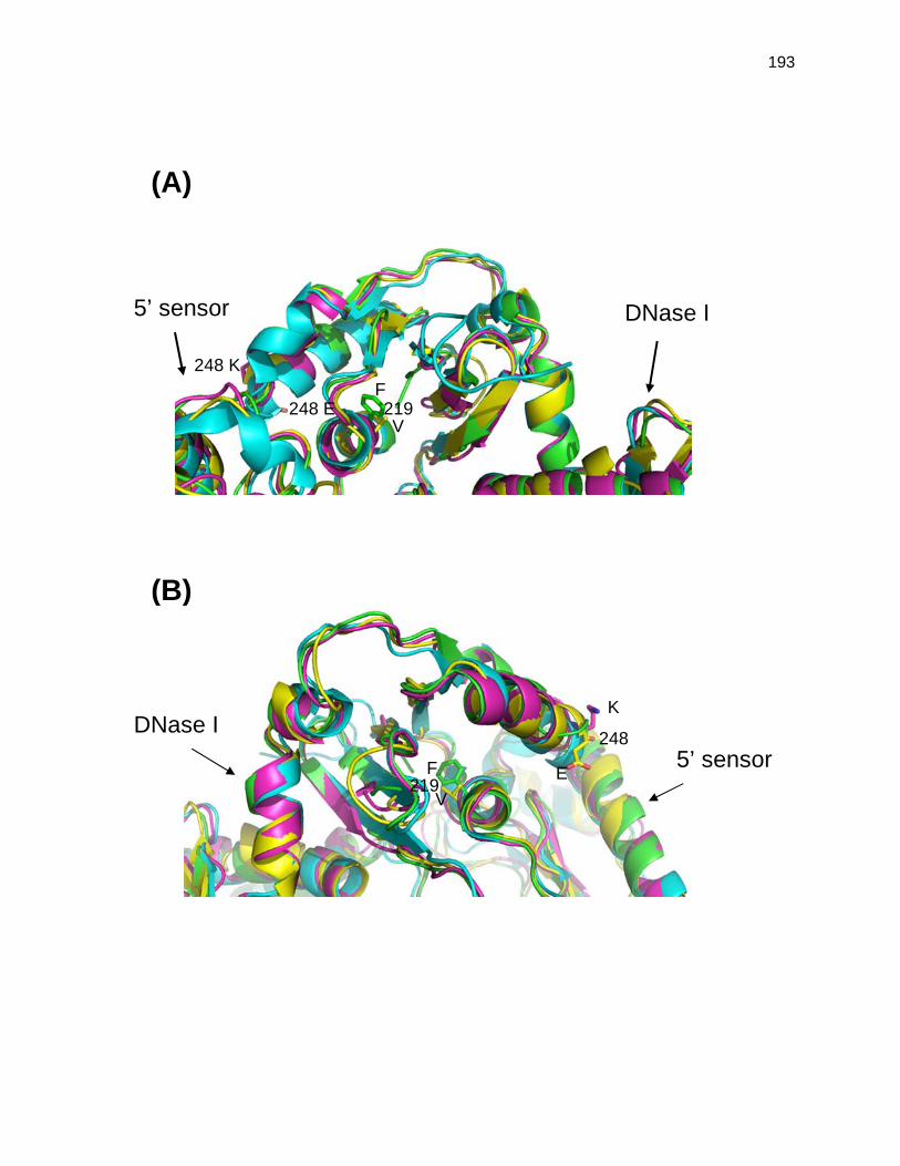
193
(B)
(A)
DNase I 5’ sensor
F
V
219 248 E
248 K
DNase I
5’ sensor
K
E
248
F 219
V

194
Figure A1.3. Analysis of 9S rRNA processing. Equivalent amounts (5 µg) of total steady-state
RNA extracted from cultures that had either been grown at 30oC or had been shifted to 44oC for
120 min, as described in Materials and Methods, were separated in a 6% polyacrylamide/7 M
urea gel, and electroblotted onto a Biotrans+ membrane. The membrane was probed with 32P-
end-labeled oligonucleotide complementary to the mature 5S rRNA [PB5S (48)]. The leftward
arrows indicate the mature 5S rRNA. (A) rne and rng alleles present in 6-8 copies/cell. SK9714
(rne+), SK9937 (rne-1), SK9987 (rne∆645), SK3541 (rng-248), and SK3543 (rng-219). (B) rne
and rng alleles present in single copy/cell. SK10143 (rne+), SK10144 (rne-1), SK2685
(rne∆645), SK3563 (rng-248), and SK3564 (rng-219). PF denotes the processed fraction, which
is defined as the amount of mature 5S rRNA divided by the total amount of the 5S rRNA
(processed and unprocessed).

195
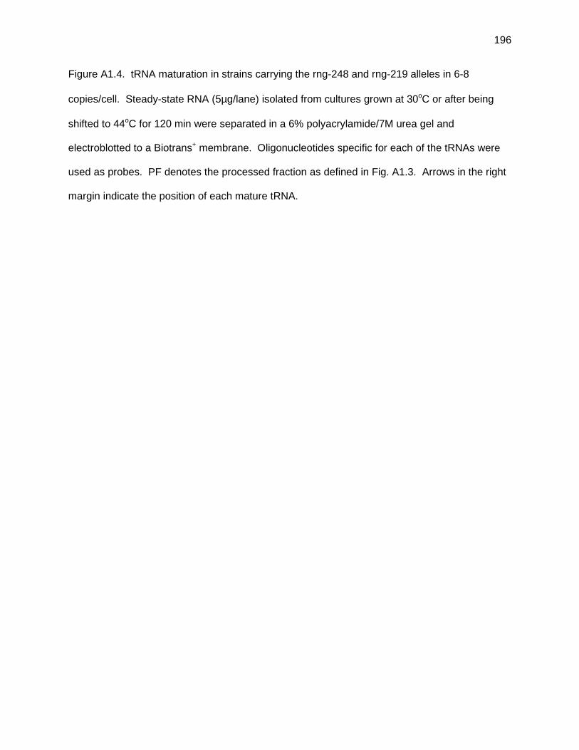
196
Figure A1.4. tRNA maturation in strains carrying the rng-248 and rng-219 alleles in 6-8
copies/cell. Steady-state RNA (5µg/lane) isolated from cultures grown at 30oC or after being
shifted to 44oC for 120 min were separated in a 6% polyacrylamide/7M urea gel and
electroblotted to a Biotrans+ membrane. Oligonucleotides specific for each of the tRNAs were
used as probes. PF denotes the processed fraction as defined in Fig. A1.3. Arrows in the right
margin indicate the position of each mature tRNA.

197