Generation and Analysis of Genetically Defined Liver...
13
10.1101/sqb.2005.70.059 Access the most recent version at doi: 2005 70: 251-261 Cold Spring Harb Symp Quant Biol L. ZENDER, W. XUE, C. CORDÓN-CARDO, et al. Carcinomas Derived from Bipotential Liver Progenitors Generation and Analysis of Genetically Defined Liver References http://symposium.cshlp.org/content/70/251.refs.html This article cites 56 articles, 22 of which can be accessed free at: service Email alerting click here the box at the top right corner of the article or Receive free email alerts when new articles cite this article - sign up in http://symposium.cshlp.org/subscriptions go to: Cold Spring Harbor Symposia on Quantitative Biology To subscribe to Copyright 2005, Cold Spring Harbor Laboratory Press Cold Spring Harbor Laboratory Press on July 2, 2009 - Published by symposium.cshlp.org Downloaded from
Transcript of Generation and Analysis of Genetically Defined Liver...
10.1101/sqb.2005.70.059Access the most recent version at doi: 2005 70: 251-261Cold Spring Harb Symp Quant Biol
L. ZENDER, W. XUE, C. CORDÓN-CARDO, et al. Carcinomas Derived from Bipotential Liver ProgenitorsGeneration and Analysis of Genetically Defined Liver
References http://symposium.cshlp.org/content/70/251.refs.html
This article cites 56 articles, 22 of which can be accessed free at:
serviceEmail alerting
click herethe box at the top right corner of the article orReceive free email alerts when new articles cite this article - sign up in
http://symposium.cshlp.org/subscriptions go to: Cold Spring Harbor Symposia on Quantitative BiologyTo subscribe to
Copyright 2005, Cold Spring Harbor Laboratory Press
Cold Spring Harbor Laboratory Press on July 2, 2009 - Published by symposium.cshlp.orgDownloaded from

Hepatocellular carcinoma (HCC) represents the fifthmost frequent neoplasm worldwide. However, due to thelack of treatment options, it constitutes the third leadingcause of cancer death (>500,000 deaths per year) (Parkinet al. 2001). The only curative treatment options for HCCare surgical resection or liver transplantation. Unfortu-nately, at the time of diagnosis the majority of patientspresent with advanced tumor growth and are therefore notcandidates for surgical therapy. Local ablative therapieslike transarterial chemoembolization (TACE) or percuta-neous ethanol injection (PEI) are being used as treatmentoptions but are only feasible in selected patients and arenot curative (Bruix et al. 2004). Systemic chemothera-peutic treatment is ineffective against HCC. For example,whereas doxorubicin or cisplatinum has marginal activ-ity, these agents have no impact on long-term patient sur-vival (Llovet et al. 2003).
More than 80% of all HCCs in humans are due to in-fection with hepatitis B virus (HBV) or hepatitis C virus(HCV) or aflatoxin B1 (AFB1) intake. The precisemechanisms by which HBV and HCV predispose to livercancer are not fully elucidated. However, it is certain thatchronic infection with HBV or HCV leads to a chronicturnover of hepatocytes, and this provides a suitable con-text for oncogenic genetic alterations. Furthermore, re-cent studies have shown that the HBx antigen of HBVcan bind and functionally inactivate the tumor suppres-sor p53 (Wang et al. 1994; Ueda et al. 1995; Huo et al.2001). In fact, mice transgenic for the HBx antigen de-velop hepatic adenomas and HCC (Ueda et al. 1995).Similar studies have been performed for proteins ofHCV. It has been reported that the HCV core proteintranscriptionally represses the p53 promoter in differentcell types (Ray et al. 1997). In non-virus-induced livercancers, p53 mutations may also play a role. For exam-
ple, liver cancers arising from aflatoxin exposure fre-quently have mutations in p53 at codon 249.
A number of other genetic alterations have been linkedto the development of HCC. For example, Rb and/or theinsulin growth factor receptor 1 (IGFR1) are frequentlymutated or deleted (Murakami et al. 1991; Kim et al.1996), whereas c-myc and cyclin D1 are often amplified(Peng et al. 1993; Nishida et al. 1994; Kusano et al.1999; Wong et al. 1999; Niketeghad et al. 2001). Alter-ations in the Wnt-signaling pathway may also be impor-tant during hepatocarcinogenesis. Thus, mutations thatactivate β-catenin, or inactivate its negative regulatoraxin, occur in 30–40% of human HCCs, and nuclearoverexpression of β-catenin occurs in 10–90% of thesetumors (Nhieu et al. 1999; Buendia 2000; Wong et al.2001). Deregulation of β-catenin leads to transcriptionalactivation of Wnt-pathway target genes like cyclin D1, c-myc, or PPARγ, which in turn promote tumor progres-sion by enhancing proliferation.
Despite its impact on human health, liver cancer is un-derstudied compared to other major lethal cancers. Inaddition, the field has a limited arsenal of model sys-tems in which to study the development of this diseaseand to test potential intervention strategies. For the mostpart, the traditional study of human cancers only pro-vides a snapshot of the final result, rather than a detailedroadmap of the route taken by the tumor cell to reach itsdestination. The ability to model intermediate stages oftumor formation is essential for a complete understand-ing of the disease—not only in terms of our understand-ing of disease biology, but also in terms of effectivelyusing biological information for the development ofnew treatments and therapies.
The use of genetically engineered mouse models tostudy cancer enables an assessment of how specific ge-
Generation and Analysis of Genetically Defined LiverCarcinomas Derived from Bipotential Liver Progenitors
L. ZENDER,* W. XUE,* C. CORDÓN-CARDO,† G.J. HANNON,* R. LUCITO,* S. POWERS,* P. FLEMMING,‡ M.S. SPECTOR,* AND S.W. LOWE*¶
*Cold Spring Harbor Laboratory, Cold Spring Harbor, New York 11724;†Division of Molecular Pathology, Memorial Sloan-Kettering Cancer Center, New York, New York 10021;
‡Department of Pathology, Medical School Hannover, Hannover, Germany;¶Howard Hughes Medical Institute, Cold Spring Harbor, New York, New York 11724
Cold Spring Harbor Symposia on Quantitative Biology, Volume LXX. © 2005 Cold Spring Harbor Laboratory Press 0-87969-773-3. 251
Hepatocellular carcinoma is a chemoresistant cancer and a leading cause of cancer mortality; however, the molecular mech-anisms responsible for the aggressive nature of this disease are poorly understood. In this study, we developed a new livercancer mouse model that is based on the ex vivo genetic manipulation of embryonic liver progenitor cells (hepatoblasts). Af-ter retroviral gene transfer of oncogenes or short hairpin RNAs targeting tumor suppressor genes, genetically altered liverprogenitor cells are seeded into the liver of otherwise normal recipient mice. We show that histopathology of the engineeredliver carcinomas reveals features of the human disease. Furthermore, representational oligonucleotide microarray analysis(ROMA) of murine liver tumors initiated by two defined genetic hits revealed spontaneously acquired genetic alterations thatare characteristic for human hepatocellular carcinoma. This model provides a powerful platform for applications like cancergene discovery or high-throughput preclinical drug testing.
251-262_29_Zender et al_Symp70 5/12/06 10:02 AM Page 251
Cold Spring Harbor Laboratory Press on July 2, 2009 - Published by symposium.cshlp.orgDownloaded from
netic alterations occurring in the human disease affect thetumor phenotype. Currently, mouse models of liver cancerare based mainly on classic transgenic approaches, tetra-cycline-regulated transgene expression or chemically in-duced carcinogenesis (Sandgren et al. 1989; Jhappan et al.1990; Murakami et al. 1993; Verna et al. 1996a,b; Deaneet al. 2001; Manickan et al. 2001; Wang et al. 2001;Harada et al. 2004; Shachaf et al. 2004). Although thesemodels have produced important insights into the molec-ular mechanisms of hepatocarcinogenesis, they have sev-eral disadvantages. First, the expression of transgenes bytissue-specific promoters may not target all epithelial cellswithin the organ (stem-cell and non-stem-cell compart-ment) and thus may alter the cell of origin from that oc-curring in the human disease. Furthermore, the expressionof the oncogene (or deletion of a tumor suppressor gene)in all cells within a tissue creates a field effect such that allcells have altered gene expression, which does not mimicthe situation of spontaneous tumorigenesis and may altera tumor’s evolutionary trajectory. Second, it is very cost-and time-intensive to generate germ-line transgenic andknockout animals, and production of compound mutantanimals can involve complicated intercrossing strategiesthat are extremely slow. Third, some lesions also producedevelopmental abnormalities resulting in embryoniclethality or developmental compensation in the resultingtissue, such that the consequences of the mutation may notreflect the acute activation or loss that occurs in tumorige-nesis. Obviously, a mouse model that allows bypass ofthese limitations would be extremely valuable.
Over the last few years, chimeric mouse models basedon the genetic manipulation and retransplantation ofhematopoietic stem cells have provided new insights intomechanisms of tumor initiation, progression, and treat-ment response in hematopoietic malignancies (see, e.g.,Pear et al. 1998; Schmitt et al. 2002a,b; Hemann et al.2005). Such models have the advantage of generating ge-netically defined tumors at a fraction of the time and costrequired to produce comparable germ-line models, al-lowing more genes and gene combinations to be studied.Because the cancers that arise in these animals are de-rived from targeting stem and/or progenitor cells, theymay more accurately reflect the evolution of correspond-ing human malignancies. Finally, these models are pro-duced by transplantation of genetically altered cells intorecipient mice, thereby yielding genetic mosaics wherethe developing cancer cell is surrounded by otherwisenormal counterparts. For example, using such systems,we have shown that mutant Myc alleles obtained fromBurkitt’s lymphoma are highly oncogenic because theyfail to induce the pro-apoptotic protein Bim (Heman et al.2005), and that p53 can influence the antitumor activityof certain oncogene-targeted therapeutics (H.G. Wendelet al., in prep.). Nevertheless, these powerful systemshave been largely limited to the hematopoietic system.
Combining our previous experience with these novelchimeric mouse model systems, and our desire to easilyand accurately model HCCs, we developed and character-ized a new mouse model for HCC that is based on the exvivo manipulation of liver progenitor cells followed by theseeding of these cells into normal recipients. These meth-
ods allowed us to rapidly produce in situ liver cancers ofdefined genetic origin. The generation of the model and itspotential applications are the topic of this report.
MATERIALS AND METHODS
Isolation, culture, and retroviral infection of E-Cad-herin-positive liver progenitor cells. Liver cell suspen-sions from fetal livers of ED12.5–15 C57BL/6 mice(Jackson Laboratories, Rochester, New York), were pre-pared as described previously (Nitou et al. 2002). Purifi-cation of E-Cadherin-positive hepatoblasts from liversuspensions was performed using the MACS® magneticcell-sorting system (Miltenyi, Auburn, California) in theindirect labeling option together with the rat anti-mouseE-Cadherin (ECCD-1) antibody (Calbiochem, SanDiego, California). Columns of the “MS” size were used.Purification was performed by a modification of the pro-tocol recommended by the manufacturer. Briefly, goatanti-rat IgG microbeads were first incubated with theECCD-1 antibody for 45 minutes at room temperaturewith gentle resuspension by pipetting every 5 minutes. 20μl of immunomagnetic beads were incubated with 4 μg ofECCD-1 antibody for later incubation with up to 107 pos-itive cells. Complexes of primary and secondary antibodywere washed using a MS column according to the manu-facturer’s instructions. Liver cell suspensions were incu-bated with the antibody complexes in DMEM 10% FBSin plastic tubes for 45 minutes at 4°C. After loading ontoMACS MS columns, cells were washed and subsequentlyeluted in 1 ml of hepatocyte growth medium (Block et al.1996). The harvest from one embryonic liver was splitinto two portions and plated in two wells of a 6-well platethat were coated with gelatin (Specialty Media, Phillips-burg, New Jersey) and contained irradiated (30 Gy) NIH-3T3 feeder layers (3 x 10³ cells/cm2). Hepatocyte growthmedium was supplemented with hepatocyte growth fac-tor (HGF)/SF (40 ng/ml) (Peprotech, Rocky Hill, NewJersey) and epidermal growth factor (EGF) (20 ng/ml)(Peprotech, Rocky Hill, New Jersey).
After 68 hours, cultured hepatoblasts were transducedwith various lentiviral or murine stem cell virus(MSCV)-based retroviral vectors as described previ-ously (Schmitt et al. 2002a), each coexpressing a greenfluorescent protein (GFP) reporter. Briefly, retroviruswas produced in Phoenix packaging cells and collectedin hepatocyte growth medium. High-titer viral super-natant was passed through a 0.45-μm filter and supple-mented with polybrene (2 μg/ml), HGF (40 ng/ml), andEGF (20 ng/ml). The infection procedure was repeatedthree times every 8 hours. 24 hours after the last infec-tion, the fraction of GFP-positive cells was assessed byGFP fluorescence microscopy.
Immunofluorescence, immunohistochemistry, andhistopathology. Mouse monoclonal antibodies against cy-tokeratin AE1 (Biocare Medical, Walnut Creek, Califor-nia) (1:100), OV-6 (R&D systems, Minneapolis, Min-nesota) (1:50), rabbit anti-mouse albumin antiserum(Biogenesis, Poole, United Kingdom) (1:200), rabbit anti-human α-fetoprotein (AFP) antiserum (Dako, Glostrup,
252 ZENDER ET AL.
251-262_29_Zender et al_Symp70 5/12/06 10:02 AM Page 252
Cold Spring Harbor Laboratory Press on July 2, 2009 - Published by symposium.cshlp.orgDownloaded from

GENETICALLY DEFINED LIVER CARCINOMAS 253
formed in accordance with the guidelines of the Institu-tional Animal Care and Use committee of Cold SpringHarbor Laboratory.
Generation of cell lines and retransplantation. Togenerate cells from primary HCCs, a tumor nodule of atleast 3 x 3 mm was removed and diced into small pieces.To obtain a single-cell suspension, minced tumor suspen-sions were incubated in HEPES-buffered DMEM con-taining 1000 U/ml dispase for 30–60 minutes at 37°C.The suspension was washed twice in DMEM to removedispase completely. Before plating, the cell suspensionwas filtered through a sterile metal sieve to removeclumps. To facilitate the initial attachment, isolated tu-mor cells were plated on gelatin-coated culture plates.
For retransplantation into recipient mice, 2 x 10⁶ cellswere injected in 100 μl of medium into the left liver lobe.Briefly, mice were anesthetized as described above. Asmall ventral midline laparotomy was performed. The leftliver lobe was retracted and 2 x 10⁶ cells were injectedsubcapsularly in 100 μl of medium. The injection site wascompressed for at least two minutes to prevent bleedingand peritoneal spreading of tumor cells. The intra-ab-dominal operation field was rinsed with distilled water toosmotically kill accidentally leaked tumor cells. The ab-dominal wall was closed using 4-0 vicryl suture material(Ethicon, Norderstedt, Germany).
RESULTS
Isolation and Transduction of Liver Progenitor Cells
There is increasing evidence that bipotential liver stemcells (oval cells in rodents and hepatic progenitor cells inhumans) are the cellular targets for transformation in thedevelopment of HCC (Braun et al. 1987; Dumble et al.2002; Alison and Lovell 2005) and eventually also biliarytract cancer (Tsao and Grisham 1987; Steinberg et al.1997). Oval cells are bipotential liver stem cells, existingin the healthy adult liver in low numbers around the peri-portal region. During chronic liver damage, oval cells canproliferate extensively (Thorgeirsson 1996; Dabeva andShafritz 2003), suggesting that they might be good candi-dates to target with genetic modifications to model livercancer. Numerous protocols are available to induce theoval-cell compartment in rodents (for review, see Dabevaand Shafritz 2003), and purification procedures for ovalcells have been established (Dumble et al. 2002; Dabevaand Shafritz 2003). However, a major disadvantage ofthese protocols is that they are based on carcinogen treat-ment, which may introduce undefined genetic mutationsthat contribute to tumorigenesis.
Bipotential hepatoblasts are the embryonic counterpartto oval cells from the adult liver (Thorgeirsson 1996;Dabeva and Shafritz 2003). These cells also give rise tocells of both the hepatocytic and bile duct lineage (Fig.1A) and share almost every marker with oval cells. Theyexist in large quantities in the murine fetal liver, whichalso contains hematopoietic cells, endothelial cells, con-nective tissue cells, and precursors of hepatic stellatecells. Recently, it was reported that hepatoblasts can bepurified from the ED12.5 mouse liver using antibodies
Denmark) (1:200), mouse monoclonal antibodies againstcytokeratin 8 (RDI, Concord, Massachusetts) (1:100), orrabbit polyclonal antibodies against GFP (1:200) (Abcam,Cambridge, Massachusetts) were used as primary anti-bodies. For immunofluorescence of cultured hepatoblasts,cells were either plated on gelatin-coated coverslips/NIH-3T3 feeder layers (CK AE1, OV-6, CK8) or on NIH-3T3feeder layers grown on gelatin-coated plastic wells (AFP,Alb). Cells were fixed with 4% PFA at room temperaturefor 15 minutes, permeabilized with 0.2% Triton, 5% goatserum in phosphate-buffered saline (PBS) for 5 minutesand subsequently washed with 0.1% PBS-T, 5% normalgoat serum. Incubation with the primary antibody was per-formed for 1 hour in 0.1% PBS-T, 5% normal goat serumat room temperature. Control samples were incubatedwith 0.1% PBS-T, 5% normal goat serum. Secondary an-tibodies used were goat anti-mouse Alexa Fluor 594-la-beled antibody (Invitrogen, Carlsbad, California) and goatanti-rabbit Alexa Fluor-labeled antibodies (Invitrogen,Carlsbad, California). Anti-GFP immunofluorescencewas performed on paraffin-embedded liver sections. Stan-dard Proteinase K antigen-retrieval was used. Detailedprotocols for anti-cytokeratin-8 immunohistochemistry onparaffin-embedded liver tumor sections are available uponrequest. Histopathological evaluation of murine liver car-cinomas was done by an experienced liver pathologist(P.F.) using paraffin-embedded liver tumor sections thatwere stained with hematoxylin/eosin (H&E) according tostandard protocols.
Conditioning of recipient mice and transplantation.Pathogen-free female C57BL/6 mice (Jackson Laborato-ries, Rochester, New York), 8–12 weeks of age, weighing20–30 g were used as recipients for genetically modifiedliver progenitor cells. Retrorsine and CCl4 stock solutionswere prepared as described previously (Guo et al. 2002).Before injection, retrorsine stock solution was diluted 1:5in sterile PBS. Mice received retrorsine, 70 mg/kg i.p. Thisdose was repeated after 7 days and the mice were allowedto recover for 10 days prior to hepatoblast transplantation.
For transplantation of genetically modified hepato-blasts, animals were anesthetized with isolflurane, and asmall left subcostal laparotomy was performed. Thespleen was retracted and the lower spleen pole loopedwith a 4-0 vicryl ligation. 2 x 10⁶ cells were injected in 0.2ml of medium in the lower pole of the spleen in an injec-tion time of 2 minutes. The lower spleen pole was ligatedand resected. The abdomen was sutured using 4-0 vicryl(Ethicon, Norderstedt, Germany). Transplanted cellswere allowed to migrate to the recipient liver and engraftthe liver. 5 days after intrasplenic cell transplantation,CCl4 was administered in a dose of 0.5 ml/kg i.p. CCl4treatment was repeated on d10 and d15 after cell trans-plantation. Animals were subsequently monitored for tu-mor formation by abdominal palpation of whole-bodyfluorescence imaging. For external GFP-tumor imaging,mice were anesthetized with isolflurane, abdominal furwas removed with depilating cream, and animals weremonitored by whole-body fluorescence imaging for theretrovirally expressed GFP reporter as described previ-ously (Schmitt et al. 2002a). All procedures were per-
251-262_29_Zender et al_Symp70 5/12/06 10:02 AM Page 253
Cold Spring Harbor Laboratory Press on July 2, 2009 - Published by symposium.cshlp.orgDownloaded from

recognizing the extracellular epitopes of E-Cadherinmolecules in combination with immunomagnetic beads(Nitou et al. 2002) to a purity of about 98%. These hepa-toblasts were capable of spontaneously differentiatinginto hepatocytes when cocultured with the nonparenchy-mal fraction of the embryonic liver (Nitou et al. 2002).We therefore reasoned that these embryonic hepatoblastswould serve as an excellent source of liver progenitorcells for further manipulation, and we set out to isolate,culture, and genetically manipulate mouse embryonichepatoblasts using procedures analogous to our previousefforts in hematopoietic systems.
We isolated E-Cadherin-positive liver progenitor cellsusing an immunomagnetic-bead-based procedure. Afterplating, aggregates of hepatoblasts attach in close prox-imity to the feeder layer cells (Fig. 1B, left). A major lim-itation to propagating pure hepatoblasts is that the growthand survival of these cells in vitro is limited due to the re-quirement for hepatoblast–nonparenchymal cell interac-tions for survival and growth (Nitou et al. 2002). How-ever, we noted that hepatoblasts grown on gelatin-coatedculture plates with irradiated NIH-3T3 feeder layers inchemically defined medium (Block et al. 1996) enrichedwith HGF and EGF are capable of extensive proliferation(Fig. 1B, middle).
Transgenes were introduced into these cells by retro-virus-mediated gene transfer using MSCV-based vectorscoexpressing GFP. Using a standard protocol, we typi-cally were able to infect 20–50% of cells, as assessed bymonitoring GFP in the target cell population (Fig. 1B,right). To determine whether these cells retained markersof bipotential liver progenitors, we examined their im-munophenotype using several well-established markers(AFP, albumin, CK8, CKAE1, and OV-6) that allow (1)the discrimination of parenchymal from non-parenchy-mal cells and (2) a classification of the parenchymal cellsinto progenitor cells, cells of the hepatocytic lineage, andcells of the bile duct lineage.
Immunofluorescence against AFP and albumin (Alb)can be performed on hepatoblasts grown on gelatin-coated plastic dishes, thus allowing a quantification of theAlb\AFP-double positive fraction. As expected, the E-Cadherin-positive liver progenitor cells displayed abun-dant expression of Alb and AFP, with >98% beingAlb\AFP-double positive cells. Note that AFP is a markerfor bipotential liver progenitor cells that is usually notfound in differentiated hepatocytes or bile duct cells. Fur-thermore, we performed immunofluorescence on the E-Cadherin-positive liver progenitor cells using a mono-clonal antibody directed against the acidic (Type 1)family of cytokeratins. This family consists of cytoker-atins 10, 14, 15, 16, and 19. Cytokeratins 10, 15, and 16are mainly found expressed in the skin. Cytokeratins 14and 19, however, are known to be markers for bipotentialliver progenitor cells (for review, see Thorgeirsson1996). Positive results were also found in immunofluo-rescence analyses using antibodies directed against the“liver cytokeratin” cytokeratin 8 and the oval-cell antigenOV-6. The expression levels of CK8, CKAE1, and OV-6were notably lower than those of Alb and AFP. The re-
sults of the characterization of the expression pattern ofthese purified E-Cadherin-positive liver progenitor cellsstrongly suggest that they are bipotential liver progenitorcells, or hepatoblasts.
Intrahepatic Seeding of Genetically ModifiedLiver Progenitor Cells
We next sought to explore whether purified hepato-blasts could be genetically modified and incorporatedinto the liver. It is known that liver cell transplantationinto the spleen of healthy recipient mice leads to an inef-
254 ZENDER ET AL.
Figure 1. (A) Schematic representation showing the embryonicderivation of hepatic cell lineages. (B) E-Cadherin-positive fetalliver progenitor cells (LPCs, also known as hepatoblasts) can bepurified by immunomagnetic beads. E-Cadherin+ fetal liver pro-genitor cells are isolated from ED = 12.5–15 fetal mouse liversusing the MACS® system with indirect labeling. Freshly har-vested hepatoblasts are grown in gelatin-coated plates on irradi-ated NIH-3T3 feeder layers. Islands of LPCs are growing inclose proximity to 3T3 feeder cells (left). E-Cadherin+ LPCsshow an extensive growth potential in vitro when grown onfeeder layers in HGF/EGF-enriched hepatocyte growth medium(middle). E-Cadherin+ LPCs can be infected efficiently withstandard retroviral vectors as shown by GFP fluorescence(right). (C) Characterization of the E-Cadherin+ liver progenitorcells with established hepatoblast/oval cell markers. E-Cad-herin+ LPCs reveal strong positivity for AFP, Alb, and CKAE1;positivity for CK8; and weak positivity for OV-6.
A
B
C
251-262_29_Zender et al_Symp70 5/12/06 10:02 AM Page 254
Cold Spring Harbor Laboratory Press on July 2, 2009 - Published by symposium.cshlp.orgDownloaded from

ficient repopulation of the host liver (0.03–0.5% of the to-tal liver mass) (Gupta et al. 1991; Ponder et al. 1991).Different approaches to facilitate the engraftment oftransplanted hepatocytes within the recipient liver havebeen described. All these approaches share the featurethat hepatocytes are transplanted into a predamaged liverwhich provides a regenerative environment for trans-planted cells (Shafritz and Dabeva 2002; Dabeva andShafritz 2003). Recently, Guo et al. (2002) reported anenhanced repopulation of the mouse liver after pretreat-ment of the animals with retrorsine. Retrorsine is an alka-loid that exerts a strong and persistent block of nativehepatocyte proliferation (Laconi et al. 1999). After hepa-tocyte transplantation, CCl4 was used to induce a regen-erative environment in which transplanted cells can pro-liferate, while proliferation of recipient cells remainsblocked. Using this protocol, Guo et al. (2002) demon-strated repopulation rates of up to 20% of the liver mass.We reasoned that such procedures in combination withtransplantation of genetically modified hepatocyteswhich exert a proliferation advantage should allow ahighly efficient engraftment of transplanted cells.
A schematic representation of our approach is shown inFigure 2A. E-Cadherin-positive bipotential liver progen-itor cells are purified and grown in primary culture. Cellsare infected with retroviral constructs, expressing onco-genes or short hairpin RNAs (shRNAs) directed againsttumor suppressor genes. After infection, 2 x 10⁶ liver pro-genitor cells are transplanted into the spleens of recipientmice that were conditioned with retrorsine. One week af-ter intrasplenic injection of the cells, a cycle of three CCl4treatments is started. The timeline of the retrorsine pre-treatment and subsequent CCl4 treatment is shown in Fig-ure 2B. One week after intrasplenic injection of GFP-tagged liver progenitor cells, livers were harvested andprocessed for immunofluorescence with an antibody di-rected against GFP. We found that approximately 1% ofthe whole liver consisted of the “seeded,” GFP-positiveliver progenitor cells. Interestingly, on H&E sectionsthese transplanted cells are indistinguishable from thesurrounding normal hepatocytes (Fig. 2C).
Generation of Liver Cancers In Situ
Our next goal was to generate primary liver carcinomaswith defined genetic lesions previously linked to the de-velopment of human HCC. For example, mutations inp53, Rb, and IGFR1 are frequently found in HCC (Mu-rakami et al. 1991; Kim et al. 1996), and the oncogenes c-myc and cyclin D1 are frequently found to be overex-pressed (Peng et al. 1993; Nishida et al. 1994). To gener-ate primary liver carcinomas of the genetic compound le-sion “p53–/–;c-myc,” we purified hepatoblasts fromp53-deficient embryonic mouse livers. These cells weretransduced with an MSCV-based retroviral vector coex-pressing c-myc with a GFP reporter and transplanted intothe spleen of retrorsine-pretreated recipient mice as de-scribed above. Animals were continuously monitored forgeneral health and tumor formation by abdominal palpa-tion and external GFP fluorescence imaging.
Figure 2D shows a tumor-bearing mouse with aswollen abdomen due to ascites (digital picture, rightpanel). The tumor is detectable by whole-body fluores-cence imaging (left panel). The asterisks indicate rem-nants of GFP-positive transplanted hepatocytes in thespleen. GFP imaging of the explanted liver (lower panel,left) shows an advanced liver carcinoma filling a wholeliver lobe. It is apparent that the external GFP signal is notproportional to the real tumor size, because about 70% ofthe GFP signal is absorbed by the ribcage/abdominalwall. Presumably, more sensitive imaging techniques,such as bioluminescence using a luciferase reporter,would enable the detection of small liver tumors and pre-cise assessment of partial responses in treatment studies.
We also generated in situ liver carcinomas by seedinggenetically manipulated adult liver cells (no separation ofadult hepatocytes from liver progenitor cells) obtained bycollagenase perfusion from livers of p53-deficient mice(data not shown). Because the infection efficiency of dif-ferentiated hepatocytes with standard retroviral vectors ismuch lower compared to hepatoblasts (data not shown),we used lentiviral vectors in this setting. Infection of p53-deficient adult liver cells with a lentivirus expressing anHA-tagged form of c-myc (Hemann et al. 2005) and seed-ing of these cells into the livers of conditioned recipientmice gave rise to in situ liver carcinomas with lower pen-etrance than the hepatoblast cultures. We also found thattumorigenicity of p53–/– embryonic liver cells isolatedfrom the late-stage mouse embryo (ED16–18) and in-fected with a c-myc-expressing retrovirus is significantlylower than tumorigenicity of cells of the same genotypethat were harvested around ED12.5 (data not shown). In-terestingly, embryonic hepatocytes that were harvestedfrom ED18 p53–/– embryonic mouse livers and infectedwith a retrovirus encoding c-myc are only immortalizedbut not transformed (0/8 tumors subcutaneously on nudemice, 0/12 tumors in situ).
We next established a protocol for the culture and ex-pansion of hepatoma cells from primary murine HCCs ofa defined genetic origin (see Materials and Methods fordetails). In most instances, this procedure allowed the es-tablishment of a stable population of cultured hepatomacells. After expansion, the cells could be directly injectedinto the liver of recipient mice. Figure 2E shows an ex-planted liver several weeks after direct infiltration of 2 x10⁶ “p53–/–;c-myc” hepatoma cells into the left liver lobe.The tumor is readily visualized by GFP imaging as shownin the left panel of the figure. The ability to generate mul-tiple tumors of exactly the same genetic backgroundshould enable side-by-side comparisons of different treat-ment regimens in tumors of the same genetic background.
Suppression of Gene Expression in Murine HCCUsing Stable RNA Interference
Historically, mouse models of human liver cancerhave involved the germ-line manipulation of eitheroncogenes or tumor suppressor genes. In the case of tu-mor suppressor genes, one either constitutively deletesthe tumor suppressor gene or produces a conditional
GENETICALLY DEFINED LIVER CARCINOMAS 255
251-262_29_Zender et al_Symp70 5/12/06 10:02 AM Page 255
Cold Spring Harbor Laboratory Press on July 2, 2009 - Published by symposium.cshlp.orgDownloaded from

“knockout” animal where the gene is flanked by loxPsites such that the tumor suppressor gene can be inacti-vated in specific settings using cre recombinase. Al-though extremely powerful, these approaches can be ex-tremely time- and cost-intensive and, in the case ofconstitutive knockout strains, are subject to develop-mental compensation that can mask aspects of the nullphenotype (Sage et al. 2003). However, over the last sev-eral years, RNA interference (RNAi) has emerged as acomplementary and effective tool to homologous recom-bination to study the consequences of gene suppressionin cultured mammalian cells and in mice (Hemann et al.2003; Hannon and Rossi 2004; Dickins et al. 2005). Fur-thermore, improved reagents for stable RNAi have beenreported where the silencing triggers mimic a natural microRNA primary transcript, and each target sequencehas been selected on the basis of thermodynamic criteriafor optimal shRNA performance (Dickins et al. 2005;Silva et al. 2005). We were therefore interested in evalu-
ating whether stable RNAi technology could be har-nessed to create dominantly acting inhibitors of tumorsuppressor function in a chimeric (genetic mosaic)model of hepatocarcinogenesis.
To test the feasibility of such an approach, we soughtto generate in situ liver carcinomas by genetic manipu-lation of bipotential liver progenitor cells with syntheticmicroRNAs directed against p53 in combination withdifferent oncogenes. The predicted folding of the pri-mary microRNA is shown in Figure 3A. The red bars in-dicate the predicted mature small interfering RNA. Fig-ure 3B shows in situ liver carcinomas derived eitherfrom p53-deficient liver progenitor cells (LPCs) over-expressing oncogenic Ras or from wild-type LPCs thatwere double infected with a microRNA-based sh-con-struct directed against p53 (MLS.1224), together with avector expressing oncogenic Ras. The infected cell pop-ulations were seeded into the liver or injected subcuta-neously into an immunocompromised mouse.
256 ZENDER ET AL.
Figure 2. Generation of genetically defined in situ liver cancers. (A) Technical outline. E-Cadherin-positive fetal liver progenitorcells (hepatoblasts) are purified and cultured as described above. Using the murine stem cell virus (MSCV) optimized to drive long-term gene expression in vivo, the cells are infected with oncogenes and/or shRNAs directed against tumor suppressor genes. Afterinfection and expansion, the cells are transplanted into the spleens of conditioned recipient mice. Because all retroviral vectors usedfor infections are GFP-tagged, development of in situ HCCs can be monitored by external GFP imaging. (B) Schematic representa-tion of the timeline of the approach. Mice undergo two pretreatments with the liver cell cycle inhibitor retrorsine. After transplan-tation of the cells into the spleen, the transplanted hepatoblasts are selectively expanded by CCl4 treatment of the mice. (C) GFP+
transplanted hepatoblasts can be detected in the recipient liver by anti-GFP immunofluorescence (right). H&E staining of an adja-cent section of the liver (left). (D) External GFP imaging of a p53; c-myc tumor-bearing mouse (left). The GFP-positive spot in thesquare represents the intrahepatic tumor mass. The two additional spots (marked by asterisks) represent transplanted cells residingin the spleen after transplantation. Mice with advanced intrahepatic tumor growth present with swollen, ascites-containing abdomenallowing detection of tumor burden by palpation. GFP imaging of the explanted liver (bottom panel) reveals an advanced intrahep-atic tumor, filling out a whole liver lobe. (E) Primary liver tumors can be outgrown in culture and retransplanted in situ into recipi-ent mice. Shown is a tumor that was retransplanted by direct liver injection of 2 x 10⁶ tumor cells. Note the extensive intrahepaticmetastasis of the transplanted cells.
A
B
C
D
E
251-262_29_Zender et al_Symp70 5/12/06 10:02 AM Page 256
Cold Spring Harbor Laboratory Press on July 2, 2009 - Published by symposium.cshlp.orgDownloaded from
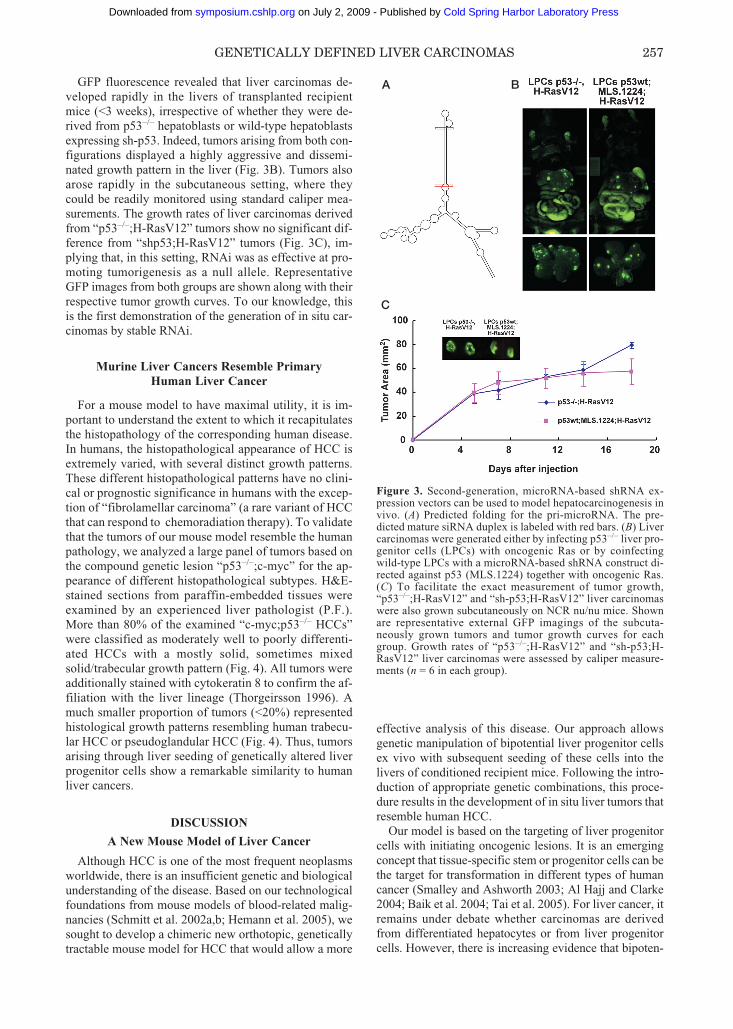
GFP fluorescence revealed that liver carcinomas de-veloped rapidly in the livers of transplanted recipientmice (<3 weeks), irrespective of whether they were de-rived from p53–/– hepatoblasts or wild-type hepatoblastsexpressing sh-p53. Indeed, tumors arising from both con-figurations displayed a highly aggressive and dissemi-nated growth pattern in the liver (Fig. 3B). Tumors alsoarose rapidly in the subcutaneous setting, where theycould be readily monitored using standard caliper mea-surements. The growth rates of liver carcinomas derivedfrom “p53–/–;H-RasV12” tumors show no significant dif-ference from “shp53;H-RasV12” tumors (Fig. 3C), im-plying that, in this setting, RNAi was as effective at pro-moting tumorigenesis as a null allele. RepresentativeGFP images from both groups are shown along with theirrespective tumor growth curves. To our knowledge, thisis the first demonstration of the generation of in situ car-cinomas by stable RNAi.
Murine Liver Cancers Resemble Primary Human Liver Cancer
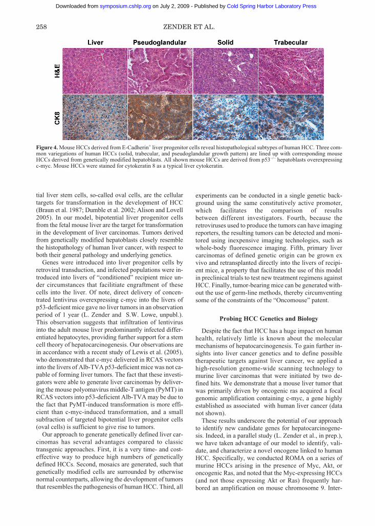
For a mouse model to have maximal utility, it is im-portant to understand the extent to which it recapitulatesthe histopathology of the corresponding human disease.In humans, the histopathological appearance of HCC isextremely varied, with several distinct growth patterns.These different histopathological patterns have no clini-cal or prognostic significance in humans with the excep-tion of “fibrolamellar carcinoma” (a rare variant of HCCthat can respond to chemoradiation therapy). To validatethat the tumors of our mouse model resemble the humanpathology, we analyzed a large panel of tumors based onthe compound genetic lesion “p53–/–;c-myc” for the ap-pearance of different histopathological subtypes. H&E-stained sections from paraffin-embedded tissues wereexamined by an experienced liver pathologist (P.F.).More than 80% of the examined “c-myc;p53–/– HCCs”were classified as moderately well to poorly differenti-ated HCCs with a mostly solid, sometimes mixedsolid/trabecular growth pattern (Fig. 4). All tumors wereadditionally stained with cytokeratin 8 to confirm the af-filiation with the liver lineage (Thorgeirsson 1996). Amuch smaller proportion of tumors (<20%) representedhistological growth patterns resembling human trabecu-lar HCC or pseudoglandular HCC (Fig. 4). Thus, tumorsarising through liver seeding of genetically altered liverprogenitor cells show a remarkable similarity to humanliver cancers.
DISCUSSION
A New Mouse Model of Liver Cancer
Although HCC is one of the most frequent neoplasmsworldwide, there is an insufficient genetic and biologicalunderstanding of the disease. Based on our technologicalfoundations from mouse models of blood-related malig-nancies (Schmitt et al. 2002a,b; Hemann et al. 2005), wesought to develop a chimeric new orthotopic, geneticallytractable mouse model for HCC that would allow a more
effective analysis of this disease. Our approach allowsgenetic manipulation of bipotential liver progenitor cellsex vivo with subsequent seeding of these cells into thelivers of conditioned recipient mice. Following the intro-duction of appropriate genetic combinations, this proce-dure results in the development of in situ liver tumors thatresemble human HCC.
Our model is based on the targeting of liver progenitorcells with initiating oncogenic lesions. It is an emergingconcept that tissue-specific stem or progenitor cells can bethe target for transformation in different types of humancancer (Smalley and Ashworth 2003; Al Hajj and Clarke2004; Baik et al. 2004; Tai et al. 2005). For liver cancer, itremains under debate whether carcinomas are derivedfrom differentiated hepatocytes or from liver progenitorcells. However, there is increasing evidence that bipoten-
GENETICALLY DEFINED LIVER CARCINOMAS 257
Figure 3. Second-generation, microRNA-based shRNA ex-pression vectors can be used to model hepatocarcinogenesis invivo. (A) Predicted folding for the pri-microRNA. The pre-dicted mature siRNA duplex is labeled with red bars. (B) Livercarcinomas were generated either by infecting p53–/– liver pro-genitor cells (LPCs) with oncogenic Ras or by coinfectingwild-type LPCs with a microRNA-based shRNA construct di-rected against p53 (MLS.1224) together with oncogenic Ras.(C) To facilitate the exact measurement of tumor growth,“p53–/–;H-RasV12” and “sh-p53;H-RasV12” liver carcinomaswere also grown subcutaneously on NCR nu/nu mice. Shownare representative external GFP imagings of the subcuta-neously grown tumors and tumor growth curves for eachgroup. Growth rates of “p53–/–;H-RasV12” and “sh-p53;H-RasV12” liver carcinomas were assessed by caliper measure-ments (n = 6 in each group).
A B
C
251-262_29_Zender et al_Symp70 5/12/06 10:02 AM Page 257
Cold Spring Harbor Laboratory Press on July 2, 2009 - Published by symposium.cshlp.orgDownloaded from
tial liver stem cells, so-called oval cells, are the cellulartargets for transformation in the development of HCC(Braun et al. 1987; Dumble et al. 2002; Alison and Lovell2005). In our model, bipotential liver progenitor cellsfrom the fetal mouse liver are the target for transformationin the development of liver carcinomas. Tumors derivedfrom genetically modified hepatoblasts closely resemblethe histopathology of human liver cancer, with respect toboth their general pathology and underlying genetics.
Genes were introduced into liver progenitor cells byretroviral transduction, and infected populations were in-troduced into livers of “conditioned” recipient mice un-der circumstances that facilitiate engraftment of thesecells into the liver. Of note, direct delivery of concen-trated lentivirus overexpressing c-myc into the livers ofp53-deficient mice gave no liver tumors in an observationperiod of 1 year (L. Zender and S.W. Lowe, unpubl.).This observation suggests that infiltration of lentivirusinto the adult mouse liver predominantly infected differ-entiated hepatocytes, providing further support for a stemcell theory of hepatocarcinogenesis. Our observations arein accordance with a recent study of Lewis et al. (2005),who demonstrated that c-myc delivered in RCAS vectorsinto the livers of Alb-TVA p53-deficient mice was not ca-pable of forming liver tumors. The fact that these investi-gators were able to generate liver carcinomas by deliver-ing the mouse polyomavirus middle-T antigen (PyMT) inRCAS vectors into p53-deficient Alb-TVA may be due tothe fact that PyMT-induced transformation is more effi-cient than c-myc-induced transformation, and a smallsubfraction of targeted bipotential liver progenitor cells(oval cells) is sufficient to give rise to tumors.
Our approach to generate genetically defined liver car-cinomas has several advantages compared to classictransgenic approaches. First, it is a very time- and cost-effective way to produce high numbers of genetically defined HCCs. Second, mosaics are generated, such thatgenetically modified cells are surrounded by otherwisenormal counterparts, allowing the development of tumorsthat resembles the pathogenesis of human HCC. Third, all
experiments can be conducted in a single genetic back-ground using the same constitutively active promoter,which facilitates the comparison of results between different investigators. Fourth, because theretroviruses used to produce the tumors can have imagingreporters, the resulting tumors can be detected and moni-tored using inexpensive imaging technologies, such aswhole-body fluorescence imaging. Fifth, primary livercarcinomas of defined genetic origin can be grown exvivo and retransplanted directly into the livers of recipi-ent mice, a property that facilitates the use of this modelin preclinical trials to test new treatment regimens againstHCC. Finally, tumor-bearing mice can be generated with-out the use of germ-line methods, thereby circumventingsome of the constraints of the “Oncomouse” patent.
Probing HCC Genetics and Biology
Despite the fact that HCC has a huge impact on humanhealth, relatively little is known about the molecularmechanisms of hepatocarcinogenesis. To gain further in-sights into liver cancer genetics and to define possibletherapeutic targets against liver cancer, we applied ahigh-resolution genome-wide scanning technology tomurine liver carcinomas that were initiated by two de-fined hits. We demonstrate that a mouse liver tumor thatwas primarily driven by oncogenic ras acquired a focalgenomic amplification containing c-myc, a gene highlyestablished as associated with human liver cancer (datanot shown).
These results underscore the potential of our approachto identify new candidate genes for hepatocarcinogene-sis. Indeed, in a parallel study (L. Zender et al., in prep.),we have taken advantage of our model to identify, vali-date, and characterize a novel oncogene linked to humanHCC. Specifically, we conducted ROMA on a series ofmurine HCCs arising in the presence of Myc, Akt, oroncogenic Ras, and noted that the Myc-expressing HCCs(and not those expressing Akt or Ras) frequently har-bored an amplification on mouse chromosome 9. Inter-
258 ZENDER ET AL.
Figure 4. Mouse HCCs derived from E-Cadherin+ liver progenitor cells reveal histopathological subtypes of human HCC. Three com-mon variegations of human HCCs (solid, trabecular, and pseudoglandular growth pattern) are lined up with corresponding mouseHCCs derived from genetically modified hepatoblasts. All shown mouse HCCs are derived from p53–/– hepatoblasts overexpressingc-myc. Mouse HCCs were stained for cytokeratin 8 as a typical liver cytokeratin.
251-262_29_Zender et al_Symp70 5/12/06 10:02 AM Page 258
Cold Spring Harbor Laboratory Press on July 2, 2009 - Published by symposium.cshlp.orgDownloaded from
estingly, by analyzing a series of human HCCs, we founda corresponding amplification on chromosome 11, whichis syntenic to mouse chromosome 9 and contained thesame set of genes. Through further cross-species analysisand expression studies, we narrowed down one gene—the cellular inhibitor of apoptosis gene cIAP1—as themost likely “driver” gene in this amplicon.
One powerful aspect of this system is that genes iden-tified as altered in the tumors can be rapidly examined foroncogenic potential in the mouse using the precise ge-netic configuration in which the spontaneous lesionarose. Thus, by returning to the mouse, we demonstratedthat enforced expression of cIAP1 in the genetic contextin which the amplification occurred (p53–/–;c-Myc) ac-celerated tumorigenesis and, conversely, suppression ofcIAP1 in cells harboring the amplicon reduced tumorgrowth. These effects were highly genotype-specific—thus, cIAP1 had no impact on promoting tumors initiatedwith Akt or Ras, and shRNAs targeting cIAP did not de-lay the growth of tumors that did not contain the cIAPamplicon. In addition to validating cIAP1 as a bona fideoncogene, these studies define an integrative approach tocancer genetics and biology that may facilitate the anno-tation of the cancer genome (for more discussion, see L.Zender et al., in prep.).
In addition to identifying new genes involved in HCCusing genome-scanning approaches followed by “reversegenetics,” our model is amenable to forward genetics ap-proaches using new, low-complexity cDNA or RNAi li-braries to identify new oncogenes and tumor suppressorgenes, respectively. In principle, one can envision intro-ducing defined “pools” of cDNAs or shRNAs into hepa-toblasts from a “sensitized” genetic background followedby transplantation into the livers of recipient mice. Inprinciple, if a given pool harbors an oncogenic cDNA orshRNA, tumors should arise more rapidly than controls.Here, we provide a proof-of-principle experiment thatmicroRNA-based shRNAs can be used to generate in situliver carcinomas. This HCC mouse model should proveto be a valuable tool to further explore molecular mecha-nisms of hepatocarcinogenesis and to help define newmolecular targets.
A Preclinical Mouse Model for TestingNew Therapies
Human liver cancer currently represents the third lead-ing cause of cancer deaths; in part, because no effectivechemotherapeutic regimens are currently available. Thus,a major effort of HCC research is to develop the molecu-lar and modeling infrastructure to identify new drug tar-gets and test their potential efficacy. As mentioned above,our chimeric mouse model of HCC has many featuresthat should enable it to provide new insights into themolecular genetics of HCC. In addition, as shown in thisstudy, all of the liver tumors we produce can be moni-tored by GFP imaging, and most can be propagated invitro or by transplanting into the livers of syngeneic re-cipients. Although more sensitive imaging techniquessuch as bioluminescence should improve monitoring ca-
pabilities, the availability of defined tumors and simpleimaging modalities should enable relatively high-throughput preclinical studies using new drugs or drugcombinations on HCC. Such approaches, particularlywith new molecularly targeted therapies, may identifynew treatment regimens that may be effective againstliver cancers with a defined set of underlying genetic le-sions.
ACKNOWLEDGMENTS
We thank Amy Brady, Christine Rosenthal, Lisa Bianco,and Maria S. Jiao for excellent technical assistance, and Dr.Eva Hernando for assistance with histology. We also ac-knowledge Michael Wigler, Lakshmi Muthuswamy, andother members of the Wigler group for providing the bioin-formatics required for the mouse ROMA chip and ROMAdata analysis, and the members of the Lowe lab for con-structive criticism and discussions throughout the course ofthis work. L.Z. was supported by the German Researchfoundation (Emmy Noether Programme). This work wasalso supported by a grant from the Alan Seligson founda-tion, and by grants CA13106, CA87497, and CA105388from the National Institutes of Health. S.W.L. and G.J.H.are Howard Hughes Medical Institute investigators.
REFERENCES
Al Hajj M. and Clarke M.F. 2004. Self-renewal and solid tumorstem cells. Oncogene 23: 7274.
Alison M.R. and Lovell M.J. 2005. Liver cancer: The role ofstem cells. Cell Prolif. 38: 407.
Baik I., Becker P.S., DeVito W.J., Lagiou P., Ballen K., Que-senberry P.J., and Hsieh C.C. 2004. Stem cells and prenatalorigin of breast cancer. Cancer Causes Control 15: 517.
Block G.D., Locker J., Bowen W.C., Petersen B.E., Katyal S.,Strom S.C., Riley T., Howard T.A., and Michalopoulos G.K.1996. Population expansion, clonal growth, and specific dif-ferentiation patterns in primary cultures of hepatocytes in-duced by HGF/SF, EGF and TGF alpha in a chemically de-fined (HGM) medium. J. Cell Biol. 132: 1133.
Braun L., Goyette M., Yaswen P., Thompson N.L., and FaustoN. 1987. Growth in culture and tumorigenicity after transfec-tion with the ras oncogene of liver epithelial cells from car-cinogen-treated rats. Cancer Res. 47: 4116.
Bruix J., Boix L., Sala M., and Llovet J.M. 2004. Focus on hep-atocellular carcinoma. Cancer Cell 5: 215.
Buendia M.A. 2000. Genetics of hepatocellular carcinoma.Semin. Cancer Biol. 10: 185.
Dabeva M.D. and Shafritz D.A. 2003. Hepatic stem cells andliver repopulation. Semin. Liver Dis. 23: 349.
Deane N.G., Parker M.A., Aramandla R., Diehl L., Lee W.J.,Washington M.K., Nanney L.B., Shyr Y., and Beauchamp R.D.2001. Hepatocellular carcinoma results from chronic cyclin D1overexpression in transgenic mice. Cancer Res. 61: 5389.
Dickins R.A., Hemann M.T., Zilfou J.T., Simpson D.R., IbarraI., Hannon G.J., and Lowe S.W. 2005. Probing tumor pheno-types using stable and regulated synthetic microRNA precur-sors. Nat. Genet. 37: 1289.
Dumble M.L., Croager E.J., Yeoh G.C., and Quail E.A. 2002.Generation and characterization of p53 null transformed hep-atic progenitor cells: Oval cells give rise to hepatocellular car-cinoma. Carcinogenesis 23: 435.
Guo D., Fu T., Nelson J.A., Superina R.A., and Soriano H.E.2002. Liver repopulation after cell transplantation in micetreated with retrorsine and carbon tetrachloride. Transplanta-tion 73: 1818.
GENETICALLY DEFINED LIVER CARCINOMAS 259
251-262_29_Zender et al_Symp70 5/12/06 10:02 AM Page 259
Cold Spring Harbor Laboratory Press on July 2, 2009 - Published by symposium.cshlp.orgDownloaded from

Gupta S., Aragona E., Vemuru R.P., Bhargava K.K., Burk R.D.,and Chowdhury J.R. 1991. Permanent engraftment and func-tion of hepatocytes delivered to the liver: Implications forgene therapy and liver repopulation. Hepatology 14: 144.
Hannon G.J. and Rossi J.J. 2004. Unlocking the potential of thehuman genome with RNA interference. Nature 431: 371.
Harada N., Oshima H., Katoh M., Tamai Y., Oshima M., andTaketo M.M. 2004. Hepatocarcinogenesis in mice with beta-catenin and Ha-ras gene mutations. Cancer Res. 64: 48.
Hemann M.T., Fridman J.S., Zilfou J.T., Hernando E., PaddisonP.J., Cordon-Cardo C., Hannon G.J., and Lowe S.W. 2003.An epi-allelic series of p53 hypomorphs created by stableRNAi produces distinct tumor phenotypes in vivo. Nat.Genet. 33: 396.
Hemann M.T., Bric A., Teruya-Feldstein J., Herbst A., NilssonJ.A., Cordon-Cardo C., Cleveland J.L., Tansey W.P., andLowe S.W. 2005. Evasion of the p53 tumour surveillance net-work by tumour-derived MYC mutants. Nature 436: 807.
Huo T.I., Wang X.W., Forgues M., Wu C.G., Spillare E.A., Gi-annini C., Brechot C., and Harris C.C. 2001. Hepatitis B virusX mutants derived from human hepatocellular carcinoma re-tain the ability to abrogate p53-induced apoptosis. Oncogene20: 3620.
Jhappan C., Stahle C., Harkins R.N., Fausto N., Smith G.H., andMerlino G.T. 1990. TGF alpha overexpression in transgenicmice induces liver neoplasia and abnormal development ofthe mammary gland and pancreas. Cell 61: 1137.
Kim S.O., Park J.G., and Lee Y.I. 1996. Increased expression ofthe insulin-like growth factor I (IGF-I) receptor gene in hepa-tocellular carcinoma cell lines: Implications of IGF-I receptorgene activation by hepatitis B virus X gene product. CancerRes. 56: 3831.
Kusano N., Shiraishi K., Kubo K., Oga A., Okita K., and SasakiK. 1999. Genetic aberrations detected by comparative ge-nomic hybridization in hepatocellular carcinomas: Their rela-tionship to clinicopathological features. Hepatology 29: 1858.
Laconi S., Curreli F., Diana S., Pasciu D., De Filippo G., SarmaD.S., Pani P., and Laconi E. 1999. Liver regeneration in re-sponse to partial hepatectomy in rats treated with retrorsine: Akinetic study. J. Hepatol. 31: 1069.
Lewis B.C., Klimstra D.S., Socci N.D., Xu S., Koutcher J.A.,and Varmus H.E. 2005. The absence of p53 promotes metas-tasis in a novel somatic mouse model for hepatocellular car-cinoma. Mol. Cell. Biol. 25: 1228.
Llovet J.M., Burroughs A., and Bruix J. 2003. Hepatocellularcarcinoma. Lancet 362: 1907.
Lucito R., Healy J., Alexander J., Reiner A., Esposito D., ChiM., Rodgers L., Brady A., Sebat J., Troge J., West J.A., Ros-tan S., Nguyen K.C., Powers S., Ye K.Q., Olshen A., Venka-traman E., Norton L., and Wigler M. 2003. Representationaloligonucleotide microarray analysis: A high-resolutionmethod to detect genome copy number variation. GenomeRes. 13: 2291.
Manickan E., Satoi J., Wang T.C., and Liang T.J. 2001. Condi-tional liver-specific expression of simian virus 40 T antigenleads to regulatable development of hepatic neoplasm intransgenic mice. J. Biol. Chem. 276: 13989.
Murakami H., Sanderson N.D., Nagy P., Marino P.A., MerlinoG., and Thorgeirsson S.S. 1993. Transgenic mouse model forsynergistic effects of nuclear oncogenes and growth factors intumorigenesis: Interaction of c-myc and transforming growthfactor alpha in hepatic oncogenesis. Cancer Res. 53: 1719.
Murakami Y., Hayashi K., Hirohashi S., and Sekiya T. 1991.Aberrations of the tumor suppressor p53 and retinoblastomagenes in human hepatocellular carcinomas. Cancer Res. 51:5520.
Nhieu J.T., Renard C.A., Wei Y., Cherqui D., Zafrani E.S., andBuendia M.A. 1999. Nuclear accumulation of mutated beta-catenin in hepatocellular carcinoma is associated with in-creased cell proliferation. Am. J. Pathol. 155: 703.
Niketeghad F., Decker H.J., Caselmann W.H., Lund P., GeisslerF., Dienes H.P., and Schirmacher P. 2001. Frequent genomicimbalances suggest commonly altered tumour genes in hu-
man hepatocarcinogenesis. Br. J. Cancer 85: 697.Nishida N., Fukuda Y., Komeda T., Kita R., Sando T., Furukawa
M., Amenomori M., Shibagaki I., Nakao K., and Ikenaga M.,et al. 1994. Amplification and overexpression of the cyclin D1gene in aggressive human hepatocellular carcinoma. CancerRes. 54: 3107.
Nitou M., Sugiyama Y., Ishikawa K., and Shiojiri N. 2002. Pu-rification of fetal mouse hepatoblasts by magnetic beadscoated with monoclonal anti-e-cadherin antibodies and theirin vitro culture. Exp. Cell Res. 279: 330.
Parkin D.M., Bray F., Ferlay J., and Pisani P. 2001. Estimating theworld cancer burden: Globocan 2000. Int. J. Cancer 94: 153.
Pear W.S., Miller J.P., Xu L., Pui J.C., Soffer B., QuackenbushR.C., Pendergast A.M., Bronson R., Aster J.C., Scott M.L.,and Baltimore D. 1998. Efficient and rapid induction of achronic myelogenous leukemia-like myeloproliferative dis-ease in mice receiving P210 bcr/abl-transduced bone marrow.Blood 92: 3780.
Peng S.Y., Lai P.L., and Hsu H.C. 1993. Amplification of the c-myc gene in human hepatocellular carcinoma: Biologic sig-nificance. J. Formos. Med. Assoc. 92: 866.
Ponder K.P., Gupta S., Leland F., Darlington G., Finegold M.,DeMayo J., Ledley F.D., Chowdhury J.R., and Woo S.L.1991. Mouse hepatocytes migrate to liver parenchyma andfunction indefinitely after intrasplenic transplantation. Proc.Natl. Acad. Sci. 88: 1217.
Ray R.B., Steele R., Meyer K., and Ray R. 1997. Transcriptionalrepression of p53 promoter by hepatitis C virus core protein.J. Biol. Chem. 272: 10983.
Sage J., Miller A.L., Perez-Mancera P.A., Wysocki J.M., andJacks T. 2003. Acute mutation of retinoblastoma gene func-tion is sufficient for cell cycle re-entry. Nature 424: 223.
Sandgren E.P., Quaife C.J., Pinkert C.A., Palmiter R.D., andBrinster R.L. 1989. Oncogene-induced liver neoplasia intransgenic mice. Oncogene 4: 715.
Schmitt C.A., Fridman J.S., Yang M., Baranov E., HoffmanR.M., and Lowe S.W. 2002a. Dissecting p53 tumor suppres-sor functions in vivo. Cancer Cell 1: 289.
Schmitt C.A., Fridman J.S., Yang M., Lee S., Baranov E., Hoff-man R.M., and Lowe S.W. 2002b. A senescence programcontrolled by p53 and p16INK4a contributes to the outcomeof cancer therapy. Cell 109: 335.
Shachaf C.M., Kopelman A.M., Arvanitis C., Karlsson A., BeerS., Mandl S., Bachmann M.H., Borowsky A.D., Ruebner B.,Cardiff R.D., Yang Q., Bishop J.M., Contag C.H., and FelsherD.W. 2004. MYC inactivation uncovers pluripotent differen-tiation and tumour dormancy in hepatocellular cancer. Nature431: 1112.
Shafritz D.A. and Dabeva M.D. 2002. Liver stem cells andmodel systems for liver repopulation. J. Hepatol. 36: 552.
Silva J.M., Li M.Z., Chang K., Ge W., Golding M.C., Rickles R.J.,Siolas D., Hu G., Paddison P.J., Schlabach M.R., Sheth N.,Bradshaw J., Burchard J., Kulkarni A., Cavet G., Sachidanan-dam R., McCombie W.R., Cleary M.A., Elledge S.J., and Han-non G.J. 2005. Second-generation shRNA libraries coveringthe mouse and human genomes. Nat. Genet. 37: 1281.
Smalley M. and Ashworth A. 2003. Stem cells and breast cancer:A field in transit. Nat. Rev. Cancer 3: 832.
Steinberg P., Frank H., Odenthal M., Dienes H.P., and Seidel A.1997. Role of the Ha-ras gene in the malignant transformationof rat liver oval cells. Int. J. Cancer 71: 680.
Tai M.H., Chang C.C., Kiupel M., Webster J.D., Olson L.K., andTrosko J.E. 2005. Oct4 expression in adult human stem cells:Evidence in support of the stem cell theory of carcinogenesis.Carcinogenesis 26: 495.
Thorgeirsson S.S. 1996. Hepatic stem cells in liver regeneration.FASEB J. 10: 1249.
Tsao M.S. and Grisham J.W. 1987. Hepatocarcinomas, cholan-giocarcinomas, and hepatoblastomas produced by chemicallytransformed cultured rat liver epithelial cells. A light- andelectron-microscopic analysis. Am. J. Pathol. 127: 168.
Ueda H., Ullrich S.J., Gangemi J.D., Kappel C.A., Ngo L., Feit-elson M.A., and Jay G. 1995. Functional inactivation but not
260 ZENDER ET AL.
251-262_29_Zender et al_Symp70 5/12/06 10:02 AM Page 260
Cold Spring Harbor Laboratory Press on July 2, 2009 - Published by symposium.cshlp.orgDownloaded from

structural mutation of p53 causes liver cancer. Nat. Genet. 9:41.
Verna L., Whysner J., and Williams G.M. 1996a. 2-Acetyl-aminofluorene mechanistic data and risk assessment: DNAreactivity, enhanced cell proliferation and tumor initiation.Pharmacol. Ther. 71: 83.
———. 1996b. N-nitrosodiethylamine mechanistic data andrisk assessment: Bioactivation, DNA-adduct formation, mu-tagenicity, and tumor initiation. Pharmacol. Ther. 71: 57.
Wang R., Ferrell L.D., Faouzi S., Maher J.J., and Bishop J.M.2001. Activation of the Met receptor by cell attachment in-duces and sustains hepatocellular carcinomas in transgenicmice. J. Cell Biol. 153: 1023.
Wang X.W., Forrester K., Yeh H., Feitelson M.A., Gu J.R., andHarris C.C. 1994. Hepatitis B virus X protein inhibits p53 se-quence-specific DNA binding, transcriptional activity, andassociation with transcription factor ERCC3. Proc. Natl.Acad. Sci. 91: 2230.
Wong C.M., Fan S.T., and Ng I.O. 2001. β-Catenin mutation andoverexpression in hepatocellular carcinoma: Clinicopatho-logic and prognostic significance. Cancer 92: 136.
Wong N., Lai P., Lee S.W., Fan S., Pang E., Liew C.T., ShengZ., Lau J.W., and Johnson P.J. 1999. Assessment of geneticchanges in hepatocellular carcinoma by comparative genomichybridization analysis: Relationship to disease stage, tumorsize, and cirrhosis. Am. J. Pathol. 154: 37.
GENETICALLY DEFINED LIVER CARCINOMAS 261
251-262_29_Zender et al_Symp70 5/12/06 10:02 AM Page 261
Cold Spring Harbor Laboratory Press on July 2, 2009 - Published by symposium.cshlp.orgDownloaded from

251-262_29_Zender et al_Symp70 5/12/06 10:02 AM Page 262
Cold Spring Harbor Laboratory Press on July 2, 2009 - Published by symposium.cshlp.orgDownloaded from


![The Time&Averaged Paleomagnetic Field · netic studies [e.g., Hospets, 1954; Cox and Doell, 1960; Irving, 1964; Opdyke and Henry, 1969] and archeomag- netic investigation [Champion,](https://static.fdocuments.in/doc/165x107/610d3346ea5efe04b0355db7/the-timeaveraged-paleomagnetic-field-netic-studies-eg-hospets-1954-cox.jpg)