General principles of fractures treatment 1
53
Transcript of General principles of fractures treatment 1
Fracture ‐loss of continuity of bone
Hairline fracture, microscopic fracture, highly comminuted fracture
Dislocation ‐loss of congruity between the articulation surface of joint
Subluxation‐articulating surface of jointare no longer congruous but still maintain contact.

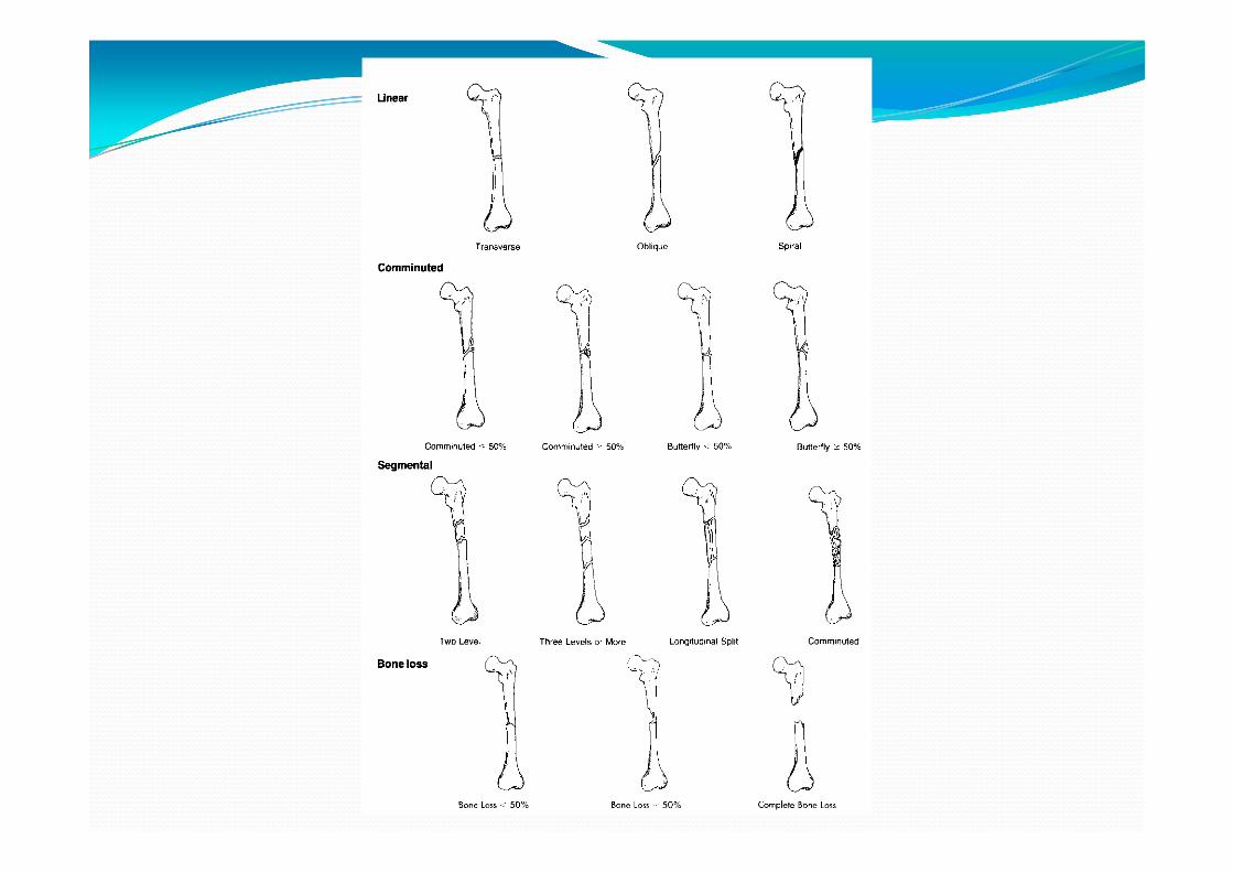
Classification of fracture According plane of # surface
Simple – closed Compound – open
According to cause traumatic pathological stress or fatigue fracture
According to number Single Multiple
Complete #, incomplete#

Mechanism of injury Spiral (twisting) Short oblique
Bending ‐ Triangular ‘butterfly’ fragment Transverse
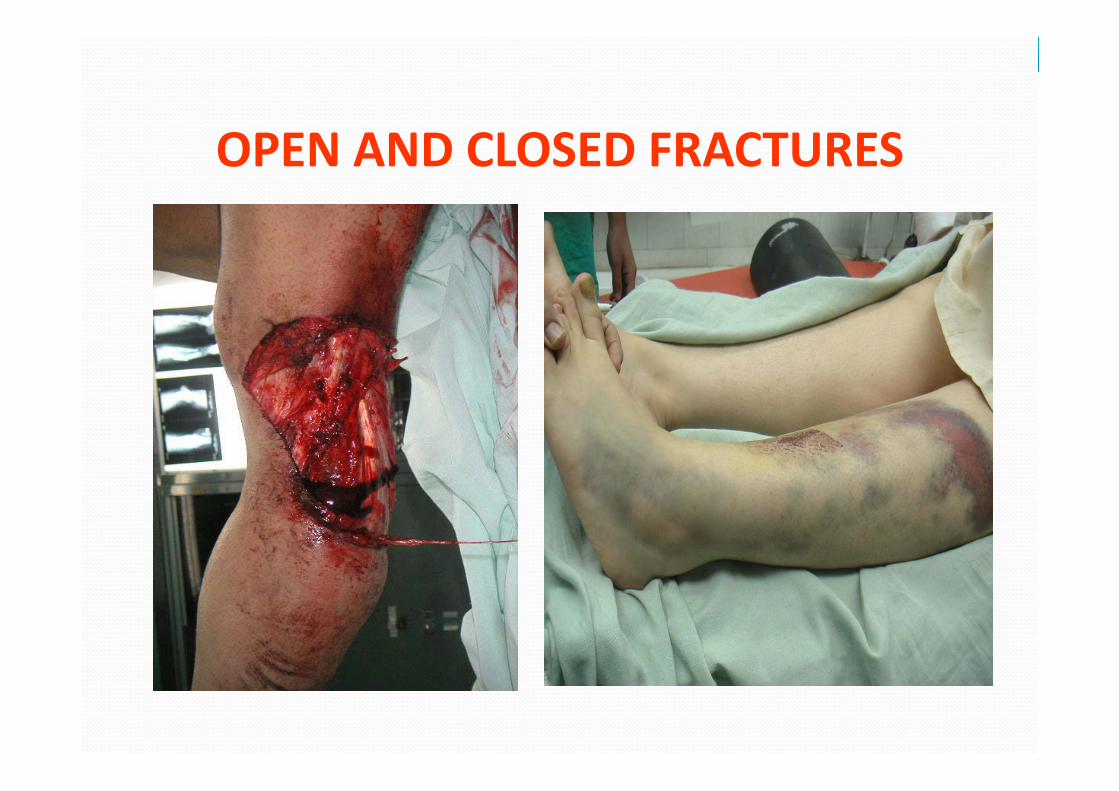
OPEN AND CLOSED FRACTURES

Traumatic fracture
Direct violence :
Indirect violence : twisting, bending,
Muscular contraction.

Pathological fracture
It is one in which a bone is broken through an area weakened by pre‐existing disease , & by a degree of force that would have left normal bone intact e.g osteoporosis , O.M. , bone tumours
Stress fracture :
Bone, like other materials, reacts to repeated loading. On occasion, it becomes fatigued & a crack develops
e.g military installations, ballet dancers & athletes.
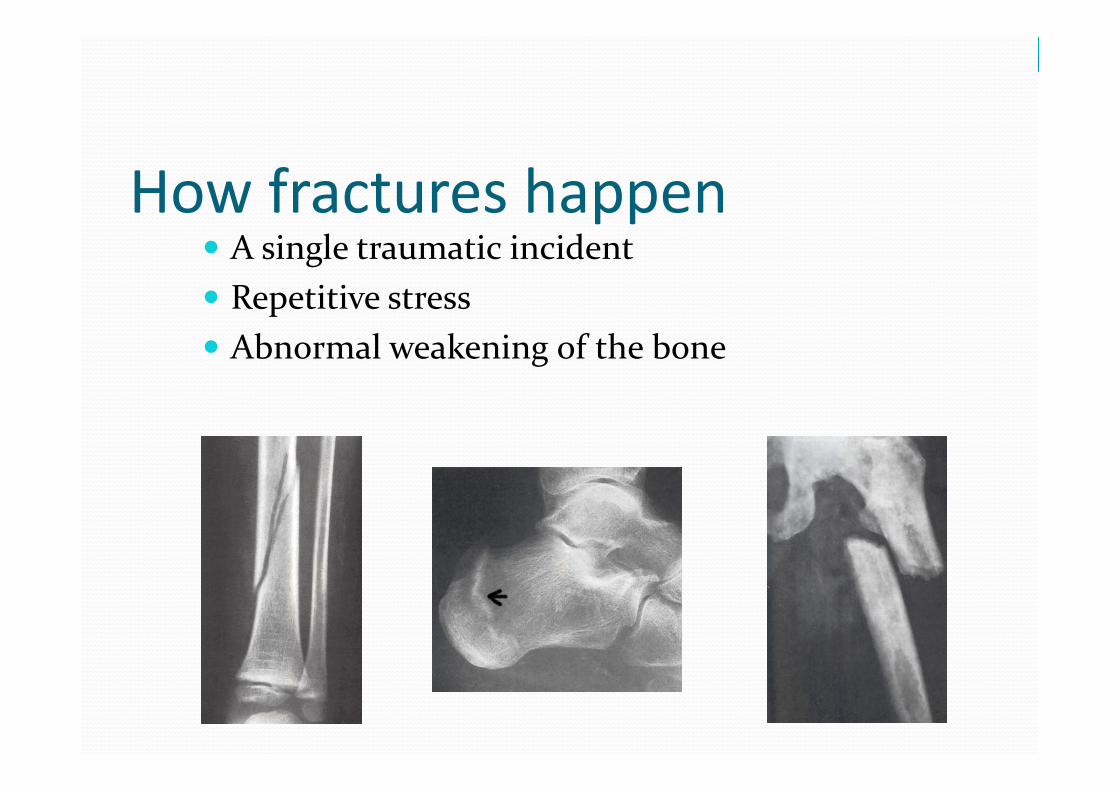
How fractures happen A single traumatic incident Repetitive stress Abnormal weakening of the bone
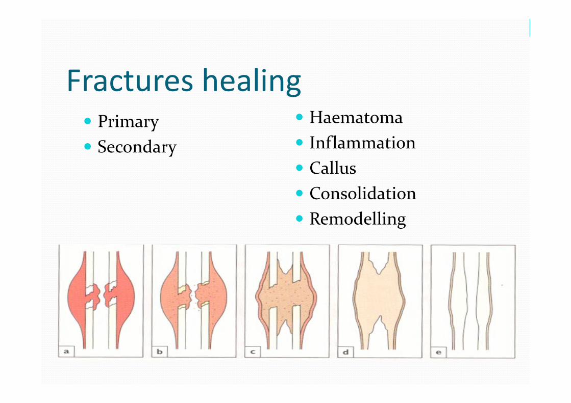
Fractures healing Primary
Secondary
Haematoma
Inflammation
Callus Consolidation
Remodelling

Fractures healing
Clinical Features of Fracture History of trauma
Symptoms & signs: 1. Pain & tenderness 2. Swelling
3. Deformity 4. Crepitus 5. Loss of function 6. Abnormal move. 7. N.V. injuries

Diagnosis
Clinical picture
Radiography

Principles of treatment
Anatomical reduction
Stable internal fixation
Preservation of blood supply
Early mobilization

Implants types Pin and wire fixation
Screw fixation
Plate and screw fixation
Intramedullary nail fixation
External fixation

Open fractures Wound debridement
Antibiotic prophylaxis
Stabilization of the fracture
Early wound cover

Principles of fractures
Fracture repair is a tissue regeneration process rather than a healing process the injured bone is replaced by bone.
The process of repair varies according to: ‐The type of bone involved. ‐The amount of movement at the fracture. ‐The closeness of the fracture surfaces.

Principles of fractures
Unfavorable factors Impairment of blood supply Infection Excessive movement Presence of tumor Synovial fluid in intraarticular Fx. Interposition of soft tissue Any form of Nicotine
Definitive fracture treatment The goal of fracture treatment is to obtain union of thefracture in the most anatomical position compatible withmaximal functional return of the extremity.
Conservative
Operative
Principles of Treatment
Treat the Patient, not only the fracture
Restriction of movement Prevention of displacement Alleviation of pain Promote soft‐tissue healing Try to allow free movement of the unaffected parts
Splint the fracture, not the entire limb
Principles of Treatment
Methods of holding reduction:
Sustained traction
Cast splintage
Functional bracing
Internal fixation
External fixation

Definitive Fracture Fixation Options
Casts and Splints Appropriate for many fractures especially hand and foot fractures
Adults typically will get plaster splints initially transitioned to fiberglass casts as swelling decreases
Kids typically will get fiberglass casts

CLOSED, UNDISPLACED CLOSED, REDUCIBLE
CONSERVATIVE TREATMENT
2- CAST
Below Knee Above Knee

Complications of cast splintage Liable to appear once the patient has left the hospital; added risk of delay before the problem is attended to
1. Tight cast 2. Pressure sores 3. Skin abrasion or laceration
4.Loose cast

Functional Bracing
Prevents joint stiffness while still permitting
fracture splintage and loading
Most commonly for fractures of the femur or tibia
Since its not very rigid, it is usually applied only when the fracture is beginning to unite
Comes out well on all four of the basic requirements: “hold” “move” “speed” “safe”
Definitive Fracture Fixation Options
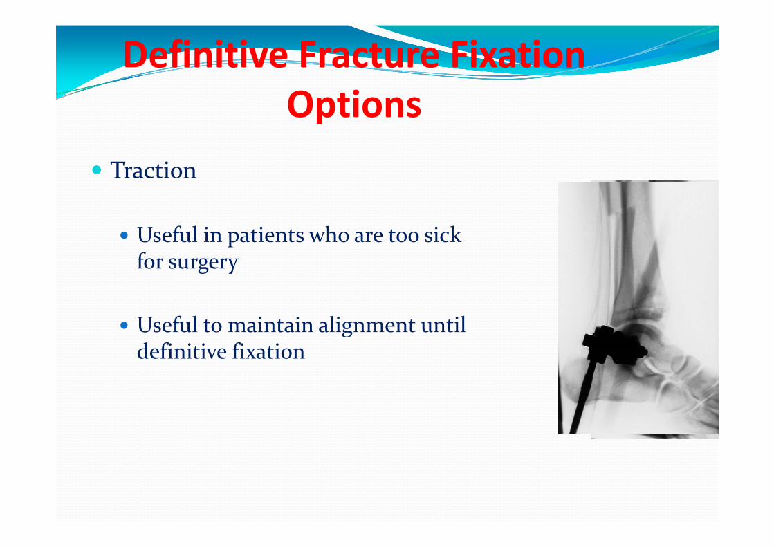
Traction
Useful in patients who are too sick for surgery
Useful to maintain alignment until definitive fixation
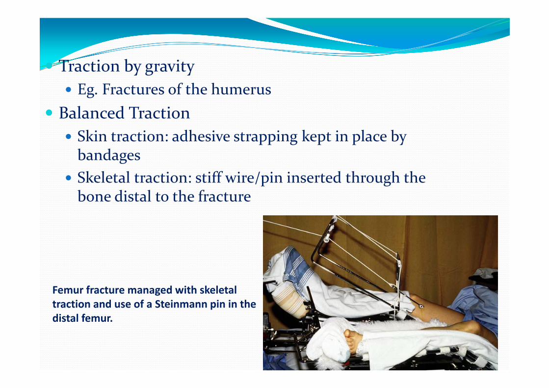
Traction by gravity Eg. Fractures of the humerus
Balanced Traction Skin traction: adhesive strapping kept in place by bandages
Skeletal traction: stiff wire/pin inserted through the bone distal to the fracture
Femur fracture managed with skeletal traction and use of a Steinmann pin in the distal femur.

Operative
ORIF (open reduction internal fixat.)
External fixation
Indications of ORIF
‐ absolute
‐ relative

Indications of ORIF ‐ Absolute Indications for ORIF of fractures
Unable to obtain an adequate reduction
Displaced intra‐articular fractures
Certain types of displaced epiphyseal fractures
Major avulsion fractures where there is loss of function of a joint ormuscle group
Non‐unions
Re‐ implantations of limbs or extremities

Indications of ORIF Relative Indications for ORIF of fractures
Delayed unions
Multiple fractures to assist in care and general management
Unable to maintain a reduction
Pathological fractures
To assist in nursing care
To reduce morbidity due to prolonged immobilisation
For fractures in which closed methods are known to be ineffective

Indications of ORIF Questionable
Fractures accompanying nerve of vessel injury
Open fractures
Cosmetic considerations
Economic considerations

Types of Internal Fixation ‐ Pin & wire fixat. ‐ Screw fixat. ‐ Plate & screws fixat. ‐ Intra‐medullary fixat.

Definitive Fracture Fixation Options
Open Reduction and Internal fixation with Plates and screws Used for many fractures especially those involving joints


Definitive Fracture Fixation Options
Intramedullary Nails Treatment of choice for most tibia and femur fractures
Used in selected humerus and forearm fractures


Internal Fixation “holds” securely with precise reduction
“movements” can begin at once (no stiffness and edema)
“speed”: patient can leave hospital as soon as wound ishealed, but full weight bearing is unsafe for some time
“safety”= biggest problem! SEPSIS!!! Risk depends on: the patient, the surgeon, the facilities

Definitive Fracture Fixation Options
Joint Replacement
Used in displaced femoral neck fractures in geriatric patients
Allows for early ambulation
Occasionally used in geriatric pts with comminuted shoulder or elbow fractures
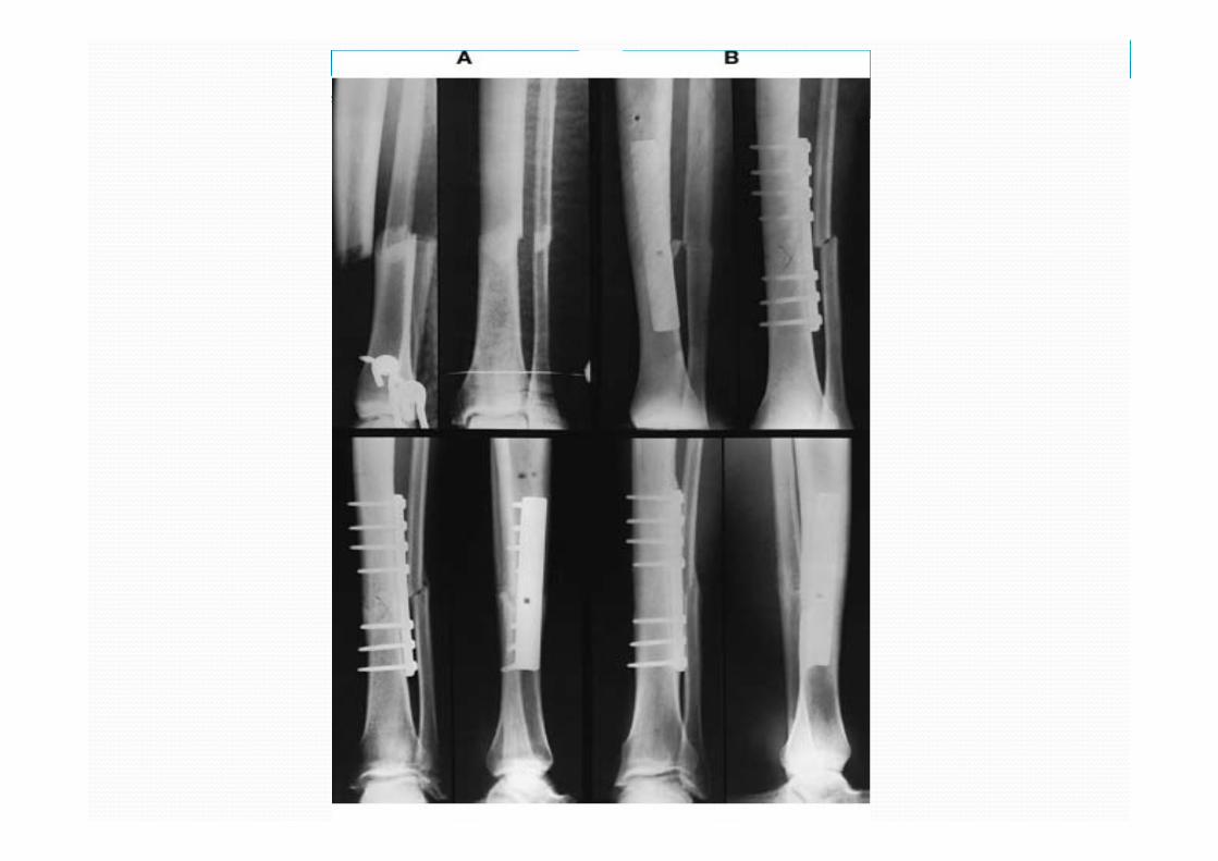
Complications of internal fixation
Most are due to poor technique, equipment, or operatingconditions
Infection ○ Iatrogenic infection is now the most common cause of chronic
osteomyelitis
Non‐union ○ Excessive stripping of the soft tissues ○ unnecessary damage to the blood supply in the course of
operative fixation ○ rigid fixation with a gap between the fragments
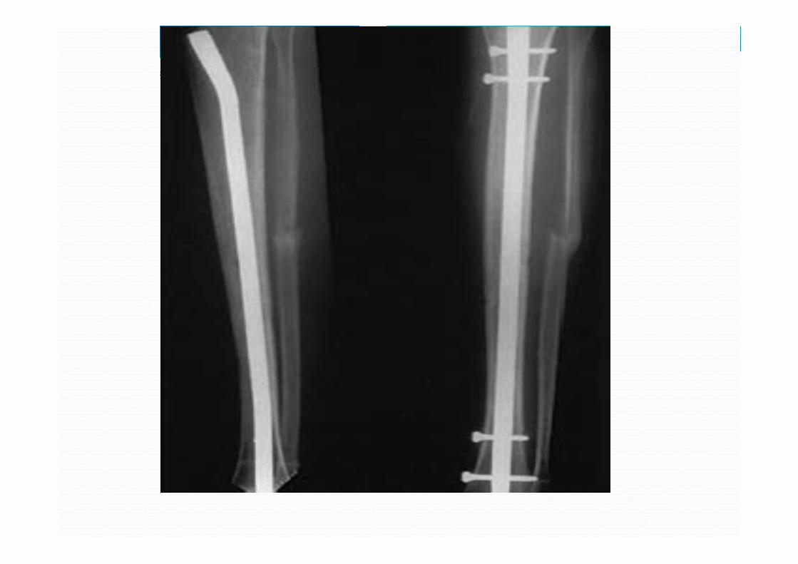
Implant failure
Refracture

Definitive Fracture Fixation Options External Fixation
Used primarily in the treatment of open fractures and pelvis fractures
Also useful as temporary stabilization prior to definitive fixation

External Fixation
Permits adjustment of length and angulation
Some allow reduction of the fracture in all 3 planes.
Especially applicable to the long bones and the pelvis.
Indications: 1. Fractures of the pelvis, which often cannot be controlled
quickly by any other method.
2. Fractures associated with severe soft‐tissue damage wherethe wound can be left open for inspection, dressing, ordefinitive coverage.

External Fixation
3. Severely comminuted and unstable fractures, which can beheld out to length until healing commences.
4. Fractures of the pelvis, which often cannot be controlledquickly by any other method.
5. Fractures associated with nerve or vessel damage.
6. Infected fractures, for which internal fixation might not besuitable.
7. Un‐united fractures, where dead or sclerotic fragments can beexcised and the remaining ends brought together in theexternal fixator; sometimes this is combined with elongationin the normal part of the shaft

Complications of external fixation
High degree of training and skill! Often used for the most difficult fractures increased likelihood of complications
Damage to soft‐tissue structures
Over‐distraction No contact between the fragments union delayed/prevented
Pin‐track infection

OPEN FRACTURES Initial Management
At the scene of the accident
In the hospital

Types of Open Fractures
‐ The incidence of wound infection
‐ correlates directly with the extent ofsoft‐tissue damage, <2% in type 1>10% in type 3
‐ rises with increasing delay inobtaining soft tissue coverage of thefracture.

Principles of Treatment of Open Fractures
All open fractures assumed to be contaminated Prevent infection!
The essentials: Prompt wound debridement
Antibiotic prophylaxis
Stabilization of the fracture
Early definitive wound cover
Repeated examination of the limb because open fractures can alsobe associated with compartment syndrome

Complications of fractures Early
‐visceral injury ‐ vascular injury ‐ nerve injury
Late ‐
delayed union ‐ non‐union ‐ avascular necrosis ‐ bed sores
‐ compartment syndrome
‐myositis ossificans ‐ tendon lesion
‐ haemarthrosis ‐ nerve compression
‐ infection ‐ gas gangrene ‐ fracture blisters ‐ plaster and pressure sores
‐muscle contracture ‐ joint instability ‐ joint stiffness ‐ algodystrophy RSD ‐ osteoarthritis

Thank you