General Pathology Adel Inflammation
50
Jhqhudo Sdwkrorj| Iru Ghqwdo Vwxghqwv= D Frqflvh Wh{w
Transcript of General Pathology Adel Inflammation
7/21/2019 General Pathology Adel Inflammation
http://slidepdf.com/reader/full/general-pathology-adel-inflammation 1/50
Jhqhudo Sdwkrorj|
Iru Ghqwdo Vwxghqwv=
D Frqflvh Wh{w
A d e l M A b d e l - A z i m
E d i t i o n 1

7/21/2019 General Pathology Adel Inflammation
http://slidepdf.com/reader/full/general-pathology-adel-inflammation 2/50
7/21/2019 General Pathology Adel Inflammation
http://slidepdf.com/reader/full/general-pathology-adel-inflammation 3/50
General Pathology for Dental
Students: A Concise Text
Adel M. Abdel-Azim, MDSC, PhD
Professor and Chairman of Oral Pathology Department,
Faculty of Dentistry, Ain Shams University, Egypt
Faculty of Dentistry, Ain-Shams University 2010
7/21/2019 General Pathology Adel Inflammation
http://slidepdf.com/reader/full/general-pathology-adel-inflammation 4/50
——————————————————————————————–
All rights reserved. No part of this publication may be reproduced, stored in a
retrieval system, or transmitted in any form or by any means, electronic, me-
chanical, photocopying, recording or otherwise, without a written permission of
the author.
Medical knowledge is constantly changing. As new information become avail-
able, changes in treatment, procedures, equipment and the use of drugs become
necessary. Readers are strongly recommended to follow up recent medical infor-
mation.
———————————————————————————————

7/21/2019 General Pathology Adel Inflammation
http://slidepdf.com/reader/full/general-pathology-adel-inflammation 5/50
iii
Preface
The aim of this book is to offer an overview of aspects of pathology, with an em-
phasis on basic facts in general practice. Intended outcomes are that, having read
this book, readers should be more aware of the immediate steps needed to make
the diagnosis and arrange patient management.
The amount of knowledge that must be assimilated by the student of today makes
it important to present the basic facts in as simple and concise a manner as pos-
sible. Each year more is added to the sum of knowledge whilst the available timeto learn for examination purposes is not increased.
The material presented is somewhat comprehensive, but still concise. The high-
lights of the book are brief outlines summarizing the features of each disease.
These summary statements serve as take-home messages and could only facili-
tate learning. Composite drawings of clinical signs and symptoms, summarizing
the principal clinical findings in important diseases or their complications are yet
another highlight. Structural writing makes the book easier to use and contribute
to its esthetic appeal. The book has a clinical orientation and the pathology find-ings are frequently correlated with the clinical data. This is especially well done
in the chapters dealing with general pathology.

7/21/2019 General Pathology Adel Inflammation
http://slidepdf.com/reader/full/general-pathology-adel-inflammation 6/50
iv

7/21/2019 General Pathology Adel Inflammation
http://slidepdf.com/reader/full/general-pathology-adel-inflammation 7/50
v
Course Description
This course has been designed to provide you with a practical approach to gen-
eral and systemic pathology. The lectures and laboratories provide information
and practice needed to develop an introductory level of proficiency in formu-
lating differential diagnoses through the interpretation of gross and microscopic
changes in various organs and their correlation with clinical, radiologic and lab-
oratory tests.
Overall, the goals of the labs are to build your knowledge, skills and abilities in:
1. Developing a medical vocabulary in order to converse with other health
professionals.
2. Synthesizing lecture material with clinical information, laboratory, and ra-
diographic examinations of systemic diseases and their etiology, pathogen-
esis and prognosis.
Learning Objectives Upon completion of this course you will be able to:
1. Formulate an orderly differential diagnosis of possible causes for a patient’s
problem, including the correct etiology, plus the other most likely and per-
tinent etiologies.
2. Prioritize the list of possible causes in your differential diagnosis based on
clinical history, physical examination findings, radiographs and laboratory
work.
3. Suggest further medical tests to help distinguish between your different di-
agnostic choices.
4. Explain how this disease will affect your dental practices.
5. Describe the basic pathogenesis of various disease entities.
6. Recognize general medical terms utilized in pathology.

7/21/2019 General Pathology Adel Inflammation
http://slidepdf.com/reader/full/general-pathology-adel-inflammation 8/50
vi
Recommended Text Books
Kumar, V. (Ed.), Cotran, R. S., Robbins, S. L. (2007). Basic Pathology (8th ed.).
Philadelphia, PA: W.B. Saunders.To enhance the meaning of the lectures, the stu-
dent is expected to read the pertinent text material PRIOR to the lecture.
7/21/2019 General Pathology Adel Inflammation
http://slidepdf.com/reader/full/general-pathology-adel-inflammation 9/50
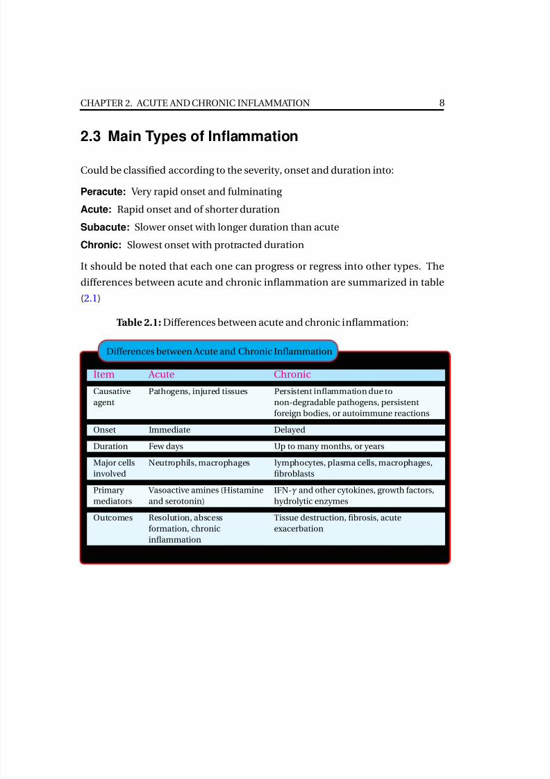
2 Acute and Chronic Inflammation
ChapterOverview 2.1 Definition
2.2 Terminology
2.3 Main Types of Inflammation
2.4 Types of Inflammatory Cells
2.5 Acute Inflammation
2.6 Chronic Inflammation
2.7 Mixed Acute & Chronic Inflammation
2.8 Granulomatous Inflammation
2.1 Definition
Inflammation is the tissue reaction against injury or irritant. Inflammation is not
a synonym for infection. Inflammation occurs only in living tissue.
2.2 Terminology
Greek root + -itis
gingivitis, metritis, pulpitis, nephritis and son on .....
7
7/21/2019 General Pathology Adel Inflammation
http://slidepdf.com/reader/full/general-pathology-adel-inflammation 10/50
CHAPTER 2. ACUTE AND CHRONIC INFLAMMATION 8
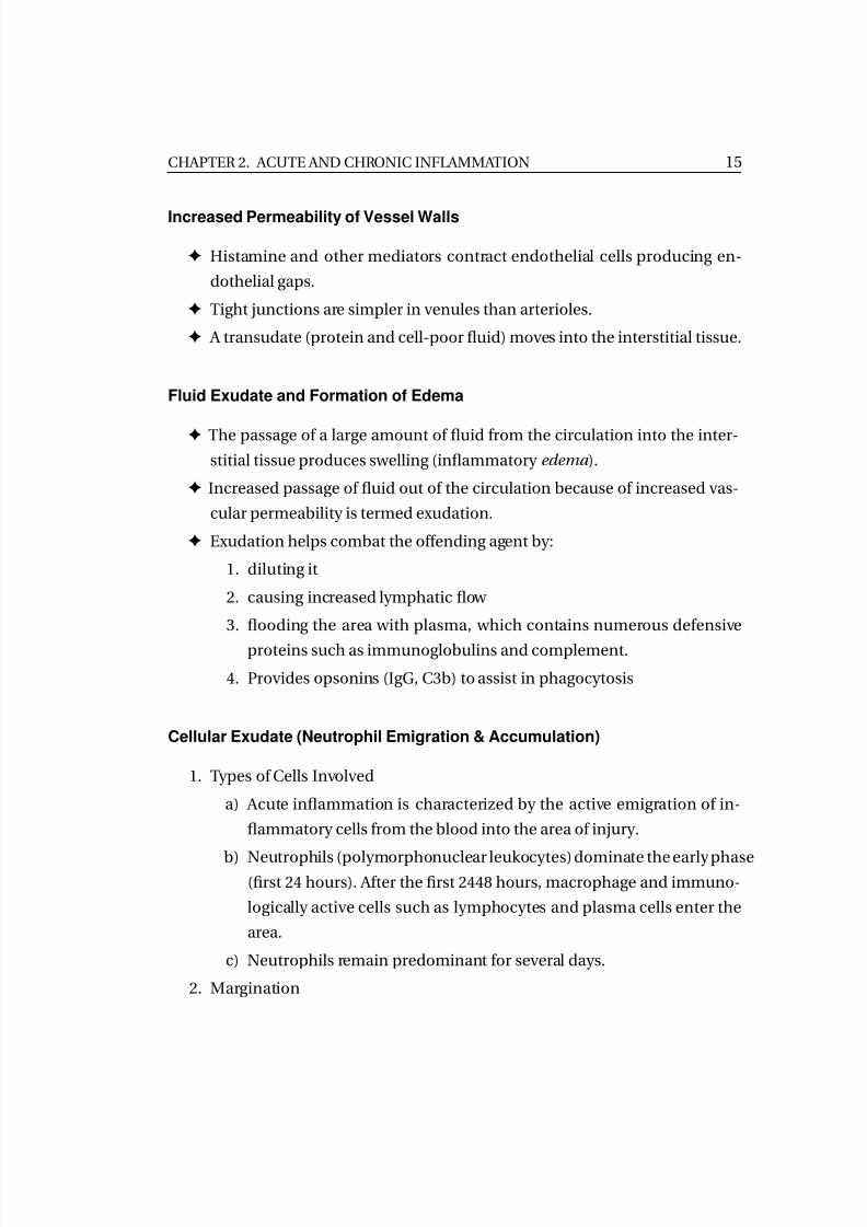
2.3 Main Types of Inflammation
Could be classified according to the severity, onset and duration into:
Peracute: Very rapid onset and fulminating
Acute: Rapid onset and of shorter duration
Subacute: Slower onset with longer duration than acute
Chronic: Slowest onset with protracted duration
It should be noted that each one can progress or regress into other types. The
differences between acute and chronic inflammation are summarized in table
(2.1)
Table 2.1: Differences between acute and chronic inflammation:
Item Acute Chronic
Causative
agent
Pathogens, injured tissues Persistent inflammation due to
non-degradable pathogens, persistent
foreign bodies, or autoimmune reactions
Onset Immediate Delayed
Duration Few days Up to many months, or years
Major cells
involved
Neutrophils, macrophages lymphocytes, plasma cells, macrophages,
fibroblasts
Primary
mediators
Vasoactive amines (Histamine
and serotonin)
IFN-γ and other cytokines, growth factors,
hydrolytic enzymes
Outcomes Resolution, abscess
formation, chronic
inflammation
Tissue destruction, fibrosis, acute
exacerbation
Differences between Acute and Chronic Inflammation

7/21/2019 General Pathology Adel Inflammation
http://slidepdf.com/reader/full/general-pathology-adel-inflammation 11/50
CHAPTER 2. ACUTE AND CHRONIC INFLAMMATION 9
2.4 Types of Inflammatory Cells
Inflammatory cells are classified based on the following basis, see also figure
(2.1):
1. Presence of absence of microscopic cytoplasmic granules:
a) Granulocytes
Polymorph nuclear leukocytes (PNL)
i. Neutrophils
ii. Esinophils
iii. Basophils
b) Agranulocytes
i. Monocytes (histiocytes, macrophages)
ii. Lymphocytes
iii. Plasma cells
2. Function (phagocytosis)
a) Phagocytic cells
i. Monocytes (histiocytes, macrophages)
ii. PNL (particularly neutrophils)
b) Non-phagocytic cells
i. Lymphocytes
ii. Plasma cells
2.5 Acute Inflammation
2.5.1 Definition
A rapid and transient response to injury or irritant

7/21/2019 General Pathology Adel Inflammation
http://slidepdf.com/reader/full/general-pathology-adel-inflammation 12/50
CHAPTER 2. ACUTE AND CHRONIC INFLAMMATION 10
Figure 2.1: Types of Inflammatory Cells
2.5.2 Cardinal Signs of Inflammation
classical 5 symptoms (Celsus 1st c. B.C., Virchow 19th c. A.D.)
1. calor heat, resulting from vasodilation of blood vessels
2. rubor redness, resulting from vasodilation of blood vessels
3. tumor swelling, resulting from edema
4. dolor pain. resulting from local release of prostaglandin and kinins
5. functio laesa loss (or impairment) of function
2.5.3 Causes of Acute Inflammation
1. Infections (e.g., bacterial or viral infection)
2. Immune reactions (e.g., reaction to a bee sting)
3. Other stimuli
Tissue necrosis (e.g., acute myocardial infarction)
Trauma, radiation, burns, foreign body (e.g., glass, splinter)

7/21/2019 General Pathology Adel Inflammation
http://slidepdf.com/reader/full/general-pathology-adel-inflammation 13/50
CHAPTER 2. ACUTE AND CHRONIC INFLAMMATION 11
2.5.4 Aims of Inflammation
Is the inflammation beneficial?
Yes, inflammation aims at elimination of the noxious agents and restora-
tion of tissue integrity.
But sometimes, one has to pay for this benefit. This payment may be repre-
sented in a form of residual scarring or other forms of inflammatory com-
plications. Always remember that “nothing in life comes for free and that
there are sacrifices that seem worthwhile”.
Inflammation may be harmful as in hypersensitivity, rheumatoid arthritis,
anaphylactic reaction and atherosclerosis.
2.5.5 Classification (Other Types of Inflammation)
Types of Inflammation Based on Exudate:
1. Suppurative (purulent) inflammation: pus
Localized proliferation of pus-forming organisms, such as Staphylo-coccus aureus (e,g., skin abscess).
S. aureus contains coagulase. which cleaves fibrinogen into fibrin and
traps bacteria and neutrophils
Pyogenic bacteria, eg, staphylococci, streptococci, gramnegative bacilli,
anaerobes.
2. Serous inflammation: effusion
Thin, watery exudate
Insufficient amount of fibrinogen to produce fibrin Example blister in second-degree burns, viral pleuritis
3. Catarrhal inflammation (inflammation of mucous membranes)
Marked secretion of mucus.
Infections, e.g., common cold (rhinovirus); allergy (eg, hay fever).
4. Fibrinous inflammation: fibrinogen → fibrin

7/21/2019 General Pathology Adel Inflammation
http://slidepdf.com/reader/full/general-pathology-adel-inflammation 14/50
CHAPTER 2. ACUTE AND CHRONIC INFLAMMATION 12
Due to increased vessel permeability. with deposition of a fibrin-rich
exudate
Often occurs on the serosal lining o f the pericardium, peritoneum, or
pleura
Danger of adhesions
Examplefibrinous pericarditis
5. Pseudomembranous inflammation: surface necrosis
Bacterial toxins damage mucosal lining, producing a membrane com-
posed of necrotic tissue
Example pseudomembranes associated with Corynebacterium diph-
theriae produces a toxin causing pseudomembrane formation in the
pharynx and trachea.
6. Necrotizing inflammation:
Marked tissue necrosis
Highly virulent organisms (bacterial, viral, fungal), eg, plague (Yersinia
pestis), anthrax (Bacillus anthracis), mucormycosis.
7. Hemorrhagic inflammation:
Destruction of blood vessel walls resulting in leakage of a large num-
ber of red blood cells resulting in the red coloration of inflammatory
exudate.
Example Epidemic hemorrhagic fever, Leptospirosis and Plague.
8. Ulcerative inflammation:
Necrosis on or near the surface leads to loss of tissue and creation of
a local defect (ulcer)
Example Ulcerative colitis
9. Granulomatous inflammation:
Is a distinct type of chronic inflammation characterized by formation
of granuloma
Example TB, syphilis, actinomycosis and leprosy

7/21/2019 General Pathology Adel Inflammation
http://slidepdf.com/reader/full/general-pathology-adel-inflammation 15/50
CHAPTER 2. ACUTE AND CHRONIC INFLAMMATION 13
Types of Inflammation Based on Histological Features
1. Nonspecific: Produce non-specific histologic picture
2. Specific: Produce a specific histologic picture that is peculiar to that type
of infection e.g. TB.
Types of Inflammation Based on Causative Agent
1. Aseptic (sterile) chemical substances, radiation
2. Septic (caused by living organisms)
2.5.6 Stages of Acute Inflammation
The process of acute inflammation occurs in stages (see figure (2.2)).
Figure 2.2: Stages of Acute Inflammation
These stages are summarized in the following paragraph.
1. Transient vasoconstriction
2. Persistent vasodilatation
3. Increased Permeability of vessel walls

7/21/2019 General Pathology Adel Inflammation
http://slidepdf.com/reader/full/general-pathology-adel-inflammation 16/50
CHAPTER 2. ACUTE AND CHRONIC INFLAMMATION 14
4. Fluid exudate (edema), (the passage of a large amount of fluid from the
circulation into the interstitial tissue producing swelling)
5. Cellular exudate (Neutrophil emigration & accumulation)
6. Resolution or progression
Table 2.2: Exudate Versus Transudate
Exudation should be distinguished from transudation. Transudation(transudate) denotes increased passage of fluid into tissues through ves-
sels of normal permeability. The force that causes outward passage of fluid
from the microcirculation into the tissues is either increased hydrostatic
pressure or decreased plasma colloid osmotic pressure. A transudate has
a composition similar to that of an ultrafiltrate of plasma.
On the other hand, Exudate is the passage of blood fluids into the inter-
stitial tissue due to defective permeability of the vascular walls as in acute
inflammation.
Always Remember
Transient Vasoconstriction
Results from neurogenic reflex that lasts only seconds
Persistent Vasodilatation and Stasis
Histamine and other vasodilators (e.g., nitric oxide) relax vascular smooth
muscle. causing initial increase in blood flow (hyperemia ).
As fluid is lost into the exudate (see below), stasis may supervene, with very
sluggish blood flow due to increased vessel caliber and increased blood vis-
cosity.
The postcapillary venule is dilated and has swollen endothelial cells.
Postcapillary venules generally show the greatest change.
7/21/2019 General Pathology Adel Inflammation
http://slidepdf.com/reader/full/general-pathology-adel-inflammation 17/50
CHAPTER 2. ACUTE AND CHRONIC INFLAMMATION 15
Increased Permeability of Vessel Walls
Histamine and other mediators contract endothelial cells producing en-
dothelial gaps.
Tight junctions are simpler in venules than arterioles.
A transudate (protein and cell-poor fluid) moves into the interstitial tissue.
Fluid Exudate and Formation of Edema
The passage of a large amount of fluid from the circulation into the inter-
stitial tissue produces swelling (inflammatory edema ).
Increased passage of fluid out of the circulation because of increased vas-
cular permeability is termed exudation.
Exudation helps combat the offending agent by:
1. diluting it
2. causing increased lymphatic flow
3. flooding the area with plasma, which contains numerous defensive
proteins such as immunoglobulins and complement.
4. Provides opsonins (IgG, C3b) to assist in phagocytosis
Cellular Exudate (Neutrophil Emigration & Accumulation)
1. Types of Cells Involved
a) Acute inflammation is characterized by the active emigration of in-
flammatory cells from the blood into the area of injury.
b) Neutrophils (polymorphonuclear leukocytes) dominate the early phase(first 24 hours). After the first 2448 hours, macrophage and immuno-
logically active cells such as lymphocytes and plasma cells enter the
area.
c) Neutrophils remain predominant for several days.
2. Margination

7/21/2019 General Pathology Adel Inflammation
http://slidepdf.com/reader/full/general-pathology-adel-inflammation 18/50
CHAPTER 2. ACUTE AND CHRONIC INFLAMMATION 16
a) Neutrophils are pushed from the central axial column to the periph-
ery (margination)
b) RBCs aggregate into rouleaux ("stacks of coins") in venules.
3. Rolling
a) Due to activation of selectin adhesion molecules on the surface of
neutrophils and endothelial cells
b) Neutrophils loosely bind to selectins and "roll" along the endothe-
lium.
4. Adhesion
Adhesion molecules firmly bind neutrophils to endothelial cells.
5. Transmigration (diapedesis)
a) Neutrophils dissolve the basement membrane and enter interstitial
tissue.
b) Fluid rich in proteins and cells (i.e., exudate) accumulates in intersti-
tial tissue.
6. Chemotaxis
a) Neutrophils follow chemical gradients that lead to the infection site.
b) Chemotactic mediators bind to neutrophil receptors.
c) Mediators include C5a, LTB4, bacterial products, and interleukin.
d) Binding causes the release of calcium, Which increases neutrophil
motility.
7. Phagocytosis
a) Multistep process, consisting of three steps
i. Opsonization
ii. Ingestion
iii. Killing
b) Opsonization
i. Opsonins attach to bacteria (or foreign bodies),
7/21/2019 General Pathology Adel Inflammation
http://slidepdf.com/reader/full/general-pathology-adel-inflammation 19/50
CHAPTER 2. ACUTE AND CHRONIC INFLAMMATION 17
A. Opsonins include IgG, C3b fragment of complement, and other
proteins (e.g., Creactive protein).
B. Neutrophils have membrane receptors for IgC and C3b.
ii. Opsonization enhances neutrophil recognition and attachment
to bacteria.
c) Ingestion
i. Neutrophils engulf (phagocytose) and then trap bacteria in phago-
cytic vacuoles.
ii. Primary lysosomes empty hydrolytic enzymes into phagocytic vac-
uoles producing phagolysosomes.
d) Bacterial killing
i. O2-dependent myeloperoxidase (MPO) systemic
A. Production of superoxide free radicals (O2)
B. Production of peroxide (H2O2)
C. Production of bleach (HOCL)
D. Chronic granulomatous disease are examples of diseases that
have a defect in the O2-dependent system.
ii. O2-independent system
A. Refers to bacterial killing from substances located in leuko-
cyte granules
B. Examples lactoferrin (binds iron necessary for bacterial re-
production)
Resolution or progression
In uncomplicated acute inflammation, tissue returns to normal in a process of
resolution (Chapter Healing & Repair), in which the exudate and cellular debris
are liquefied and removed by macrophages and lymphatic flow.

7/21/2019 General Pathology Adel Inflammation
http://slidepdf.com/reader/full/general-pathology-adel-inflammation 20/50
CHAPTER 2. ACUTE AND CHRONIC INFLAMMATION 18
2.5.7 Chemical Mediators of Inflammation
1. They derive from plasma, leukocytes, local tissue, bacterial products.
Example arachidonic acid mediators are released from membrane phos-
pholipids in macrophages, endothelial cells, and platelets).
2. They have short half-lives (e.g., seconds to minutes).
3. They may have local and systemic effects.
Example histamine may produce local signs of itching or systemic signs of
anaphylaxis.
4. They have diverse functions:
a) Vasodilation; Examples histamine, nitric oxide.
b) Vasoconstriction; Example thromboxane.
c) Increase vessel permeability; Example histamine, bradykinin.
d) Produce pain; Example bradykinin
e) Produce fever; Examples IL-1, T N F
f ) Chemotactic; Examples IL8
2.5.8 Factors Involved in the Termination of Acute Inflammation
Usually, when the causative agents are removed, inflammation resolves sponta-
neously due to the action of the following factors:
1. Short half-life of inflammatory mediators
2. Lipoxins
a) Anti-inflammatory mediators
b) Derive from arachidonic acid metabolites
c) Inhibit transmigration and chemotaxis
d) Signal macrophages to phagocytose apoptotic bodies
3. Resolvins
a) Synthesized from omega-3 fatty acids
7/21/2019 General Pathology Adel Inflammation
http://slidepdf.com/reader/full/general-pathology-adel-inflammation 21/50
CHAPTER 2. ACUTE AND CHRONIC INFLAMMATION 19
b) Inhibit production and recruitment of inflammatory cells to the site
of inflammation
4. Clearance of neutrophils by apoptosis
2.5.9 Outcomes (Consequences) of acute inflammation
1. Resolution The inflammatory exudate is reabsorbed and the tissue restored
to normal, e.g. lobar pneumonia. This presumes that there has been mini-
mum or no tissue destruction.2. Healing by repair or regeneration where tissue has been destroyed, scar
may be formed.
3. Progression into chronic inflammation.
4. Spread
a) Direct-e.g. cellulitis
b) Lymphaticlymphangitis progressing to acute lymphadenitis
c) Blood vessels
i. Pyaemia-spread of pyogenic organisms in infected micro-thrombivia the blood stream possibly giving rise to secondary (metastatic)
abscesses
ii. Septicaemia-multiplication of organisms in the blood stream in
the absence of adequate host defenses
5. Death resulting from
a) Toxemia, e.g. endotoxic shock and its complications
b) Involvement of vital organs, e.g. encephalitis, myocarditis
2.5.10 Systemic Manifestations of Inflammation
(General Responses To Inflammation)
Occurs mainly in severe or prolonged inflammation:

7/21/2019 General Pathology Adel Inflammation
http://slidepdf.com/reader/full/general-pathology-adel-inflammation 22/50
CHAPTER 2. ACUTE AND CHRONIC INFLAMMATION 20
1. Pyrexia (fever) resulting from the effects on the temperature regulation cen-
tre in the hypothalamus of a small molecular weight protein, endogenous
pyrogen. Endogenous pyrogen is produced by polymorphs and cells of the
mononuclear phagocyte system after they have been activated by:
a) Phagocytosis
b) Infective agents
c) Endotoxins
d) Pyrogenic steroids
e) Indirectly by immune-complexes
The endogenous pyrogen is then released into the circulation and acts on
the hypothalamus. Body temperature rises as a consequence of heat con-
servation and increased production.
2. Negative nitrogen balance
3. Increased erythrocyte sedimentation rate
4. Anemia as a result of:
a) Blood loss from inflammatory lesions
b) Haemolysis
c) Toxic depression of the bone marrow
5. Leucocytosis:
a) Neutrophilia in
i. Pyogenic infections
ii. Tissue breakdown-myocardial infarction, mesenteric infarction
b) Eosinophilia in
i. Allergic disorders-hay fever, drug allergy
ii. Parasitic infestation-trichinosis, schistosomiasis, filariasis, hydatid
disease, strongyloides
iii. Skin diseases-some cases of exfoliative dermatitis, dermatitis her-
petiformis, pemphigus, eczema, psoriasis, scabies
iv. Pulmonary eosinophilia-Loeffler’s syndrome (simple pulmonary
eosinophilia), prolonged pulmonary eosinophilia, tropical eosinophilia
7/21/2019 General Pathology Adel Inflammation
http://slidepdf.com/reader/full/general-pathology-adel-inflammation 23/50
CHAPTER 2. ACUTE AND CHRONIC INFLAMMATION 21
v. Polyarteritis nodosa
c) Lymphocytosis in
i. Chronic infection-tuberculosis, secondary syphilis brucellosis, ty-
phoid fever
ii. Viral infection-influenza, rubella, mumps, measles, chicken-pox,
infectious mononucleosis
iii. Whooping-cough
iv. Acute infectious lymphocytosis
d) Monocytosis in some cases of
i. Bacterial infections-tuberculosis, typhoid fever, brucellosis, sub-
acute bacterial endocarditis
ii. Protozoal and rickettsial infections-malaria, leishmaniasis, try-
panosomiasis, Rocky Mountain spotted fever
6. Reactive hyperplasia of the reticuloendothelial and lymphoid systems (es-
pecially with chronic inflammation)
a) Enlargement of regional lymph nodes
b) Hepatomegaly (and ’non-specific reactive hepatitis’)
c) Splenomegaly
7. Degenerative changes in other organs as a result of persistent toxemia, e.g.
fatty change and hydropic vacuolation in the liver
8. Constitutional symptoms-malaise, anorexia, headache, loss of weight, etc.
2.6 Chronic Inflammation
2.6.1 Definition
Is the persistence of inflammation with attempts of repair resulting from persis-
tence of the injurious agent.
7/21/2019 General Pathology Adel Inflammation
http://slidepdf.com/reader/full/general-pathology-adel-inflammation 24/50
CHAPTER 2. ACUTE AND CHRONIC INFLAMMATION 22
2.6.2 Causes
1. Persisting infection or prolonged exposure to irritants (intracellular surviv-
ing of agents e.g. TB)
2. Repeated acute inflammations (otitis, rhinitis)
3. Primary chronic inflammation low virulence, sterile inflammations (silico-
sis)
4. Autoimmune reactions (rheumatoid arthritis, glomerulonephritis, multi-
ple sclerosis)
2.6.3 Mechanisms
The injurious agent may persist because:
1. There is a defective acute inflammatory response
a) Poor blood supply
b) Poor general nutrition
c) Abnormal neutrophil function
d) Anti-inflammatory drugs, especially corticosteroids
2. The agent is resistant to phagocytosis and/or intracellular destruction
a) Intracellular infectious agents, e.g. tuberculosis, salmonellosis, bru-
cellosis, viral infections
b) Foreign-body reactions. These act as a nidus for persistent infection
or as tissue irritants which directly provoke a chronic inflammatory
reaction. Such irritants can be divided into: a. Endogenous, e.g. necrotic
adipose tissue, cholesterol crystals, uric acid crystals in gout b. Exoge-
nous, e.g. suture material, metallic fragments, silica, asbestos fibers
3. The provoking agent is a body constituent as in:
a) Auto-immune diseases, e.g. diffuse lymphocytic thyroiditis (Hashimoto’s
disease), auto-immune atrophic gastritis, adrenal atrophy, etc.
b) Reactions to altered self-antigens, e.g. contact dermatitis to rubber,
nickel, etc.
7/21/2019 General Pathology Adel Inflammation
http://slidepdf.com/reader/full/general-pathology-adel-inflammation 25/50
CHAPTER 2. ACUTE AND CHRONIC INFLAMMATION 23
2.6.4 Classification
1. Clinical
a) Following acute inflammation, e.g. chronic osteomyelitis
b) Arising de novo, e.g. brucellosis, tuberculosis
2. Histological
a) Specific having a reproducible histological pattern, e.g. tuberculosis,
syphilis, leprosy
b) Non-specific showing only the general features of inflammation, e.g.chronic pulpitis, chronic cholecystitis, chronic pyelonephritis
2.6.5 General Features
1. Continuing some features of acute inflammation
a) Polymorph infiltration
b) Fibrinous exudation
c) Increased vascularity
2. Features of healing-repair and/or regeneration (formation of granulation
tissue)
3. Infiltration by chronic inflammatory cells
a) Lymphocytes
b) Plasma cells
c) Macrophages
d) Eosinophils
2.6.6 Cells of Chronic Inflammation
A. Lymphocytes and plasma cells
Small lymphocytes can be divided into two reactive populations
7/21/2019 General Pathology Adel Inflammation
http://slidepdf.com/reader/full/general-pathology-adel-inflammation 26/50
CHAPTER 2. ACUTE AND CHRONIC INFLAMMATION 24
1. T-lymphocyes which are thymus dependent and are ŋresponsible for cell-
mediated immunity
2. B-lymphocytes which are responsible for antibody mediŋated (humoral)
immunity
On contact with the appropriate antigen both types of lymphocyte, if already
primed or sensitized, will undergo blast cell transformation. B-cells develop into
plasma cells which then produce the corresponding antibody, and T-cells pro-
duce soluble factors, lymphokines, which are important in the mediation of chronic
inflammation. There is co-operation between T-cells and macrophages in therecognition, concentration and processing of antigen prior to a B-cell response.
A third population of lymphocytes does not give conventional results in tests for
T and B cells and these cells have been designated ’null cells’. This population
also includes cells capable of antibody ŋdependent cytotoxicity (K cells) and nat-
ural killer cells (NK cells) which spontaneously destroy tumor cells in vitro.
1T-lymphocyte sub-populations In the normal individual about 75% of the pe-
ripheral blood lymphocytes are T-cells. About 2/3 of these belong to the helperŋinducer
subset, and the other 1/3 are of the cytotoxic or suppressor type.
For normal development T-lymphocytes require a period of maturation in the
thymus. Precursor cells originating in the bone marrow arrive in the thymus
pass through three maturation stages before appearing in the peripheral blood
as small lymphocytes.
2Lymphokines These are soluble products of varying molecular weightsecreted
by sensitized T-cells when they react with antigen. They function as an amplifi-
cation system in the cellular immune response and have the following actions:
1. Recruitment of macrophages
a) Monocyte Chemotactic factor attracts monocytes out of the circula-
tion to the site of chronic inflammation

7/21/2019 General Pathology Adel Inflammation
http://slidepdf.com/reader/full/general-pathology-adel-inflammation 27/50
CHAPTER 2. ACUTE AND CHRONIC INFLAMMATION 25
b) Macrophage Migration Inhibition Factor retains monoŋcytes/macrophages
at the inflammatory site
c) Macrophage Activation Factors enhance the phagocytoŋsis and killing
of bacteria
d) Macrophage Arming Factors enhance the cells cytotoxic effects
e) Lymphokines augment macrophage secretion of comŋplement com-
ponents and prostaglandins
2. Recruitment of other lymphocytes
a) Lymphocyte Mitogenic Factor stimulates other nonŋcommitted lym-
phocytes to produce lymphokines at the inflammatory site
b) Lymphocyte potentiators augment the reaction of other sensitized lym-
phocytes to specific antigens
3. Production of interferon In addition to the synthesis of interferon by lym-
phocytes as a direct response to viral stimulation, a lymphokine is released
which enhances macrophage production of interferon.
3. B-cell stimulation Certain antigens are capable of directly stimulating B-
lymphocytes to undergo blast transformation. Such antigens, for example bac-
terial lipo-polysaccharides, are termed T-independent antigens. The majority of
antigens, however, require co-operation between macrophages, Tand Blympho-
cytes to produce a response (T-dependent antigens). One possible mode of co-
operation is that macrophages phagocytose antigen and present it to helper T-
cells by direct cell-to-cell interaction. Such interaction is controlled by the major
histocompatibility genes. Processed antigen on the T-cell surface is then pre-
sented to the B-cell by antigen bridging, and this induces B-cell transformation.
Alternatively, a factor may be secreted by sensitized helper lymphocytes follow-
ing their interaction with macrophages which induces a reaction in B-lymphocytes.
4. Role of antibodies in chronic inflammation
1. Antigen binding followed by complement activation
7/21/2019 General Pathology Adel Inflammation
http://slidepdf.com/reader/full/general-pathology-adel-inflammation 28/50
CHAPTER 2. ACUTE AND CHRONIC INFLAMMATION 26
2. Bacterial agglutination
3. Opsonization of bacteria or foreign cells via Fc binding of phagocytic cells
4. Neutralization of toxins and virus infectivity
B. Macrophages
Although monocyte emigration is a feature of the later stages of acute inflamma-
tion, their accumulation in chronic inflammation is frequently conspicuous and
they may constitute the predominant cell type. When macrophages are the dom-inant cell, and in particular when they are found in circumscribed aggregates, the
inflammatory reaction is termed granulomatous. The aggregates themselves are
termed granulomas. Granulomas with a high turnover of cells recruit macrophages
from the circulating monocyte pool. The demands of low-turnover granulomas
can be met by proliferation of local tissue macrophages.
The mononuclear phagocytic system
A system composed of macrophages and their precursors Ostoeclasts and other multinucleated giant cells are derived from this sys-
tem
2. Functions of the macrophage
1. Phagocytosis
a) Ingestion and destruction of bacteria (particularly after a lymphokine
response)
b) Removal of effete cells or necrotic cell debris
c) Storage of irritant substances, e.g. carbon particles
2. Antigen handling
a) Uptake and processing of antigen with production of fragments (? cou-
pled with RNA) which are stimulatory for helper lymphocytes
b) Direct cell-to-cell binding with specifically sensitized T-lymphocytes

7/21/2019 General Pathology Adel Inflammation
http://slidepdf.com/reader/full/general-pathology-adel-inflammation 29/50
CHAPTER 2. ACUTE AND CHRONIC INFLAMMATION 27
3. Enzyme production
a) Neutral proteases Collagenase Elastase Plasminogen activator Angiotensin
convertase
b) Acid hydrolases Lipases Acid proteases Ribonucleases Phosphatases
Glycosidases Sulphatases
c) Lysozyme (anti-bacterial activity)
4. Synthesis
a) Complement components
b) Arachidonic acid metabolites Prostaglandins Thromboxane Leukotrienes
c) Binding proteins Fibronectin Transferrin Transcobalamin
d) Endogenous pyrogens
e) Enzyme inhibitors Plasmin inhibitors n2-macroglobulin
5. Growth promoting factors for
a) Lymphocytes
b) Fibroblasts
c) Endothelial cells
d) Erythroid and myeloid precursors
6. Production of interferon
3. Special forms of macrophage
1. Epithelioid cells-enlarged macrophages with finely granular eosinophilic
cytoplasm found in tuberculosis, sarcoidosis, Crohn’s granulomas, etc.
2. Siderophagesmacrophages laden with hemosiderin and found in: a. Ar-
eas of hemorrhage b. Chronic venous congestion of the lung (heart failurecells’) c. Hemosiderosis
3. Melanophages-melanin-laden macrophages found in the interstices of a
malignant melanoma, pigmented nevus, etc.
4. Lipophages-macrophages with ’ground-glass’ cytoplasm after phagocyto-
sis of: a. Altered fat, e.g. in traumatic fat necrosis b. Cholesterol, e.g. in
atherosclerosis, cholesterolosis of the gall-bladder, etc.
7/21/2019 General Pathology Adel Inflammation
http://slidepdf.com/reader/full/general-pathology-adel-inflammation 30/50
CHAPTER 2. ACUTE AND CHRONIC INFLAMMATION 28
5. Muciphages-macrophages which have ingested mucin following its release
from damaged epithelium, e.g. in the lamina propria of the large intestine
after an episode of inflammatory bowel disease
4. Giant cells
In some circumstances macrophages fuse and give rise to multinucleated
giant-cells
Examples are:
1. Specific infections
a) Tuberculosis (Langhans giant cells)
b) Syphilis
c) Fungal infections
2. Foreign body reactions
3. Lipid phagocytosis ( Touton giant-cells) in xanthogranuloma, fibro-
histiocytoma, etc.
4. Collagen diseases
a) Rheumatic fever (Aschoff giant-cells)
b) Rheumatoid nodules
c) Giant-cell arteritis
5. Granulomatous diseases of unknown aetiology
a) Sarcoidosis
b) Crohn’s disease
c) Wegener’s granulomatosis
C. Eosinophils
Whilst eosinophils are seen in certain acute inflammatory responses such as atopic
hypersensitivity reactions, they are more characteristic of chronic inflammation.
They are poorly phagocytic cells whose granules have a high content of an arginine-

7/21/2019 General Pathology Adel Inflammation
http://slidepdf.com/reader/full/general-pathology-adel-inflammation 31/50
CHAPTER 2. ACUTE AND CHRONIC INFLAMMATION 29
rich cationic protein in addition to the usual granulocyte enzymes. They possess
receptors for IgG, C3b and C3d.
1Functions (possible)
1. Neutralization of leukotrienes
2. Inhibition of histamine release from mast cells via the cyclic AMP system,
probably by production of E, and E2 prostaglandins
3. Production of enzymes capable of killing helminths
4. Killing of antibody-coated cells
2Chemotaxis and Eosinophils
Eosinophils respond to the same chemotactic factors as neutrophil poly-
morphs with the following important additions:
1. Eosinophil chemotactic factor of anaphylaxis (ECF-A) released from
sensitized tissues on exposure to antigen
2. A specific eosinophil chemotactic factor produced by sensitized T-lymphocytes on exposure to antigen
3. A pre-formed factor released from mast-cell granules (ECG-M) which
may be identical with ECG-A
4. A complement dependent factor found in guinea-pig serum, ECG-C
D. Basophils
Basophils are the least common of the granulocytes, representing about 0.01% to0.3% of circulating leukocytes (white blood cells).
They contain large cytoplasmic granules which obscure the cell nucleus under
the microscope.
Functions (possible)
7/21/2019 General Pathology Adel Inflammation
http://slidepdf.com/reader/full/general-pathology-adel-inflammation 32/50
CHAPTER 2. ACUTE AND CHRONIC INFLAMMATION 30
Releases histamine, proteoglycans (e.g. heparin and chondroitin), and pro-
teolytic enzymes (e.g. elastase and lysophospholipase).
They also secrete leukotrienes, and several cytokines.
Basopenia (a low basophil count) is difficult to demonstrate as the normal
basophil count is so low; it has been reported in association with autoim-
mune urticaria (a chronic itching condition).
Basophilia is also uncommon but may be seen in some forms of leukaemia
or lymphoma.
2.7 Mixed Acute & Chronic Inflammation
Features of both types of inflammation may coexist in certain circumstances, as
in chronic suppurative inflammation and recurring acute inflammation. Two ex-
amples will follow:
2.7.1 Chronic Suppurative Inflammation
It is difficult to remove the large amounts of pus associated with chronic
suppurative inflammation. Infectious agents in pus are basically inacces-
sible to the actions of antimicrobial drugs and host defense mechanisms
because the pus material is avascular.
The surrounding viable tissue responds with a longstanding inflammatory
process in which areas of suppuration (liquefied necrotic tissue and neu-
trophils) alternate with areas of chronic inflammation (lymphocytes, plasma
cells, macrophages) and fibrosis. Such a pattern occurs in chronic suppu-rative osteomyelitis and pyelonephritis.
If the area of suppuration localizes to an abscess that remains over a long
period, a fibrous wall of increasing thickness forms. The difference be-
tween an acute and a chronic abscess lies in the thickness of the fibrous
wall; both forms are filled with pus.
7/21/2019 General Pathology Adel Inflammation
http://slidepdf.com/reader/full/general-pathology-adel-inflammation 33/50
7/21/2019 General Pathology Adel Inflammation
http://slidepdf.com/reader/full/general-pathology-adel-inflammation 34/50
CHAPTER 2. ACUTE AND CHRONIC INFLAMMATION 32
Parasites: Schistosomiasis
Fungi: Histoplasmosis, Blastomycosis
Foreign body Granulomas
Endogenous ( keratin, necrotic bone or adipose tissue uric acid crys-
tals)
Exogenous (wood, silica, asbestos, silicone, suture)
Unknown cause such as sarcoidosis
2.8.4 Mechanism of Granuloma Formation
The classic example for the immune granuloma is that caused by the bacillus of
tuberculosis. In this disease, the granuloma is referred to as a tubercle and is
classically characterized by the presence of central caseous necrosis. Caseating
necrosis is rare in other granulomatous diseases.
There are many atypical presentations that it is always necessary to identify the
specific etiologic agent by: special stains for organisms (acid-fast stains for tuber-
cle bacilli), culture methods (tuberculosis, fungal disease), and serologic studies
(syphilis). In sarcoidosis, the etiologic agent is unknown.

7/21/2019 General Pathology Adel Inflammation
http://slidepdf.com/reader/full/general-pathology-adel-inflammation 35/50
CHAPTER 2. ACUTE AND CHRONIC INFLAMMATION 33
Granuloma: bacilli are inhaled by droplets
↓Bacteria are phagocytosed by alveolar macrophages. Macrophages fail to
digest the phagocytosed bacteria and accumulate at the site of injury.
↓
A localized inflammatory response recruits more mononuclear cells
↓
The granuloma consists of a kernel of infected macrophages surroundedby foamy macrophages and a ring of lymphocytes and a fibrous cuff.
↓
The granuloma may caseates, ruptures and spills into the airway

7/21/2019 General Pathology Adel Inflammation
http://slidepdf.com/reader/full/general-pathology-adel-inflammation 36/50
CHAPTER 2. ACUTE AND CHRONIC INFLAMMATION 34

7/21/2019 General Pathology Adel Inflammation
http://slidepdf.com/reader/full/general-pathology-adel-inflammation 37/50
3 Healing and Repair
ChapterOverview 3.1 Definitions
3.2 Major Causes of Tissue Destruction
3.3 Regeneration
3.4 Repair
3.5 Wound Healing
3.1 Definitions
Healing is the replacement of destroyed or lost tissue by viable tissue. Healing is
achieved in two ways:
1. Regeneration: Is the replacement of the damaged tissue by the same tissue
type as was originally there.
2. Repair: The proliferation and migration of connective tissue cells leading
to fibrosis and scar formation.
Most organs will heal using a mixture of both mechanisms.
3.2 Major Causes of Tissue Destruction
1. Loss of blood supply (ischemic) necrosis
35
7/21/2019 General Pathology Adel Inflammation
http://slidepdf.com/reader/full/general-pathology-adel-inflammation 38/50
CHAPTER 3. HEALING AND REPAIR 36
2. Inflammatory agents
a) By direct physical or toxic effects
b) Indirectly as a result of the host response
3. Traumatic excision
a) Accidental
b) Surgical
4. Radiotherapy
3.3 Regeneration
The capacity of damaged tissue to respond by regeneration varies considerably.
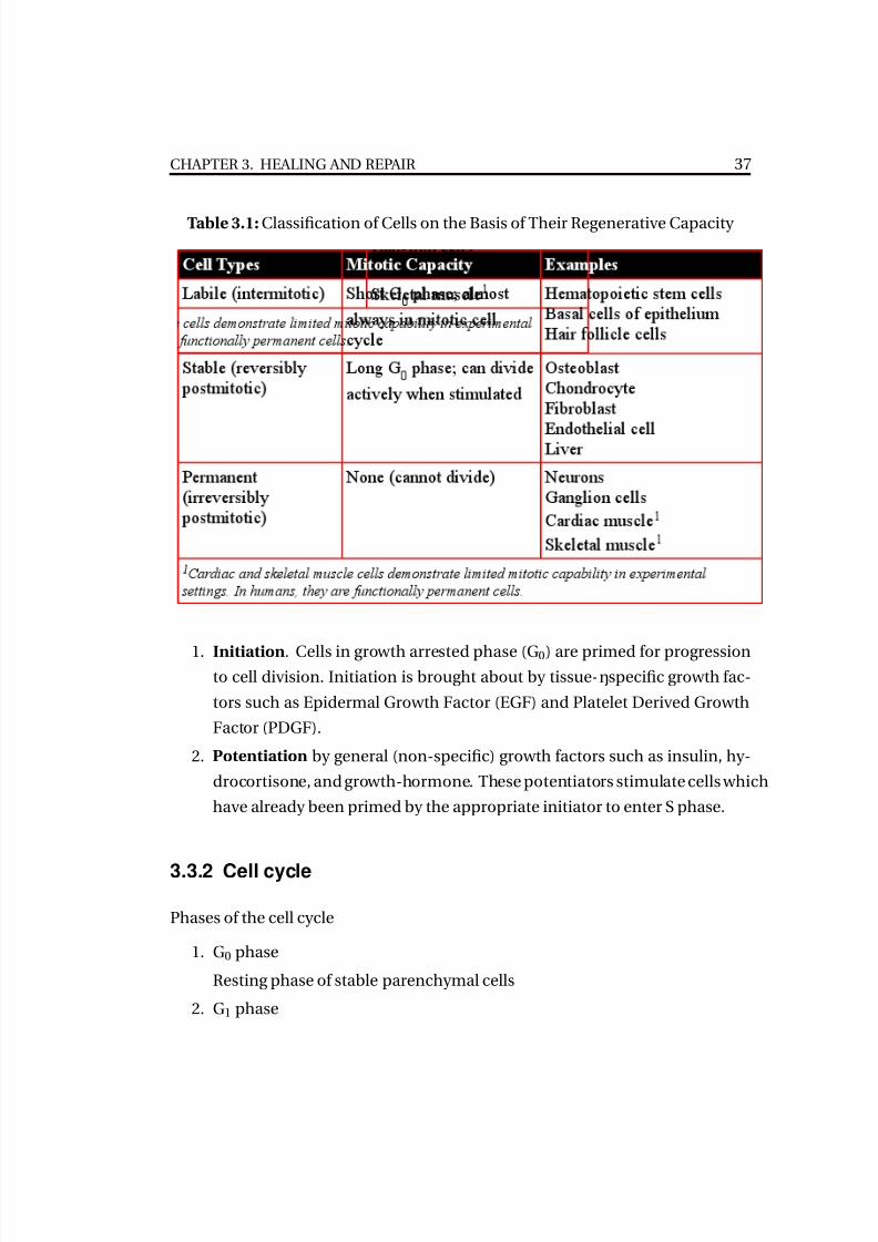
Tissues can be allocated to one of three categories, see table (3.1):
1. Labile cells (intermitotic ) which continue to proliferate throughout life, e.g.
epidermis, lining epithelia, endothelium, connective tissue, haemopoietic
tissue, endothelial cells
2. Stable cells (reversibly postmitotic ) which retain the capacity to regenerateand occasionally exhibit mitoses by virtue of normal cell-turnover, e.g. ,
liver, renal tubular epithelium, smooth muscle
3. Permanent cells (irreversibly postmitotic ) which cannot reproduce them-
selves after attaining maturity, e.g. neurones of the C.N.S., sensory organs,
renal glomeruli, striated muscle, adrenal medulla.
Labile tissues heal by regeneration with little or no repair. Permanent tissues are
incapable of regeneration and heal entirely by repair. Most organs show evidence
of both processes.
3.3.1 Control of Regeneration
Regeneration appears to be controlled by stimulatory and inhibitory factors. Stim-
ulation is a two-stage process:
7/21/2019 General Pathology Adel Inflammation
http://slidepdf.com/reader/full/general-pathology-adel-inflammation 39/50
CHAPTER 3. HEALING AND REPAIR 37
Table 3.1: Classification of Cells on the Basis of Their Regenerative Capacity
1. Initiation. Cells in growth arrested phase (G0) are primed for progressionto cell division. Initiation is brought about by tissue-ŋspecific growth fac-
tors such as Epidermal Growth Factor (EGF) and Platelet Derived Growth
Factor (PDGF).
2. Potentiation by general (non-specific) growth factors such as insulin, hy-
drocortisone, and growth-hormone. These potentiators stimulate cells which
have already been primed by the appropriate initiator to enter S phase.
3.3.2 Cell cycle
Phases of the cell cycle
1. G0 phase
Resting phase of stable parenchymal cells
2. G1 phase

7/21/2019 General Pathology Adel Inflammation
http://slidepdf.com/reader/full/general-pathology-adel-inflammation 40/50
CHAPTER 3. HEALING AND REPAIR 38
Synthesis of RNA, protein, organelles, and cyclin D
3. S (synthesis) phase
Synthesis of DNA, RNA, protein
4. G2 phase
Synthesis of tubulin, which is necessary for formation of the mitotic spin-
dle
5. M (mitotic) phase
Two daughter cells are produced.
3.4 Repair
Before discussing the nature of repair, it is necessary to appreciate the role of the
fibroblast in the biosynthesis of proteoglycans and collagen.
3.4.1 Biosynthesis of proteoglycans
The proteoglycans or “ground substance” of connective tissues are, as their name
implies, macromolecules composed of a protein core to which carbohydrate is
attached. The carbohydrate moieties take the form of long linear polysaccharides
which are attached radially around the protein molecule. They can be divided
into sulphated and non-sulphated types:
Sulphated:
Heparan sulphate, Keratan sulphate, Chondroitin sulphates A, B, and C
Non-sulphated:
Hyaluronic acid Chondroitin
The protein moiety is synthesis ed in the rough ER of the fibroblast and to this
core the hexose sugars are sequentially added to form the polysaccharide attach-
ments. Sulphation follows as a separate step.
7/21/2019 General Pathology Adel Inflammation
http://slidepdf.com/reader/full/general-pathology-adel-inflammation 41/50
CHAPTER 3. HEALING AND REPAIR 39
3.4.2 Biosynthesis of collagen
Collagen is the most abundant protein in the body and forms the major struc-
tural component of many organs. Collagen molecules consist of three polypep-
tide chains arranged in a triple helix and whilst the basic polypeptide structure
is straightforward, the molecule undergoes a complex series of post-translational
modifications and interactions with proteoglycans which greatly modifies its prop-
erties.
3.4.3 Types of collagen
There are seven types of collagen chain and these have been designated -1 (I-
V)and a2 (1) and (11). They are combined to form at least five isotypes of colla-
gen:
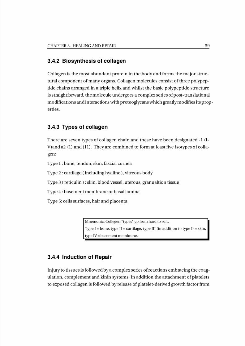
Type 1 : bone, tendon, skin, fascia, cornea
Type 2 : cartilage ( including hyaline ), vitreous body
Type 3 ( reticulin ) : skin, blood vessel, uterous, granualtion tissue
Type 4 : basement membrane or basal lamina
Type 5: cells surfaces, hair and placenta
Mnemonic: Collegen "types" go from hard to soft.
Type I = bone, type II = cartilage, type III (in addition to type I) = skin,
type IV = basement membrane.
3.4.4 Induction of Repair
Injury to tissues is followed by a complex series of reactions embracing the coag-
ulation, complement and kinin systems. In addition the attachment of platelets
to exposed collagen is followed by release of platelet-derived growth factor from
7/21/2019 General Pathology Adel Inflammation
http://slidepdf.com/reader/full/general-pathology-adel-inflammation 42/50
CHAPTER 3. HEALING AND REPAIR 40
their granules. This factor initiates fibroblast replication which is the predomi-
nant feature of early repair. Repair involves two overlapping processes:
1. Organization
2. Progressive fibrosis
1. Organization
This is the replacement of dead tissue or hematoma by granulation tissue.
Organization is seen in:
1. Hematomas in wound and fracture healing
2. Thrombi
3. Infarcts
4. Fibrinous exudates
Granulation tissue:
1. Demolition. Monocytes migrate into the area, take on the properties of
macrophages, and phagocytose cell debris, fibrin and red blood cells. Clear-
ance of dead tissue is facilitated by the secretion of proteolytic enzymes by
macrophages (e.g. collagenase, elastase) and other secretory products are
important in promoting repair, e.g. fibronectin
2. Fibroblast activity. Local resting fibroblasts (fibrocytes) proliferate rapidly
and migrate into the area where they continue to divide and commence
synthetic activity. Initially the activated fibroblasts produce proteoglycans
but as they mature switch over to collagen synthesis. At the same time,
some fibroblasts develop bundles of microfilaments in their cytoplasm and
acquire contractile properties. Such modified fibroblasts are termed my-
ofibroblasts.
3. lngrowth of capillaries. Endothelial cells in the severed blood vessels of sur-
rounding viable tissue undergo rapid proliferation and grow into the area
as solid cords. These endothelial ’buds’:
7/21/2019 General Pathology Adel Inflammation
http://slidepdf.com/reader/full/general-pathology-adel-inflammation 43/50
CHAPTER 3. HEALING AND REPAIR 41
a) Link up to form arcades
b) Canalize. This occurs within hours of formation
c) Become freely permeable to plasma, RBCs, leukocytes and platelets
d) Differentiate into arterioles and venules
2. Progressive fibrosis
Continued accumulation of intercellular collagen and diminution of vas-
cularity and cellularity 1. Collagen re-orientation along lines of stress-remodeling
2. Diminished cellularity
3. Formation of an avascular, hypocellular scar
Further changes in scars:
1. Cicatrization -a late diminution in size resulting in deformity
2. Calcification
3. Ossification
3.4.5 Cell-Matrix Interactions
Whilst it has long been appreciated that certain cells require to be attached to a
substrate before they can proliferate normally, it has only recently been estab-
lished that such attachment is brought about by a series of specific binding pro-
teins. These proteins are particularly important in the proliferation of connective
tissue cells:
1. Fibronectin attaches fibroblasts to collagen
2. Chondronectin binds chondrocytes to Type II collagen, the matrix of carti-
lage
3. Laminin binds epithelial cells to the Type IV collagen of basement mem-
branes

7/21/2019 General Pathology Adel Inflammation
http://slidepdf.com/reader/full/general-pathology-adel-inflammation 44/50
CHAPTER 3. HEALING AND REPAIR 42
4. Osteonectin binds hydroxy-apatite and calcium ions to Type I collagen (bone
matrix) and initiates mineralization
3.5 Wound Healing
In considering the healing of a skin wound two types are usually distinguished:
1. A clean wound with closely apposed margins-an incised wound (healing
by first intention)2. An open or excised wound (healing by second intention).
There are no fundamental differences between these two types, they merely dif-
fer in the degree to which the various stages apply (healing by second intention).
3.5.1 Stages in wound healing
1. Escape of blood and exudate
2. Acute inflammatory response at the margins
3. Hardening of the surface forming a scab
4. Demolition by macrophages with phagocytosis of cellular debris and re-
moval of dead tissue. In addition macrophages secrete products which
are important in the early stages of repair such as prostaglandins and fi-
bronectin.
5. Organization:
a) Platelet derived growth factor initiates fibroblastic proliferation
b) Activated fibroblasts secrete proteoglycans and fibronectin
c) Fibroblasts produce Type III collagen (reticulin) fibers and migrate
along this “scaffold”.
d) Fibronectin-mediated attachment of fibroblasts to collagen is followed
by enhanced proliferation
e) Simultaneous proliferation and migration of endothelial cells

7/21/2019 General Pathology Adel Inflammation
http://slidepdf.com/reader/full/general-pathology-adel-inflammation 45/50
CHAPTER 3. HEALING AND REPAIR 43
6. Epidermal proliferation. By mitotic activity and migration, epidermal cells
grow in from the margins and undermine the surface scab. When they meet
in the centre of the wound, mitosis and migration cease presumably as a
result of some cell-to-cell signal. This phenomenon is known as “contact
inhibition”.
7. Contraction of the wound an early diminution in size brought about by the
inward movement of the skin margins which greatly reduces the volume of
repair tissue required for healing. Myofibroblasts are thought to be respon-
sible for wound contraction, and the same cells provide the tensile strength
of the wound at this stage
8. Progressive increase in collagen fibers
9. Loss of vascularity and shrinkage of the scar
3.5.2 Healing by First Intention:
Occurs in small wounds that close easily.
Epithelial regeneration predominates over fibrosis
Healing is fast, with minimal scarring/infection
Examples:
Paper cuts
Well-approximated surgical incisions
Replaced periodontal flaps
3.5.3 Healing by second Intention:
Their is a substantial loss of tissue in the wound requiring formation of large
quantity of granulation tissue with subsequent scar formation
The healing of an excised wound differs from that of an incised wound in that
there is:
1. Greater tissue loss
7/21/2019 General Pathology Adel Inflammation
http://slidepdf.com/reader/full/general-pathology-adel-inflammation 46/50
CHAPTER 3. HEALING AND REPAIR 44
2. More inflammatory exudate and necrotic tissue to remove
3. Wound contraction is necessary
4. More granulation tissue is required, a bigger scar is formed and this may
result in deformity
5. Slower process
6. Increased liability to infection
Key Facts For Healing by Second Intention: Occurs in larger wounds that have gaps between wound margins
Fibrosis predominates over epithelial regeneration
Healing is slower, with more inflammation and granulation tissue
formation, and more scarring
Examples: large burns and ulcers, extraction sockets, external-
bevel gingivectomies
3.5.4 Factors influencing wound healing
Local factors adversely affecting healing
1. Type of wounding agent; blunt, crushing, tearing etc.
2. Infection
3. Foreign bodies in wound
4. Poor blood supply
5. Excessive movement
6. Poor apposition of margins, e.g. large hematoma formation
7. Poor wound contraction due to tissue tethering, e.g. skin over tibia
8. Infiltration by tumor
9. Previous irradiation
7/21/2019 General Pathology Adel Inflammation
http://slidepdf.com/reader/full/general-pathology-adel-inflammation 47/50
CHAPTER 3. HEALING AND REPAIR 45
General factors adversely affecting healing
1. Poor nutrition
a) Deficiency of protein. This results in a lack of the sulfur-containing
amino acids methionine and cysteine which are essential for the syn-
thesis of collagen
b) Lack of ascorbic acid (vitamin C) results in abnormal granulation tis-
sue and deficient collagen production
c) Zinc deficiency
2. Excessive glucocorticosteroid production or administration
3. Fall in temperature
4. Jaundice
3.5.5 Factors accelerating wound healing
1. Ultraviolet light
2. Administration of anabolic steroids, deoxycorticosterone acetate, and growth
hormone
3. Rise in temperature
4. Hyperbaric oxygen
3.5.6 Complications of wound healing
1. Wound rupture
2. Infection
3. Implantation of epidermal cells giving rise to keratin-filled epidermoid cyst
4. Weak scars with possible development of incisional hernia
5. Cicatrization and deformity
6. Keloid formation: The production of an elevated scar by excessive fibrosis
7/21/2019 General Pathology Adel Inflammation
http://slidepdf.com/reader/full/general-pathology-adel-inflammation 48/50
CHAPTER 3. HEALING AND REPAIR 46
7. Proud flesh: The swollen flesh that surrounds a healing wound, caused by
excessive granulation tissue. May results from persistence of foreign bodies
or contamination with some bacteria or fungi.
8. Malignant change. The development of squamous carcinoma in old healed
incisions is a recognized but rare complication
3.5.7 Healing of Fractures
Steps in the healing of a fractured long bone are:
1. Hemorrhage: This is due to torn blood vessels.
2. Hematoma formation
3. Transient inflammatory reaction: This is due to damage of the cells. This
reaction should subside within few days otherwise complications will re-
sults
4. Organization of the clot: In which granulation tissue will begin to invade
the clot
5. Osteoclastic activity: To remove the sharp edges and bone debris, this ac-tion is responsible for widening of the fracture line after 2 weeks of the
trauma.
6. Osteoid tissue formation: In which undifferentiated mesenchymal cells dif-
ferentiate into osteoblasts. Osteoblasts begin to lay down the special bone
matrix named osteoid which consists of a ground substance and a special
types of collagen, collagen type I.
7. Calcification of osteoid: Once osteoid tissue is calcified it is termed callus.
Callus consists of woven bone.
8. Remodeling: In which callus is resorbed and replaced by lamellar bone.
3.5.8 Healing of tooth socket
Healing of tooth socket is considered as a healing by second intention. The steps
of healing will be as follows:
7/21/2019 General Pathology Adel Inflammation
http://slidepdf.com/reader/full/general-pathology-adel-inflammation 49/50
CHAPTER 3. HEALING AND REPAIR 47
1. Following extraction of a tooth, the socket fills with extravasated blood which
then colts
2. The blood clot is organized to form granulation tissue
3. Transient inflammatory reaction which should subside within few days oth-
erwise complications will results
4. Osteoclastic resorption of the crestal bone and small specules of bone de-
tached during extraction commences at this time
5. Gingival epithelial proliferation and migration occurs across the defect.
Epithelial continuity is restored 10-14 days after extraction
6. Osteoblasts begin to appear in the granulation tissue toward the base of the
socket and the granulation tissue is gradually replaced by woven bone
7. After approximately 6 weeks, the outline of the socket can be viewed both
histologically and radiographically
8. Woven bone is remodeled with the formation of cortical and cancellous
bone and disappearance of the lamina dura. Remodeling also includes a
reduction in the height of the alveolar bone in the area of the extraction
9. Radiographically, the socket is generally obliterated between 20 and 30 weeksafter extraction.
3.5.9 Complications of fracture healing
1. Delayed union
2. Mal-union
a) Angulation
b) Shortening 3. Fibrous union resulting from
a) Excessive movement which may lead to the development of a false
joint (pseudoarthrosis)
b) Infection which may also give rise to osteomyelitis
c) Ischemia.
7/21/2019 General Pathology Adel Inflammation
http://slidepdf.com/reader/full/general-pathology-adel-inflammation 50/50
CHAPTER 3. HEALING AND REPAIR 48
4. Non-union if soft-tissues such as muscle or fat are interposed between the
severed ends
3.5.10 Pathological Fractures
These are fractures occurring spontaneously (that is with normal stresses) be-
cause of intrinsic disease of the bone.
Causes:
1. Osteoporosis, especially steroid induced
2. Metastatic tumors
3. Primary tumors (benign and malignant)
4. Paget’s disease
5. Bone lesions of hyperparathyroidism
6. Osteogenesis imperfecta