General Certificate of Education (Advanced Level)nie.lk/pdffiles/other/eOM...
62
1 General Certificate of Education (Advanced Level) Handout for unit 02 Chemical and cellular basis of life Department of Science National Institute of Education Maharagama Sri Lanka www.nie.lk
-
Upload
trinhduong -
Category
Documents
-
view
221 -
download
0
Transcript of General Certificate of Education (Advanced Level)nie.lk/pdffiles/other/eOM...

1
General Certificate of Education (Advanced Level)
Handout for unit 02 Chemical and cellular basis of life
Department of Science
National Institute of Education
Maharagama
Sri Lanka
www.nie.lk

2
Unit 02
Chemical and cellular basis of life
2.1.1. Elemental composition of living matter
There are about ninety two elements naturally occur in earth’s crust. Of which, about 20-25%
elementsare essential to continue healthy life and reproduction. (about 25- elements are essential for
humans and about 17 for plants).
Oxygen (O), Carbon (C), Hydrogen (H), and Nitrogen (N) make up 96% of living matter.
Calcium (Ca), Phosphorous (P), potassium (K) and sulphur (S)- make up most of the remaining 4% of
the mass of the organism.
In humans, C, H, O, N- accounts for 96.3% of the body mass and Ca, P, K, S, Na, Cl, Mg and trace
elementsaccounts for the remaining 3.7%. (e.g. B (Boron) , Co (Cobalt), Cu (Copper), Cr
(Chromium), F (Fluorine), I (Iodine), Fe (Iron),
Mo (Molybdenum), Mn (Manganese), Se (Selenium), Si (Silicon), Sn (Tin), V (Vanadium), Zn (Zinc)

3
2.1.2. Physical and chemical properties of water important for life
Wateris a vital inorganic molecule; life could not exist on this planet without water. It is important
due to following reasons,
1. Vital chemical constituent of living cell
2. Provides a biological medium for all organisms
Most of above properties are based on the chemical structure of water molecule.Physical and
chemical properties of water molecule provide the ability to render the vitality.Water molecule is a
small, polar and angular molecule.
Polarity is an uneven charge distribution within a molecule. In water molecule, oxygen atom is
slightly negative and hydrogen atom is slightly positive. Weak attractions between the slightly polar
hydrogen atom of one water molecuole and the slightly polar oxygen atom of adjacent water
molecuoleare known as hydrogen bonds.Thesehydrogen bonds play a major role in maintaining all the
properties of water.
The properties of water arise due to attractions of different water molecules. When the water is in
liquid form its H bonds are very fragile. H bonds form, break and reform with great frequency.
Four major properties of water to maintain life on earth
1. Cohesive behavior
2. Ability to moderate temperature
3. Expansion upon freezing
4. Versatility as a solvent
Properties of water related to functions
δ+ partial positive
δ-partial negative
Fig 2.1: Chemical structure of the water molecule
Fig 2.2: Hydrogen bonding in water

4
1. Cohesive behavior
Attraction between water molecules due to hydrogen bonding is known as cohesion. Attraction
between water molecules and other substances are known as adhesion. Both of the aboveproperties of
water allow it to act as a transport medium.
Due to cohesion between water molecules, water and dissolved substances such as minerals and
nutrients transport through vascular tissues, xylem and phloem against gravity.
Adhesion between water molecules and cell walls also helps in conduction of water and dissolved
substances.
Water has a high surface tension. This ability is given to water molecules, due to cohesion between
the water molecules. Therefore, in an aquatic system, upper surface water molecules are attracted by
lower surface molecules and it forms a water film. Small insects e.g. water skaters can walk on the
surface of a pond.
2. Ability to moderate temperature
Water can absorb or release a relatively high amount of heat energy by a slight change in its own
temperature.
Due to the high specific heat, water will function as thermal buffer in living system and aquatic bodies
during the temperature fluctuations on earth.
Due to the high heat of vapourization, with the minimum loss of water an organism can release much
heat energy. Therefore, body surface ofan organism maintained as cool surface.
e.g. Prevent from overheating.
Evaporation of sweat from human skin helps to maintain the body temperature at constant
level.
Transpiration in plants keeps the plant body surface as a cool surface and prevent from becoming too
warm in the sunlight.
3. Expansion upon freezing
Generally, in an increase in temperature of any substances, reduces their density and on the other
hand, in a decrease in temperature increases their density. Whenthe temperature of water falls below 4
˚C, it begins to freeze and forms a crystalline lattice called ice cubes. Therefore water has the
maximum density at 4˚C.Hence, ice floats on the surface of water bodies. It is an important property
of water in polar regions, where, organisms in aquatic bodies can survive during the winter.
4. Versatility as a solvent
This ability is given to water due to their polarity. Polar molecules (e.g. Glucose), non polar ionic (e.g.
NaCl), both polar and ionic (e.g. lysozymes) can dissolve in water, because water molecules surround
each of the solute molecules and form hydrogen bonds with them. Solubility depends on polarity and
not in their ionic nature

5
2.1.3. Chemical Nature and
Organisms
Carbohydrates
Most abundant group of organic compound on earth is carbohydrates. Major elemental composition is
C, H, and O. Hydrates of carbon contain
General formula is Cx(H2O)y. Three major groups of carbohydrates
disaccharides and polysaccharides.
Generally carbohydrates include sugars
Monosaccharides
The simplest form of carbohydrates
aremonosaccharide. Where C varies from 3
and occur in crystalline form.
According to the number of carbon atoms,t
3C- Triose e.g.Glyceraldehydes ( 4C- Tetrosee.g.Erythrose(rare in nature 5C- Pentosese.g. Ribose, Deoxyribose, 6C- Hexoses e.g. Glucose, F
According to the type of carbonyl (Keto, aldo)
a. Aldoses-glucose, galactoseb. Ketoses-fructose
Aldose
Ketose
Fig 2.3: Solid form of glucose
Fig 2.5: Solid form of fructose
ature and Functions of Main Organic Compounds
organic compound on earth is carbohydrates. Major elemental composition is
ydrates of carbon contain the same proportion of H: O which equals to 2:1 as in water.
Three major groups of carbohydrates aremonosaccharides,
Generally carbohydrates include sugars (monosaccharides and disaccharides) and polysaccharide
form of carbohydrates having general molecular formula as (CH2O)n
Where C varies from 3-7 . All monosaccharide are reducing sugars,
g to the number of carbon atoms,they are named as;
lyceraldehydes (Phosphoglyceraldehyde is a derivative of Trioserare in nature)
eoxyribose, Ribulose (RUBP is a derivative of ribulose)Fructose, Galactose
type of carbonyl (Keto, aldo)group, they are classified as; glucose, galactose
Aldehyde
group
Keto group
Fig 2.3: Solid form of glucose Fig 2.4: Aqueous form of Glucose molecule
Fig 2.6: Aqueous form of fructose
ompounds of
organic compound on earth is carbohydrates. Major elemental composition is
which equals to 2:1 as in water.
ides,
and polysaccharides.
, water soluble
riose)
bulose)
Fig 2.4: Aqueous form of Glucose molecule
Fig 2.6: Aqueous form of fructose

6
In aqeous media some monosaccharides are in ring form (No need to memorize the chemical
structures)
Disaccharides
They are sugars formed by joining two monosaccharides by a glycosidic bond.
(no need to memorize the chemical structures)
Glycosidic bond is formed by removal of a water molecule from two adjacent monosaccharides by a
condensation reaction. Water molecule is formed from OH group of one monosaccharide molecule
and H from adjoining monosaccharide molecule.
Glucose + Glucose Maltose + H20
Glucose + fructose Sucrose + H20
Glucose + Galactose Lactose + H20
Maltose and lactose are reducing sugars and sucrose is a non reducing sugar.
Polysaccharides
They are macromolecules and biopolymers. Polysaccharides are made up of few hundred to a few
thousand monosaccharide subunits
They arenon crystalline, water insoluble, and not considered as sugars.
Some polysaccharides act as storage components where others contribute to the structure of living
organisms. Based on their function they are categorized as Storage polysaccharides and structural
polysaccharides.
1. Storage- Starch, glycogen
2. Structural- Cellulose, Hemicellulose, Pectin
Based on their architecture they are categorized as
1. Linear forms- Cellulose, Amylose
2. Branched forms- Glycogen, Amylopectin, Hemicellulose
Condensation
condensation
Condensation
Fig 2.7: Formation of sucrose Fig 2.8: Formation of maltose
7
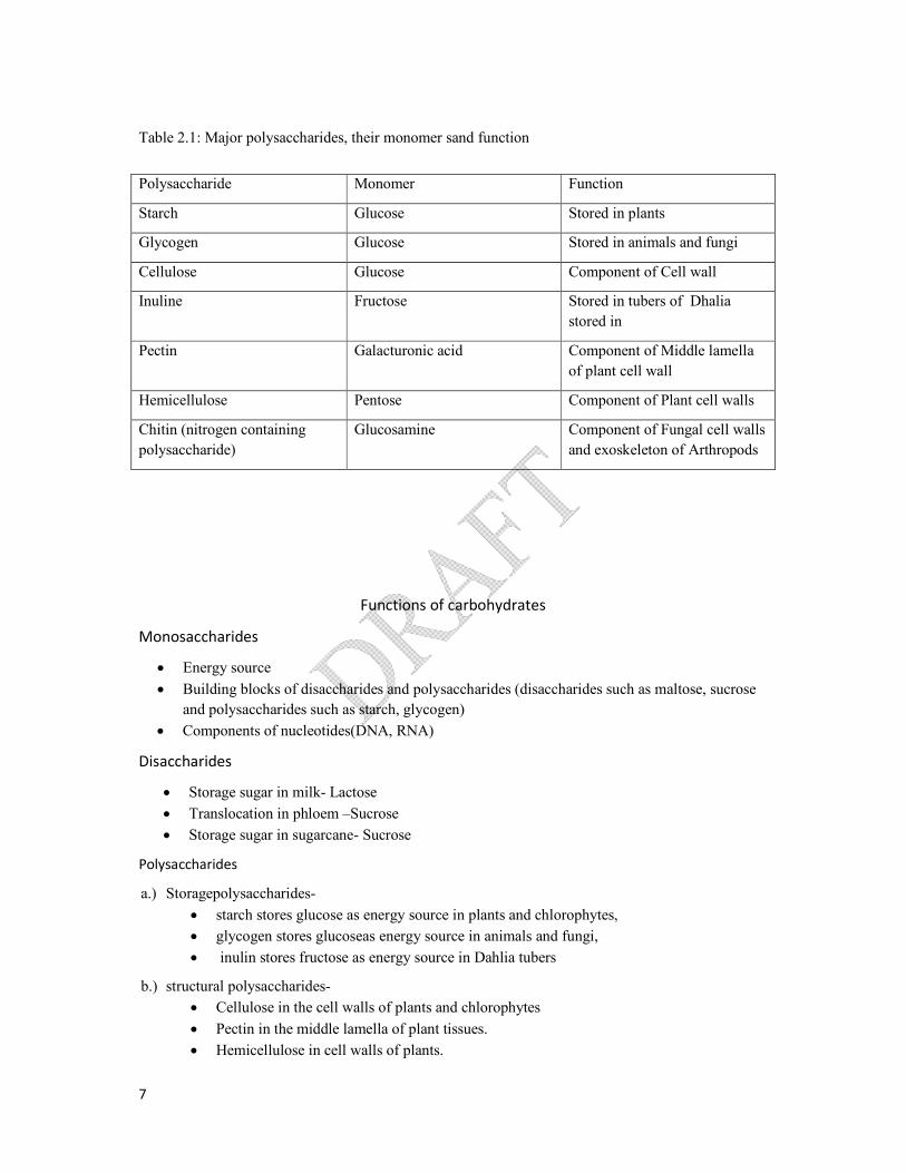
Table 2.1: Major polysaccharides, their monomer sand function
Functions of carbohydrates
Monosaccharides
Energy source
Building blocks of disaccharides and polysaccharides (disaccharides such as maltose, sucrose
and polysaccharides such as starch, glycogen)
Components of nucleotides(DNA, RNA)
Disaccharides
Storage sugar in milk- Lactose
Translocation in phloem –Sucrose
Storage sugar in sugarcane- Sucrose
Polysaccharides
a.) Storagepolysaccharides-
starch stores glucose as energy source in plants and chlorophytes,
glycogen stores glucoseas energy source in animals and fungi,
inulin stores fructose as energy source in Dahlia tubers
b.) structural polysaccharides-
Cellulose in the cell walls of plants and chlorophytes
Pectin in the middle lamella of plant tissues.
Hemicellulose in cell walls of plants.
Polysaccharide Monomer Function
Starch Glucose Stored in plants
Glycogen Glucose Stored in animals and fungi
Cellulose Glucose Component of Cell wall
Inuline Fructose Stored in tubers of Dhalia
stored in
Pectin Galacturonic acid Component of Middle lamella
of plant cell wall
Hemicellulose Pentose Component of Plant cell walls
Chitin (nitrogen containing
polysaccharide)
Glucosamine Component of Fungal cell walls
and exoskeleton of Arthropods

8
Peptidoglycan in the cell walls of prokaryotes.
Chitin in the cell walls of fungi and in exoskeleton in Arthropods.
Lipids
Diverse group of hydrophobic molecules
Large biological molecules but not considered as polymers or macromolecules.
Consist of C, H, O and H:O ratio is not 2:1. Comparatively more H are present.
Biologically important types of lipids: Fats, Phospholipids and Steroids.
Fats
Fats are made up of glycerol and fatty acids; Glycerol belongs to alcohol group having 3 carbons
where each of them bear single hydroxyl group. Fatty acids are hydrocarbon chains with long (16-18)
carbon skeleton with a carboxyl group at its one terminal.
Fatty acid molecules bind to each hydroxyl group of glycerol by ester bond. Resulting fat molecules
are called as triacylglycerol.
Fig 2.9: Formation of Triacylglycerol
Hydrocarbon chains of fatty acids contribute to the hydrophobic nature of the fats. Based on the
nature of hydrocarbon chains of fatty acids, they are categorized as
a) Saturated fats- fats made up of saturated fatty acids: fatty acids with hydrocarbons having no any
double bonds. Usually animal fats come under this category. They are mostly solid at room
temperature. e.g: butter
b) Unsaturated fats- fats made up of unsaturated fatty acids- fatty acids with hydrocarbons having one
or more double bonds. Usually plant fats come under this category. They are mostly liquid in room
temperature. e.g: Vegetable Oils. Unsaturated fats may classify based on the nature of their double
bonds. a) Cis Unsaturated fat b) Trans Unsaturated fat
Consumption of excess saturated fats and trans unsaturated fats contribute arthrosclerosis.
Phospholipids
Phospholipids are major components of the cell membranes. They are composed of two fatty acids
and one phosphate group attached to one glycerol molecule. The phosphate group gives the negative

9
electrical charge to the phospholipid molecule. Typically an additional polar molecule or small
charged molecule is also linked to the phosphate group e.g. choline.
The two ends of the phospholipids show different behavior. The hydrocarbon tails are hydrophobic
while phosphate group and its attachment (head) are hydrophilic.
Functions of Lipids
food reserve as energy source (triglycerides such as fats and oils)
maintain the fluidity of plasma membrane (phospholipids, cholestrol)
act as signaling molecules (eg. Hormones) that travel through the body
found as components of animal cell membrane (cholesterol)
Proteins
Proteins are made up of amino acids. Twenty different amino acids are involved in the formation of
proteins. Elemental composition is C, H,O,N and S. At the centre of the amino acid is an asymmetric
carbon atom except in glycine. Each amino acid is composed of an amino group, a carboxyl group, a
hydrogen atom and a variable group symbolized by R, which is an alkyl group. In the case of glycine
R is replaced by H atom. The R group also called the ‘side chain’ differs with each amino acid where
as the other groups are in the ‘ back bone’ (including the H atom).
Amino acids may have one or more carboxyl groups and amino groups. Amino group has alkaline
nature and carboxyl group has acidic nature. When both characteristics are found in one molecule
they are known as amphoteric molecules. Therefore, amino acids are as amphoteric.
Two Amino acids undergo condensation reactionby removing a water molecule from both and result a
bond known as peptide bond;
Fig 2.10: Structure of an Amino acid molecule
Fig 2.11: Formation of peptide bond
Alky
Carbox
Amin

10
Protein is composed of one or more polypeptide chains which are composed of amino acids.
Levels of protein structures
There are four levels of structure which play important roles in their functions;
a) Primary
b) Secondary
c) Tertiary
d) Quaternary
a) Primary structure
The unique sequence of linearly arranged amino acids linked by peptide bonds is the primary structure
of proteins.
b). Secondary structure
The primary structure of a single polypeptide chain coils and folds, as a result of intra molecular
hydrogen bonds between the oxygen atoms and the hydrogen atoms attached to the nitrogen atoms, of
the same poly peptide chain backbone, to form the secondary structure, which is either β pleated or
alpha helical.
Alpha helix- e.g.Keratin.
β pleated sheete.g.spider’ssilk fiber
11
b) Tertiary structure
Usually the secondary polypeptide chain bends and folds extensively forming a precise compact
unique, functional and three-dimensional shape resulting from following interactions between the side
chain/ R-group of amino acids;
1. H bonds
2. Disulphidebonds
3. Ionic bonds
4. Van der Waals interactions/ Hydrophobic interactions
5.
e.g.most of the enzymes, myoglobin, albumin
c) Quaternary structure
Aggregation of two or more polypeptide chains involve in the formation of one functional protein.
Separate chains are called protein subunits which were held together by inter and intra-molecular
interactions.
e.g.Haemoglobin, Collagen
Denaturation of proteins
Denaturation of protein is the loss of specific chemical three dimensional shape due to the
alteration of weak chemical bonds and interactions.
.
Agents affecting the denaturation
1. High temperature and high energy radiation
2. Strong acids, alkaline and high concentrations of salts
3. Heavy metals
4. Organic solvents and detergents
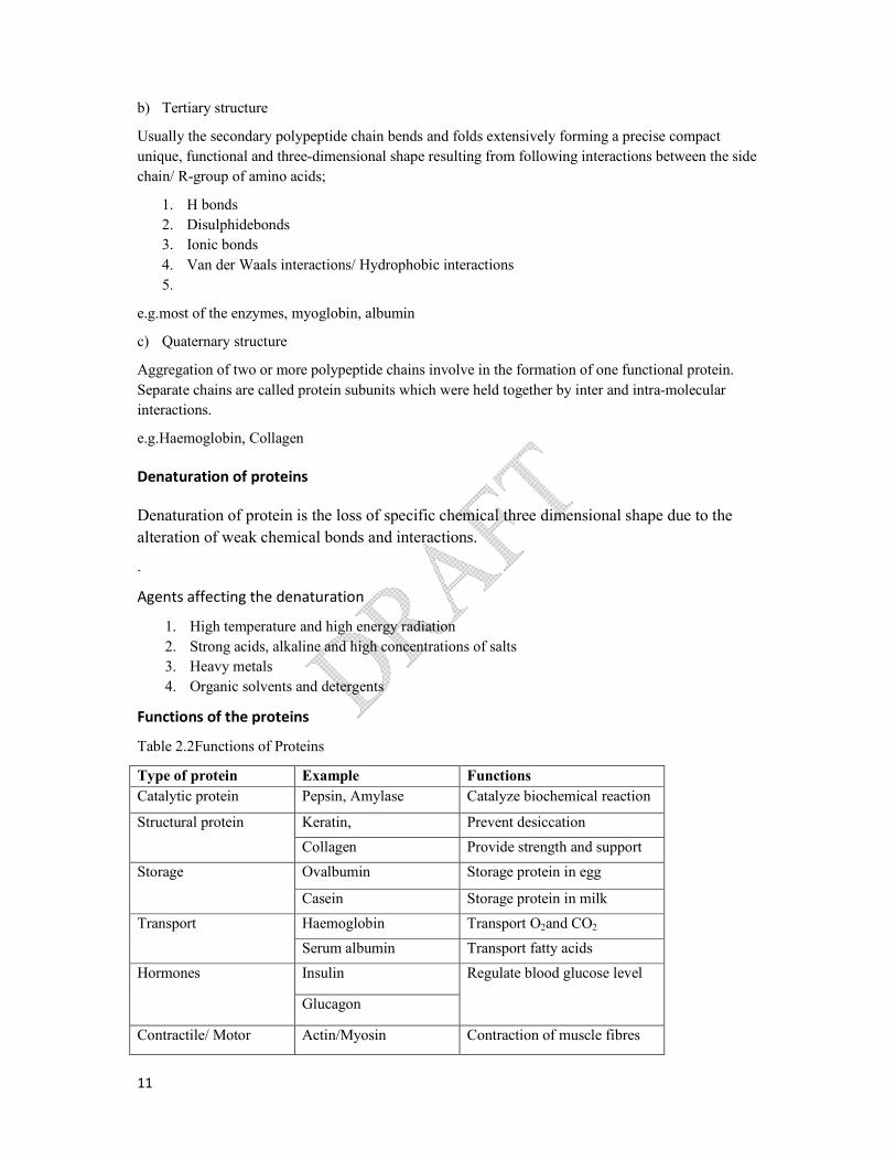
Functions of the proteins
Table 2.2Functions of Proteins
Type of protein Example Functions
Catalytic protein Pepsin, Amylase Catalyze biochemical reaction
Structural protein Keratin, Prevent desiccation
Collagen Provide strength and support
Storage Ovalbumin Storage protein in egg
Casein Storage protein in milk
Transport Haemoglobin Transport O2and CO2
Serum albumin Transport fatty acids
Hormones Insulin Regulate blood glucose level
Glucagon
Contractile/ Motor Actin/Myosin Contraction of muscle fibres

12
Defensive Immunoglobins Eliminate foreign bodies
Nucleic acids
Nucleic acids are Polymers exist as polynucleotides made up of monomers called nucleotides. They
contain C, H, O, N and P. Nucleic acidsare macromolecules, biopolymers. There are two types of
Nucleic acids: DNA (Deoxyribo nucleic acids) and RNA (Ribonucleic acids).
Structure of nucleotides
Nucleotides have 3 components; namely pentose sugar, nitrogenous base and a phosphate group
A nucleotide without a phosphate group is called a nucleoside.e.g. Adenosine, Guanosine
Pentose sugar
Pentose sugars are two types; namely Deoxy ribose and ribose (in deoxyriboseone oxygen atom is less
than in ribose)
Nitrogenous bases
There are two major groups of nitrogenous bases:
1. Purines- larger in size with two rings
2. Pyrimidines- smaller in size with a single ring
In purines there are two types; namely Adenine, Guanine. In pyrimidens there are three types,
Thyamine, Uracil and Cytocine. Bases are commonly represented by letters A, G, T, U and C
respectively.
Phosphate group
It gives the nucleic acids the acidic nature.
Formation of nucleic acids
Millions of nucleotides join by phospho-di-ester bond to form polynucleotide chains by condensation
between the –OH group of the phosphate of one nucleotide with the –OH attached to 3rd carbon of
pentose sugar of the other. These bonds results in a backbone with a repeating pattern of sugar-
phosphate units. Nucleic acids are linear polymers of nucleotides. There are two kinds of nucleic acids
depending on the type of the sugar molecules involved. If the sugar molecule in the nucleotide is
deoxyribose,the nucleic acid is (DNA). If the pentose sugar is ribose, then the nucleic acid is RNA.
Fig 2.15: Structure of nucleotides

13
DNA contains Adenine, Thymine, Guanine and Cytosine and RNA contains Adenine, Guanine,
Cytosine and Uracil.
Structure of DNA molecule (Watson and Crick model)
DNA molecules have twoanti-parallel polynucleotide chains that spiral around an imaginary axis,
forming a double helix. The two sugar-phosphate backbones run in opposite directions from each
other, and the arrangement is referred to as anti-parallel. The sugar phosphate backbones are on the
outside of the helix, and the nitrogenous bases are paired in the interior of the helix. The two strands
are held together by hydrogen bonds between the paired nitrogen bases.
Base pair rule
Always a purine base, pairs with a specific,pyrimidine base,
A=T (2 hydrogen bonds)
G≡C (3 hydrogen bonds)
Hence two chains (strands) are said to be complementary to each other.These pairs are known as
complementary base pairs.In this original double helical structure, one complete turn consists of ten
base pairs as shown in the diagram.
Functions of DNA
Store and transmit genetic information from one generation to the next generation
Store the genetic information for protein synthesis
Structure of RNA
This is normally a single strandednucleic acid composed of ribo-nucleotides containing bases, Uracil
(U), Cytosine (C ), Guanine (G), Adenine (A). Complementary base pairing between two RNA
molecules or within the same molecule may occur in some. Complementary base pairing facilitates
three dimensional shapes essential for their functioning. Adenine binds with Uracil with two
hydrogen bonds and Guanine binds with Cytosine with three hydrogen bonds. There are three types
of RNA present in cells,
I. Messenger RNA (mRNA)
II. Transfer RNA (tRNA)
III. Ribosomal RNA (rRNA)
1. Messenger RNA
Messenger RNA is a linear molecule and is the least abundant type of RNAin a cells comparatively.
There are two functions;
1. Copies the genetic information stored in DNA molecule as a sequence of nitrogenous bases
2. Transports genetic information from nucleoplasam to the site of protein synthesis (ribosome)
through nucleopores
2. Transfer RNA (tRNA)

14
Smallest RNA molecule. Linear, but forms three- looped structure as shown in the diagram.
Function - transportation of amino acids to the site of protein synthesis.
3. Ribosomal RNA
It is the most abundant type of RNA. rRNA has a complex irregular structure. It provides the site
where polypeptide chains are assembled.
Differences between DNA and RNA
1. DNA is double stranded molecule while RNA is a single stranded molecule.
2. DNA consists of A, T, G and Cand absence of U, while RNA consists of A,U, G and C and
absence of T
3. Sugar molecule in RNA is ribose, while in DNA it is deoxyribose.
Nucleotides other than those found in nucleic acids
ATP, NAD+, NADP+, FAD and their functions
Functions of ATP
Universal energy carrier
Functions of NAD+
Act as a coenzyme
Act as an electron carrier
Function as an oxidizing agent during respiration
Functions of NADP+
Fig 2.17: Structure of the tRNA molecule

15
Act as coenzymes
Act as an electron carrier
NADP+ act as a reducing agent in photosynthesis
Functions of FAD
Act as a coenzyme
Act as an electron carrier

16
2.2.1. Contribution of microscope to the expansion of knowledge on cells and
cellular organization
Advancement of the cytology is mostly based on the microscopy. The discovery and early study of
cells progressed with the invention of microscope.
Light microscope
Visible light is passed through the specimen and then through glass lenses. The lenses refract the light
in such a way that the image of the specimen is magnified as it is projected into the eye.The simplest
microscope is a single lens.
The compound light microscope
Compound light microscopes are commonly used in school laboratories and it is used in medical
laboratories as a diagnostic tool.
Resolution power and magnification are important parameters which can be seen in a microscope.
Magnification is ratio of an object’s image size to its actual size. Usually the maximum magnification
of light microscope is 1000 times the actual size of the specimen)
Resolution power is minimum distance between two points that can be distinguished as separate
points (resolution power of light microscope is 0.2µm). It is a measure of the clarity of the image.
Magnification is limited due to the resolution.
Light from an object (specimen on the slide) passes first through objective lens. Then produce a
magnified image.
Above image then acts as an object for the second lens (the eye piece lens) which further magnifies it.
The total magnification is hence the product of the magnification of each lens.
Total magnification =
e.g.If magnification of Objective lens = ×40, eyepiece =×15
Total is =15 × 40= ×600 time magnified
Magnification of objective
lens
Magnification of
eyepiece

17
The Electron Microscope
The limitation imposed upon the resolution power of the light microscope by the wavelength of light.
The resolution power is inversely proportional to the wavelength. Due to this, scientists considered
the use of other forms of radiations with comparatively shorter wavelengths.
As a result, electron microscopes were developed. In electron microscopy, a beam of electrons is
focusedthrough the specimen or on to its surface.
This means, that in theory, the electron microscope should be able to magnify objects up to
1×10⁸times. In practice, it magnifies just over 5×10⁵ times.
Electron microscopes have revealed many organelles and other sub cellular structures those were
impossible to resolve with the light microscopes.
There are two types of electron microscopes.
1. Transmission electron microscopes (TEM)
2. Scanning electron microscopes(SEM)
Transmission electron microscopes
It is used to study the internal structures of cells.In this microscope, a beam of electrons is passed
through a thin,especially prepared slice of material. A very thin specimen is used. Specimens stained
with heavy metals which attach more to certain cellular structures than other areas. Image reflects the
pattern of electrons passed through the specimen, displays on a screen. While electrons pass through
the specimen, more electrons may get displayed in regions where structures were densely stained.
Scanning electron microscopes
In this instrument, a fine beam of electrons is reflected from the surface ofspecimen. Specimen is
mostly coated with gold prior to observation. Here the specimen scatters many electrons whereas
others are absorbed. This instrument is ideal to observe the surface viewinthree
dimensionalappearances.
Table 2.3: Differences between light and electron microscope
Light Microscope Electron microscope
Glass lenses are usedto focus the light rays Powerful magnets are used to focus beam of
electrons
Image is directly detected by naked eye Not directrly detected by naked eye, micrographs
are used
Living and non living objects can be observed Only non-living objects are observed
Actual color of the object can be observed Actual color cannot be observed. Images are
developed
Dyes used to stain the object Heavy metals are used to stain the object

18
Historical background of cell and the structure and functions of the subcellular
units
Cell theory
All organisms are composed of cells.
Recall the hierarchy of life, the levels of organization mentioned earlier. The basic unit which can be
called “living” is the cell, which may form a single celled organism (e.g.Chlamydomonas, Yeast) or a
multi-cellular plant or animal. The cell is the basic structural and functional unit of life.
The level of organization of matter represented by a cell shows all the characteristics of life. Any
stage below level of a cell cannot be considered living, whether it is a single celled organism or multi-
cellular plant or an animal.
Robert Hooke (1665) examined a cork using simple microscope and gave the term “CELL” to
describe the basic units.
Anton Van Leeuwenhook (1650), a contemporary of Robert Hooke, was the first to describe and
recordliving single celled organisms, Euglena& bacteria
Matthias Schleiden (1831), a botanist, studying plant tissues concluded that all plants are made up of
cells.
Theodore Schwann a zoologist (1839) concluded that animal tissues are also made up of cells.
Rudolf Virchow (1855) showed that all cells arise from pre-existing cells by cell division,
Schleiden, Schwann and Virchow presented the ‘Cell Theory’ which included the following.
1.All organisms are composed of one or more cells.
2.The basic structural and functional unit of organisms is the cell.
3.All cells arise from pre-existing cells.
Organization of cells
There are two kinds of cellular organization - Prokaryotic and Eukaryotic
All cells share certain basic features. They are;
All cells are bounded by a plasma membrane which is a selective barrier
Within the cell have, a semifluid, jelly like substance which is called cytosol. Subcellular
components are suspended within the cytosol.
They carry DNA as genetic materials.
Ribosomes are found in all cells
19
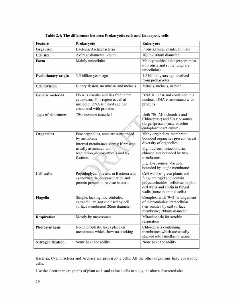
Table 2.4: The differences between Prokaryotic cells and Eukaryotic cells
Bacteria, Cyanobacteria and Archaea are prokaryotic cells. All the other organisms have eukaryotic
cells.
Use the electron micrographs of plant cells and animal cells to study the above characteristics
Feature Prokaryote Eukaryote
Organism Bacteria, Archaebacteria Protists,Fungi, plants, animals
Cell size Average diameter 1-5µm 10µm-100µm diameter
Form Mainly unicellular Mainly multicellular (except most of protista and some fungi are unicellular)
Evolutionary origin 3.5 billion years ago 1.8 billion years ago ,evolved from prokaryotes
Cell division Binary fission, no mitosis and meiosis Mitosis, meiosis, or both;
Genetic material DNA is circular and lies free in the cytoplasm. This region is called nucleoid, DNA is naked and not associated with proteins
DNA is linear and contained in a nucleus. DNA is associated with proteins
Type of ribosomes 70s ribosome (smaller) Both 70s (Mitochondria and Chloroplast) and 80s ribosomes (larger)present (may attachto endoplasmic reticulum)
Organelles Few organelles, none are surrounded by membrane
Internal membranes scares; if present usually associated with respiration,photosynthesis and N2
fixation.
Many organelles, membrane bounded organelles present. Great diversity of organelles.
E.g. nucleus, mitochondria, chloroplasts bounded by two membranes.
E.g. Lysosomes, Vacuole, bounded by single membrane.
Cell walls Peptidoglycan present in Bacteria and cyanobacteria, polysaccharide and protein present in Archae bacteria
Cell walls of green plants and fungi are rigid and contain polysaccharides; cellulose in plant cell walls and chitin in fungal walls (none in animal cells)
Flagella Simple, lacking microtubules; extracellular (not enclosed by cell surface membrane) 20nm diameter
Complex, with ‘9+2’ arrangement of microtubules; intracellular (surrounded by cell surface membrane) 200nm diameter
Respiration Mostly by mesosomes Mitochondria for aerobic respiration
Photosynthesis No chloroplasts; takes place on membranes which show no stacking
Chloroplasts containing membranes which are usually stacked into lamellae or grana
Nitrogen fixation Some have the ability None have the ability

20
.
Fig 2.18: Structure of an animal cell

21
Fig 2.19: Structure of plant cell

22
Structures and functions of organelles and other subcellular components
Plasma membranePlasmamembrane is the outer limit of cytoplasm. All cellular membranes
resemble the ultra structure of plasma membrane. In 1972, Singer and Nicolson put forward the fluid
mosaic model ofcell membrane. It is mainly composed of;
1. Phospholipids (most abundant type of lipid in plasma membrane)
2. Protein
The Plasma membrane has the following features. It is about 7nm thick. Itismainly made up of a
phospholipid bilayer. Phospholipids are amphipathic molecules. The hydrophilic heads of the
phospholipids face outwards into the aqueous environment of both inside and outside of the cell.
The hydrophobic hydrocarbon tails face inwards and create a hydrophobic interior.
Plasmamembrane is compared to the fluid mosaic model. Since phospholipid molecules are
moveable, they provide the fluid nature to the membrane.
Protein molecules embedded randomly contribute to its mosaic nature.
Some of the protein molecules penetrate all the way through the membrane, called transmembrane
proteins and some others penetrate only part of the way into the membrane. These are called integral
proteins.
Most of the integral proteins are transmembrane proteins which have hydrophilic channels. These act
as pores through which ions and certain polar molecules can pass.
Some proteins are not embedded in the lipid bilayer at all, and are loosely bound to the inner surface
of the membrane, called peripheral proteins.
Some proteins and lipids have short branching carbohydrate chains like antennae, forming
glycoprotein and glycolipids, respectively.
Animal’s cell membrane may contain few cholesterol molecules randomly integrated into the lipid
bilayer.
These cholesterol molecules provide flexibility and stability to the membrane by reducing membrane
fluidity at moderate temperatures and prevent membrane solidification at low temperatures.
The two sides of the membrane may differ in composition and function.

23
Functions
The plasma membrane surrounds the cytoplasm of living cell physically separating the
intracellular components from the extracellular environment.
Plasma membrane is selectively permeable and able to regulate the exchange of material
needed for survival.
Proteins embedded in the plasma membrane identify the cell, enabling nearby cells to
communicate with each other (involved in cell recognition).
Some protein molecules act as receptor molecules for interacting with specific biochemical,
such as hormones, neurotransmitters and immune proteins.
Some proteins in the cell membrane attach to some cytoskeletal fibers and help to maintain
the shape of the cell.
Some proteins in the membrane act as enzymes. (e.g.Microvillus on epithelial cell lining of
some parts of the gut contains digestive enzymes in their cell surface membrane.)
Fig 2.20: The fluid mosaic structure of the cell membrane

24
Subcellular components
There are many sub-cellular components in the cell. Some of them are organelles, which are bound by
membranes and suspended in the cytosol of eukaryotic cell to perform specialized functions.
Nucleus
Most prominent organelle, consist most of the genes, having an average diameter of 5m and
enclosed by a double membrane cover called nuclear envelope.
Nuclear envelope- composed of two membranes, inner and outer membranes, separated by a
space of 20-40 nm. Nuclear envelope is perforated by nuclear pores which has pore complex
to regulate the entry and exit of substances. It has nuclear lamina, made up of protein
filaments which line the interior side of the nuclear envelope.
nuclear matrix made up of protein filaments and extended throughout the interior of the
nucleus. Chromatin and nucleolus are embedded in the nuclear matrix.
Nucleolus- appears as darkly stained granules with fibers adjoining part of the chromatin.
Chromatin –appears as a diffused mass in electron micrographs of non dividing cells. It is a
complex of DNA and proteins. During nuclear divisions, chromatin condenses, tightly coils
and form threads, calledchromosomes. Each species has a constant number of chromosomes.
(e.g. typical human cell has 46 chromosomes).
Functions
Control all cellular activities.
Synthesize DNA to produce new nuclei for cell divisions.
SynthesizerRNAs and ribosomal subunits required for protein synthesis, through nucleolus.
Synthesize mRNA and tRNA according to the information present on the DNA.
Store and transport genetic information.
Ribosomes
These are subcellular components whichcarryout protein synthesis. They consist of two subunits;
larger subunit and smaller subunit. They are composed of rRNA and protein. Ribosomes are found in
two types; 70S and 80S. 70S ribosomes are found freely on the cytoplasam of prokaryotes,matrix of
mitochondria and stroma of chloroplasts. 80S ribosomes are found only in eukaryotes. Based on the
nature of presence, 80S ribosomes are categorized as two types; free ribosomes and bound ribosomes.
Free ribosomes: freely available as group in cytoplasam. Bound ribosomes are attached to the
membrane surface of rough endoplasmic reticulum.
Functions
Protein synthesis
Endoplasmic reticulum

25
It is a network of internal membranes forming flattened or tubular sacs separating cytosol from ER
lumen. It is continuous with the outer membrane of nuclear envelope. There are two types of ER;
Rough ER and Smooth ER
Rough ER
Rough ER consists of flattened sacs, and ribosomes bound to surface. Proteins synthesized by
ribosomes move into lumen of ER.
Functions
Transport protein synthesized by ribosomes
Synthesizing glycoproteins
Produce transport vesicles
Facilitate the growth of own membrane by adding phospholipids proteins and carbohydrates.
Therefore called as membrane factory
Smooth ER
Smooth ER is a network of tubular sacs without ribosomes. Membrane bound enzymes are present.
Functions
It synthesizes lipids including oils, steroids and phospholipids.
Metabolism of carbohydrates.
Produce transport vesicles to transport within cell.
Involves in detoxification.
Stores Ca2+ ions.
Fig 2.21: structure of the endoplasmic reticulum

26
Golgi apparatus
Golgi apparatus is a stacks of flattened sacs or Cisternae.Inner and outer surfaces can be identified as
cis face and transface respectively. Cis face is located near the E.R to receive vesicles from E.R. Trans
face give rise to secretory vessicles which budded off and travel other side. Golgi complex is
abundant in secretory cells.
Functions
Collecting, packaging and distribution of materials
Manufacturing cellulose and non cellulose cell wall components such as pectin
Produce lysosomes
Lysosomes
Structure of the golgi apparatus
27
They are single membrane bounded vesicles contributing to digestive activity. They contain
hydrolytic enzymes which catalyze breakdown of carbohydrates, proteins, lipids and nucleic acids.
Functions
Digest food particles received by phagocytosis
Transport residue material out of cell by exocytosis.
Digest worn out organelles
Autolysis causing cell death.
28
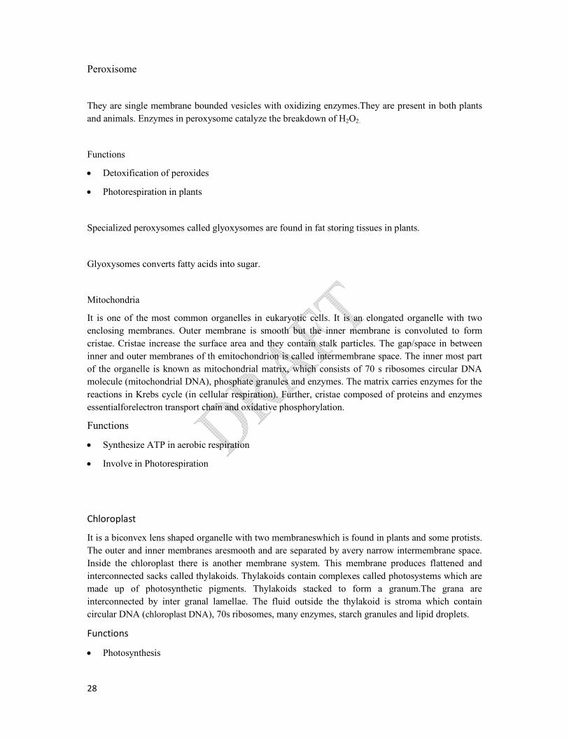
Peroxisome
They are single membrane bounded vesicles with oxidizing enzymes.They are present in both plants
and animals. Enzymes in peroxysome catalyze the breakdown of H2O2.
Functions
Detoxification of peroxides
Photorespiration in plants
Specialized peroxysomes called glyoxysomes are found in fat storing tissues in plants.
Glyoxysomes converts fatty acids into sugar.
Mitochondria
It is one of the most common organelles in eukaryotic cells. It is an elongated organelle with two
enclosing membranes. Outer membrane is smooth but the inner membrane is convoluted to form
cristae. Cristae increase the surface area and they contain stalk particles. The gap/space in between
inner and outer membranes of th emitochondrion is called intermembrane space. The inner most part
of the organelle is known as mitochondrial matrix, which consists of 70 s ribosomes circular DNA
molecule (mitochondrial DNA), phosphate granules and enzymes. The matrix carries enzymes for the
reactions in Krebs cycle (in cellular respiration). Further, cristae composed of proteins and enzymes
essentialforelectron transport chain and oxidative phosphorylation.
Functions
Synthesize ATP in aerobic respiration
Involve in Photorespiration
Chloroplast
It is a biconvex lens shaped organelle with two membraneswhich is found in plants and some protists.
The outer and inner membranes aresmooth and are separated by avery narrow intermembrane space.
Inside the chloroplast there is another membrane system. This membrane produces flattened and
interconnected sacks called thylakoids. Thylakoids contain complexes called photosystems which are
made up of photosynthetic pigments. Thylakoids stacked to form a granum.The grana are
interconnected by inter granal lamellae. The fluid outside the thylakoid is stroma which contain
circular DNA (chloroplast DNA), 70s ribosomes, many enzymes, starch granules and lipid droplets.
Functions
Photosynthesis
29
Cytoskeleton
Cytoskeleton is the supporting structure of the cell and maintains its shape. It is more important for
animal cells which lack cell walls. Cytoskeleton is made out of microtubules and protein filaments.
Additionally, it is Dynamic hence, has the ability to break and reform as needed.
There are three types of componentsin the Cytoskeleton as follows;
Microtubules
Actin filaments orMicrofilaments,
Intermediate filaments
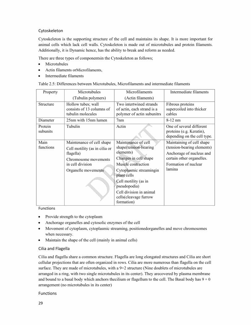
Table 2.5: Differences between Microtubules, Microfilaments and intermediate filaments
Property Microtubules
(Tubulin polymers)
Microfilaments
(Actin filaments)
Intermediate filaments
Structure Hollow tubes; wall consists of 13 columns of tubulin molecules
Two intertwined strands of actin, each strand is a polymer of actin subunitrs
Fibrous proteins supercoiled into thicker cables
Diameter 25nm with 15nm lumen 7nm 8-12 nm
Protein subunits
Tubulin Actin One of several different proteins (e.g. Keratin), depending on the cell type.
Main functions
Maintenance of cell shape
Cell motility (as in cilia or flagella)
Chromosome movements in cell division
Organelle movements
Maintenance of cell shape(tension-bearing elements)
Changes in cell shape
Muscle contraction
Cytoplasmic streamingin plant cells
Cell motility (as in pseudopodia)
Cell division in animal cells(cleavage furrow formation)
Maintaining of cell shape (tension-bearing elements)
Anchorage of nucleus and certain other organelles.
Formation of nuclear lamina
Functions
Provide strength to the cytoplasm
Anchorage organelles and cytosolic enzymes of the cell
Movement of cytoplasm, cytoplasmic streaming, positionedorganelles and move chromosomes
when necessary.
Maintain the shape of the cell (mainly in animal cells)
Cilia and Flagella
Cilia and flagella share a common structure. Flagella are long elongated structures and Cilia are short
cellular projections that are often organized in rows. Cilia are more numerous than flagella on the cell
surface. They are made of microtubules, with a 9+2 structure (Nine doublets of microtubules are
arranged in a ring, with two single microtubules in its center). They arecovered by plasma membrane
and bound to a basal body which anchors thecilium or flagellum to the cell. The Basal body has 9 + 0
arrangement (no microtubules in its center)
Functions

30
Act as locomotor appendages
Can move fluid over the surface of the tissue
Cilia liningin oviducts helpmove an egg toward the uterus
Centrioles
Centriole is made up of cylindrically arranged microtubuleswhich are non membrane bounded
subcellular component present only in animal cells. Each centriole composed of nine sets of triplet
microtubules arranged in a ring (9+0). A pair of centrioles which arranged perpendicular to each other
are located in a region called centrosome near the nucleus.
Functions
Produce aster and spindle in cell division
Central Vacuole
Central vacuole is a large structure, bound by tonoplast, filled with liquid called cell sap found in
plant cells. The composition of sap differs from cytosol and it contains water, ions such as Potassium
and Chloride and sometimes water soluble colored pigments such as anthocyanin.
Functions
stores water and other materials such as sugars, ions and pigments.
Maintains water balance of the cell
Gives turgidity and support to cell.
Produce colours in some plants with sap pigments
Stores soluble substances needed for cellular activities.
Extracellular components
1. Cell wall
Cell wall is an extracellular structure of plant cells. Animal cells do not have cell walls. However,
prokaryotes, fungi and some protists also have a thin and flexible cell wall .The chemical composition
of the wall greatly varies from speciesto species and even from one cell type to another even in the
sameplant. Nevertheless in Plants, cell wall isgenerally made up of cellulose,pectin, hemicellulose,
lignin and suberin (in some plant cells only).
Plants generate two types of cell walls: primary and secondary walls. Young cells first secrete
primarycell wall: it is the wall laid down during plant cell division.
Just outside the primary wall there is a thin layer (middle lamella) which is rich in sticky
polysaccharidescalled pectins (magnesium and calcium pectate).Middle lamella glues adjacent cells
together. Due to the deposition of hardening substances on the primary wall a secondarycell wall is
generated secondarily.
31
Primary cell wall is permeable, relatively thin, flexible, composed mainly of cellulose fibers which are
laid unevenly running through the extracellular matrix (middle lamella)Water can move freely
through the free spaces of cell wall
Secondary cell wall lies between plasma membrane and primarycell wall. It contains several layers of
hard materials, forming a rigid structure. In addition to cellulose, impermeable substances such as
lignin and suberine are also incorporated in to the secondary wall. Lignin cement anchors cellulose
fibers together providing hard and rigid matrix, giving the cell wall an extra support.
Cell wall has pits through which cytoplasm of adjoining cells join through plasmodesmata.
Functions
Protection and support
Allows development of turgidity when water enters the cell
Prevents bursting during turgidity
Limits and control cell growth
Component of appoplast pathway
Maintaining cell shape
hold the plant up against the force of gravity
2. Cell junctions
Cell junctions are structures at which neighbouring plasma membranes are joined. They are
also interact and communicate via sites of direct physical contacts. There are three types of
cell junctions in animal cells.
There are three types of cell junctions in animal cells
Tight junctions – connect the plasma membranes of adjacent cells tightly bound by specific
proteins forming continous seals around the cells. Prevent leakages of extracellular fluids
through intercellular space. e.g. skin epithelium
Desmosomes/Anchor junctions – mechanically attach the cytoskeletons of adjoining cells by
intermediate filaments for strong binding.e.g. muscle tissue
Gap junctions /Communicating junctions – provide cytoplasmic channels from one cell to an
adjescentcell. Gap junctions consists of special membrane proteins that surround the pore
through which ions, sugars amino acids may pass. They allow signal and material exchange
between adjacent cells through direct connections. e.g.heart muscles, animal embryo.
Plasmodesmata
Microscopic channels which runs through plant cell walls. They are cytoplasmic living
connections between cytoplasam of adjoining cells. These are membrane lined channels filled
with cytoplasam.
3. Extracellular matrix of animal cells

32
Although animal cells lack cell walls they do have elaborate extracellular matrix (ECM). Main
components of the ECM are glycoproteins and other carbohydrates containing molecules secreted by
the cells. Most abundant glycoprotein in the ECM of most animal cell is collagen which forms strong
fibres outside the cell. The collagen fibres are embedded in a network woven out of proteoglycan
secreted by cells.
Functions
Forms a protective layer over the cell surface
Linking extra cellular matrix and cytoskeleton.
Influences the cell behavior by Involving in the mechanical and chemical signaling.

33
Cell cycle and the process of the Cell division
Cell cycle
The sequence of events that takes place in the cell from the end of one cell division to the end of the
next cell division is referred to as cell cycle. At the end of the cell division, two genetically identical
daughter cells resembling the parent cell are produced in mitosis.
Eukaryotic cell cycle
Mitosis
Eukaryotic cell cycle may divided into two major phases.
Interphase
Mitotic phase/ M-phase
Interphase is the longer phase of cell division. It covers about 90% of the cell cycle.
Interphase could be divided into three phases;
G1 phase (first gap phase)
S phase (synthetic phase)
G2 phase (second gap phase)
G1 phase
In this phase synthesis of proteins and production of cellular organelles leading to cell growth occur.
Proteins essential for S phase are produced during this phase.
S phase
DNA replication occurs and synthesis of histone proteins takes place. DNA wind around histone
beads and form chromatin.
G2 phase
Cells continue to grow through protein synthesis as well as cellular organelles. Proteins essential for
mitotic phase will be synthesized. Duplication of centrosomes takes place.
There are cell cycle-controlling checkpoints available at G1, G2 and M phases to ensure that the cell is
ready for moving into upcoming phases of cell division. Some cells receive a go-head signal at the G1
check point, it will usually complete the G1, S, G2 and M phases and divide. If it does not receive a go
head signal at that point it may exit the cycle, entering into a non dividing stage called the Go phase.
The most cells of the human body are actually in the Go phase. e.g. nerve cells and muscle cells.
Mitotic phase/ M phase
M phase covers only about 10% of cell cycle. This includes mitosis and cytokinesis.
34
Mitosis
Mitosis is referred to the nuclear division which gives rise to two genetically identical daughter nuclei
from a mother nucleus. This may get divided into five stages; prophase, prometaphase, metaphase,
anaphase and telophase in order to ease the learning of activities of cell cycle.
1. Prophase
Chromatin fibers get condensed by shortening and thickening and transformed into chromosomes. As
a result chromosomes will be visible through light microscope. Nucleoli get disappeared and
chromosomes appear with two sister chromatids attached at the centromere. Chromosomal arms of
sister chromatids attached by special proteins called cohesion. The formation of mitotic spindles
begins. Spindle includes the centrosomes, the spindle microtubules and the aster.
Centrosomes move toward opposite poles of the cell due to the lengthening of microtubules between
them.
2. Prometaphase
The nuclear envelope fragments. Chromosomes get even more condensed. A special protein called
kinetochore attaches the sister chromatids of each chromosome at their centromere. Some of the
microtubules that attach to the kinetochore of the chromosomes move the chromosomes back and
forth. Microtubules which are not attached to the kinetochore interact with those from the opposite
poles.
3. Metaphase
Centrosomes reach the opposite poles. The chromosomes have arrived to a place called metaphase
plate which is located in equal distance from each pole. The centromeres of all chromosomes are
located in the metaphase plate. At the end of this phase, each chromosome of the cell get attached to
the kinetochore microtubule at their centromere and aligned at the metaphase plate.
4. Anaphase
Sister chromatids are separated at the centromere. Microtubules attached to kinetochore get shorten
and pull sister chromatids towards the opposite poles. Cell elongates as the non kinetochore
microtubules are lengthen. By the end of anaphase equal and complete set of chromosomes found at
each pole of the cell.
5. Telophase
Nuclear envelope reforms around each set of chromosomes at opposite poles. Nucleoli reappears.
Spindle microtubules get deplolymerized. Chromosomes unwind and become less condense to form
chromatin. Two genetically identical daughter nuclei are formed.
Cytokinesis
The division of the cytoplasam starts at the end of the telophase. Therefore at the end of the mitosis
two genetically identical daughter cells are produced.
In animal cells- a cleavage furrow forms. This produces two genetically identical daughter cells.
In plant cells- cell plate forms as a result of vesicle produced by golgi apparatus. This divides the
cytoplasm in to two and generates two genetically identical daughter cells to the parent cell.
35
Significances of mitosis
1. Maintains the genetic stability
2. Growth and development
3. Cell repair, replacement and regeneration
4. Asexual reproduction
Meiosis
Sexually reproducing organisms undergo different type of cell division called meiosis.
Meiosis
Meiosis is a type of nuclear division which gives rise to four haploid, genetically non identical
daughter nuclei, from a diploid mother nucleus.
Meiosis involves two consecutive nuclear divisions, Meiosis I and Meiosis II.
Meiosis I is a reduction division and Meiosis II is similar to mitosis, each stage consists of four sub-
phases: prophase, metaphase, anaphase, and telophase.
Before meiosis one cell is in interphase, during S phase of the interphase DNA replication occur.
Meiosis I
1. Prophase I
Cell enters to the prophase I from interphase. Chromosomes begin to condense. Nucleolus begins to
disappear. Next the formation of zipper like structure called the synaptonemal complex by a specific
proteins holds two homolg tightly together. The pairing and physical connection of homologous
chromosomes is called synapsis.
During synapsis part of the DNA molecule of non-sister chromatids paired homologous
chromosomes break, exchange and rejoin at corresponding point. This process is called crossing over.
These points of crossing over become visible as chiasmata after the synaptonemal complex
dissembles and the homologous chromosomes slightly apart from each other..
Nuclear envelop breaks. Centrosomes move towards opposite poles forming spindle in animal cells.
The kinetochore of each homologue attach to microtubule from one pole or the other. The
homologous pair then moves toward the metaphase plate.
2. Metaphase
The pair of homologous chromosomes get arranged on the metaphase plate with one chromosome of
each pair faces each pole.Both chromatids of a homologue are attached to kinetochore microtubules
from one pole and those of the other homolog are attached to kinetochore microtubules from the
opposite pole. Homologous chromosome arrange randomly at metaphase plate.
3. Anaphase I
36
Kinetochore microtubules of the spindle get shorten. Homologous pair separates and one chromosome
of each pair moves towards the opposite pole. Sister chromatids of each chromosome remain attached
at the centromere and move as a single unit towards the same pole.
4. Telophase
One complete haploid set of chromosomes accumulate at each pole. Nuclear envelope reforms around
each set of chromosomes.Nucleoli reappear. Spindle disintegrates. Chromosomes decondensed into
chromatin.Genetically non identical,haploid, two daughter nuclei are formed within one cell.
Cytokinesis
Usually occurs simultaneously with telophase I. Genetically non identical, haploid, two daughter cells
are formed. In animal cells, cleavage furrow is formed. In plant cells a cell plate is formed.
No DNA replication occurs between meiosisand meiosis.
Meiosis
1. Prophase
Centrosomes start producing spindle apparatus (spindle fibers, aster centrosome). Chromatin fibers
condense and produce chromosomes with two sister chromatids. Nuclearenvelope breaks down into
fragments. Nucleolus disappears. During the late prophase II centromere of the chromosomes are
moved to the metaphase plate.
2. Metaphase
All Chromosomes get attached to the microtubules at their centromere and aligned on the metaphase
plate. Kinetochores of sisterchromatids are attached to microtubules extending from both poles.
Due to the crossing over in meiosis, the two sister chromatids of each chromosome are not
genetically identical.
Meiosis II usually takes place in the perpendicular direction of MeiosisI. Therefore, metaphase plate
of meiosis II is perpendicular to the metaphase plate of meiosisI.
3. Anaphase
Due to the breakdown of proteins attaching sister chromatids, they are separated at centromere. As a
result of shortening of microtubules , sister chromatids of each chromosome move towards opposite
poles.
4. Telophase
Nuclear envelope and nucleolus reform.Chromosomes decondense into chromatin.Spindle
disassembles.Genetically non identical,haploid, four daughter nuclei are formed from one parent cell.

37
Cytokinesis
Cytokinesis occurs as in mitosis. Genetically non identical, haploid, four daughter cells are formed.
These four daughter cells are not even identical to their parent cell.
Centrosomes or centrioles are not available in plant cells. However, spindle is formed during cell
division from accumulated microtubule complex.
Significance of meiosis
Maintain the constant number of chromosomes through generations in sexually reproducing
species.
Produce new genetic variations leading to evolution.
Genetic variation occur due to crossing over ,recombination and independent assortment.
Tumor, cancer and galls
Tumors and cancers in animals
Cell division is driven by external and internal factors. They may be chemical or
physical factors
Chemical factors such as growth factors, physical factors such as density dependent
inhibition and anchorage dependence are needed for cell division normally.
Normal animal cells stop deviding when they come into contact with crowded cells.
this phenomena is known as density dependent inhibition.
Most animal cells divide, if they are attached to an external surface such as the surface
of a cell next to them. This is known as anchorage dependence inhibition.
Cancer cells exhibit neither density dependent inhibition nor anchorage dependent
inhibition.
Cancer cells do not respond to normally to the body’s control mechanism
They divide excessively and invade other tissues. If unchecked they can kill the
organism.
Cancer cells do not consider the normal signals that regulate the cell cycle.
They do not need growth factors. They may make required growth factors themselves
or giving signals to continue for the cell cycle without growth factors. Another
possiblility is an abnormal cell cycle control system.
The problem begins when a single cell in a tissue undergoes transformation, the
process converts a normal cell to abnormal cell.
If the body immune system can not recognize and destroy it this leads proliferate cells
and form a tumor.
If the abnormal cells remain at the original site, the lump is called benign tumor. Most
benign tumors do not cause serious problems and can be completely removed by a
surgery.

38
A malignant tumor becomes invasive and attack one or more organs. An individual
with a malignant tumor is said to have a cancer.
A few tumor cells may separate from the original tumor, enter blood vessels or lymph
vessels and travel to other parts of the body. They may proliferate and form a new
tumor.
This spread of cancer cells to locations distant from their original site is called
metastasis.
Galls in plants
This occurs due to uncontrolled mitotic division of plant cell.
The plant cell division is controlled by maintaining a proper balance between plant
growth regulators such as auxins and cytokinins. When this balance is lost plant cells
produce undifferentiated mass of cells.
Galls are the bumbs and growths that develop on different parts of plants after being
invaded by some very unique organisms.
Galls have range of causes, including viruses, fungi, bacteria, insects and mites.
Usually the gall causers in some way attack or penetrate the plants growing tissues
and causes the host to reorganize its cells and to develop an abnormal growth.

39
Metabolism
Energy relationships in metabolic process
Sum of all biochemical reactions of living being is known as the metabolism and it consists of all
catabolic and anabolic reactions.
Catabolism is breaking down of complex molecules into simple molecules by releasing free energy.
Thereforeit is an exergonic reaction. Anabolism is making complex molecules from the simple
molecules by absorbing free energy.Hence it is an endergonic reaction.
Biochemical reactions involved in usage of energy released by catabolic reactions in living system are
called as anabolic reactions. ATP acts as the energy carrier in all living organism including the
simplest bacteria. Thereforethe ATP is known as the universal currency of energy transactions.
Energy can be defined as the capacity to do work. All living organisms require energy for their living
process in many ways. Such processes are;
Synthesis of substances
Active transport across plasma membrane
Transmission of nerve impulses
Muscle contraction
Beating of cilia and flagella
Bioluminescence
Electrical discharges.
Overall idea of the energy relations of living system on biosphere is composed of following steps.
1. Energy flows into biological systems from the environment through solar radiation. (Primary
energy source is the Sun)
2. Light energy is captured in the cells having photosynthetic pigments (chlorophyll) by the
process of photosynthesis and stored as chemical energy in the organic compounds such as
carbohydrates
3. Stored energy in organic food is transformed into chemical energy in ATP by a process called
cellular respiration.
4. The energy stored in ATP is utilized in various energy requiring processes.
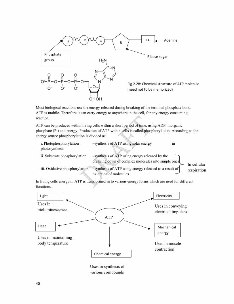
ATP (Adenosine Tri Phosphate)
ATP is a nucleotide, consisting of,
Ribose- sugar
Adenine - nitreogenous base
A chain of three phosphate groups.
During the hydrolysis of ATP, ADP and Pi are produced. As a result, a very high energy is released.
This is because the reactants (ATP and water) contain more energy in comparison to products (ADP
and Pi). Therefore it yields energy and is an exergonic reaction.
When ATP is hydrolyzed, the free energy yield of eachof the two end phosphate groups is -
30.5kJ/mol.
40
Most biological reactions use the energy released during breaking of the terminal phosphate bond.
ATP is mobile. Therefore it can carry energy to anywhere in the cell, for any energy consuming
reaction.
ATP can be produced within living cells within a short period of time, using ADP, inorganic
phosphate (Pi) and energy. Production of ATP within cells is called phosphorylation. According to the
energy source phosphorylation is divided as;
i. Photophosphorylation –synthesis of ATP using solar energy in
photosynthesis
ii. Substrate phosphorylation –synthesis of ATP using energy released by the
breaking down of complex molecules into simple ones.
iii. Oxidative phosphorylation –synthesis of ATP using energy released as a result of
oxidation of molecules.
In living cells energy in ATP is transformed in to various energy forms which are used for different
functions..
R AA P P P
Ribose sugar Phosphate
group
Adenine
In cellular
respiration
Heat
Chemical energy
Uses in
bioluminescence
Uses in synthesis of
various compounds
Uses in muscle
contraction
Fig 2.28: Chemical structure of ATP molecule
(need not to be memorized)
Light
Mechanical
energy
Electricity
Uses in maintaining
body temperature
Uses in conveying
electrical impulses ATP
41
Role of Enzyme
An enzyme is a macromolecule, which act
cells/
General characteristics of an enzyme
1. Most of the enzymes are globular proteins.
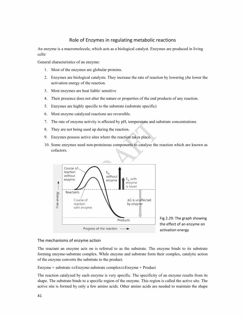
2. Enzymes are biological catalysts.
activation energy of the reaction.
3. Most enzymes are heat liable/
4. Their presence does not alter the nature or properties of the end products of any reac
5. Enzymes are highly specific
6. Most enzyme catalyzed reaction
7. The rate of enzyme activity
8. They are not being used up during the reaction.
9. Enzymes possess active sites where the reactio
10. Some enzymes need non-prote
cofactors.
The mechanisms of enzyme action
The reactant an enzyme acts on is referred to as the substrate
forming enzyme-substrate complex. While enzyme and substrate
of the enzyme converts the substrate to the
Enzyme + substrate Enzyme-substrate complex
The reaction catalyzed by each enzyme is very specific. The specificity of an enzyme results from its
shape. The substrate binds to a specific region of the enzyme
active site is formed by only a few amino acids.
Enzymes in regulating metabolic reactions
, which acts as a biological catalyst. Enzymes are produced in living
of an enzyme:
globular proteins.
atalysts. They increase the rate of reaction by lowering yhe
reaction.
liable/ sensitive
Their presence does not alter the nature or properties of the end products of any reac
specific to the substrate (substrate specific)
catalyzed reactions are reversible.
The rate of enzyme activity is affected by pH, temperature and substrate concentrations
They are not being used up during the reaction.
Enzymes possess active sites where the reaction takes place.
proteinous components to catalyse the reaction which are known as
of enzyme action
is referred to as the substrate. The enzyme binds to its substrate
substrate complex. While enzyme and substrate form their complex, catalytic action
of the enzyme converts the substrate to the product.
substrate complexEnzyme + Product
The reaction catalyzed by each enzyme is very specific. The specificity of an enzyme results from its
specific region of the enzyme. This region is called the active site. The
active site is formed by only a few amino acids. Other amino acids are needed to maintain the shape
Fig 2.29: The graph showing
the effect of an enzyme on
activation energy
. Enzymes are produced in living
increase the rate of reaction by lowering yhe lower the
Their presence does not alter the nature or properties of the end products of any reaction.
substrate concentrations.
reaction which are known as
The enzyme binds to its substrate
catalytic action
The reaction catalyzed by each enzyme is very specific. The specificity of an enzyme results from its
is called the active site. The
Other amino acids are needed to maintain the shape
Fig 2.29: The graph showing
the effect of an enzyme on
activation energy

42
of the enzyme molecule. The shape of the active site is complementary to the shape of the specific
substrate of the enzyme, and hence important in the substrate specificity of the enzyme.
the active site of an enzyme is not
rigid structures, the interactions between substrate and active site may slight
active site, so that the substrate and the active site
induced fit mechanism. The tight fit not only brings the substrate molecules and the active site close
to each other, but also ensures the correct orientation of the molecules to help the reaction to proceed
and catalyzes the conversion of substrate to product. Thereafter, the
site of the enzyme. The enzyme is then free to take another substrate
Cofactors
Non-proteinuos components which are essential for the catalytic activities o
called cofactors.
These cofactors bind to the enzymes in two ways. Some tightly bind and remain
others loosely bind temporarily. Loosely bound co
Organic cofactors are called co-enzymes. e.
Inorganic co-factors – e.g. Zn2+, Fe2+
Factors affecting the rate of enzymatic reactions
1. Temperature
2. pH
3. Substrate concentration
4. Inhibitors
Temperature
Increase intemperature increases molecular motion. Therefore the
both enzymes as well as the substrate will be accelerated. T
for both enzyme active sites and substrate molecules. More collision between the enzyme active sites
and substrate molecules generate greater chances for the reaction to occur.
certain point, after which there is a rapid
optimum temperature. This may vary
Fig 2.30: Induced fit between an enzyme and its substrate
. The shape of the active site is complementary to the shape of the specific
and hence important in the substrate specificity of the enzyme.
not always fully complementary to its substrate. As enzymes are not
rigid structures, the interactions between substrate and active site may slightly change the shape of the
e substrate and the active site become complementary to each other.
The tight fit not only brings the substrate molecules and the active site close
lso ensures the correct orientation of the molecules to help the reaction to proceed
version of substrate to product. Thereafter, the product departs from the active
. The enzyme is then free to take another substrate molecule into its active site.
components which are essential for the catalytic activities of certain enzymes are
factors bind to the enzymes in two ways. Some tightly bind and remain permanently
d temporarily. Loosely bound cofactors are reversible under certain circumstances.
enzymes. e.g. derivatives of vitamins e.g. NAD, FAD and biotin
2+, Cu2+
Factors affecting the rate of enzymatic reactions
Substrate concentration
Increase intemperature increases molecular motion. Therefore the speed of the moving molecules of
e substrate will be accelerated. This will enhance the colliding probability
for both enzyme active sites and substrate molecules. More collision between the enzyme active sites
ules generate greater chances for the reaction to occur. This can continue up to a
rapid decline in enzyme activity. This point is referred to as
This may vary from organism to organism.
Fig 2.30: Induced fit between an enzyme and its substrate
. The shape of the active site is complementary to the shape of the specific
and hence important in the substrate specificity of the enzyme. The shape of
its substrate. As enzymes are not
ge the shape of the
complementary to each other. This is called
The tight fit not only brings the substrate molecules and the active site close
lso ensures the correct orientation of the molecules to help the reaction to proceed
product departs from the active
molecule into its active site.
f certain enzymes are
permanently and
factors are reversible under certain circumstances.
e.g. NAD, FAD and biotin
speed of the moving molecules of
his will enhance the colliding probability
for both enzyme active sites and substrate molecules. More collision between the enzyme active sites
This can continue up to a
decline in enzyme activity. This point is referred to as

43
e.g. most of the human enzymes have optimum temperature around the body temperature (35˚C-
40˚C). Optimum temperature of bacteria in hot springs is about 70˚C.
When the temperature increases beyond the optimum temperature, the hydrogen bonds, ionic and
other weak chemical bonds of enzyme active sites may be disrupted. This will result a change in the
shape of the active site of enzyme which will alter the complementary nature of the active site of
enzyme molecules. Therefore, the complementary binding of enzyme active sites and substrate
molecules will be prevented. The above event is called as denaturation of enzyme molecules.
Therefore the rate of enzyme catalyzed reaction will start to decline when the temperature increases
beyond the optimum temperature and stops completely at certain temperature, although rate of
collision will keep on increasing.
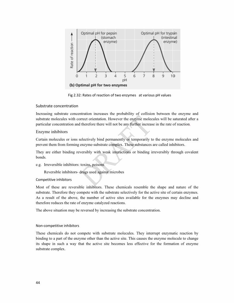
pH
Enzymes function most efficiently within a certain pH range despite maintaining temperature of the
environmentconstant.
The narrow range of pH in which a particular enzyme catalyzed reaction takes place is named as the
pH range. The pH at which the highest rate of reaction occurs is the optimum pH of the enzyme.The
alteration in pH above or below the optimum pH may lead to decline in enzyme activity. This is due
to the alteration of chemical bonds involving in formation of enzyme substrate complex. In most
enzymes optimum pH range is 6-8, but there are exceptions. Pepsin works best at pH 2 and optimum
pH for Trypsin is 8.
44
Substrate concentration
Increasing substrate concentration increases the probability of collision between the enzyme and
substrate molecules with correct orientation. However the enzyme molecules will be saturated after a
particular concentration and therefore there will not be any further increase
Enzyme inhibitors
Certain molecules or ions selectively bind
prevent them from forming enzyme
They are either binding reversibly with weak interactions or binding irreversibly through covalent
bonds.
e.g. Irreversible inhibitors: toxins, poisons
Reversible inhibitors- drugs used against microbes
Competitive inhibitors
Most of these are reversible inhib
substrate. Therefore they compete with the substrate selectively for the active site of certain enzymes.
As a result of the above, the number of active site
therefore reduces the rate of enzyme catalyzed reactions.
The above situation may be reversed by increasing the substrate concentration.
Non-competitive inhibitors
These chemicals do not compete with
binding to a part of the enzyme other than the active site
its shape in such a way that the ac
substrate complex.
Fig 2.32: Rates of reaction of two enzymes at various pH values
concentration increases the probability of collision between the enzyme and
substrate molecules with correct orientation. However the enzyme molecules will be saturated after a
particular concentration and therefore there will not be any further increase in the rate of reaction
Certain molecules or ions selectively bind permanently or temporarily to the enzyme molecules and
prevent them from forming enzyme-substrate complex. These substances are called inhibitors.
They are either binding reversibly with weak interactions or binding irreversibly through covalent
oxins, poisons
drugs used against microbes
inhibitors. These chemicals resemble the shape and nature of the
substrate. Therefore they compete with the substrate selectively for the active site of certain enzymes.
number of active sites available for the enzymes may de
therefore reduces the rate of enzyme catalyzed reactions.
The above situation may be reversed by increasing the substrate concentration.
These chemicals do not compete with substrate molecules. They interrupt enzymatic reaction by
other than the active site. This causes the enzyme molecule to change
such a way that the active site becomes less effective for the formation of enzyme
Fig 2.32: Rates of reaction of two enzymes at various pH values
concentration increases the probability of collision between the enzyme and
substrate molecules with correct orientation. However the enzyme molecules will be saturated after a
in the rate of reaction.
or temporarily to the enzyme molecules and
substrate complex. These substances are called inhibitors.
They are either binding reversibly with weak interactions or binding irreversibly through covalent
the shape and nature of the
substrate. Therefore they compete with the substrate selectively for the active site of certain enzymes.
the enzymes may decline and
nterrupt enzymatic reaction by
. This causes the enzyme molecule to change
less effective for the formation of enzyme

45
Fig 2.35: noncompetitive inhibitors.35: noncompetitive inhibitors

46
Regulation mechanism of enzymatic activity in cells
Allosteric regulation of enzymes
In many cases, the molecules that naturally regulate enzyme activity in a cell behave like reversible non-
competitive inhibitors. Regulatory molecules (either activators or inhibitors) bind to specific regulatory
sites elsewhere (other than the active site) of the molecule via non-covalent interactions and affect the
shape and function of the enzyme. It may result in either inhibition or stimulation of an enzyme activity.
a.) Allosteric activation and inhibition
most enzymes regulated by allosteric regulation are made from two or more subunits. Each sub unit
composed of a polypeptide chain with its own active site. The entire complex oscillates between two
different shapes one catalyzing active and other inactive. In this two forms regulatory molecules bind to a
regulatory site called allosteric site, often located where subunits join.
When an activator binds with this regulatory site, stabilizes the shape with functional active sites. Wheras
the inhibitor binds with the regulatory site, it stabilizes the inactive form of enzyme. Subunits of enzyme
arranged in a way through which they transmit the signals quickly other subunits. Through the interaction
of subunits even a single activator or inhibitor molecule that bind to one regulatory site will affect the
active site of all sub units. e.g. ADP function as allosteric activator bind to the enzyme and stimulates the
production of ATP by catabolism. If the supply of ATP exceed demand catabolism shows down as ATP
bind to the same enzyme as inhibitor.
b.) ccoperativity
This is another type of allosteric activation. Binding of one substrate molecule can stimulate binding or
activity at other active site. Thereby increase the catalytic activity.
e.g. hemoglobin (not an enzyme) is made up of four subunits each with an O2 binding site. The binding of
an one molecule of O2 to one binding site increases the affinity for O2 of the remaining binding site.
c.) Feed back inhibition
in feed back inhibition, a metabolic pathway is stopped by the inhibitory binding of its end product of a
process to an enzyme. Thereby limit the production of more end products than required and thus wasting
chemical resources.
Feedback inhibition
Feedback inhibition is an essential process regulates the end products produced in metabolism.
e.g. ADP function as allosteric activator and stimulates the production of ATP during the catabolism.
In case ATP supply exceeds demand, catabolism slows down as ATP molecules function as allosteric inhibitor.
Energy needed for all living processes is obtained directly from ATP. ATP is mainly produced by a
process call cellular respiration, in living cells.

47
Cellular respiration and its importance
Cellular respiration is the process by which chemical energy in organic molecules such as
carbohydrates is released by stepwise oxidative process, catalyzed by enzymes and made available in
living cells in the form of ATP. Cellular respiration is divided as
a) aerobic respiration
b) anaerobic respiration
Aerobic respiration:-
The process of synthesize ATP from the respiratory substrates such as glucose in the presence of
molecular oxygen (O2) known as aerobic respiration. Glucose is found to be the major respiratory
substrate in living cells.
The aerobic respiration of glucose molecules can be represented by the following balanced chemical
equation.
C6H12O6 + 6O2(g) 6CO2(g) + 6H2O + Energy (ATP+ heat)
This process consist three main steps. They are;
a) Glycolysis
b) Pyruvate oxidation and citric acid cycle (Kreb’s cycle)
c) Oxydative phosphorylation (Electron transport chain)
Glycolysis
It takes place in the cytosol of the cell, because all enzymes that catalyze reactions of the glycolysis
are found in the cytosol of the cell. This process does not depend on O2.
During the above process a six carbon (6C) glucose molecule is broken down step by step into two
three-carbon (3C) pyruvate molecules.
Two ATP molecules are used up to initiate the process.
Four hydrogen molecules and electrons released from glucose breakdown reduce two NAD+and
produce two NADH.At the end of glycolysis there will be four ATP molecules produced. Since two
ATP molecules were used up for the initiative step, the net yield will be two ATP molecules.
Only when O2is present, the pyruvate molecules will enter the mitochondria and further steps will take
place.
Oxidation of Pyruvate/ Link reaction
These two pyruvate molecules enter mitochondrion by active transport through the membrane. In the
matrix of mitochondria, Pyruvate is converted to acetyl group by releasing two CO2molecules. Then
this acetyl group combines with co-enzyme A to produce Acetyl co-A. In this reactiontwoNAD+ is
converted to two NADH molecules. Therefore this step can be represented as follows.
2C3H4O3 + 2CoA + 2NAD+ 2 Acetyl Co-A + 2CO2+ 2 NADH
Oxidation of pyruvate is a linking reaction of glycolysis and citric acid cycle.
Acetyl Co-A will feed its acetyl group for citric acid cycle.

48
Citric acid cycle
This takes place in the matrix of mitochondria using specific enzymes. As the first product of this
cyclic pathway is citric acid, it is known as citric acid cycle. The pathway was discovered by a
German-British scientist Hans Krebs. Hence,it is named as Kreb’s cycle. Citric acid contains three
carboxylic acid groups. This cycle is also known as Tricarboxylic Acid cycle or TCA cycle.In the
citric acid cycle 4 C compound oxalo acetate combines with 2 C compound acetyl Co -A to form 6 C
compound, citric acid. Then citric acid undergoes a series of reactions to regenerate oxaloacetate by
releasing two CO2 molecules by decarboxylation reaction. One ATP molecule is produced by
substrate level phosphorylation. One FADH2 and three NADH will be generated as a result
ofoxidation reactions. These are the products of a single acetyl group led into citric acid cycle and
hence these numbers should double when the yield for a glucose molecule is considered.
Electron transport chain
This step is taken place across the inner membrane (cristae) of mitochondria. The folding of cristae
increases surface area for oxidative phosphorylation. NADH and FADH2 products in the early stages
of aerobic respiration are oxidized by transferring electrons, through the electron transport chain
andfinally to molecular oxygen (O2). The electron transport chain is located in the inner membrane of
mitochondrion and composed of series of protein and non-protein molecules involving in the
movement of electrons and protons across cristae. Therefore, the Molecular oxygen(O2)is the final
electron acceptor in aerobic respiration. In the electron transport chain, ATP is synthesized by
oxidative phosphorylation.
In this electron transport chain, energy is released progressively from NADH and FADH2and that
energy is used to synthesize ATP. When one molecule of NADH is oxidized in the electron transport
chain, 2.5 molecules of ATP in average are generated due to oxidative phosphorylation. When one
molecule of FADH2 is oxidized 1.5 molecules of ATP in averageare produced due to oxidative
phosphorylation. Total number ATP that is produced in this step is 28.
This is true in the active cells such as liver cells and cardiac muscle cells but not in other cells where
two ATP produced in glucolysis is used to transport 2NADH from cytosol to mitochondrial matrix. In
those cells total number of ATP produced by one molecule of glucose is (32-2) = 30 ATP.
Total number of ATP molecules produced from one molecule of glucose, during aerobic respiration.
In glycolysis;
As ATP 2ATP
From 2NADH 5ATP (oxidation phosphorylation)
In pyruvate oxidation;
From 2NADH 5ATP (oxidative phosphorylation)
In Citric acid cycle;
As ATP ATP (substrate level phosphorylation)
From 6NADH 15ATP (oxidative phosphyrlation)
49
From 2 FADH2 3ATP
Total Number ATP = 32 ATP
Anaerobic respiration
Anaerobic respiration is breaking down of glucose in the absence of molecular oxygen which is
regulated by enzymes of the cells occurring in cytosol. In the absence of molecular oxygen pyruvate
molecules cannot be broken down further. ATP generated isutilized to fulfill energy requirements.
However, NADH produced during glycolysiscannot be utilized. Therefore, since NAD+is limited it is
essential for the cell to recycle NADH to enhance the availability of NAD+ other than anaerobic
respiration fermentation is a method of production of ATP in the absence of O2 . There are many types
of fermentation, differing from end products formed by pyruvate. The two common types are;
1. Ethyl alcohol fermentation
2. Lactic acid fermentation
Ethyl alcohol fermentation
Like in aerobic respiration, the first step of this is also Glycolysis.
Therefore one molecule of glucose is converted to 2 molecules of pyruvate giving 2
molecules of ATP and two molecules of NADH
Then this pyruvate involve in two steps. In the 1st step pyruvate is converted Acetyldehyde,
releasing a molecule of CO2.
in the second step acetyldehyde is reduced to ethanol using NADH that is produced in
Glycolysis.
Therefore final hydrogen acceptor in ethyl alcohol fermentation is acetylaldehyde (organic
compound)
Many bacteria carry out ethyl alcohol fermentation. The most common organism which
carries out ethyl alcohol fermentation is yeast.
Lactic acid fermentation
As in ethyl alcohol fermentation, Glycolysis takes place as the first step of lactic acid
fermentation.
Therefore one molecule of glucose produces two molecules of pyruvate, two molecules of
ATP and two molecules of NADH.
Then pyruvate is reduced directly by NADH for lactic acid as an end product with no release
of CO2. Therefore final H acceptor is also organic compounds.
Certain fungi and bacteria carryout lactic acid fermentation but the most common organisms
are lactic acid bacteria involved in formation of yoghurt and curd.
Respiratory quotient
It is the ratio of CO2 evolved and the volume of O2 consumed in a given time for the respiratory
substrate.
RQ= V CO2/ VO2
RQ of respiration of carbohydrates, fats and proteins are 1.0, 0.7 and 0.8 respectively.
Use of Proteins, Carbohydrates andFats in respiration

50
NH3
Amino
Acid
Proteins
GLYCOLYSIS
Glucose
Glyceraldehyde 3-P
Pyruvate
Sugars Glycerol
Acetyl Co-A
CITRIC ACID CYCLE
OXIDATIVE
PHOSPHORYLATION
Carbohydrates Fats
Fatty acids

51
Photosynthesis as an Energy fixing Mechanism
Photosynthesis
Photosynthesis is a metabolic process by which light energy is trapped and converted to chemical
energy. Chemical energy is stored in chemical bonds of carbohydrates, fats, oils, and proteins. All life
on Earth depends on photosynthesis either directly or indirectly. Photosynthesis also occurs in algae
and certain prokaryotes.
Global importance of photosynthesis;
All life on earth depends on photosynthesis, directly or indirectly
Fulfill both carbon and energy requirements of organisms
Provide O2 for respiration of aerobic organisms
Maintain O2 and CO2 balance in the atmosphere
Production of fossil fuel
Maintenance of global temperature
During photosynthesis CO2is reduced by the H of H2O and simple sugars are made using light energy.
In eukaryotic photosynthetic cells, chloroplasts are the sites of photosynthesis.
Process of photosynthesis consists of two main stages and they are integrated.
1. Light-dependent reaction
2. Calvin cycle
There are two types of photosynthetic mechanisms (path ways) based on the number of C atoms of the
first stable product of the CO2 fixation.
1. C3 Mechanism – No of C atom of the first stable compound is three
2. C4 Mechanism – No of C atom of the first stable compound is four
Light-dependent reactions of photosynthesis take place in the membrane system of thylakoids. They
are flattened fluid-filled sacs, which form stacks called grana at intervals. Chlorophylls, carotenoids
and electron acceptors are located on this membrane system of thylakoids.
Stroma is a gel like structure containing soluble enzymes and other chemicals, which is the site of the
Calvin cycle.
Photosynthetic pigments are substances which absorb visible light. In a leaf we see green colour
because chlorophylls absorb violet, blue and red light and therefore, they transmit and reflect green
colour. Different pigments absorb different wavelengths of light. In chloroplast, there are two types of
chloroplast pigments such as chlorophylls and carotenoids. Chlorophyll a is the key light capturing
pigment and they participate directly in the light reaction of photosynthesis.
According to the action spectrum, chlorophyll a is more effective for blue and red light. Chlorophyll b
and caraotenoids (carotenes and xanthophylls) are effective in absorption of specific range
wavelengths of corresponding to different colours.

52
Other important function of some carotenoids is photoprotection. Photoprotection is absorption and
dissipation of excessive light energy, if not that excessive light may cause damage to the chlorophylls
or interact with oxygen and form reactive oxidative molecules which are dangerous to the cell.
Absorption spectrum
An absorption spectrum is a graph of the relative amounts of light absorbed at different wavelengths
by a pigment.
Action spectrum
An action spectrum is a graph showing the effectiveness of different wave lengths of light in
stimulating the photosynthesis.

53
Excitation of chlorophyll by light
When a molecule of chlorophyll or other photosynthetic pigment absorbs light it becomes
excited. The energy from the light is used to boost electrons to a higher level and become
positively charged. The excited state is unstable and returns to their original lower energy
state. The excited electrons may pass through several electron carriers until they reach the
final electron acceptor.
Chlorophyll Chlorophyll+ + e- (electron)
Therefore chlorophyll is oxidized and electron acceptor is reduced.
Photosystems
Chlorophyll molecules, other organic molecules and proteins are organized into complexes in
the thylakoid membrane of chloroplasts. They are called photosystems.
A photosystem contains a reaction centre complex and light harvesting complexes. The
reaction centre complex also contains a primary electron acceptor.
There are two types of photosystems found in the thylakoid membrane. They are
Photosystem I (PS I) and photosystem II (PS II).In the PS I the chlorophyll a molecule is
known as P700 since they absorb light at 700nm wave length effectively. In the PS II the
reaction centre contains a chlorophyll a molecule which is known as P680 which absorbs
light having a wavelength of 680 nm.
Light-dependent reaction /Light reaction photosynthesis
Linear electron flow
Light is absorbed by the photosynthetic pigments and synthesize ATP and NADPH due to the
excitation of Photosystem I and Photosystem II which are embedded in the thylakoid
membrane of chloroplast. The key to this energy transformation is a flow of the electron in
one direction through the photosystems and other molecular components built in the
thylakoid. This process is called linear electron flow.
The striking of photons of light on the pigments results in the excitation of electrons from the
photosystem II to the higher energy state.
These electrons will be accepted by the primary electron acceptor of photosystem II.
Splitting of water takes place as a result of an enzyme catalyzed reaction and yields O2 (g),
H+ ions and electrons.
Electrons released as a result of hydrolysis may neutralize excited photosystem II (P680).
Light energy
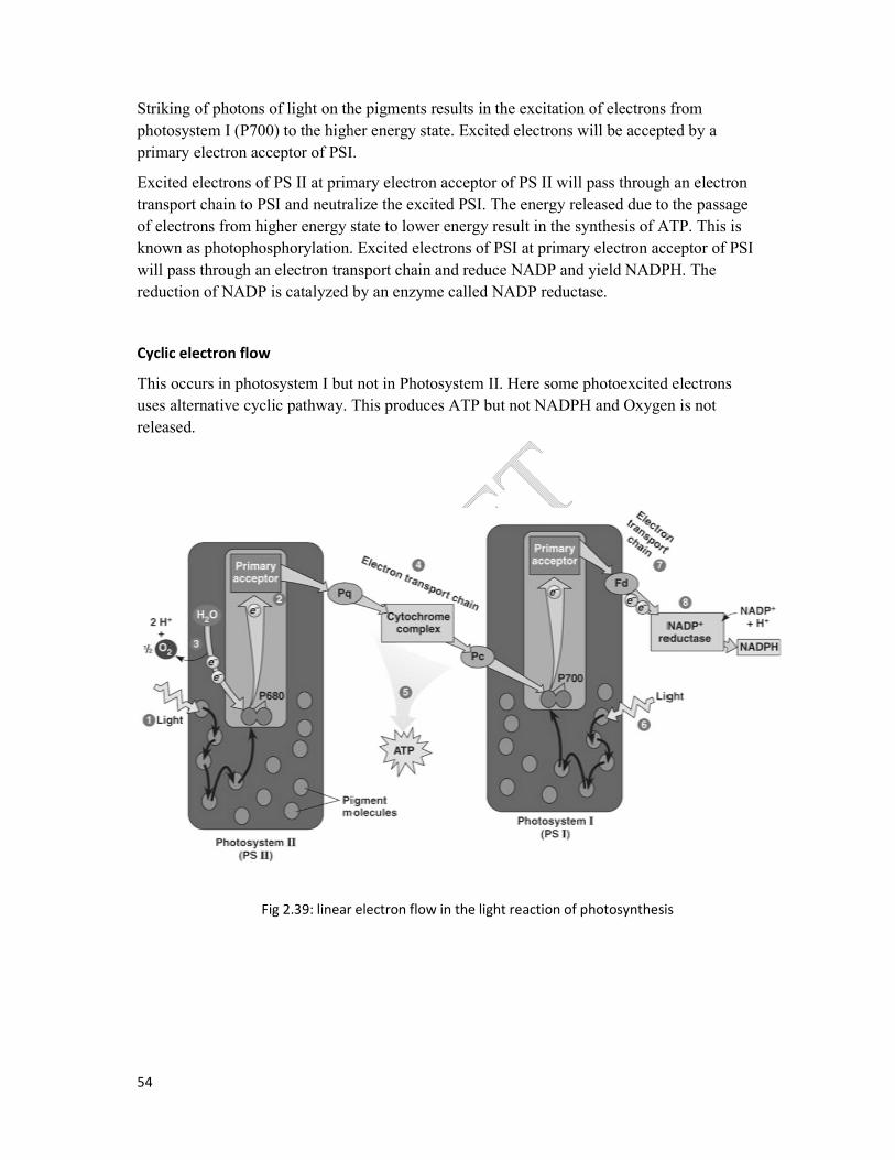
54
Striking of photons of light on the pigments results in the excitation of electrons from
photosystem I (P700) to the higher energy state. Excited electrons will be accepted by a
primary electron acceptor of PSI.
Excited electrons of PS II at primary electron acceptor of PS II will pass through an electron
transport chain to PSI and neutralize the excited PSI. The energy released due to the passage
of electrons from higher energy state to lo
known as photophosphorylation. Excited electrons of PSI at primary electron acceptor of PSI
will pass through an electron transport chain and reduce NADP and yield NADPH. The
reduction of NADP is catalyzed by an enzyme called NADP reductase.
Cyclic electron flow
This occurs in photosystem I but not in Photosystem II. Here some photoexcited electrons
uses alternative cyclic pathway. This produces ATP but not NADPH
released.
Fig 2.39: linear electron flow in the light reaction of photosynthesis
Striking of photons of light on the pigments results in the excitation of electrons from
photosystem I (P700) to the higher energy state. Excited electrons will be accepted by a
ectron acceptor of PSI.
Excited electrons of PS II at primary electron acceptor of PS II will pass through an electron
transport chain to PSI and neutralize the excited PSI. The energy released due to the passage
of electrons from higher energy state to lower energy result in the synthesis of ATP. This is
known as photophosphorylation. Excited electrons of PSI at primary electron acceptor of PSI
will pass through an electron transport chain and reduce NADP and yield NADPH. The
by an enzyme called NADP reductase.
This occurs in photosystem I but not in Photosystem II. Here some photoexcited electrons
uses alternative cyclic pathway. This produces ATP but not NADPH and Oxygen is not
: linear electron flow in the light reaction of photosynthesis
Striking of photons of light on the pigments results in the excitation of electrons from
photosystem I (P700) to the higher energy state. Excited electrons will be accepted by a
Excited electrons of PS II at primary electron acceptor of PS II will pass through an electron
transport chain to PSI and neutralize the excited PSI. The energy released due to the passage
wer energy result in the synthesis of ATP. This is
known as photophosphorylation. Excited electrons of PSI at primary electron acceptor of PSI
will pass through an electron transport chain and reduce NADP and yield NADPH. The
This occurs in photosystem I but not in Photosystem II. Here some photoexcited electrons
Oxygen is not
: linear electron flow in the light reaction of photosynthesis

55
The Calvin cycle
The Calvin cycle takes place in the stroma of the chloroplast. Energy from ATP and NADPH
produced by the light reaction are used to reduce CO2. The reactions are catalyzed by
enzymes and their sequence was discovered by scientist Calvin. This is an anabolic reaction.
The first stable product of the Calvin cycle is glyceraldehyde 3-phosphate (G3P). For the net
synthesis of one molecule of G3P, the cycle must take place three times.
The Calvin cycle of photosynthesis can be described in three steps;
Carboxylation (Carbon fixation)
Reduction
Regeneration of carbondioxide acceptor
Carbon fixation
The CO2 acceptor is a 5 C sugar, Ribulose bisphosphate (RuBP). The addition of CO2 to a
RuBP is called carboxylation. The enzyme involves in this reaction is RuBP carboxylase
oxygenase or Rubisco.
The first product of RuBP carboxylation is a 6C molecule which is unstable and breaks down
immediately in to two molecules of 3-phosphoglycerate (3-PGA). This is the first stable
product of photosynthesis. The enzyme RuBP carboxylase oxygenase (Rubisco) is present in
large amounts in the chloroplast stroma.
Reduction phase
1,3-Bisphosphoglycerate will be reduced to Glyceraldehyde 3- phosphate (G3P) through step
by step. Enzyme catalyzed reactions utilizing NADPH and ATP from light reaction. G3P will
act as a precursor for carbohydrate synthesis (glucose).
Regeneration of RuBP
RuBP is regenerated by undergoing a series of complex reactions. This process uses energy
from ATP generated in light reaction.
Photorespiration
As its name suggests, Rubisco is capable of catalyzing two distinct reactions, acting as both a
carboxylase and as an oxygenase.
In the oxygenase reaction of Rubisco uses the same substrate, RUBP, but reacts it with O2.
The reaction is catalyzed on the same active site as the carboxylation reaction. Thus CO2 and
O2 are competitive substrates. Therefore CO2 inhibits the oxygenase and O2 inhibits the
carboxylase reaction.

56
The oxygenase reaction forms just one molecule of 3-PGA plus a two carbon product, 2-
phosphoglycolate which is of no immediate use in the Calvin cycle and in higher
concentrations it is toxic for the plant. It therefore has to be processed in a metabolic pathway
called photorespiration. The photorespiratory pathway involves enzymes in the chloroplasts,
peroxisome and mitochondria. (detail of this pathway is not expected).
Photorespiration is not only energy demanding, but furthermore leads to a net loss of CO2.
Each time Rubisco reacts with O2 instead of CO2 the plants makes 50% less 3-PGA than it
would have done if CO2 had been used. This potentially eliminates the net gain in
photosynthetic carbon and loose the productivity.
The proportion of photosynthetically fixed carbon that is lost through photorespiration will
principally depend on the relative rates of the carboxylase and oxygenase reactions of
Rubisco. This ratio is determined by the ratio of the substrates CO2 and O2 in solution in the
cell as well as the inherent specificity factor of Rubisco, that is the affinity of Rubisco for O2
is less sensitive to temperature than is the affinity for CO2.
The (CO2) to (O2) ratio in solution is influenced by temperature. The solubility of a gas
decreases with increasing temperature. As the solubility of CO2 decreases more than that of
O2 with increasing temperature the ratio of (CO2) to (O2) decreases as the temperature rises.
These two factors result in an increase in photorespiration relative to photosynthesis so that
an increasing proportion of carbon is lost as the temperature rises.
The CO2 required for photosynthesis enters a leaf via stomata. However, stomata are also the
main avenues of transpiration. On a hot, dry day, most plants close their stomata in order to
conserves water. At the same time O2 released from the light reactions begins to increase and
this leads to further reduction of (CO2) to (O2) ratio in the cytosol. These conditions within
the leaf favor a wasteful process photorespiration under high temperature, dryness and high
light intensities.
Therefore plants developed different way to cope with this situation during the evolution that
resulted a most successful solution to concentrate CO2 around Rubisco provided by C4
photosynthetic pathway.
The establishment of C4 photosynthetic pathway includes several biochemical and
anatomical modifications that allow plants with this pathway to concentrate CO2 at the site of
Rubisco. Thereby its oxygenase reaction and the following photorespiration are largely
repressed in C4 plants.
In most C4 plants the CO2 concentration mechanism is achieved by a division of labor
between two distinct specialized leaf cell types, the mesophyll and the bundle sheath cells.
Compared to C3 plants the bundle sheath cells of C4 plants have expanded physiological
functions. This is reflected by the enlargement and higher organelle content of these cells in
C4 species. For the efficient function of the C4 pathway a close contact between mesophyll
and bundle sheath cells are tightly interconnected to each other by high numbers of
plasmodesmata. The bundle sheath cells enclose the vascular bundles and are themselves
surrounded by the mesophyll cells and this type of leaf anatomy was termed Kranz anatomy.

57
Since Rubisco can operate under high CO2 concentrations in the bundle sheath cells, it works
more efficiently than in C3 plants. Because of the CO2 concentration mechanism they can
acquire enough CO2 even when keeping their stomata more closed and minimize the water
loss by transpiration.

58
C4pathway of photosynthesis
In the mesophyll cells of C4 plants CO2 is converted to bicarbonate by carbonic anhydrase
and initially fixed by phosphoenolpyruvate carboxylase using PEP as CO2 acceptor. The
resulting oxaloacetate (OAA) is composed of four carbon atoms, which is the basis for the
name of this metabolic pathway. Oxaloacetate is rapidly converted to the more stable C4
acids malate or aspertate that diffuse to the bundle sheath cells. Here, CO2 is released by
decarboxylating enzymes and the released CO2 is refixed by Rubisco, which exclusively
operates in the bundle sheath cells in C4 plants.
Chloroplasts found in mesophyll cells are different in anatomy in comparison to chloroplasts
of bundle sheath cells.
Since chloroplasts of mesophyll cells carryout only light reaction, they are rich in grana. The
grana of mesophyll chloroplasts are large and highly differentiated for light reaction. Bundle
sheath chloroplasts possess a very few, less differentiated grana or grana are absent.
Moreover, that PSII in the bundle sheath cells are depleted in order to lower oxygen
production in these cells.
CO2 + H2O carbonic anhydrase HCO3- + H+
PEP + HCO3- PEP carboxylase Oxaloacetate (4C)
This PEP carboxylase enzyme is much more efficient than the enzyme of RUBP carboxylase
for two reasons.
1. It reacts with bicarbonate (HCO3-) rather than with CO2. The advantage of this is that
there is a 50-fold higher concentration of HCO3- than CO2 in solution in the cytosol.
2. It has no affinity for O2

59
Fig 2.40: The CFig 2.40: The C4 pathway

60
Significance of the C4 pathway
Helps plants to improve the efficiency of CO2 fixation at lower CO2 concentrations by
preventing the gateways for photorespiration by spatially separating Rubisco.
In hot-dry climate, it is essential for the stomata to close to prevent water loss through
transpiration. This reduces CO2 intake of particular plants. Therefore, plants in
tropical zones or hot climate may suffer from CO2 deficiency. At lower CO2
concentrations, C4 mechanism increases the efficiency of photosynthesis by
concentrating CO2 in the bundle sheath cells.
C4 plants exhibit better water-use efficiency than C3 plants because of the CO2
concentration mechanism they can acquire enough CO2 even when keeping their
stomata more closed. Thus water loss by transpiration is reduced.
Since Rubisco can operate under high CO2 concentrations in the bundle sheath cells, it
woks more efficiently than in C3 plants, consequently C4 plants need less of this
enzyme, this leads to a better nitrogen-use efficiency of C4 compared to C3 plants.
Table 2.6: Comparisons of C3 and C4 plants
Characteristics C3 plants C4 plants
1. Representative species Wheat, rice barley Maize, sugarcane, grasses
2. Temperature
optimum for
photosynthesis (0C)
15-25
50% greater at 35 ˚C
3. CO2 fixation Occurs once Occurs twice, first in
mesophyll cells, second in
bundle sheath cells
4. CO2 acceptor 5C, RuBP 3C, PEP mesophyll cells
5C RuBP in bundle sheath
cells
5. CO2 fixing enzyme Rubisco PEP carboxylase in mesophyll
cells which is very efficient
Rubisco in bundle sheath
cells, working efficiently
under high CO2 concentration
6. First product of CO2
fixation
C3 compound, 3-
phosphoglycerate (3-PGA)
4C compund, oxaloacetate
(OAA)
7. Leaf anatomy Bundle sheath cells, if
present, are not green
(nonphotosynthetic),
photosynthesis occurs in
Mesophyll cells
Kranz anatomy with
photosynthesis occurring in
both mesophyll cells and
bundle sheath cells
8. Productivity Yield is usually lower Yield is usually high

61
Factors affecting photosynthesis
The rate of photosynthesis is an important factor in crop production. Rate is affected by
various factors.
e.g. light intensity, CO2 concentration, temperature,
The photosynthesis involves a series of reactions. Therefore various factors are involved in it.
Blackman who is the scientist first proposed the idea of principal of limiting factors.
When a chemical process is affected by more than one factor, its rate is limited by the factor
which is nearest its minimum value.
e.g. Light intensity
Light Intensity
The rate of photosynthesis increases linearly with increasing light intensity. Gradually the
rate of increase falls off as the other factors become limiting. Very high light intensities
chlorophyll may bleach and slow down photosynthesis. However, plants exposed to such
conditions are usually protected by devices such as thick cuticles, hairy leaves.
Under normal conditions, CO2 is the major limiting factor in photosynthesis. Increase in
photosynthetic rate is achieved by increasing CO2 concentration. For example some
greenhouse crops such as tomatoes are grown in CO2 enriched atmosphere.
Light intensity
Fig 2.41: Rate of photosynthesis with light intensity at different temperatures
Rate of
photosynthesis

62