General base catalyzed deprotonation of a thiamin- … · II Abstract General base catalyzed...
51
General base catalyzed deprotonation of a thiamin- derived intermediate: Evidence for sequential proton transfer in pyridine catalyzed decarboxylation by Steven Rathgeber A thesis submitted in conformity with the requirements for the degree of Master of Science Graduate department of Chemistry University of Toronto © Copyright by Steven Rathgeber 2009
Transcript of General base catalyzed deprotonation of a thiamin- … · II Abstract General base catalyzed...
General base catalyzed deprotonation of a thiamin-
derived intermediate: Evidence for sequential proton
transfer in pyridine catalyzed decarboxylation
by
Steven Rathgeber
A thesis submitted in conformity with the requirements
for the degree of Master of Science
Graduate department of Chemistry
University of Toronto
© Copyright by Steven Rathgeber 2009
II
Abstract
General base catalyzed deprotonation of a thiamin-derived intermediate: Evidence
for sequential proton transfer in pyridine catalyzed decarboxylation
Steven Rathgeber
Master of Science, 2009
University of Toronto, Department of Chemistry
The conjugate acid of pyridine had been found to catalyze decarboxylation of α-
mandelylthiamin (MTh). It was proposed this occurs by association between the substrate
and pyridinium ion in a π-stacked complex prior to cleavage of the C-C bond. Despite the
evidence for selective acid catalyzed decarboxylation of MTh with pyridine and its
derivatives, the nature of proton transfer occuring after the C-C bond breaks and before
the final products form had not been investigated. General base catalyzed deprotonation
of hydroxybenzylthiamin (HBnTh) has been applied as a model for the reverse reaction
of acid-catalyzed decarboxylation. Kinetic analysis of this process suggests the
acceleration by a preassociated pyridinium ion and the product-determining step in the
decarboxylation of MTh are facilitated by independent sequential proton transfers.
III
Acknowledgments
I would like to thank all the students and postdoctoral fellows in Professor Ronald
Kluger’s lab for their support throughout my undergraduate and graduate years at the
University of Toronto. In particular, I would like to thank Scott Mundle for contributing
to my project and my understanding of chemistry by sharing his experience through
many valuable discussions.
For his support, advice, and guidance throughout my undergraduate and graduate
career in research, I am grateful for having Professor Ronald Kluger as a supervisor. His
insight and experience as a scientist has provided me with a practical context of the
biological and physical sciences that will be beneficial to my professional development.
IV
Table of Contents
Abstract.............................................................................................................................. II
Acknowledgments.............................................................................................................III
Table of Contents ............................................................................................................. IV
List of figures ..................................................................................................................VII
List of schemes ................................................................................................................. IX
List of equations .................................................................................................................X
List of Tables .................................................................................................................... XI
List of Abbreviations .......................................................................................................XII
1 Introduction................................................................................................................ 1
1.1 Thiamin diphosphate dependent decarboxylation.................................................... 1
1.2 Mechanism of thiamin diphosphate decarboxylation ............................................... 2
1.3 Dynamic proton transfer in enzymatic catalysis ....................................................... 8
1.4 Synthetic thiamin derived intermediates ................................................................... 9
1.5 Fragmentation of HBnTh.......................................................................................... 10
1.6 Pyridine catalyzed decarboxylation of MTh............................................................ 12
1.7 Sequence of proton transfer in pyridine catalyzed decarboxylation..................... 13
2 Experimental ............................................................................................................ 15
2.1 Materials ..................................................................................................................... 15
2.2 Synthesis of N1’-methyl-2-(1-hydroxybenzylthiamin) (MHBnTh)........................ 15
V
2.2.1 Condensation of thiamin hydrochloride with benzaldehyde................................................. 15
2.2.2 N1’ methylation of 2-(1-hydroxybenzyl) thiamin................................................................. 16
2.3 pKA determination ..................................................................................................... 17
2.4 Kinetics........................................................................................................................ 19
2.4.1 Buffer preparation................................................................................................................. 19
2.4.2 Ultraviolet spectroscopy ....................................................................................................... 19
2.4.3 Kinetic data analysis ............................................................................................................. 20
3 Results....................................................................................................................... 22
3.1 pKA determination ..................................................................................................... 22
3.2 Kinetics........................................................................................................................ 23
3.3 Brønsted linear free energy relationship.................................................................. 25
4 Discussion................................................................................................................. 26
4.1 General base catalyzed deprotonation of HBnTh ................................................... 26
4.1.1 No change in rate-limiting step with tertiary amines............................................................ 26
4.1.2 Steric effect in general base catalyzed deprotonation of HBnTh with pyridine derivatives: A
stereoelectronic phenomenon.............................................................................................................. 27
4.2 Sequential proton transfer in decarboxylation of MTh.......................................... 28
4.2.1 Selective acid catalysis in the decarboxylation of MTh ....................................................... 28
4.2.2 Consistency in selective acid-catalyzed decarboxylation of MTh and general base-catalyzed
deprotonation of HBnTh ..................................................................................................................... 30
4.2.3 Mechanism for acid catalyzed decarboxylation of MTh ...................................................... 32
4.3 Implications of sequential proton transfer in enzymatic decarboxylation ........... 33
4.3.1 Suppressing fragmentation of enzyme-bound ThDP............................................................ 33
5 Conclusions and future work .................................................................................. 34

VI
6 References ................................................................................................................ 36
VII
List of figures
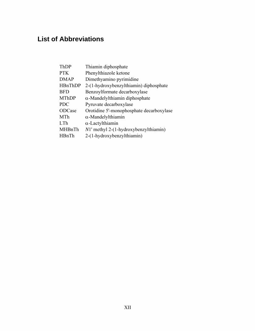
Figure 1.1 Numbered structure of thiamin diphosphate.................................................. 2
Figure 1.2 Orientation of the C2α carboxylate prior to decarboxylation of MThDP to
ensure maximum overlap of the nascent carbanion into the adjacent thiazolium ring.. 4
Figure 1.4 Protonation of a preassociated Brønsted acid in the active site prevents
internal return of CO2 (k-1) and suppresses fragmentation (kf). The protonated
intermediate can then undergo elimination to yield benzaldehyde as the product and
regenerate the cofactor. ..................................................................................................... 8
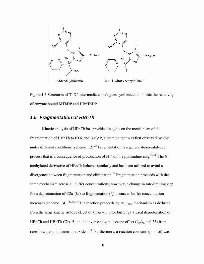
Figure 1.5 Structures of ThDP intermediate analogues synthesized to mimic the
reactivity of enzyme bound MThDP and HBnThDP...................................................... 10
Figure 2.1 Wavelength scans of HBnTh buffered with 2,4-lutidine over a 24 hour
period. Solid lines represent wavelength scans at 1 hour intervals and the dotted line
corresponds to a scan performed after 24 hours. ........................................................... 20
Figure 3.1 Wavelength scan of pyridine in acid (solid line), base (dashed line), and
buffered solution (dot-dash line). .................................................................................... 22
Figure 3.2 First order rate plot for general base catalyzed deprotonation of HBnTh by
pyridine. The concentration dependence on both base (circles) and buffer (triangles) is
shown................................................................................................................................ 24
Figure 3.3 Brønsted plot with a slope of β = 0.85 for the general base catalyzed
deprotonation of HBnTh with unhindered pyrdines (black circles), hindered (white
circles) pyridines, and imidazole (grey circle). Unhindered bases (pKa): Pyridine (4.92),
VIII
3-picoline (5.35), 3,5-lutidine (5.82), 4-picoline (5.94), imidazole (6.70). Hindered bases
(pKa): 2-Picoline (5.54), 2,6-lutidine (6.26), 2,4-lutidine (6.42), 2,4,6-collidine (6.94).25
Figure 4.1 Stereoelectronic constraints in the deprotonation of acetone resulting in the
observed steric effect in general base catalysis by substituted pyridines. ...................... 28
Figure 4.2 π-stacked pyridinium acting as a spectator catalyst in the decarboxylation of
MTh by protonating the incipient carbanion following C-C bond breaking................. 29
Figure 4.3 pH-rate profile for the decarboxylation of MTh in catalytic and non-
catalytic buffers (1 M). The ionic strength was maintained at 1 in all cases. All kinetic
runs at pH 4 were buffered with acetate/acetic acid....................................................... 29
IX
List of schemes
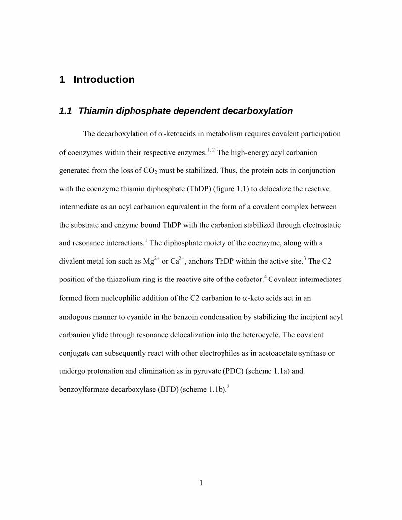
Scheme 1.1 Decarboxylation of an a-keto acid by enzyme bound ThDP (a. PDC, b.
BFD). .................................................................................................................................. 2
Scheme 1.2 Fragmentation of the carbanion/enamine into PTK and DMAP following
decarboxylation of MThDP. .............................................................................................. 4
Scheme 1.3 Enzymic deprotonation at C2 of thiazolium to generate the nucleophilic
zwitterions that can react with the substrate α-keto acid. ................................................ 5
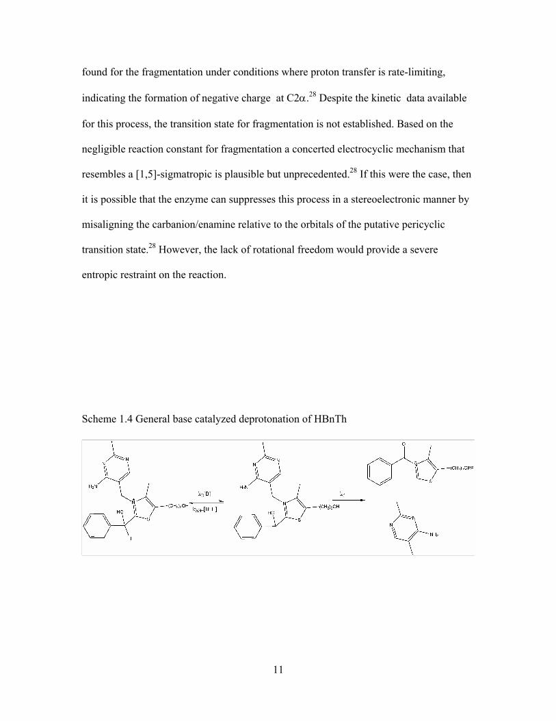
Scheme 1.4 General base catalyzed deprotonation of HBnTh....................................... 11
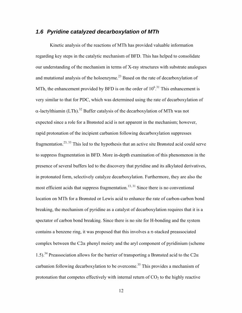
Scheme 1.5 Pyridine catalyzed decarboxylation of MTh................................................ 13
Scheme 4.1 Coincident (upper) and sequential (lower) proton transfer routes in acid
catalyzed decarboxylation. ............................................................................................... 31
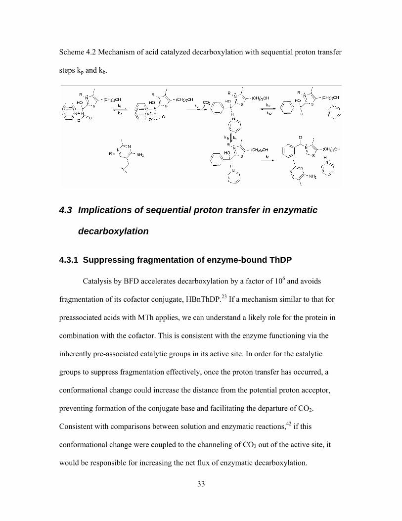
Scheme 4.2 Mechanism of acid catalyzed decarboxylation with sequential proton
transfer steps kp and kb..................................................................................................... 33
X
List of equations
Equation 2.1 Activity coefficient of the acid component for each buffer at 40 oC and I =
1......................................................................................................................................... 18
Equation 2.2 Calculation of the pKa using the activity coefficient and the pH of the
buffer at 40 oC. ................................................................................................................. 18
Equation 2.3 Debye-Huckel relationship used to determine the pKa at variable ionic
strength. A = Temperature constant (A = 0.5262 at 40 oC), z = charge of conjugate
acid.................................................................................................................................... 18
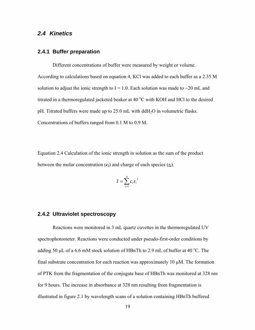
Equation 2.4 Calculation of the ionic strength in solution as the sum of the product
between the molar concentration (ci) and charge of each species (zi). .......................... 19
Equation 2.5 Equation for the observed first order rate constant for deprotonation of
HBnTh.............................................................................................................................. 21
Equation 2.6 Approximated equation for the observed first order rate constant for
deprotonation of HBnTh. ................................................................................................ 21
XI
List of Tables
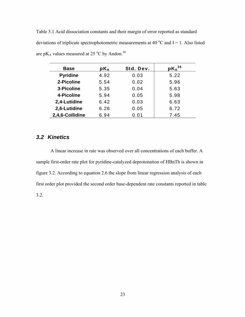
Table 3.1 Acid dissociation constants and their margin of error reported as standard
deviations of triplicate spectrophotometric measurements at 40 oC and I = 1. Also listed
are pKA values measured at 25 oC by Andon.36............................................................... 23
Table 3.2 Second order base-dependent rate constants for general base catalyzed
deprotonation of HBnTh ................................................................................................. 24
XII
List of Abbreviations
ThDP Thiamin diphosphate PTK Phenylthiazole ketone DMAP Dimethyamino pyrimidine HBnThDP 2-(1-hydroxybenzylthiamin) diphosphate BFD Benzoylformate decarboxylase MThDP α-Mandelylthiamin diphosphate PDC Pyruvate decarboxylase ODCase Orotidine 5'-monophosphate decarboxylase MTh α-Mandelylthiamin LTh α-Lactylthiamin MHBnTh N1' methyl 2-(1-hydroxybenzylthiamin) HBnTh 2-(1-hydroxybenzylthiamin)
1
1 Introduction
1.1 Thiamin diphosphate dependent decarboxylation
The decarboxylation of α-ketoacids in metabolism requires covalent participation
of coenzymes within their respective enzymes.1, 2 The high-energy acyl carbanion
generated from the loss of CO2 must be stabilized. Thus, the protein acts in conjunction
with the coenzyme thiamin diphosphate (ThDP) (figure 1.1) to delocalize the reactive
intermediate as an acyl carbanion equivalent in the form of a covalent complex between
the substrate and enzyme bound ThDP with the carbanion stabilized through electrostatic
and resonance interactions.1 The diphosphate moiety of the coenzyme, along with a
divalent metal ion such as Mg2+ or Ca2+, anchors ThDP within the active site.3 The C2
position of the thiazolium ring is the reactive site of the cofactor.4 Covalent intermediates
formed from nucleophilic addition of the C2 carbanion to α-keto acids act in an
analogous manner to cyanide in the benzoin condensation by stabilizing the incipient acyl
carbanion ylide through resonance delocalization into the heterocycle. The covalent
conjugate can subsequently react with other electrophiles as in acetoacetate synthase or
undergo protonation and elimination as in pyruvate (PDC) (scheme 1.1a) and
benzoylformate decarboxylase (BFD) (scheme 1.1b).2
2
Figure 1.1 Numbered structure of thiamin diphosphate.
Scheme 1.1 Decarboxylation of an a-keto acid by enzyme bound ThDP (a. PDC, b.
BFD).
1.2 Mechanism of thiamin diphosphate decarboxylation
Site-directed mutagenesis of ThDP dependent holoenzymes has emphasized the
importance of the cofactor in catalysis. Despite evidence from X-ray crystal structures of
BFD that suggest the importance of certain residues in the catalytic mechanism,
mutations at any site fail to inactivate the enzyme completely.5-12 Residual activity of the
enzyme with non-conserved active site mutations illustrates the robust role of ThDP.
Although the cofactor can function independently of the active site, the protein provides a
106-fold acceleration over and above the non-enzymic rate when the intermediate is
3
produced synthetically.13 The rate enhancement is obtained from a number of sequential
and consecutive processes provided by multiple active residues and conformational
changes. In particular, the mechanism for the ThDP-dependent enzyme, BFD, begins
when the cofactor is activated by deprotonation of C2 and added to the substrate,
benzoylformate. The covalent intermediate undergoes decarboxylation, leaving the C2α
carbanion/enamine intermediate, which is protonated and subsequently eliminated
following deprotonation of the C2α hydroxyl (scheme 1.1).1
The efficiency of the holoenzyme is achieved by meeting a number of challenges.
First, the weakly acidic C2 (pKa 17-18)3 thiazolium carbon must be deprotonated to react
with substrate to form α-mandelylthiamin diphosphate (MThDP). Second, proper
orientation of the substrate to both enforce nucleophilic attack by the ylide and enable
stabilization of the nascent carbanion through delocalization into the thiazolium ring must
be achieved (figure 1.2). Finally, the C2α carbon must be protonated following the loss
of CO2. Depending on the enzyme, the intermediate following decarboxylation exists
predominantly as either a highly reactive zwitterionic C2α carbanion (pKa 15-16) or a
less reactive, but still unstable, enamine (figure 1.1).14-17 These entities can also be
considered as resonance contributors of a single common structure with rehybridization.
The reactivity of both species necessitates a mechanism that irreversibly quenches the
nucleophilic C2α position by protonation, producing 2-(1-hydroxybenzylthiamin)
diphosphate (HBnThDP). This will serve to both increase the commitment by preventing
internal return of CO2 (kp) and suppress the destruction of the carbanion/enamine cofactor
conjugate by fragmentation (kf) to a phenyl thiazole ketone (PTK) and dimethylamino
pyrimidine (DMAP) (scheme 1.2).18

4
Figure 1.2 Orientation of the C2α carboxylate prior to decarboxylation of MThDP to
ensure maximum overlap of the nascent carbanion into the adjacent thiazolium ring.
Scheme 1.2 Fragmentation of the carbanion/enamine into PTK and DMAP following
decarboxylation of MThDP.
Deprotonation of C2 is accomplished by cooperation of the enzyme active site
and the aminopyrimidine moiety of the cofactor.3, 17, 19 The pyrimidine is in equilibrium
with its tautomer, 1’,4’-iminopyrimidine, which is stabilized by three active site-derived
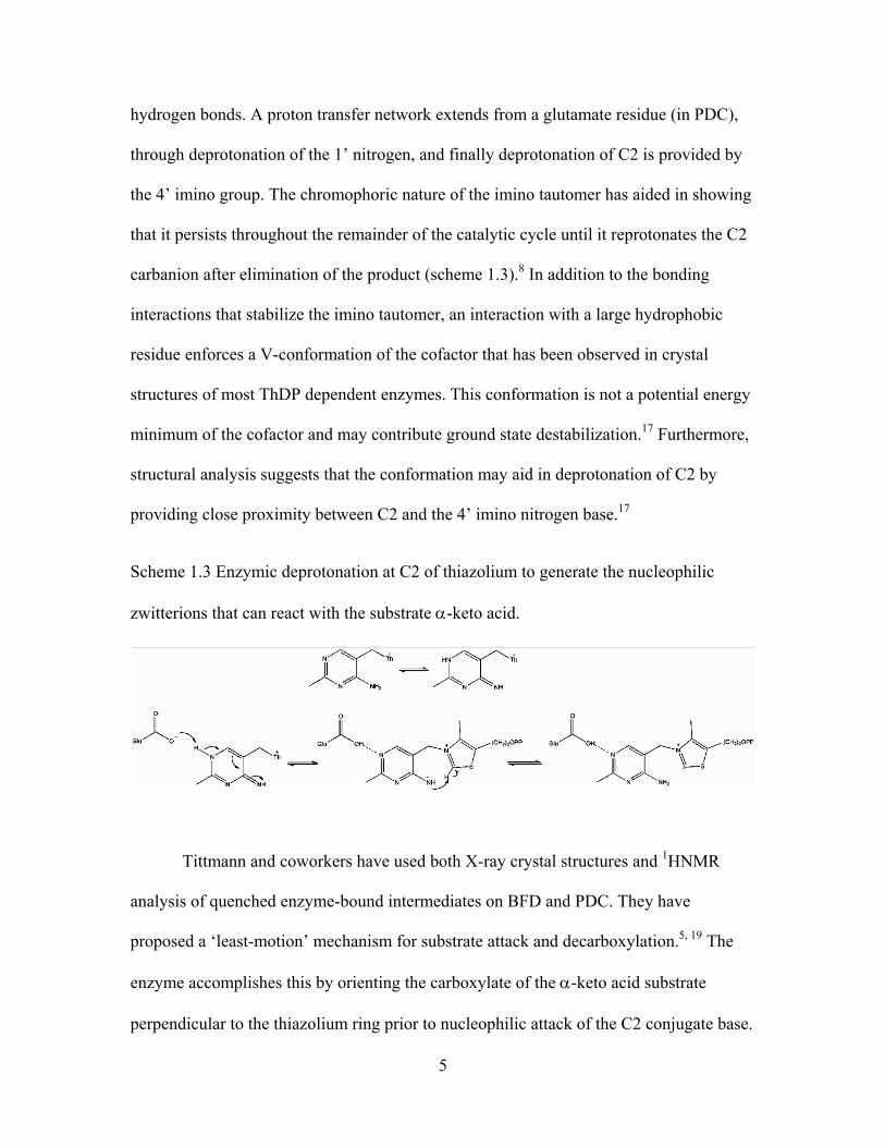
5
hydrogen bonds. A proton transfer network extends from a glutamate residue (in PDC),
through deprotonation of the 1’ nitrogen, and finally deprotonation of C2 is provided by
the 4’ imino group. The chromophoric nature of the imino tautomer has aided in showing
that it persists throughout the remainder of the catalytic cycle until it reprotonates the C2
carbanion after elimination of the product (scheme 1.3).8 In addition to the bonding
interactions that stabilize the imino tautomer, an interaction with a large hydrophobic
residue enforces a V-conformation of the cofactor that has been observed in crystal
structures of most ThDP dependent enzymes. This conformation is not a potential energy
minimum of the cofactor and may contribute ground state destabilization.17 Furthermore,
structural analysis suggests that the conformation may aid in deprotonation of C2 by
providing close proximity between C2 and the 4’ imino nitrogen base.17
Scheme 1.3 Enzymic deprotonation at C2 of thiazolium to generate the nucleophilic
zwitterions that can react with the substrate α-keto acid.
Tittmann and coworkers have used both X-ray crystal structures and 1HNMR
analysis of quenched enzyme-bound intermediates on BFD and PDC. They have
proposed a ‘least-motion’ mechanism for substrate attack and decarboxylation.5, 19 The
enzyme accomplishes this by orienting the carboxylate of the α-keto acid substrate
perpendicular to the thiazolium ring prior to nucleophilic attack of the C2 conjugate base.
6
The orientation provides maximum π-overlap to stabilize the C2α carbanion following
decarboxylation.5 This is an extension of Dunathan’s hypothesis regarding the
mechanism of pyridoxal dependent enzymes.20 Site-directed mutageneis has implicated
H70 as a residue that may hydrogen bond with the carbonyl of the α-keto acid substrate
for the purpose of both activating it as an electrophile and to orient it perpendicular to the
thiazolium ring.6, 10
The intermediate following decarboxylation must be protonated rapidly to allow
the forward commitment of the process. With a high local concentration of CO2 adjacent
to the reactive C2α conjugate base, internal return may proceed with little or no enthalpic
barrier.21 Furthermore, rapid protonation of the C2α carbanion/enamine is necessary to
prevent fragmentation of the cofactor, which occurs in solution at a rate 100 times that of
enzymatic decarboxylation.5 The reactivity of the C2α carbanion as a nucleophile
towards CO2 and as a precursor of fragmentation emphasizes the importance of proton
transfer that occurs at a rate that suppresses these processes and contributes to the overall
efficiency of the enzyme. X-ray and mutagenesis studies have shown that active site
histidine residues could serve this purpose. This is consistent with the ~100 fold decrease
in kcat from mutation of H281 of BFD to alanine.6 Although the role for H281 has been
called into question based on its distance of >5 Å from C2α,8 the residue is part of a
flexible loop on the enzyme that is likely to undergo a conformational change when the
substrate binds. This phenomenon has been observed in other ThDP dependent
enzymes.6, 7, 15 Based on the previous discussion, rapid protonation from histidine will
increase the commitment of the enzymatic reaction by inhibiting reversion by internal
return of CO2. Rapid protonation to produce the HBnThDP (in BFD) also serves to
7
suppress fragmentation of the carbanion/enamine since protonation will occur faster in
the preassociated complex than the cofactor conjugate can fragment.
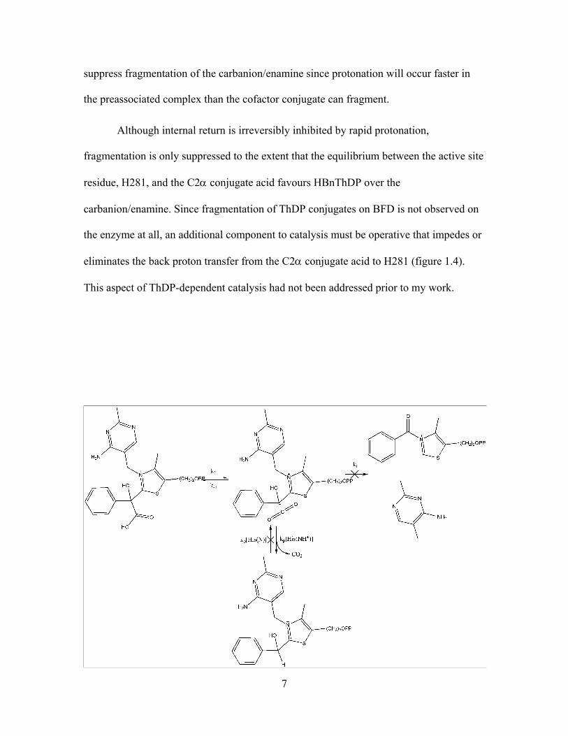
Although internal return is irreversibly inhibited by rapid protonation,
fragmentation is only suppressed to the extent that the equilibrium between the active site
residue, H281, and the C2α conjugate acid favours HBnThDP over the
carbanion/enamine. Since fragmentation of ThDP conjugates on BFD is not observed on
the enzyme at all, an additional component to catalysis must be operative that impedes or
eliminates the back proton transfer from the C2α conjugate acid to H281 (figure 1.4).
This aspect of ThDP-dependent catalysis had not been addressed prior to my work.
8
Figure 1.4 Protonation of a preassociated Brønsted acid in the active site prevents internal
return of CO2 (k-1) and suppresses fragmentation (kf). The protonated intermediate can
then undergo elimination to yield benzaldehyde as the product and regenerate the
cofactor.
1.3 Dynamic proton transfer in enzymatic catalysis
As previously discussed, the decrease in kcat of the H281A by two orders of
magnitude indicates this residue is important in catalysis; however, the residual kcat of 1-2
s-1 is still 10 000 times that of the non-enzymic counterpart.6, 10 In order to understand
how enzyme catalysis still proceeds in the absence of the proposed proton donor adjacent
to the incipient carbanion intermediate; the notion of proton donation can be extended
beyond discreet hydrogen bonding donor-acceptor interactions. In this extension, enzyme
active sites are composed of electrostatic networks where bimolecular equilibria between
donor and acceptor are part of a larger flux of protons moving from regions of low to
high potential along the lowest energy paths. This type of dynamic process is implicated
in the mechanism of OMP decarboxylase (ODCase), and provides an illustration of the
interdependence of multiple residues in the process of protonating a carbanion following
decarboxylation. Composed of alternating lysine and aspartate residues, this network is
important for catalysis as indicated by decreases in kcat of up to five orders of magnitude
upon mutation of one amino acid in the chain. Considering that ODCase accelerates
decarboxylation of OMP ~1017 times above the spontaneous reaction, a decrease in the
rate by 105 is considerable. However, it indicates that the enzyme is still capable of
significantly lowering transition state energy in its altered form.17, 22 In BFD, despite the

9
loss of an important catalytic group, the dynamic interactions between enzyme and
substrate compensate by providing a proton from an alternative site. Although an
analogous network is not as evident in the active site of BFD, the retention of ~10 000
fold acceleration following mutation of H281 indicates that the enzyme benefits from a
similar network of proton sources.
1.4 Synthetic thiamin derived intermediates
Studying the mechanism of ThDP dependent enzymes by subjecting the
holoenzyme to experiments such as structure determination by X-ray diffraction in the
presence of substrate analogues, kinetic analysis of mutant enzymes, and 1HNMR
analysis of covalent intermediates bound to quenched enzymes has provided important
mechanistic information. Included in this data is the identity of enzyme bound thiamin
conjugates that indicate the stable intermediates along the catalytic path. Both MThDP
and HBnThDP have been observed as intermediates in the catalytic cycle of BFD.5 In
order to complement the understanding gained from observing the reactivity of the native
substrates and intermediates within the active site of an enzyme, analysis of synthetic
analogues of enzyme-bound intermediates have provided the means for observing the
catalytic function of thiamin in a controlled environment. Both α-mandelylthiamin
(MTh) and 2-(1-hydroxybenzylthiamin) (HBnTh) have been synthesized for this purpose
(figure 1.5).23, 24 These intermediate analogues differ from the natural enzymic
intermediates only by the absence of the diphosphate moiety, a group that is not
implicated in catalysis.
10
Figure 1.5 Structures of ThDP intermediate analogues synthesized to mimic the reactivity
of enzyme bound MThDP and HBnThDP.
1.5 Fragmentation of HBnTh
Kinetic analysis of HBnTh has provided insights on the mechanism of the
fragmentation of HBnTh to PTK and DMAP, a reaction that was first observed by Oka
under different conditions (scheme 1.2).25 Fragmentation is a general-base-catalyzed
process that is a consequence of protonation of N1’ on the pyrimidine ring.26-28 The N-
methylated derivative of HBnTh behaves similarly and has been utilized to avoid a
divergence between fragmentation and elimination.29 Fragmentation proceeds with the
same mechanism across all buffer concentrations; however, a change in rate-limiting step
from deprotonation of C2α (kB) to fragmentation (kf) occurs as buffer concentration
increases (scheme 1.4).18, 27, 30 The reaction proceeds by an E1CB mechanism as deduced
from the large kinetic isotope effect of kH/kD = 5.8 for buffer catalyzed deprotonation of
HBnTh and HBnTh-C2α-d and the inverse solvent isotope effect (kH/kD = 0.33) from
rates in water and deuterium oxide.18, 30 Furthermore, a reaction constant (ρ = 1.6) was
11
found for the fragmentation under conditions where proton transfer is rate-limiting,
indicating the formation of negative charge at C2α.28 Despite the kinetic data available
for this process, the transition state for fragmentation is not established. Based on the
negligible reaction constant for fragmentation a concerted electrocyclic mechanism that
resembles a [1,5]-sigmatropic is plausible but unprecedented.28 If this were the case, then
it is possible that the enzyme can suppresses this process in a stereoelectronic manner by
misaligning the carbanion/enamine relative to the orbitals of the putative pericyclic
transition state.28 However, the lack of rotational freedom would provide a severe
entropic restraint on the reaction.
Scheme 1.4 General base catalyzed deprotonation of HBnTh
12
1.6 Pyridine catalyzed decarboxylation of MTh
Kinetic analysis of the reactions of MTh has provided valuable information
regarding key steps in the catalytic mechanism of BFD. This has helped to consolidate
our understanding of the mechanism in terms of X-ray structures with substrate analogues
and mutational analysis of the holoenzyme.23 Based on the rate of decarboxylation of
MTh, the enhancement provided by BFD is on the order of 106.31 This enhancement is
very similar to that for PDC, which was determined using the rate of decarboxylation of
α-lactylthiamin (LTh).32 Buffer catalysis of the decarboxylation of MTh was not
expected since a role for a Brønsted acid is not apparent in the mechanism; however,
rapid protonation of the incipient carbanion following decarboxylation suppresses
fragmentation.23, 33 This led to the hypothesis that an active site Brønsted acid could serve
to suppress fragmentation in BFD. More in-depth examination of this phenomenon in the
presence of several buffers led to the discovery that pyridine and its alkylated derivatives,
in protonated form, selectively catalyze decarboxylation. Furthermore, they are also the
most efficient acids that suppress fragmentation.13, 31 Since there is no conventional
location on MTh for a Brønsted or Lewis acid to enhance the rate of carbon-carbon bond
breaking, the mechanism of pyridine as a catalyst of decarboxylation requires that it is a
spectator of carbon bond breaking. Since there is no site for H-bonding and the system
contains a benzene ring, it was proposed that this involves a π-stacked preassociated
complex between the C2α phenyl moiety and the aryl component of pyridinium (scheme
1.5).34 Preassociation allows for the barrier of transporting a Brønsted acid to the C2α
carbanion following decarboxylation to be overcome.35 This provides a mechanism of
protonation that competes effectively with internal return of CO2 to the highly reactive
13
carbanion (pKa 15-16).13, 31, 17 Overall, this should lead to an increase in the commitment
towards loss of CO2 and a larger rate. This is an important observation that correlates
with the previously mentioned mutational analysis of BFD where H281 has been
identified to be in the correct proximity and orientation to fulfill the role of a
preassociated Brønsted acid poised for rapid protonation of the C2α carbanion.
Scheme 1.5 Pyridine catalyzed decarboxylation of MTh.
1.7 Sequence of proton transfer in pyridine catalyzed
decarboxylation
The importance of proton transfer for both catalysis and preservation of the
cofactor in the mechanism of BFD has been established from analysis of both HBnTh and
MTh. Protonation by pre-associated pyridine-derived acids suppresses fragmentation of
the conjugate base of HBnTh following departure of CO2. However, unlike
decarboxylation, this depends on both the acidity and degree of alkyl substitution of the
pyridine-derived acid and other Brønsted acids.13 Although decarboxylation and
suppression of fragmentation both involve a proton transfer, it is apparent that the
structural requirements for promoting decarboxylation and preventing fragmentation are
14
different. Since removal of a proton from HBnTh is the microscopic reverse of
protonation after separation of CO2, the kinetics of formation of the C2α carbanion with a
wide range of Brønsted bases has been investigated. From the Brønsted catalysis law, the
transition state for general base catalyzed deprotonation of HBnTh with both pyridine and
non-pyridine buffers is deduced in the current study and compared to our understanding
of the transition state for selective acid catalysis of decarboxylation via preassociation.
Identifying how these transition states intersect in the mechanism of decarboxylation of
MTh provides important insight into the sequence of proton transfers in ThDP-dependent
decarboxylation.
15
2 Experimental
2.1 Materials
Commercial reagents were used as purchased without further purification. The pH
of buffer solutions was monitored in titrations by a glass Ag/AgCl electrode in KCl
calibrated with IUPAC standards. Kinetic measurements were performed on a GBC
Cintra 40 UV/Vis with a peltier thermostat.
2.2 Synthesis of N1’-methyl-2-(1-hydroxybenzylthiamin)
(MHBnTh)
2.2.1 Condensation of thiamin hydrochloride with benzaldehyde
HBnTh was synthesized by condensing thiamin hydrochloride with benzaldehyde
according to the procedure of Doughty.24 Under an argon atmosphere, two equivalents of
sodium ethoxide (80.1 mmol of sodium in 135 mL) was added slowly to 40 mmol
(10.62g) thiamin. Benzaldehyde (80.1 mmol, 8.1 mL) was dissolved in 135 mL of
ethanol and added in one portion to the thiamin/ethoxide solution and stirred for 10
minutes. The reaction was quenched with two equivalents of concentrated hydrochloric
acid (80.1 mmol, 6.7 mL), filtered, and dried under vacuum. The solid was washed with
dichloromethane and the aqueous fraction was frozen and lyophilized. The resulting solid
was recrystallized from ddH2O. 2-(1-hydroxybenzylthiamin) (HBnTh) was obtained in
15% yield (2.21 g).

16
1HNMR (400MHz, DMSO-d6) δ: 7.90 (1H, s), 7.50-7.47 (m, 2H), 7.29-7.26 (m,
3H), 7.15 (s, 1H), 6.45 (s, 1H), 5.74 (d, 1H, 2J = 17.8 Hz), 5.47 (d, 1H, 2J = 17.9 Hz),
3.73-3.64 (m, 2H), 3.06-3.03 (t, 2H), 2.50 (s, 3H), 2.30 (s, 3H).
2.2.2 N1’ methylation of 2-(1-hydroxybenzyl) thiamin
The N1’ position on the pyrimidine of HBnTh was methylated according to a
procedure adapted from that of Zoltewicz.29 HBnTh (2.21 g, 6.0 mmol) was dissolved in
ddH2O (9.2 mL), and both one equivalent (6.0 mmol) of HCO3 and a half equivalent of
CaCO3 (3.0 mmol) were added slowly to the aqueous solution. The pH was adjusted to
6.5 with 1M HCl and solid NaHCO3 and 2.5 equivalents of dimethylsulfate (DMS) (14.9
mmol, 1.4 mL) was added in three portions. Thirty minutes of reaction time separated the
addition of the first two quarter equivalents (3.7 mmol, 0.35 mL). The solution was
adjusted to pH 6.5 before the addition of the second quarter equivalent and again after
another 30 minutes of reaction. The remaining half equivalent (7.4 mmol, 0.7 mL) was
added and the reaction was allowed to proceed for an additional 1.5 hours. The reaction
was filtered, chilled on ice, and 6.4 equivalents (38.1 mmol) of NaClO4 as a 6M solution
was added slowly. The crude product was isolated by filtration after crystallizing
overnight at 4 oC. The crude product, N1’-methyl 2-(1-hydroxybenzylthiamin)
(MHBnTh), was recrystallized from 1% HClO4 and obtained in a yield of 81% (1.86 g).
17
1HNMR (400MHz, DMSO-d6) δ: 9.26 (s, 1H), 8.42 (s, 1H), 7.72 (s, 1H), 7.40-7.38 (m,
2H), 7.30-7.23 (m, 3H), 6.72 (s, 1H), 6.30 (s, 1H), 5.31 (s, 2H), 3.80-3.67 (m, 2H), 3.52
(s, 3H), 3.14-3.00 (m, 2H), 2.50 (s, 3H), 2.29 (s, 3H).
13CNMR (100MHz, DMSO-d6) δ: 178.5, 162.2, 160.6, 143.9, 143.2, 138.5, 135.0,
129.8, 129.5, 128.5, 108.5, 71.0, 60.1, 46.8, 42.1, 30.3, 21.9, 11.9.
ESIMS [C20H25N4O2S]+, calculated: 385.1692, observed: 385.2.
2.3 pKA determination
Acid dissociation constants for all pyridine derivatives were measured at 40 oC
and ionic strength (I) of 1.0 using the procedure of Andon and Cox.36 Stock solutions of
the pyridine derivative (0.05 M) in 3 mL quartz cuvettes were prepared with both 0.1 M
KOH and 0.015 M HCl to a final concentration of 1.67 x 10⁻⁴ M. Three samples of each
acid and base were prepared at 40 oC and scanned from 230 to 290 nm. A third solution
with the same concentration of base was prepared at 40 oC in various ratios of 0.015 M
potassium acetate/0.005 M acetic acid. The buffer ratio was adjusted until three
wavelength scans of the base were approximately halfway between the scans in acid and
base resulting in a pKa was with an error equal to or less than 0.05. The pKa was
calculated by first identifying λmax for each base and subtracting Aλmax from the baseline
at A290 for each of the acid (ΔAAcid), base (ΔAbase), and buffer solutions (ΔABuffer). The
pH of each buffer solution was measured at 40 oC and the pKa was calculated according
18
to equation 1 and 2 for each of three independent scans of the pyridine base in acid, base,
and buffer.
Equation 2.1 Activity coefficient of the acid component for each buffer at 40 oC and I =
1.
⎟⎟⎠
⎞⎜⎜⎝
⎛
+−=+
II
BH 15262.0logγ
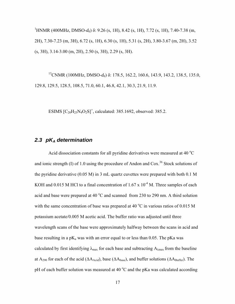
Equation 2.2 Calculation of the pKa using the activity coefficient and the pH of the buffer
at 40 oC.
++
⎟⎟⎠
⎞⎜⎜⎝
⎛
Δ−ΔΔ−Δ
−= BH
BufferAcid
BaseBuffera
AAAA
pHpK γlog1log
The pKa for imidazole at 40 oC and I = 1 was calculated using the temperature
coefficient (dpKa/dT) of -0.022/oC and the Debye-Hückel relationship (equation 2.3).37, 38
Equation 2.3 Debye-Huckel relationship used to determine the pKa at variable ionic
strength. A = Temperature constant (A = 0.5262 at 40 oC), z = charge of conjugate acid.
⎥⎥⎦
⎤
⎢⎢⎣
⎡−
+−+== I
IIAzpKpK aIa 1.0
)1()12()1(
19
2.4 Kinetics
2.4.1 Buffer preparation
Different concentrations of buffer were measured by weight or volume.
According to calculations based on equation 4, KCl was added to each buffer as a 2.35 M
solution to adjust the ionic strength to I = 1.0. Each solution was made to ~20 mL and
titrated in a thermoregulated jacketed beaker at 40 oC with KOH and HCl to the desired
pH. Titrated buffers were made up to 25.0 mL with ddH2O in volumetric flasks.
Concentrations of buffers ranged from 0.1 M to 0.9 M.
Equation 2.4 Calculation of the ionic strength in solution as the sum of the product
between the molar concentration (ci) and charge of each species (zi).
∑=
=n
iii zcI
1
2
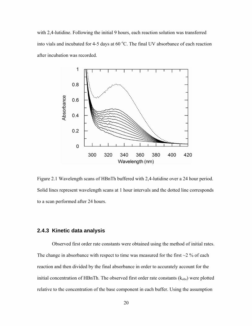
2.4.2 Ultraviolet spectroscopy
Reactions were monitored in 3 mL quartz cuvettes in the thermoregulated UV
spectrophotometer. Reactions were conducted under pseudo-first-order conditions by
adding 50 μL of a 6.6 mM stock solution of HBnTh to 2.9 mL of buffer at 40 oC. The
final substrate concentration for each reaction was approximately 10 μM. The formation
of PTK from the fragmentation of the conjugate base of HBnTh was monitored at 328 nm
for 9 hours. The increase in absorbance at 328 nm resulting from fragmentation is
illustrated in figure 2.1 by wavelength scans of a solution containing HBnTh buffered
20
with 2,4-lutidine. Following the initial 9 hours, each reaction solution was transferred
into vials and incubated for 4-5 days at 60 oC. The final UV absorbance of each reaction
after incubation was recorded.
Figure 2.1 Wavelength scans of HBnTh buffered with 2,4-lutidine over a 24 hour period.
Solid lines represent wavelength scans at 1 hour intervals and the dotted line corresponds
to a scan performed after 24 hours.
2.4.3 Kinetic data analysis
Observed first order rate constants were obtained using the method of initial rates.
The change in absorbance with respect to time was measured for the first ~2 % of each
reaction and then divided by the final absorbance in order to accurately account for the
initial concentration of HBnTh. The observed first order rate constants (kobs) were plotted
relative to the concentration of the base component in each buffer. Using the assumption

21
that kf (8 x 10³ s-1) >> k-1([BH] + kH2O) and k1[B] > kOH-[-OH], equation 2.5 was
simplified to equation 2.6. This allowed for the second order rate constants (kb) to be
calculated from the slope of the linear regression for the relationship between kobs and
base concentration. The second order rate constants were used along with the measured
pKA’s to construct a Brønsted plot.
Equation 2.5 Equation for the observed first order rate constant for deprotonation of
HBnTh.
kobs =(kb[B]+ k
OH − [OH − ])k f
k−b[BH + ]+ kH2O + k f
Equation 2.6 Approximated equation for the observed first order rate constant for
deprotonation of HBnTh.
kobs = kb[B]

22
3 Results
3.1 pKA determination
Spectrophotometric analysis of each buffer was performed in triplicate. A sample
wavelength scan of pyridine in the presence of acid, base, and buffer is shown in figure
3.1.
Figure 3.1 Wavelength scan of pyridine in acid (solid line), base (dashed line), and
buffered solution (dot-dash line).
Applying equations 2.1 and 2.2 to the data in each wavelength scan provided
three pKA measurements for each base that were averaged. The resulting constants for
seven pyridine derivatives used in the current study are reported in table 3.1.
23
Table 3.1 Acid dissociation constants and their margin of error reported as standard
deviations of triplicate spectrophotometric measurements at 40 oC and I = 1. Also listed
are pKA values measured at 25 oC by Andon.36
Base pKA Std. Dev. pKA36
Pyridine 4.92 0.03 5.22 2-Picoline 5.54 0.02 5.96 3-Picoline 5.35 0.04 5.63 4-Picoline 5.94 0.05 5.98
2,4-Lutidine 6.42 0.03 6.63 2,6-Lutidine 6.26 0.05 6.72
2,4,6-Collidine 6.94 0.01 7.45
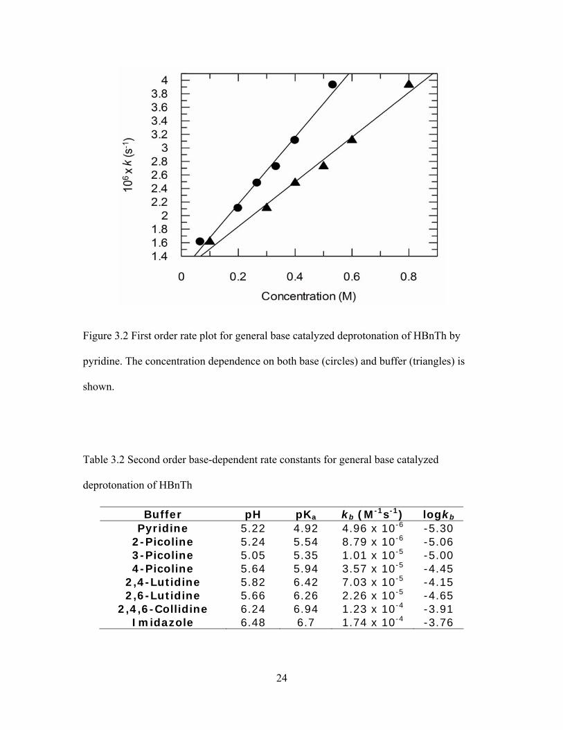
3.2 Kinetics
A linear increase in rate was observed over all concentrations of each buffer. A
sample first-order rate plot for pyridine-catalyzed deprotonation of HBnTh is shown in
figure 3.2. According to equation 2.6 the slope from linear regression analysis of each
first order plot provided the second order base-dependent rate constants reported in table
3.2.
24
Figure 3.2 First order rate plot for general base catalyzed deprotonation of HBnTh by
pyridine. The concentration dependence on both base (circles) and buffer (triangles) is
shown.
Table 3.2 Second order base-dependent rate constants for general base catalyzed
deprotonation of HBnTh
Buffer pH pKa kb (M-1s-1) logkb Pyridine 5.22 4.92 4.96 x 10⁻⁶ -5.30
2-Picoline 5.24 5.54 8.79 x 10⁻⁶ -5.06 3-Picoline 5.05 5.35 1.01 x 10⁻⁵ -5.00 4-Picoline 5.64 5.94 3.57 x 10⁻⁵ -4.45
2,4-Lutidine 5.82 6.42 7.03 x 10⁻⁵ -4.15 2,6-Lutidine 5.66 6.26 2.26 x 10⁻⁵ -4.65
2,4,6-Collidine 6.24 6.94 1.23 x 10⁻⁴ -3.91 Imidazole 6.48 6.7 1.74 x 10⁻⁴ -3.76
25
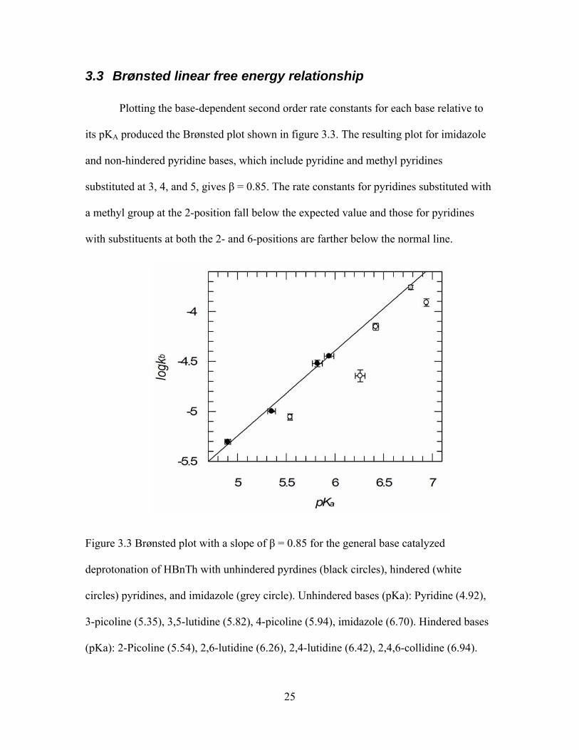
3.3 Brønsted linear free energy relationship
Plotting the base-dependent second order rate constants for each base relative to
its pKA produced the Brønsted plot shown in figure 3.3. The resulting plot for imidazole
and non-hindered pyridine bases, which include pyridine and methyl pyridines
substituted at 3, 4, and 5, gives β = 0.85. The rate constants for pyridines substituted with
a methyl group at the 2-position fall below the expected value and those for pyridines
with substituents at both the 2- and 6-positions are farther below the normal line.
Figure 3.3 Brønsted plot with a slope of β = 0.85 for the general base catalyzed
deprotonation of HBnTh with unhindered pyrdines (black circles), hindered (white
circles) pyridines, and imidazole (grey circle). Unhindered bases (pKa): Pyridine (4.92),
3-picoline (5.35), 3,5-lutidine (5.82), 4-picoline (5.94), imidazole (6.70). Hindered bases
(pKa): 2-Picoline (5.54), 2,6-lutidine (6.26), 2,4-lutidine (6.42), 2,4,6-collidine (6.94).
26
4 Discussion
4.1 General base catalyzed deprotonation of HBnTh
4.1.1 No change in rate-limiting step with tertiary amines
In the general base catalyzed deprotonation of HBnTh by anionic buffers such as
phosphate, saturation of the rate is observed with increasing concentration, indicating a
change in rate-limiting step is occurring. Tertiary amine buffers did not indicate a
concentration dependent change in rate-limiting step. First order rate plots of phosphate
catalyzed deprotonation saturate at high buffer concentrations. This observation is
attributed to a change from rate-limiting deprotonation at low buffer concentrations to
rate-limiting fragmentation at high buffer concentrations. Under all conditions of this
study, first order kinetic plots were linear as illustrated for pyridine catalyzed
deprotonation in figure 3.2. Considering that this is a one step reaction, there must be a
base dependent change in the transition state for deprotonation when the catalyst is
changed from an oxyanion to a tertiary amine. This is also evident from comparison of
the Brønsted coefficients of β = 0.85 for tertiary amine derivatives and β = 0.5 for acetate
derivates in the deprotonation of HBnTh.18 The magnitudes of the Brønsted coefficients
suggest that proton transfer is later in the case of tertiary amines relative to acetates,
which indicates that the former case is thermodynamically less favourable. This can be
justified by considering electrostatic interactions in the transition state for the two cases.
Anionic oxygen sites acquire a proton from the C2α carbon acid producing a neutral
27
carboxylic acid and a carbanion. Alternatively, a neutral pyridine (or imidazole) nitrogen
will acquire a proton from the C2α carbon acid to become a positively charged
pyridinium (or imidazolium) ion that is adjacent to a negatively charged carbanion. The
electrostatic interaction of the carbanion paired with a pyridinium ion provides a lower
barrier for reprotonation over reaction of a neutral acid, justifying the proposal of a late
transition state in tertiary amine-catalyzed deprotonation of HBnTh. The slower rate of
deprotonation with tertiary amines explains the absence of saturation in the first order
rate plots since that rate does not exceed that of fragmentation under these conditions.
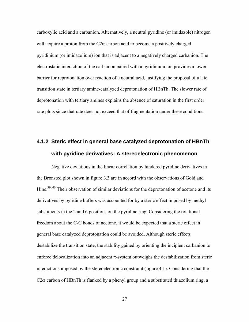
4.1.2 Steric effect in general base catalyzed deprotonation of HBnTh
with pyridine derivatives: A stereoelectronic phenomenon
Negative deviations in the linear correlation by hindered pyridine derivatives in
the Brønsted plot shown in figure 3.3 are in accord with the observations of Gold and
Hine.39, 40 Their observation of similar deviations for the deprotonation of acetone and its
derivatives by pyridine buffers was accounted for by a steric effect imposed by methyl
substituents in the 2 and 6 positions on the pyridine ring. Considering the rotational
freedom about the C-C bonds of acetone, it would be expected that a steric effect in
general base catalyzed deprotonation could be avoided. Although steric effects
destabilize the transition state, the stability gained by orienting the incipient carbanion to
enforce delocalization into an adjacent π-system outweighs the destabilization from steric
interactions imposed by the stereoelectronic constraint (figure 4.1). Considering that the
C2α carbon of HBnTh is flanked by a phenyl group and a substituted thiazolium ring, a
28
steric effect in deprotonation by substituted pyridines is a reasonable expectation. This is
due to both the steric bulk and extended π-systems of these groups that will readily
collide with alkyl substituents on pyridine derivatives and enforce a perpendicular
orientation between the incipient carbanion p-orbital and their conjugated systems,
respectively.
Figure 4.1 Stereoelectronic constraints in the deprotonation of acetone resulting in the
observed steric effect in general base catalysis by substituted pyridines.
4.2 Sequential proton transfer in decarboxylation of MTh
4.2.1 Selective acid catalysis in the decarboxylation of MTh
It was pointed out earlier that MTh does not contain a location for a Brønsted acid
to act as a catalyst. Alternatively, decarboxylation can be catalyzed by preassociation of a
spectator Brønsted acid in a π-stacked complex (figure 4.2). The spectator Brønsted acid
in this case is pyridinium and is alkyl derivatives. The positive deviation from the pH-rate
profile in figure 4.3 by both hindered and non-hindered pyridine derivatives illustrates

29
that their catalytic ability is independent of substitution. The absence of a similar
deviation in the presence of imidazole shows that it is not catalytic.41
Figure 4.2 π-stacked pyridinium acting as a spectator catalyst in the decarboxylation of
MTh by protonating the incipient carbanion following C-C bond breaking.
Figure 4.3 pH-rate profile for the decarboxylation of MTh in catalytic and non-catalytic
buffers (1 M). The ionic strength was maintained at 1 in all cases. All kinetic runs at pH 4
were buffered with acetate/acetic acid.
30
The observed first order rate coefficient for the decarboxylation of MTh in acetate
buffer at pH 4 is 6.4 x 10⁻⁴ s-1. With 1.0 M imidazolium in acetate buffer, the rate is
unchanged but with 1.0 M pyridinium the observed rate coefficient increases to 8.8 x
10-4 s-1, consistent with a preassociation mechanism that permits protonation of the
carbanion by pyridinium without diffusion, preventing reaction of the carbanion with
CO2. As a result, the sequence of departure of CO2 and transfer of the proton from a pre-
associated acid is important for our understanding of how the process is both enforced to
proceed in the forward direction and how fragmentation is inhibited in the presence of
catalyst.
4.2.2 Consistency in selective acid-catalyzed decarboxylation of MTh
and general base-catalyzed deprotonation of HBnTh
We note that in the transfer of a proton from the product, HBnTh, there is a steric
effect from α-methyl groups on the catalyst, while the rates for unhindered pyridines
conform to the Brønsted plot. These results require that the proton transfer that
accelerates decarboxylation is completed prior to that which leads to the final
protonation. The latter competes with fragmentation rather than with carboxylation.13, 23
In other words, upon loss of CO2, the HBnTh is associated with a Brønsted base that can
accept the C2α proton in the absence of CO2 (and lead to fragmentation), or which can
separate and lead to formation of HBnTh. Analysis of proton transfer from HBnTh
provides information only about the process in the absence of carbanion-associated CO2
while catalysis of the decarboxylation of MTh provides information about proton transfer
in the presence and then absence of associated CO2.

31
Irreversible loss of CO2 connects the sequential proton transfer steps. Nominally,
the C2α conjugate base can be accessed by loss of a proton from HBnTh or loss of CO2
from MTh. However, in reality there is a significant difference. Starting from MTh, an
electrophilic substitution occurs through an unstable C2α carbanion that is either
quenched by a proton or by internal return of CO2. A preassociated acid is required to
accelerate irreversible loss of CO2, presumably by enforcing the progress towards the
protonated product. However, if the C2α conjugate base is generated from HBnTh, no
pre-association occurs and there are conventional steric effects in the direct transfer of a
proton to a Brønsted base. This process is the microscopic reverse of the subsequent
proton transfer after departure of CO2 in the decarboxylation of MTh. Scheme 4.1
illustrates the difference between coincident proton transfer and sequential proton transfer
steps in decarboxylation. Only the latter is consistent with my results.
Scheme 4.1 Coincident (upper) and sequential (lower) proton transfer routes in acid
catalyzed decarboxylation.
32
4.2.3 Mechanism for acid catalyzed decarboxylation of MTh
We can consolidate our understanding of the mechanism for formation of the C2α
conjugate base from both MTh and HBnTh into a mechanism for decarboxylation of
MTh (Scheme 4.2). This involves C-C bond breaking, with or without catalytic
protonation. The resulting complex in the absence of CO2 equilibrates a proton in the step
where formation of HBnTh competes with fragmentation. The role of pyridine is evident
in the mechanism as a preassociated acid that accelerates the electrophilic substitution of
CO2 by enforcing the evolution of CO2 (kp) in competition with its internal return (k-1).
The highly exergonic process is independent of the acidity and steric character of the pre-
associated catalyst. Following the loss of CO2, the conjugate base of the catalyst is
present in a high local concentration relative to the C2α carbon acid. The basicity and
steric constraints of the catalyst determine the final products that are the result of a
partition between diffusion of the base (kD) and removal of a proton from the carbon acid
(kb).
33
Scheme 4.2 Mechanism of acid catalyzed decarboxylation with sequential proton transfer
steps kp and kb.
4.3 Implications of sequential proton transfer in enzymatic
decarboxylation
4.3.1 Suppressing fragmentation of enzyme-bound ThDP
Catalysis by BFD accelerates decarboxylation by a factor of 106 and avoids
fragmentation of its cofactor conjugate, HBnThDP.23 If a mechanism similar to that for
preassociated acids with MTh applies, we can understand a likely role for the protein in
combination with the cofactor. This is consistent with the enzyme functioning via the
inherently pre-associated catalytic groups in its active site. In order for the catalytic
groups to suppress fragmentation effectively, once the proton transfer has occurred, a
conformational change could increase the distance from the potential proton acceptor,
preventing formation of the conjugate base and facilitating the departure of CO2.
Consistent with comparisons between solution and enzymatic reactions,42 if this
conformational change were coupled to the channeling of CO2 out of the active site, it
would be responsible for increasing the net flux of enzymatic decarboxylation.
34
5 Conclusions and future work
Covalent thiamin conjugates provide a realistic model to investigate the
mechanism of ThDP dependent enzymes. HBnTh, in this case, provided the means to
study the reverse of acid-catalyzed deprotonation. By comparing the structure-reactivity
characteristics of both processes it is apparent that the transition state that accelerates
decarboxylation of MTh is not the microscopic reverse of deprotonation of HBnTh. The
selectivity for pyridine buffers and the absence of steric effects in pyridine-catalyzed
decarboxylation is what precludes this process from being directly related to general
base-catalyzed deprotonation of HBnTh. Unlike decarboxylation, the rates of
deprotonation for planar tertiary amines are subject to steric effects and produce a linear
Brønsted plot. These differences indicate that two independent sequential proton transfers
are responsible for accelerating decarboxylation and then for providing a partition in the
product determining step between fragmentation and protonation. Since fragmentation
only results from the second proton transfer following the irreversible loss of CO2, the
absence of fragmentation on an enzyme is simply avoided by the protonation that is part
of catalysis. A conformational change resulting in displacement of the conjugate base of
H281 to inhibit an equilibrium with the C2α carbon acid will prevent formation of the
carbanion leading to fragmentation.
Further studies with different classes of Brønsted bases is of interest for the
purpose of understanding the extent of delocalization of the C2α carbanion in the
transition state for deprotonation of HBnTh. This information would provide a better
understanding of the energetics for internal return of CO2 in the decarboxylation of MTh.
35
A more localized carbanion will result in a lower barrier for internal return, which would
emphasize the importance of preassociated acid-catalysis both on and off the enzyme.
Further study with the holoenzyme enzyme, BFD, is necessary to apply the
understanding gained from current and previous work with covalent thiamin
intermediates. Previous mechanistic studies with BFD have focused on rates of catalysis
and substrate affinity, which has been important for our understanding of catalysis.
However, in consideration of the current work, it would be valuable to focus on the
enzyme’s ability to suppress destruction of thiamin by fragmentation. Site-directed
mutagenesis of residues flanking the catalytic H281 residue may inhibit a necessary
conformation change resulting in deprotonation of enzyme-bound HBnTh leading to
fragmentation. Since the C2α carbanion intermediate is instrumental for catalysis of
many ThDP dependent enzymes, it is important that the mechanism by which these
enzymes avoid this destructive path is understood to clarify their catalytic mechanisms.
36
6 References
1. R. Kluger and K. Tittmann, Chemical Reviews, 2008, 108, 1797-1833.
2. R. B. Silverman, The Organic Chemistry of Enzyme-Catalyzed Reactions,
Academic Press, San Diego, 2002.
3. A. Schellenberger, Biochim. Biophys. Acta-Protein Struct. Molec. Enzym., 1998,
1385, 177-186.
4. R. Breslow, J Am Chem Soc, 1957, 79, 1762-1763.
5. M. Bruning, M. Berheide, D. Meyer, R. Golbik, H. Bartunik, A. Liese and K.
Tittmann, Biochemistry, 2009, 48, 3258-3268.
6. S. Polovnikova Elena, J. McLeish Michael, A. Sergienko Eduard, T. Burgner
John, L. Anderson Natalie, K. Bera Asim, F. Jordan, L. Kenyon George and S.
Hasson Miriam, Biochemistry, 2003, 42, 1820-1830.
7. G. Schenk, F. J. Leeper, R. England, P. F. Nixon and R. G. Duggleby, Eur. J.
Biochem., 1997, 248, 63-71.
8. G. S. Brandt, M. M. Kneen, S. Chakraborty, A. T. Baykal, N. Nemeria, A. Yep,
D. I. Ruby, G. A. Petsko, G. L. Kenyon, M. J. McLeish, F. Jordan and D. Ringe,
Biochemistry, 2009, 48, 3247-3257.
9. R. Kluger and S. Rathgeber, Febs Journal, 2008, 275, 6089-6100.
10. A. Yep, G. L. Kenyon and M. J. McLeish, Proc. Natl. Acad. Sci. U. S. A., 2008,
105, 5733-5738.
37
11. M. S. Hasson, A. Muscate, G. T. M. Henehan, P. F. Guidinger, G. A. Petsko, D.
Ringe and G. L. Kenyon, Protein Sci., 1995, 4, 955-959.
12. M. S. Hasson, A. Muscate, M. J. McLeish, L. S. Polovnikova, J. A. Gerlt, G. L.
Kenyon, G. A. Petsko and D. Ringe, Biochemistry, 1998, 37, 9918-9930.
13. R. Kluger, G. Ikeda, Q. Hu, P. Cao and J. Drewry, J Am Chem Soc, 2006, 128,
15856-15864.
14. G. Barletta, W. P. Huskey and F. Jordan, J Am Chem Soc, 1992, 114, 7607-7608.
15. C. L. Berthold, C. G. Toyota, P. Moussatche, M. D. Wood, F. Leeper, N. G. J.
Richards and Y. Lindqvist, Structure, 2007, 15, 853-861.
16. F. Jordan and N. S. Nemeria, Bioorg. Chem., 2005, 33, 190-215.
17. F. Jordan, Nat. Prod. Rep., 2003, 20, 184-201.
18. R. Kluger and I. F. Moore, J Am Chem Soc, 2000, 122, 6145-6150.
19. K. Tittmann, R. Golbik, K. Uhlemann, L. Khailova, G. Schneider, M. Patel, F.
Jordan, D. M. Chipman, R. G. Duggleby and G. Hubner, Biochemistry, 2003, 42,
7885-7891.
20. H. C. Dunathan, Proc. Natl. Acad. Sci. U. S. A., 1966, 55, 712-&.
21. J. L. Gao, Curr. Opin. Struct. Biol., 2003, 13, 184-192.
22. B. G. Miller, M. J. Snider, R. Wolfenden and S. A. Short, J. Biol. Chem., 2001,
276, 15174-15176.
23. Q. Y. Hu and R. Kluger, J Am Chem Soc, 2002, 124, 14858-14859.
24. M. B. Doughty, G. E. Risinger and S. J. Jungk, Bioorg. Chem., 1987, 15, 15-30.

38
25. Y. Oka, S. Kishimot and H. Hirano, Chem. Pharm. Bull., 1970, 18, 527-&.
26. R. Kluger, J. F. Lam, J. P. Pezacki and C. M. Yang, J Am Chem Soc, 1995, 117,
11383-11389.
27. I. F. Moore and R. Kluger, Organic Letters, 2000, 2, 2035-2036.
28. I. F. Moore and R. Kluger, J Am Chem Soc, 2002, 124, 1669-1673.
29. J. A. Zoltewicz and T. D. Baugh, Synthesis, 1980, 217-218.
30. G. Ikeda and R. Kluger, J. Phys. Org. Chem., 2004, 17, 507-510.
31. Q. Y. Hu and R. Kluger, J Am Chem Soc, 2005, 127, 12242-12243.
32. R. Kluger, J. Chin and T. Smyth, J Am Chem Soc, 1981, 103, 884-888.
33. Q. Y. Hu and R. Kluger, J Am Chem Soc, 2004, 126, 68-69.
34. K. S. Venkatasubban and R. L. Schowen, Journal of Organic Chemistry, 1984,
49, 653-655.
35. W. P. Jencks, Accounts of Chemical Research, 1980, 13, 161-169.
36. R. J. L. Andon, J. D. Cox and E. F. G. Herington, Trans. Faraday Soc., 1954, 50,
918-927.
37. R. J. Beynon and J. S. Easterby, Buffer solutions, Oxford University Press,
Oxford, 1996.
38. D. D. Perrin, Aust. J. Chem., 1964, 17, 484-&.
39. J. A. Feather and V. Gold, Journal of the Chemical Society, 1965, 1752-&.

39
40. J. Hine, J. G. Houston, J. H. Jensen and J. Mulders, J Am Chem Soc, 1965, 87,
5050-&.
41. Data submitted for publication.
42. M. Garcia-Viloca, J. Gao, M. Karplus and D. G. Truhlar, Science, 2004, 303, 186-
195.


![Thiamin Confers Enhanced Tolerance to Oxidative …Thiamin Confers Enhanced Tolerance to Oxidative Stress in Arabidopsis1[W][OA] Meral Tunc-Ozdemir, Gad Miller, Luhua Song, James Kim,](https://static.fdocuments.in/doc/165x107/5e26da2839fe9f578b1b0980/thiamin-confers-enhanced-tolerance-to-oxidative-thiamin-confers-enhanced-tolerance.jpg)