
GENE THERAPY 101 - Achroma Corp · GENE THERAPY 101 Shannon Boye, Ph.D. Assistant Professor...
21
GENE THERAPY 101 Shannon Boye, Ph.D. Assistant Professor Department of Ophthalmology Achromacorp Conference 6/2014
Transcript of GENE THERAPY 101 - Achroma Corp · GENE THERAPY 101 Shannon Boye, Ph.D. Assistant Professor...

GENE THERAPY 101
Shannon Boye, Ph.D.Assistant ProfessorDepartment of OphthalmologyAchromacorp Conference 6/2014

Let’s start from the beginning…..

Achromatopsia is caused by defects in genes that encode proteinsexpressed in cone photoreceptors
Vitreous
humorLens
Optic
Nerve
Fovea
Pupil
Iris
Cornea
Ciliary body
Anterior
chamber
Sclera
Choroid
GCL
INL
ONL
IS
OSRPE
ILMShannon Boye
Retina
photoreceptors

RODS CONES
Photoreceptors are where vision begins
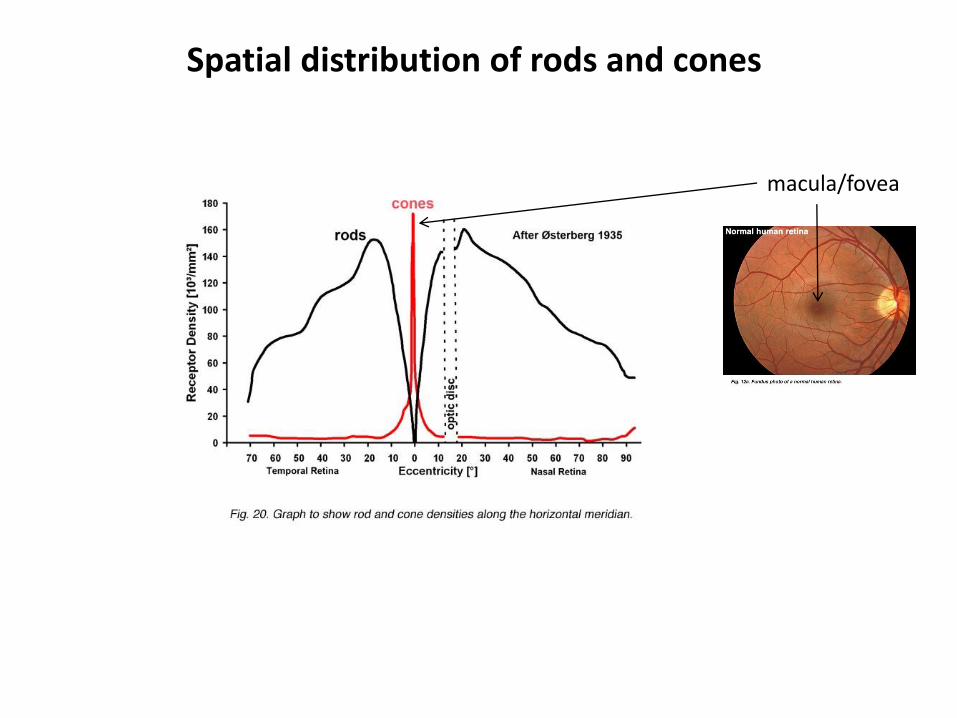
Spatial distribution of rods and cones
macula/fovea
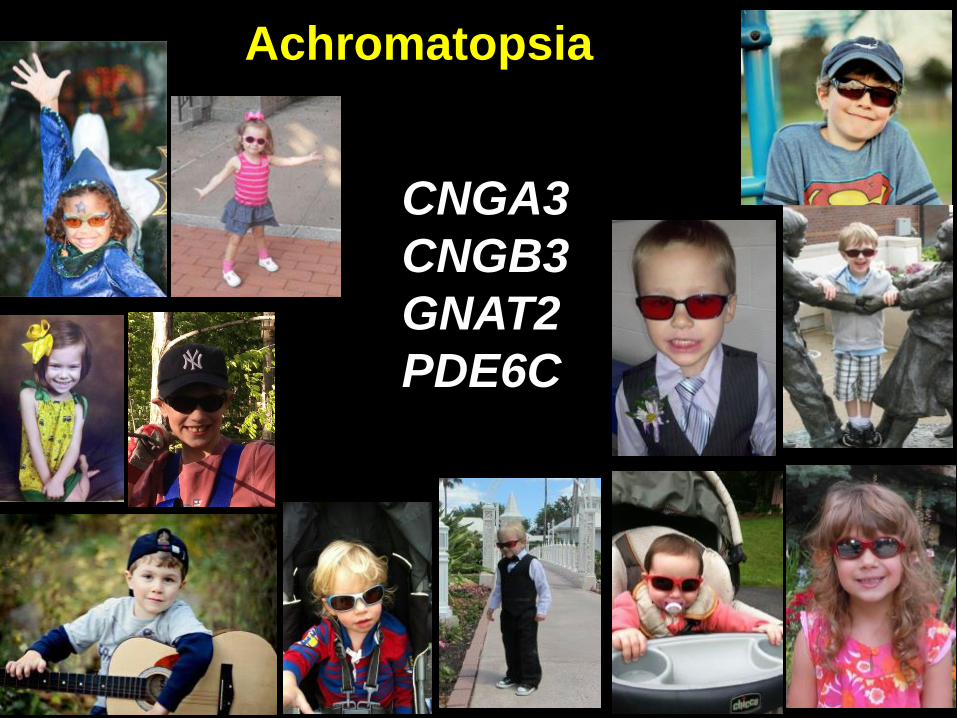
CNGA3
CNGB3
GNAT2
PDE6C
Achromatopsia

cytoplasm IPMphotoreceptor disk membrane
NCKX
K+,Ca
++
Phototransduction
cascade
GTP
CNGopen
cations
CaM
CaM
GCAP
GC
GCAP
-Ca2+
GC*Arr
RP
REC
Rhk
Rhk*
-Ca2+
Ca++
Ca++ R*
PDE
g g
cGMP
GMP
closed
CNG
Ta*
GTP
PDE*
Tbg
T
Ta*
GTP
rgs9
Gb5
Ta
GDP
CaM -Ca2+
R
hn
ret
ret
GDP
GNAT2
PDE6C
CNGA3/CNGB3
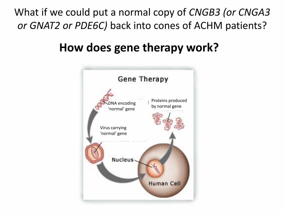
DNA encoding ‘normal’ gene
Virus carrying ‘normal’ gene
Proteins producedby normal gene
How does gene therapy work?
What if we could put a normal copy of CNGB3 (or CNGA3or GNAT2 or PDE6C) back into cones of ACHM patients?

Adeno-associated virus (AAV)
• non pathogenic
• non immunogenic
• drives efficient, sustained expression of genes
• can infect cone photoreceptors
CLINICALLY RELEVANT
a-1-Antitrypsin deficiency (AAT)Alzheimer's
Rheumatoid arthritisCystic Fibrosis
Duchenne muscular dystrophyEpilepsy
HemophiliaParkinson’s
Batten’s Leber congential amaurosis 2 (LCA2)
wet AMDchoroideremia
Ocular gene therapies

We remove the native (wild type) genes of the virus and
replace them with our gene of interest
An example:
green fluorescent protein (GFP)
Green fluorescent protein

Testing vectorin cells in a dish

RPEChoroidSclera
Subretinal
injection
How to get vector into cone photoreceptors of a living organism?

Testing vector in mouse retina
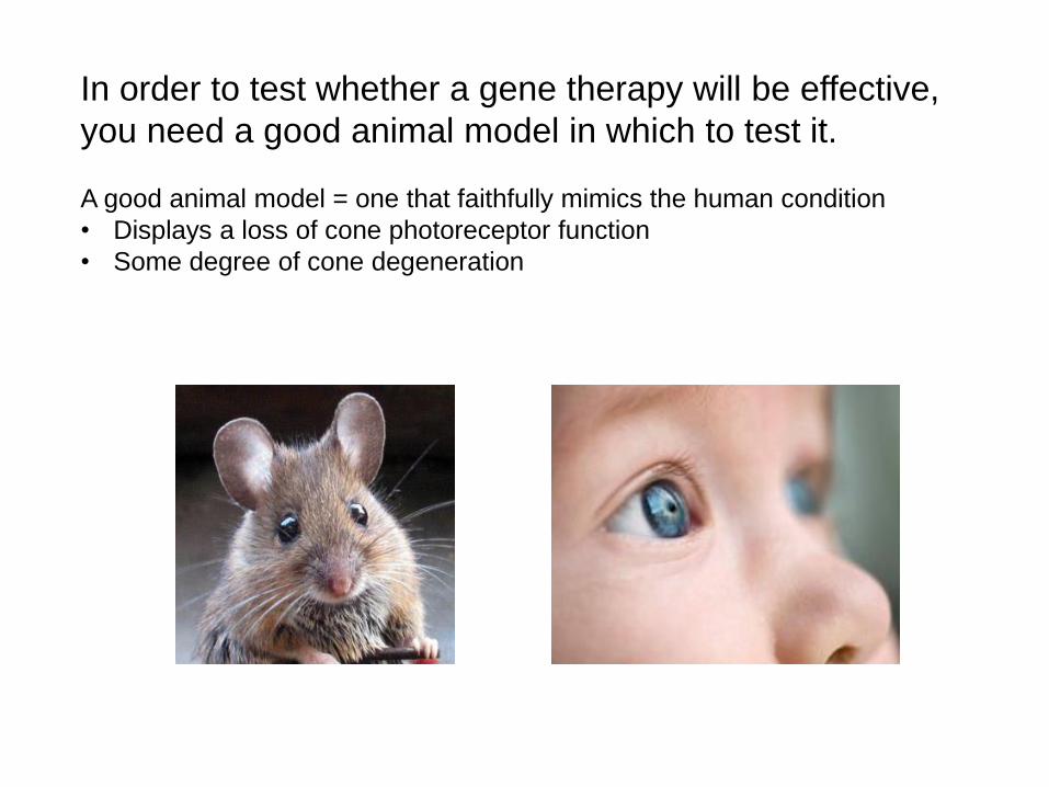
In order to test whether a gene therapy will be effective,
you need a good animal model in which to test it.
A good animal model = one that faithfully mimics the human condition
• Displays a loss of cone photoreceptor function
• Some degree of cone degeneration
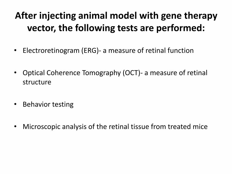
After injecting animal model with gene therapy vector, the following tests are performed:
• Electroretinogram (ERG)- a measure of retinal function
• Optical Coherence Tomography (OCT)- a measure of retinal structure
• Behavior testing
• Microscopic analysis of the retinal tissue from treated mice

Gene replacement therapy is dependent on preservation of target cells and knowledge of patient’s gene mutation
Alternatives to gene replacement therapy:• Optogenetics• Stem cells• Retinal prosthesis

Signals originating in photoreceptors are transmittedthrough ganglion cells to the optic nerve and thento the brain where they are translated as “vision”
Optic nervehead

Optogenetics• Deliver a gene that encodes a light-sensitive protein
• Forces the surviving retinal cells to become light sensitive
• If the retina is light sensitive, it can send signals to the brain which can be processed as vision.
Signals coming from light sensitive middle retinaare sent to the brain and processed as vision
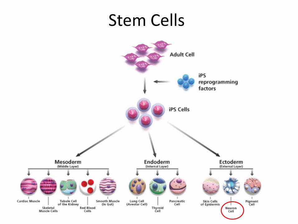
Stem Cells

Treat photoreceptors derived from stem cells with gene therapy vector then
deliver treated cells to the patient’s retina

Injection of stem-cell derived photoreceptors Integration of functioning, transplanted photoreceptors
