Gene duplications and the evolution of c-type lysozyme...
14
ADVANCES IN CICHLID RESEARCH II Gene duplications and the evolution of c-type lysozyme during adaptive radiation of East African cichlid fish Shiho Takahashi-Kariyazono . Hirokazu Tanaka . Yohey Terai Received: 22 February 2016 / Revised: 14 June 2016 / Accepted: 18 June 2016 / Published online: 1 July 2016 Ó Springer International Publishing Switzerland 2016 Abstract The adaptive radiation of African cichlids is a prime model system for studying vertebrate speciation. Cichlid species are distributed in lakes and rivers with various water conditions such as various pH values. The innate immune system in fish is particularly important because water contains a wide range of pathogenic microorganisms. To investigate the evolution of the host defense system in cichlids, we isolated the c-type lysozyme gene, which functions in the innate immune system of fish. Southern blot and sequence analyses showed that the lysozyme gene underwent several gene duplication events and evolved with amino acid replacements during the adaptive radiation of cichlids. The inferred 3D struc- ture revealed that the amino acid substitutions were localized on the lysozyme surface. Moreover, more than half of the surface substitutions changed the charge of amino acid residues, suggesting changes in the optimum pH for enzymatic activity. In African cichlids, the lysozyme genes may have played and still play an important role in defense against pathogens. Keywords c-Type lysozyme Gene duplication Adaptation East African cichlids Introduction Gene duplication is a major source of genetic variation and can lead to the generation of functional genes (Ohno, 1970; Zhang, 2003; Conant & Wolfe, 2008; Makino & Kawata, 2012). One copy of a duplicated gene retains the ancestral function and the other copy is freed from purifying selection and may acquire a new function (Zhang, 2003; Conant & Wolfe, 2008). The evolution of such redundant copies has con- tributed to adaptation to environmental variation by enabling the acquisition of new gene functions (Kon- drashov, 2012). Indeed, the proportion of duplicated genes is related to environmental diversity in Droso- phila species, indicating that variation due to gene Shiho Takahashi-Kariyazono and Yohey Terai have contributed equally to this work. Guest editors: S. Koblmu ¨ller, R. C. Albertson, M. J. Genner, K. M. Sefc & T. Takahashi / Advances in Cichlid Research II: Behavior, Ecology and Evolutionary Biology S. Takahashi-Kariyazono Y. Terai (&) Department of Evolutionary Studies of Biosystems, SOKENDAI (The Graduate University for Advanced Studies), Shonan Village, Hayama, Japan e-mail: [email protected] H. Tanaka Department of Biology and Geosciences, Graduate School of Science, Osaka City University, 3-3-138, Sugimoto, Sumiyoshi-ku, Osaka 558-8585, Japan Y. Terai Graduate School of Bioscience and Biotechnology, Tokyo Institute of Technology, 4259 Nagatsuta-cho, Midori-ku, Yokohama 226-8501, Japan 123 Hydrobiologia (2017) 791:7–20 DOI 10.1007/s10750-016-2892-6
Transcript of Gene duplications and the evolution of c-type lysozyme...
ADVANCES IN CICHLID RESEARCH II
Gene duplications and the evolution of c-type lysozymeduring adaptive radiation of East African cichlid fish
Shiho Takahashi-Kariyazono .
Hirokazu Tanaka . Yohey Terai
Received: 22 February 2016 / Revised: 14 June 2016 / Accepted: 18 June 2016 / Published online: 1 July 2016
� Springer International Publishing Switzerland 2016
Abstract The adaptive radiation of African cichlids
is a prime model system for studying vertebrate
speciation. Cichlid species are distributed in lakes and
rivers with various water conditions such as various
pH values. The innate immune system in fish is
particularly important because water contains a wide
range of pathogenic microorganisms. To investigate
the evolution of the host defense system in cichlids, we
isolated the c-type lysozyme gene, which functions in
the innate immune system of fish. Southern blot and
sequence analyses showed that the lysozyme gene
underwent several gene duplication events and
evolved with amino acid replacements during the
adaptive radiation of cichlids. The inferred 3D struc-
ture revealed that the amino acid substitutions were
localized on the lysozyme surface. Moreover, more
than half of the surface substitutions changed the
charge of amino acid residues, suggesting changes in
the optimum pH for enzymatic activity. In African
cichlids, the lysozyme genes may have played and still
play an important role in defense against pathogens.
Keywords c-Type lysozyme � Gene duplication �Adaptation � East African cichlids
Introduction
Gene duplication is a major source of genetic variation
and can lead to the generation of functional genes
(Ohno, 1970; Zhang, 2003; Conant & Wolfe, 2008;
Makino & Kawata, 2012). One copy of a duplicated
gene retains the ancestral function and the other copy
is freed from purifying selection and may acquire a
new function (Zhang, 2003; Conant & Wolfe, 2008).
The evolution of such redundant copies has con-
tributed to adaptation to environmental variation by
enabling the acquisition of new gene functions (Kon-
drashov, 2012). Indeed, the proportion of duplicated
genes is related to environmental diversity in Droso-
phila species, indicating that variation due to gene
Shiho Takahashi-Kariyazono and Yohey Terai have
contributed equally to this work.
Guest editors: S. Koblmuller, R. C. Albertson, M. J. Genner,
K. M. Sefc & T. Takahashi / Advances in Cichlid Research II:
Behavior, Ecology and Evolutionary Biology
S. Takahashi-Kariyazono � Y. Terai (&)
Department of Evolutionary Studies of Biosystems,
SOKENDAI (The Graduate University for Advanced
Studies), Shonan Village, Hayama, Japan
e-mail: [email protected]
H. Tanaka
Department of Biology and Geosciences, Graduate School
of Science, Osaka City University, 3-3-138, Sugimoto,
Sumiyoshi-ku, Osaka 558-8585, Japan
Y. Terai
Graduate School of Bioscience and Biotechnology, Tokyo
Institute of Technology, 4259 Nagatsuta-cho, Midori-ku,
Yokohama 226-8501, Japan
123
Hydrobiologia (2017) 791:7–20
DOI 10.1007/s10750-016-2892-6

duplication might be a determinant of the limits of
range expansions (Makino & Kawata, 2012).
Lakes Victoria, Malawi, and Tanganyika in the East
African Rift Valley harbor roughly 700, 700, and 250
endemic species of cichlid fish, respectively (Turner
et al., 2001; Koblmuller et al., 2008b). These fish have
fascinated evolutionary biologists as an example of a
vertebrate adaptive radiation (Fryer & Iles, 1972;
Kocher, 2004). LakeMalawi (with an estimated age of
2–5 million years) (Delvaux, 1995) and Lake Victoria
(with an estimated age of 250,000–750,000 years)
(Johnson et al., 1996) are younger than the oldest Lake
Tanganyika (with an estimated age of 9–12 million
years) (Cohen et al., 1993, 1997). Cichlid species are
distributed in lakes and rivers with pH values ranging
from 6.0 to 10.0 (Fryer & Iles, 1972) in Africa.
Molecular analyses have revealed the phylogenetic
relationships among the major lineages of African
cichlids (Salzburger et al., 2005; Takahashi &
Koblmuller, 2011; Friedman et al., 2013; Weiss
et al., 2015; Takahashi & Sota, 2016). Reflecting the
relative age of the lake, the cichlid lineage of Lake
Tanganyika is the oldest and is derived from riverine
species (Mayer et al., 1998; Terai et al., 2003;
Takahashi & Sota, 2016). A few Lake Tanganyika
species reinvaded rivers and their progenies indepen-
dently colonized Lakes Malawi and Victoria (Sal-
zburger et al., 2005). The fish in each lake underwent
several independent radiations after colonization
(Salzburger et al., 2005; Genner et al., 2007;
Koblmuller et al., 2008b). It is not clear why some
lineages successfully reinvaded riverine habitats and
expanded their distribution. A genomic study of five
African cichlids revealed an excess of gene duplica-
tions in the East African lineage (Brawand et al.,
2014), suggesting the importance of gene duplication
events during their radiation.
Lysozymes are hydrolytic enzymes that cleave the
peptidoglycan in bacterial cell walls (Callewaert &
Michiels, 2010). Three types of lysozymes have been
identified in the animal kingdom, c-type (chicken),
g-type (goose type), and i-type (invertebrate type)
(Callewaert & Michiels, 2010). The enzymatic activ-
ity of lysozymes is widely used for antibacterial host
defense and digestion (Dobson et al., 1984; Jolles
et al., 1996). In fish, lysozyme functions in the innate
immune system and is considered to be the first line of
defense against various bacteria (Saurabh & Sahoo,
2008). The fish innate immune system is particularly
important because water contains a wide range of
pathogenic microorganisms (Hikima et al., 2001).
Transgenic zebrafish expressing the chicken lysozyme
gene show resistance to pathogenic bacterial infec-
tions, indicating the role of the innate immune system
in defense against bacteria (Yazawa et al., 2006).
Cichlid lysozyme activity was detected from gill,
serum, liver, plasma, skin, and mucus samples of
Oreochromis niloticus Linnaeus, 1758 (On; Sankaran
& Gurnani, 1972; Taoka et al., 2006; Welker et al.,
2007). Considering the important role of the innate
immune system in fish, genetic variation in the
lysozyme gene may be important in the process of
their radiation, especially when they have colonized in
new water environments during the radiation in East
African lakes and rivers. However, the lysozyme gene
has not been analyzed in cichlids from the Great
Lakes.
In this study, we determined the c-type lysozyme
gene from African riverine and lake cichlids. Lyso-
zyme genes have undergone gene duplications during
the adaptive radiation of African cichlids, which may
have contributed to their current distribution in various
water conditions.
Materials and methods
Specimens
Three riverine species, ten Lake Tanganyika species,
six Lake Malawi species, and three Lake Victoria
species were used for analyses (Table 1). All of these
fish were obtained from a commercial source. The
animal protocols and procedures were approved by the
Institutional Animal Care and Use Committees of the
Tokyo Institute of Technology and SOKENDAI. All
surgery was performed under anesthesia, and all
efforts were made to minimize suffering.
DNA and RNA extraction
Genomic DNA was extracted from caudal fin or
muscular tissues using DNeasy blood and tissue kit
(Qiagen, Hilden, Germany). The whole body of fish
was frozen in liquid nitrogen, and then fragmented to
small size. Total RNA was extracted from fragmented
whole body using the RNeasy mini kit (Qiagen).
8 Hydrobiologia (2017) 791:7–20
123

Table 1 Species and detected haplotypes
Species names IDs Determined haplotypes from cDNA (expressed
haplotypes)
Number of bands in
Southern blot
Oreochromis niloticus O. niloticus#1 On1 On2 –
O. niloticus#2 – 2
Steatocranus casuarius S. casuarius#1 Sc1 –
Tilapia buttikoferi T. buttikoferi#1 Tb1 –
Neolamprologus brichardi N. brichardi#1 Nb1 –
Neolamprologus leleupi N. leleupi#1 Nl1 –
Altolamprologus calvus A. calvus#1 Ac1 –
A. calvus#2 – 1
Variabilichromis moorii V. moorii#1 Vm1 –
Lamprologus ocellatus L. ocellatus#1 Lo1 –
Cyphotilapia frontosa C. frontosa#1 Cf1 –
Xenotilapia ochrogenys X. ochrogenys#1 Xo1 –
Cyprichromis leptosoma C. leptosoma#1 Cl1 Cl2 –
C. leptosoma#2 – 1
Spathodus erythrodon S. erythrodon#1 Se1 Se2 –
S. erythrodon#2 – 1
Tropheus duboisi T. duboisi#1 Td1 –
T. duboisi#2 – 1
Aulonocara sp. Aulonocara sp.#1 Au1 Au2 –
Pseudotropheus lombardoi P. lombardoi#1 Pl1 –
P. lombardoi#2 Pl2 –
Pseudotropheus sp. Pseudotropheus sp.#1 Ps1 Ps2 –
Labidochromis caeruleus L. caeruleus#1 Lc2 –
L. caeruleus#2 Lc1 –
L. caeruleus#3 Lc1 Lc3 –
L. caeruleus#4 – 4
Dimidiochromis compressiceps D. compressiceps#1 Dc1 Dc5 –
D. compressiceps#2 Dc6, and four other partial haplotypes (Fig. 5) –
D. compressiceps#3 Dc3 Dc4 –
D. compressiceps#4 Dc4 –
D. compressiceps#5 Dc2 Dc4
D. compressiceps#6 Determined from genomic DNA: Dc1 Dc2 Dc3
Dc6 and one other haplotypes (Fig. 5)
4
Melanochromis auratus M. auratus#1 Ma1 –
Haplochromis brownae H. brownae#1 Hb1 Hb2 –
H. nigricans H. nigricans#1 Hn1 Hn2 –
H. sp. ‘redtail sheller’ H. sp. ‘redtail sheller’#1 Hr1 –
H. sp. ‘redtail sheller’#2 – 2
Hydrobiologia (2017) 791:7–20 9
123
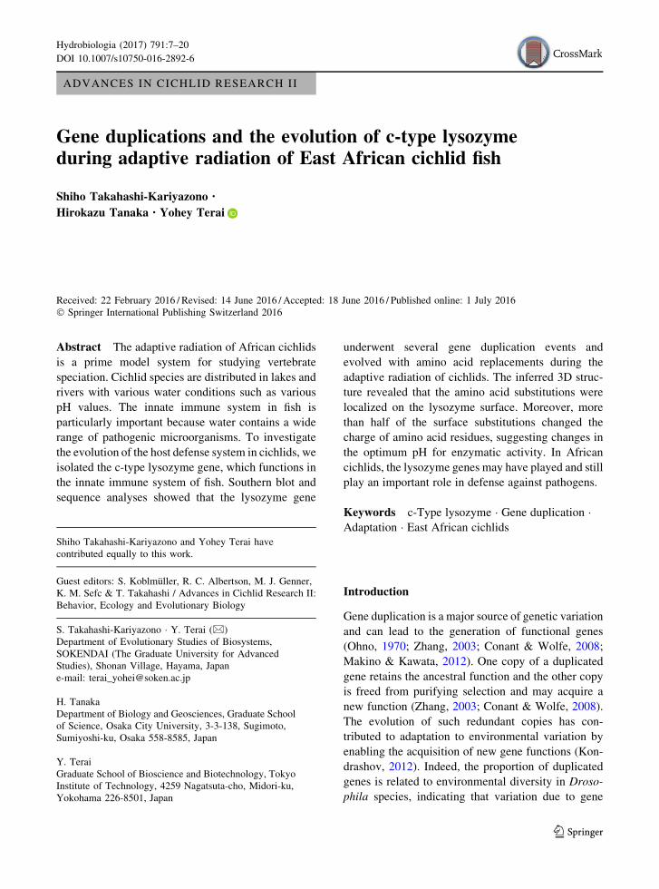
Isolation of lysozyme cDNA and gene
cDNAs were synthesized using a cDNA synthesis kit
(Takara, Shiga, Japan). A partial cDNA of cichlid
lysozymewas cloned by RT-PCR using primers LysdF1
(50-TCTTCCAGCGCTGTGAATGG-30) and LysdR1
(50-TATTGATCTGAAAGATGCCATAGTC-30), andfull-length cDNAs were isolated from the RNA of
Labidochromis caeruleus Fryer, 1956 by 30- and 50
RACE using the nested primers LysU1 (50-CAAACGGGATGGATGGCTATCGT-30), LysU2 (50-ACAAAGGCAACAAATCGTAACACTGATG-30), LysD1 (50-CATCAGTGTTACGATTTGTTGCCTTTGT-30), and
LysD2 (50-CTGACACCACGATAGCCATCCATC-30).PCR was performed in a PTC-100 programmable
thermal controller (MJ Research, Waltham, MA, USA)
and the GeneAmp PCR system 9700 (Applied Biosys-
tems, Carlsbad, CA, USA). The PCR program for
isolation of partial cDNAconsisted of a denaturation step
for 3 min at 94�C, followed by 30 cycles of denaturationfor 1 min at 94�C, annealing for 1 min at 55�C, andextension for 1 min at 72�C. The 30- and 50 RACE were
performed with two rounds of nested PCR using LysU1
and LysD1 for the first round, and LysU2 and LysD2 for
the second round. The first and second PCR were
performed under the same conditions consisting of a
denaturation step for 3 min at 94�C, followed by 30
cycles of denaturation for 1 min at 94�C, annealing for
1 min at 65�C, and extension for 1 min at 72�C. Thelysozyme gene was amplified using LysF1 (50-ATCT-GAACCCAGACAGTCACAG-30) and LysR1 (50-GTCACCCAATGTGTTTTCCTT-30) primers and geno-
mic DNA of L. caeruleus as a template. The PCR
program for isolation of the lysozyme gene consisted of a
denaturation step for 3 min at 94�C, followed by 30
cycles of denaturation for 1 min at 94�C, annealing for
1 minat 55�C, andextension for 3 min at 72�C.ThePCR
product was purified and determined by direct sequenc-
ing using the Applied Biosystems automated 3130
sequencer.
Determination of the lysozyme cDNA sequences
The cDNA fragments containing lysozyme coding
region were amplified by PCR using LysF1 and LysR1
primers and the cDNAof all species used in this study as
templates. The positions of primers are described in
Fig. 1. The PCR program for amplification of the
lysozyme cDNA consisted of a denaturation step for
3 min at 94�C, followed by30 cycles of denaturation for1 min at 94�C, annealing for 1 min at 55�C, and
extension for 1 min at 72�C. The PCR product was
purified and determined by direct sequencing. After the
analysis of the sequences, we used the double peaks in
the sequencing result (color waves) as an indicator of
which PCR product contains two or more sequences.
When we found double peaks in the sequencing results,
we cloned the PCR products into the pGEM-T plasmid
vector (Promega, Madison, WI, USA) and determined
the sequences of several clones to obtain haplotype
information. To amplify lysozyme haplotypes without
annealing bias of PCR primers, we designed primers in
the conserved regions among haplotypes from Lake
Malawi species (Fig. 1, Lys_cnsv_F1: 50-GTTTTCTTGCTTTTGATAACTGTG-30, Lys_exon2F1: 50-TTGCCTGACCAAACATGAGTCAAAC-30, and Lys_cnsv_R1: 50-TTGTTSCCTAACGATACGTTTG-30). The
DNA fragment from exons 1 to 3 was amplified using
Lys_cnsv_F1 and Lys_cnsv_R1 from cDNA of Dimid-
iochromis compressicepsBoulenger, 1908 (Dc,D. com-
pressiceps#2) (Table 1).TheDNAfragment fromexons
2 to 3 with intron 2 was amplified using Lys_exon2F1
and Lys_cnsv_R1 from genomic DNA of D. compres-
siceps#6 (Table 1). The PCR program for amplification
exon 1 exon 2 exon 3 exon 4
200 bp
coding region 429 bp
Lysozyme cDNA
intron 1: probe
LysF1
LysR1
Lys_cnsv_F1 Lys_cnsv_R1Lys_exon2F1_F1
LysF1
LysR1
Lysozyme geneFig. 1 Structure of the
cichlid c-type lysozyme.
Exon positions are indicated
by numbers. Intron 1 was
used as a probe for Southern
blots. The positions of the
primers used for
amplification are indicated
by arrows. Scale bar
represents 200 bp
10 Hydrobiologia (2017) 791:7–20
123

of those fragments consisted of a denaturation step for
3 min at 94�C, followed by30 cycles of denaturation for1 min at 94�C, annealing for 1 min at 55�C, and
extension for 30 s at 72�C. The PCR products were
cloned and determined the sequences as described
above. The nucleotide sequences were deposited in
GenBank under accession numbers LC012556–
LC012592 and LC126611–LC126614.
Southern blot analysis
The partial sequence of lysozyme intron 1 (698 bp)
was amplified using LysintF1 (50-CCAAAATAAG-CAGTTCACCATTG-30) and LysintR1 (50-ACTAACTACTTCTCACATGCTGACACT-30) primers and
the genomic DNA of L. caeruleus as a template. The
PCR product was labeled by [a-32P]dCTP (NEN
Research Products, Boston, MA, USA) and used as a
probe. Genomic DNAs (10 lg each) of Cyprichromis
leptosomaBoulenger, 1898 (Cl) and L. caeruleuswere
digested by EcoRI, HindIII, and PstI, and those of H.
sp. ‘redtail sheller,’ Dc, Tropheus duboisi Marlier,
1959, Spathodus erythrodon Boulenger, 1900 (Se),
Altolamprologus calvus Poll, 1978, and On were
digested by EcoRI and PstI. The digested fragments
were subjected to electrophoresis in a 1% agarose gel.
After electrophoresis, DNA fragments were trans-
ferred from gels to GeneScreen Plus Membranes in
0.4 M NaOH and 0.6 M NaCl. Membranes were
neutralized in 0.5 M Tris–HCl (pH 7.0) and 1 M
NaCl, and then dried. Hybridization was performed at
45�C overnight in a solution of 69 SSC (SSC is
0.15 M NaCl and 0.015 M trisodium citrate, pH 7.0),
1% (w/v) sodium dodecyl sulfate (SDS), 59 Den-
hardt’s reagent [l9 Denhardt’s reagent is 0.02% (w/v)
Ficoll 400, 0.02% (w/v) polyvinylpyrrolidone, and
0.02% (w/v) bovine serum albumin], and 100 pg/ml
herring DNA.Washing was performed in 29 SSC plus
1% SDS at 60�C for 60 min. The signals were detected
by BAS-2000 (Fijix, Fuji Film, Tokyo, Japan).
Genetic analysis
The coding sequences of lysozyme cDNA (429 bp)
were aligned using Genetyx (ver. 10) and was
subjected to a phylogenetic analysis with 1,000
bootstrap replications. A neighbor-joining analysis
(Saitou & Nei, 1987) was performed with MEGA 5
(Tamura et al., 2011) using all substitution sites (87
sites). The sequences from O. niloticus (On2) and
Tilapia buttikoferi Hubrecht, 1881 (Tb1) were used as
outgroup sequences.
Positions of substitutions in the lysozyme 3D
structure
The lysozyme amino acid sequences were aligned
using Genetyx (ver. 10), and all variable positions
were identified. The positions of hen egg white
lysozyme corresponding to variable positions of
cichlid lysozyme were identified from an alignment
of both sequences. Those positions in the 3D structure
of hen egg white lysozyme were observed using Cn3D
software version 4.3 (Wang et al., 2000).
Results
Determination of cichlid lysozyme
We isolated the partial lysozyme sequence from cDNA
of L. caeruleus, and the full-length coding region by
50- and 30 RACE. The coding region was 429 bp in
length. We amplify the lysozyme gene from genomic
DNA of L. caeruleus by PCR, and determined the
sequences. We compared the lysozyme gene
sequences determined from cDNA and genomic
DNA. The genomic structure of the lysozyme gene
consisted of four exons (Fig. 1). The coding regionwas
similar to that of Oreochromis aureus Steindachner,
1864 c-type lysozyme mRNA (EU836689) (420/
429 bp identity). To investigate the evolution of the
lysozyme gene in cichlids, we amplified lysozyme
cDNA from ten Lake Tanganyika species, six Lake
Malawi species, and three Lake Victoria species. In
total, we determined 37 different sequences. We
detected 87 variable sites including 64 nonsynony-
mous and 23 synonymous sites (Fig. 2). Substitutions
in 64 nonsynonymous sites correspond to amino acid
replacements in 47 positions, because several pairs of
nonsynonymous substitutions were located in the same
codon (e.g., substitution in sites 49 and 50 in Fig. 2).
More than two lysozyme haplotypes were isolated
within a species of Lake Malawi cichlids, whereas one
or two haplotypes in Lake Tanganyika cichlids
(Table 1).
Hydrobiologia (2017) 791:7–20 11
123

Lysozyme copy number variation in cichlids
To identify copy number variation of the lysozyme
gene, we performed Southern blot analysis using
intron 1 of the gene as a probe (Fig. 1). First, we used
digested genomic DNA from each of one Lakes
Tanganyika and Malawi species with each of three
restriction enzymes (EcoRI, HindIII, and PstI). We
detected only one signal per lane of a Lake Tan-
ganyika species (Fig. 3A, left panel). In a Lake
Malawi species, four bands were observed in the
digested DNA with EcoRI and PstI, whereas large
sizes of three bands were observed in the lane of
HindIII (Fig. 3A, right panel). The digestion of
genomic DNA with HindIII was not appropriate for
Southern blot analysis, because restriction enzyme
sites were not located near the lysozyme gene. We
additionally analyzed the copy number in one riverine
species, three Lake Tanganyika species, one Lake
Malawi species, and one Lake Victoria species using
two restriction enzymes (EcoRI and PstI). As shown in
Fig. 3B, we detected two signals from each lane of a
Lake Victoria
Lake Malawi
Lake Tanganyika
Riverine
Lam
prol
ogin
i
syn./nonsyn.
nucleotide site
11111111111111222222222222222222222222333333333333333333334444444 12345666666677778889900113456778889013334444555557777888999122344556678888899990000122 698990012378967890190279138509362780730694789016783469678089324646897890138902483456645 snnsnnsnnsnnsnnsnnsnsnnnsnsnnnnnnnnnssnnnnnnsnnnnssnssnnsnnnnnssnnnnnnnnsnnnsnnnnnsnnnnHn1 GTTGAAGCGCGACCGTAAGCAAACTGCATACCGACGCCGGGGCGCCGCATCACTAACTACCATTGGACGTGCTGCAGGCAAGGCCGGHn2 ........C.....A........G....................................T....C....A..............C.Hb1 .......................G..T...........A..................G............A....G.........C.Hb2 ..................................................T.........T.........A................Hr1 .......................G.........................C..........T.........A..............C.Lc1 .......................G........................G.......T...T.C..C....A..............C.Lc3 ..............A........G........................G.......T...T.C..C....A..............C.Lc2 .....T..........................................G.....G.T........C....A................Ma1 ....................................T...........G.....G.T.............A..............C.Au1 .......................G........................G.....................A................Au2 ........CT....A........G........................G......G.........C....A..............C.Ps1 A....T.................G........................G.....G.T........C....A................Ps2 .....T.................G........................G.....G.T........C....A................Dc1 ....G.......A..........A........A......TCC......G.G.....T..TT.........A.............T..Dc2 ....G.......A..........A........A...............G.......T..TT.........A.............T..Dc3 ............A..........A........................G.....G.T...T.........A..............C.Dc4 ............A...................................G.....G.T.............A..............C.Dc5 .......................A........................G.G.....T...T.........A................Dc6 ........................C.............................G.T...T.........A..............C.Pl1 ....................................T.................G.T.............A..............C.Pl2 ................................................G.....G.T.............A..............C.Td1 .....................................T................................A..............C.Cl1 ............A................G..............................T.........AA.............C.Cl2 ............A....................................G....G.T...G.........AA.............C.Se1 ............A.........TG....................................T........................C.Se2 ............A.........TG....................................T......................G...Xo1 ............................................................T.........A..............C.Cf1 ............................C...............................T.........AA.............C.Ac1 .A....CAAG.C....G......G.A.......C.AT......A...AC..G.CG.TA..TG..CC..TAAAAA..AAAT.CCT..ALo1 ..C.C.CAAGAC.....T.....G.........C.AT......A...AC..GGCG.TA..TG..CC.GTAAAAAA.AAAT.CCT..ANb1 ....C..AAG.C.....T.....G.........CGA.........A...A....G.T...T....C..TAAAAA..A....C.T.C.Nl1 ....C...AG.C....GT..G..G.......A...AT........AT.........T...T...CCG.TAAAAA..A.A..C.T...Vm1 ....C..AAG.C.....T.....G.......A...A.........A........G.T...T....C..TAAAAA..A....C.T.C.Sc1 .....................G....................................G.T..C......AA...............Tb1 ...C.........AAA...A.......G.....C.A......G.T..A............T..C......AA......A......C.On1 ..................A.........................T..A.......G..G.T.........AA........G..T.C.On2 .............AAA...A.......G..A..C.A......G.T..A............T..C......AA......A......C.
Fig. 2 A nucleotide alignment of the cichlid c-type lysozyme.
Nucleotide sites are numbered according to the sequence of the
cichlid c-type lysozyme. The dots and letters indicate conserved
and variable nucleotides, respectively, compared with the
reference sequence on the top line. The abbreviations of each
species are as follows:O. niloticus (On), Steatocranus casuarius
Poll, 1939 (Sc), T. buttikoferi (Tb), Neolamprologus brichardi
Poll, 1974 (Nb), Neolamprologus leleupi Poll, 1956 (Nl),
Altolamprologus calvus Poll, 1978 (Ac), Variabilichromis
moorii Boulenger, 1898 (Vm), Lamprologus ocellatus
Steindachner, 1909 (Lo), Cyphotilapia frontosa Boulenger,
1906 (Cf), Xenotilapia ochrogenys Boulenger, 1914 (Xo), C.
leptosoma (Cl), S. erythrodon (Se), T. duboisi (Td), Aulonocara
sp. (Au), Maylandia lombardoi (synonym: Pseudotropheus
lombardoi) Burgess, 1977 (Pl), Pseudotropheus sp. (Ps), L.
caeruleus (Lc), D. compressiceps (Dc),Melanochromis auratus
Boulenger, 1897 (Ma), Haplochromis brownae Greenwood,
1962 (Hb), Haplochromis nigricans Boulenger, 1906 (Hn), and
H. sp. ‘redtail sheller’ (Hr). Nonsynonymous substitutions are
shown in a gray background
12 Hydrobiologia (2017) 791:7–20
123

riverine species (On). Three Lake Tanganyika species
showed a single signal in each lane (Fig. 3B, T.
duboisi, Se, and A. calvus). Each of one Lakes Malawi
and Victoria species showed four and two signals,
respectively (Fig. 3B). In total, one riverine, four Lake
Tanganyika, two Lake Malawi, and one Lake Victoria
species possess two, one, four, and two copies of the
lysozyme gene, respectively. Based on the phyloge-
netic relationships among African cichlids (Salzburger
et al., 2002, 2005; Terai et al., 2003) and the Southern
blot results, we inferred the evolutionary transitions in
lysozyme copy number (Fig. 4).
We detected multiple haplotypes of lysozyme
genes from a Lake Malawi species. Using the primers
in the conserved regions among haplotypes from six
Lake Malawi species, we amplified DNA fragments
from cDNA (from exons 1 to 3) and genomic DNA
(from exons 2 to 3 with intron 2) (Fig. 1). These DNA
fragments were amplified using cDNA from D.
compressiceps#2, and genomic DNA from D. com-
pressiceps#6 (Table 1). We cloned and determined 28
and 24 sequences from cDNA and genomic DNA
templates, respectively, and found each of five hap-
lotypes from cDNA and genomic DNA (Fig. 5;
Table 1).
The evolution of lysozyme in cichlids
We identified the location of each variable amino acid
position using the 3D structure of hen egg white c-type
lysozyme. As shown in Fig. 6A, 45 out of 47 variable
amino acid positions were located on the surface of
lysozyme (shown in light-gray, nonsurface replace-
ments are surrounded by a rectangle in Fig. 6B).
D. com
pres
sicep
s
H.
T. du
boisi
S. e
ryth
rodo
n
A. ca
lvus
O. nilo
ticus
L. caeruleus (L. Malawi)
C. leptosoma (L. Tanganyika)
E: EcoRI
H: HindIII
P: PstI
E H P E H P
(L. Victoria) (L. Malawi) (L. Tanganyika) (L. Tanganyika) (L. Tanganyika) (Riverine)
(A)
(B)
2 kb
9.4 kb
2 kb
9.4 kb
2 kb
9.4 kb
2 kb
9.4 kb
2 kb
9.4 kb
2 kb
9.4 kb
2 kb
9.4 kb
2 kb
9.4 kb
PE PE PE PE PE PE
Fig. 3 The results of Southern blot analysis. Genomic DNAswere digested by EcoRI,HindIII, andPstI (A), or EcoRI and PstI (B). Thepositions of the DNA marker are shown by arrows
Hydrobiologia (2017) 791:7–20 13
123

Among 45 surface replacement positions, 35 positions
substantially changed the charge of residues (posi-
tively charged–uncharged, positively charged–nega-
tively charged, negatively charged–uncharged, or vice
versa) (Fig. 6B, highlighted in gray). Among
sequences from Lakes Malawi and Victoria species,
12 replacements (marked by asterisks in Fig. 6B) out
of 19 surface replacements had a large effect on the
charge of residues.
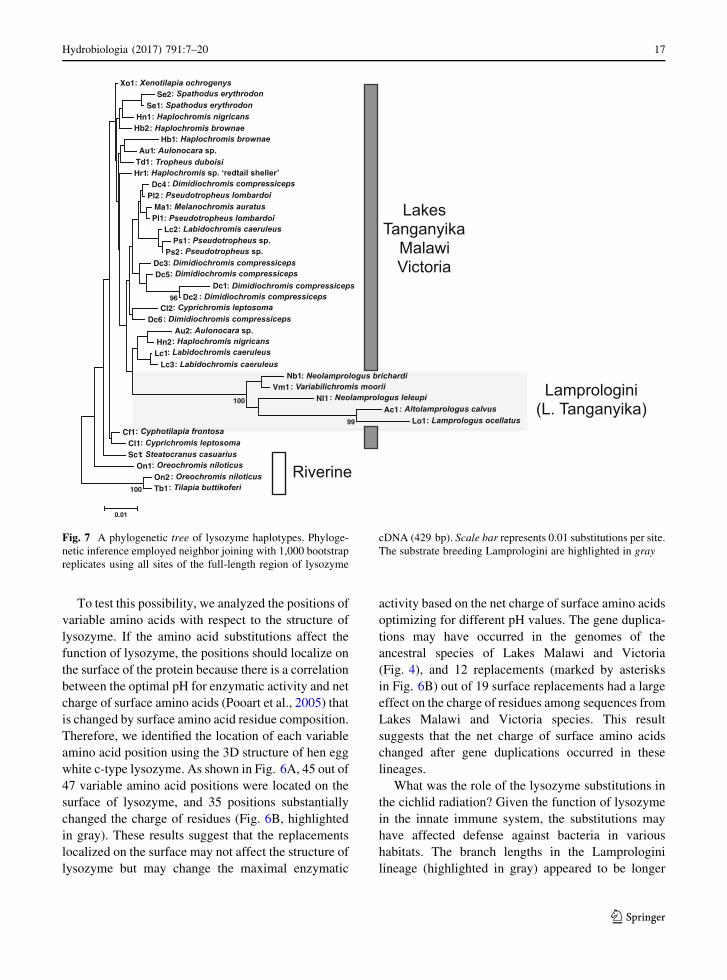
We constructed a phylogenetic tree using all sites
(Fig. 7). The branch lengths in the Lamprologini
lineage (highlighted in gray) appeared to be longer
than the other branches in the phylogenetic tree
(Fig. 7).
Discussion
Isolation and characterization of cichlid lysozyme
In fish, the innate immune system is important for
defense against bacteria in various water environ-
ments (Saurabh & Sahoo, 2008). Lysozyme is
involved in the innate immune system; therefore, we
investigated the evolution of the lysozyme gene during
the radiation of East African cichlids. We determined
37 different sequences from Great Lakes and riverine
species. Among 87 variable sites, 64 were nonsyn-
onymous sites (Fig. 2), suggesting that this gene has
evolved with amino acid replacements during radia-
tion. Surprisingly, we isolated more than two lyso-
zyme haplotypes within a species of Lake Malawi
cichlids (L. caeruleus and Dc), whereas one or two
haplotypes were detected in each Lake Tanganyika
cichlid (Fig. 2; Table 1). This result raised the possi-
bility that the lysozyme copy number may differ
among cichlid species; therefore, we further analyzed
the gene copy number.
Lysozyme copy number varied during evolution
of cichlids
Based on the results of Southern blot analysis, the copy
numbers of lysozyme gene were estimated as two, one,
four, and two in one riverine, four Lake Tanganyika,
two Lake Malawi, and one Lake Victoria species,
respectively. This estimation includes a possibility of
underestimation of copy numbers, because Southern
blot analysis would not distinguish different copies in
the restriction enzyme fragments if they had the same
size. The sequence analysis from a single individual of
Lake Malawi species also supported the multicopy of
lysozyme genes. Each of the five haplotypes was
determined from cDNA and genomic DNA of Dc
M. auratusD. compressiceps
L. caeruleus
Pseudotropheus. sp.P. lombardoiAulonocara sp.
H. brownae
H. nigricansH.
T. duboisi
S. erythrodonC. frontosa
N. brichardiN. leleupi
L. ocellatus
X. ochrogenys
A. calvus
C. leptosoma
V. moorii
T. buttikoferiS. casuariusO. niloticus
gene duplication
gene duplication(s)
Lake Victoria
Lake Malawi
Lake Tanganyika
RiverineLam
prologini
Copy numbers
4
4
2
1
2
1*
1
1
Fig. 4 The positions of gene duplications on the phylogenetic
tree of cichlids. The lysozyme gene copy numbers based on
Southern blot and sequence analyses are shown on the left side
of each species. The positions of gene duplications are shown on
the branches of the phylogenetic tree. The phylogenetic tree is
based on previous studies (Salzburger et al., 2002, 2005; Terai
et al., 2003). *The possibility of two copies in C. leptosoma was
inferred from sequence analysis (see ‘‘Discussion’’ section)
14 Hydrobiologia (2017) 791:7–20
123

(Fig. 5). The number of sequences from a single
individual indicates multiple copies of lysozyme
genes in the genome of Dc. Since cichlids are diploid
(Kuroiwa et al., 2013), we estimated that a minimum
of three lysozyme genes are present in the genome of
Dc, considering that all sequences are allelic varia-
tions. Two of five sequences amplified from both
cDNA and genomic DNA may be allelic variation,
because the results of Southern blot analysis showed
four copies of gene in Dc (Fig. 3B). Among the
nucleotide variation in six haplotypes determined
from five Dc individuals (Dc1–6), A at site 109
(109A), 111T, and 313C were not observed in
sequences determined from a single individual cDNA
(D. compressiceps#2), and 313C was not observed in
sequences from genomic DNA (D. compressiceps#6)
(Figs. 2, 5). The difference of nucleotide variation
between individuals may be due to the haplotype
variation within a species.
The copy number of the lysozyme gene in the
whole-genome sequence data from a Lake Malawi
cichlid did not match our Southern blot results from
Lake Malawi species (Fig. 3). The genome sequence
of Maylandia zebra Boulenger, 1899 (M_ze-
bra_UMD1: http://www.ncbi.nlm.nih.gov/) contains
two copies of the lysozyme gene (XP_014270133,
XP_014263224).Wemapped the short read sequences
(SRR077291) that were used for the assembly of the
M. zebra genome on the genome sequence (aligned
throughout the genome). In the two lysozyme genes,
the variants among Dc individuals at position 109,
257, 286, 299, 313, and 416 (Fig. 5) were observed at
the corresponding sites in the aligned short read
sequences. This result suggests a possibility that some
lysozyme genes are missing from the genome assem-
bly, because highly similar multicopy genes are dif-
ficult to assemble (Mariano et al., 2015).
To amplify the full-length of lysozyme coding
region from cDNA, we used a set of primers (LysF1
and LysR1) locating on the 50- and 30 UTRs for all
species used in this study. In this case, there was a
possibility that we could not amplify particular
haplotypes with mutations in primer biding positions,
indicating the possibility of underestimation of the
number of haplotypes.
In Lake Tanganyika species, two haplotypes were
determined from Cl (Cl1 and Cl2) and Se (Se1 and
Se2) (Fig. 2; Table 1), while a single band was
observed from each species (Fig. 3A, B). In the case
of Se, two haplotypes (Se1 and Se2) may represent
allelic variants, because the two haplotypes clustered
together in the phylogenetic tree (Fig. 7), and the
difference between these two haplotypes was only
one mutation (Fig. 2). In contrast, two haplotypes
from Cl (Cl1 and Cl2) were distantly related in the
tree (Fig. 7), and the difference between two
haplotypes was five mutations (Fig. 2). According
to these results, Cl seemed to possess two copies of
lysozyme gene. Lysozyme is one of the immune
system genes, and synonymous substitutions (posi-
tions 69 and 288) as well as nonsynonymous
substitutions in the sequences from Cl were shared
among species from different lineages (Fig. 2).
Therefore, we could not rule out the possibility that
the distantly related haplotypes of lysozyme have
been retained from an ancestral lineage, similar to
what has been previously reported for class II major
111222222 2344 46018334578 9112 99912694736 9364Dc1 GAATATCCGGA----------TTTGDc2 .....GGG.C.----------....Dc3 A...GGGG.CG----------C.CCDc4 A.C.GGGG.CG----------CCCCDc5 AC..GGGG...----------C.C.Dc6 ACCCGGGGACG----------C.CC
c1 ..CC.......----------..--c6 ..CC......G----------..--c7 ACCCGGGGACG----------C.--c12 ACCC......G----------..--c25 ACCC.......----------..--
g3 ----GGGGACG-intron 2-C.--g5 ----.GGG.C.-intron 2-..--
g15 ----....ACG-intron 2-C.--g17 ----GGGG.CG-intron 2-C.--
Nucleotide site
cDN
AG
enom
ic D
NA
Fig. 5 An alignment of all variable amino acid positions of
lysozyme haplotypes from D. compressiceps. The nucleotide
sites are shown on the top of the alignment. Dots indicate the
nucleotides that are identical with those in the top line.
HaplotypesDc1–6 were determined from fiveD. compressiceps
individuals. Haplotypes c1–25 were determined from cDNA of
a single individual (D. compressiceps#2). Haplotypes g3–17
were determined from genomic DNA of a single individual (D.
compressiceps#6)
Hydrobiologia (2017) 791:7–20 15
123

histocompatibility complex genes in Lake Malawi
cichlids (Klein et al., 1993).
According to the results of Southern blot and
sequence analyses, the copy number of lysozyme
varied between one and four in African cichlids. The
evolutionary transitions in lysozyme copy number was
inferred based on the phylogenetic relationships
among African cichlids (Salzburger et al.,
2002, 2005; Terai et al., 2003) and the Southern blot
(Fig. 3) and sequence analyses. After colonization in
Lake Tanganyika, some species reinvaded riverine
habitats and their progeny independently colonized
Lakes Malawi and Victoria (Salzburger et al., 2005).
The gene duplications may have occurred in the
genomes of reinvaders and the ancestral species of
Lake Malawi (Fig. 4).
The evolution of lysozyme during cichlid adaptive
radiation
The adaptive evolution of lysozyme for optimum
enzymatic activity in various pH environments has
been reported (Stewart et al., 1987; Jolles et al., 1990;
Swanson et al., 1991; Irwin et al., 1992; Irwin, 1995;
Kornegay, 1996). Cichlid species are distributed in
lakes and rivers with pH values ranging from 6.0 to
10.0 (Fryer & Iles, 1972) in Africa. Therefore, we
assumed that the lysozyme gene might have evolved
rapidly to gain enzymatic activity in tissues facing
water environments with different pH values and salt
concentrations during radiation in different rivers and
lakes in East Africa.
Lake Victoria
Lake Malawi
Lake Tanganyika
Riverine
Lam
prol
ogin
i
amino acid position
(A)
(B) 1111111111 1111111 122222333 3345556667 7888889999 0001122223 3333334 7701367016 7897891347 9023461267 0585603780 1235692Hn1 FKERDRKANY RGKKATRTDR WCDRRHCNNV TPNRETVARQ GQDRPSGHn2 ...P.H.... G......... .......... .S..Q..T.. ......RHb1 .......... G........H .........G .......T.R ......RHb2 .......... .......... .......... .S.....T.. .......Hr1 .......... G......... .......... .S.....T.. ......RLc1 .......... G......... .....R.... .S..Q..T.. ......RLc3 .....H.... G......... .....R.... .S..Q..T.. ......RLc2 .I........ .......... .....R..D. ....Q..T.. .......Ma1 .......... .......... .....R..D. .......T.. ......RAu1 .......... G......... .....R.... .......T.. .......Au2 ...P.H.... G......... .....R..S. ....Q..T.. ......RPs1 .I........ G......... .....R..D. ....Q..T.. .......Ps2 .I........ G......... .....R..D. ....Q..T.. .......Dc1 .E..E..... S.....H... LSH..RW... IS.....T.. .....F.Dc2 .E..E..... S.....H... .....R.... IS.....T.. .....F.Dc3 ....E..... S......... .....R..D. .S.....T.. ......RDc4 ....E..... .......... .....R..D. .......T.. ......RDc5 .......... S......... .....RW... .S.....T.. .......Dc6 .......... .......... ........D. .S.....T.. ......RPl1 .......... .......... ........D. .......T.. ......RPl2 .......... .......... .....R..D. .......T.. ......RTd1 .......... .......... .......... .......T.. ......RCl1 ....E..... ...E...... .......... .S.....N.. ......RCl2 ....E..... .......... .....Q..D. .A.....N.. ......RSe1 ....E....F G......... .......... .S........ ......RSe2 ....E....F G......... .......... .S........ ....A..Xo1 .......... .......... .......... .S.....T.. ......RCf1 .......... .......... .......... .S.....N.. ......RAc1 I.DKA.E... GD.....PN. ...H.T.DDE .SDTQ.YKH. DKVTS.DLo1 .QDKT.M... G......PN. ...H.T.EDE .SDTQRYKHK DKVTS.DNb1 .Q.KA.M... G......RN. ....SQ..D. .S..Q.YKH. ...TS.RNl1 .Q.QA.V... G....K..N. ....I..... .S.TQAYKH. .K.TS..Vm1 .Q.KA.M... G....K..N. ....S...D. .S..Q.YKH. ...TS.RSc1 ........S. .......... .......... AS.....N.. .......Tb1 .....K.E.. ..E....PN. ...G.N.... .S.....N.. .K....ROn1 .......... .......... .....N..S. AS.....N.. ...GS.ROn2 .....K.E.. ..E.E..PN. ...G.N.... .S.....N.. .K....R
* * * * * * * * * * * *
Fig. 6 Characteristics of
variable lysozyme amino
acids among cichlids. A The
variable amino acid
positions of lysozyme in the
3D structure. The variable
amino acids are shown in
light gray. Right and left
panels show the view from
opposite sides. B An amino
acid alignment of the cichlid
c-type lysozyme. The dots
and letters indicate
conserved and variable
amino acids, respectively,
compared with the reference
on the top line. The
abbreviations for each
species are the same as in
Fig. 2. Amino acid
substitutions that change the
charge of residues are shown
in a gray background.
Nonsurface replacements
are surrounded by a
rectangle. The replacements
that occurred in Lakes
Malawi and Victoria
lineages are marked by
asterisks
16 Hydrobiologia (2017) 791:7–20
123
To test this possibility, we analyzed the positions of
variable amino acids with respect to the structure of
lysozyme. If the amino acid substitutions affect the
function of lysozyme, the positions should localize on
the surface of the protein because there is a correlation
between the optimal pH for enzymatic activity and net
charge of surface amino acids (Pooart et al., 2005) that
is changed by surface amino acid residue composition.
Therefore, we identified the location of each variable
amino acid position using the 3D structure of hen egg
white c-type lysozyme. As shown in Fig. 6A, 45 out of
47 variable amino acid positions were located on the
surface of lysozyme, and 35 positions substantially
changed the charge of residues (Fig. 6B, highlighted
in gray). These results suggest that the replacements
localized on the surface may not affect the structure of
lysozyme but may change the maximal enzymatic
activity based on the net charge of surface amino acids
optimizing for different pH values. The gene duplica-
tions may have occurred in the genomes of the
ancestral species of Lakes Malawi and Victoria
(Fig. 4), and 12 replacements (marked by asterisks
in Fig. 6B) out of 19 surface replacements had a large
effect on the charge of residues among sequences from
Lakes Malawi and Victoria species. This result
suggests that the net charge of surface amino acids
changed after gene duplications occurred in these
lineages.
What was the role of the lysozyme substitutions in
the cichlid radiation? Given the function of lysozyme
in the innate immune system, the substitutions may
have affected defense against bacteria in various
habitats. The branch lengths in the Lamprologini
lineage (highlighted in gray) appeared to be longer
Lamprologini (L. Tanganyika)
Lakes Tanganyika
Malawi Victoria
Riverine
Xo1 Se2
Se1 Hn1
Hb2 Hb1
Au1 Td1 Hr1
Dc4 Pl2
Ma1 Pl1
Lc2 Ps1
Ps2 Dc3 Dc5
Dc1 Dc2
Cl2 Dc6
Au2 Hn2 Lc1
Lc3 Nb1
Vm1 Nl1
Ac1 Lo1
Cf1 Cl1 Sc1
On1 On2 Tb1
99
100
100
96
0.01
: Oreochromis niloticus: Oreochromis niloticus
: Steatocranus casuarius
: Tilapia buttikoferi
: Neolamprologus brichardi
: Altolamprologus calvus
: Variabilichromis moorii
: Lamprologus ocellatus: Cyphotilapia frontosa
: Cyprichromis leptosoma
: Cyprichromis leptosoma
: Spathodus erythrodon: Spathodus erythrodon
: Tropheus duboisi
: Aulonocara sp.
: Aulonocara sp.
: Pseudotropheus lombardoi
: Pseudotropheus lombardoi
: Labidochromis caeruleus: Labidochromis caeruleus
: Labidochromis caeruleus: Pseudotropheus sp.
: Pseudotropheus sp.
: Dimidiochromis compressiceps
: Dimidiochromis compressiceps: Dimidiochromis compressiceps
: Dimidiochromis compressiceps: Dimidiochromis compressiceps
: Dimidiochromis compressiceps
: Melanochromis auratus
: Haplochromis brownae: Haplochromis brownae
: Haplochromis nigricans
: Haplochromis nigricans
: Haplochromis
: Xenotilapia ochrogenys
: Neolamprologus leleupi
Fig. 7 A phylogenetic tree of lysozyme haplotypes. Phyloge-
netic inference employed neighbor joining with 1,000 bootstrap
replicates using all sites of the full-length region of lysozyme
cDNA (429 bp). Scale bar represents 0.01 substitutions per site.
The substrate breeding Lamprologini are highlighted in gray
Hydrobiologia (2017) 791:7–20 17
123
than the other branches (Fig. 7). The amino acid
replacements in this lineage may be related to
certain living environments or traits specific to
Lamprologini species. All the species in this tribe
spawn eggs on the surface of the rock or inside the
empty shells of Neothauma tanganyikense Smith,
1880 (substrate-brooder) (Coulter, 1991; Salzburger
et al., 2002; Clabaut et al,. 2005; Koblmuller et al.,
2008a; Sturmbauer et al., 2010), where the risk of
bacterial infections may be high. However, the
correlation between egg spawning and the evolution
of lysozyme is unlikely, because all the haplotypes
were isolated from adult cDNAs. The other envi-
ronments such as water condition, depth, and
habitat may not be largely different between
Lamprologini and other Lake Tanganyika species.
The functional analysis of lysozyme from haplo-
types with long branches will be a key to find the
role of this gene in Lamprologini species.
In this study, we demonstrated a possible role of
gene duplications and amino acid replacements of
lysozyme that may contribute to the defense mecha-
nism against bacteria in environments with different
salt concentrations, pH values, temperatures, and
organic substances. Although the knowledge of the
role for gene duplication is still limited, the cichlid
genome sequences will facilitate the identification of
candidate genes with copy number variation among
lineages of African cichlids. The functional analysis of
the duplicated gene products will demonstrate the
importance of gene duplication during the adaptive
radiation of cichlid fish.
Acknowledgments This work was supported by the Ministry
of Education, Culture, Sports, Science, and Technology of Japan
Grants to Y.T. (Nos. 23570269 and 26440209), an internal
SOKENDAI Grant to Y.T., and the Center for the Promotion of
Integrated Sciences (CPIS) of SOKENDAI Grant to Y.T. I thank
Dr. Tetsumi Takahashi (Graduate School of Science, Kyoto
University, Japan; present address: Institute of Natural and
Environmental Sciences, University of Hyogo) for identification
of Lake Tanganyika cichlids, and Dr. Norihiro Okada (Graduate
School of Bioscience and Biotechnology, Tokyo Institute of
Technology; present address: Foundation of Advancement of
International Science, Japan; National Cheng Kung University,
Taiwan) for providing laboratory space and experimental
equipment.
Author contributions ST.K.: determination and analysis of
lysozyme sequences, manuscript editing. H.T.: manuscript
editing. Y.T.: research concept, research planning, all experi-
ments, data analysis, and manuscript preparation.
References
Brawand, D., C. E. Wagner, Y. I. Li, M. Malinsky, I. Keller, S.
Fan, O. Simakov, A. Y. Ng, Z. W. Lim, E. Bezault, J.
Turner-Maier, J. Johnson, R. Alcazar, H. J. Noh, P. Russell,
B. Aken, J. Alfoldi, C. Amemiya, N. Azzouzi, J.
F. Baroiller, F. Barloy-Hubler, A. Berlin, R. Bloomquist,
K. L. Carleton, M. A. Conte, H. D’Cotta, O. Eshel, L.
Gaffney, F. Galibert, H. F. Gante, S. Gnerre, L. Greuter, R.
Guyon, N. S. Haddad, W. Haerty, R. M. Harris, H.
A. Hofmann, T. Hourlier, G. Hulata, D. B. Jaffe, M. Lara,
A. P. Lee, I. MacCallum, S. Mwaiko, M. Nikaido, H.
Nishihara, C. Ozouf-Costaz, D. J. Penman, D. Przybylski,
M. Rakotomanga, S. C. Renn, F. J. Ribeiro, M. Ron, W.
Salzburger, L. Sanchez-Pulido, M. E. Santos, S. Searle, T.
Sharpe, R. Swofford, F. J. Tan, L. Williams, S. Young, S.
Yin, N. Okada, T. D. Kocher, E. A. Miska, E. S. Lander, B.
Venkatesh, R. D. Fernald, A. Meyer, C. P. Ponting, J.
T. Streelman, K. Lindblad-Toh, O. Seehausen & F. Di
Palma, 2014. The genomic substrate for adaptive radiation
in African cichlid fish. Nature 513: 375–381.
Callewaert, L. & C. W. Michiels, 2010. Lysozymes in the ani-
mal kingdom. Journal of Biosciences 35: 127–160.
Clabaut, C., W. Salzburger & A. Meyer, 2005. Comparative
phylogenetic analyses of the adaptive radiation in Lake
Tanganyika cichlid fishes: nuclear sequences are less
homoplasious but also less informative than mitochondrial
DNA. Journal of Molecular Evolution 31: 666–681.
Cohen, A. S., M. J. Soreghan & C. A. Scholz, 1993. Estimating
the age of formation of lakes: an example from Lake
Tanganyika, East African Rift system. Geology 21:
511–514.
Cohen, A. S., K. E. Lezzar, J. J. Tiercelin & M. Soreghan, 1997.
New palaeogeographic and lake-level reconstructions of
Lake Tanganyika: implications for tectonic, climatic and
biological evolution in a rift lake. Basin Research 9:
107–132.
Conant, G. C. & K. H. Wolfe, 2008. Turning a hobby into a job:
how duplicated genes find new functions. Nature Reviews
Genetics 9: 938–950.
Coulter, G. W., 1991. The Benthic Fish Community. Oxford
University Press, London.
Delvaux, D., 1995. Age of Lake Malawi (Nyasa) and Water
Level Fluctuations. Muses Royal de l’Afrique Centrale,
Tervuren (Belgium), Department of Geology and Miner-
alogy, Rapp Annual: 99–108.
Dobson, D. E., E. M. Prager & A. C. Wilson, 1984. Stomach
lysozyme of ruminants. Journal of Biological Chemistry
259: 11607–11616.
Friedman, M., B. P. Keck, A. Dornburg, R. I. Eytan, C.
H. Martin, C. D. Hulsey, P. C. Wainwright & T. J. Near,
2013. Molecular and fossil evidence place the origin of
cichlid fishes long after Gondwanan rifting. Proceedings of
the Royal Society of London B: Biological Sciences 280:
20131733.
Fryer, G. & T. D. Iles, 1972. The Cichlid Fishes of the Great
Lakes of Africa. Oliver and Boyd, Edinburgh.
Genner, M. J., O. Seehausen, D. H. Lunt, D. A. Joyce, P.
W. Shaw, G. R. Carvalho & G. F. Turner, 2007. Age of
18 Hydrobiologia (2017) 791:7–20
123

cichlids: new dates for ancient lake fish radiations.
Molecular Biology and Evolution 24: 1269–1282.
Hikima, J.-I., S. Minagawa, I. Hirono & T. Aoki, 2001.
Molecular cloning, expression and evolution of the Japa-
nese flounder goose-type lysozyme gene, and the lytic
activity of its recombinant protein. Biochimica Biophysica
Acta 1520: 35–44.
Irwin, D. M., 1995. Evolution of the bovine lysozyme gene
family: changes in gene expression and reversion of
function. Journal of Molecular Evolution 41: 299–312.
Irwin, D. M., E. M. Prager & A. C. Wilson, 1992. Evolutionary
genetics of ruminant lysozymes.AnimalGenetics 23: 193–202.
Johnson, T. C., C. A. Scholz,M. R. Talbot, K.Kelts, R. D. Ricketts,
G. Ngobi, K. Beuning, I. I. Ssemmanda& J.W.McGill, 1996.
Late Pleistocene desiccation of Lake Victoria and rapid
evolution of cichlid fishes. Science 273: 1091–1093.
Jolles, J., E. M. Prager, E. S. Alnemri, P. Jolles, I. M. Ibrahimi &
A. C.Wilson, 1990. Amino-acid-sequences of stomach and
nonstomach lysozymes of ruminants. Journal of Molecular
Evolution 30: 370–382.
Jolles, J., A. Fiala-Medioni & P. Jolles, 1996. The ruminant
digestion model using bacteria already employed early in
evolution by symbiotic molluscs. Journal of Molecular
Evolution 43: 523–527.
Klein, D., H. Ono, C. O’hUigin, V. Vincek, T. Goldschmidt & J.
Klein, 1993. Extensive MHC variability in cichlid fishes of
Lake Malawi. Nature 364: 330–334.
Koblmuller, S., U. K. Schliewen, N. Duftner, K. M. Sefc, C.
Katongo & C. Sturmbauer, 2008a. Age and spread of the
haplochromine cichlid fishes in Africa. Molecular Phylo-
genetics and Evolution 49: 153–169.
Koblmuller, S., K. M. Sefc & C. Sturmbauer, 2008b. The Lake
Tanganyika cichlid species assemblage: recent advances in
molecular phylogenetics. Hydrobiologia 615: 5–20.
Kocher, T. D., 2004. Adaptive evolution and explosive specia-
tion: the cichlid fish model. Nature Reviews Genetics 5:
288–298.
Kondrashov, F. A., 2012. Gene duplication as a mechanism of
genomic adaptation to a changing environment. Proceed-
ings of the Royal Society of London B: Biological Sciences
279: 5048–5057.
Kornegay, J. R., 1996. Molecular genetics and evolution of
stomach and nonstomach lysozymes in the Hoatzin. Jour-
nal of Molecular Evolution 42: 676–684.
Kuroiwa, A., Y. Terai, N. Kobayashi, K. Yoshida, M. Suzuki, A.
Nakanishi, Y. Matsuda, M. Watanabe & N. Okada, 2013.
Construction of chromosome markers from the Lake Vic-
toria cichlid Paralabidochromis chilotes and their appli-
cation to comparative mapping. Cytogenetic and Genome
Research 142: 112–120.
Makino, T. & M. Kawata, 2012. Habitat variability correlates
with duplicate content of Drosophila genomes. Molecular
Biology and Evolution 29: 3169–3179.
Mariano, D. C., F. L. Pereira, P. Ghosh, D. Barh, H.
C. Figueiredo, A. Silva, R. T. Ramos & V. A. Azevedo,
2015. MapRepeat: an approach for effective assembly of
repetitive regions in prokaryotic genomes. Bioinformation
11: 276–279.
Mayer, W. E., H. Tichy & J. Klein, 1998. Phylogeny of African
cichlid fishes as revealed by molecular markers. Heredity
80: 702–714.
Ohno, S., 1970. Evolution by Gene Duplication. Springer, New
York.
Pooart, J., T. Torikata & T. Araki, 2005. Enzymatic properties of
rhea lysozyme. Bioscience, Biotechnology, and Bio-
chemistry 69: 103–112.
Saitou, N. &M. Nei, 1987. The neighbor-joining method: a new
method for reconstructing phylogenetic trees. Molecular
Biology and Evolution 4: 406–425.
Salzburger, W., A. Meyer, S. Baric, E. Verheyen & C. Sturm-
bauer, 2002. Phylogeny of the Lake Tanganyika cichlid
species flock and its relationship to the Central and East
African haplochromine cichlid fish faunas. Systematic
Biology 51: 113–135.
Salzburger,W., T.Mack, E. Verheyen&A.Meyer, 2005. Out of
Tanganyika: genesis, explosive speciation, key-innova-
tions and phylogeography of the haplochromine cichlid
fishes. BMC Evolutionary Biology 5: 17.
Sankaran, K. & S. Gurnani, 1972. On the variation in the cat-
alytic activity of lysozyme in fishes. Indian Journal of
Biochemistry and Biophysics 9: 162–165.
Saurabh, S. & P. K. Sahoo, 2008. Lysozyme: an important
defence molecule of fish innate immune system. Aqua-
culture Research 39: 223–239.
Stewart, C. B., J. W. Schilling & A. C. Wilson, 1987. Adaptive
evolution in the stomach lysozymes of foregut fermenters.
Nature 330: 401–404.
Sturmbauer, C., W. Salzburger, N. Duftner, R. Schelly & S.
Koblmuller, 2010. Evolutionary history of the Lake Tan-
ganyika cichlid tribe Lamprologini (Teleostei: Perci-
formes) derived from mitochondrial and nuclear DNA
data. Molecular Phylogenetics and Evolution 57: 266–284.
Swanson, K. W., D. M. Irwin & A. C. Wilson, 1991. Stomach
lysozyme gene of the langur monkey: tests for convergence
and positive selection. Journal of Molecular Evolution 33:
418–425.
Takahashi, T. & S. Koblmuller, 2011. The adaptive radiation of
cichlid fish in Lake Tanganyika: a morphological per-
spective. International Journal of Evolutionary Biology
2011: 620754.
Takahashi, T. & T. Sota, 2016. A robust phylogeny among
major lineages of the East African cichlids. Molecular
Phylogenetics and Evolution 100: 234–242.
Tamura, K., D. Peterson, N. Peterson, G. Stecher, M. Nei & S.
Kumar, 2011. MEGA5: molecular evolutionary genetics
analysis using maximum likelihood, evolutionary distance,
and maximum parsimony methods. Molecular Biology and
Evolution 28: 2731–2739.
Taoka, Y., H. Maeda, J. Y. Jo, S. M. Kim, S. I. Park, T. Yosh-
ikawa & T. Sakata, 2006. Use of live and dead probiotic
cells in tilapiaOreochromis niloticus. Fisheries Science 72:
755–766.
Terai, Y., K. Takahashi, M. Nishida, T. Sato & N. Okada, 2003.
Using SINEs to probe ancient explosive speciation: ‘‘hid-den’’ radiation of African cichlids? Molecular Biology and
Evolution 20: 924–930.
Turner, G. F., O. Seehausen, M. E. Knight, C. J. Allender & R.
L. Robinson, 2001. How many species of cichlid fishes are
there in African lakes? Molecular Ecology 10: 793–806.
Wang, Y., L. Geer, C. Chappey, J. A. Kans & S. H. Bryant, 2000.
Cn3D: sequence and structure views for Entrez. Trends in
Biochemical Sciences 6: 300–302.
Hydrobiologia (2017) 791:7–20 19
123

Weiss, J. D., F. P. Cotterill & U. K. Schliewen, 2015. Lake
Tanganyika – A ‘Melting Pot’ of ancient and young cichlid
lineages (Teleostei: Cichlidae)? PLoS One 10: e0125043.
Welker, T. L., C. Lim, M. Yildirim-Aksoy & P. H. Klesius,
2007. Growth, immune function and disease and stress
resistance of juvenile Nile tilapia (Oreochromis niloticus)
fed graded level of bovine lactoferrin. Aquaculture 262:
156–162.
Yazawa, R., I. Hirono & T. Aoki, 2006. Transgenic zebrafish
expressing chicken lysozyme show resistance against
bacterial diseases. Transgenic Research 15: 385–391.
Zhang, J., 2003. Evolution by gene duplication: an update.
Trends in Ecology and Evolution 18: 292–298.
20 Hydrobiologia (2017) 791:7–20
123


![RepeatabilityandHeritabilityofBehaviouralTypesin …behav.zoology.unibe.ch/sysuif/uploads/files/esh/pdf...and reproductively mature [39]). Behavioural types in this species may also](https://static.fdocuments.in/doc/165x107/5f0c63a57e708231d43528dc/repeatabilityandheritabilityofbehaviouraltypesin-behav-and-reproductively-mature.jpg)