Gene-Based Risk Stratification for Cardiac Disorders in ... · Correspondence to Takeru Makiyama,...
23
1 T he LMNA (lamin A/C) gene encodes the A-type lamin proteins, lamin A and C—the major components of the nuclear membrane in vertebrates—and mutations in LMNA have been reported to cause a variety of clinical phenotypes, including cardiac disorders, 1 Emery–Dreifuss muscular dys- trophy, 2,3 limb-girdle muscular dystrophy, 4 Charcot–Marie– Tooth type 2, 5 familial partial lipodystrophy, 6,7 and premature aging. 8,9 The cardiac phenotypes associated with LMNA muta- tions are characterized by cardiac conduction disturbance (CCD), atrial fibrillation, ventricular tachyarrhythmia (VT), and dilated cardiomyopathy. 10–14 Risk stratification for LMNA-related cardiomyopathy has been assessed previously in some reports. Approximately 90% of LMNA mutation carriers >30 years of age have some type of arrhythmia, and the subjects have a risk of sudden arrhythmic death before the development of cardiac failure 15 Recently, Kumar et al 16 reported the long-term follow-up data of 122 consecutive LMNA mutation carriers and showed that LMNA-related heart diseases had a malignant course; most Background—Mutations in LMNA (lamin A/C), which encodes lamin A and C, typically cause age-dependent cardiac phenotypes, including dilated cardiomyopathy, cardiac conduction disturbance, atrial fibrillation, and malignant ventricular arrhythmias. Although the type of LMNA mutations have been reported to be associated with susceptibility to malignant ventricular arrhythmias, the gene-based risk stratification for cardiac complications remains unexplored. Methods and Results—The multicenter cohort included 77 LMNA mutation carriers from 45 families; cardiac disorders were retrospectively analyzed. The mean age of patients when they underwent genetic testing was 45±17, and they were followed for a median 49 months. Of the 77 carriers, 71 (92%) were phenotypically affected and showed cardiac conduction disturbance (81%), low left ventricular ejection fraction (<50%; 45%), atrial arrhythmias (58%), and malignant ventricular arrhythmias (26%). During the follow-up period, 9 (12%) died, either from end-stage heart failure (n=7) or suddenly (n=2). Genetic analysis showed truncation mutations in 58 patients from 31 families and missense mutations in 19 patients from 14 families. The onset of cardiac disorders indicated that subjects with truncation mutations had an earlier occurrence of cardiac conduction disturbance and low left ventricular ejection fraction, than those with missense mutations. In addition, the truncation mutation was found to be a risk factor for the early onset of cardiac conduction disturbance and the occurrence of atrial arrhythmias and low left ventricular ejection fraction, as estimated using multivariable analyses. Conclusions—The truncation mutations were associated with manifestation of cardiac phenotypes in LMNA-related cardiomyopathy, suggesting that genetic analysis might be useful for diagnosis and risk stratification. (Circ Cardiovasc Genet. 2017;10:e001603. DOI: 10.1161/CIRCGENETICS.116.001603.) Key Words: arrhythmia ◼ cardiomyopathies ◼ death, sudden, cardiac ◼ heart failure ◼ lamin type A ◼ mutation ◼ prognosis © 2017 American Heart Association, Inc. Circ Cardiovasc Genet is available at http://circgenetics.ahajournals.org DOI: 10.1161/CIRCGENETICS.116.001603 Received September 6, 2016; accepted September 25, 2017. The Data Supplement is available at http://circgenetics.ahajournals.org/lookup/suppl/doi:10.1161/CIRCGENETICS.116.001603/-/DC1. Correspondence to Takeru Makiyama, MD, PhD, Department of Cardiovascular Medicine, Kyoto University Graduate School of Medicine, 54 Kawahara-cho, Shogoin, Sakyo-ku, Kyoto 606–8507, Japan, E-mail [email protected] or Takeshi Aiba, MD, PhD, Division of Arrhythmia and Electrophysiology, National Cerebral and Cardiovascular Center, 5-7-1 Fujishiro-dai, Suita, Osaka, 565–8565 Japan, E-mail [email protected] Gene-Based Risk Stratification for Cardiac Disorders in LMNA Mutation Carriers Suguru Nishiuchi, MD; Takeru Makiyama, MD, PhD; Takeshi Aiba, MD, PhD; Kenzaburo Nakajima, MD; Sayako Hirose, MD; Hirohiko Kohjitani, MD; Yuta Yamamoto, DVM, PhD; Takeshi Harita, MD; Mamoru Hayano, MD; Yimin Wuriyanghai, MD; Jiarong Chen, PhD; Kenichi Sasaki, MD, PhD; Nobue Yagihara, MD, PhD; Taisuke Ishikawa, DVM, PhD; Kenji Onoue, MD, PhD; Nobuyuki Murakoshi, MD, PhD; Ichiro Watanabe, MD, PhD; Kimie Ohkubo, MD, PhD; Hiroshi Watanabe, MD, PhD; Seiko Ohno, MD, PhD; Takahiro Doi, MD, PhD; Satoshi Shizuta, MD, PhD; Tohru Minamino, MD, PhD; Yoshihiko Saito, MD, PhD; Yasushi Oginosawa, MD, PhD; Akihiko Nogami, MD, PhD; Kazutaka Aonuma, MD, PhD; Kengo Kusano, MD, PhD; Naomasa Makita, MD, PhD; Wataru Shimizu, MD, PhD; Minoru Horie, MD, PhD; Takeshi Kimura, MD, PhD Original Article See Editorial by Sinagra et al See Clinical Perspective by guest on December 16, 2017 http://circgenetics.ahajournals.org/ Downloaded from by guest on December 16, 2017 http://circgenetics.ahajournals.org/ Downloaded from by guest on December 16, 2017 http://circgenetics.ahajournals.org/ Downloaded from by guest on December 16, 2017 http://circgenetics.ahajournals.org/ Downloaded from by guest on December 16, 2017 http://circgenetics.ahajournals.org/ Downloaded from by guest on December 16, 2017 http://circgenetics.ahajournals.org/ Downloaded from by guest on December 16, 2017 http://circgenetics.ahajournals.org/ Downloaded from by guest on December 16, 2017 http://circgenetics.ahajournals.org/ Downloaded from by guest on December 16, 2017 http://circgenetics.ahajournals.org/ Downloaded from by guest on December 16, 2017 http://circgenetics.ahajournals.org/ Downloaded from by guest on December 16, 2017 http://circgenetics.ahajournals.org/ Downloaded from by guest on December 16, 2017 http://circgenetics.ahajournals.org/ Downloaded from by guest on December 16, 2017 http://circgenetics.ahajournals.org/ Downloaded from
Transcript of Gene-Based Risk Stratification for Cardiac Disorders in ... · Correspondence to Takeru Makiyama,...
1
The LMNA (lamin A/C) gene encodes the A-type lamin proteins, lamin A and C—the major components of the
nuclear membrane in vertebrates—and mutations in LMNA have been reported to cause a variety of clinical phenotypes, including cardiac disorders,1 Emery–Dreifuss muscular dys-trophy,2,3 limb-girdle muscular dystrophy,4 Charcot–Marie–Tooth type 2,5 familial partial lipodystrophy,6,7 and premature aging.8,9 The cardiac phenotypes associated with LMNA muta-tions are characterized by cardiac conduction disturbance (CCD), atrial fibrillation, ventricular tachyarrhythmia (VT), and dilated cardiomyopathy.10–14
Risk stratification for LMNA-related cardiomyopathy has been assessed previously in some reports. Approximately 90% of LMNA mutation carriers >30 years of age have some type of arrhythmia, and the subjects have a risk of sudden arrhythmic death before the development of cardiac failure15 Recently, Kumar et al16 reported the long-term follow-up data of 122 consecutive LMNA mutation carriers and showed that LMNA-related heart diseases had a malignant course; most
Background—Mutations in LMNA (lamin A/C), which encodes lamin A and C, typically cause age-dependent cardiac phenotypes, including dilated cardiomyopathy, cardiac conduction disturbance, atrial fibrillation, and malignant ventricular arrhythmias. Although the type of LMNA mutations have been reported to be associated with susceptibility to malignant ventricular arrhythmias, the gene-based risk stratification for cardiac complications remains unexplored.
Methods and Results—The multicenter cohort included 77 LMNA mutation carriers from 45 families; cardiac disorders were retrospectively analyzed. The mean age of patients when they underwent genetic testing was 45±17, and they were followed for a median 49 months. Of the 77 carriers, 71 (92%) were phenotypically affected and showed cardiac conduction disturbance (81%), low left ventricular ejection fraction (<50%; 45%), atrial arrhythmias (58%), and malignant ventricular arrhythmias (26%). During the follow-up period, 9 (12%) died, either from end-stage heart failure (n=7) or suddenly (n=2). Genetic analysis showed truncation mutations in 58 patients from 31 families and missense mutations in 19 patients from 14 families. The onset of cardiac disorders indicated that subjects with truncation mutations had an earlier occurrence of cardiac conduction disturbance and low left ventricular ejection fraction, than those with missense mutations. In addition, the truncation mutation was found to be a risk factor for the early onset of cardiac conduction disturbance and the occurrence of atrial arrhythmias and low left ventricular ejection fraction, as estimated using multivariable analyses.
Conclusions—The truncation mutations were associated with manifestation of cardiac phenotypes in LMNA-related cardiomyopathy, suggesting that genetic analysis might be useful for diagnosis and risk stratification. (Circ Cardiovasc Genet. 2017;10:e001603. DOI: 10.1161/CIRCGENETICS.116.001603.)
Key Words: arrhythmia ◼ cardiomyopathies ◼ death, sudden, cardiac ◼ heart failure ◼ lamin type A ◼ mutation ◼ prognosis
© 2017 American Heart Association, Inc.
Circ Cardiovasc Genet is available at http://circgenetics.ahajournals.org DOI: 10.1161/CIRCGENETICS.116.001603
Received September 6, 2016; accepted September 25, 2017.The Data Supplement is available at http://circgenetics.ahajournals.org/lookup/suppl/doi:10.1161/CIRCGENETICS.116.001603/-/DC1.Correspondence to Takeru Makiyama, MD, PhD, Department of Cardiovascular Medicine, Kyoto University Graduate School of Medicine, 54
Kawahara-cho, Shogoin, Sakyo-ku, Kyoto 606–8507, Japan, E-mail [email protected] or Takeshi Aiba, MD, PhD, Division of Arrhythmia and Electrophysiology, National Cerebral and Cardiovascular Center, 5-7-1 Fujishiro-dai, Suita, Osaka, 565–8565 Japan, E-mail [email protected]
Gene-Based Risk Stratification for Cardiac Disorders in LMNA Mutation Carriers
Suguru Nishiuchi, MD; Takeru Makiyama, MD, PhD; Takeshi Aiba, MD, PhD; Kenzaburo Nakajima, MD; Sayako Hirose, MD; Hirohiko Kohjitani, MD;
Yuta Yamamoto, DVM, PhD; Takeshi Harita, MD; Mamoru Hayano, MD; Yimin Wuriyanghai, MD; Jiarong Chen, PhD; Kenichi Sasaki, MD, PhD; Nobue Yagihara, MD, PhD;
Taisuke Ishikawa, DVM, PhD; Kenji Onoue, MD, PhD; Nobuyuki Murakoshi, MD, PhD; Ichiro Watanabe, MD, PhD; Kimie Ohkubo, MD, PhD; Hiroshi Watanabe, MD, PhD;
Seiko Ohno, MD, PhD; Takahiro Doi, MD, PhD; Satoshi Shizuta, MD, PhD; Tohru Minamino, MD, PhD; Yoshihiko Saito, MD, PhD; Yasushi Oginosawa, MD, PhD; Akihiko Nogami, MD, PhD; Kazutaka Aonuma, MD, PhD; Kengo Kusano, MD, PhD;
Naomasa Makita, MD, PhD; Wataru Shimizu, MD, PhD; Minoru Horie, MD, PhD; Takeshi Kimura, MD, PhD
Original Article
See Editorial by Sinagra et al See Clinical Perspective
by guest on Decem
ber 16, 2017http://circgenetics.ahajournals.org/
Dow
nloaded from
by guest on Decem
ber 16, 2017http://circgenetics.ahajournals.org/
Dow
nloaded from
by guest on Decem
ber 16, 2017http://circgenetics.ahajournals.org/
Dow
nloaded from
by guest on Decem
ber 16, 2017http://circgenetics.ahajournals.org/
Dow
nloaded from
by guest on Decem
ber 16, 2017http://circgenetics.ahajournals.org/
Dow
nloaded from
by guest on Decem
ber 16, 2017http://circgenetics.ahajournals.org/
Dow
nloaded from
by guest on Decem
ber 16, 2017http://circgenetics.ahajournals.org/
Dow
nloaded from
by guest on Decem
ber 16, 2017http://circgenetics.ahajournals.org/
Dow
nloaded from
by guest on Decem
ber 16, 2017http://circgenetics.ahajournals.org/
Dow
nloaded from
by guest on Decem
ber 16, 2017http://circgenetics.ahajournals.org/
Dow
nloaded from
by guest on Decem
ber 16, 2017http://circgenetics.ahajournals.org/
Dow
nloaded from
by guest on Decem
ber 16, 2017http://circgenetics.ahajournals.org/
Dow
nloaded from
by guest on Decem
ber 16, 2017http://circgenetics.ahajournals.org/
Dow
nloaded from
2 Nishiuchi et al Gene-Based Risk Stratification for Laminopathy
patients experienced arrhythmia, heart block, embolic events, or heart failure within 7 years of diagnosis.
Several studies have also explored genotype–phenotype correlations; frameshift mutations were often associated with the cardiac disorders in laminopathies,17 and splice-site muta-tions were one of the independent risk factors for sudden cardiac death.13 Recently, van Rijsingen et al18 revealed that nonmissense mutations were (ins-del/truncating or mutations affecting splicing) one of the major independent risk factors for malignant ventricular arrhythmias (MVAs) in a large European cohort of 269 LMNA mutation carriers. These results indicated that patients with truncation LMNA mutations, which result in haploinsufficiency of A-type lamins, might be at significantly higher risk of sudden cardiac death because of MVAs, than missense mutation carriers. However, other cardiac complica-tions, such as left ventricular systolic dysfunction and CCD, in LMNA mutation carriers have not yet been fully investigated in terms of the type of mutations. Based on the available litera-ture, we hypothesized that truncation mutations might be asso-ciated with poor prognosis in LMNA mutation carriers because of CCD, MVAs, and heart failure. To test this hypothesis, we performed a multicenter retrospective cohort study.
MethodsStudy DesignThe cohort for the present study consisted of genotyped LMNA muta-tion carriers and their relatives who had been introduced to the Kyoto University Hospital (Kyoto, Japan), Shiga University of Medical Science (Shiga, Japan), Nagasaki University Graduate School of Biomedical Sciences (Nagasaki, Japan), Niigata University Graduate School of Medical and Dental Sciences (Niigata, Japan), University of Tsukuba (Ibaraki, Japan), and the National Cerebral and Cardiovascular Center (Osaka, Japan) for genetic screening of LMNA between April 2008 and October 2015. Carriers of compound heterozygous mutations in LMNA and SCN5A were not included in this cohort. All subjects willingly provided informed consent for participating in this study. After providing informed consent, the patients were subjected to clini-cal screening and underwent peripheral blood sampling for genetic testing. Detailed clinical information was obtained from each subject by cardiologists in the respective institutes. After the genetic diagno-sis, LMNA mutation carriers were followed up by a cardiologist from one of the participating institutes or by a private doctor. The primary end point in this analysis was all-cause death during the follow-up pe-riod. The secondary end point was a composite of cardiac events and diagnosis, including the first occurrence of CCD, atrial arrhythmias, low left ventricular ejection fraction (LVEF), or MVAs. The protocol of this study was approved by the Institutional Ethics Committee and performed in accordance with its guidelines (Kyoto University, G194; National Cerebral and Cardiovascular Center, M24-031-4).
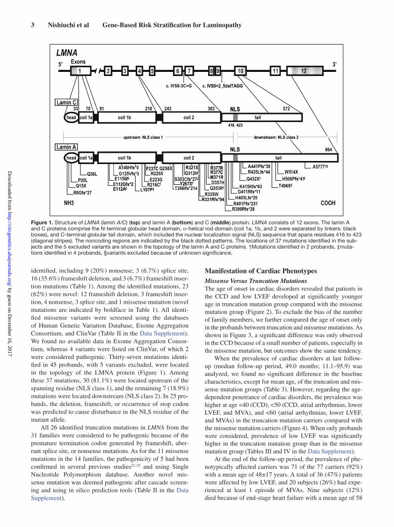
Genetic AnalysisThe methods followed for DNA extraction, for generating the PCR primers, and for mutational screening are described in Appendix in the Data Supplement and Table I in the Data Supplement. The muta-tions were divided into 2 groups: truncation (including splice site, frameshift insertion, frameshift deletion, and nonsense mutations) and missense mutations. We also classified the mutations based on the site: mutations located upstream (nuclear localization signal [NLS] class 1) of the spanning residues 416 to 423 and mutations located downstream (NLS class 2; Figure 1).13,19 If a frameshift or nonsense mutation was located upstream of the NLS residue, or a missense mu-tation disturbed the base sequence of the NLS residue, it was defined as an NLS-disturbed mutation. All the other mutations that did not disturb the NLS sequence were defined as NLS-conserved mutations.
Bioinformatic AnalysisVariants in LMNA were screened by genetic variant databases, a splic-ing site prediction tool, and in silico predictions (details in Appendix in the Data Supplement). Additionally, we confirmed the pathogenic-ity of mutations by using cascade screening, after accommodating relatives. Of the 84 carriers from 51 families genotyped by screen-ing, 7 carriers from 6 families were excluded (Table 1; Table II in the Data Supplement) because unreported LMNA variants without enough clinical or genetic data of relatives were considered variants of unknown significance.
Clinical DefinitionsLow LVEF was defined as LVEF <50%. dilated cardiomyopathy was defined as low LVEF or left ventricular enlargement, as per published normal values.20 MVAs were defined as sustained VT, ventricular fi-brillation, sudden cardiac death, cardiopulmonary resuscitation, and appropriate implantable cardioverter defibrillator (ICD) treatment (an ICD discharge for termination of ventricular fibrillation/VT and anti-tachycardia pacing for sustained VT). Nonsustained VT was defined as ≥3 consecutive ventricular beats at >120 bpm21 and a duration for <30 seconds. A history of sudden cardiac death within a family was considered positive, if at least 1 relative (up to the fourth degree) had died suddenly before the age of 60 years. Atrioventricular blocks were classified into first, second, or third degree. First-degree atrioventric-ular block was defined by a PQ interval >200 ms. Sinus node dys-function was defined as heart rate <45 per minute or sinus pause >3 seconds. CCD was defined as having sinus node dysfunction or any degree of atrioventricular block. Atrial arrhythmia included atrial fi-brillation, atrial flutter, and paroxysmal supraventricular tachycardia.
Statistical AnalysisThe JMP Pro 12.2.0 software (SAS Institute, Inc, Cary, NC) was used for statistical analyses. Continuous variables were expressed as the mean and SD or as the median with interquartile range. Categorical variables were expressed as numbers and percentages. We compared continuous variables using the Student t test or the Welch t test, based on the type of distribution. We compared categorical variables using the Pearson χ2 test when appropriate or the Fisher exact test. Logistic regression models were used to investigate the association of the type of mutation with events. Odds ratios and 95% confidence intervals were calculated. All statistical analyses were 2-tailed, and P values <0.05 were considered statistically significant.
ResultsStudy Population and Clinical CharacteristicsThis study included a multicenter cohort of 45 probands with pathogenic LMNA mutations and 32 genotypically affected relatives (77 carriers in total from 45 different families). Of these 77 LMNA mutation carriers, 49 (63.6%) were men, and the mean age at entry was 45±16 years for men and 41±17 years for women. The cascade screening identified truncation mutations in 58 subjects (75%) from 31 families and missense mutations in 19 subjects (25%) from the remaining 14 fami-lies (Table 1). As shown in Table 2, there were no significant differences in the baseline characteristics, except for mean age between truncation and missense mutation groups. However, the prevalence of low LVEF (<50%) was significantly higher in the truncation mutation group than in the missense muta-tion group among the probands, at first clinical contact (Table III in the Data Supplement).
Genotype AnalysisIn the cohort of this study, 11 missense mutations in 14 probands and 26 truncation mutations in 31 probands were
by guest on Decem
ber 16, 2017http://circgenetics.ahajournals.org/
Dow
nloaded from
3 Nishiuchi et al Gene-Based Risk Stratification for Laminopathy
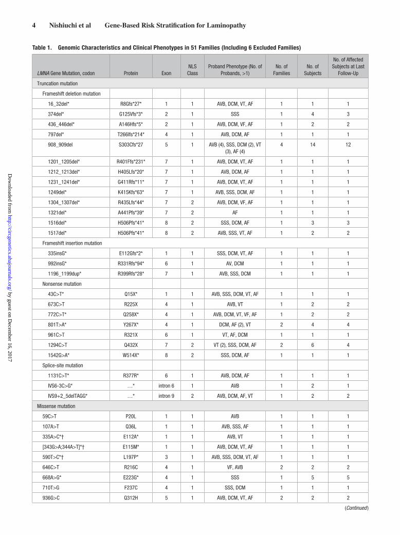
identified, including 9 (20%) nonsense, 3 (6.7%) splice site, 16 (35.6%) frameshift deletion, and 3 (6.7%) frameshift inser-tion mutations (Table 1). Among the identified mutations, 23 (62%) were novel: 12 frameshift deletion, 3 frameshift inser-tion, 4 nonsense, 3 splice site, and 1 missense mutation (novel mutations are indicated by boldface in Table 1). All identi-fied missense variants were screened using the databases of Human Genetic Variation Database, Exome Aggregation Consortium, and ClinVar (Table II in the Data Supplement). We found no available data in Exome Aggregation Consor-tium, whereas 4 variants were listed on ClinVar, of which 2 were considered pathogenic. Thirty-seven mutations identi-fied in 45 probands, with 5 variants excluded, were located in the topology of the LMNA protein (Figure 1). Among these 37 mutations, 30 (81.1%) were located upstream of the spanning residue (NLS class 1), and the remaining 7 (18.9%) mutations were located downstream (NLS class 2). In 25 pro-bands, the deletion, frameshift, or occurrence of stop codon was predicted to cause disturbance in the NLS residue of the mutant allele.
All 26 identified truncation mutations in LMNA from the 31 families were considered to be pathogenic because of the premature termination codon generated by frameshift, aber-rant splice site, or nonsense mutations. As for the 11 missense mutations in the 14 families, the pathogenicity of 5 had been confirmed in several previous studies22–25 and using Single Nucleotide Polymorphism database. Another novel mis-sense mutation was deemed pathogenic after cascade screen-ing and using in silico prediction tools (Table II in the Data Supplement).
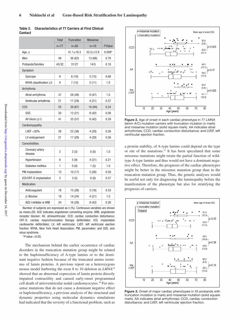
Manifestation of Cardiac PhenotypesMissense Versus Truncation MutationsThe age of onset in cardiac disorders revealed that patients in the CCD and low LVEF developed at significantly younger age in truncation mutation group compared with the missense mutation group (Figure 2). To exclude the bias of the number of family members, we further compared the age of onset only in the probands between truncation and missense mutations. As shown in Figure 3, a significant difference was only observed in the CCD because of a small number of patients, especially in the missense mutation, but outcomes show the same tendency.
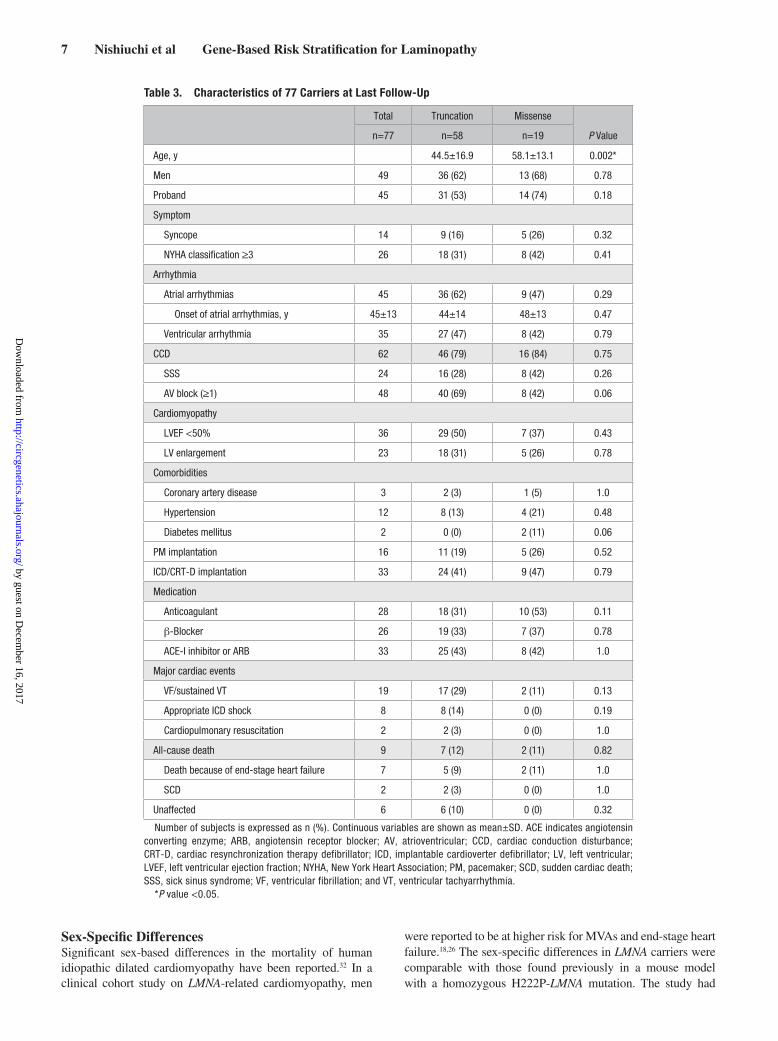
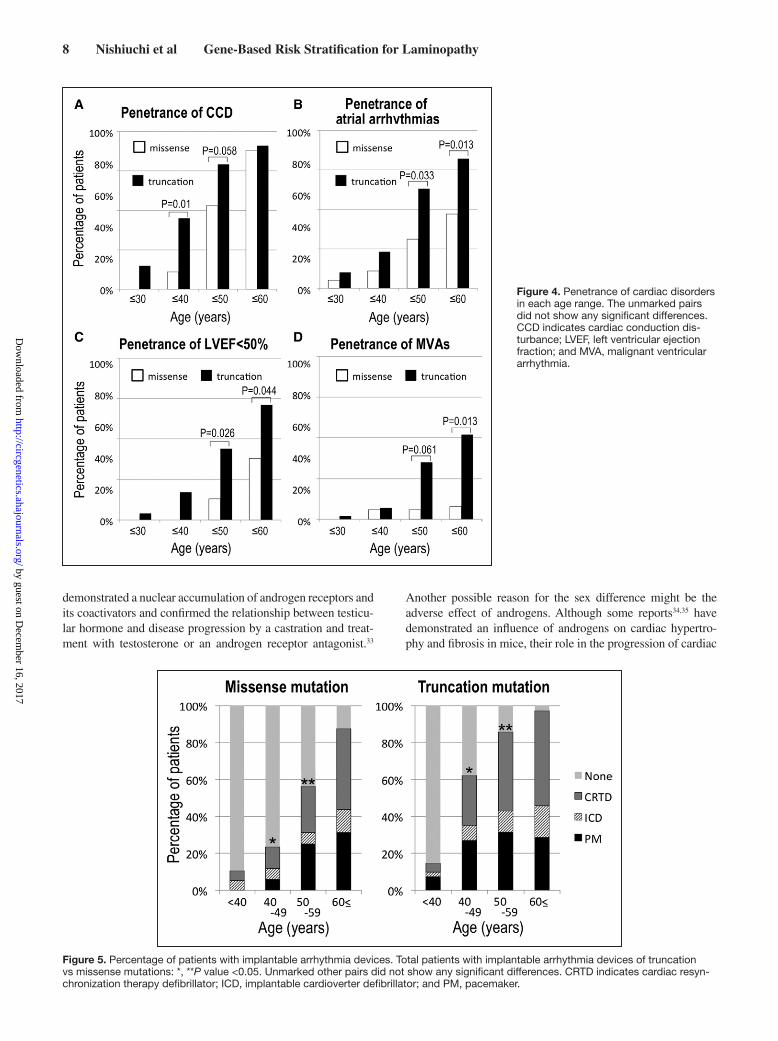
When the prevalence of cardiac disorders at last follow-up (median follow-up period, 49.0 months; 11.1–95.9) was analyzed, we found no significant difference in the baseline characteristics, except for mean age, of the truncation and mis-sense mutation groups (Table 3). However, regarding the age-dependent penetrance of cardiac disorders, the prevalence was higher at age <40 (CCD), <50 (CCD, atrial arrhythmias, lower LVEF, and MVA), and <60 (atrial arrhythmias, lower LVEF, and MVAs) in the truncation mutation carriers compared with the missense mutation carriers (Figure 4). When only probands were considered, prevalence of low LVEF was significantly higher in the truncation mutation group than in the missense mutation group (Tables III and IV in the Data Supplement).
At the end of the follow-up period, the prevalence of phe-notypically affected carriers was 71 of the 77 carriers (92%) with a mean age of 48±17 years. A total of 36 (47%) patients were affected by low LVEF, and 20 subjects (26%) had expe-rienced at least 1 episode of MVAs. Nine subjects (12%) died because of end-stage heart failure with a mean age of 58
Figure 1. Structure of LMNA (lamin A/C) (top) and lamin A (bottom) and C (middle) protein. LMNA consists of 12 exons. The lamin A and C proteins comprise the N-terminal globular head domain, α-helical rod domain (coil 1a, 1b, and 2 were separated by linkers: black boxes), and C-terminal globular tail domain, which included the nuclear localization signal (NLS) sequence that spans residues 416 to 423 (diagonal stripes). The noncoding regions are indicated by the black dotted patterns. The locations of 37 mutations identified in the sub-jects and the 5 excluded variants are shown in the topology of the lamin A and C proteins. †Mutations identified in 2 probands, ‡muta-tions identified in 4 probands, §variants excluded because of unknown significance.
by guest on Decem
ber 16, 2017http://circgenetics.ahajournals.org/
Dow
nloaded from
4 Nishiuchi et al Gene-Based Risk Stratification for Laminopathy
Table 1. Genomic Characteristics and Clinical Phenotypes in 51 Families (Including 6 Excluded Families)
LMNA Gene Mutation, codon Protein ExonNLS
ClassProband Phenotype (No. of
Probands, >1)No. of
FamiliesNo. of
Subjects
No. of Affected Subjects at Last
Follow-Up
Truncation mutation
Frameshift deletion mutation
16_32del* R8Gfs*27* 1 1 AVB, DCM, VT, AF 1 1 1
374del* G125Vfs*3* 2 1 SSS 1 4 3
436_446del* A146Hfs*5* 2 1 AVB, DCM, VF, AF 1 2 2
797del* T266Ifs*214* 4 1 AVB, DCM, AF 1 1 1
908_909del S303Cfs*27 5 1 AVB (4), SSS, DCM (2), VT (3), AF (4)
4 14 12
1201_1205del* R401Ffs*231* 7 1 AVB, DCM, VT, AF 1 1 1
1212_1213del* H405Lfs*20* 7 1 AVB, DCM, AF 1 1 1
1231_1241del* G411Rfs*11* 7 1 AVB, DCM, VT, AF 1 1 1
1249del* K415Kfs*63* 7 1 AVB, SSS, DCM, AF 1 1 1
1304_1307del* R435Lfs*44* 7 2 AVB, DCM, VF, AF 1 1 1
1321del* A441Pfs*39* 7 2 AF 1 1 1
1516del* H506Pfs*41* 8 2 SSS, DCM, AF 1 3 3
1517del* H506Pfs*41* 8 2 AVB, SSS, VT, AF 1 2 2
Frameshift insertion mutation
335insG* E112Gfs*2* 1 1 SSS, DCM, VT, AF 1 1 1
992insG* R331Rfs*94* 6 1 AV, DCM 1 1 1
1196_1199dup* R399Rfs*28* 7 1 AVB, SSS, DCM 1 1 1
Nonsense mutation
43C>T* Q15X* 1 1 AVB, SSS, DCM, VT, AF 1 1 1
673C>T R225X 4 1 AVB, VT 1 2 2
772C>T* Q258X* 4 1 AVB, DCM, VT, VF, AF 1 2 2
801T>A* Y267X* 4 1 DCM, AF (2), VT 2 4 4
961C>T R321X 6 1 VT, AF, DCM 1 1 1
1294C>T Q432X 7 2 VT (2), SSS, DCM, AF 2 6 4
1542G>A* W514X* 8 2 SSS, DCM, AF 1 1 1
Splice-site mutation
1131C>T* R377R* 6 1 AVB, DCM, AF 1 1 1
IVS6-3C>G* …* intron 6 1 AVB 1 2 1
IVS9+2_5delTAGG* …* intron 9 2 AVB, DCM, AF, VT 1 2 2
Missense mutation
59C>T P20L 1 1 AVB 1 1 1
107A>T Q36L 1 1 AVB, SSS, AF 1 1 1
335A>C*† E112A* 1 1 AVB, VT 1 1 1
[343G>A;344A>T]*† E115M* 1 1 AVB, DCM, VT, AF 1 1 1
590T>C*† L197P* 3 1 AVB, SSS, DCM, VT, AF 1 1 1
646C>T R216C 4 1 VF, AVB 2 2 2
668A>G* E223G* 4 1 SSS 1 5 5
710T>G F237C 4 1 SSS, DCM 1 1 1
936G>C Q312H 5 1 AVB, DCM, VT, AF 2 2 2
(Continued )
by guest on Decem
ber 16, 2017http://circgenetics.ahajournals.org/
Dow
nloaded from

5 Nishiuchi et al Gene-Based Risk Stratification for Laminopathy
years, and 2 men carrying the truncation mutations died sud-denly. The 6 phenotypically unaffected mutation carriers were relatively younger than the affected relatives (17±13 versus 34±18 years old; P=0.002). In addition, we found no correla-tion between the type of mutations and the manifestation of cardiac phenotypes in relatives.
Device Therapy in LMNA-Related CardiomyopathyDuring the follow-up period, additional 7 subjects developed CCD, 7 showed a progression in CCD, 12 received a pacemaker, 9 underwent an ICD implantation, and 24 underwent cardiac resynchronization therapy defibrillator implantation (including upgraded cases) in all carriers. Among the 28 patients initially implanted with pacemaker, 25 of them had not been genetically diagnosed as carriers of LMNA mutations. After the genetic diagnosis, 12 of these 28 were upgraded to defibrillator dur-ing the follow-up period. The remaining 16 patients were alive without any MVA event, but 1 patient died because of end-stage heart failure. The percentage of patients with implantable devices increased substantially with age, and the proportion of cardiac resynchronization therapy defibrillator–implanted patients increased in the older population (Figure 5).
Probands Versus RelativesThe age of onset in cardiac disorders was comparable between probands and relatives (Figure I in the Data Supplement). The prevalence of cardiac disorders also did not differ between them at the last follow-up.
NLS-Disturbed Mutations Versus NLS-Conserved MutationsRegarding the site of mutations, the majority of subjects with cardiomyopathy and CCD had mutations upstream of the NLS, spanning residues 416 to 423. This region was reported to be essential for the structure and the translocational regula-tion of the nucleus.19 In this study, the onset of cardiac disor-ders was not associated with disturbance of the NLS residue (Figure II in the Data Supplement).
Sex DifferenceMale sex was reported to be at higher risk for MVAs and end-stage heart failure in laminopathy.18,26 We also assessed the impact of sex differences on clinical outcome (Figure III in the
Data Supplement); however, the onset of cardiac disorders was comparable between male and female carriers. Therefore, sex difference did not affect the clinical outcomes in this cohort.
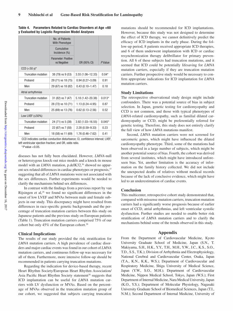
Risk StratificationWe performed multivariable analysis using logistic regres-sion model to evaluate which clinical character, such as type of mutations, sex, and probands, is actually associated with occurrence of cardiac disorders. As shown in Figure 4, the penetrance of CCD at age <60 years was ≈100% both in the truncation and missense mutations. Therefore, only for CCD, we evaluated those with age <50 years. As a result of mul-tivariable analysis, the truncation mutation carriers were at higher risk for the occurrence of CCD under 50-year-old and that of atrial arrhythmias and low LVEF under 60-year-old compared with the missense carriers (Table 4). The event rate of MVAs in missense mutations was small during the follow-up period in this study, and risk evaluation for MVAs was not fully convinced by results of this study. On the contrary, pro-band and male sex did not reach statistical significance.
DiscussionOur multicenter cohort study showed the age-dependent and high prevalence of cardiac manifestations in LMNA mutation carriers. To the best of our knowledge, this is the first study on LMNA-related cardiomyopathy in an Asian country to provide extensive information on the progressive course of cardiac phenotypes. The high penetrance in LMNA mutation carriers found in this study is similar to that found in other studies on caucasians.13,27,28 Further-more, truncation mutations carriers showed a significantly ear-lier manifestation of the cardiac phenotypes of laminopathy than missense mutation carriers, suggesting that genetic analysis may play an important role in both the diagnosis and risk strati-fication in probands with LMNA mutations and their relatives.
Genotype–Phenotype CorrelationsPrevious reports have suggested that the type or site of LMNA mutations might be related to the occurrence and prognosis of the cardiac phenotypes.13,16,18,19 According to results of this study, the truncation mutations were associated with the onset of CCD, atrial arrhythmia, and low LVEF (Figure 2; Table 4).
1003C>T R335W 6 1 SSS, DCM 1 2 2
1058A>G Q353R 6 1 AF, VT 2 2 2
1069G>C D357H 6 1 AVB, VT, AF 1 1 1
1112T>G M371R 6 1 AVB, AF 1 1 1
1129C>T R377C 6 1 AVB, AF, DCM 1 1 1
1486A>T*† T496S* 8 2 VT 1 1 1
1729G>A*† A577T* 11 2 VT, VF, AMI 2 3 2
NLS class: classification described in Methods. AF indicates atrial fibrillation; AMI, acute myocardial infarction; AVB, atrioventricular block; DCM, dilated cardiomyopathy; LMNA, lamin A/C gene; NLS, nuclear localization signal; SSS, sick sinus syndrome; VF, ventricular fibrillation; and VT, ventricular tachycardia.
*Novel variants.†Variants excluded because of unknown significance.
Table 1. Continued
LMNA Gene Mutation C p. ExonNLS
ClassProband Phenotype (No. of
Probands, >1)No. of
FamiliesNo. of
Subjects
No. of Affected Subjects at Last
Follow-Up
by guest on Decem
ber 16, 2017http://circgenetics.ahajournals.org/
Dow
nloaded from
6 Nishiuchi et al Gene-Based Risk Stratification for Laminopathy
The mechanism behind the earlier occurrence of cardiac disorders in the truncation mutation group might be related to the haploinsufficiency of A-type lamins or to the domi-nant negative fashion because of the truncated amino termi-nus of lamin proteins. A previous report on a heterozygous mouse model harboring the exon 8 to 10 deletion in LMNA29 showed that an abnormal expression of lamin protein directly impaired contractility and caused early-onset programmed cell death of atrioventricular nodal cardiomyocytes.30 For mis-sense mutations that do not cause a dominant negative effect or haploinsufficiency, a previous analysis of the structural and dynamic properties using molecular dynamics simulations had indicated that the severity of a functional problem, such as
a protein stability, of A-type lamins could depend on the type or site of the mutations.31 It has been speculated that some missense mutations might retain the partial function of wild-type A-type lamins and thus would not have a dominant nega-tive effect. Therefore, the prognosis of the cardiac phenotypes might be better in the missense mutation group than in the truncation mutation group. Thus, the genetic analyses would be useful not only for diagnosing the laminopathy before the manifestation of the phenotype but also for stratifying the prognosis of carriers.
Table 2. Characteristics of 77 Carriers at First Clinical Contact
Total Truncation Missense
P Valuen=77 n=58 n=19
Age, y 41.1±16.5 52.2±12.8 0.009*
Men 49 36 (62) 13 (68) 0.78
Probands/families 45/32 31/27 14/5 0.18
Symptom
Syncope 9 6 (10) 3 (15) 0.68
NYHA classification ≥3 9 7 (12) 2 (11) 1.0
Arrhythmia
Atrial arrhythmia 37 28 (48) 9 (47) 1.0
Ventricular arrhythmia 21 17 (29) 4 (21) 0.57
CCD 55 39 (67) 16 (84) 0.24
SSS 20 12 (21) 8 (42) 0.08
AV block (≥1) 41 33 (57) 8 (42) 0.29
Cardiomyopathy
LVEF <50% 26 22 (38) 4 (20) 0.26
LV enlargement 21 17 (29) 4 (20) 0.56
Comorbidities
Coronary artery disease
2 2 (3) 0 (0) 1.0
Hypertension 9 5 (9) 4 (21) 0.21
Diabetes mellitus 1 0 (0) 1 (5) 1.0
PM implantation 15 10 (17) 5 (26) 0.50
ICD/CRT-D implantation 3 3 (5) 0 (0) 0.57
Medication
Anticoagulant 18 15 (26) 3 (16) 0.53
β-Blocker 18 14 (24) 4 (21) 1.0
ACE-I inhibitor or ARB 24 16 (28) 8 (42) 0.26
Number of subjects are expressed as n (%). Continuous variables are shown as mean±SD. ACE indicates angiotensin converting enzyme; ARB, angiotensin receptor blocker; AV, atrioventricular; CCD, cardiac conduction disturbance; CRT-D, cardiac resynchronization therapy defibrillator; ICD, implantable cardioverter defibrillator; LV, left ventricular; LVEF, left ventricular ejection fraction; NYHA, New York Heart Association; PM, pacemaker; and SSS, sick sinus syndrome.
*P value <0.05.
Figure 2. Age of onset in each cardiac phenotype in 77 LMNA (lamin A/C) mutation carriers with truncation mutation (x mark) and missense mutation (solid square mark). AA indicates atrial arrhythmias; CCD, cardiac conduction disturbance; and LVEF, left ventricular ejection fraction.
Figure 3. Onset of major cardiac phenotypes in 45 probands with truncation mutation (x mark) and missense mutation (solid square mark). AA indicates atrial arrhythmias; CCD, cardiac conduction disturbance; and LVEF, left ventricular ejection fraction.
by guest on Decem
ber 16, 2017http://circgenetics.ahajournals.org/
Dow
nloaded from
7 Nishiuchi et al Gene-Based Risk Stratification for Laminopathy
Sex-Specific DifferencesSignificant sex-based differences in the mortality of human idiopathic dilated cardiomyopathy have been reported.32 In a clinical cohort study on LMNA-related cardiomyopathy, men
were reported to be at higher risk for MVAs and end-stage heart failure.18,26 The sex-specific differences in LMNA carriers were comparable with those found previously in a mouse model with a homozygous H222P-LMNA mutation. The study had
Table 3. Characteristics of 77 Carriers at Last Follow-Up
Total Truncation Missense
P Valuen=77 n=58 n=19
Age, y 44.5±16.9 58.1±13.1 0.002*
Men 49 36 (62) 13 (68) 0.78
Proband 45 31 (53) 14 (74) 0.18
Symptom
Syncope 14 9 (16) 5 (26) 0.32
NYHA classification ≥3 26 18 (31) 8 (42) 0.41
Arrhythmia
Atrial arrhythmias 45 36 (62) 9 (47) 0.29
Onset of atrial arrhythmias, y 45±13 44±14 48±13 0.47
Ventricular arrhythmia 35 27 (47) 8 (42) 0.79
CCD 62 46 (79) 16 (84) 0.75
SSS 24 16 (28) 8 (42) 0.26
AV block (≥1) 48 40 (69) 8 (42) 0.06
Cardiomyopathy
LVEF <50% 36 29 (50) 7 (37) 0.43
LV enlargement 23 18 (31) 5 (26) 0.78
Comorbidities
Coronary artery disease 3 2 (3) 1 (5) 1.0
Hypertension 12 8 (13) 4 (21) 0.48
Diabetes mellitus 2 0 (0) 2 (11) 0.06
PM implantation 16 11 (19) 5 (26) 0.52
ICD/CRT-D implantation 33 24 (41) 9 (47) 0.79
Medication
Anticoagulant 28 18 (31) 10 (53) 0.11
β-Blocker 26 19 (33) 7 (37) 0.78
ACE-I inhibitor or ARB 33 25 (43) 8 (42) 1.0
Major cardiac events
VF/sustained VT 19 17 (29) 2 (11) 0.13
Appropriate ICD shock 8 8 (14) 0 (0) 0.19
Cardiopulmonary resuscitation 2 2 (3) 0 (0) 1.0
All-cause death 9 7 (12) 2 (11) 0.82
Death because of end-stage heart failure 7 5 (9) 2 (11) 1.0
SCD 2 2 (3) 0 (0) 1.0
Unaffected 6 6 (10) 0 (0) 0.32
Number of subjects is expressed as n (%). Continuous variables are shown as mean±SD. ACE indicates angiotensin converting enzyme; ARB, angiotensin receptor blocker; AV, atrioventricular; CCD, cardiac conduction disturbance; CRT-D, cardiac resynchronization therapy defibrillator; ICD, implantable cardioverter defibrillator; LV, left ventricular; LVEF, left ventricular ejection fraction; NYHA, New York Heart Association; PM, pacemaker; SCD, sudden cardiac death; SSS, sick sinus syndrome; VF, ventricular fibrillation; and VT, ventricular tachyarrhythmia.
*P value <0.05.
by guest on Decem
ber 16, 2017http://circgenetics.ahajournals.org/
Dow
nloaded from
8 Nishiuchi et al Gene-Based Risk Stratification for Laminopathy
demonstrated a nuclear accumulation of androgen receptors and its coactivators and confirmed the relationship between testicu-lar hormone and disease progression by a castration and treat-ment with testosterone or an androgen receptor antagonist.33
Another possible reason for the sex difference might be the adverse effect of androgens. Although some reports34,35 have demonstrated an influence of androgens on cardiac hypertro-phy and fibrosis in mice, their role in the progression of cardiac
Figure 4. Penetrance of cardiac disorders in each age range. The unmarked pairs did not show any significant differences. CCD indicates cardiac conduction dis-turbance; LVEF, left ventricular ejection fraction; and MVA, malignant ventricular arrhythmia.
Figure 5. Percentage of patients with implantable arrhythmia devices. Total patients with implantable arrhythmia devices of truncation vs missense mutations: *, **P value <0.05. Unmarked other pairs did not show any significant differences. CRTD indicates cardiac resyn-chronization therapy defibrillator; ICD, implantable cardioverter defibrillator; and PM, pacemaker.
by guest on Decem
ber 16, 2017http://circgenetics.ahajournals.org/
Dow
nloaded from
9 Nishiuchi et al Gene-Based Risk Stratification for Laminopathy
diseases has not fully been elucidated. However, LMNA-null or heterozygous knock-out mice models and a knock-in mouse model with an LMNA mutation, p.delK32,36 showed no appar-ent sex-related differences in cardiac phenotypes or prognosis,37 suggesting that all of LMNA mutations were not associated with the sex differences. Further experiments would be needed to clarify the mechanisms behind sex differences.
In contrast with the findings from a previous report by van Rijsingen et al,26 we found no significant differences in the onset of low LVEF and MVAs between male and female sub-jects in our study. This discrepancy might have resulted from differences in race-specific genetic backgrounds and the per-centage of truncation mutation carriers between this study on Japanese patients and the previous study on European patients (Table 1). Truncation mutation carriers comprised 75% of our cohort but only 45% of the European cohort.26
Clinical ImplicationsThe results of our study provided the risk stratification for LMNA mutation carriers. A high prevalence of cardiac disor-ders and major cardiac events was found in our cohort of LMNA mutation carriers, and continuous follow-up was necessary for all of them. Furthermore, more intensive follow-up should be recommended in patients carrying truncation mutations.
Regarding the indication for device-based therapy, recent Heart Rhythm Society/European Heart Rhythm Association/Asia Pacific Heart Rhythm Society statement38 suggests that ICD implantation can be useful for LMNA mutation car-riers with LV dysfunction or MVAs. Based on the percent-age of MVAs observed in the truncation mutation group of our cohort, we suggested that subjects carrying truncation
mutations should be recommended for ICD implantations. However, because this study was not designed to determine the effect of ICD therapy, we cannot definitively predict the efficacy of ICD implants in the early phase. During the fol-low-up period, 8 patients received appropriate ICD therapies, and 6 of them underwent implantation with ICD or cardiac resynchronization therapy defibrillator for primary preven-tion. All 6 of these subjects had truncation mutations, and it seemed that ICD could be potentially lifesaving for LMNA mutation carriers, especially if they are truncation mutation carriers. Further prospective study would be necessary to con-firm appropriate indications for ICD implantation for LMNA mutation carriers.
Study LimitationsThe retrospective observational study design might include confounders. There was a potential source of bias in subject selection. In Japan, genetic testing for cardiomyopathy and CCD is not common, and those with typical phenotypes of LMNA-related cardiomyopathy, such as familial dilated car-diomyopathy or CCD, might be preferentially referred for genetic testing. Therefore, this study does not entirely clarify the full view of how LMNA mutations manifest.
Second, LMNA mutation carriers were not screened for sarcomeric genes, which might have influenced the dilated cardiomyopathy phenotype. Third, some of the mutations had been observed in a large number of subjects, which might be another potential source of bias. Fourth, the cohort was derived from several institutes, which might have introduced unfore-seen bias. Yet, another limitation is the accuracy of infor-mation on the family history collected. We did not include the unexpected deaths of relatives without medical records because of the lack of conclusive evidence, which might have led to an underestimation of cardiac events.
ConclusionThis multicenter, retrospective cohort study demonstrated that, compared with missense mutation carriers, truncation mutation carriers had a significantly worse prognosis because of earlier onset of CCD, atrial arrhythmias, and left ventricular systolic dysfunction. Further studies are needed to enable better risk stratification of LMNA mutation carriers and to clarify the mechanisms behind some of the trends observed in this study.
AppendixFrom the Department of Cardiovascular Medicine, Kyoto University Graduate School of Medicine, Japan (S.N., T. Makiyama, S.H., H.K., Y.Y., T.H., M.H., Y.W., J.C., K.S., S.O., T.D., S.S., T.K.); Division of Arrhythmia and Electrophysiology, National Cerebral and Cardiovascular Center, Osaka, Japan (T.A., K.N., K.K., W.S.); Department of Cardiovascular and Respiratory Medicine, Shiga University of Medical Science, Japan (Y.W., S.O., M.H.); Department of Cardiovascular Medicine, Nippon Medical School, Tokyo, Japan (W.S.); First Department of Internal Medicine, Nara Medical University, Japan (K.O., Y.S.); Department of Molecular Physiology, Nagasaki University Graduate School of Biomedical Sciences, Japan (T.I., N.M.); Second Department of Internal Medicine, University of
Table 4. Parameters Related to Cardiac Disorders at Age <60 y Evaluated by Logistic Regression Model Analyses
No. of Patients With Phenotype
OR (95% CI) P Value
Cumulative Incidence (%)
Parameter: Positive vs Negative
CCD (<50 y)*
Truncation mutation 38 (79) vs 9 (53) 3.55 (1.06–12.33) 0.04*
Proband 29 (71) vs 18 (75) 0.94 (0.27–3.09) 0.91
Men 29 (67) vs 18 (82) 0.43 (0.10–1.47) 0.18
Atrial arrhythmias
Truncation mutation 31 (82) vs 7 (47) 5.18 (1.42–20.38) 0.013*
Proband 28 (72) vs 10 (71) 1.13 (0.24–4.95) 0.87
Men 25 (69) vs 13 (76) 0.62 (0.13–2.56) 0.52
Low LVEF (≤50%)
Truncation mutation 24 (71) vs 5 (38) 3.92 (1.03–16.55) 0.045*
Proband 22 (67) vs 7 (50) 2.28 (0.59–9.33) 0.23
Men 18 (58) vs 11 (69) 1.76 (0.46–7.62) 0.41
CCD indicates cardiac conduction disturbance; CI, confidence interval; LVEF, left ventricular ejection fraction; and OR, odds ratio.
*P value <0.05.
by guest on Decem
ber 16, 2017http://circgenetics.ahajournals.org/
Dow
nloaded from
10 Nishiuchi et al Gene-Based Risk Stratification for Laminopathy
Occupational and Environmental Health, Fukuoka, Japan (Y.O.); Cardiovascular Division, University of Tsukuba, Ibaraki, Japan (N.M., A.N., K.A.); Department of Cardiovascular Biology and Medicine, Niigata University Graduate School of Medical and Dental Sciences, Japan (N.Y., H.W., T. Minamino); and Division of Cardiology, Department of Medicine, Nihon University School of Medicine, Tokyo, Japan (I.W., K.O.).
AcknowledgmentsWe would like to thank the probands and relatives for their partici-pation in this study. We are also grateful for the outstanding effort of all collaborators in this clinical research (Appendix in the Data Supplement), Koko Asakura for the statistical direction, and also thank Aya Umehara for the technical assistance.
Sources of FundingThis work was supported by JSPS KAKENHI (25461054 to Dr Makiyama), a Grant-in-Aid for Scientific Research from the Ministry of Education, Culture, Sports, Science and Technology (C; 16K09499 to Dr Makiyama, 15K09150 to Dr Aiba), Health Labour Sciences Research Grant from the Ministry of Health, Labour, and Welfare of Japan (H24-033, H26-040, and H27-032 to Drs Aiba, Horie, and Shimizu), and a re-search grant from Japan Agency for Medical Research and Development (AMED) (15km0305015h0101 to Dr Makita), (16ek0210073h0001 to Dr Aiba), and Hoansha Foundation (Dr Makiyama).
DisclosuresNone.
References 1. Rankin J, Ellard S. The laminopathies: a clinical review. Clin Genet.
2006;70:261–274. doi: 10.1111/j.1399-0004.2006.00677.x. 2. Bonne G, Di Barletta MR, Varnous S, Bécane HM, Hammouda EH, Mer-
lini L, et al. Mutations in the gene encoding lamin A/C cause autosomal dominant Emery-Dreifuss muscular dystrophy. Nat Genet. 1999;21:285–288. doi: 10.1038/6799.
3. Raffaele Di Barletta M, Ricci E, Galluzzi G, Tonali P, Mora M, Morandi L, et al. Different mutations in the LMNA gene cause autosomal dominant and autosomal recessive Emery-Dreifuss muscular dystrophy. Am J Hum Genet. 2000;66:1407–1412.
4. Muchir A, Bonne G, van der Kooi AJ, van Meegen M, Baas F, Bol-huis PA, et al. Identification of mutations in the gene encoding lamins A/C in autosomal dominant limb girdle muscular dystrophy with atrio-ventricular conduction disturbances (LGMD1B). Hum Mol Genet. 2000;9:1453–1459.
5. De Sandre-Giovannoli A, Chaouch M, Kozlov S, Vallat JM, Tazir M, Kassouri N, et al. Homozygous defects in LMNA, encoding lamin A/C nuclear-envelope proteins, cause autosomal recessive axonal neuropathy in human (Charcot-Marie-Tooth disorder type 2) and mouse. Am J Hum Genet. 2002;70:726–736. doi: 10.1086/339274.
6. Cao H, Hegele RA. Nuclear lamin A/C R482Q mutation in canadian kin-dreds with Dunnigan-type familial partial lipodystrophy. Hum Mol Genet. 2000;9:109–112.
7. Shackleton S, Lloyd DJ, Jackson SN, Evans R, Niermeijer MF, Singh BM, et al. LMNA, encoding lamin A/C, is mutated in partial lipodystrophy. Nat Genet. 2000;24:153–156. doi: 10.1038/72807.
8. Eriksson M, Brown WT, Gordon LB, Glynn MW, Singer J, Scott L, et al. Recurrent de novo point mutations in lamin A cause Hutchinson-Gilford progeria syndrome. Nature. 2003;423:293–298. doi: 10.1038/nature01629.
9. De Sandre-Giovannoli A, Bernard R, Cau P, Navarro C, Amiel J, Boccac-cio I, et al. Lamin a truncation in Hutchinson-Gilford progeria. Science. 2003;300:2055. doi: 10.1126/science.1084125.
10. Lin F, Worman HJ. Structural organization of the human gene encoding nu-clear lamin A and nuclear lamin C. J Biol Chem. 1993;268:16321–16326.
11. Taylor MR, Fain PR, Sinagra G, Robinson ML, Robertson AD, Carniel E, et al; Familial Dilated Cardiomyopathy Registry Research Group. Natural history of dilated cardiomyopathy due to lamin A/C gene mutations. J Am Coll Cardiol. 2003;41:771–780.
12. van Tintelen JP, Hofstra RM, Katerberg H, Rossenbacker T, Wiesfeld AC, du Marchie Sarvaas GJ, et al; Working Group on Inherited Cardiac Disor-ders, line 27/50, Interuniversity Cardiology Institute of The Netherlands. High yield of LMNA mutations in patients with dilated cardiomyopathy and/or conduction disease referred to cardiogenetics outpatient clinics. Am Heart J. 2007;154:1130–1139. doi: 10.1016/j.ahj.2007.07.038.
13. Pasotti M, Klersy C, Pilotto A, Marziliano N, Rapezzi C, Serio A, et al. Long-term outcome and risk stratification in dilated cardiolaminopathies. J Am Coll Cardiol. 2008;52:1250–1260. doi: 10.1016/j.jacc.2008.06.044.
14. Fatkin D, MacRae C, Sasaki T, Wolff MR, Porcu M, Frenneaux M, et al. Missense mutations in the rod domain of the lamin A/C gene as causes of dilated cardiomyopathy and conduction-system disease. N Engl J Med. 1999;341:1715–1724. doi: 10.1056/NEJM199912023412302.
15. Meune C, Van Berlo JH, Anselme F, Bonne G, Pinto YM, Duboc D. Pri-mary prevention of sudden death in patients with lamin A/C gene muta-tions. N Engl J Med. 2006;354:209–210. doi: 10.1056/NEJMc052632.
16. Kumar S, Baldinger SH, Gandjbakhch E, Maury P, Sellal JM, Androula-kis AF, et al. Long-term arrhythmic and nonarrhythmic outcomes of la-min A/C mutation carriers. J Am Coll Cardiol. 2016;68:2299–2307. doi: 10.1016/j.jacc.2016.08.058.
17. Benedetti S, Menditto I, Degano M, Rodolico C, Merlini L, D’Amico A, et al. Phenotypic clustering of lamin A/C mutations in neuromus-cular patients. Neurology. 2007;69:1285–1292. doi: 10.1212/01.wnl.0000261254.87181.80.
18. van Rijsingen IA, Arbustini E, Elliott PM, Mogensen J, Hermans-van Ast JF, van der Kooi AJ, et al. Risk factors for malignant ventricular arrhyth-mias in lamin a/c mutation carriers a European cohort study. J Am Coll Cardiol. 2012;59:493–500. doi: 10.1016/j.jacc.2011.08.078.
19. Hegele R. LMNA mutation position predicts organ system involvement in laminopathies. Clin Genet. 2005;68:31–34. doi: 10.1111/j.1399- 0004.2005.00447.x.
20. Lang RM, Badano LP, Mor-Avi V, Afilalo J, Armstrong A, Ernande L, et al. Recommendations for cardiac chamber quantification by echocardiog-raphy in adults: an update from the American Society of Echocardiogra-phy and the European Association of Cardiovascular Imaging. J Am Soc Echocardiogr. 2015;28:1–39.e14. doi: 10.1016/j.echo.2014.10.003.
21. Grimm W, Christ M, Bach J, Müller HH, Maisch B. Noninvasive arrhyth-mia risk stratification in idiopathic dilated cardiomyopathy: results of the Marburg Cardiomyopathy study. Circulation. 2003;108:2883–2891. doi: 10.1161/01.CIR.0000100721.52503.85.
22. Ben Yaou R, Bécane HM, Demay L, Laforet P, Hannequin D, Bohu PA, et al. [Autosomal dominant limb-girdle muscular dystrophy associated with conduction defects (LGMD1B): a description of 8 new families with the LMNA gene mutations]. Rev Neurol (Paris). 2005;161:42–54.
23. Stallmeyer B, Koopmann M, Schulze-Bahr E. Identification of novel mutations in LMNA associated with familial forms of dilated cardio-myopathy. Genet Test Mol Biomarkers. 2012;16:543–549. doi: 10.1089/gtmb.2011.0214.
24. Astejada MN, Goto K, Nagano A, Ura S, Noguchi S, Nonaka I, et al. Emerinopathy and laminopathy clinical, pathological and molecular fea-tures of muscular dystrophy with nuclear envelopathy in Japan. Acta Myol. 2007;26:159–164.
25. Fujimori Y, Okimatsu H, Kashiwagi T, Sanda N, Okumura K, Takagi A, et al. Molecular defects associated with antithrombin deficiency and dilated cardiomyopathy in a Japanese patient. Intern Med. 2008;47:925–931.
26. van Rijsingen IA, Nannenberg EA, Arbustini E, Elliott PM, Mogensen J, Hermans-van Ast JF, et al. Gender-specific differences in major cardiac events and mortality in lamin A/C mutation carriers. Eur J Heart Fail. 2013;15:376–384. doi: 10.1093/eurjhf/hfs191.
27. van Berlo JH, de Voogt WG, van der Kooi AJ, van Tintelen JP, Bonne G, Yaou RB, et al. Meta-analysis of clinical characteristics of 299 car-riers of LMNA gene mutations: do lamin A/C mutations portend a high risk of sudden death? J Mol Med (Berl). 2005;83:79–83. doi: 10.1007/s00109-004-0589-1.
28. Kourgiannidis G, Anastasakis A, Lampropoulos K, Iliopoulos T. A pa-tient with ventricular tachycardia due to a novel mutation of the la-min A/C gene: case presentation and mini review. Hellenic J Cardiol. 2013;54:326–330.
29. Sullivan T, Escalante-Alcalde D, Bhatt H, Anver M, Bhat N, Nagashima K, et al. Loss of A-type lamin expression compromises nuclear envelope integrity leading to muscular dystrophy. J Cell Biol. 1999;147:913–920.
30. Wolf CM, Wang L, Alcalai R, Pizard A, Burgon PG, Ahmad F, et al. La-min A/C haploinsufficiency causes dilated cardiomyopathy and apop-tosis-triggered cardiac conduction system disease. J Mol Cell Cardiol. 2008;44:293–303. doi: 10.1016/j.yjmcc.2007.11.008.
by guest on Decem
ber 16, 2017http://circgenetics.ahajournals.org/
Dow
nloaded from
11 Nishiuchi et al Gene-Based Risk Stratification for Laminopathy
31. Gangemi F, Degano M. Disease-associated mutations in the coil 2B domain of human lamin A/C affect structural properties that mediate dimerization and intermediate filament formation. J Struct Biol. 2013;181:17–28. doi: 10.1016/j.jsb.2012.10.016.
32. Towbin JA, Bowles NE. The failing heart. Nature. 2002;415:227–233. doi: 10.1038/415227a.
33. Arimura T, Helbling-Leclerc A, Massart C, Varnous S, Niel F, Lacène E, et al. Mouse model carrying H222P-Lmna mutation develops muscular dys-trophy and dilated cardiomyopathy similar to human striated muscle lami-nopathies. Hum Mol Genet. 2005;14:155–169. doi: 10.1093/hmg/ddi017.
34. Gao XM, Agrotis A, Autelitano DJ, Percy E, Woodcock EA, Jennings GL, et al. Sex hormones and cardiomyopathic phenotype induced by cardiac beta 2-adrenergic receptor overexpression. Endocrinology. 2003;144:4097–4105. doi: 10.1210/en.2002-0214.
35. Li Y, Kishimoto I, Saito Y, Harada M, Kuwahara K, Izumi T, et al. An-drogen contributes to gender-related cardiac hypertrophy and fibrosis
in mice lacking the gene encoding guanylyl cyclase-A. Endocrinology. 2004;145:951–958. doi: 10.1210/en.2003-0816.
36. Bertrand AT, Renou L, Papadopoulos A, Beuvin M, Lacène E, Massart C, et al. DelK32-lamin A/C has abnormal location and induces incomplete tissue maturation and severe metabolic defects leading to premature death. Hum Mol Genet. 2012;21:1037–1048. doi: 10.1093/hmg/ddr534.
37. Nikolova V, Leimena C, McMahon AC, Tan JC, Chandar S, Jogia D, et al. Defects in nuclear structure and function promote dilated cardiomyopa-thy in lamin A/C-deficient mice. J Clin Invest. 2004;113:357–369. doi: 10.1172/JCI19448.
38. Priori SG, Wilde AA, Horie M, Cho Y, Behr ER, Berul C, et al; Docu-ment Reviewers; Heart Rhythm Society; European Heart Rhythm Asso-ciation; Asia Pacific Heart Rhythm Society. Executive summary: HRS/EHRA/APHRS expert consensus statement on the diagnosis and manage-ment of patients with inherited primary arrhythmia syndromes. Europace. 2013;15:1389–1406. doi: 10.1093/europace/eut272.
CLINICAL PERSPECTIVEMutations in LMNA (lamin A/C) gene are associated with several cardiac phenotypes, including cardiac conduction distur-bance, atrial or ventricular tachyarrhythmias, and dilated cardiomyopathy, resulting in heart failure or sudden cardiac death; however, risk stratification for LMNA-related cardiomyopathy has been still controversial. This multicenter cohort study has shown a high prevalence of these cardiac disorders and major cardiac events in the LMNA mutation carriers, and multivari-able analyses revealed that the truncation mutation in LMNA gene was associated with a risk for earlier onset of cardiac conduction disturbance and the occurrence of atrial arrhythmias and low left ventricular ejection fraction, suggesting more intensive follow-up is necessary in patients carrying truncation mutations. On the contrary, even though a missense muta-tion, the LMNA mutation carriers have some risk for the cardiac disorders and significant cardiac mortality. Moreover, this study included only for subjects with typical LMNA-related cardiomyopathy called laminopathy and their relatives but not included forme fruste LMNA carriers. Therefore, this study does not entirely clarify the full view of how LMNA mutations manifest. Regarding the indication for device-based therapy, we would recommend the subjects carrying LMNA truncation mutations to receive implantable cardioverter defibrillators (or cardiac resynchronization therapy defibrillators) because of their high incidence of ventricular tachyarrhythmias observed over 40 years old. However, this study was not designed to determine the effect of implantable cardioverter defibrillator therapy on mortality or quality of daily activity, we cannot completely define the efficacy of prophylactic implantable cardioverter defibrillator implantation in the early age. Further prospective study may disclose appropriate indications for implantable cardioverter defibrillator (or cardiac resynchroniza-tion therapy defibrillator) implantation for LMNA mutation carriers.
by guest on Decem
ber 16, 2017http://circgenetics.ahajournals.org/
Dow
nloaded from
Takeshi KimuraKazutaka Aonuma, Kengo Kusano, Naomasa Makita, Wataru Shimizu, Minoru Horie and
Satoshi Shizuta, Tohru Minamino, Yoshihiko Saito, Yasushi Oginosawa, Akihiko Nogami,Murakoshi, Ichiro Watanabe, Kimie Ohkubo, Hiroshi Watanabe, Seiko Ohno, Takahiro Doi, Jiarong Chen, Kenichi Sasaki, Nobue Yagihara, Taisuke Ishikawa, Kenji Onoue, Nobuyuki
Hirohiko Kohjitani, Yuta Yamamoto, Takeshi Harita, Mamoru Hayano, Yimin Wuriyanghai, Suguru Nishiuchi, Takeru Makiyama, Takeshi Aiba, Kenzaburo Nakajima, Sayako Hirose,
Mutation CarriersLMNAGene-Based Risk Stratification for Cardiac Disorders in
Print ISSN: 1942-325X. Online ISSN: 1942-3268 Copyright © 2017 American Heart Association, Inc. All rights reserved.
Dallas, TX 75231is published by the American Heart Association, 7272 Greenville Avenue,Circulation: Cardiovascular Genetics
doi: 10.1161/CIRCGENETICS.116.0016032017;10:Circ Cardiovasc Genet.
http://circgenetics.ahajournals.org/content/10/6/e001603World Wide Web at:
The online version of this article, along with updated information and services, is located on the
http://circgenetics.ahajournals.org/content/suppl/2017/12/12/CIRCGENETICS.116.001603.DC1Data Supplement (unedited) at:
http://circgenetics.ahajournals.org//subscriptions/
is online at: Circulation: Cardiovascular Genetics Information about subscribing to Subscriptions:
http://www.lww.com/reprints Information about reprints can be found online at: Reprints:
document. Permissions and Rights Question and Answer information about this process is available in the
requested is located, click Request Permissions in the middle column of the Web page under Services. FurtherCenter, not the Editorial Office. Once the online version of the published article for which permission is being
can be obtained via RightsLink, a service of the Copyright ClearanceCirculation: Cardiovascular Geneticsin Requests for permissions to reproduce figures, tables, or portions of articles originally publishedPermissions:
by guest on Decem
ber 16, 2017http://circgenetics.ahajournals.org/
Dow
nloaded from
SUPPLEMENTAL MATERIAL
Methods
Genetic analysis
Genomic deoxyribonucleic acid was extracted from peripheral blood leukocytes using a
DNA isolation kit for Mammalian Blood (Roche Diagnostics, Basel, Switzerland). Standard
polymerase chain reaction (PCR) primers were derived from intronic sequences (Table S1)
to amplify the 12 protein-coding exons of LMNA. Mutational screenings of PCR amplicons
were performed by direct sequencing on an ABI PRISM 3130x Genetic Analyzer (Thermo
Fisher Scientific, Waltham, Massachusetts) by using BigDye Terminator chemistry (v1.1 or
3.1) according to standard protocols. Reference sequences used in this study are as follows:
LMNA gene: NCBI NC_000001; LMNA messenger ribonucleic acid: NCBI NM_170707;
lamin A protein: NCBI NP_733821; LMNC messenger ribonucleic acid: NCBI NM_005572;
lamin C protein: NCBI NP_005563. Mutations were categorized in two ways based on type
and site.
Bioinformatic analysis
Mutations present in the dbSNP build 146 or published in the literature were identified. All
non-matching variants were filtered using a minor allele frequency threshold <0.3% based
on the Human Genetic Variation Database (http://www.genome.med.kyoto-u.ac.jp/SnpDB/),
which included the Japanese population, Exome Aggregation Consortium
(http://exac.broadinstitute.org), and ClinVar (http://www.ncbi.nlm.nih.gov/clinvar/). All
single base substitutions without changes in the coding amino acid were screened by a
splicing site prediction tool (Berkeley Drosophila Genome Project: http://www.fruitfly.org),
and the possibility of aberrant splicing was estimated. The candidate variant was considered
as a pathogenic mutation if it generated a stop codon, a frameshift of the open reading frame,

or an aberrant splice site. The pathogenicity of novel missense variants was screened by in
silico predictions using Polyphen2 (http://genetics.bwh.harvard.edu/pph2/), SIFT
(http://sift.jcvi.org/), and Condel (http://bg.upf.edu/fannsdb/). A novel missense variant was
considered pathogenic if classified as ‘probably damaging’ by Polyphen2, ‘damaging’ by
SIFT, or predicted to be ‘deleterious’ by Condel.
List of Participating Institutes and Investigators for this cohort study:
National Cerebral and Cardiovascular Center: Kenzaburo Nakajima, Koko Asakura, Takeshi
Aiba, Kengo Kusano
Kyoto University Graduate School of Medicine: Takeru Makiyama, Takahiro Doi, Satoshi
Shizuta, Takeshi Kimura
Shiga University of Medical Science: Tetshuhisa Hattori, Seiko Ohno, Minoru Horie
Nagasaki University Graduate School of Biomedical Science: Taisuke Ishikawa, Naomasa
Makita
University of Tsukuba: Nobuyuki Murakoshi, Akihiko Nogami, Kazutaka Aonuma
Nippon Medical School Hospital: Wataru Shimizu
Niigata University Graduate School of Medical and Dental Sciences: Nobue Yagihara,
Hiroshi Watanabe, Tohru Minamino
Yokohama Rosai Hospital: Nobuyuki Murakoshi, Akihiko Nogami
Nara Medical University Hospital: Kenji Onoue, Yoshihiko Saito
University of Occupational and Environmental Health Hospital: Yasushi Oginosawa
Nihon University Itabashi Hospital: Ichiro Watanabe, Kimie Ohkubo
Hokkaido University: Naomasa Makita
Tenri Hospital: Kazuhiro Kaitani, Yoshihisa Nakagawa
Oita University Hospital: Naohiko Takahashi
Chiba University Hospital: Motoi Nishimura

Kitano Hospital: Tetsuya Haruna, Kenichi Sasaki
Japanese Red Cross Hadano Hospital: Yusuke Matsumoto
Ijin-kai Takeda Hospital: Takeru Makiyama
Dokkyo Medical University Koshigaya Hospital: Naofumi Tsukada
Fujimoto-chuo Hospital: Koji Sakata
Iida Municipal Hospital: Yuichi Katagiri
Jichi Medical School: Hiroaki Watanabe
JCHO Yamato Koriyama Hospital: Tetsuhisa Hattori
Saiseikai Kyoto Hospital: Kazuya Ishibashi
Fujita Health University Hospital: Eiichi Watanabe
Showa University Fujigaoka Hospital: Daisuke Wakatsuki, Yukei Higashi
Japanese Red Cross Otsu Hospital: Hirooki Higami, Takashi Konishi (in order of descending
subject prevalence)
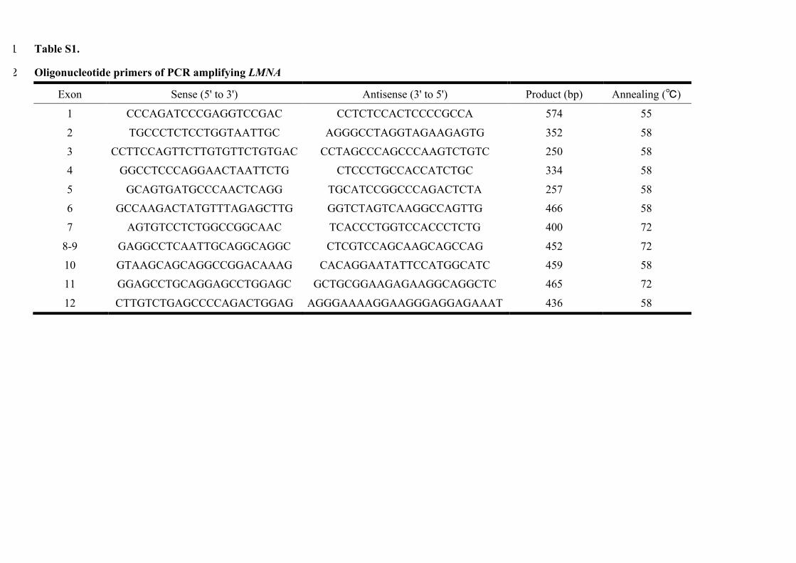
Table S1. 1
Oligonucleotide primers of PCR amplifying LMNA 2
Exon Sense (5' to 3') Antisense (3' to 5') Product (bp) Annealing (�)
1 CCCAGATCCCGAGGTCCGAC CCTCTCCACTCCCCGCCA 574 55
2 TGCCCTCTCCTGGTAATTGC AGGGCCTAGGTAGAAGAGTG 352 58
3 CCTTCCAGTTCTTGTGTTCTGTGAC CCTAGCCCAGCCCAAGTCTGTC 250 58
4 GGCCTCCCAGGAACTAATTCTG CTCCCTGCCACCATCTGC 334 58
5 GCAGTGATGCCCAACTCAGG TGCATCCGGCCCAGACTCTA 257 58
6 GCCAAGACTATGTTTAGAGCTTG GGTCTAGTCAAGGCCAGTTG 466 58
7 AGTGTCCTCTGGCCGGCAAC TCACCCTGGTCCACCCTCTG 400 72
8-9 GAGGCCTCAATTGCAGGCAGGC CTCGTCCAGCAAGCAGCCAG 452 72
10 GTAAGCAGCAGGCCGGACAAAG CACAGGAATATTCCATGGCATC 459 58
11 GGAGCCTGCAGGAGCCTGGAGC GCTGCGGAAGAGAAGGCAGGCTC 465 72
12 CTTGTCTGAGCCCCAGACTGGAG AGGGAAAAGGAAGGGAGGAGAAAT 436 58
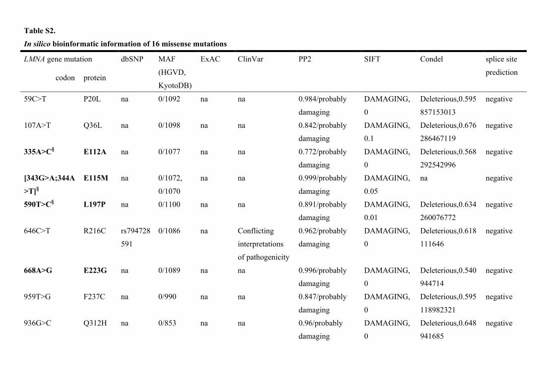
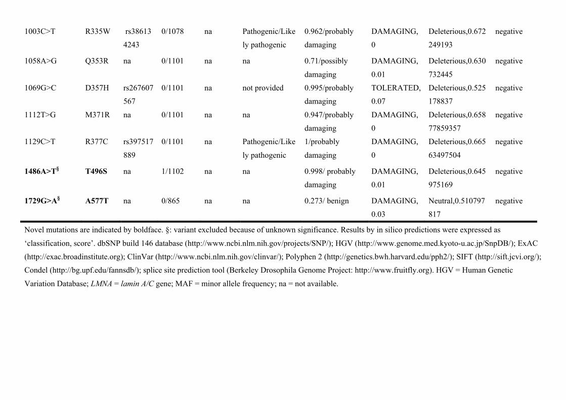
Table S2.
In silico bioinformatic information of 16 missense mutations
LMNA gene mutation dbSNP MAF
(HGVD,
KyotoDB)
ExAC ClinVar PP2 SIFT Condel splice site
prediction codon protein
59C>T P20L na 0/1092 na na 0.984/probably
damaging
DAMAGING,
0
Deleterious,0.595
857153013
negative
107A>T Q36L na 0/1098 na na 0.842/probably
damaging
DAMAGING,
0.1
Deleterious,0.676
286467119
negative
335A>C§ E112A na 0/1077 na na 0.772/probably
damaging
DAMAGING,
0
Deleterious,0.568
292542996
negative
[343G>A;344A
>T]§
E115M na 0/1072,
0/1070
na na 0.999/probably
damaging
DAMAGING,
0.05
na negative
590T>C§ L197P na 0/1100 na na 0.891/probably
damaging
DAMAGING,
0.01
Deleterious,0.634
260076772
negative
646C>T R216C rs794728
591
0/1086 na Conflicting
interpretations
of pathogenicity
0.962/probably
damaging
DAMAGING,
0
Deleterious,0.618
111646
negative
668A>G E223G na 0/1089 na na 0.996/probably
damaging
DAMAGING,
0
Deleterious,0.540
944714
negative
959T>G F237C na 0/990 na na 0.847/probably
damaging
DAMAGING,
0
Deleterious,0.595
118982321
negative
936G>C Q312H na 0/853 na na 0.96/probably
damaging
DAMAGING,
0
Deleterious,0.648
941685
negative
1003C>T R335W rs38613
4243
0/1078 na Pathogenic/Like
ly pathogenic
0.962/probably
damaging
DAMAGING,
0
Deleterious,0.672
249193
negative
1058A>G Q353R na 0/1101 na na 0.71/possibly
damaging
DAMAGING,
0.01
Deleterious,0.630
732445
negative
1069G>C D357H rs267607
567
0/1101 na not provided 0.995/probably
damaging
TOLERATED,
0.07
Deleterious,0.525
178837
negative
1112T>G M371R na 0/1101 na na 0.947/probably
damaging
DAMAGING,
0
Deleterious,0.658
77859357
negative
1129C>T R377C rs397517
889
0/1101 na Pathogenic/Like
ly pathogenic
1/probably
damaging
DAMAGING,
0
Deleterious,0.665
63497504
negative
1486A>T§ T496S na 1/1102 na na 0.998/ probably
damaging
DAMAGING,
0.01
Deleterious,0.645
975169
negative
1729G>A§ A577T na 0/865 na na 0.273/ benign DAMAGING,
0.03
Neutral,0.510797
817
negative
Novel mutations are indicated by boldface. §: variant excluded because of unknown significance. Results by in silico predictions were expressed as
‘classification, score’. dbSNP build 146 database (http://www.ncbi.nlm.nih.gov/projects/SNP/); HGV (http://www.genome.med.kyoto-u.ac.jp/SnpDB/); ExAC
(http://exac.broadinstitute.org); ClinVar (http://www.ncbi.nlm.nih.gov/clinvar/); Polyphen 2 (http://genetics.bwh.harvard.edu/pph2/); SIFT (http://sift.jcvi.org/);
Condel (http://bg.upf.edu/fannsdb/); splice site prediction tool (Berkeley Drosophila Genome Project: http://www.fruitfly.org). HGV = Human Genetic
Variation Database; LMNA = lamin A/C gene; MAF = minor allele frequency; na = not available.
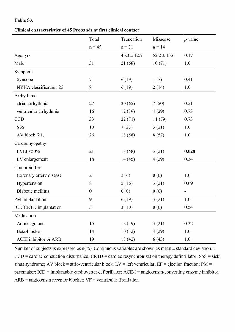
Table S3.
Clinical characteristics of 45 Probands at first clinical contact
Total Truncation Missense p value
n = 45 n = 31 n = 14
Age, yrs 46.3 ± 12.9 52.2 ± 13.6 0.17
Male 31 21 (68) 10 (71) 1.0
Symptom
Syncope 7 6 (19) 1 (7) 0.41
NYHA classification �3 8 6 (19) 2 (14) 1.0
Arrhythmia
atrial arrhythmia 27 20 (65) 7 (50) 0.51
ventricular arrhythmia 16 12 (39) 4 (29) 0.73
CCD 33 22 (71) 11 (79) 0.73
SSS 10 7 (23) 3 (21) 1.0
AV block (�1) 26 18 (58) 8 (57) 1.0
Cardiomyopathy
LVEF<50% 21 18 (58) 3 (21) 0.028
LV enlargement 18 14 (45) 4 (29) 0.34
Comorbidities
Coronary artery disease 2 2 (6) 0 (0) 1.0
Hypertension 8 5 (16) 3 (21) 0.69
Diabetic mellitus 0 0 (0) 0 (0) -
PM implantation 9 6 (19) 3 (21) 1.0
ICD/CRTD implantation 3 3 (10) 0 (0) 0.54
Medication
Anticoagulant 15 12 (39) 3 (21) 0.32
Beta-blocker 14 10 (32) 4 (29) 1.0
ACEI inhibitor or ARB 19 13 (42) 6 (43) 1.0
Number of subjects is expressed as n(%). Continuous variables are shown as mean ± standard deviation. ;
CCD = cardiac conduction disturbance; CRTD = cardiac resynchronization therapy defibrillator; SSS = sick
sinus syndrome; AV block = atrio-ventricular block; LV = left ventricular; EF = ejection fraction; PM =
pacemaker; ICD = implantable cardioverter defibrillator; ACE-I = angiotensin-converting enzyme inhibitor;
ARB = angiotensin receptor blocker; VF = ventricular fibrillation
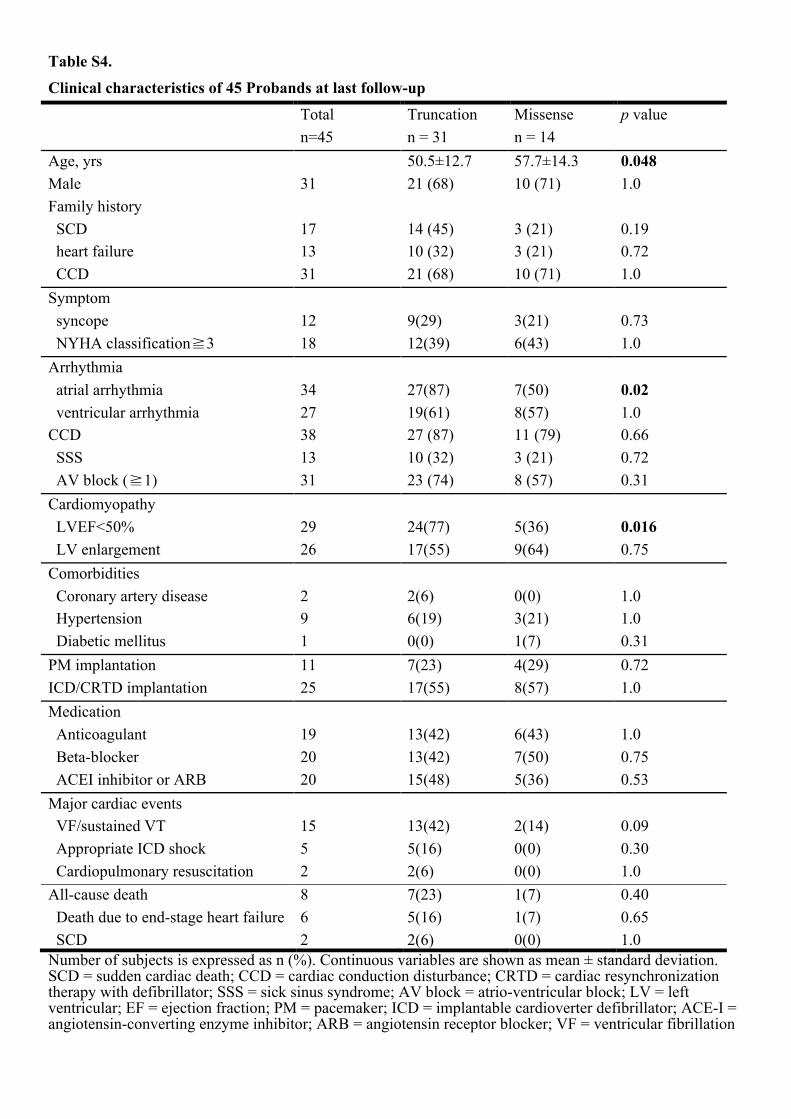
Table S4.
Clinical characteristics of 45 Probands at last follow-up
Total Truncation Missense p value n=45 n = 31 n = 14
Age, yrs 50.5±12.7 57.7±14.3 0.048 Male 31 21 (68) 10 (71) 1.0 Family history SCD 17 14 (45) 3 (21) 0.19 heart failure 13 10 (32) 3 (21) 0.72 CCD 31 21 (68) 10 (71) 1.0 Symptom syncope 12 9(29) 3(21) 0.73 NYHA classification�3 18 12(39) 6(43) 1.0 Arrhythmia atrial arrhythmia 34 27(87) 7(50) 0.02 ventricular arrhythmia 27 19(61) 8(57) 1.0 CCD 38 27 (87) 11 (79) 0.66 SSS 13 10 (32) 3 (21) 0.72 AV block (�1) 31 23 (74) 8 (57) 0.31 Cardiomyopathy LVEF<50% 29 24(77) 5(36) 0.016 LV enlargement 26 17(55) 9(64) 0.75 Comorbidities Coronary artery disease 2 2(6) 0(0) 1.0 Hypertension 9 6(19) 3(21) 1.0 Diabetic mellitus 1 0(0) 1(7) 0.31 PM implantation 11 7(23) 4(29) 0.72 ICD/CRTD implantation 25 17(55) 8(57) 1.0 Medication Anticoagulant 19 13(42) 6(43) 1.0 Beta-blocker 20 13(42) 7(50) 0.75 ACEI inhibitor or ARB 20 15(48) 5(36) 0.53 Major cardiac events VF/sustained VT 15 13(42) 2(14) 0.09 Appropriate ICD shock 5 5(16) 0(0) 0.30 Cardiopulmonary resuscitation 2 2(6) 0(0) 1.0 All-cause death 8 7(23) 1(7) 0.40 Death due to end-stage heart failure 6 5(16) 1(7) 0.65 SCD 2 2(6) 0(0) 1.0 Number of subjects is expressed as n (%). Continuous variables are shown as mean ± standard deviation. SCD = sudden cardiac death; CCD = cardiac conduction disturbance; CRTD = cardiac resynchronization therapy with defibrillator; SSS = sick sinus syndrome; AV block = atrio-ventricular block; LV = left ventricular; EF = ejection fraction; PM = pacemaker; ICD = implantable cardioverter defibrillator; ACE-I = angiotensin-converting enzyme inhibitor; ARB = angiotensin receptor blocker; VF = ventricular fibrillation
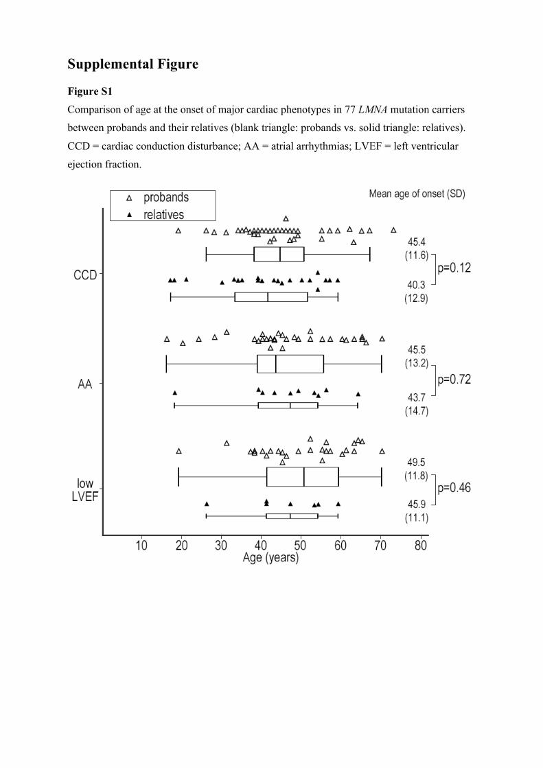
Supplemental Figure
Figure S1
Comparison of age at the onset of major cardiac phenotypes in 77 LMNA mutation carriers
between probands and their relatives (blank triangle: probands vs. solid triangle: relatives).
CCD = cardiac conduction disturbance; AA = atrial arrhythmias; LVEF = left ventricular
ejection fraction.
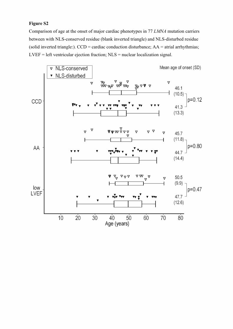
Figure S2
Comparison of age at the onset of major cardiac phenotypes in 77 LMNA mutation carriers
between with NLS-conserved residue (blank inverted triangle) and NLS-disturbed residue
(solid inverted triangle:). CCD = cardiac conduction disturbance; AA = atrial arrhythmias;
LVEF = left ventricular ejection fraction; NLS = nuclear localization signal.
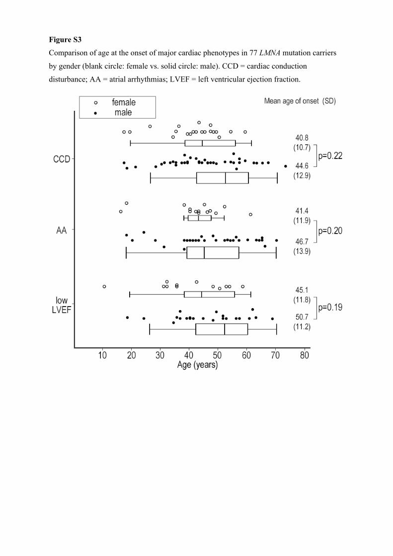
Figure S3
Comparison of age at the onset of major cardiac phenotypes in 77 LMNA mutation carriers
by gender (blank circle: female vs. solid circle: male). CCD = cardiac conduction
disturbance; AA = atrial arrhythmias; LVEF = left ventricular ejection fraction.